ppt of sensory organ
TRANSCRIPT
By –Mr. ASHOK BISHNOILecturer, JINR

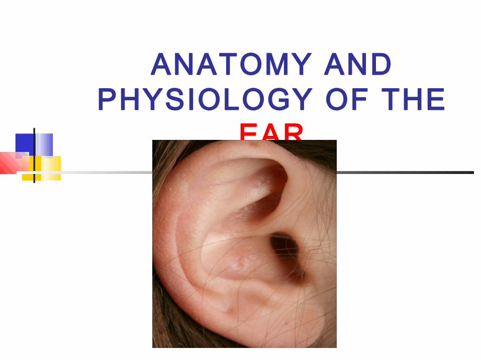
ANATOMY AND PHYSIOLOGY OF THE
EAR

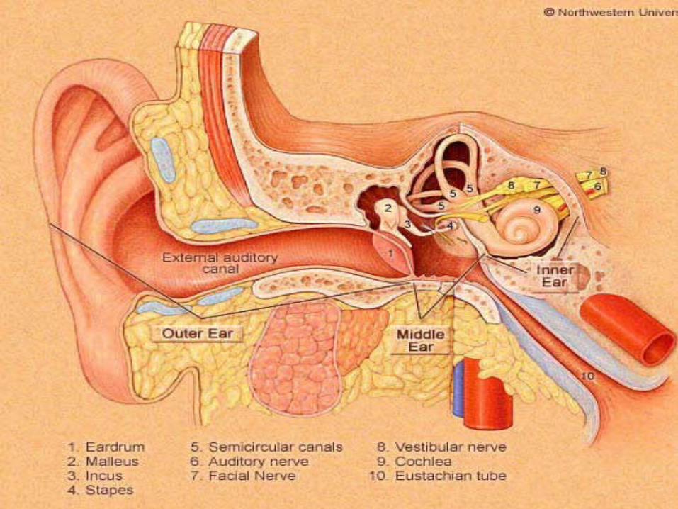
Divided into “3” parts:-
1. Outer Ear
2. Middle Ear (Tympanic cavity)
3. Inner Ear

Major Divisions of the EarPeripheral Mechanism Central Mechanism
Outer Ear
Middle Ear
Inner Ear
VIII Cranial Nerve
Brain
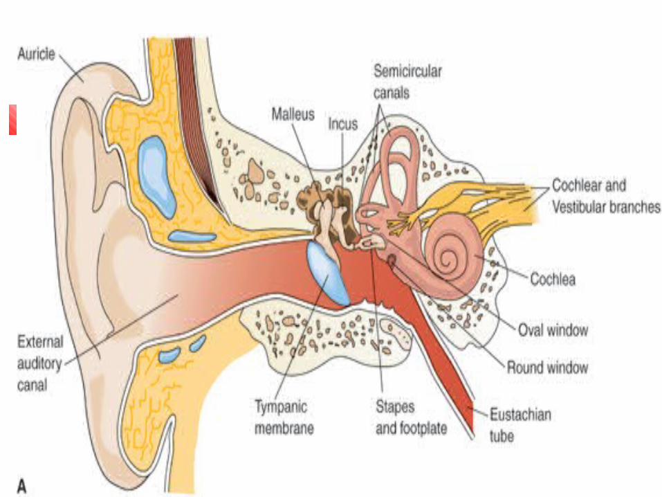
It consist of the auricle (Pinna) & External acoustic meatus ( Auditory canal)
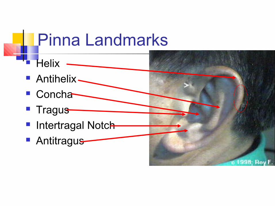
Pinna Landmarks Helix Antihelix Concha Tragus Intertragal Notch Antitragus
Auricle (Pinna):-
It is visible part of ear It composed of fibro elastic cartilage
convered with skin. Helix- it is outer ridge part Lobule (Ear lobe) –Soft pliable part
External acoustic meatus ( Auditory canal)
Slightly “S” Shaped tube About 2.5 cm long ( 1 inch ) External from auricle to tympanic
membrane There numerous ceruminous gland &
hair follicles Numerous gland are modified sweat
gland that secret cerumen (Earwax)
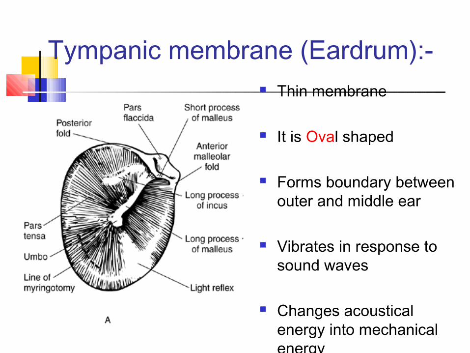
Tympanic membrane (Eardrum):- Thin membrane
It is Oval shaped
Forms boundary between outer and middle ear
Vibrates in response to sound waves
Changes acoustical energy into mechanical energy

Mastoid Process of Temporal Bone
Bony ridge behind the auricle.
Hardest bone in body, protects cochlea and vestibular system.
Provides support to the external ear and posterior wall of the middle ear cavity.
Contains air cavities which can be reservoir for infection
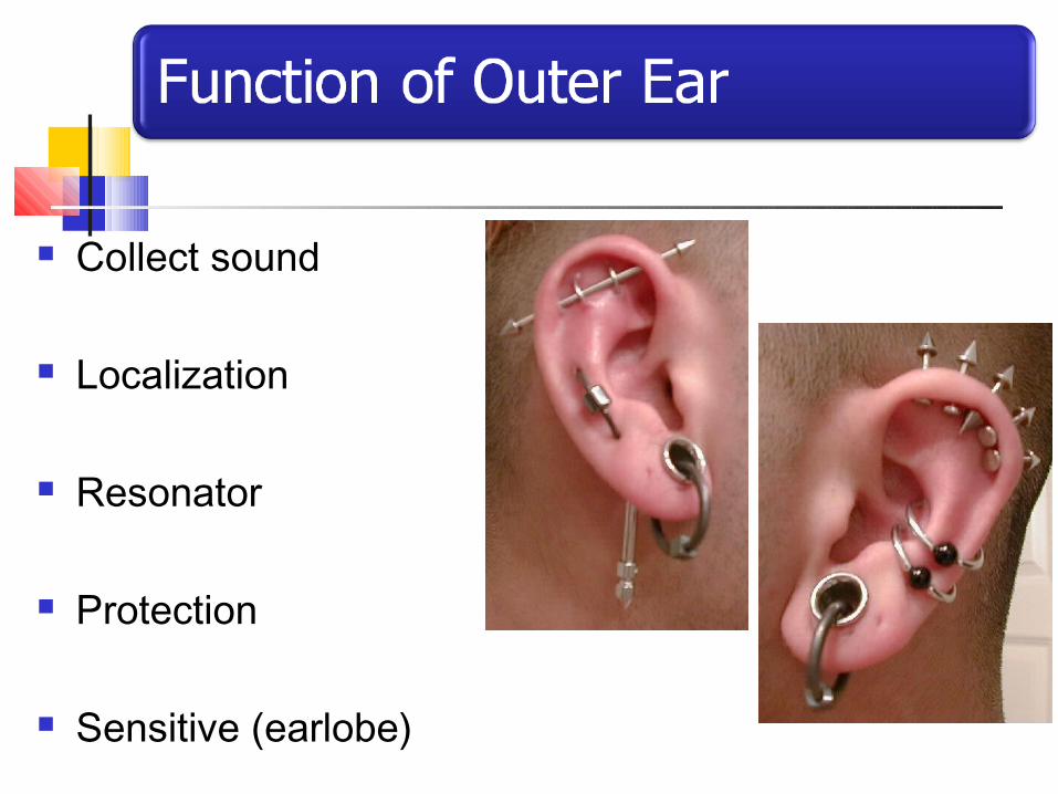
Collect sound
Localization
Resonator
Protection
Sensitive (earlobe)
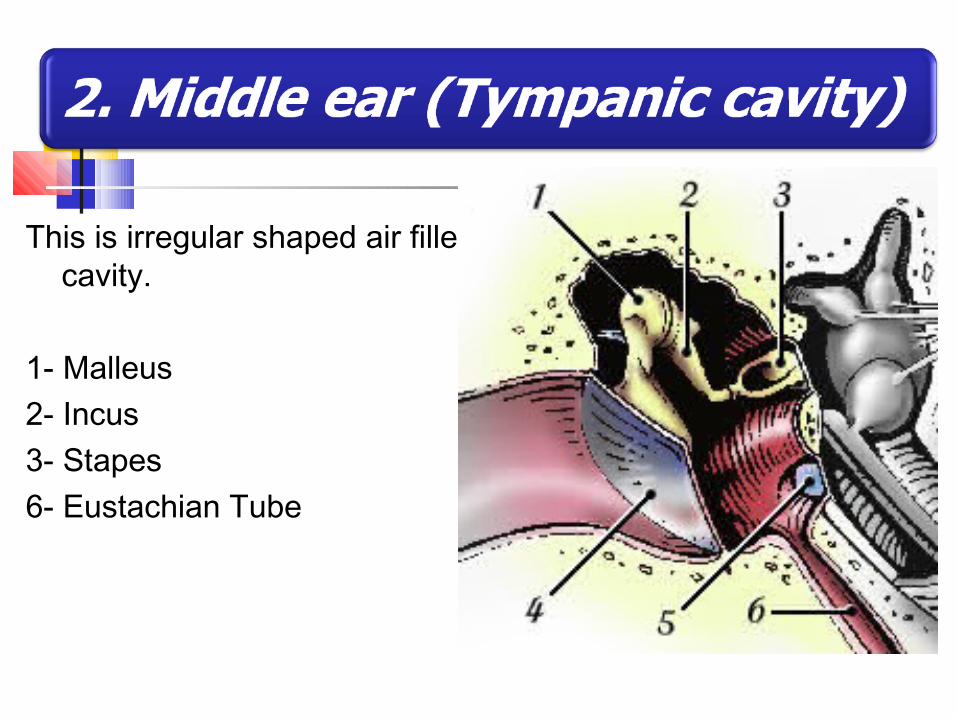
This is irregular shaped air filled cavity.
1- Malleus2- Incus 3- Stapes 6- Eustachian Tube
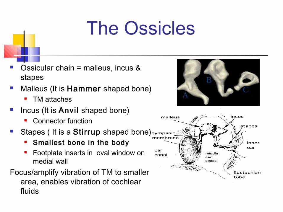
The Ossicles
Ossicular chain = malleus, incus & stapes
Malleus (It is Hammer shaped bone) TM attaches
Incus (It is Anvil shaped bone) Connector function
Stapes ( It is a Stirrup shaped bone) Smallest bone in the body Footplate inserts in oval window on
medial wallFocus/amplify vibration of TM to smaller
area, enables vibration of cochlear fluids
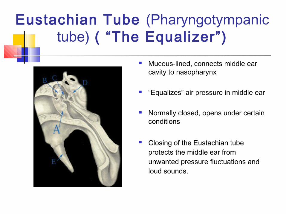
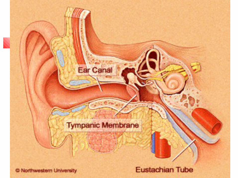
Eustachian Tube (Pharyngotympanic tube) ( “The Equalizer”)
Mucous-lined, connects middle ear cavity to nasopharynx
“Equalizes” air pressure in middle ear
Normally closed, opens under certain conditions
Closing of the Eustachian tube protects the middle ear from unwanted pressure fluctuations and loud sounds.

Conduction Conduct sound from the outer ear to the inner ear
Protection Creates a barrier that protects the middle and inner areas
from foreign objects Transducer
Converts acoustic energy to mechanical energy Converts mechanical energy to hydraulic energy
Amplifier Transformer action of the middle ear
The inner ear contain the organ of hearing & balance
It consist of – Vestibule (Balance) Cochlea Semicircular canalCochlea - Snail-shaped organ with a series of fluid-filled
tunnels; converts mechanical energy into electr ical energy

22
� Oval Window – located at the footplate of the stapes; when the footplate vibrates, the cochlear fluid is set into motion
� Round Window – functions as the pressure relief port for the fluid set into motion initially by the movement of the stapes in the oval window
23
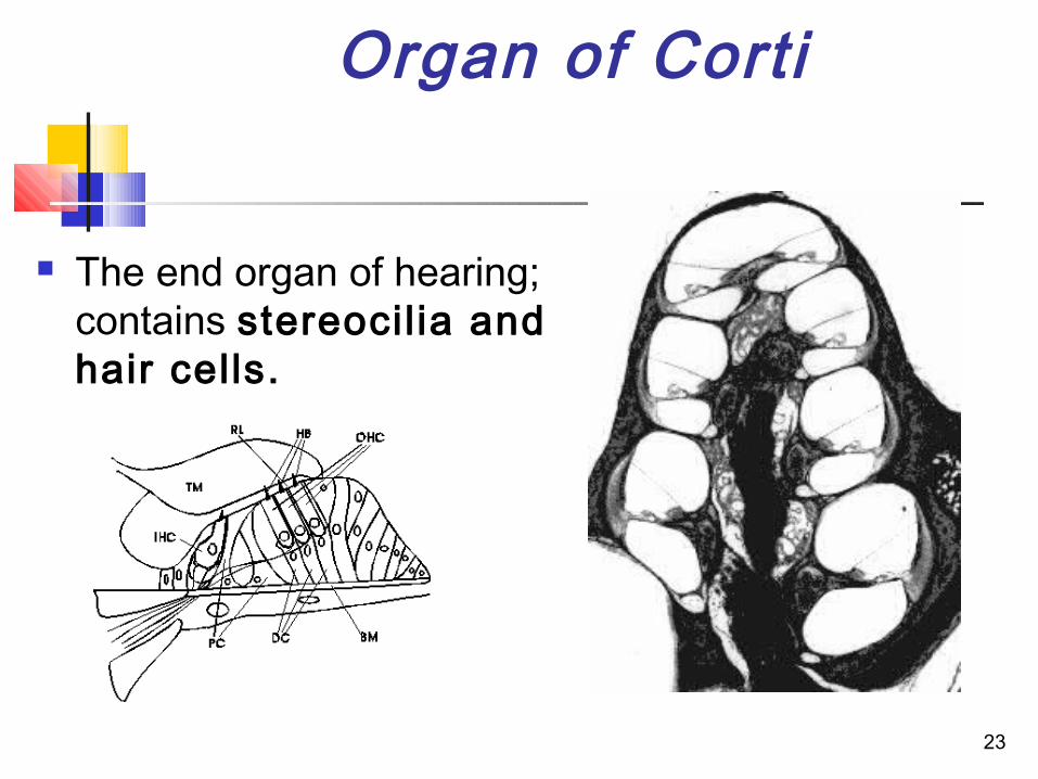
Organ of Corti
The end organ of hearing; contains stereocil ia and hair cells.
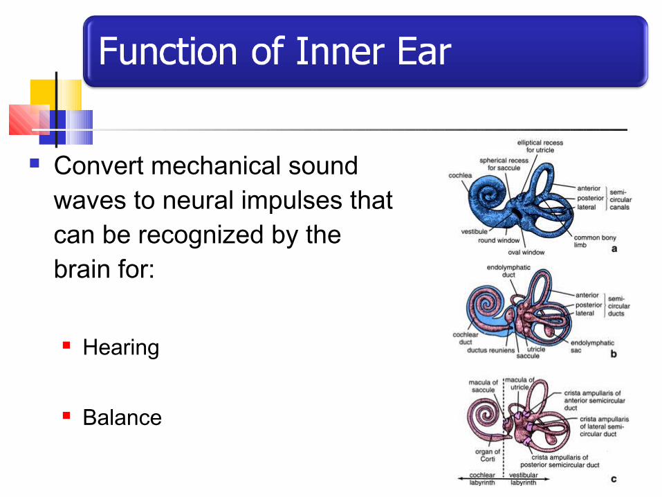
Convert mechanical sound waves to neural impulses that can be recognized by the brain for:
Hearing
Balance
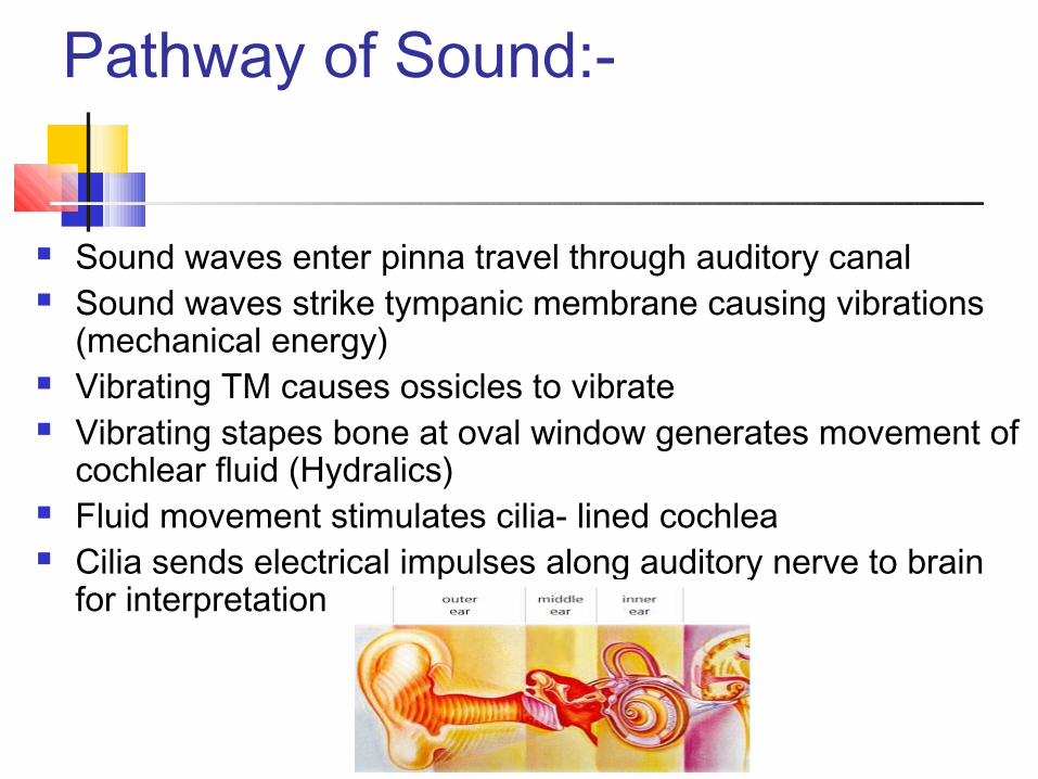
Pathway of Sound:-
Sound waves enter pinna travel through auditory canal Sound waves strike tympanic membrane causing vibrations
(mechanical energy) Vibrating TM causes ossicles to vibrate Vibrating stapes bone at oval window generates movement of
cochlear fluid (Hydralics) Fluid movement stimulates cilia- lined cochlea Cilia sends electrical impulses along auditory nerve to brain
for interpretation
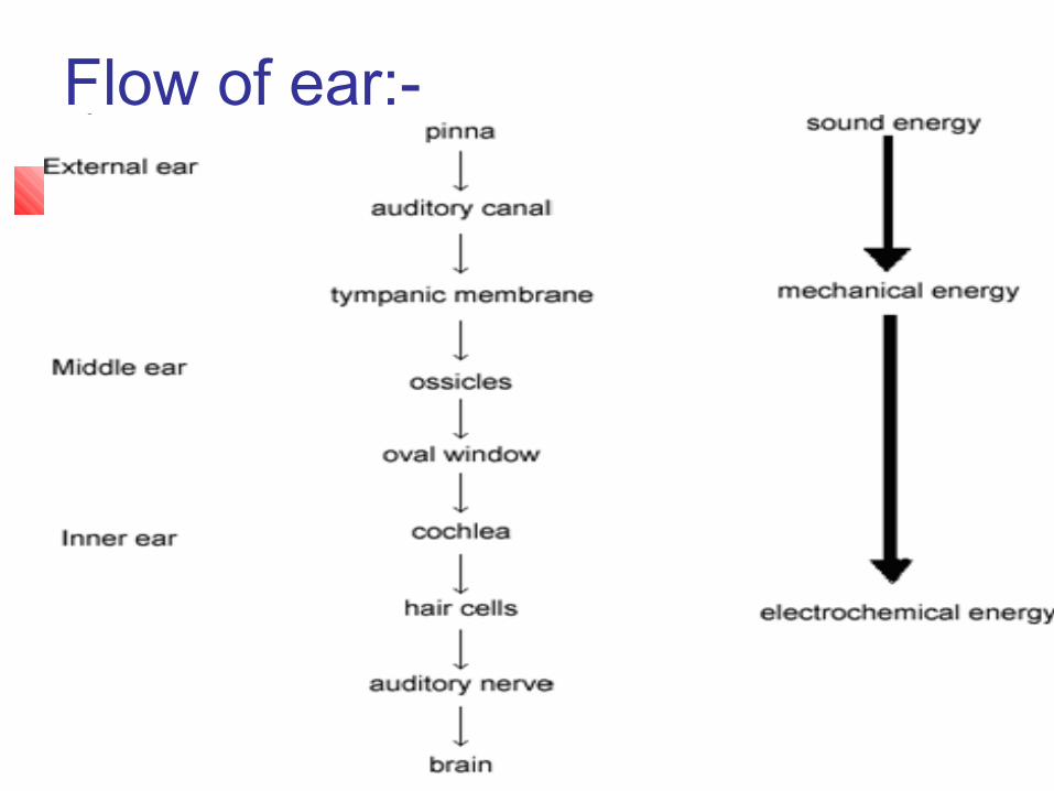
Flow of ear:-
Hearing
Sound Conduction and Transmission
Balance and Equilibrium