pp jurnal thorak new
TRANSCRIPT

8/22/2019 Pp Jurnal Thorak New
http://slidepdf.com/reader/full/pp-jurnal-thorak-new 1/93
Missed Breast Carcinoma:
Pitfalls and Pearls Presented by : dr Rivani Kurniawan
Lecturer : dr Hari Soekersi SpRad(K)
Aneesa S. Majid, MD et all
RSNA,• radiographics.rsnajnls.org

8/22/2019 Pp Jurnal Thorak New
http://slidepdf.com/reader/full/pp-jurnal-thorak-new 2/93
Introduction
• Mammography is the standard of reference for the
early detection of breast cancer
• Screening mammography is performed to detect anabnormality, whereas diagnostic mammography isused to further evaluate the abnormality or a clinical
problem.

8/22/2019 Pp Jurnal Thorak New
http://slidepdf.com/reader/full/pp-jurnal-thorak-new 3/93
• According to data from the Breast Cancer DetectionDemonstration Project, the false-negative rate ofmammography is approximately 8%– 10%
• After evaluating retrospective versus blindedinterpretations of mammograms, others have
concluded that the rate of missed breast cancers is ashigh as 35%

8/22/2019 Pp Jurnal Thorak New
http://slidepdf.com/reader/full/pp-jurnal-thorak-new 4/93
• In a series of 150 mammograms read by 10
radiologists, immediate work-up of the true cancerswas recommended in 74%– 96% of cases
• Recent studies have emphasized the use ofalternative imaging modalities to detect and diagnose
breast carcinoma, including ultrasonography (US),magnetic resonance (MR) imaging, and nuclear medicine studies

8/22/2019 Pp Jurnal Thorak New
http://slidepdf.com/reader/full/pp-jurnal-thorak-new 5/93
Breast cancers may be missed
because of ( 7 tools ) :
•Dense parenchyma that obscures a lesion
•Subtle features of malignancy
•A slowly changing malignancybreast
•Poor potitioning
•Poor techniquetechnologist
•Lack of perception of an abnormality that ispresent
•Incorrect interpretation of a suspect findingradiologist
8/22/2019 Pp Jurnal Thorak New
http://slidepdf.com/reader/full/pp-jurnal-thorak-new 6/93
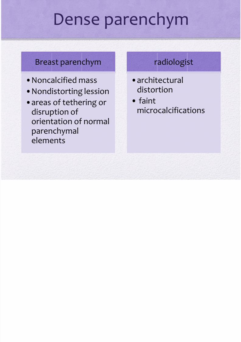
Dense parenchym
Breast parenchym
•
Noncalcified mass•Nondistorting lession
•areas of tethering or disruption oforientation of normalparenchymalelements
radiologist
•
architecturaldistortion
• faintmicrocalcifications
8/22/2019 Pp Jurnal Thorak New
http://slidepdf.com/reader/full/pp-jurnal-thorak-new 7/93
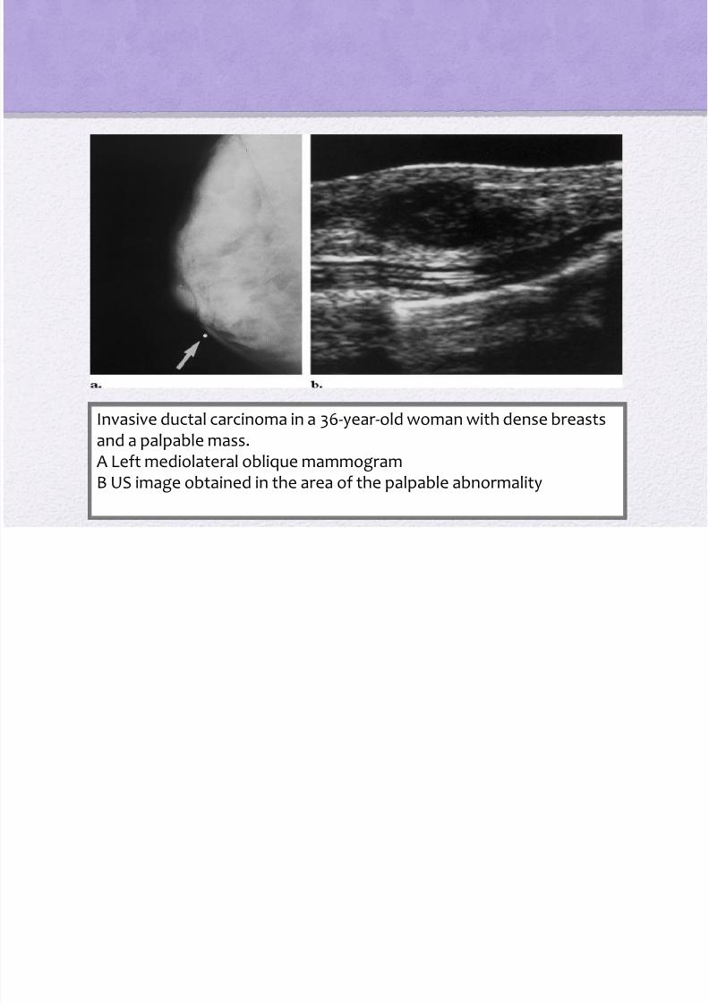
Invasive ductal carcinoma in a 36-year-old woman with dense breastsand a palpable mass.A Left mediolateral oblique mammogramB US image obtained in the area of the palpable abnormality

8/22/2019 Pp Jurnal Thorak New
http://slidepdf.com/reader/full/pp-jurnal-thorak-new 8/93
U l t r a
s o n o r
a p h y
determining the presence of a solid mass thatcorresponds to an area of distortion
characterizing palpable masses in dense tissue
evaluation of asymmetric densities seen atmammography
8/22/2019 Pp Jurnal Thorak New
http://slidepdf.com/reader/full/pp-jurnal-thorak-new 9/93
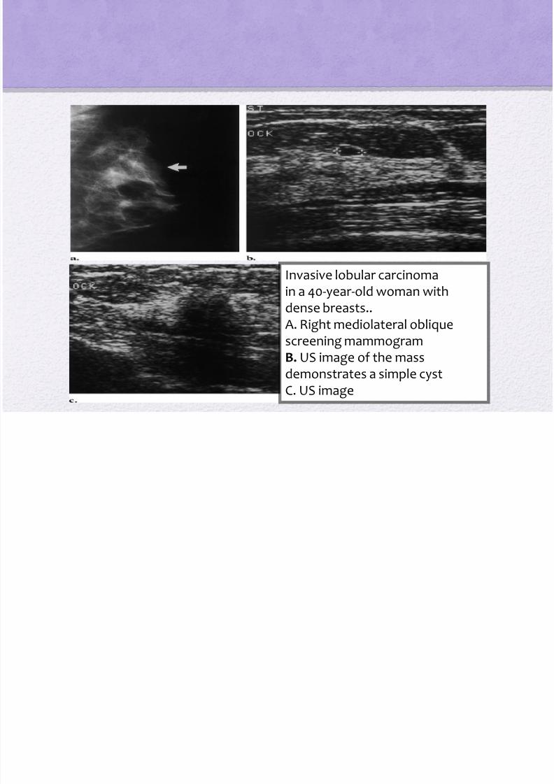
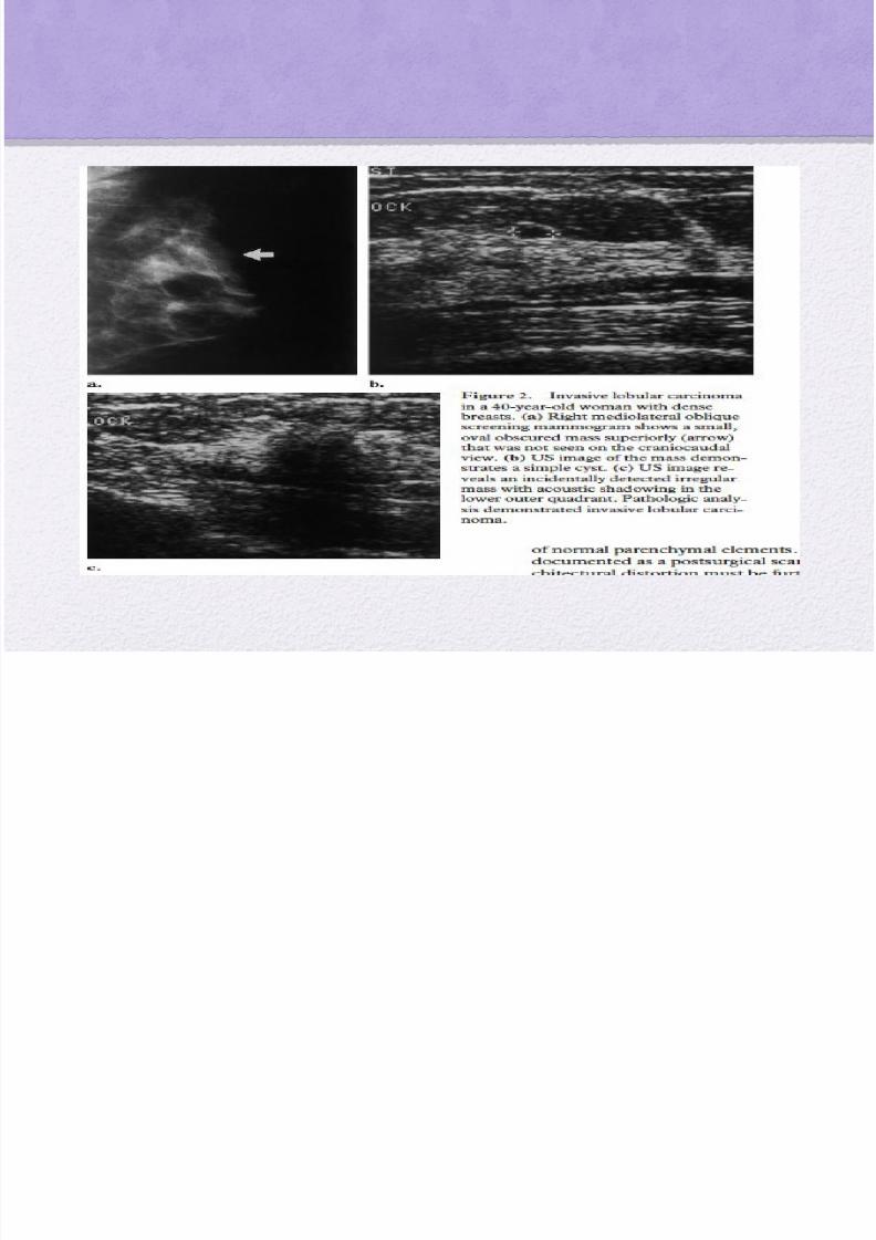
Invasive lobular carcinoma
in a 40-year-old woman withdense breasts..A. Right mediolateral obliquescreening mammogramB. US image of the massdemonstrates a simple cyst
C. US image
8/22/2019 Pp Jurnal Thorak New
http://slidepdf.com/reader/full/pp-jurnal-thorak-new 10/93
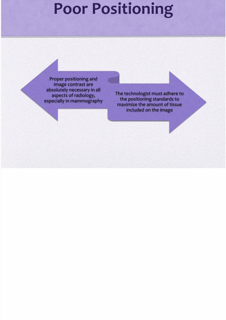
Poor Positioning
Proper positioning andimage contrast are
absolutely necessary in allaspects of radiology,
especially in mammography
The technologist must adhere tothe positioning standards to
maximize the amount of tissueincluded on the image
8/22/2019 Pp Jurnal Thorak New
http://slidepdf.com/reader/full/pp-jurnal-thorak-new 11/93

8/22/2019 Pp Jurnal Thorak New
http://slidepdf.com/reader/full/pp-jurnal-thorak-new 12/93

8/22/2019 Pp Jurnal Thorak New
http://slidepdf.com/reader/full/pp-jurnal-thorak-new 13/93

8/22/2019 Pp Jurnal Thorak New
http://slidepdf.com/reader/full/pp-jurnal-thorak-new 14/93
Proper positioning(a) Left mediolateral oblique (left) and craniocaudal
(right) mammograms(b) On a left mediolateral oblique mammogram
obtained with improved positioning

8/22/2019 Pp Jurnal Thorak New
http://slidepdf.com/reader/full/pp-jurnal-thorak-new 15/93
• Creative positioning may be necessary to includeareas of palpable abnormalities on the images :
Radiopaque markers
Spot compression

8/22/2019 Pp Jurnal Thorak New
http://slidepdf.com/reader/full/pp-jurnal-thorak-new 16/93
Creative positioning may also be helpful in :
patients who are tense
who have suffered a stroke
who have shoulder problems
other debilitating factors that limit visualization of theposterior breast on standard mediolateral obliqueviews

8/22/2019 Pp Jurnal Thorak New
http://slidepdf.com/reader/full/pp-jurnal-thorak-new 17/93
Creative positioning for lesion detection(a) Bilateral mediolateral obliqueMammograms(b) On a right lateromedialMammogram(c) Spot magnificationmammogram
P T h i

8/22/2019 Pp Jurnal Thorak New
http://slidepdf.com/reader/full/pp-jurnal-thorak-new 18/93
Poor Technique
optimize image contrast to avoidobtaining over or
underpenetrated images
Careful attention to dailyprocessor quality control is alsonecessary to optimize contrast
Proper positioning of thephotocell is necessary to achieve
correct optical density on theimage
The technologist should alwaysreview the images under proper
mammographic viewingconditions to assess the adequacy
of imaging technique
technologist

8/22/2019 Pp Jurnal Thorak New
http://slidepdf.com/reader/full/pp-jurnal-thorak-new 19/93
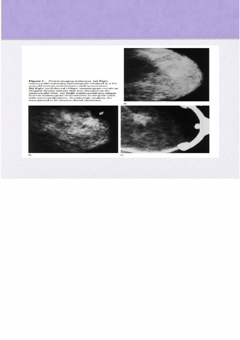
Proper imaging technique
(a) Rightcraniocaudal screening mammogram(b) Right mediolateral obliquemammogram(c) Right craniocaudal spot magnificationmammogram

8/22/2019 Pp Jurnal Thorak New
http://slidepdf.com/reader/full/pp-jurnal-thorak-new 20/93
lack of perception
- Two major causes ofmissed breast cancers are
related to radiologisterror
- Perception error occurswhen the lesion is included in
the field of view and is evidentbut is not recognized by the
radiologist

8/22/2019 Pp Jurnal Thorak New
http://slidepdf.com/reader/full/pp-jurnal-thorak-new 21/93
• The lesion may or may not have subtle features ofmalignancy that cause it to be less visible.
Small nonspiculated masses,
Areas of architectural distortion and asymmetry,
Small clusters of amorphous or faint
microcalcifications may all be difficult to perceive.
8/22/2019 Pp Jurnal Thorak New
http://slidepdf.com/reader/full/pp-jurnal-thorak-new 22/93
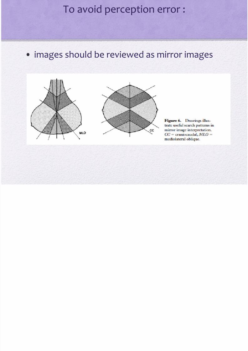
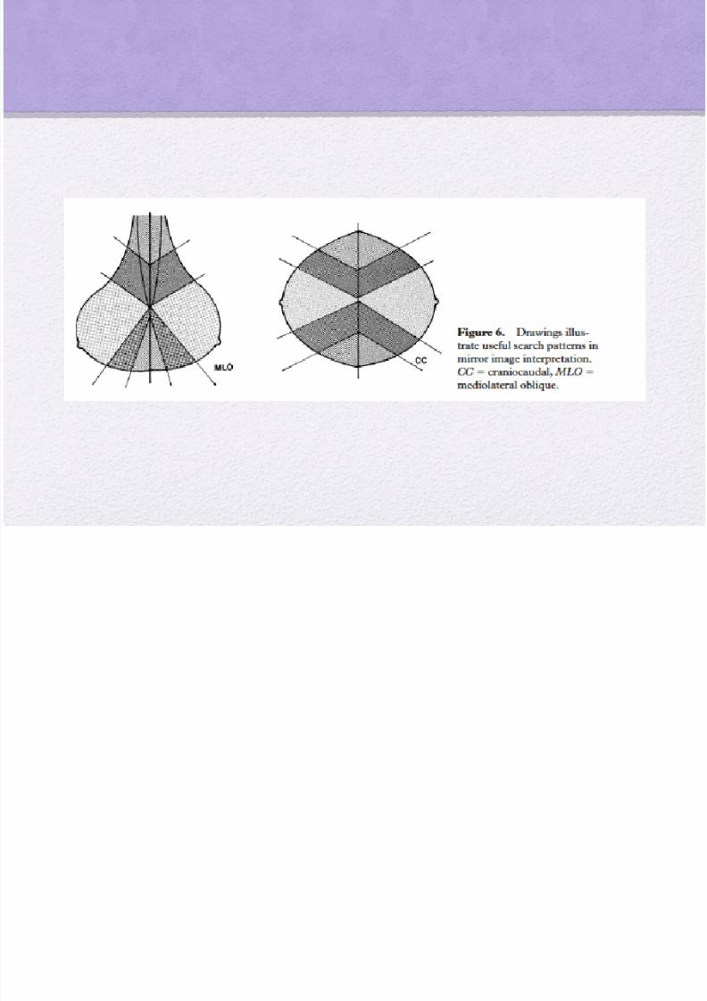
To avoid perception error :
• images should be reviewed as mirror images

8/22/2019 Pp Jurnal Thorak New
http://slidepdf.com/reader/full/pp-jurnal-thorak-new 23/93
• The radiologist should compare like areas onthe side-by-side images to identify any focalasymmetric density or low-density mass
Mirror image interpretation(a) Bilateral mediolateraloblique mammograms(b, c) On left craniocaudalspot compression
mammograms, the posterior (b) and anterior (c)

8/22/2019 Pp Jurnal Thorak New
http://slidepdf.com/reader/full/pp-jurnal-thorak-new 24/93
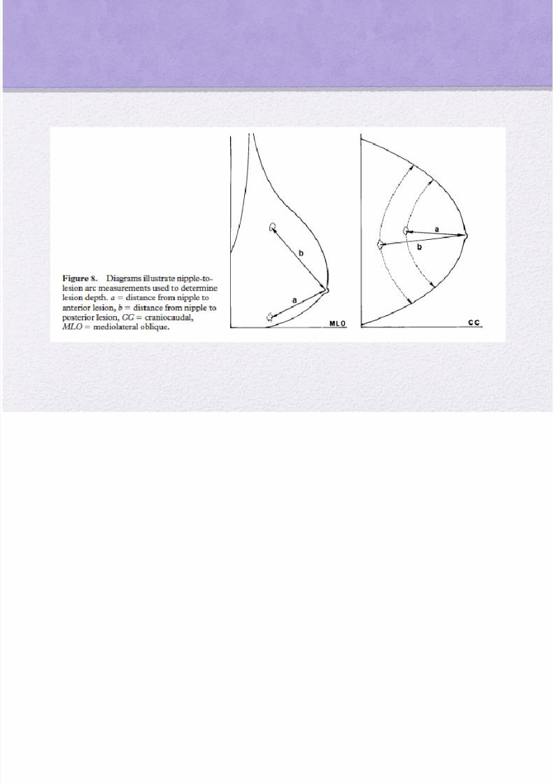
• Identification of a focal density should prompta search for this density on the correspondingview in the same arc from the nipple
8/22/2019 Pp Jurnal Thorak New
http://slidepdf.com/reader/full/pp-jurnal-thorak-new 25/93
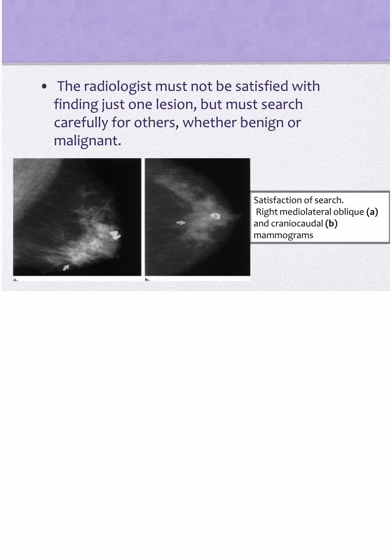
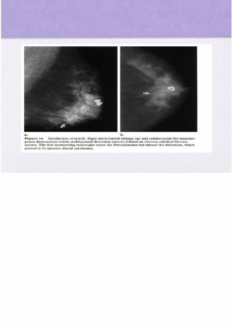
• The radiologist must not be satisfied withfinding just one lesion, but must searchcarefully for others, whether benign or
malignant.
Satisfaction of search.Right mediolateral oblique (a)
and craniocaudal (b)mammograms

8/22/2019 Pp Jurnal Thorak New
http://slidepdf.com/reader/full/pp-jurnal-thorak-new 26/93
• MR imaging has been useful in identifying the primarycarcinoma when a metastatic node is found in theaxilla and mammographic findings are negative
8/22/2019 Pp Jurnal Thorak New
http://slidepdf.com/reader/full/pp-jurnal-thorak-new 27/93
Incorect interpretation
- The second major cause ofmissed breast cancers that isrelated to radiologist error isincorrect interpretation of a
lesion
- which occurs when anabnormality with suspect featuresis observed but is misinterpreted
as being definitely or at leastprobably benign

8/22/2019 Pp Jurnal Thorak New
http://slidepdf.com/reader/full/pp-jurnal-thorak-new 28/93
Several factors may lead to misinterpretation,such as
lack of experience
fatigue,
Inattention

8/22/2019 Pp Jurnal Thorak New
http://slidepdf.com/reader/full/pp-jurnal-thorak-new 29/93
• Characterization of a lesion that is identified at
screening mammography should be based ondiagnostic mammographic findings and not onscreening findings alone.
S btle Sig s of
8/22/2019 Pp Jurnal Thorak New
http://slidepdf.com/reader/full/pp-jurnal-thorak-new 30/93
Subtle Signs ofMalignancy
The cancers that are the most challenging todiagnose and that most often lead to
interpretation errors are those with subtle or indistinct features of malignancy

8/22/2019 Pp Jurnal Thorak New
http://slidepdf.com/reader/full/pp-jurnal-thorak-new 31/93
These features include
• areas of architectural distortion
• small groups of amorphous or punctatemicrocalcifi cations,
• focal asymmetric densities
• dilated ducts
• relatively well circumscribed masses

8/22/2019 Pp Jurnal Thorak New
http://slidepdf.com/reader/full/pp-jurnal-thorak-new 32/93
Circumscribed cancer in a 63-year-old woman.Right exaggerated craniocaudal lateral mammogram
8/22/2019 Pp Jurnal Thorak New
http://slidepdf.com/reader/full/pp-jurnal-thorak-new 33/93
US is helpful in predicting the likelihood ofmalignancy in a circumscribed mass
•
Simple cysts seen at US constitute a benignfinding.
• Solid lesions that are smooth, elliptic, and
wider than they are tall are probably benign
8/22/2019 Pp Jurnal Thorak New
http://slidepdf.com/reader/full/pp-jurnal-thorak-new 34/93
• masses that have irregular or angulated margins, aremarkedly hypo echogenic, and are taller than they arewide are probably malignant
• If, however, the mass is seen at US as a solid lesionwith worrisome features such as a “ taller-than-wide”
shape or irregular margins , biopsy is indicated

8/22/2019 Pp Jurnal Thorak New
http://slidepdf.com/reader/full/pp-jurnal-thorak-new 35/93
• Clinical history is important in evaluating focal
areas of asymmetry
• In the absence of tumor or infection, focaldeveloping densities should prompt further
assessment and, usually, biopsy

8/22/2019 Pp Jurnal Thorak New
http://slidepdf.com/reader/full/pp-jurnal-thorak-new 36/93
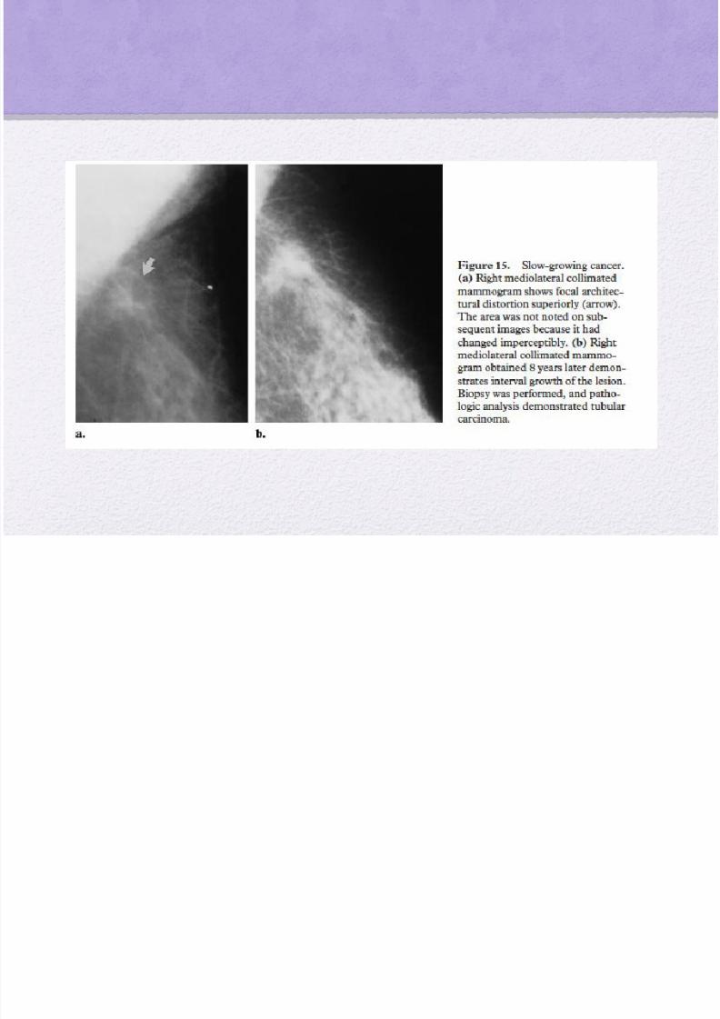
Slow-growing Cancers
• The doubling time for breast cancers has been
reported to range from 44 to 1,869 days
• However, malignant calcifi cations have beenreported to be stable at mammography for as
long as 63 months

8/22/2019 Pp Jurnal Thorak New
http://slidepdf.com/reader/full/pp-jurnal-thorak-new 37/93
• Therefore, a slowly changing cancer may go
undetected if the radiologist fails to compare findingswith those on older images
• A lesion with features that strongly suggestmalignancy but that has been stable for 1– 2 years still
requires biopsy because it may represent a slowlychanging cancer
8/22/2019 Pp Jurnal Thorak New
http://slidepdf.com/reader/full/pp-jurnal-thorak-new 38/93

8/22/2019 Pp Jurnal Thorak New
http://slidepdf.com/reader/full/pp-jurnal-thorak-new 39/93
Role of Double Reading
.Double reading ofmammograms hasbeen shown toincrease thedetection rate for breast cancer byup to 15% (29,30)
.Computer-aideddetection (CAD)represents arelatively newtechnology thathas beenimplemented insomemammography
facilities for double reading
.The sensitivity ofthe CAD systems isgreater for detectingcalcifications thanfor detectingmasses

8/22/2019 Pp Jurnal Thorak New
http://slidepdf.com/reader/full/pp-jurnal-thorak-new 40/93
Conclusions
• Although mammography is the standard of
reference for the detection of early breastcancer, as many as 30% of breast cancers maybe missed

8/22/2019 Pp Jurnal Thorak New
http://slidepdf.com/reader/full/pp-jurnal-thorak-new 41/93
To reduce the possibility of missing a cancer, theradiologist should take the following steps wheninterpreting mammographic fi ndings:
• 1. Do not rely on screening views alone to diagnose adetected abnormality; complete the evaluation withdiagnostic mammography.
•
2. Review clinical data and use US to help assess apalpable or mammographically detected mass

8/22/2019 Pp Jurnal Thorak New
http://slidepdf.com/reader/full/pp-jurnal-thorak-new 42/93
• 3. Be strict about positioning and technicalrequirements to optimize image quality.
• 4. Be alert to subtle features of breast cancers.
• 5. Compare current images with multiple prior studiesto look for subtle increases in lesion size.
•
6. Look for other lesions when one abnormality isseen.
• 7. Judge a lesion by its most malignant features.

8/22/2019 Pp Jurnal Thorak New
http://slidepdf.com/reader/full/pp-jurnal-thorak-new 43/93

8/22/2019 Pp Jurnal Thorak New
http://slidepdf.com/reader/full/pp-jurnal-thorak-new 44/93
TERIMA KASIH

8/22/2019 Pp Jurnal Thorak New
http://slidepdf.com/reader/full/pp-jurnal-thorak-new 45/93

8/22/2019 Pp Jurnal Thorak New
http://slidepdf.com/reader/full/pp-jurnal-thorak-new 46/93
8/22/2019 Pp Jurnal Thorak New
http://slidepdf.com/reader/full/pp-jurnal-thorak-new 47/93

8/22/2019 Pp Jurnal Thorak New
http://slidepdf.com/reader/full/pp-jurnal-thorak-new 48/93

8/22/2019 Pp Jurnal Thorak New
http://slidepdf.com/reader/full/pp-jurnal-thorak-new 49/93

8/22/2019 Pp Jurnal Thorak New
http://slidepdf.com/reader/full/pp-jurnal-thorak-new 50/93

8/22/2019 Pp Jurnal Thorak New
http://slidepdf.com/reader/full/pp-jurnal-thorak-new 51/93

8/22/2019 Pp Jurnal Thorak New
http://slidepdf.com/reader/full/pp-jurnal-thorak-new 52/93
8/22/2019 Pp Jurnal Thorak New
http://slidepdf.com/reader/full/pp-jurnal-thorak-new 53/93

8/22/2019 Pp Jurnal Thorak New
http://slidepdf.com/reader/full/pp-jurnal-thorak-new 54/93
Breast cancer risk factor
Gender
Aging
Genetic
Personal history of breast cancer
Race and ethnicity
Dense breast tissue
Menstrual periods
Previous chest radiation

8/22/2019 Pp Jurnal Thorak New
http://slidepdf.com/reader/full/pp-jurnal-thorak-new 55/93
BIRADS

8/22/2019 Pp Jurnal Thorak New
http://slidepdf.com/reader/full/pp-jurnal-thorak-new 56/93

8/22/2019 Pp Jurnal Thorak New
http://slidepdf.com/reader/full/pp-jurnal-thorak-new 57/93
BREAST PARENCHYMAL DENSITIY
• I. The breast is almost entirely fat
• II. There are scattered fibroglandular densities.
• III. The breast is heterogeneously dense. Thismay lower the sensitivity of mammography.
• IV. The breast tissue is extremely dense, whichcould obscure a lesion in mammography

8/22/2019 Pp Jurnal Thorak New
http://slidepdf.com/reader/full/pp-jurnal-thorak-new 58/93

8/22/2019 Pp Jurnal Thorak New
http://slidepdf.com/reader/full/pp-jurnal-thorak-new 59/93

8/22/2019 Pp Jurnal Thorak New
http://slidepdf.com/reader/full/pp-jurnal-thorak-new 60/93
• Findings on the mediolateral oblique view thatindicate proper positioning include:
visualization of the pectoralis muscle to the level ofthe nipple
A convex appearance of the pectoralis major muscle
Complete visualization of posterior breast tissue
Breast tissue that is well compressed and positioned inan up-and-out orientation
An open inframammary fold

8/22/2019 Pp Jurnal Thorak New
http://slidepdf.com/reader/full/pp-jurnal-thorak-new 61/93
Breast cancers may be missed

8/22/2019 Pp Jurnal Thorak New
http://slidepdf.com/reader/full/pp-jurnal-thorak-new 62/93
ybecause of ( 7 tools ) :
Dense parenchyma that obscures a lesion
Poor positioning or technique
lesion location outside the field of view
Lack of perception of an abnormality that is present
Incorrect interpretation of a suspect finding
Subtle features of malignancy
A slowly changing malignancy

8/22/2019 Pp Jurnal Thorak New
http://slidepdf.com/reader/full/pp-jurnal-thorak-new 63/93
Causes of Missed Breast Cancers
Dense Parenchyma
• Breast parenchyma that is inherently dense
compromises the ability to detect a mass,especially a noncalcified, nondistorting lesion
• The radiologist must be particularly attentive
in searching for areas of architecturaldistortion or faint microcalcifications

8/22/2019 Pp Jurnal Thorak New
http://slidepdf.com/reader/full/pp-jurnal-thorak-new 64/93

8/22/2019 Pp Jurnal Thorak New
http://slidepdf.com/reader/full/pp-jurnal-thorak-new 65/93
• the tissue must be intensely evaluated for any areas oftethering or disruption of orientation of normal
parenchymal elements
• Unless it is documented as a postsurgical scar, an areaof architectural distortion must be further evaluated
with additional views (eg, spot compression, magnification, off-angle)
8/22/2019 Pp Jurnal Thorak New
http://slidepdf.com/reader/full/pp-jurnal-thorak-new 66/93
• US may also be helpful in determining the presence ofa solid mass that corresponds to an area of distortion
• US is very important in the evaluation ofmammographic abnormalities, being useful incharacterizing palpable masses in dense tissue and
circumscribed isodense masses

8/22/2019 Pp Jurnal Thorak New
http://slidepdf.com/reader/full/pp-jurnal-thorak-new 67/93
• US can be especially helpful in the evaluation ofasymmetric densities seen at mammography becauseit can help identify the density as either breast tissueor a true mass
• However, a palpable mass that appears solid at US
warrants further evaluation with biopsy
8/22/2019 Pp Jurnal Thorak New
http://slidepdf.com/reader/full/pp-jurnal-thorak-new 68/93

8/22/2019 Pp Jurnal Thorak New
http://slidepdf.com/reader/full/pp-jurnal-thorak-new 69/93
Poor Positioning
• Proper positioning and image contrast are absolutelynecessary in all aspects of radiology, especially inmammography
• The technologist must adhere to the positioningstandards to maximize the amount of tissue included
on the image

8/22/2019 Pp Jurnal Thorak New
http://slidepdf.com/reader/full/pp-jurnal-thorak-new 70/93
• At craniocaudal imaging, the technologist shouldverify that the breast is pulled straight forward and
not exaggerated laterally, and that the breast tissue iswell compressed.
• The difference between the posterior nipple line
measurement on the mediolateral oblique andcraniocaudal views should not exceed 1 cm

8/22/2019 Pp Jurnal Thorak New
http://slidepdf.com/reader/full/pp-jurnal-thorak-new 71/93

8/22/2019 Pp Jurnal Thorak New
http://slidepdf.com/reader/full/pp-jurnal-thorak-new 72/93

8/22/2019 Pp Jurnal Thorak New
http://slidepdf.com/reader/full/pp-jurnal-thorak-new 73/93
Poor Technique
• The technologist must:
optimize image contrast to avoid obtaining over or underpenetrated images
Proper positioning of the photocell is necessary to
achieve correct optical density on the image

8/22/2019 Pp Jurnal Thorak New
http://slidepdf.com/reader/full/pp-jurnal-thorak-new 74/93
• Careful attention to daily processor quality control isalso necessary to optimize contrast
• The technologist should always review the imagesunder proper mammographic viewing conditions toassess the adequacy of imaging technique .
• Image blur is problematic, particularly in theassessment of microcalcifi cations.
8/22/2019 Pp Jurnal Thorak New
http://slidepdf.com/reader/full/pp-jurnal-thorak-new 75/93

8/22/2019 Pp Jurnal Thorak New
http://slidepdf.com/reader/full/pp-jurnal-thorak-new 76/93
Lack of Perception
• Two major causes of missed breast cancers arerelated to radiologist error
• Perception error occurs when the lesion is included inthe field of view and is evident but is not recognizedby the radiologist

8/22/2019 Pp Jurnal Thorak New
http://slidepdf.com/reader/full/pp-jurnal-thorak-new 77/93
To avoid perception error :
o images should be reviewed as mirror images
o The radiologist should compare like areas on the side-by-side images to identify any focal asymmetricdensity or low-density mass.
o Identification of a focal density should prompt asearch for this density on the corresponding view in
the same arc from the nipple .
o Additional views may be needed to verify thepresence of a true lesion
8/22/2019 Pp Jurnal Thorak New
http://slidepdf.com/reader/full/pp-jurnal-thorak-new 78/93

8/22/2019 Pp Jurnal Thorak New
http://slidepdf.com/reader/full/pp-jurnal-thorak-new 79/93

8/22/2019 Pp Jurnal Thorak New
http://slidepdf.com/reader/full/pp-jurnal-thorak-new 80/93
• The radiologist should compare like areas onthe side-by-side images to identify any focalasymmetric density or low-density mass.
8/22/2019 Pp Jurnal Thorak New
http://slidepdf.com/reader/full/pp-jurnal-thorak-new 81/93

8/22/2019 Pp Jurnal Thorak New
http://slidepdf.com/reader/full/pp-jurnal-thorak-new 82/93
• Failure to diagnose multifocal and multicentric breastcancers can directly affect patient treatment.
• The radiologist must not be satisfied with finding justone lesion, but must search carefully for others,whether benign or malignant.
8/22/2019 Pp Jurnal Thorak New
http://slidepdf.com/reader/full/pp-jurnal-thorak-new 83/93

8/22/2019 Pp Jurnal Thorak New
http://slidepdf.com/reader/full/pp-jurnal-thorak-new 84/93
• The radiologist must not be satisfied withfinding just one lesion, but must searchcarefully for others, whether benign or
malignant.

8/22/2019 Pp Jurnal Thorak New
http://slidepdf.com/reader/full/pp-jurnal-thorak-new 85/93
• The primary breast cancer may be occult and either not observed or very subtle at mammography
• Careful attention to mirror image abnormalities or focal asymmetric densities is important in identifyingthe primary lesion

8/22/2019 Pp Jurnal Thorak New
http://slidepdf.com/reader/full/pp-jurnal-thorak-new 86/93
Incorrect Interpretation
• The second major cause of missed breast cancers thatis related to radiologist error is incorrect
interpretation of a lesion
• which occurs when an abnormality with suspectfeatures is observed but is misinterpreted as being
definitely or at least probably benign
8/22/2019 Pp Jurnal Thorak New
http://slidepdf.com/reader/full/pp-jurnal-thorak-new 87/93
• Misinterpretation may also occur if the radiologistfails to obtain all the views needed to assess thecharacteristics of a lesion or if the lesion is slowgrowing and prior images are not used for comparison.
•
The radiologist may erroneously judge theabnormality by its most benign features and missimportant malignant features that necessitate biopsy

8/22/2019 Pp Jurnal Thorak New
http://slidepdf.com/reader/full/pp-jurnal-thorak-new 88/93
Subtle Signs of

8/22/2019 Pp Jurnal Thorak New
http://slidepdf.com/reader/full/pp-jurnal-thorak-new 89/93
gMalignancy
The cancers that are the most challenging to
diagnose and that most often lead tointerpretation errors are those with subtle or indistinct features of malignancy

8/22/2019 Pp Jurnal Thorak New
http://slidepdf.com/reader/full/pp-jurnal-thorak-new 90/93
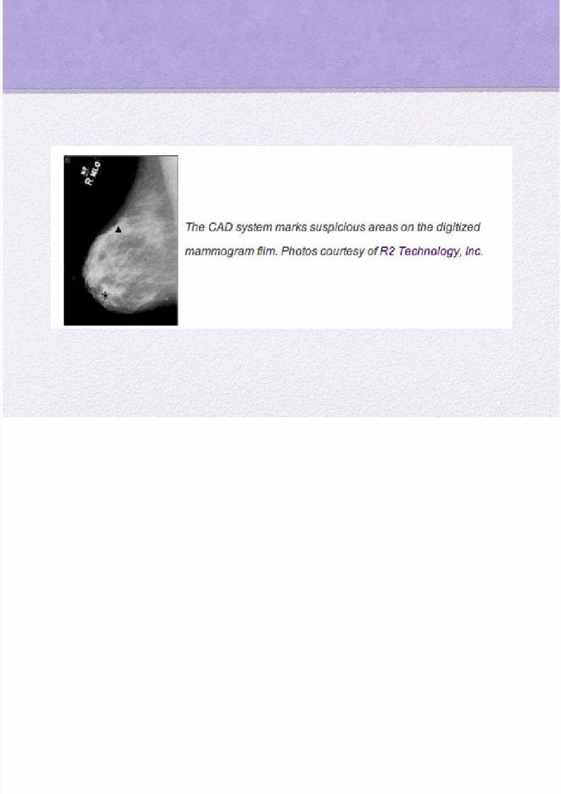
Role of Double Reading
• Double reading of mammograms has beenshown to increase the detection rate for breast cancer by up to 15% (29,30)
• Computer-aided detection (CAD) represents arelatively new technology that has beenimplemented in some mammography facilitiesfor double reading

8/22/2019 Pp Jurnal Thorak New
http://slidepdf.com/reader/full/pp-jurnal-thorak-new 91/93
• The sensitivity of the CAD systems is greater for detecting calcifications than for detectingmasses

8/22/2019 Pp Jurnal Thorak New
http://slidepdf.com/reader/full/pp-jurnal-thorak-new 92/93
• Asymmetric densities are frequently seen atmammography.
• These findings in isolation have a low positivepredictive value for malignancy
•
however, when they are associated withmicrocalcifications or architectural distortion, the riskof malignancy is increased

8/22/2019 Pp Jurnal Thorak New
http://slidepdf.com/reader/full/pp-jurnal-thorak-new 93/93