potential legal implications of advances in … · s laureys, f perrion, s bredart,...
TRANSCRIPT
POTENTIAL LEGAL IMPLICATIONS OF ADVANCES IN NEUROIMAGING TECHNIQUES
FOR THE CLINICAL MANAGEMENT OF PATIENTS WITH DISORDERS OF CONSCIOUSNESS
Dr Stephanie Pywell, The Open University Law School, England
INTRODUCTION
Voluntary communication with others is a fundamental characteristic of human beings. It serves myriad
purposes, and depriving someone of the ability to communicate by gagging and isolation has long been
recognised as a harsh form of punishment. This paper considers whether recent developments in
neuroimaging, and the United Kingdom’s legal obligations, particularly those arising under human rights
treaties, could combine to require greater efforts to be made to enable people whom serious brain
damage has rendered unable to communicate by conventional methods to make their wishes and
feelings known.
Most adult patients are able to inform clinicians when they are in pain or distress. Patients who are
unable to speak may exhibit non-verbal forms of communication – such as responsive tears or an
involuntary reaction to palpation – that can be the trigger for overt sympathy or the administration of
pain-killers. People suffering from the disorders of consciousness (DoCs) – coma, vegetative state (VS)
and minimally conscious state (MCS) – are usually unable to communicate verbally or non-verbally, and
the traditional view has been that such patients cannot make their feelings and wishes known. In a
fourth condition, locked-in syndrome (LIS), patients are fully conscious but almost completely paralysed,
1
and hence unable to respond to most stimuli, but they may be able to communicate via tiny
movements, usually of the eyes or eyelids.1
Several studies have suggested that a significant number of patients are misdiagnosed as having a more
severe DoC than is actually the case, meaning that their already profound suffering may be exacerbated
by their being inappropriately treated as though they were unconscious. Recent advances in the use of
neuroimaging techniques including electroencephalograms (EEGs), functional magnetic resonance
imaging (fMRI) and positron emission tomography (PET) potentially offer a means of enabling some MCS
patients, and LIS patients who are totally paralysed, to communicate. If neuroimaging techniques could
be refined and made available to assess all patients with DoCs, physicians would have another potential
means of confirming diagnoses that had been reached by conventional means. Further development of
some recently developed neuroimaging paradigms could potentially enable doctors to ascertain
whether some patients with DoCs were in pain and, if so, whether and how they would like it to be
treated.
This paper outlines the DoCs and reviews some important neuroimaging studies on patients who suffer
from them. It goes on to discuss whether, in the future, doctors could be under an ethical duty to use
neuroimaging techniques to assess all such patients and to endeavour to facilitate communication by
some patients. It concludes with consideration of whether the UK’s accession to the European
Convention on Human Rights and the United Nations Convention on the Rights of Persons with
Disabilities could eventually mean that its emanation, the National Health Service (NHS), could be
deemed to have a legal duty to exploit neuroimaging techniques in order to improve the psychological
and physical care afforded to some of its most vulnerable adult citizens.
1 JB Posner and others, ‘Plum and Posner’s diagnosis of stupor and coma’ (Oxford University Press: New York, 4th edn, 2007).
2
CLINICAL CONSIDERATIONS
Signs of consciousness
Consciousness is vital to most people’s understanding of what it is to be human, but there is no
completely satisfactory, universally accepted definition of it.2 It has been defined as including minimal
sentience and the capacity to experience suffering.3 Many sources regard it as denoting knowledge and
awareness of oneself and the environment,4 and view it as consisting of two components: wakefulness
and awareness.5 Wakefulness occurs when a person’s eyes are open and his or her brain is emitting a
predictable pattern of electrical activity; awareness, because of its subjective nature, is much more
difficult to determine by pure observation, and relies on a positive response from the individual.6 In
people who are completely paralysed – and hence wholly unresponsive to standard clinical instructions
such as ‘blink’ or ‘squeeze my hand’ – the only way that such a response may be elicited is via
neuroimaging.
Disorders of consciousness
2 S Laureys, F Perrion, S Bredart, ‘Self-consciousness in non-communicative patients’ (2007) Conscious Cogn. 16(3), 722–41. 3 DJ Wilkinson and others, ‘Functional neuroimaging and withdrawal of life-sustaining treatment from vegetative patients’ (2009) J. Med. Ethics 35, 508–511. 4 JB Posner and others, ‘Plum and Posner’s diagnosis of stupor and coma’ (Oxford University Press: New York, 4th edn, 2007). 5 S Laureys, F Perrion, S Bredart, ‘Self-consciousness in non-communicative patients’ (2007) Conscious Cogn. 16(3), 722–41. 6 AM Owen, ‘Detecting Consciousness: A Unique Role for Neuroimaging’ (2013) Annual Review of Pyschology 64, 109–133.
3
Three DoCs are diagnosed and mutually differentiated, with some difficulty, by established criteria. 7, 8
All of them can be brought about by traumatic brain injury such as a blow to the head, an organic illness
such as meningitis, a stroke or an episode of anoxia. They are outlined below in decreasing order of
severity.
A deep coma is characterised by a complete absence of unaided eye-opening and voluntary movement;
patients’ only motor responses are reflexes.9 A diagnosis is reached if there is a 40–50% loss of brain
metabolic activity lasting at least an hour.10 This condition is usually transient, though it must last at
least six hours11 and most patients progress to VS or MCS within a few weeks.12,13 Medically induced
comas, such as the one effected in December 2013 to facilitate the recovery of former Formula One
champion, Michael Schumacher, can last for longer periods of time, but an increased duration of
complete unconsciousness increases the probability of significant residual damage.
Patients in VS have periods of apparent sleep and wakefulness, because they open and close their eyes
in a cyclical way.14 They may moan, smile or even utter occasional single words, but all these actions are
thought to be involuntary15 and there is no evidence that they are aware of themselves or their
7 S Laureys, AM Owen, ND Schiff, ‘Brain function in coma, vegetative state, and related disorders’ (2004) The Lancet Neurology 3(9), 537–546. 8 C Schnakers, J Giacino, S Laureys, ‘Coma: Detecting signs of consciousness in severely brain injured patients recovering from coma’ in JH Stone, M Blouin (eds), International Encyclopedia of Rehabilitation (2010). 9 Ibid. 10 Ibid. 11 Royal College of Physicians, Prolonged disorders of consciousness: National clinical guidelines. (RCP: London, 2013). 12 S Laureys, AM Owen, ND Schiff, ‘Brain function in coma, vegetative state, and related disorders’ (2004) The Lancet Neurology 3(9), 537–546. 13 C Schnakers, J Giacino, S Laureys, ‘Coma: Detecting signs of consciousness in severely brain injured patients recovering from coma’ in JH Stone, M Blouin (eds), International Encyclopedia of Rehabilitation (2010). 14 Royal College of Physicians, Prolonged disorders of consciousness: National clinical guidelines. (RCP: London, 2013). 15 C Schnakers, J Giacino, S Laureys, ‘Coma: Detecting signs of consciousness in severely brain injured patients recovering from coma’ in JH Stone, M Blouin (eds), International Encyclopedia of Rehabilitation (2010).
4

surroundings.16,17 They are doubly incontinent.18 They may grind their teeth, roll their eyes or shed
tears, but all these actions appear to be spontaneous rather than responsive. The pejorative
connotations of the word ‘vegetative’ led to a proposal that VS be re-named ‘unresponsive wakefulness
syndrome’ (UWS),19 but most studies still refer to ‘VS’, and the Royal College of Physicians (RCP) prefers
this term because ‘UWS’ has not been fully defined.20
MCS was defined as being a state with a diagnosis distinct from that of VS in 2002.21 MCS may be
diagnosed if the patient can consistently follow simple commands, articulate or indicate positive and
negative responses (irrespective of accuracy), speak intelligibly or exhibit purposeful behaviour such as
reaching out in the direction of an object.22 Emotional responses such as crying and smiling can be
elicited by environmental triggers, and patients’ eyes have been observed as following things.23 MCS can
be a stage of the recovery from coma to consciousness – in which case it can be termed ‘transient MCS’
– and people are deemed to have emerged from it if they can answer six simple ‘yes’/‘no’ questions
correctly on two consecutive assessments, or if they can correctly use – or attempt to use – two
different objects, such as a comb or pencil, on two consecutive evaluations.24 In 2011, MCS was sub-
classified into MCS+ and MCS– : patients in the former state are able to follow commands, speak
16 S Laureys, AM Owen, ND Schiff, ‘Brain function in coma, vegetative state, and related disorders’ (2004) The Lancet Neurology 3(9), 537–546. 17 Royal College of Physicians, Prolonged disorders of consciousness: National clinical guidelines. (RCP: London, 2013). 18 Royal College of Physicians, ‘The Vegetative State: Guidance on diagnosis and management’ (RCP: London, 2003). https://www.rcplondon.ac.uk/sites/default/files/documents/vegetative-state.pdf. 19 S Laureys and others, ‘Unresponsive wakefulness syndrome: a new name for the vegetative state or apallic syndrome’ (2010) BMC Medicine 2010 8, 68. 20 Royal College of Physicians, Prolonged disorders of consciousness: National clinical guidelines. (RCP: London, 2013). 21 JT Giacino and others, ‘The minimally conscious state: Definition and diagnostic criteria (2002) Neurology 58 (3), 349–353. http://www.neurology.org/content/58/3/349.full. 22 Ibid. 23 C Schnakers, J Giacino, S Laureys, ‘Coma: Detecting signs of consciousness in severely brain injured patients recovering from coma’ in JH Stone, M Blouin (eds), International Encyclopedia of Rehabilitation (2010). 24 JT Giacino and others, ‘The minimally conscious state: Definition and diagnostic criteria (2002) Neurology 58 (3), 349–353. http://www.neurology.org/content/58/3/349.full.
5
intelligibly and indicate ‘yes’ or ‘no’, while those in the latter state are able only to identify the location
of a source of discomfort, follow objects with their eyes, cry or smile responsively and reach, touch or
hold objects.25
VS and MCS can be enduring states, and their duration provides a basis for sub-classification. It was
suggested that VS be characterised as ‘persistent’ if it lasted for more than one month and ‘permanent’
if it lasted more than three months or were deemed to be irreversible.26, 27, 28 This terminology has not
been consistently adopted, and the RCP has pointed out the potential confusion because both sub-
classifications could be abbreviated to ‘PVS’. It therefore proposes the use of ‘continuing’ to denote a
state of VS or MCS that has existed for four weeks or more. ‘Permanent’ would be used for a VS of at
least six months’ duration if it were caused by anoxia or a metabolic brain injury, and at least 12 months’
duration if it were caused by a traumatic injury. MCS would be ‘permanent’ if it had lasted at least five
years or, in some specified circumstances – such as having been triggered by protracted anoxia – three
years, after which a return to consciousness is deemed very improbable.29
Patients with LIS are fully conscious but behaviourally unresponsive due to paralysis. Some of them are
able to establish reliable, if painstakingly slow, means of communication with their clinicians and carers,
but there are concerns that approximately 40% of LIS patients are not recognised as being conscious or
able to communicate, and the percentage is likely to be higher in ‘total LIS’ patients who are unable to
25 Marie-Aurélie Bruno and others. ‘From unresponsive wakefulness to minimally conscious PLUS and functional locked-in syndromes: recent advances in our understanding of disorders of consciousness’ (2011) Journal of Neurology 258(7), 1373-1384. 26 Multi-Society Task Force on P.V.S., ‘Medical Aspects of the Persistent Vegetative State (2)’ (1994) New Engl. J. Med. 330 , 1572-1579. 27 S Laureys, AM Owen, ND Schiff, ‘Brain function in coma, vegetative state, and related disorders’ (2004) The Lancet Neurology 3(9), 537–546. 28 C Schnakers, J Giacino, S Laureys, ‘Coma: Detecting signs of consciousness in severely brain injured patients recovering from coma’ in JH Stone, M Blouin (eds), International Encyclopedia of Rehabilitation (2010). 29 Royal College of Physicians, Prolonged disorders of consciousness: National clinical guidelines. (RCP: London, 2013).
6
move even their eyes or eyelids.30, 31 As was illustrated by the highly publicised case of Tony
Nicklinson,32 some LIS patients – even those who have devoted and compassionate carers with whom
they can communicate – find their situation unbearable because they are fully conscious of their
helplessness and indignity. One can only imagine the degree of distress experienced by those in total LIS.
The plight of these patients highlights the need for diagnostic accuracy: if total LIS can be mistaken for
MCS – or even VS – it is possible that patients with no cognitive impairments overhear doctors
discussing them as if they were, quite literally, not there.
Diagnostic challenges
Accurate diagnostic differentiation between the DoCs, which is reached by detecting signs of
consciousness, is acknowledged as being challenging.33, 34 It has been suggested that more comatose
patients than formerly believed may regain some signs of consciousness.35 One study found that 43% of
40 patients admitted to a rehabilitation unit had been misdiagnosed as being in VS, and a further 33% of
the 40 patients slowly emerged from VS.36
The RCP recognises that misdiagnosis of DoCs is ‘a significant problem’, and recommends that diagnosis
should occur after ‘evaluation by a multidisciplinary team of clinicians’ using ‘both detailed clinical
30 AM Owen, MR Coleman, ‘Functional Neuroimaging of the Vegetative State’ (2008) 9 Nat. Rev. Neurosci. 235–243. 31 C Schnakers and others, ‘Detecting consciousness in a total locked-in syndrome: an active event-related paradigm’ (2009) Neurocase 15, 271–277. 32 R. (on the application of Nicklinson) v Ministry of Justice; R. (on the application of A.M.) v Director of Public Prosecutions and others [2012] EWHC 2381 (Admin) 33 Royal College of Physicians, Prolonged disorders of consciousness: National clinical guidelines. (RCP: London, 2013). 34 J Stender and others, ‘Diagnostic precision of PET imaging and functional MRI in disorders of consciousness: a clinical validation study’ (2014) Lancet 384 (9942), 514–522. 35 S Laureys and others, ‘Unresponsive wakefulness syndrome: a new name for the vegetative state or apallic syndrome’ (2010) BMC Medicine 2010 8, 68. 36 K Andrews and others, ‘Misdiagnosis of the vegetative state: retrospective study in a rehabilitation unit’ (1996) BMJ 313, 13–16.
7

evaluation and validated structured assessment tools’.37 Because of the inconsistency inherent in MCS
patients’ responses, the structured assessments should be repeated about 10 times over a two- or
three-week period.38 This approach maximises the chances of gaining insight into the various factors
that may mask or prevent patients from indicating that they are minimally conscious.
The significance of diagnosis
It is difficult to overstate the importance of accurate diagnosis: the label of VS or MCS affects clinical
decisions, treatment planning and, potentially, decisions to withdraw treatment and allow a patient to
die.39, 40
In Airedale NHS Trust v Bland41 the High Court ruled, and the Court of Appeal affirmed, that the
withdrawal of artificial nutrition and hydration (ANH) would be in the best interests of Tony Bland, who
had been in PVS for over three-and-a-half years. In the House of Lords, which unanimously upheld the
judgments of the lower courts, Lord Mustill observed (at 899):
… I still believe that the proposed conduct is ethically justified, since the continued treatment of
Anthony Bland can no longer serve to maintain that combination of manifold characteristics
which we call a personality … I have no doubt that the best interests of Anthony Bland no longer
37 Royal College of Physicians, Prolonged disorders of consciousness: National clinical guidelines. (RCP: London, 2013) at pp. 14, 16. 38 Royal College of Physicians, Prolonged disorders of consciousness: National clinical guidelines. (RCP: London, 2013) 39 S Laureys, AM Owen, ND Schiff, ‘Brain function in coma, vegetative state, and related disorders’ (2004) The Lancet Neurology 3(9), 537–546. 40 Royal College of Physicians, Prolonged disorders of consciousness: National clinical guidelines. (RCP: London, 2013). 41 [1993] A.C. 789, [1993] 2 W.L.R. 316.
8
demand the continuance of his present care and treatment. This is not at all to say that I would
reach the same conclusion in less extreme cases, where the glimmerings of awareness may give
the patient an interest which cannot be regarded as null.
The difference between VS and MCS can be regarded as the ‘glimmerings of awareness’ to which Lord
Mustill referred and, since Bland, the UK courts have generally permitted the withdrawal of ANH from
patients in VS, but not from those in MCS. The latter practice was evident in the High Court’s decision in
W. v M.,42 where the patient had been diagnosed as being in MCS; experts differed as to whether she
was at the higher level within this condition, or at the boundary between MCS and VS. Several members
of the patient’s close family asserted that she had formerly made it known that she would not have
wished to be kept alive in such circumstances, but Baker J. (at para 221) ordered that ANH be continued
on the basis that it was in the patient’s best interests. He described the patient thus:
She is sensate, clinically stable, aware of herself and her environment, able to respond to
people, and to music, and also, in a very limited way, to communicate about her needs. In short,
she is recognisably alive in a way that a patient in VS is not.
The term ‘recognisably alive’ is another vivid indicator of the difference between VS and MCS: by
implication, people in VS are regarded almost as though they were dead. This emphasises the crucial
importance of not misdiagnosing a patient as being in VS when he or she may have even the slightest
‘glimmering of awareness’.
42 [2011] EWHC 2443. 9
The courts’ different approaches to the withdrawal of ANH in patients in VS and MCS were highlighted
in An NHS Trust v J.43 where the patient had been diagnosed as being in VS. The High Court’s approval of
the application to withdraw ANH was delayed because of the Official Solicitor’s concern – following the
publication of a ground-breaking study in which fMRI had detected apparent consciousness in a patient
who had been diagnosed as being in VS44 – that the patient might in fact be in MCS.
Another powerful argument for accurate differentiation between the various DoCs is that the
perception of pain appears to differ between patients in MCS and those in VS. One study found that the
brains of patients in MCS process pain in a more integrated way: they responded similarly to those of
healthy control subjects when the patients were subjected to electrical stimulation of the nerves in their
wrists. Although no such responses were observed in patients in VS, the researchers were mindful of the
limitations of their results, which had been based on group responses.45 A study using a revised version
of the Nociception Coma Scale – which relies on patients’ observable motor, verbal and facial responses
to unpleasant stimulation, such as pressure applied to the nailbed – yielded similar results. Using a
responsiveness scale of 0–12 – between 0 and 3 marks were awarded for each of four subscores: the
intensity of patients’ motor, verbal and visual responses, and facial expressions – it was proposed that
pain would be assumed to exist at a score of 4 for MCS patients, and 3 for VS patients. MCS patients’
responses were in the range 2–8, while those for VS patients were in the range 1–4.46 This suggests that,
although it may be reasonable to regard patients in VS as being unable to feel pain, a different approach
must be adopted towards those in MCS.
43 [2006] EWHC 3152 (Fam.), [2006] All E.R. (D.) 73 (Dec.). 44 AM Owen and others , ‘Detecting awareness in the vegetative state’ (2006) Science 313, 1402. 45 M Boly and others, ‘Perception of pain in the minimally conscious state with PET activation: an observational study’ (2008) Lancet Neurol. 7, 1013–20. 46 C Chatelle and others, ‘A sensitive scale to assess nociceptive pain in patients with disorders of consciousness’ (2012) Journal of Neurology, Neurosurgery & Psychiatry 83 (12), 1233–1237.
10
In W. v M.47 an expert witness expressed her belief that the patient had hypersensitivity, and that she
was experiencing significant discomfort and occasional pain, probably caused by her spasticity, joint
deformities and double incontinence. Professional carers estimated that she was in pain about 25–30%
of the time; her family thought she was in pain or discomfort more frequently. Baker J. concluded (at
para. 232) that it was very difficult to determine whether a person in MCS was in pain and, if so, the
extent of that pain. He concluded that the patient could feel pain, that she was sometimes in pain and
sometimes not in pain, and held that these factors were part of the ‘balancing exercise’ that must be
performed when considering whether to withdraw her ANH.
The usual causes of DoCs – sudden, intense impacts or medical conditions that cause severe
inflammation or swelling of the brain – seem likely to be associated with chronic pain. The treatments –
such as tube-feeding and ventilation – that are administered to many patients with DoCs are believed by
some people to be burdensome and painful.48 The consequences of prolonged incontinence and
immobility, such as skin irritation and bedsores, are likely to cause serious discomfort. All these factors
indicate that, if the condition of a person in a DoC is such that he or she can feel pain, there is a high
likelihood that he or she does in fact feel pain.
There is now a widespread belief that people in MCS can experience pain; this has led to the assertion
that: ‘we are invited to greater humility in discussing pain perception (and awareness more generally) in
disorders of consciousness’.49 It therefore seems clear that clinicians managing patients in MCS and VS
should administer prophylactic analgesia when there is reason to suppose that a patient may experience
a higher-than-usual level of pain. This would apply when a patient is undergoing planned surgery or an
47 Above n. 42. 48 L Skene and others. ‘Neuroimaging and the withdrawal of life-sustaining treatment from patients in vegetative state’ (2009) Med.L. Rev. 17(2), 245–261. 49 A Demertzi and others, ‘Pain Perception in Disorders of Consciousness: Neuroscience, Clinical Care, and Ethics in Dialogue’ (2011) Neuroethics 6(1), 37-50 at p.44.
11
invasive treatment,50 or when suffering from additional disorders such as dental abscesses or ingrowing
toenails.51 One commentator has suggested that people in MCS should be treated as though they were
conscious, and hence should receive the same clinical support, including pain relief, as would be given to
fully conscious patients.52 This accords with the suggestion that all people who have phenomenal
consciousness, which means that they can experience states such as pain and joy, should be regarded as
‘moral patients’ whose sentience means that there is a duty to minimise their suffering.53 The cost of
providing analgesia or anaesthesia when patients in MCS are undergoing procedures which would
produce pain in healthy patients is low, particularly when set against the risk that non-provision could
result in unnecessary suffering.54
NEUROIMAGING
Techniques and their limitations
The three most established neuroimaging techniques used in studies of people with DoCs are functional
magnetic resonance imaging (fMRI), positron emission tomography (PET) scanning and
electroencephalography (EEG). Diffusion tensor imaging (DTI) and functional near-infrared spectroscopy
(fNRIS) also appear to have the potential to offer insights into the brain structure and function of such
patients.
50 M Boly and others, ‘Perception of pain in the minimally conscious state with PET activation: an observational study’ (2008) Lancet Neurol. 7, 1013–20. 51 Royal College of Physicians, Prolonged disorders of consciousness: National clinical guidelines. (RCP: London, 2013). 52 J Sleigh, CE Warnaby. ‘Functional brain imaging: gatecrashing the clinical party?’ (2014) Lancet 384 (9942) 476–477. 53 N Levy, J Savulescu, ‘Moral significance of phenomenal consciousness’ in S Laureys, ND Schiff and AM Owen (eds), Progress in Brain Research, Vol 177, Coma Science: Clinical and Ethical Implications (Elsevier: The Netherlands, 2009), 361–370. 54 J Whyte ‘Clinical implications of the integrity of the pain matrix’ (2008) Lancet Neurology 7(11), 979–980.
12
A scanner used in fMRI contains a powerful magnet, typically weighing around a ton, which measures
the magnetic properties of oxygenated and deoxygenated blood. As the flow of blood to any part of the
brain increases when that part is active, there will be both types of blood in all active regions. The
scanner will indicate this fact, and the resulting image will indicate any regions of brain activity.55 The
process is non-invasive: unlike some types of scan, it does not involve the injection of any contrast
agent. It has spatial and temporal limitations: the images reflect the blood flow to groups of several
hundreds of thousands of neurons – nerve cells within the brain – whose activity cannot be
differentiated from those of nearby neurons because they receive blood at the same time, and rapid
changes in oxygenation are not identifiable because blood flows relatively slowly.56 fMRI consists of a
series of images taken every few seconds over 30 to 40 minutes while the subject is engaged in a
particular activity, and analysis of the numerous sequential images takes place later, so the technique
does not provide ‘real time’ data.57
There appear to be no concerns about the general safety of fMRI.58 People can potentially be injured by
loose ferromagnetic items that become airborne due to their magnetisation, and patients can suffer
temporary hearing loss, the risk of which can be reduced by ear protectors, and excitation of peripheral
nerves.59 fMRI is not, however, suitable for everyone: patients with any kind of metallic implant cannot
be scanned, and there are practical difficulties associated with patients who need frequent suctioning or
55 W Glannon, Bioethics and the Brain (Oxford University Press: New York, 2006). 56 G Rees, ‘The scope and limits of neuroimaging’ in The Royal Society, Brain Waves Module 1: Neuroscience, society and policy (The Royal Society: London, 2011). 57 Ibid. 58 US Food and Drug Administration, MRI (Magnetic Resonance Imaging ) (last updated2014). Online: http://www.fda.gov/radiation-emittingproducts/radiationemittingproductsandprocedures/medicalimaging/ucm200086.htm#rb, accessed 9 October 2014. 59 S Currie, ‘Understanding MRI: basic MR physics for physicians’ (2013) Postgrad. Med. J. 89, 209–223.
13

exhibit involuntary movements.60 Being transferred to a scanner may be traumatic for patients,61 and
the results will be unreliable if patients are unable to keep still.
It is not currently practical to recommend fMRI as a routine clinical procedure62. Most, if not all,
published studies are conducted by highly specialised teams working in carefully controlled conditions
that would not easily be replicated in general hospitals,63 and most MRI scanners are not currently
operated by staff who have the expertise to administer and evaluate the necessary assessment
paradigms.64
Most fMRI studies report the use of a 3-tesla MRI scanner, which costs a total of around £0.5 million to
install in an existing scanner room, and scanning is logistically complex65 and expensive: it cost around
£500 per hour in 2011.66 European Union health data indicated that, in 2011, there were an estimated
365 NHS-owned MRI scanner units in the UK67. In 2014, there were around 2,300 NHS hospitals in the
UK,68 suggesting that roughly one in every six hospitals has such a scanner. As the scanners are not
easily portable, patients need to be taken to them. Although some MRI scanners may not have the
60 Royal College of Physicians, Prolonged disorders of consciousness: National clinical guidelines. (RCP: London, 2013). 61 AM Owen, ‘Detecting Consciousness: A Unique Role for Neuroimaging’ (2013) Annual Review of Pyschology 64, 109–133. 62 Royal College of Physicians, Prolonged disorders of consciousness: National clinical guidelines. (RCP: London, 2013). 63 AM Owen, ‘Detecting Consciousness: A Unique Role for Neuroimaging’ (2013) Annual Review of Pyschology 64, 109–133. 64 Royal College of Physicians, Prolonged disorders of consciousness: National clinical guidelines. (RCP: London, 2013). 65 J Stender and others, ‘Diagnostic precision of PET imaging and functional MRI in disorders of consciousness: a clinical validation study’ (2014) Lancet 384 (9942), 514–522. 66 G Rees, ‘The scope and limits of neuroimaging’ in The Royal Society, Brain Waves Module 1: Neuroscience, society and policy (The Royal Society: London, 2011). 67 Eurostat Data Explorer, Medical Technology: Magnetic Resonance Imaging Units in hospitals and ambulatory care providers (undated). The online statistical table from which this datum was taken was accessed, via a search of http://epp.eurostat.ec.europa.eu/portal/page/portal/eurostat/home, on 7 May 2014. By 25 July 2014, the estimated figure had been removed from the table, and there were no data for the United Kingdom. 68 NHS Confederation, Key statistics on the NHS (2014). Online: http://www.nhsconfed.org/priorities/political-engagement/Pages/NHS-statistics.aspx, accessed 9/10/14.
14

capacity to provide the detailed imagery necessary for fMRI, some modern 1.5T scanners can be used
for fMRI.69
PET scanning is invasive: it involves injecting patients with a radioactive substance, commonly 18F-
fluordeoxyglucose. The rate of radioactive decay indicates the rate at which the substance is being
metabolised; this is generally proportional to the level of brain activity because active areas of the brain
consume glucose more rapidly.70 The main limitations of this technique are that images are taken
roughly 30 seconds apart, so rapid changes are not detected, and the risks of radioactivity mean that
only a small number of images can be produced for each patient.71
PET scanning involves some of the same practical difficulties as fMRI scanning: scanners cost around £4
million to buy72 and are available only at larger hospitals and some research centres,73 where each scan
costs approximately £1,00074.
EEG collates data from a large number of electrodes that are stuck to a person’s scalp to measure the
difference in electrical potential that occurs when the large groups of neurons transmit information to
adjacent brain cells. The results are recorded as visual traces on a graph-paper-style grid.
Electroencephalographs are inexpensive, widely available and typically small enough to fit on a bedside
locker, and portable versions are used to record people’s brain activity as they carry out their normal
69 Siemens (undated). Online: http://www.healthcare.siemens.co.uk/magnetic-resonance-imaging/0-35-to-1-5t-mri-scanner/magnetom-aera/use, accessed 25 July 2014. The scanner in question is the Magnetom Aera, for which the ‘Clinical use’ tab mentions fMRI. 70 J Stender and others, ‘Diagnostic precision of PET imaging and functional MRI in disorders of consciousness: a clinical validation study’ (2014) Lancet 384 (9942), 514–522. 71 G Rees, ‘The scope and limits of neuroimaging’ in The Royal Society, Brain Waves Module 1: Neuroscience, society and policy (The Royal Society: London, 2011). 72 Clinic Compare (undated). Online: http://petscans.cliniccompare.co.uk/pet-scan-cost, accessed 9 October 2014. 73 NHS Choices, ‘PET Scan: Introduction’ (2013). Online: http://www.nhs.uk/conditions/PET-scan/Pages/Introduction.aspx, accessed 9 October 2014. 74 Clinic Compare (undated). Online: http://petscans.cliniccompare.co.uk/pet-scan-cost, accessed 9 October 2014.
15

lives. EEG is non-invasive, and does not involve subjecting patients to any significant electrical or
magnetic forces. EEG is more tolerant of patients’ movements than are fMRI and PET, and a method has
been proposed to separate EEG data that are due to head movements.75 It has, however, been
suggested that the quality of recordings may be adversely affected by patients’ involuntary movements
or other factors,76 and abnormal EEG results can be difficult to interpret with certainty.77
DTI – which, like fMRI, is a variant of magnetic resonance imaging – senses the movement of water
within the brain, mainly along the axes of bundles of the white matter that constitutes most of the
brain. This can yield important information about the integrity of the brain’s structure. One study
reported that DTI could differentiate between MCS and VS patients with 95% accuracy, leading the
researchers to claim that it may prove to be a very accurate method for diagnosing these two DoCs.78
Since this technique involves magnetic resonance imaging, it seems inevitable that it will be subject to
the same constraints as are fMRI and PET.
fNRIS relies on the fact that oxygenated and deoxygenated blood differ in the amount of near-infrared
light that they absorb so, like fMRI, this technique provides a way of detecting the rate of blood flow in
different regions of the brain.79 It is able to penetrate to a depth of around 3cm, which includes the
upper parts of the cortices.80 The necessary equipment is portable, almost silent and suitable for
75 S O’Regan, S Faul, W Marnane, ‘Automatic detection of EEG artefacts arising from head movements’ (2010) Engineering in Medicine and Biology Society, 2010 Annual International Conference of the IEEE. Online: http://ieeexplore.ieee.org/xpls/icp.jsp?arnumber=5627282, accessed 9 October 2014. 76 Dorothée Lulé and others, ‘Probing command following in patients with disorders of consciousness using a brain–computer interface’ (2013) Clinical Neurophysiology 134, 101–106. 77 AM Owen, ‘Detecting Consciousness: A Unique Role for Neuroimaging’ (2013) Annual Review of Pyschology 64, 109–133. 78 D Fernandez-Espejo and others, ‘Diffusion weighted imaging distinguishes the vegetative state from the minimally conscious state’ (2010) Neuroimage 54, 103–112. 79 AM Owen, ‘Detecting Consciousness: A Unique Role for Neuroimaging’ (2013) Annual Review of Pyschology 64, 109–133. 80 Ibid.
16
patients who have metallic implants.81 Its spatial resolution is better than that of EEG, and it has been
shown in some studies to be an efficient way of detecting brain activity in non-responsive patients.82 It is
not yet established as offering a means of communication with patients diagnosed as being in PVS, but it
seems to have significant potential.83
Some significant studies
One fMRI study suggested that patients in MCS can experience some emotional responses that are
similar to those in healthy individuals. Bekinschtein found that the amygdala – a centre of emotion in
the brain – of a 17-year-old patient in transient MCS responded differently to recordings of his mother’s
voice and a stranger’s voice.84
An fMRI study on healthy volunteers offers some insight into the effect of sedation on people’s response
to words. Individuals’ brains were scanned when they were fully awake, lightly sedated and heavily
sedated.85 While they were in each of these states, the researchers played recordings of sentences that
included ambiguous words, non-ambiguous sentences and non-verbal noise. Even under heavy
sedation, the subjects showed brain responses to the sentences, but there was additional brain activity
in the frontal and posterior temporal cortices – which constitute part of the higher-functioning grey
matter of the brain – in response to the ambiguous sentences only when they were fully awake. This
study suggested that, although some kinds of brain response to words can occur in the absence of
consciousness, the cognitive processes that are required to interpret ambiguous sentences are impaired
81 Ibid. 82 Ibid. 83 Ibid. 84 T Bekinschtein and others. ‘Emotion processing in the minimally conscious state’ (2004) J. Neurol. Neurosurg. Psychiatry 75, 788. 85 MH Davis and others, ‘Dissociating speech perception and comprehension at reduced levels of awareness’ (2007) Proc. Natl. Acad. Sci. USA 104(41), 16032–16037.
17
by sedation. This means that only studies that require patients deliberately to choose to think about
something specific can be taken as indicating that they have a recognisable level of conscious
awareness.
Owen reports that fMRI has identified signs of consciousness in some patients who had been diagnosed
with VS. Two of 17 patients with this diagnosis were invited to visualise playing tennis and walking
around their homes.86, 87 This paradigm, which was chosen because the two visualisations activate
different parts of the brain, had been validated on healthy volunteers.88 Both patients who showed signs
of consciousness had received the diagnosis of VS less than six months after suffering their brain
injuries, which is the period when most changes occur. The study points out that, behaviourally, the
diagnosis of VS was correct, and proposes a new term – ‘non-behavioural minimally conscious state’ or
‘non-behavioural fully conscious state’ – to cover patients whose consciousness is discernible only via
fMRI. Both patients progressed to showing behavioural signs of MCS within a year of entering VS, which
suggests that fMRI be useful in determining which VS patients may progress to MCS.
An fMRI study using a similar paradigm appeared to establish a means of eliciting responses from a
patient who had been diagnosed with PVS. Fifty-four patients in VS or MCS were invited to visualise the
activities specified by Professor Owen.89 Five patients’ brains showed activity similar to that in healthy
subjects engaged in the same visualisations, which was taken as an indication that they could wilfully
modulate their brain activity. One of these patients had already been diagnosed with MCS. Of the four
who had been diagnosed with PVS, two had already been found to have some signs of awareness. No
86 AM Owen and others , ‘Detecting awareness in the vegetative state’ (2006) Science 313, 1402. 87 AM Owen, MR Coleman, ‘Functional Neuroimaging of the Vegetative State’ (2008) 9 Nat. Rev. Neurosci. 235–243. 88 M Boly and others, ‘When thoughts become action: An fMRI paradigm to study volitional brain activity in non-communicative brain injured patients’ (2007) Neuroimage 36, 979–992. 89 MM Monti and others, ‘Willful Modulation of Brain Activity in Disorders of Consciousness’ (2010) N. Engl. J. Med. 362, 579-589.
18
sign of consciousness had been detected in the other two patients. Their responsiveness provided a
mechanism via which they could potentially be invited to respond positively or negatively to questions,
visualising playing tennis to indicate ‘yes’ and walking round their homes to indicate ‘no’ or vice versa.
One of the unresponsive patients showed that he could use the visualisation technique to give correct
answers to five out of six simple autobiographical questions – such as ‘Do you have any brothers?’. He
did not respond at all to the sixth question; the researchers noted that it was impossible to decide
whether he fell asleep, did not hear the question, chose not to respond to it or lost consciousness. This
patient had been repeatedly diagnosed with PVS, but behavioural re-assessment after the fMRI scan
showed ‘reproducible but highly fluctuating and inconsistent signs of awareness … consistent with the
diagnosis of a minimally conscious state’.90 Despite the improvement in his condition, it remained
impossible to establish two-way communication with him by normal bedside methods, so fMRI was the
sole means by which he could be enabled to express himself.
A subsequent study indicates that it would be wrong to regard a patient as unconscious purely because
of the non-production of meaningful fMRI results in response to requests to perform cognitive tasks.
Researchers gave tasks of varying complexity to 14 healthy control subjects and six patients. They found
that two patients in MCS and one in LIS demonstrated the anticipated patterns of brain activity when
visualising playing tennis or swimming, but two patients with MCS and one who had emerged from MCS
showed no such response. Only one of the three patients in the former group showed the expected
pattern of brain activity when combining visualisation with the task of identifying her mother’s name. In
a more complex task involving identifying a previously memorised playing card, this patient gave
discernible but incorrect responses. The researchers note that her positive responses appeared to be
elicited by the options that were presented to her immediately after the correct options, and
90 Ibid. at p. 585. 19
acknowledge the possibility that her clinical condition meant that she had attempted to respond to the
correct option, but had been unable to do so within the 20-second interval allowed. The researchers
suggest various interpretations of the results and conclude that ‘the most parsimonious choice … is that
Subject 1 was able to carry out the mental imagery to communicate’, indicating their confidence that
her results were not a ‘false positive’. The study did, however, appear to produce ‘false negatives’: two
patients who could respond accurately to the tasks using gestures or verbal communication did not
produce detectable brain responses, even though they later indicated to the researchers that they had
tried to perform the task. This led the researchers to conclude that their results pose ‘significant
challenges’ to the prospect of using fMRI to communicate with DoC patients.91
In November 2012 it was widely reported that a 39-year-old Canadian man believed to have been in VS
for 12 years was able, using the visualisation paradigm devised by Professor Owen, to inform doctors
that he was not in pain.92
Although fMRI may potentially enable communication with DoC patients, its ability to enable them to
indicate only positive and negative responses is limiting. It does not provide for the possibility of their
asking questions, or of their giving an equivocal reply – such as ‘Don’t know’ or ‘Maybe’ – or any
conditional response. The answers to any questions obtained in this way must therefore be interpreted
in the light of each patient’s history and clinical condition; they cannot, given the current state of
technology, constitute informed consent.93 It has been argued that a simple ‘yes’ or ‘no’ response could
not constitute evidence of informed consent because such consent requires the ability to discuss and
91 Jonathan C. Bardin and others. ‘Dissociations between behavioural and functional magnetic resonance imaging-based evaluations of cognitive function after brain injury’ (2011) Brain 134 (3), 769–782. 92 F Walsh, ‘Vegetative patient Scott Routley says “I’m not in pain”’ (2012) BBC News. Online: http://www.bbc.co.uk/news/health-20268044, accessed 9 October 2014. 93 JJ Fins, ND Schiff. ‘In the Blink of the Mind’s Eye’ (2010) Hastings Center Report (May–June) 40(3), 21–23.
20

clearly express wishes about options.94 Given that patients with DoCs may well have limited cognitive
abilities, it would in any case be almost impossible to determine whether they fully understood the
nature and quality of what they were being asked to consent to.
One EEG study showed that three out of 16 patients diagnosed with VS could generate regular P3
brainwaves, which denote cognitive processing, when asked to imagine moving their hand or toes,
suggesting that, contrary to their diagnosis, they had some degree of consciousness .95
A further study by Cruse and colleagues suggested that the cause of the DoC may be a factor in
determining whether observation and neuroimaging will lead to the same diagnosis. Twenty-three
patients in MCS were asked to visualise movements of their fingers or toes, because the resulting EEG
images would show whether they were able to follow simple commands. All five of the patients who
followed the command had entered MCS following a traumatic injury. This suggests that there may be a
higher level of cognitive functioning in patients whose brain injuries were caused by trauma rather than
disease.96 This is consistent with the fact that the chances of recovery are higher if the VS was triggered
by a traumatic injury.97
94 W Glannon ‘Ethical issues with brain-computer interfaces’ ( 2014) Front. Syst. Neurosci. 8, 136. 95 D Cruse and others, ‘Bedside detection of awareness in the vegetative state: a cohort study’(2011) Lancet 378(9809), 2088–94. The statistics that underlay this paper’s findings were criticised as being invalid, because the results could have been affected by ‘fluctuating artefact and arousal state’ in patients, but not in healthy control subjects (Andrew M Goldfine and others, ‘Reanalysis of ‘Bedside detection of awareness in the vegetative state: a cohort study’ (2013) Lancet 381 (9863), 289–291. The original authors defended their methods (Damian Cruse and others, ‘Reanalysis of “Bedside detection of awareness in the vegetative state: a cohort study” – Authors’ reply’ (2013) Lancet 381 (9863), 291–292, indicating that there is still controversy about the robustness of at least some of the science on which these apparent advances depend.
96 D Cruse and others, ‘The relationship between aetiology and covert cognition in the minimally conscious state’ (2012) Neurology 78, 816–22. 97 Multi-Society Task Force on P.V.S., ‘Medical Aspects of the Persistent Vegetative State (2)’ (1994) New Engl. J. Med. 330 , 1572-1579.
21
EEG has been used to study the electrophysiological features of sleep in a small group of patients in MCS
and VS. It was found that all six patients in MCS showed brain activity during sleep that mirrored that of
healthy people – including, in five cases, periods of rapid-eye-movement (REM) and non-REM sleep.
There was no such finding in the five patients in VS, although four of them had sustained periods during
which their eyes were closed, thus giving the appearance of a normal sleep–wakefulness cycle.98 This
study suggests that measuring the electrophysiological features of sleep could be a useful way of
differentiating between patients in MCS and those in VS.
A 2013 EEG study in which patients were asked to count the number of times that an experimenter said
‘yes’ or ‘no’, in a study where they also said ‘pass’ and ‘end’, yielded fairly consistent results in 14 out of
16 healthy control subjects, and results of potential interest for one out of two patients with LIS, one out
of 13 patients in MCS and none of the three patients in VS.99 The researchers concluded that it is
currently impossible to determine whether EEGs offer a potentially useful means of communication with
patients with DoCs, and suggest that studies should be conducted on patients with LIS, who are known
to be conscious, in order to determine whether consciousness means that the patient can respond in
such a way as to give positive results.
The relative reliability of PET and fMRI scanning in diagnosing MCS and VS, thereby providing more
accurate prognoses, was assessed in 2014.100 The 126 patients, whose scan results were compared with
those of 39 healthy control subjects, had received settled clinical diagnoses of VS, MCS or LIS. Each
patient had been diagnosed by the hospital that had referred them to University Hospital Liège, where
98 E Landsness and others, ‘Electrophysiological correlates of behavioural changes in vigilance in vegetative state and minimally conscious state’ (2011) Brain 134, 2222–2232. 99 Dorothée Lulé and others, ‘Probing command following in patients with disorders of consciousness using a brain–computer interface’ (2013) Clinical Neurophysiology 134, 101–106. 100 J Stender and others, ‘Diagnostic precision of PET imaging and functional MRI in disorders of consciousness: a clinical validation study’ (2014) Lancet 384 (9942), 514–522.
22
the study took place, and by the Coma Recovery Scale – Revised (CRS–R), which is a detailed and reliable
behavioural assessment tool. The PET scans measured glucose uptake in the areas of the brain
associated with consciousness, notably the associative frontal-parietal cortices, and the fMRI scans were
taken while patients were being asked to visualise playing tennis or walking round their homes; the
results of either type of scan were omitted from the analysis if a patient moved too much. The
prognostic accuracy of each type of scan was assessed against follow-up data obtained a year later, by
which time several patients had died from causes apparently unrelated to their brain functioning. PET
imaging results for 112 patients led to diagnoses that were consistent with those obtained using the
CRS–R in 85% of patients, and correctly predicted outcomes in 74% of cases, with significantly greater
reliability for negative outcomes (92%) than for positive outcomes (67%). fMRI results for 70 patients
accorded with the CRS–R diagnosis in 63% of patients and correctly predicted outcomes in 56% of cases
(63% correct for positive outcomes; 52% for negative outcomes). Both methods of neuroimaging
detected brain activity in all the LIS patients, but PET scans showed 95% sensitivity to MCS, which was
significantly higher than the level achieved by agreed clinical diagnosis (67%) or fMRI (45%), the latter
having been confounded by a large number of false negatives. PET scans correctly predicted all the late
recoveries of patients clinically diagnosed as being in VS, of whom four had been unresponsive for a year
at the time of the assessment. It is suggested that these patients may in fact have been in ‘a border zone
… non-behavioural minimally conscious states could be used to characterise [their] clinical situation’.101
This study highlights the need for repeated standardised assessment tests, such as the robust and
replicable CRS–R. The researchers acknowledge that fMRI remains a useful tool for identifying cognitive
capacity, and that it is a potential tool for assisted communication, suggesting that PET and fMRI may
have complementary roles in assessing patients with DoCs.
101 J Stender and others, ‘Diagnostic precision of PET imaging and functional MRI in disorders of consciousness: a clinical validation study’ (2014) Lancet 384 (9942), 514–522 at p. 519.
23
It is impossible to eliminate all elements of subjectivity from the interpretation of neuroimaging results,
and there are outstanding questions over the validity of some studies, particularly negative findings,102
since about 20% of healthy volunteers do not generate discernible brain activity when undertaking
imagery tasks.103 A few patients whose behavioural diagnoses were that they were in MCS were
identified by PET scans as being in VS, and a higher number of patients in MCS gave false negative
results in fMRI scans.104 This is powerful evidence that, assuming that the aim is to diagnose any trace of
consciousness in order to offer patients appropriate care, neuroimaging must always be
complementary to, rather than a substitute for, repeated testing with a robust assessment tool such as
CRS–R.
The assertion made in 2004 by Steven Laureys, who is currently director of the Coma Science Group at
the University of Liège – ‘Functional neuroimaging will never replace the clinical assessment of patients
with altered states of consciousness.’105– probably remains true. Professor Laureys’ participation over
the last decade in ground-breaking neuroimaging studies, including many of those identified in this
paper, is nonetheless testament to his belief that such studies constitute a very useful tool for increasing
our understanding of disorders of consciousness.
POTENTIAL ETHICAL AND LEGAL IMPLICATIONS
Underlying ethical principles
102 J Stender and others, ‘Diagnostic precision of PET imaging and functional MRI in disorders of consciousness: a clinical validation study’ (2014) Lancet 384 (9942), 514–522. 103 Royal College of Physicians, Prolonged disorders of consciousness: National clinical guidelines. (RCP: London, 2013). 104 J Stender and others, ‘Diagnostic precision of PET imaging and functional MRI in disorders of consciousness: a clinical validation study’ (2014) Lancet 384 (9942), 514–522. 105 S Laureys, AM Owen, ND Schiff, ‘Brain function in coma, vegetative state, and related disorders’ (2004) The Lancet Neurology 3(9), 537–546 at p. 544.
24
Doctors’ duties, which are grounded in medical ethics, include taking ‘all possible steps to alleviate pain
and distress whether or not a cure be possible’,106 and avoiding harm, which can involve stopping,
withdrawing or not starting treatment.107 The requirement to respect patients’ autonomy108 is reflected
in the duty to take account of patients’ ‘history (including the symptoms and psychological, spiritual,
social and cultural factors), their views and values’.109 These duties are as relevant to patients with DoCs
as they are to fully conscious patients, so doctors should strive to fulfil them, even though they may
never know with certainty whether they have succeeded; one commentator has expressly stated that
doctors’ ethical obligation to provide compassionate and empathetic care to all patients includes those
who cannot report their pain.110
The need for regular re-assessment
It is essential that each patient’s diagnosis continues to be correct; as discussed above, this has very
significant implications for the treatment and care that are manifestations of the extent to which
doctors have fulfilled their ethical duties. Clinicians acknowledge that they must re-assess DoC patients
regularly in order to check whether their levels of consciousness have improved in ways that are not
apparent by routine clinical observation. RCP suggests that an assessment of each patient should be
carried out at least annually by specialised centres; at present there are inadequate resources to enable
106 GMC. Good medical practice (GMC: London, 2013). Online: http://www.gmc-uk.org/guidance/good_medical_practice.asp, accessed 9 October 2014, at Domain 1, para. 16(c). 107 Royal College of Physicians, Prolonged disorders of consciousness: National clinical guidelines. (RCP: London, 2013). 108 TL Beauchamp, JF Childress, Principles of Biomedical Ethics (Oxford University Press: New York, 7th edn, 2013). 109 GMC. Good medical practice (GMC: London, 2013). Online: http://www.gmc-uk.org/guidance/good_medical_practice.asp, accessed 9 October 2014, at Domain 1, para. 15(a). 110 L Munglani, ‘Pain perception in altered states of consciousness’ (2013) Journal of Observational Pain Medicine 1(2).
25
this to occur.111 The RCP Guidelines include a detailed structured approach to assessment112 which
assesses similar factors to those identified in the CRS–R. The study by Dr Stender and colleagues
indicates that it would be preferable for annual reassessments to be carried out using the CRS–R, since
this has a very high replicability between different clinicians and on different days.113 If a patient shows
some signs of being in MCS, but is unable to communicate by conventional means, it would be ideal if
his or her cognitive functioning could be assessed via neuroimaging in order to avoid the despair that
seems likely to engulf a conscious person who is unable to communicate. At present, logistical
constraints make it impracticable for this to be standard practice, but the emergence of techniques such
as fNRIS give grounds for hope that it may become possible to assess all patients by both structured
assessments and neuroimaging techniques, in order to maximise the chances of identifying any signs of
consciousness.
Regular re-assessment would indirectly benefit all patients with DoCs, because the resulting data could
be used to develop a register of information about the number of people with DoCs and, in time, a
database providing longitudinal data about the outcomes of DoCs.114 These resources would provide
concrete evidence that there is a need to establish additional ‘high-cost/low volume “specialised
services”’, in keeping with the recommendations of the Carter report.115 This would mean that staff with
relevant expertise and experience could be concentrated in centres where patients with DoCs were
111 Royal College of Physicians, Prolonged disorders of consciousness: National clinical guidelines. (RCP: London, 2013). 112 Royal College of Physicians, Prolonged disorders of consciousness: National clinical guidelines. Electronic annex 2a: Full formal clinical assessment of people with PDOC. Online: https://www.rcplondon.ac.uk/sites/default/files/annex_2a_full_formal_clinical_assessment.pdf, accessed 9 October 2014. 113 J Stender and others, ‘Diagnostic precision of PET imaging and functional MRI in disorders of consciousness: a clinical validation study’ (2014) Lancet 384 (9942), 514–522. 114 Royal College of Physicians, Prolonged disorders of consciousness: National clinical guidelines. (RCP: London, 2013). 115 Royal College of Physicians, Prolonged disorders of consciousness: National clinical guidelines. (RCP: London, 2013) at p. 92.
26

treated, increasing the chances that patients would be physically close to scanning equipment and
people who were able to operate it. The RCP acknowledges that this would be costly, but argues that
the cost is justified because of the extreme vulnerability of patients with DoCs, their families’ distress,
and the potential cost savings in other acute care services.116 Specialised centres could also carry out
research in areas including neuroimaging which could potentially benefit significant numbers of people
who have DoCs.
The importance of research
The recognition that patients with DoCs cannot give informed consent to research has led to a
suggestion that conducting research on such patients is unethical.117 Against this must be set the fact
that any treatment that such patients are receiving or have received is inevitably non-consensual. If they
were found to be able to indicate simple responses via fMRI, an early question could be whether they
consented to taking part in research and whether they wished their present treatment regime to
continue. For reasons already discussed,118 this would not constitute proper informed consent, but – at
least arguably – it would be better than nothing.
It has been argued that such concerns can be overcome if one balances the ethical imperative of
protecting non-communicative patients from harm with a recognition of the potential importance of
research studies that increase the chances of diagnostic clarity, which carries the attendant possibility
116 Royal College of Physicians, Prolonged disorders of consciousness: National clinical guidelines. (RCP: London, 2013). 117 S Laureys, AM Owen, ND Schiff, ‘Brain function in coma, vegetative state, and related disorders’ (2004) The Lancet Neurology 3(9), 537–546 118 Above n. 93 and n.94.
27

that some non-communicative patients may become able to express their own views.119 It is unclear
whether the patient who answered five out of six questions correctly by visualisation120 would have
been re-assessed so thoroughly using the established clinical methods had he not been taking part in the
fMRI study. If not, he would have continued to be mis-labelled as being in PVS rather than MCS. If
technological research is a trigger for detailed clinical re-assessment, it confers a benefit on patients in
addition to, potentially, enabling doctors to respect the autonomy of these patients to a greater extent
than is possible at present.
Patients who (actually or ostensibly) lack mental capacity
Some legal guidance as to the appropriate way of treating patients with DoCs can be found in the
Mental Capacity Act 2005. This provides that ‘a person is assumed to have mental capacity unless it is
established that he lacks capacity’ (subsection 1(2)), and that no-one should ‘be treated as unable to
make a decision unless all practical steps to help him to do so have been taken without success’
(subsection 1(3)). It has been stated that: ‘By definition, a person in PDOC [persistent disorder of
consciousness] will lack the mental capacity to make decisions regarding their welfare or treatment’.121 I
would argue that this is not necessarily the case: although people in MCS have suffered ‘an impairment
of … the mind or brain’ (subsection 2(1)), in the sense that they have lost the brain-driven ability to
communicate, and are unable to communicate through normal means, they may not have the required
inability to make decisions. This inability exists if the person cannot understand, retain, use or evaluate
relevant information, or is unable to communicate the decision by ‘talking, using sign language or any
other means’ (subsection 3(1)). If the neuroimaging techniques discussed above are eventually
119 S Laureys, AM Owen, ND Schiff, ‘Brain function in coma, vegetative state, and related disorders’ (2004) The Lancet Neurology 3(9), 537–546. 120 Above, n. 89. 121 Royal College of Physicians, Prolonged disorders of consciousness: National clinical guidelines. (RCP: London, 2013).
28
developed to a point where they are robust and replicable by a significant number of centres and
clinicians, it could be a breach of subsections 1(2) and 1(3) not to use them to attempt to determine
whether patients with DoCs do have capacity. Neuroimaging techniques that can facilitate
communication could fall within the ‘any other means’ specified in subsection 3(1). This means that
techniques currently confined to research studies could ultimately be a key determinant of whether
someone is deemed to have mental capacity.
The Act provides that decisions taken on behalf of those who lack capacity must be taken in the
patient’s best interests, which’ includes considering ‘so far as is reasonably ascertainable … the person’s
past and present wishes and feelings’ (subsection 4(6)). This aspect of the determination of best
interests was evident in the Supreme Court’s judgment in Aintree University Hospitals NHS Foundation
Trust v James,122 where Lady Hale stated (at para. 45) that the test of a patient’s wishes was subjective:
The purpose of the best interests test is to consider matters from the patient’s point of view …
Insofar as it is possible to ascertain the patient’s wishes and feelings, his beliefs and values or
the things which were important to him, it is those which should be taken into account because
they are a component in making the choice which is right for him as an individual human being.
A person’s past wishes and feelings may be ascertained from any oral or written statements made
before the brain impairment occurred, and an indication of his or her current wishes and feelings might
be ascertained by neuroimaging. James was applied in Sheffield Teaching Hospitals NHS Foundation
Trust v T.H. and T.R.,123 in which the Court of Protection had to decide whether it was in the best
interests of T.H., a patient in MCS, to have life-sustaining treatment. Hayden J. stressed (at para. 56)
122 [2013] UKSC 67. 123 [2014] EWCOP 4.
29
that: ‘whatever the ultimate weight to be given to TH's views it is important to be rigorous and
scrupulous in seeking them out’. It can be argued that rigour includes affording patients all possible
opportunities to communicate, but it is more likely that this would be interpreted as encompassing a
requirement of reasonableness that would not extend to the current logistical and other difficulties
involved in neuroimaging.
The possible relevance of human rights treaties
Article 3 of the European Convention on Human Rights (ECHR) – which, at 15 words, is by far the
shortest substantive Article – provides that: ‘No one shall be subjected to torture or to inhuman or
degrading treatment or punishment’. Like all the rights enshrined in the ECHR, Article 3 is – as Lady Hale
(at para. 36) pointed out in P. (by his litigation friend the Official Solicitor) v Cheshire West and Chester
Council and another; P. and Q. (by their litigation friend, the Official Solicitor) v Surrey County Council124
(Cheshire West – universal in character and ‘premised on the inherent dignity of all human beings
whatever their frailty or flaws’, so it applies equally to disabled and non-disabled people. Hohfeld’s
seminal analysis125 provides that the jural correlative of an individual’s absolute right is a state’s
absolute duty, which means that Article 3 imposes on all Contracting States an absolute duty to prevent
their citizens from being subjected to torture or inhuman or degrading treatment. The absolute nature
of the protection afforded by Article 3 is evident in a number of the European Court of Human Rights’
(ECtHR) judgments, and no derogation from it is permitted, ‘even in the event of a public emergency
threatening the life of the nation’ (Ireland v United Kingdom126 at para. 163). It is therefore necessary to
124 [2014] UKSC 19. 125 WN Hohfeld, Fundamental Legal Conceptions as Applied in Judicial Reasoning: And Other Essays, WW Cook (ed). (Yale University Press: New Haven, 1964). 126 (App. no. 5310/71) [1978] 2 EHRR 25.
30
consider whether leaving a minimally conscious person in unnecessary pain, or treating such a person as
being in PVS, would be a breach of a Contracting State’s duty under Article 3.
In two cases involving prisoners, the ECtHR has held that Article 3 includes a duty to treat pain. In
Kupczak v Poland127 the ECtHR held that it was a breach of Article 3 for the responsible authorities to fail
to repair, over a period of two-and-a-half years, the morphine pump of a prisoner with chronic pain. In
Gurenko v Russia,128 the ECtHR stated (at para. 80) that ill-treatment in breach of Article 3 would involve
‘a minimum level of severity [that] usually involves actual bodily injury or intense physical or mental
suffering’. Part of the ECtHR’s reasoning for holding that Article 3 had been breached was (at para. 88)
that the prison authorities had ‘failed to send the applicant to a cardiologist for an assessment and
medical management of his serious condition’. This could be interpreted as suggesting that, in order to
comply with the ECHR, all Contracting States are under a duty to assess and manage the pain of
individuals whom they know, or believe, to be in pain.
Article 3 could also potentially be relevant to the argument that patients in MCS are undergoing a form
of mental torture not experienced by those in VS, because the former group may be aware of their total
dependence on others.129, 130 It has been pointed out that the term ‘minimally conscious’ may be
misleading, as patients may be fully able to experience sensations, even though their cognitive capacity
is unclear.131 This is a sobering thought: if patients in ‘MCS’ are actually fully conscious, with a degree of
cognitive capacity, they may be deeply distressed at their plight yet wholly unable to communicate that
127 (App. no. 2627/09) [2011] ECHR 127. 128 (App. no. 41828/10) (unreported) 5 February 2013, ECtHR. 129 Royal College of Physicians, Prolonged disorders of consciousness: National clinical guidelines. (RCP: London, 2013). 130 LSM Johnson, ‘The right to die in the minimally conscious state’ (2011) J. Med. Ethics 37, 175–178. 131 Ibid.
31
distress to anyone. Some neuroimaging studies suggest that people in MCS can experience emotion,132
but a meta-analytical study of pain in people with DoCs concludes that it is uncertain whether the brain
activity that has been observed really does indicate conscious emotion.133 It has been argued that
evidence that people in MCS can perceive pain and experience emotions gives rise to a moral duty to
treat such patients.134 If people in MCS are misdiagnosed as being in VS, and treated accordingly, this
misdiagnosis would inevitably add to their suffering, and it is certainly arguable that this could feel like
torture, despite its being inflicted inadvertently. It is established that Article 3 ECHR affords protection
from psychological torture in circumstances where there is also physical suffering: in Ilaşcu and others v
Moldova and Russia,135 the ECtHR noted that an applicant’s anguish was exacerbated by the arbitrary
nature of his trial and the arbitrary exercise of powers relating to correspondence and visits during his
detention. In N. v United Kingdom136 (at para. 29), the ECtHR observed:
The suffering which flows from naturally occurring illness, physical or mental, may be covered by
Article 3, where it is, or risks being, exacerbated by treatment, whether flowing from conditions
of detention, expulsion or other measures, for which the authorities can be held responsible…
This express allusion to Contracting States’ duty not to worsen the mental suffering of people who are ill
certainly seems applicable to the situation that would arise if a person were incorrectly diagnosed by the
NHS – an emanation of the state – as being without the ‘glimmerings of awareness’ to which Lord
Mustill referred in Airedale NHS Trust v Bland137.
132 Above, n. 84. 133 A Demertzi and others, ‘Pain Perception in Disorders of Consciousness: Neuroscience, Clinical Care, and Ethics in Dialogue’ (2011) Neuroethics 6(1), 37-50. 134 LSM Johnson, ‘The right to die in the minimally conscious state’ (2011) J. Med. Ethics 37, 175–178. 135 (App. no. 48787/99) ECHR 2004-VII 179 136 (App. no. 26565/05) ECHR 2008-III 227 137 Above n. 41.
32
One potential avenue of redress for an MCS patient who was treated as being in VS or left in treatable
pain because no-one had taken all possible steps to assess his or her condition or to attempt to
ascertain his or her views would be a claim under Article 14 in conjunction with Article 3 ECHR. In
practical terms, an action would have to be started by a litigation friend, who would need to argue
that the patient’s rights under Article 3 had been breached because of his or her disability; if the patient
had been able to communicate easily with clinicians, they would have been aware of, and would
presumably have treated, the pain. The ECtHR confirmed in Glor v Switzerland138 that: ‘there is no doubt
that the scope of this provision includes discrimination based on disability’ (at para. 80).
Although there are no readily identifiable cases where an applicant has linked disability discrimination
contrary to Article 14 to a claim under Article 3, there are cases where claimants have linked Articles 3
and 14. This indicates that a combined claim could be advanced if someone’s Article 3 rights were
denied as a result of that person’s disability. The applicants in Pentiacova and 48 others v Moldova139
had renal failure, and so would be classified as disabled, but their claim for a violation of Article 14 in
conjunction with Article 3 alleged discrimination on the basis of the place where they lived, rather than
on their health status. The decision illustrates two rather surprising aspects of the ECtHR’s approach.
The first concerns the issues to be determined: the applicants had sought to discontinue their initial
claim for a breach of their right to a private and family life under Article 8, but the ECtHR asserted its
right to be ‘master of the characterisation to be given in law to the facts of a case’. It considered and
rejected the claim that the applicants’ Article 8 rights had been violated by Moldova’s failure to finance
an optimal regime of haemodialysis, noting that Contracting States had a margin of appreciation in
striking a balance between the competing interests of individuals and the community as a whole:
138 (App. no. 13444/04) (unreported) 30 April 2009, ECtHR. 139 (App. no. 14462/03) ECHR 2005-I 357.
33
While it is clearly desirable that everyone should have access to a full range of medical
treatment, including life-saving medical procedures and drugs, the lack of resources means that
there are, unfortunately, in the Contracting States many individuals who do not enjoy them,
especially in cases of permanent and expensive treatment… the Court is of the opinion that in
the circumstances of the present case it cannot be said that the respondent State failed to strike
a fair balance between the competing interests of the applicants and the community as a whole.
(Sub-heading ‘The law: B. Alleged violation of Article 8 of the Convention’.)
Article 8 is a qualified right; the derogations under Article 8(2) include ‘the protection of the rights and
freedoms of others’. This leads to the second interesting aspect: the ECtHR relied upon its dismissal of
the Article 8 claim to dismiss without any discussion the applicants’ claim that their absolute right to
freedom from torture etc under Article 3 had been violated. It seems that the ECtHR takes a pragmatic
view of Contracting States’ resources, and is prepared to impose its view of the ‘affordability’ of a right
– via consideration of a claim not even raised by the applicant – in order to defeat a claim for the
violation of an absolute right.
In N. v United Kingdom140 the ECtHR observed that ‘inherent in the whole of the Convention is a search
for a fair balance between the demands of the general interest of the community and the requirements
of the protection of the individual’s fundamental rights’. This case involved the right of an asylum-
seeker to remain in order to continue to receive effective anti-retroviral therapy for Aids-related
illnesses, and the ECtHR’s judgment (at para. 24) suggests that her Article 3 right would not have been
balanced against others’ interests if she had been a UK national:
140 Above n. 136. 34
The practical effect of extending Article 3 to cover the applicant’s case would be to grant her,
and countless others afflicted by Aids and other fatal diseases, a right to remain and to continue
to benefit from medical treatment within a Contracting State. It was inconceivable that the
Contracting States would have agreed to such a provision … To enable an applicant to claim
access to health care by the ‘back door’ of Article 3 would leave the State with no margin of
appreciation and would be entirely impractical and contrary to the intention behind the
Convention.
The United Kingdom is also a State Party to the United Nations Convention on the Rights of Persons with
Disabilities (CRPD). Lady Hale pointed out in Cheshire West 141 (at para. 45) that CRPD is recognised by
the ECtHR as being part of the international law within which the ECHR should be interpreted, and
opined that it is ‘axiomatic that people with disabilities, both mental and physical, have the same human
rights as the rest of the human race’. CRPD includes several provisions that could be relevant to patients
with DoCs. Article 4 1(g) provides that States Parties must undertake or promote the research and
development of new technologies, including communications technologies that are suitable for people
with disabilities. It could be argued that this includes neuroimaging, though it seems likely that, at
present, such technology would fall foul of the requirement that priority be given to ‘technologies at an
affordable cost’. More abstract provisions are included in Article 12, whose title is ‘Equal recognition
before the law’. This provides (at 2) that people with disabilities have equal legal capacity with others,
and (at 3) that States Parties must ‘take appropriate measures to provide access by persons with
disabilities to the support they may require in exercising their legal capacity.’ These requirements could
be interpreted as meaning that every State Party must do what it can to facilitate communication with
141Above n. 124. 35
disabled people who cannot use conventional means to make known their clinical needs and their
wishes regarding treatment.
Principles from UK case-law
Most case-law involving patients with DoCs concerns the artificial prolongation of life. They are based on
reasoning that is not directly relevant to the alleviation of psychological suffering or physical pain, but
some elements of that reasoning are based on ethical principles that are equally applicable to those
considerations.
In NHS Trust A v M., NHS Trust B v H. 142 Butler-Sloss P. held (at p. 813) that neither continuing nor
withdrawing futile treatment could constitute torture or punishment. She further stated that Article 3
applied only to individuals who were either aware that they were being subjected to inhuman and
degrading treatment or were ‘in a state of mental or physical suffering’ (at para. 49). She therefore
concluded (at para. 49) that Article 3 was inapplicable to ‘An insensate patient suffering from permanent
vegetative state’. Recalling these dicta in W. v M.143 (at para. 92), Baker J. stated that the increase in the
medical understanding of VS that had occurred in the intervening decade meant that Butler-Sloss P.’s
conclusion ‘may … not now be applied without qualification’. As discussed above, he went on to rule
that the ANH of a patient in MCS should be continued, on the basis that it was in her best interests.
Whatever one’s view of Baker J.’s decision, the focus was, at least ostensibly, on what is best for the
patient.
142 [2001] 1 All E.R. 801. 143 Above n. 42.
36

In Airedale NHS Trust v Bland144, some of the reasoning was based on interests that were not those of
the patient. Lords Browne-Wilkinson and Mustill questioned (at A.C. pp. 879 and 896 respectively)
whether it was appropriate to spend vast amounts of public money on keeping alive someone who was
unaware of his own existence. Lord Mustill’s remark came under a sub-heading ‘Best interests of the
community’, which reflects the idea that social or distributive justice can be used as a rationale for
allocating healthcare resources. Policy-makers generally favour large-scale, low-cost health
interventions, rather than high-cost treatments that benefit a small number of individuals, so it is
appropriate to consider whether such an approach would support neuroimaging of all suitable patients
with DoCs and pain relief for those who were found to be able to communicate a need for it.
The cost-effectiveness of neuroimaging
In the United Kingdom, quality-adjusted life-years (QALYs) constitute the criterion by which the National
Institute for Health and Care Excellence (NICE) assesses the clinical effectiveness of treatments.145 QALYs
are a relative, rather than absolute, unit of measurement so, if the patient’s life expectancy would not
be predictably shortened by neuroimaging and the administration of pain-killing drugs, and the
alleviation of pain would lead to an improvement in the quality of his or her life, the QALY would be a
positive number. This is so even if the patient’s quality of life – with and without pain relief – is rated as
being worse than death.
The following example, which is a modified version of the one given on the NICE website, shows how the
calculation would be made.
144 Above n. 41. 145 National Institute for Health and Care Excellence. ‘Measuring effectiveness and cost effectiveness: the QALY’ (2010). Online: https://www.nice.org.uk/proxy/?sourceurl=http://www.nice.org.uk/newsroom/features/measuringeffectivenessandcosteffectivenesstheqaly.jsp, accessed 9 October 2014.
37
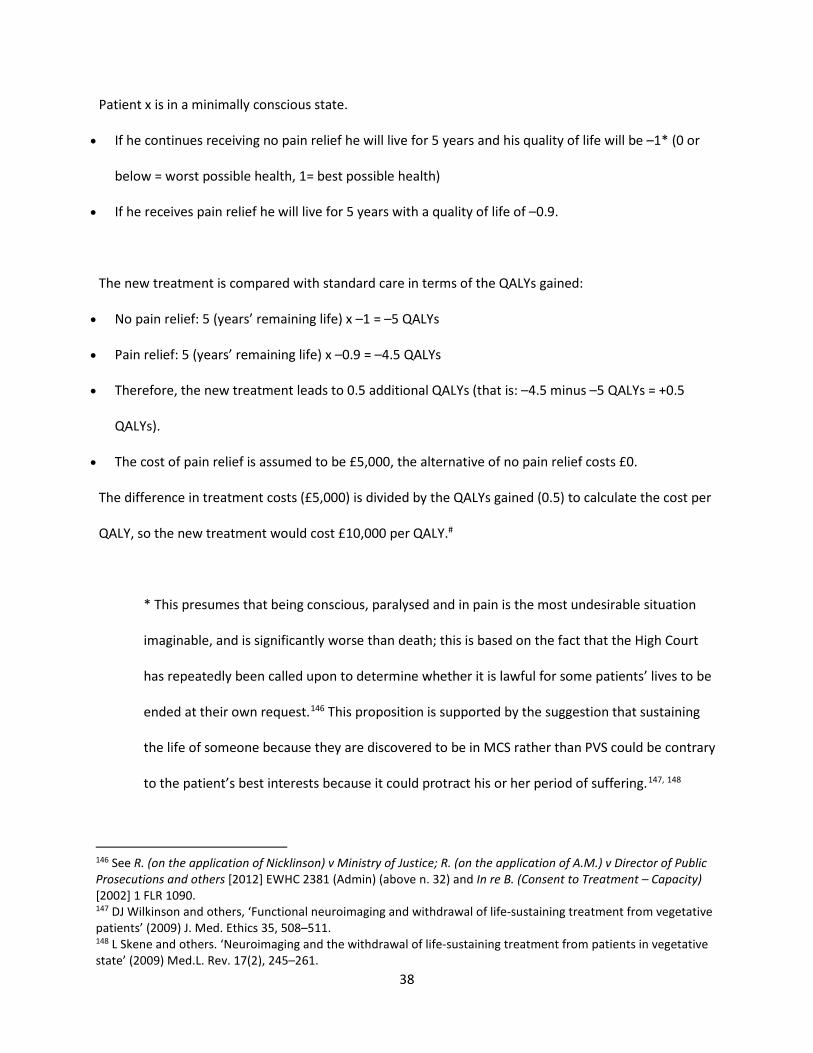
Patient x is in a minimally conscious state.
• If he continues receiving no pain relief he will live for 5 years and his quality of life will be –1* (0 or
below = worst possible health, 1= best possible health)
• If he receives pain relief he will live for 5 years with a quality of life of –0.9.
The new treatment is compared with standard care in terms of the QALYs gained:
• No pain relief: 5 (years’ remaining life) x –1 = –5 QALYs
• Pain relief: 5 (years’ remaining life) x –0.9 = –4.5 QALYs
• Therefore, the new treatment leads to 0.5 additional QALYs (that is: –4.5 minus –5 QALYs = +0.5
QALYs).
• The cost of pain relief is assumed to be £5,000, the alternative of no pain relief costs £0.
The difference in treatment costs (£5,000) is divided by the QALYs gained (0.5) to calculate the cost per
QALY, so the new treatment would cost £10,000 per QALY.#
* This presumes that being conscious, paralysed and in pain is the most undesirable situation
imaginable, and is significantly worse than death; this is based on the fact that the High Court
has repeatedly been called upon to determine whether it is lawful for some patients’ lives to be
ended at their own request.146 This proposition is supported by the suggestion that sustaining
the life of someone because they are discovered to be in MCS rather than PVS could be contrary
to the patient’s best interests because it could protract his or her period of suffering.147, 148
146 See R. (on the application of Nicklinson) v Ministry of Justice; R. (on the application of A.M.) v Director of Public Prosecutions and others [2012] EWHC 2381 (Admin) (above n. 32) and In re B. (Consent to Treatment – Capacity) [2002] 1 FLR 1090. 147 DJ Wilkinson and others, ‘Functional neuroimaging and withdrawal of life-sustaining treatment from vegetative patients’ (2009) J. Med. Ethics 35, 508–511. 148 L Skene and others. ‘Neuroimaging and the withdrawal of life-sustaining treatment from patients in vegetative state’ (2009) Med.L. Rev. 17(2), 245–261.
38

# A treatment is generally not considered by NICE to be cost-effective if it costs more than
£20,000–£30,000 per QALY, so this treatment would be considered cost-effective.
The life expectancy of five years and treatment cost of £5,000 are, in the absence of relevant data, very
rough estimates. There are no collated statistics on the life expectancy of patients following a diagnosis
of a DoC, but Cathy Kelly has been in PVS since 1989,149 so five years could be a realistic figure for some
patients, especially if the DoC had been caused by a traumatic injury. The cost of morphine for pain
control in cases of advanced cancer is only £1.25 per patient per week,150 but its use necessitates
additional nursing care to monitor and alleviate such side-effects as nausea and constipation. The
Quality, Innovation, Productivity and Prevention program estimates that the average cost of a hospital
death, following palliative care, is £3,000 ,151 so £5,000 may represent the total marginal cost, over five
years, of the additional nursing care that is required when a DoC patient – who already requires
constant care – is being treated with morphine.
The cost-effectiveness of the treatment would be significantly affected if the cost of neuroimaging were
included. The marginal cost of a scan appears to be between £500 and £1,000.152 £3,000 per patient
therefore seems likely to cover the associated transport and other costs, as well as the scan itself.
Incorporation of this into the above calculation leads to a cost per QALY for the same patient of £16,000,
which would still meet NICE’s criteria for cost-effectiveness. For many patients, however, neither life
expectancy nor quality of life would be affected by having been assessed by neuroimaging. For such
patients, there would be zero additional QALYs, meaning that the cost per QALY would be, theoretically,
149 R Fraser, ‘Taking care’ (2014) NLJ 164, 13. 150 J Silberner, ‘Morphine: The cheap, effective pain-relief drug denied to millions’ (2012) BBC News Magazine, 7 December. 151 T Hughes-Hallett and others, Palliative Care Funding Review (2011). Online; https://www.gov.uk/government/publications/independent-palliative-care-funding-review, accessed 9 October 2014. 152 Above n. 71 and n. 72.
39
infinite. Given the current state of neuroimaging techniques and their relative scarcity, therefore, it
seems certain that, across the population of people with DoCs, a blanket neuroimaging programme
would not be deemed cost-effective.
CONCLUSION
It is difficult, but vitally important, to distinguish patients who are unconscious due to coma or being in
VS from those who, because they either are in MCS or have LIS, have some awareness of themselves
and the world. Misdiagnosis can lead to inappropriate treatment and inaccurate prognoses, and it
carries the risk that those who are mistakenly classified as unconscious will undergo psychological
suffering and treatable pain. It is therefore a matter for significant concern that some studies have
suggested that approximately 40% of people diagnosed as being in VS are actually in MCS.
Several neuroimaging studies appear to have detected a degree of awareness in patients who, to all
normal bedside observations, appear to be unconscious. At present, there are significant practical
difficulties associated with fMRI, PET scanning and DTI and it does not seem realistic to recommend
these for universal use amongst patients with DoCs in the near future. These difficulties do not apply to
EEG, which some recent studies have suggested can discern signs of consciousness. fNRIS is also more
practical than magnetic scanning; it may, potentially, offer some of the same opportunities for
facilitating communication with visually unresponsive patients as does fMRI. Although the studies
outlined above seem to have enormous potential, it is important that the science that underlies them
should not be relied upon until it has been proved to be robust.
40

The ethical imperative of providing the best care to all patients requires regular re-assessment of
patients with DoCs using the best methods available. At present, this seems to be a proven structured
assessment tool such as the CRS–R, but within the next few years it may include at least one form of
neuroimaging. Such re-assessments would enable the compilation of an accurate database of patients
with DoCs, which is essential in order to ensure the provision of adequate specialised care facilities.
Although there are valid ethical concerns about conducting research on patients who are unable to
indicate their consent to it, it seems that the potential benefits to these patients – both individually and
as a group – outweigh them.
Cases decided by the ECtHR indicate that Article 3 of the ECHR could include protection from both
psychological suffering occasioned by incorrect diagnosis and physical pain that could be, but was not,
treated. Article 3 is an absolute right, so conventional jurisprudence would suggest that Contracting
States have an absolute duty to protect their citizens from these forms of suffering. The ECtHR has,
however, been pragmatic about Contracting States’ resources, and this consideration has also been
evident in some cases decided in the UK courts.
In the UK, QALYs are used to determine whether treatments are clinically effective. For any individual
patient in MCS, fMRI scanning might be cost-effective if it resulted in the administration of pain relief
that very slightly improved the quality of that patient’s life. Across the population of patients with DoCs,
however, such assessment would certainly not be deemed effective at the current stage of knowledge. If
future developments in any of the neuroimaging techniques had the effect of reducing cost, establishing
reliability and thus leading to benefits for a significant percentage of patients, the situation could change
dramatically. If this occurred, the UK – via its emanation, the NHS – could have an obligation under
human rights treaties to undertake regular neuroimaging re-assessments of all patients with DoCs, and
41

to attempt to facilitate communication with those who were in MCS or total LIS but unable to
communicate by conventional means. One would hope that anything that could reduce the risk of any
unnecessary suffering by these extremely vulnerable people would be offered without their needing to
resort to the law in order to enforce their human rights.
42