possible reversibility between epithelioid and sarcomatoid
TRANSCRIPT

RESEARCH Open Access
Possible reversibility between epithelioidand sarcomatoid types of mesothelioma isindependent of ERC/mesothelin expressionMasataka Kojima1,2, Kazunori Kajino1* , Shuji Momose3, Nadila Wali1, May Thinzar Hlaing1, Bo Han1, Liang Yue1,Masaaki Abe1, Tomoaki Fujii1,4, Katsuhisa Ikeda2 and Okio Hino1
Abstract
Background: Mesothelioma is histologically divided into three subgroups: epithelioid, sarcomatoid, and biphasictypes. The epithelioid or sarcomatoid type is morphologically defined by polygonal or spindle-like forms of cells,respectively. The biphasic type consists of both components. It is not yet understood how histologicaldifferentiation of mesothelioma is regulated. ERC/mesothelin is expressed in most cases of the epithelioid type, butnot in the sarcomatoid type of mesothelioma. Consequently, its expression is well correlated to the histologicalsubtype. We hypothesized that ERC/mesothelin expression influences the histological differentiation ofmesothelioma, and tested this hypothesis.
Methods: We performed studies using the overexpression or knockdown of ERC/mesothelin in mesothelioma cellsto examine its effect on cellular morphology, growth kinetics, or migration/invasion activity, in vitro. We thentransplanted ERC/mesothelin-overexpressing and control cells into the intraperitoneal space of mice. We examinedthe effect of ERC/mesothelin overexpression on mouse survival and tumor phenotype.
Results: In vitro cell culture manipulations of ERC/mesothelin expression did not affect cellular morphology orproliferation, although its overexpression enhanced cellular adhesion and the migration/invasion activity ofmesothelioma cells. The survival rate of mice following intraperitoneal transplantation of ERC/mesothelin-overexpressing mesothelioma cells was significantly lower than that of mice with control cells. The histologicalevaluation of the tumors, however, did not show any morphological difference between two groups, and ourhypothesis was not validated. Unexpectedly, both groups (ERC/mesothelin-overexpressing and control) ofmesothelioma cells that were morphologically monophasic and spindle-like in vitro differentiated into a biphasictype consisting of polygonal and spindle-like components in the transplanted tumor, irrespective of ERC/mesothelinexpression.
Conclusions: These results suggested that the histological transition of mesothelioma between epithelioid andsarcomatoid types may be reversible and regulated not by ERC/mesothelin, but by other unknown mechanisms.
Keywords: Mesothelioma, ERC/mesothelin, Epithelioid type, Sarcomatoid type, Histological reversibility
© The Author(s). 2020 Open Access This article is licensed under a Creative Commons Attribution 4.0 International License,which permits use, sharing, adaptation, distribution and reproduction in any medium or format, as long as you giveappropriate credit to the original author(s) and the source, provide a link to the Creative Commons licence, and indicate ifchanges were made. The images or other third party material in this article are included in the article's Creative Commonslicence, unless indicated otherwise in a credit line to the material. If material is not included in the article's Creative Commonslicence and your intended use is not permitted by statutory regulation or exceeds the permitted use, you will need to obtainpermission directly from the copyright holder. To view a copy of this licence, visit http://creativecommons.org/licenses/by/4.0/.The Creative Commons Public Domain Dedication waiver (http://creativecommons.org/publicdomain/zero/1.0/) applies to thedata made available in this article, unless otherwise stated in a credit line to the data.
* Correspondence: [email protected] of Pathology and Oncology, Juntendo University Faculty ofMedicine, 2-1-1, Hongo, Bunkyo-ku, Tokyo 113-8421, JapanFull list of author information is available at the end of the article
Kojima et al. Respiratory Research (2020) 21:187 https://doi.org/10.1186/s12931-020-01449-2

BackgroundExpressed in Renal Carcinoma (ERC) was first identifiedin a renal cell carcinoma of an Eker rat [1, 2], and is thehomolog of human mesothelin (MSLN) [3] or megakar-yocyte potentiating factor [4]. ERC/mesothelin isexpressed in normal mesothelium, and its expression isenhanced in epithelioid mesothelioma, ovarian cancer,and other malignancies [3–9]. Functionally, it is reportedto enhance cellular adhesive or invasive activities [10–12]. As for cellular proliferation, several groups have de-scribed how ERC/mesothelin has positive effects [13–15]; however, the other groups report no significant ef-fects [11, 12].Mesothelioma most commonly arises in the pleura,
and, at much lower frequency, also occurs in the peri-toneum, pericardium, and tunica vaginalis testis. It ishistologically divided into three subgroups: epithelioid,sarcomatoid, and biphasic (containing two components)types, consisting of approximately 60, 20, and 20%, re-spectively, of the pleural mesothelioma [16–18]. Mor-phologically, mesothelioma cells in the epithelioid typetake on a polygonal or cobblestone-like form, and thosein the sarcomatoid take on a spindle-like shape. The me-dian survival time after surgical therapy is 15–19, 4–10,and 10–12 months [17–19] in these groups, respectively,indicating that the prognosis of the sarcomatoid type ispoorer than that of the epithelioid one. At present, theunderlying mechanism that defines the histological dif-ferentiation into these subgroups is not yet known.ERC/mesothelin is expressed in most (75–100%) cases
of the epithelioid, but not sarcomatoid, type [20–22].Consequently, ERC/mesothelin expression and the histo-logical subtype of mesothelioma are well correlated. Wehypothesized that the expression status of ERC/mesothe-lin influences the morphological phenotype of meso-thelioma. To test this hypothesis, we examined theeffects of ERC/mesothelin overexpression or knockdownon cell morphology, as well as growth kinetics, adhesion,and migration/invasion of mesothelial cells in vitro. Wethen intraperitoneally transplanted ERC/mesothelin-overexpressing and control cells into mice, and exam-ined the effect of ERC/mesothelin on mouse survivaland tumor phenotype.
MethodsCell lines and antibodiesThe human mesothelioma cell lines NCI-H2452(H2452) and NCI-H226 (H226) were obtained from theAmerican Type Culture Collection (ATCC; Rockville,MD, USA), and ACC-MESO-4 (MESO4), which wasestablished at the Aichi Cancer Research Center Insti-tute [23], was obtained from RIKEN BioResource Center(RIKEN BRC; Tokyo, Japan). H226 and MESO4expressed endogenous ERC/mesothelin, but H2452 did
not (Fig. 1b). All cell lines were cultured in RPMI-1640medium supplemented with 10% fetal calf serum (FCS)at 37 °C in a 95% air/5% CO2 atmosphere.A mouse monoclonal anti-human C-ERC/mesothelin
antibody, 22A31, has been described previously [24].Other antibodies used in this study included mousemonoclonal anti-vimentin (clone V9), anti-cytokeratin(clone AE1/AE3), anti–E-cadherin (clone NCH-38), andanti–Ki-67 (clone MIB-1; Dako, Glostrup, Denmark);rabbit polyclonal anti-integrin α5 (#4705), anti-integrinβ1 (#4706), rabbit monoclonal anti-matrixmetalloproteinase-9 (MMP-9) (clone D603H; Cell Sig-naling Technology Japan, Tokyo, Japan); rabbit poly-clonal anti-Twist (H81) (Santa Cruz Biotechnology, CA,USA); and rabbit polyclonal anti-ZEB1 (HPA027524;Sigma–Aldrich/ Merck, KGaA, Darmstadt, Germany).
Western blottingA cellular lysate (30 μg) was harvested in 2% SDS, 10%glycerol, 50 mM Tris-HCl (pH 6.8), and 100mM dithio-threitol. After boiling for 2 min, samples were electro-phoresed on 10% polyacrylamide gels and transferredonto polyvinylidene difluoride membranes (Immobilon-P, Merck Millipore, Burlington, MA, USA). The mem-branes were blocked in 1% skim milk in phosphate-buffered saline (PBS) with 0.1% Tween-20 (PBS-T) for 1h at room temperature. The membranes were then incu-bated with anti–C-ERC/mesothelin (1 μg/mL), anti-vimentin (1:200), anti–E-cadherin (1:250), anti-integrinα5, anti-integrin β1, anti-MMP-9 (1:1000), anti-Twist (1:200), or anti-ZEB1 (1:250) at room temperature for 1 hin 1% skim milk in PBS-T. Goat anti-mouse or anti-rabbit Ig conjugated with peroxidase labeled-dextranpolymer (Envision+ System, Dako) was used as a sec-ondary antibody at a dilution of 1:100 in 1% skim milkin PBS-T at room temperature for 1 h. An ECL detectionsystem (GE Healthcare, Chicago, IL, USA) was employedto visualize proteins on membranes. ECL signals weredetected and quantified by a ChemiDoc MP imaginganalyzer (Bio-Rad, Tokyo, Japan). The expression levelof ß-actin was used as an internal control for the deter-mination of equal loading.
ImmunohistochemistryThree–micrometer thick tissue sections were preparedfrom formalin-fixed, paraffin-embedded specimens.After deparaffinization, tissue sections were heated in10mM citrate buffer (pH 6) for antigen retrieval andtreated with 3% hydrogen peroxide. The sections wereincubated with primary antibodies diluted in Tris-buffered saline with 0.1% Tween 20 overnight at 4 °C.Anti–E-cadherin, anti–Ki-67, and anti-vimentin anti-bodies were diluted at 1:200, anti-AE1/AE3 antibodieswas diluted at 1:400, and anti-Twist and anti-ZEB1 were
Kojima et al. Respiratory Research (2020) 21:187 Page 2 of 11

diluted at 1:100. Anti–C-ERC/mesothelin antibody wasused at 2 μg/mL. Immunohistochemistry (IHC) usingmouse monoclonal antibodies was performed with aHistofine Mouse Stain kit (Nichirei, Tokyo, Japan), andthat using the anti-rabbit antibody was performed withan Envision+ System secondary antibody (Dako). Diami-nobenzidine was used as the substrate.
ERC/mesothelin overexpression in H2452 cells usinglentivirus vectorHEK293T, which was used as the packaging cell line,was cotransfected with Precision LentiORF for MSLNand trans-lentiviral packaging vectors (Thermo ScientificOpen Biosystems, Waltham, MA, USA). A LentiORF-MSLN vector encoded ERC/mesothelin and Turbo greenfluorescent protein (GFP). A vector in which ERC/mesothelin was replaced with Turbo red fluorescent pro-tein (RFP) was used as a negative control. Sixteen hoursafter transfection, we microscopically confirmed thepresence of GFP- or RFP-positive cells, and the medium
was changed to that with 5% FCS. Forty-eight hoursafter medium change, supernatants were harvested andtheir infectivity on H2452 cells was titrated by countingthe number of TurboGFP- or TurboRFP-positive cells.To establish stable ERC/mesothelin- or RFP-expressingcells, we infected H2452 cells with the titrated super-natant at a multiplicity of infection of 2.0, and selectedcells that were resistant to 2.0 μg/mL blasticidin S.
SiRNA transfection to knock down ERC/mesothelin inH226 cellsON-TARGET plus Human MSLN siRNAs, including(5′-CAUUGGACCUGCUGCUAUU-3′), (5′-ACAUGAACGGGUCCGAAUA-3′), and (5′-GAUGAGCUCUACCCACAAG-3′), and ON-TARGET plus Non-targeting Pool siRNA (negative control) were purchasedfrom Dharmacon/GE Healthcare (Lafayette, CO, USA).H226 cells were seeded at 7.5 × 10⁴ in 3-cm plates.Twenty-four hours later, the cells were transfected with10 nM siRNA or with transfection reagent
Fig. 1 Effects of ERC/mesothelin expression on mesothelioma cells. a, Effects of ERC/mesothelin overexpression (left column) or knockdown (rightcolumn) on the morphology of H2452 or H226 cells, respectively, as observed by phase-contrast microscopy (objective lens × 10). Top panels:parental H2452 or H226; Middle panels: H2452 overexpressing ERC/mesothelin or H226 treated with siRNA of ERC/mesothelin; Bottom panels:H2452 or H226 treated with control vector or control siRNA. b, Effects of ERC/mesothelin overexpression on the expression of E-cadherin,vimentin and MMP-9 assessed by western blotting. Note that MESO4 (ACC-MESO-4) and H226 cells were used as positive and negative controls,respectively, for E-cadherin. c, Effects of three siRNAs of ERC/mesothelin (si5, si6, si7) on expression. RFP, red fluorescent protein; MMP-9,matrix metalloproteinase-9
Kojima et al. Respiratory Research (2020) 21:187 Page 3 of 11
(Lipofectamine RNAiMAX; Invitrogen, Carlsbad, CA,USA) alone. In the following 96 h, cellular morphologyand proliferative states were observed. For western blot-ting, cell lysates were harvested 48 h after siRNAtransfection.
Cell adhesion assayFlat 96-well plates were coated with Matrigel (Corning,Corning, NY, USA; 100 μg/mL, 100 μL/well) or fibronec-tin (Corning; 20 μg/mL, 100 μL/well) and then incubatedat 37 °C in a 5% CO2 atmosphere for 1 h. The coatedwells were washed twice with 0.1% bovine serum albu-min (BSA), blocked with 0.5% BSA for 1 h at 37 °C in a5% CO2 atmosphere, and then washed with 0.1% BSAagain. Cells were seeded at 2 × 10⁴ cells/well and incu-bated for 1 h at 37 °C in a 5% CO2 atmosphere. Theywere then washed twice with PBS, fixed with 4% parafor-maldehyde for 10 min, and then washed with PBS again.The cells were stained with 1% Crystal Violet at roomtemperature for 10 min. Solubilization of Crystal Violetwas performed in 33% acetic acid, and the absorbancewas measured at 550 nm. The measurements were con-ducted in triplicate for each experimental group.
Scratch wound migration/invasion assaysIncuCyte ImageLock 96-well plates (Essen BioScience,Tokyo, Japan) were coated with Matrigel at 100 μg/mLand incubated overnight at 37 °C. Cells were seeded at6 × 104 cells/well and allowed to adhere on top of a thinlayer of Matrigel for 4 h at 37 °C. A wound was createdwith a 96-well WoundMaker (Essen Bioscience). MoreMatrigel (6 mg/mL, 50 μL/well) was overlaid on top ofthe cells to create a three-dimensional matrix. Finally, anIncuCyte ZOOM live-cell imaging and analysis platform(Essen Bioscience) was used to quantify invading cells inthe wound area.
Cell proliferation assayCells (1 × 10³ cells/well) in RPMI-1640 with 10% FCSwere seeded in flat 96-well dishes, and incubated at37 °C in a 5% CO2 atmosphere. The area of proliferatingcells was scanned and quantified by the IncuCyteZOOM system (Essen Bioscience) every 3 h for 96 h.
Animal experimentsAll in vivo studies were approved by the Institute Ani-mal Care and Use Committee of Juntendo University.Female BALB/c athymic nude (BALB/c nu/nu) mice at6 weeks of age were purchased from Charles River Japan(Yokohama, Japan). After 14 days of acclimatization,2.5 × 106 of ERC/mesothelin-overexpressing or controlH2452 cells were injected into the intraperitoneal (IP)space of the mice. The mice were euthanized when theyshowed moribund sign, or on day 70 after injection. The
IP space was opened, and any tumors present were har-vested. All mice were maintained under specificpathogen-free conditions.
Statistical analysisWe used Student’s t test to evaluate differences betweentwo groups. Data represent the mean ± standard devi-ation (SD). The survival rate of mice was compared bythe Kaplan–Meier method, and log-rank tests were usedto estimate statistical significance between two groups.P < 0.05 was considered statistically significant.
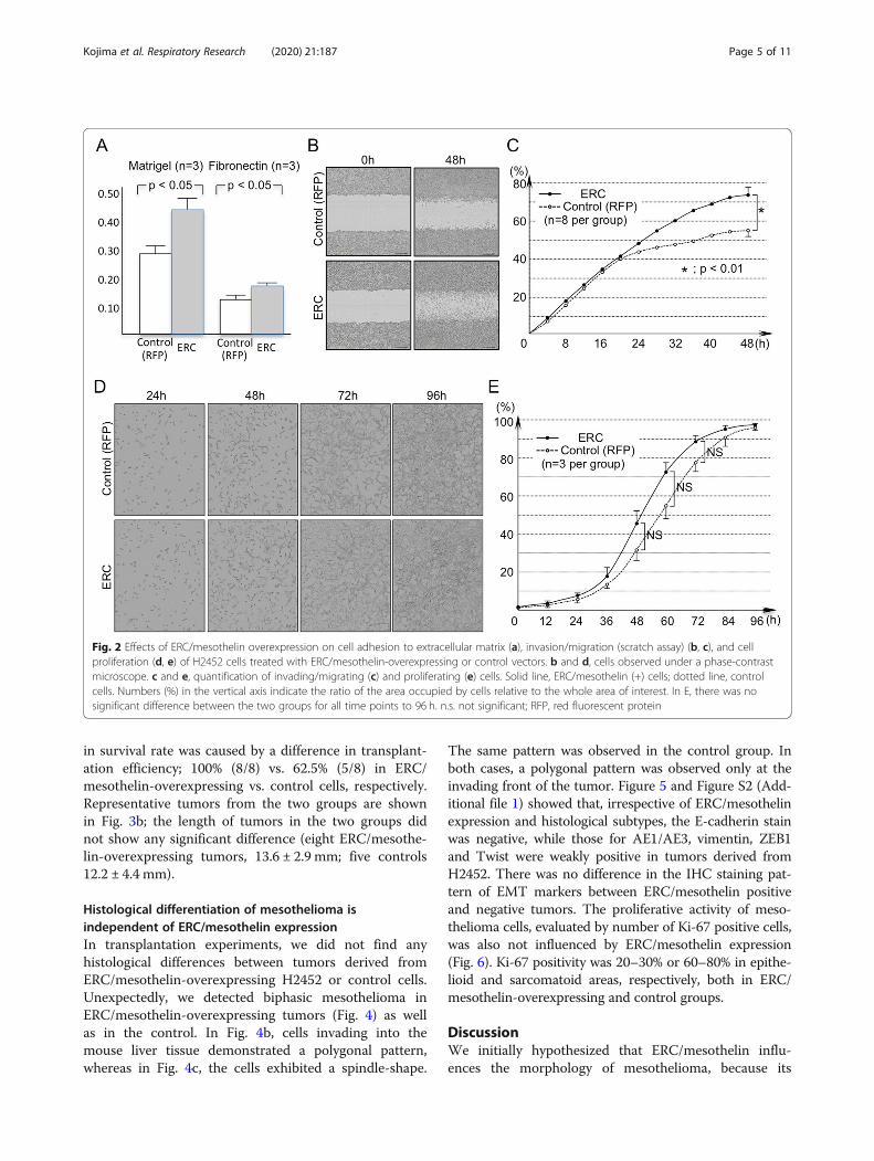
ResultsThe effects of ERC/mesothelin expression onmesothelioma cells in vitroWe hypothesized that the expression of ERC/mesothelininfluences the morphology of cells, as its expression iswell correlated to the histological subtypes of mesotheli-oma. Thus, we examined the effect of ERC/mesothelinoverexpression or knockdown on cellular morphology.As shown in Fig. 1a, ERC/mesothelin overexpression inH2452 (spindle-shaped) or knockdown in H226 (polyg-onal) cells did not affect cell morphology. The overex-pression or knockdown of ERC/mesothelin wasconfirmed in Fig. 1b and c. The manipulation of ERC/mesothelin expression did not have any effect on epithe-lial–mesenchymal transition (EMT) markers such as E-cadherin, vimentin (Fig. 1b), or ZEB1, or Twist (Add-itional file 1: Figure S1). We then examined the effectsof ERC/mesothelin on cellular activities. We found thatERC/mesothelin overexpression enhanced cellular adhe-sion (Fig. 2a) and migration/invasion (Fig. 2b and c) withregard to the extracellular matrix (ECM), but did not in-fluence cellular proliferation (Fig. 2d and e). The expres-sion of MMP-9 was enhanced in ERC/mesothelin-overexpressing cells (Fig. 1b), but that of integrin α5 andintegrin β1 remained unchanged (Additional file 1: Fig-ure S1).
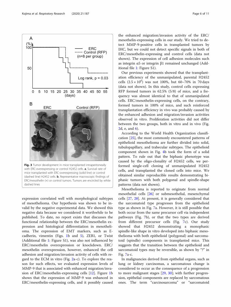
The effect of ERC/mesothelin expression onmesothelioma cells in vivoFollowing transplantation of ERC/mesothelin-overex-pressing H2452 cells into the IP space of nude mice(n = 8), we observed that the overall survival period ofthese mice was significantly shorter than those trans-planted with control H2452 (n = 8; Fig. 3a). By 40 days’post-transplantation, all eight mice transplanted withERC/mesothelin-overexpressing H2452 were euthanizedbecause of moribund sign, or found dead without thesign. On the other hand, three out of eight control micesurvived until 70 days’ post-transplantation, when theywere euthanized and tumors were not found. All 13mice that were euthanized or found dead before 70 dayshad developed tumors in IP spaces. Thus, the difference
Kojima et al. Respiratory Research (2020) 21:187 Page 4 of 11
in survival rate was caused by a difference in transplant-ation efficiency; 100% (8/8) vs. 62.5% (5/8) in ERC/mesothelin-overexpressing vs. control cells, respectively.Representative tumors from the two groups are shownin Fig. 3b; the length of tumors in the two groups didnot show any significant difference (eight ERC/mesothe-lin-overexpressing tumors, 13.6 ± 2.9 mm; five controls12.2 ± 4.4 mm).
Histological differentiation of mesothelioma isindependent of ERC/mesothelin expressionIn transplantation experiments, we did not find anyhistological differences between tumors derived fromERC/mesothelin-overexpressing H2452 or control cells.Unexpectedly, we detected biphasic mesothelioma inERC/mesothelin-overexpressing tumors (Fig. 4) as wellas in the control. In Fig. 4b, cells invading into themouse liver tissue demonstrated a polygonal pattern,whereas in Fig. 4c, the cells exhibited a spindle-shape.
The same pattern was observed in the control group. Inboth cases, a polygonal pattern was observed only at theinvading front of the tumor. Figure 5 and Figure S2 (Add-itional file 1) showed that, irrespective of ERC/mesothelinexpression and histological subtypes, the E-cadherin stainwas negative, while those for AE1/AE3, vimentin, ZEB1and Twist were weakly positive in tumors derived fromH2452. There was no difference in the IHC staining pat-tern of EMT markers between ERC/mesothelin positiveand negative tumors. The proliferative activity of meso-thelioma cells, evaluated by number of Ki-67 positive cells,was also not influenced by ERC/mesothelin expression(Fig. 6). Ki-67 positivity was 20–30% or 60–80% in epithe-lioid and sarcomatoid areas, respectively, both in ERC/mesothelin-overexpressing and control groups.
DiscussionWe initially hypothesized that ERC/mesothelin influ-ences the morphology of mesothelioma, because its
Fig. 2 Effects of ERC/mesothelin overexpression on cell adhesion to extracellular matrix (a), invasion/migration (scratch assay) (b, c), and cellproliferation (d, e) of H2452 cells treated with ERC/mesothelin-overexpressing or control vectors. b and d, cells observed under a phase-contrastmicroscope. c and e, quantification of invading/migrating (c) and proliferating (e) cells. Solid line, ERC/mesothelin (+) cells; dotted line, controlcells. Numbers (%) in the vertical axis indicate the ratio of the area occupied by cells relative to the whole area of interest. In E, there was nosignificant difference between the two groups for all time points to 96 h. n.s. not significant; RFP, red fluorescent protein
Kojima et al. Respiratory Research (2020) 21:187 Page 5 of 11
expression correlated well with morphological subtypesof mesothelioma. Our hypothesis was shown to be in-valid by the negative experimental data. We showed thisnegative data because we considered it worthwhile to bepublished. To date, no report exists that discusses thefunctional relationship between the ERC/mesothelin ex-pression and histological differentiation in mesotheli-oma. The expression of EMT markers, such as E-cadherin, vimentin (Figs. 1b and 5), ZEB1, or Twist(Additional file 1: Figure S1), was also not influenced byERC/mesothelin overexpression or knockdown. ERC/mesothelin overexpression, however, enhanced the celladhesion and migration/invasion activity of cells with re-gard to the ECM in vitro (Fig. 2a-c). To explore the rea-son for such effects, we evaluated expression levels ofMMP-9 that is associated with enhanced migration/inva-sion of ERC/mesothelin-expressing cells [12]. Figure 1bshows that the expression of MMP-9 was enhanced inERC/mesothelin-expressing cells, and it possibly caused
the enhanced migration/invasion activity of the ERC/mesothelin-expressing cells in our study. We tried to de-tect MMP-9-positive cells in transplanted tumors byIHC, but we could not detect specific signals in both ofERC/mesothelin-expressing and control cells (data notshown). The expression of cell adhesion molecules suchas integrin α5 or integrin β1 remained unchanged (Add-itional file 1: Figure S1).Our previous experiments showed that the transplant-
ation efficiency of the unmanipulated, parental H2452cells (2.5 × 10⁶) was not 100%, but 60–70% in 70 days(data not shown). In this study, control cells expressingRFP formed tumors in 62.5% (5/8) of mice, and a fre-quency was almost identical to that of unmanipulatedcells. ERC/mesothelin-expressing cells, on the contrary,formed tumors in 100% of mice, and such reinforcedtransplantation efficiency in vivo was probably caused bythe enhanced adhesion and migration/invasion activitiesobserved in vitro. Proliferation activities did not differbetween the two groups, both in vitro and in vivo (Fig.2d, e, and 6).According to the World Health Organization classifi-
cation [25], the most commonly encountered patterns ofepithelioid mesothelioma are further divided into solid,tubulopapillary, and trabecular subtypes. The epithelioidcomponent shown in Fig. 4b took the form of a solidpattern. To rule out that the biphasic phenotype wascaused by the oligo-clonality of H2452 cells, we per-formed single-cell cloning of unmanipulated H2452cells, and transplanted the cloned cells into mice. Weobtained similar reproducible results demonstrating bi-phasic tumors with both polygonal and spindle-shapepatterns (data not shown).Mesothelioma is reported to originate from normal
mesothelial cells [26] or submesothelial, mesenchymalcells [27, 28]. At present, it is generally considered thatthe sarcomatoid type progresses from the epithelioidtype as shown in Fig. 7a. However, it is still possible thatboth occur from the same precursor cell via independentpathways (Fig. 7b), or that the two types are derivedfrom different precursor cells (Fig. 7c). Our studyshowed that H2452 demonstrating a monophasicspindle-like shape in vitro developed into biphasic meso-thelioma with both epithelioid (polygonal) and sarcoma-toid (spindle) components in transplanted mice. Thissuggests that the transition between the epithelioid andsarcomatoid types may be reversible, as shown by “?” inFig. 7a-c.In malignancies derived from epithelial organs, such as
lung or kidney carcinomas, a sarcomatous change isconsidered to occur as the consequence of a progressionto more malignant stages [29, 30]; with further progres-sion, epithelial components are replaced by sarcomatousones. The term “carcinosarcoma” or “sarcomatoid
Fig. 3 Tumor development in mice transplanted intraperitoneallywith ERC-overexpressing or control H2452 cells. a, Survival rate ofmice transplanted with ERC-overexpressing (solid line) or control(dashed line) H2452 cells. b, Representative macroscopic findings ofERC/mesothelin (+) or control tumors. Tumors are encircled by whitedashed lines
Kojima et al. Respiratory Research (2020) 21:187 Page 6 of 11

carcinoma” refers to biphasic states in which both theepithelial and sarcomatous components coexist. The fre-quency of carcinosarcoma is very low, less than 1% of alllung or kidney malignancies [31, 32]. In contrast, incases of mesothelioma, the biphasic type represents as
much as 20% of all cases [16–18]. This high frequencyof the biphasic type suggests that epithelioid and sarco-matoid types may be interchangeable or reversible.H2452 (NCI-H2452) is a cell line established from epi-
thelioid mesothelioma according to the ATCC. In
Fig. 4 Histology of a representative tumor derived from transplanted H2452 cells that overexpressed ERC/mesothelin. a, Hematoxylin & eosin(HE)-stained findings at lower magnification. Higher magnification images of the boxed areas are shown in b and c. The white dashed linesdemarcate the border between invading mesothelioma cells and mouse liver. Scale bars, 100 μm in all three figures
Fig. 5 Hematoxylin & eosin (HE) staining and immunostaining for ERC/mesothelin, E-cadherin, AE1/AE3 and vimentin, in epithelioid orsarcomatoid areas in both ERC/mesothelin-overexpressing and control tumors derived from H2452. In images of epithelioid areas (top and thirdrows), the white dashed lines demarcate the border between invading mesothelioma cells (lower right) and mouse liver (upper left). Scale bars,50 μm in all figures
Kojima et al. Respiratory Research (2020) 21:187 Page 7 of 11

in vitro culture, cells exhibited a fibroblastic form andbehaved like sarcomatoid cells. This phenomenon alsoimplied the histological reversibility of mesothelioma.H2452 has multiple mutations in tumor suppressorgenes, including a missense mutation in BAP1 [33], atruncation of p53 [34], and a homozygous deletion ofCDKN2A [35] and NF2 [36]. It was significant for usthat the cell line, which harbored mutations and showedmonophasic morphology in cell culture, became biphasicin vivo.Several reports have investigated the expression of
EMT and mesenchymal–epithelial transition (MET)markers in epithelioid and sarcomatoid mesotheliomas[37, 38]. The significance of EMT/MET in the develop-ment of mesothelioma is still controversial. In our study,the histological differentiation of H2452 to polygonaland spindle-shaped components occurred withoutchanges in expression of EMT markers such as E-cadherin, AE1/AE3, vimentin (Fig. 5), and ZEB1 andTwist (Additional file 1: Figure S2).
Fig. 6 Immunostaining for Ki-67 in epithelioid or sarcomatoid areasin both ERC/mesothelin-overexpressing and control tumors derivedfrom H2452 cells. Representative areas in each tumor are shown.Scale bars, 100 μm in all figures
Fig. 7 Three possible pathways (a, b, c) for progression from precursor cells to epithelioid or sarcomatoid mesothelioma. Question marks (?) in a,b, and c indicate possible reversibility of the two subtypes
Kojima et al. Respiratory Research (2020) 21:187 Page 8 of 11

Our data suggested that the morphological differenti-ation of mesothelioma is reversible. What kind of mech-anisms are regulating it? We must consider not onlyintrinsic factors of mesothelioma cells, but also microen-vironmental factors associating with them. As for the in-trinsic ones, multiple studies showed that normalmesothelial cells have the ability to change phenotypeand behave like multipotent stem cells that can differen-tiate to smooth muscle cells or fibroblasts [39–41]. Con-sidering these findings, it is possible that mesotheliomamaintains the characteristics of multipotency even afterthe acquirement of a malignant character. In humanmesothelioma cases, CAM5.2 and AE1/AE3, both ofwhich are usually used as the epithelial markers, areexpressed in the sarcomatoid type [42, 43], and vimen-tin, which is one of the mesenchymal markers, isexpressed in the epithelioid one [21]. Therefore, the ex-pression of molecules conventionally used as epithelialor mesenchymal markers are not well associated withthe morphology of mesothelioma. There should be someother unknown molecules that regulate its differenti-ation. Several studies analyzed differences in gene ex-pression patterns between epithelioid and sarcomatoidsubtypes. Lopez-Rios et al. reported that uroplakins 1B,3B and kallikrein 11 are more prominently expressed inthe epithelioid types [44]. De Rienzo et al. showed thatmolecules associated with tyrosine kinase signaling,germ cell development, and regulation of cell prolifera-tion are upregulated in the epithelioid mesothelioma[45]. At present, it is not known whether any of thesemolecules are working as the regulating factors for thedifferentiation of mesothelioma. They compared geneexpression in mesotheliomas with different genetic back-grounds, that could induce some nonspecific effects. Weare currently examining the expression and mutationpatterns of genes in epithelioid or sarcomatoid compo-nents with an identical genetic background, using ourexperimental systems and laser microdissection. As forthe microenvironmental factors relevant to the differen-tiation of mesothelioma, Fig. 4 showed interesting find-ings. The transplanted H2452 took a polygonal,epithelioid pattern at the invasion front where it con-tacted with the host hepatocytes, and in the distant areafrom the front the cell took a spindle-like, sarcomatoidpattern. Polygonal host hepatocytes seemed to havesome effect on the morphology of the adjacent meso-thelioma cells with unknown mechanisms. Matsukumaet al. observed that metastatic cancer to the pancreasshowed the morphology resembling to that of primarypancreatic cancer, and proposed the concept of “mim-icry” of the metastatic cells to the primary carcinoma inthe site of metastasis [46]. Shepherd and Hall also re-ported the similar findings in metastatic cancer in thecolon [47]. The findings in Fig. 4 may be reflecting the
phenomenon of “mimicry”, although its molecularmechanisms are not yet known.
ConclusionsOur initial hypothesis that ERC/mesothelin regulates thehistological differentiation of mesothelioma was not sup-ported by the experimental data. Instead, mesotheliomacells with a monophasic morphology in culture devel-oped into biphasic cells in a mouse model, regardless ofthe expression of ERC/mesothelin. These results sug-gested that the histological differentiation of mesotheli-oma (epithelioid vs. sarcomatoid) may be reversible andregulated by mechanisms other than those for ERC/mesothelin or EMT/MET. Further molecular studiesboth of intrinsic factors in mesothelioma cells and mi-croenvironmental factors associating with them are re-quired to elucidate the the mechanisms of differentiationof mesothelioma.
Supplementary informationSupplementary information accompanies this paper at https://doi.org/10.1186/s12931-020-01449-2.
Additional file 1 : Figure S1. Effects of ERC/mesothelin overexpressionon the expression of EMT markers (ZEB1 and Twist), Integrins α5 and β1,assessed by western blotting. Figure S2. HE staining andimmunostaining for ZEB1 and Twist, in the epithelioid or sarcomatoidareas in both of ERC/mesothelin-overexpressing and control tumorsderived from H2452. In figures of epithelioid area (top and third figures ineach column), the white dotted lines demarcate the border between theinvading mesothelioma cell (lower) and mouse liver (upper). Scale bars,50 μm in all figures.
AbbreviationsATCC: American type culture collection; BAP1: BRCA1 associated protein-1;CDKN2A: Cyclin-dependent kinase inhibitor 2A; ECM: Extracellular matrix;EMT: Epithelial-mesenchymal transition; ERC: Expressed in renal carcinoma;FCS: Fetal calf serum; GFP: Green fluorescent protein;IHC: Immunohistochemistry; MET: Mesenchymal-epithelial transition; MMP-9: Matrix metalloproteinase-9; MSLN: Mesothelin; NF2: Neurofibromin 2;PBS: Phosphate-buffered saline; PBS-T: PBS with 0.1% Tween-20; RFP: Redfluorescent protein; RIKEN BRC: RIKEN bio-resource center; SDS: Sodiumdodecyl sulfate; SD: Standard deviation; siRNA: Small interfering RNA;ZEB1: Zinc finger E-box-binding homeobox 1
AcknowledgementsThe authors thank Ms. T. Ikegami, Ms. T. Ikeda, Ms. M. Kikkawa, and Mr. T.Takagaki for their technical assistance in experiments.
Authors’ contributionsMK established the ERC/mesothelin-overexpressing cells and those withknocked down ERC/mesothelin and studied effects on mesothelioma cells.KK designed the study and drafted the manuscript. SM, TF, KI, and OHsubstantially contributed to the analysis and interpretation of data. NW, MTH,BH, and LY contributed to the acquisition of data. MA performed the animalexperiments. All authors read and approved the final manuscript.
FundingThis study was supported in part by a Grant-in-Aid (221S0001) for ScientificResearch on Innovative Areas from the Japan Society for the Promotion ofScience, Grants-in-Aid (S1311011 and S1511008L) from the Foundation ofStrategic Research Projects in Private Universities of the Ministry of Education,Culture, Sports, Science and Technology of Japan (MEXT), and grants fromShizuoka Medical Research Center for Disaster of Juntendo University
Kojima et al. Respiratory Research (2020) 21:187 Page 9 of 11

Shizuoka Hospital, and from the Institute for Environmental and Gender-Specific Medicine of Juntendo University Urayasu Hospital.
Availability of data and materialsData sharing is not applicable to this article as no datasets were generatedor analysed during the current study.
Ethics approval and consent to participateAll animal experiments in this study were approved by the Institute AnimalCare and Use Committee of Juntendo University.
Consent for publicationNot applicable.
Competing interestsThe authors declare that they have no competing interests.
Author details1Department of Pathology and Oncology, Juntendo University Faculty ofMedicine, 2-1-1, Hongo, Bunkyo-ku, Tokyo 113-8421, Japan. 2Department ofOtorhinolaryngology, Juntendo University Faculty of Medicine, 2-1-1, Hongo,Bunkyo-ku, Tokyo 113-8421, Japan. 3Department of Pathology, SaitamaMedical Center, Saitama Medical University, 1981, Kamoda, Kawagoe, Saitama350-8550, Japan. 4Division of Animal Genetics, Laboratory Animal ResearchCenter, Institute of Medical Science, University of Tokyo, Tokyo 108-8639,Japan.
Received: 29 January 2020 Accepted: 8 July 2020
References1. Hino O, Kobayashi E, Nishizawa M, Kubo Y, Kobayashi T, Hirayama Y, et al.
Renal carcinogenesis in the Eker rat. J Cancer Res Clin Oncol. 1995;121:602–5.
2. Yamashita Y, Yokoyama M, Kobayashi E, Takai S, Hino O. Mapping anddetermination of the cDNA sequence of the Erc gene preferentiallyexpressed in renal cell carcinoma in the Tsc2 gene mutant (Eker) rat model.Biochem Biophys Res Commun. 2000;275:134–40.
3. Chang K, Pastan I. Molecular cloning of mesothelin, a differentiation antigenpresent on mesothelium, mesotheliomas, and ovarian cancers. Proc NatlAcad Sci U S A. 1996;93:136–40.
4. Kojima T, Oh-Eda M, Hattori K, Taniguchi Y, Tamura M, Ochi N, et al.Molecular cloning and expression of megakaryocyte potentiating factorcDNA. J Biol Chem. 1995;270:21984–90.
5. Scholler N, Fu N, Yang Y, Ye Z, Goodman GE, Hellstrom KE, et al. Solublemember(s) of the mesothelin/megakaryocyte potentiating factor family aredetectable in sera from patients with ovarian carcinoma. Proc Natl Acad SciU S A. 1999;96:11531–6.
6. Frierson HF Jr, Moskaluk CA, Powell SM, Zhang H, Cerilli LA, Stoler MH, et al.Large-scale molecular and tissue microarray analysis of mesothelinexpression in common human carcinomas. Hum Pathol. 2003;34:605–9.
7. Cao D, Ji H, Ronnett BM. Expression of mesothelin, fascin, and prostate stemcell antigen in primary ovarian mucinous tumors and their utility indifferentiating primary ovarian mucinous tumors from metastatic pancreaticmucinous carcinomas in the ovary. Int J Gynecol Pathol. 2004;24:67–72.
8. Hough CD, Sherman-Baust CA, Pizer ES, Montz FJ, Im DD, Rosenshein NB,et al. Large-scale serial anarysis of gene expression reveals genesdifferentially expressed in ovarian cancer. Cancer Res. 2000;60:6281–7.
9. Argani P, Iacobuzio-Donahue C, Ryu B, Rosty C, Goggins M, Wilentz RE, et al.Mesothelin is overexpressed in the vast majority of ductal adenocarcinomasof the pancreas: identification of a new pancreatic cancer marker by serialanalysis of gene expression (SAGE). Clin Cancer Res. 2001;7:3862–8.
10. Rump A, Morikawa Y, Tanaka M, Minami S, Umesaki N, Takeuchi M, et al.Binding of ovarian cancer antigen CA125/MUC16 to mesothelin mediatescell adhesion. J Biol Chem. 2004;279:9190–8.
11. Chen S-H, Hung W-C, Wang P, Paul C, Konstantopoulos K. Mesothelinbinding of CA125/MUC16 promotes pancreatic cancer cell motility andinvasion via MMP-7 activation. Sci Rep. 2013;3:1870. https://doi.org/10.1038/srep01870.
12. Servais EL, Colovos C, Rodriguez L, Bograd AJ, Nitadori J, Sima C, et al.Mesothelin overexpression promotes mesothelioma cell invasion and MMP-
9 secretion in an orthotopic mouse model and in epithelioid pleuralmesothelioma patients. Clin Cancer Res. 2012;18:2478–89.
13. Bharadwaj U, Li M, Chen C, Yao Q. Mesothelin-induced pancreatic cancercell proliferation involves alteration of cyclin E via activation of signaltransducer and activator of transcription protein 3. Mol Cancer Res. 2008;6:1755–65.
14. Yin D-D, You L-H, Yuan Q-X, Liang X-D, Wang N, Wang L-T, et al. Mesothelinpromotes cell proliferation in the remodeling of neonatal rat pancreas.World J Gastroenterol. 2014;20:2219–40.
15. Wang K, Bodempudi V, Liu Z, Borrego-Diaz E, Yamoutpoor Y, Meyer A, et al.Inhibition of mesothelin as a novel strategy for targeting cancer cells. PLoSONE. 2012;7:e33214.
16. Inai K. Pathology of mesothelioma. Environ Health Prev Med. 2008;13:60–4.17. Yap TA, Aerts JG, Popat S, Fennell DA. Novel insights into mesothelioma
biology and implications for therapy. Nat Rev Cancer. 2017;17:475–88.18. Milano MT, Zhang H. Malignant pleural mesothelioma: a population-based
study of survival. J Thorac Oncol. 2010;5:1841–8.19. Meyerhoff RR, Yang C-FJ, Speicher PJ, Gulack BC, Hartwig MG, D'Amico TA, et al.
Impact of mesothelioma histologic subtype on outcomes in the surveillance,epidemiology, and end results database. J Surg Res. 2015;196:23–32.
20. Ordonez NG. Value of mesothelin immunostaining in the diagnosis ofmesothelioma. Mod Pathol. 2003;16:192–7.
21. Kushitani K, Takeshima Y, Amatya VJ, Furonaka O, Sakatani A, Inai K.Immunohistochemical marker panels for distinguishing between epithelioidmesothelioma and lung carcinoma. Pathol Int. 2007;57:190–9.
22. Miettinen M, Sarlomo-Rikala M. Expression of calretinin, thrombomodulin,keratin 5, and mesothelin in lung carcinomas of different types: animmunohistochemical analysis of 596 tumors in comparison withepithelioid mesotheliomas of the pleura. Am J Surg Pathol. 2003;27:150–8.
23. Usami N, Fukui T, Kondo M, Taniguchi T, Yokoyama Y, Mori S, et al.Establishment and characterization of four malignant pleural mesotheliomacell lines from Japanese patients. Cancer Sci. 2006;97:387–94.
24. Ishikawa K, Segawa T, Hagiwara Y, Maeda M, Abe M, Hino O. Establishmentof novel mAb to human ERC/mesothelin useful for study and diagnosis ofERC/mesothelin-expressing cancers. Pathol Int. 2009;59:161–6.
25. Galateau-Salle F, Dacic S, Ordonez NG, Churg A, Hammar S, Rice DC, et al.Epithelioid mesothelioma. In: Travis WD, Brambilla E, Burke AP, Marx A,Nicholson AG, editors. WHO classification of tumours of the lung, pleura,thymus and heart. 4th ed. Lyon: International Agency for Research onCancer; 2015. p. 156–64.
26. Henderson DW, Shilkin KB, Whitaker D. Reactive mesothelial hyperplasia vsmesothelioma, including mesothelioma in situ. Am J Clin Pathol. 1998;110:397–404.
27. Bolen JW, Hammar SP, McNutt MA. Reactive and neoplastic serosal tissue. Alight-microscopic, ultrastructural and immunocytochemical study. Am J SurgPathol. 1986;10:34–47.
28. Bolen JW, Hammar SP, McNutt MA. Serosal tissue: reactive tissue as a modelfor understanding mesothelioma. Ultrastruct Pathol. 1987;11:251–62.
29. Terra SBSP, Jang JS, Bi L, Kipp BR, Jen J, Yi ES, et al. Molecularcharacterization of pulmonary sarcomatoid carcinoma: analysis of 33 cases.Mod Pathol. 2016;29:824–31.
30. Wei S, Al-Saleem T. The pathology and molecular genetics of sarcomatoidrenal cell carcinoma: a mini-review. J Kidney Cancer VHL. 2017;4:19–23.
31. Huang S-Y, Shen S-J, Li X-Y. Pulmonary sarcomatoid carcinoma: aclinicopathologic study and prognostic analysis of 51 cases. World J SurgOncol. 2013;11:252.
32. Ozturk H. Multiple carcinosarcomas of the kidney: a case report and reviewof the literature. Mol Clin Oncol. 2015;3:212–6.
33. Sacco JJ, Kenyani J, Butt Z, Carter R, Chew HY, Cheeseman LP, et al. Loss ofthe deubiquitylase BAP1 alters class I histone deacetylase expression andsensitivity of mesothelioma cells to HDAC inhibitors. Oncotarget. 2015;6:13757–71.
34. Romagnoli S, Fasoli E, Vaira V, Falleni M, Pellegrini C, Catania A, et al.Identification of potential therapeutic targets in malignant mesotheliomausing cell-cycle gene expression analysis. Am J Pathol. 2009;174:762–70.
35. Di Marzo D, Forte IM, Indovina P, Di Gennaro E, Rizzo V, Giorgi F, et al.Pharmacological targeting of p53 through RITA is an effective antitumoralstrategy for malignant pleural mesothelioma. Cell Cycle. 2014;13:652–65.
36. Sekido Y, Pass HI, Bader S, Mew DJY, Christman MF, Gazdar AF, et al.Neurofibromatosis type 2 (NF2) gene is somatically mutated inmesothelioma but not in lung cancer. Cancer Res. 1995;55:1227–31.
Kojima et al. Respiratory Research (2020) 21:187 Page 10 of 11

37. Iwanami T, Uramoto H, Nakagawa M, Shimokawa H, Yamada S, Kohno K,et al. Clinical significance of epithelial-mesenchymal transition-associatedmarkers in malignant pleural mesothelioma. Oncology. 2014;86:109–16.
38. Merikallio H, Paakko P, Salmenkivi K, Kinnula V, Harju T, Soini Y. Expression ofsnail, twist, and Zeb1 in malignant mesothelioma. APMIS. 2013;121:1–10.
39. Rinkevich Y, Mori T, Sahoo D, Xu P-X, Bermingham JR, Weissman IL.Identification and prospective isolation of a mesothelial precursor lineagegiving rise to smooth muscle cells & fibroblasts for mammalian internalorgans, and their vasculature. Nat Cell Biol. 2012;14:1251–60.
40. Colunga T, Hayworth M, Kreβ S, Reynolds DM, Chen L, Nazor KL, et al.Human pluripotent stem cell-derived multipotent vascular progenitors ofthe mesothelium lineage have utility in tissue engineering and repair. CellRep. 2019;26:2566–79.
41. Morimoto M, Liu Z, Cheng H-T, Winters N, Bader D, Kopan R. Canonicalnotch signaling in the developing lung is required for determination ofarterial smooth muscle cells and selection of Clara versus ciliated cell fate. JCell Sci. 2010;123:213–24.
42. Lucas DR, Pass HI, Madan SK, Adsay NV, Wali A, Tabaczka P, et al.Sarcomatoid mesothelioma and its hitological mimics: a comparativeimmunohistochemical study. Histopathol. 2003;42:270–9.
43. Klebe S, Brownlee NA, Mahar A, Burchette JL, Sporn TA, Vollmer RT, et al.Sarcomatoid mesothelioma: a clinical-pathologic correlation of 326 cases.Mod Pathol. 2010;23:470–9.
44. Lopez-Rios F, Chuai S, Flores R, Shimizu S, Ohno T, Wakahara K, et al. Globalgene expression profiling of pleural mesotheliomas: overexpression ofAurora kinases and p16/CDKN2A deletion as prognostic factors and criticalevaluation of microarray-based prognostic prediction. Cancer Res. 2006;66:2970–9.
45. De Rienzo A, Richards WG, Yeap BY, Coleman MH, Sugarbaker PE, ChirieacLR, et al. Sequential binary gene ratio tests define a novel moleculardiagnostic strategy for malignant pleural mesothelioma. Clin Cancer Res.2013;19:2493–502.
46. Matsukuma S, Suda K, Abe H, Ogata S, Wada R. Metastatic cancer involvingpancreatic duct epithelium and its mimicry of primary pancreatic cancer.Histopathology. 1997;30:208–13.
47. Shepherd NA, Hall PA. Epithelial-mesenchymal interactions can influencethe phenotype of carcinoma metastases in the mucosa of the intestine. JPathol. 1990;160:103–9.
Publisher’s NoteSpringer Nature remains neutral with regard to jurisdictional claims inpublished maps and institutional affiliations.
Kojima et al. Respiratory Research (2020) 21:187 Page 11 of 11