pollen, tapetum and orbicule development in - annals of botany
TRANSCRIPT

Pollen, Tapetum and Orbicule Development in Modiolastrum malvifolium(Malvaceae)
BEATRIZ G. GALATI*, FEDERICO MONACCI, MARINA M. GOTELLI and SONIA ROSENFELDT
Depto. Biodiversidad y Biologıa Experimental, Facultad de Ciencias Exactas y Naturales, Universidad de Buenos Aires,Pab. II, Ciudad Universitaria, C1428EHA, Buenos Aires, Argentina
Received: 3 November 2006 Revision requested: 29 November 2006 Accepted: 11 December 2006 Published electronically: 12 March 2007
† Background and Aims Although orbicular functions are still a matter of debate, they are considered by mostauthors to be exclusively formed by a secretory tapetum. However, the presence of orbicules on a peritapetal mem-brane associated with a plasmodial tapetum has been described for Abutilon pictum (Malvaceae) in a previous study.Thus, studies on other species of Malvaceae are necessary to corroborate the presence of such bodies in othermembers of the family. Pollen and microsporangium development of Modiolastrum malvifolium has been studiedin this work.† Methods Anthers at different stages of development were processed for transmission electron microscopy and lightmicroscopy. Membranes and pollen walls resistant to acetolysis were isolated from whole anthers.† Key Results Microspore tetrads have a tetrahedral arrangement. Pollen grains are shed at the bicellular stage. Thetapetum is invasive, non-syncytial and a peritapetal membrane with orbicules is formed.† Conclusions This is the first report of the presence of orbicules on a peritapetal membrane in a species with atapetum of an invasive, non-syncytial type. Taking into consideration all the information on the subject, it canbe concluded that the presence of orbicules is not a stable criterion to differentiate between a secretory or plasmo-dial, or intermediate invasive, non-syncytial tapetum.
Key words: Modiolastrum malvifolium, invasive non-syncytial tapetum, orbicules, peritapetal membrane.
INTRODUCTION
A previous ultrastructural ontogenetic study on pollendevelopment of Abutilon pictum has revealed the presenceof a peritapetal membrane with Ubisch bodies or orbiculesin the mature anther (Strittmatter et al., 2000). In thisspecies, orbicules as well as the membrane are formedfrom a plasmodial-type tapetum. The presence of orbiculesassociated with a peritapetal membrane had not beendescribed before for any other species of Angiospermae.
Although the functions of orbicules are still under discus-sion, they are considered by most authors to be exclusive ofsecretory tapeta (Raghavan, 1997; Huysmans et al., 1998;Furness and Rudall, 2001).
Ultrastructural studies on other species are required inorder to ascertain whether orbicules are present in othermembers of the Malvaceae family. Since nothing wasknown about the embryology of Modiolastrum (Johriet al., 1992), pollen and microsporangium development ofM. malvifolium has been studied under light and trans-mission electron microscope.
MATERIALS AND METHODS
Samples of Modiolastrum malvifolium (Griseb.) K. Schum,were collected from garden populations in Zarate, Provinceof Buenos Aires, and were fixed in FAA (formalin : ethanol :acetic acid : water, 10 : 50 : 5 : 35 voucher : B. G. Galati. 691.Department of Biodiversidad y Biologıa Experimental,FCEyN, UBA).
For transmission electron microscopy (TEM), anthers atdifferent developmental stages were pre-fixed in 3 % glutar-aldehyde in phosphate buffer (pH 7.2) at 2 8C for 2 h andthen post-fixed in OSO4 at 2 8C in the same buffer for3 h. They were dehydrated in an ethanol series andembedded in Spurr’s resin (O’Brien and McCully, 1981).Fine sections were prepared using a Sorval ultramicrotome,and stained with uranyl acetate and lead citrate. They wereobserved and photographed using a JEOL 100C TEM(JOEL USA, Inc. Peabody, MA).
For light microscopy, 1.5-mm thick sections ofresin-embedded tissue were prepared and stained with tolui-dine blue. Anthers were also fixed in FAA, dehydrated in anethanol series and embedded in paraffin. Sections were cutwith a rotatory microtome, stained with safranin-fast greenor crystal violet, mounted in synthetic resin and photo-graphed with a Zeiss photomicroscope.
The callosic walls were stained with 0.01 % aniline blue,which imparts a yellow fluorescence to callose (O’Brienand McCully, 1981).
Membranes and pollen walls resistant to acetolysis wereisolated from whole anthers. The acetolysis was carried outby a modification of Erdtman’s method (1960; Geniseet al., 1990). Structures resistant to acetolysis were washedwith water and mounted in glycerine-jelly. A Zeiss fluor-escence microscope was used to study autofluorescence.
RESULTS
Anther ontogeny of M. malvifolium presents six stages ofdevelopment, thus allowing further comprehension of the* For correspondence. E-mail [email protected]
# The Author 2007. Published by Oxford University Press on behalf of the Annals of Botany Company. All rights reserved.
For Permissions, please email: [email protected]
Annals of Botany 99: 755–763, 2007
doi:10.1093/aob/mcm011, available online at www.aob.oxfordjournals.org
Dow
nloaded from https://academ
ic.oup.com/aob/article/99/4/755/2769327 by guest on 30 N
ovember 2021
ultrastructural details of the formation of pollen grains, thetapetum and the Ubisch bodies.
Stage 1: microspore mother cell (MMC)
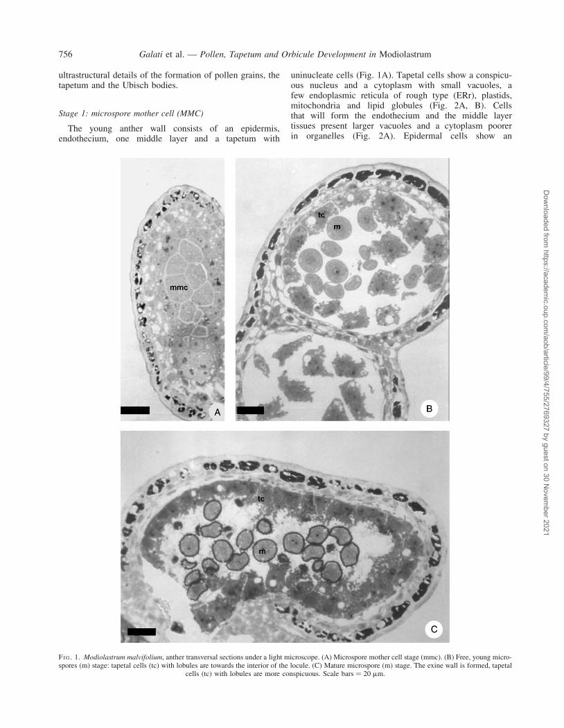
The young anther wall consists of an epidermis,endothecium, one middle layer and a tapetum with
uninucleate cells (Fig. 1A). Tapetal cells show a conspicu-ous nucleus and a cytoplasm with small vacuoles, afew endoplasmic reticula of rough type (ERr), plastids,mitochondria and lipid globules (Fig. 2A, B). Cellsthat will form the endothecium and the middle layertissues present larger vacuoles and a cytoplasm poorerin organelles (Fig. 2A). Epidermal cells show an
FI G. 1. Modiolastrum malvifolium, anther transversal sections under a light microscope. (A) Microspore mother cell stage (mmc). (B) Free, young micro-spores (m) stage: tapetal cells (tc) with lobules are towards the interior of the locule. (C) Mature microspore (m) stage. The exine wall is formed, tapetal
cells (tc) with lobules are more conspicuous. Scale bars ¼ 20 mm.
Galati et al. — Pollen, Tapetum and Orbicule Development in Modiolastrum756
Dow
nloaded from https://academ
ic.oup.com/aob/article/99/4/755/2769327 by guest on 30 N
ovember 2021

electron-dense cytoplasm and the tangential external wall isthickened (Fig. 1A).
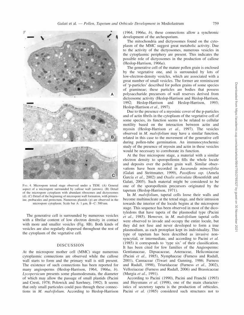
The microspore mother cell possesses a nucleus of greatvolume with a conspicuous nucleoli and a dense cytoplasm(Fig. 2A). The latter is filled with numerous small vacuolesand mitochondria, some of these in division. Abundant ERrand some dictyosomes are also observed. There are numer-ous cytoplasmic connections between these cells and a cal-losic wall is beginning to form between the plasmalemmaand the primary wall (Fig. 2A, B).
Stage 2: microspore tetrads
Tapetal cells walls are degraded and the tapetal nucleidivide. The middle layers are compressed (Fig. 3A).
The ultrastructure of the cytoplasm of tapetal cells issimilar to that presented in the previous stage.
Microspore mother cells undergo simultaneous meiosis,forming tetrads with a tetrahedral arrangement (Fig. 3A).
These are surrounded by a thick callosic wall that isfluorescent when stained with aniline blue and irradiatedwith 365-nm light (Fig. 3B). The cytoplasm of micro-spores shows numerous free ribosomes, some mitochon-dria, and abundant plastids and dictyosomes (Fig. 4A,B). At this stage, the primexine fibrilar matrix is startingto develop. On it, the future basal layer, probacula andprotectum can be seen as more electron-dense zones(Fig. 4C).
Stage 3: free, young microspores
At this stage, in the inner tangential faces of the tapetalcells an incipient lobe starts to grow towards the locule(Fig. 1B). The tapetal cytoplasm shows a great amount ofERr, whose cisterns appear in a parallel disposition.Abundant dictyosomes are also observed (Fig. 5A, B).
A membrane is formed over the outer tangential wall ofthe tapetal cells and in touch with the middle layer. On itsinner surface, future orbicules are observed aselectron-dense corpuscles (Fig. 5A).
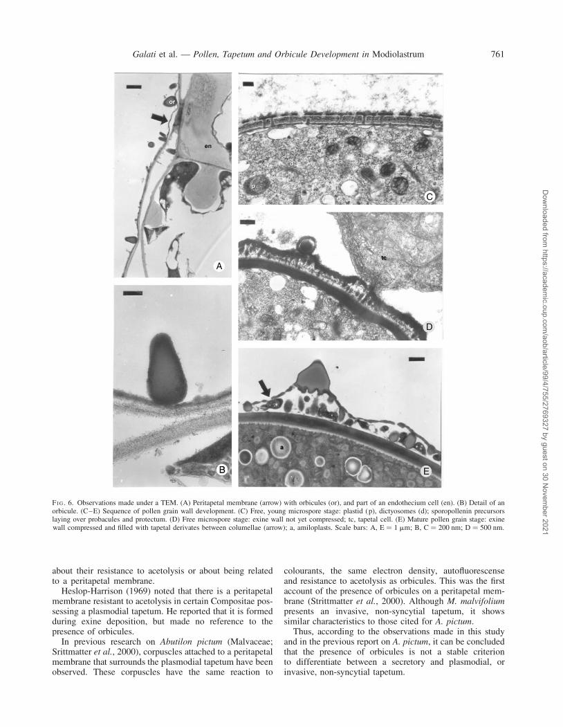
Once the callosic wall disintegrates, microspores arefreed. An electron-dense fibrilar substance fills the loculeand deposits over the pobacules and protectum (Figs 5B,6C). There are no differences between the cytoplasm ofthe microspores at this stage and at the previous stage(Fig. 6C).
Stage 4: microspores with a developed exine wall
The lobe of the tapetal cell grows towards the antherlocule (Fig. 1C); however, the ultrastructural characteristicsof the tapetum remain the same.
The exine wall of the microspores is formed by a basallayer, numerous columellae close to each other, and athick tectum (Fig. 6D). At this stage, the apertures of thefuture pollen grains and the intine oncus are formed.
Stage 5: young pollen grain
Tapetal cells do not lose their individuality as theyinvade the anther locule and surround pollen grains(Fig. 7A).
The peritapetal membrane is folded and veryelectron-dense corpuscules are observed on it (Fig. 5C).
The generative cell formed by a mitotic division of themicrospore occupies a parietal position. Pollen grainsshow a very thick wall with an echinate tectum (Fig. 7A).
Stage 6: mature pollen grain
At this stage, tapetal and middle layer cells are mostlydegraded (Fig. 7B). The peritapetal membrane is nolonger folded, due to an increase in volume of the anther,and it is in direct contact with the endothecium (Figs 7B,6A). In the latter, cells develop lignified thickenings ofthe secondary wall, formed as bands through their radialand tangential walls (Figs 7B, 6A).
Solid orbicules, without a central core and of piriformaspect are found on the peritapetal membrane (Fig. 6B).
FI G. 2. Microspore mother cell stage observed with a TEM. (A)Microspore mother cell (mmc), tapetal cells (tc), middle layer (ml), endo-thecium (en). (B) Detail of the microspore mother cell: cytoplasmic con-nections (arrow), mitochondria (m), nucleus (n) and dictyosomes (d).
Scale bars ¼ 2 mm.
Galati et al. — Pollen, Tapetum and Orbicule Development in Modiolastrum 757
Dow
nloaded from https://academ
ic.oup.com/aob/article/99/4/755/2769327 by guest on 30 N
ovember 2021
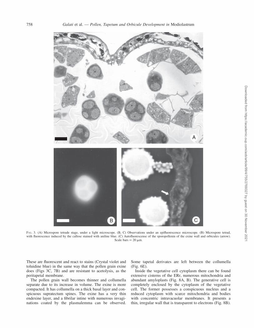
These are fluorescent and react to stains (Crystal violet andtoluidine blue) in the same way that the pollen grain exinedoes (Figs 3C, 7B) and are resistant to acetolysis, as theperitapetal membrane.
The pollen grain wall becomes thinner and collumellaseparate due to its increase in volume. The exine is morecompacted. It has collumella on a thick basal layer and con-spicuous supratectum spines. The exine has a very thinendexine layer, and a fibrilar intine with numerous invagi-nations coated by the plasmalemma can be observed.
Some tapetal derivates are left between the collumella(Fig. 6E).
Inside the vegetative cell cytoplasm there can be foundextensive cisterns of the ERr, numerous mitochondria andabundant amyloplasts (Fig. 8A, B). The generative cell iscompletely enclosed by the cytoplasm of the vegetativecell. The former possesses a conspicuous nucleus and areduced cytoplasm with scarce mitochondria and bodieswith concentric intravacuolar membranes. It presents athin, irregular wall that is transparent to electrons (Fig. 8B).
A
B C
FI G. 3. (A) Microspore tetrade stage, under a light microscope. (B, C) Observations under an epifluorescence microscope. (B) Microspore tetrad,with fluorescence induced by the callose stained with aniline blue. (C) Autofluorescense of the sporopollenin of the exine wall and orbicules (arrow).
Scale bars ¼ 20 mm.
Galati et al. — Pollen, Tapetum and Orbicule Development in Modiolastrum758
Dow
nloaded from https://academ
ic.oup.com/aob/article/99/4/755/2769327 by guest on 30 N
ovember 2021
The generative cell is surrounded by numerous vesicleswith a fibrilar content of low electron density in contactwith more and smaller vesicles (Fig. 8B). Both kinds ofvesicles are also regularly dispersed throughout the rest ofthe cytoplasm of the vegetative cell.
DISCUSSION
At the microspore mother cell (MMC) stage numerouscytoplasmic connections are observed while the callosewall starts to form and the primary wall is still present.The existence of such connections has been reported formany angiosperms (Heslop-Harrison, 1964, 1966a, b).Lycopersicum presents some plasmodesmata, the diameterof which may allow the passage of small plastids (Paciniand Cresti, 1978; Polowick and Sawhney, 1992). It seemsthat only small particules could pass through these connec-tions in M. malvifolium. According to Heslop-Harrison
(1964, 1966a, b), these connections allow a synchronicdevelopment of the archesporium.
The mitochondria and dictyosomes found on the cyto-plasm of the MMC suggest great metabolic activity. Dueto the activity of the dictyosomes, numerous vesicles inthe cytoplasmic periphery are present. This indicates thepossible role of dictyosomes in the production of callose(Heslop-Harrison, 1966a).
The generative cell of the mature pollen grain is enclosedby the vegetative one, and is surrounded by lots oflow-electron-density vesicles, which are associated with agreat number of small vesicles. The former are reminiscentof ‘p-particles’ described for pollen grains of some speciesof gramineae; these particles are bodies that possesspolysaccharide precursors of wall reserves derived fromdictyosome activity (Heslop-Harrison and Heslop-Harrison,1992; Heslop-Harrison and Heslop-Harrison, 1993;Heslop-Harrison et al., 1997).
Due to the presence of a myosinic cover of the p-particlesand of actin fibrils in the cytoplasm of the vegetative cell ofsome species, its function seems to be related to cellularmotility based on the interaction between actin andmyosin (Heslop-Harrison et al., 1997). The vesiclesobserved in M. malvifolium may have a similar function,related in this case to the movement of the generative cellduring pollen-tube germination. An immunocytochemicstudy of the presence of myosin and actin in these vesicleswould be necessary to corroborate its function.
At the free microspore stage, a material with a similarelectron density to sporopollenin fills the whole loculeand deposits over the pollen grain wall. Similar obser-vations have been recorded in Jacaranda mimosifolia(Galati and Strittmatter, 1999), Passiflora ssp. (AmelaGarcıa et al., 2002) and Oxalis articulata (Rosenfeldt andGalati, 2005). Such material might be considered to beone of the sporopollenin precursors originated by thetapetum (Heslop-Harrison, 1971).
In M. malvifolium, tapetal cells loose their walls andbecome multinucleate at the tetrad stage, and their intrusiontowards the interior of the locule begins at the microsporestage. This sequence has been observed in most of the dico-tyledons that have tapeta of the plasmodial type (Paciniet al., 1985). However, in M. malvifolium tapetal cellswere observed to invade and occupy the entire locule, butthey did not fuse and never developed to form a trueplasmodium, as each protoplast kept its individuality. Thistype of tapetum has been described as invasive non-syncytial, or intermediate, and according to Pacini et al.(1985) it corresponds to ‘type six’ of their classification.It has been cited for few families of the Angiosperms:Gentianaceae, Dipsacaceae, Asteraceae, Heliconiaceae(Pacini et al., 1985), Nymphaceae (Furness and Rudall,2001), Cannaceae (Tiwari and Gunning, 1986; Furnessand Rudall, 1998), Triuridaceae (Furness et al., 2002),Velloziaceae (Furness and Rudall, 2006) and Brassicaceae(Murgia et al., 1991).
According to Pacini (1990), Pacini and Franchi (1993)and Huysmans et al. (1998), one of the main character-istics of secretory tapeta is the production of orbicules.Pacini et al. (1985) considered such structures not to
FI G. 4. Microspore tetrad stage observed under a TEM. (A) Generalaspect of a microspore surrounded by callose wall (arrows). (B) Detailof the microspore cytoplasm with abundant ribosomes and dictyosomes(d). (C) Detail of the beginning of microspore wall formation, with primex-ine, probacules and protectum. Numerous plastids (p) are observed in the
microspore cytoplasm. Scale bar A: 1 mm, B–C: 500 nm.
Galati et al. — Pollen, Tapetum and Orbicule Development in Modiolastrum 759
Dow
nloaded from https://academ
ic.oup.com/aob/article/99/4/755/2769327 by guest on 30 N
ovember 2021

appear in species characterized by a plasmodial tapetum.However, there are three reports on the presence of cor-puscles with electron density and structure similar to orbi-cules (although not confirmed as such) in plasmodial
tapeta: Gentiana acaulis (Lombardo and Carraro, 1976),Butomus umbellatus (Fernando and Cass, 1994) andTradescantia virginiana (Tiwari and Gunning, 1986;Raghavan, 1997). These reports do not indicate anything
FI G. 5. Observations made under a TEM. (A, B) Free, young microspore stage. (A) Detail of a three-nuclei (n) tapetal cell with elaioplasts (e), abundantERr and pro-Ubisch (arrow) developing in towards the middle layer, young endothecium. (B) Detail of the radial and tangential inner wall of two tapetalcells (tc) and part of a microspore (m). (C) Microspore with exine wall formed, the peritapetal membrane folded with orbicules (or), and part of a tapetal
cell (tc). Scale bars ¼ 1 mm.
Galati et al. — Pollen, Tapetum and Orbicule Development in Modiolastrum760
Dow
nloaded from https://academ
ic.oup.com/aob/article/99/4/755/2769327 by guest on 30 N
ovember 2021
about their resistance to acetolysis or about being relatedto a peritapetal membrane.
Heslop-Harrison (1969) noted that there is a peritapetalmembrane resistant to acetolysis in certain Compositae pos-sessing a plasmodial tapetum. He reported that it is formedduring exine deposition, but made no reference to thepresence of orbicules.
In previous research on Abutilon pictum (Malvaceae;Srittmatter et al., 2000), corpuscles attached to a peritapetalmembrane that surrounds the plasmodial tapetum have beenobserved. These corpuscles have the same reaction to
colourants, the same electron density, autofluorescenseand resistance to acetolysis as orbicules. This was the firstaccount of the presence of orbicules on a peritapetal mem-brane (Strittmatter et al., 2000). Although M. malvifoliumpresents an invasive, non-syncytial tapetum, it showssimilar characteristics to those cited for A. pictum.
Thus, according to the observations made in this studyand in the previous report on A. pictum, it can be concludedthat the presence of orbicules is not a stable criterionto differentiate between a secretory and plasmodial, orinvasive, non-syncytial tapetum.
FI G. 6. Observations made under a TEM. (A) Peritapetal membrane (arrow) with orbicules (or), and part of an endothecium cell (en). (B) Detail of anorbicule. (C–E) Sequence of pollen grain wall development. (C) Free, young microspore stage: plastid (p), dictyosomes (d); sporopollenin precursorslaying over probacules and protectum. (D) Free microspore stage: exine wall not yet compressed; tc, tapetal cell. (E) Mature pollen grain stage: exinewall compressed and filled with tapetal derivates between columellae (arrow); a, amiloplasts. Scale bars: A, E ¼ 1 mm; B, C ¼ 200 nm; D ¼ 500 nm.
Galati et al. — Pollen, Tapetum and Orbicule Development in Modiolastrum 761
Dow
nloaded from https://academ
ic.oup.com/aob/article/99/4/755/2769327 by guest on 30 N
ovember 2021

LITERATURE CITED
Amela Garcıa MT, Galati BG, Anton AM. 2002. Microsporogenesis,microgametogenesis and pollen morphology of Passiflora spp.(Passifloraceae). Botanical Journal of the Linnean Society 139:383–394.
Fernando DD, Cass DD. 1994. Plasmodial tapetum and pollen wall devel-opment in Butomus umbellatus (Butomaceae). American Journal ofBotany 8: 1592–1600.
Furness CA, Rudall PJ. 1998. The tapetum and systematics in monocoty-ledons. Botanical Review 64: 201–239.
Furness CA, Rudall PJ. 2001. The tapetum in basal Angiosperms: earlydiversity. International Journal of Plant Science 162: 375–392.
Furness CA, Rudall PJ. 2006. Comparative structure and development ofpollen and tapetum in Pandanales. International Journal of PlantScience 167: 331–348.
Furness CA, Rudall PJ, Eastman A. 2002. Contribution of pollen andtapetal characters to the systematics of Triuridaceae. PlantSystematics and Evolution 235: 209–218.
Galati BG, Strittmatter LI. 1999. Correlation between pollen develop-ment and Ubisch bodies ontogeny in Jacaranda mimosifolia(Bignoniaceae). Beitrage zur Biologie der Planzen 71: 249–260.
Genise J, Palacios RA, Hoc PS, Carrizo R, Moffat L, Mom MP, et al.1990. Observaciones sobre la biologıa floral de Prosopis(Leguminosae, Mimosoideae). II. Fases florales y visitantes en el dis-trito chaqueno serrano. Darwiniana 30: 71–85.
Heslop-Harrison J. 1964. Cell walls, cell membranes, and protoplasmicconnections during meiosis and pollen development. In: LinskensHF, ed. Pollen physiology and fertilization. Amsterdam:North-Holland, 39–47.
Heslop-Harrison J. 1966a. Cytoplasmic connections during spore for-mation in flowering plants. Endeavour 25: 65–72.
Heslop-Harrison J. 1966b. Cytoplasmic connections between angiospermmeiocytes. Annals of Botany 30: 221–230.
Heslop-Harrison J. 1969. An acetolysis-resistant membrane investingtapetum and sporogenous tissue in the anthers of certainCompositae. Canadian Journal of Botany 47: 541–542.
Heslop-Harrison J. 1971. The pollen wall structure and development. In:Heslop-Harrison J, ed. Pollen: development and physiology. London:Butterworths, 75–98.
Heslop-Harrison J, Heslop-Harrison Y. 1993. Vapour phase activation,intracellular motility and germination in the triporate pollen ofEpilobium angustifolium. Botanica Acta 106: 331–337.
Heslop-Harrison J, Heslop-Harrison Y, Heslop-Harrison JS. 1997.Motility in ungerminated grass pollen: association of myosin withpolysaccharide-containing wallprecursor bodies (p-particles). SexualPlant Reproduction 10: 65–66.
Heslop-Harrison Y, Heslop-Harrison J. 1992. Germination of monocol-pate Angiosperm pollen evolution of the actin cytoskeleton and wallduring hydratation activation and tube emergence. Annals of Botany69: 385–394.
FI G. 8. Mature pollen grain observed under a TEM. (A) General aspect ofthe pollen grain: generative cell (arrow) surrounded by the vegetative cell;vn, vegetative nucleus. (B) Detail of the generative cell, with a conspicuousnucleus (gn), reduced cytoplasm with some plastids (p), thin,electron-transparent irregular wall. The vegetative cell cytoplasm presentsabundant thickened cisterns of the ERr, mitochondria (m) and amiloplast
(a). Scale bars: A ¼ 2 mm; B ¼ 500 nm.
FI G. 7. Anther transversal section observed under a light microscope. (A)Young pollen grain (pg) stage: tapetal cells (tc) invading the locule. (B)Mature pollen grain stage. Remains of the tapetum, anther wall: epidermis,mature endothecium and peritapetal membrane with orbicules (arrow).
Scale bars ¼ 20 mm.
Galati et al. — Pollen, Tapetum and Orbicule Development in Modiolastrum762
Dow
nloaded from https://academ
ic.oup.com/aob/article/99/4/755/2769327 by guest on 30 N
ovember 2021

Huysmans S, El-Ghazaly G, Smets E. 1998. Orbicules in Angiosperms:morphology, function, distribution, and relation with tapetum type.Botanical Review 64: 240–272.
Johri BM, Ambegaokar KB, Srivastava PS. 1992. Comparative embry-ology of Angiosperms, Vol. 1. Berlin, Heidelberg: Springer-Verlag,504–509.
Lombardo G, Carraro L. 1976. Tapetal ultrastructural changes duringpollen development I. Studies on Antirrhinum majus. Caryologia29: 113–125.
Murgia M, Charzynska M, Rourgier M, Cresti M. 1991. Secretorytapetum of Brassica oleraceae L.: polarity and ultrastructural features.Sexual Plant Reproduction 4: 28–35.
O’Brien TP, McCully ME. 1981. The study of plant structure. Principlesand selected methods. Melbourne, Australia: Termarcarphi Pty Ltd.
Pacini E. 1990. Tapetum and microspore function. In: Blackmore S, KnoxRB, eds. Microspores: evolution and ontogeny. London: AcademicPress.
Pacini E, Cresti M. 1978. Ultrastructural characteristics of tapetum andmicrospore mother cells in Lycopersicum peruvianum during
meiotic prophase. Societe Botanique de France. ActualitesBotaniques 125: 121–128.
Pacini E, Franchi GG. 1993. Role of the tapetum in pollen and spore dis-persal. Plant Systematics and Evolution, Supplement 7: 1–11.
Pacini E, Franchi GG, Hesse M. 1985. The tapetum: its form, function,and possible phylogeny in Embryophyta. Plant Systematics andEvolution 149: 155–185.
Polowick PL, Sawhney VK. 1992. Ultrastructural changes in the cell wall,nucleus and cytoplasm of pollen mother cells during meiotic prophaseI in Lycopersicum esculentum (Mill.). Protoplasma 169: 139–147.
Raghavan V. 1997. Molecular embryology of flowering plants.Cambridge: University Press, 33–35.
Rosenfeldt S, Galati BG. 2005. Ubisch bodies and pollen ontogeny inOxalis articulata Savigny. Biocell 29: 271–278.
Strittmatter LI, Galati BG, Monacci F. 2000. Ubisch bodies in peritape-tal membrane of Abutilon pictum Gill. (Malvaceae). Beitrage zurBiologie der Pflanzen 71: 393–402.
Tiwari SC, Gunning BES. 1986. Development of tapetum and micro-spores in Canna L.: an example of an invasive but non-syncytialtapetum. Annals of Botany 57: 557–563.
Galati et al. — Pollen, Tapetum and Orbicule Development in Modiolastrum 763
Dow
nloaded from https://academ
ic.oup.com/aob/article/99/4/755/2769327 by guest on 30 N
ovember 2021