pneumothorax
TRANSCRIPT

Dr Nirav Dhinoja

Pneumothorax is the accumulation of extrapulmonary air within the chest.
Most commonly from leakage of air from within the lung.

Term “pneumothorax” was first coined by Itard in
1803.
Laennec described the clinical picture of
pneumothorax occurring in patients with
pulmonary tuberculosis in 1819.
Description of primary spontaneous pneumothorax
occurring in healthy people was provided by
Kjaergard in 1932
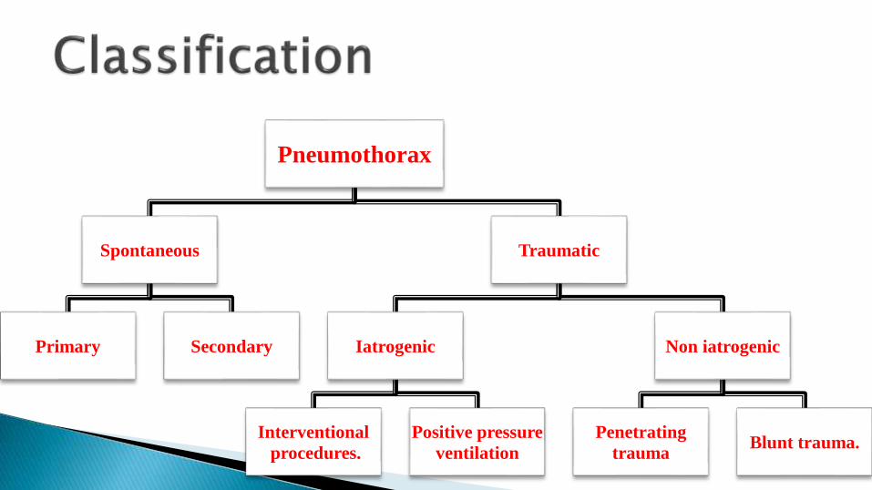
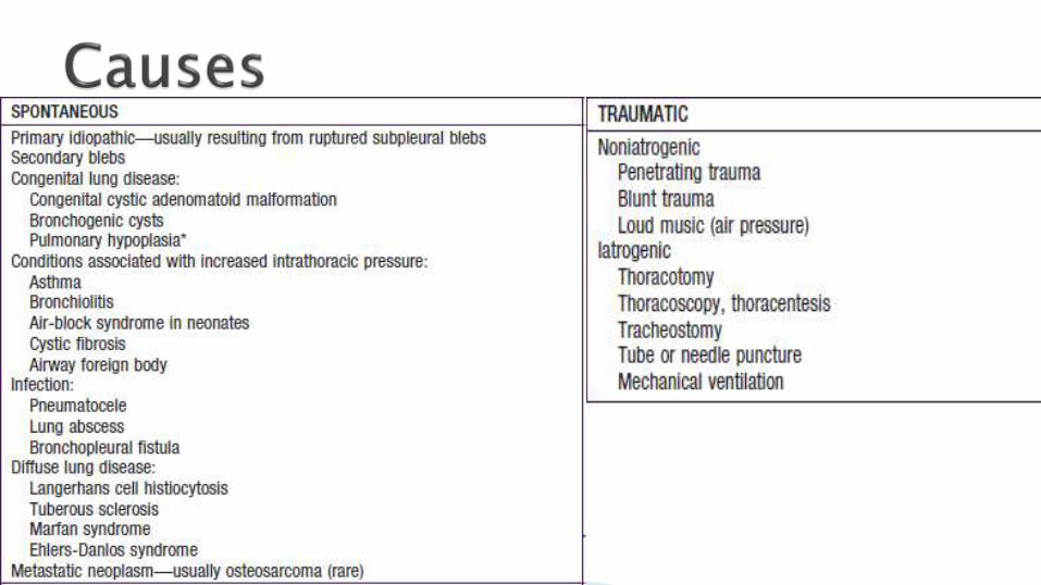
Pneumothorax
Spontaneous
Primary Secondary
Traumatic
Iatrogenic
Interventional
procedures.
Positive pressure
ventilation
Non iatrogenic
Penetrating
traumaBlunt trauma.

In normal people, the pressure in pleural space is negative during the entire respiratory cycle.
Two opposite forces result in negative pressure in pleural space.
Inherent outward pull of the chest wall and inherent elastic recoil of the lung.
The negative pressure will be disappeared if any communication develops .
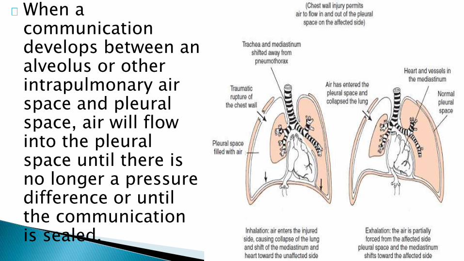
When a communication develops between an alveolus or other intrapulmonary air space and pleural space, air will flow into the pleural space until there is no longer a pressure difference or until the communication is sealed.
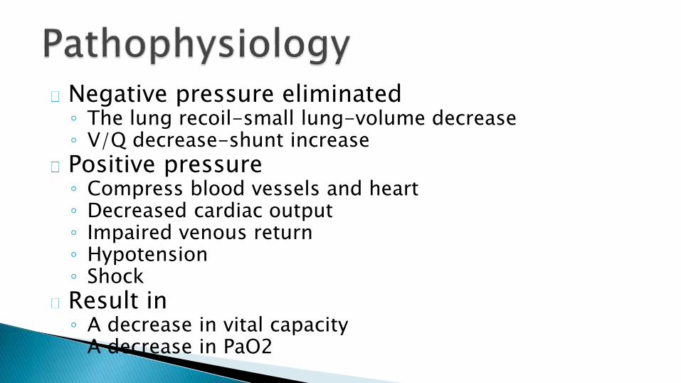
Negative pressure eliminated◦ The lung recoil-small lung-volume decrease◦ V/Q decrease-shunt increase
Positive pressure◦ Compress blood vessels and heart◦ Decreased cardiac output◦ Impaired venous return◦ Hypotension ◦ Shock
Result in ◦ A decrease in vital capacity ◦ A decrease in PaO2
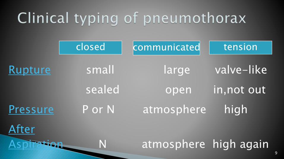
9
closed communicated tension
Rupture small large valve-like
sealed open in,not out
Pressure P or N atmosphere high
After
Aspiration N atmosphere high again

Abrupt onset.
Severity depends on :◦ Extent of lung collapse.
◦ Amount of pre-existing lung disease.
Pain – severity of pain does not reflect extent of collapse.
Dyspnea.
Cyanosis.

Tension pneumothorax◦ Distressed with rapid labored respiration
◦ Cyanosis
◦ Marked tachycardia
◦ Profuse diaphoresis
Patient who suddenly deteriorate clinically,should be suspected in the patient with◦ Mechanical ventilation
◦ Cardiopulmonary resuscitation
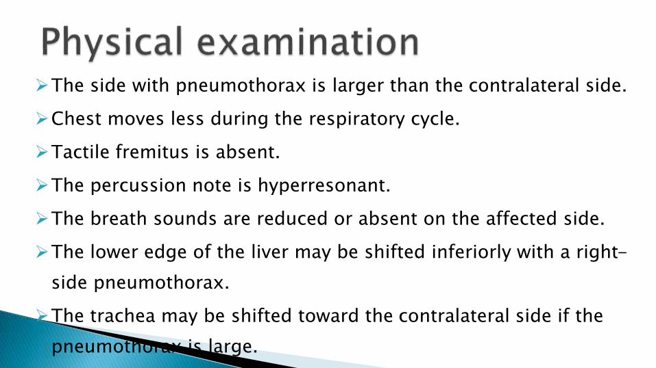
The side with pneumothorax is larger than the contralateral side.
Chest moves less during the respiratory cycle.
Tactile fremitus is absent.
The percussion note is hyperresonant.
The breath sounds are reduced or absent on the affected side.
The lower edge of the liver may be shifted inferiorly with a right-
side pneumothorax.
The trachea may be shifted toward the contralateral side if the
pneumothorax is large.
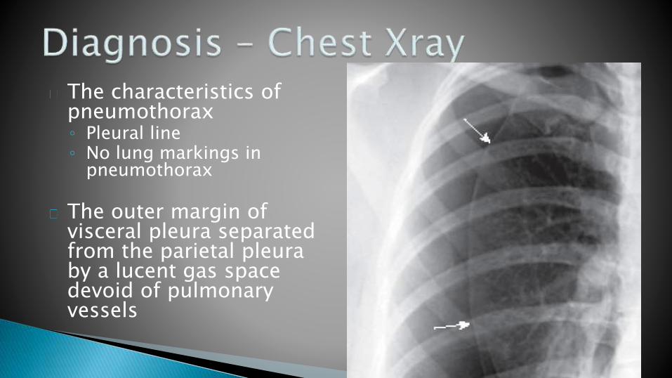
The characteristics of pneumothorax◦ Pleural line◦ No lung markings in
pneumothorax
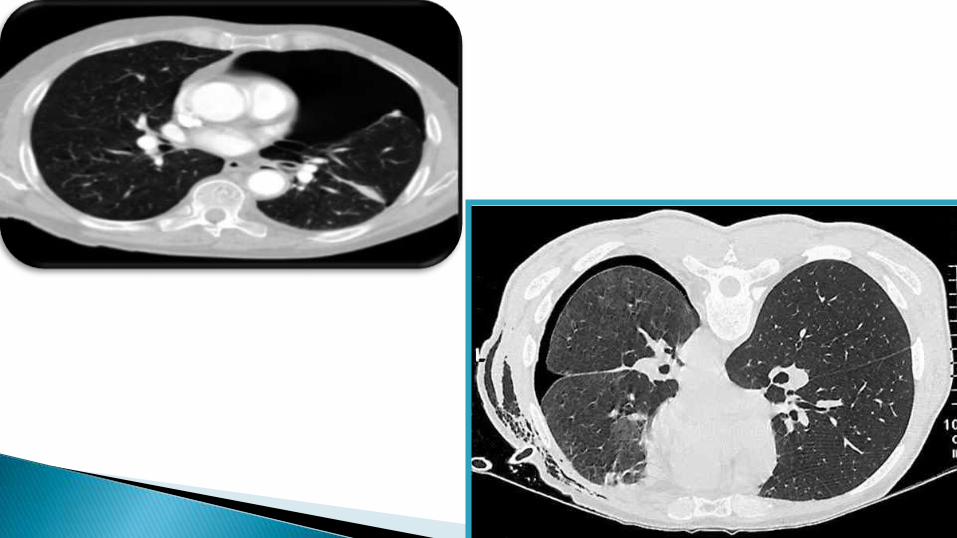
The outer margin of visceral pleura separated from the parietal pleura by a lucent gas space devoid of pulmonary vessels
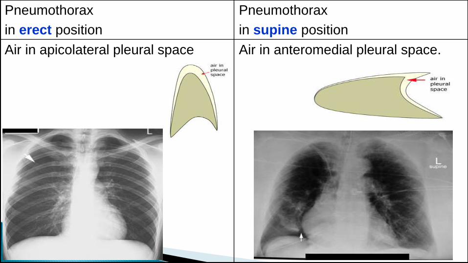
Pneumothorax
in erect position
Pneumothorax
in supine position
Air in apicolateral pleural space Air in anteromedial pleural space.

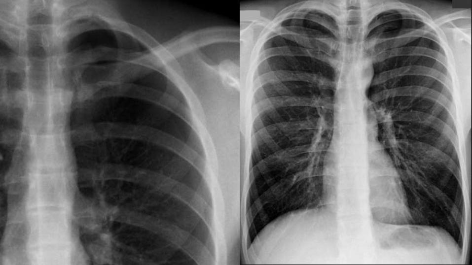
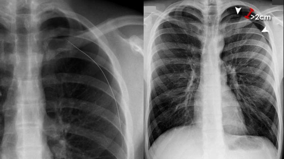
Pneumothorax
Erect
Smallpneumothorax
Apical lucency
Visceral pleural line
Largepneumothorax
Apical lucency(>2cm in width)
Visceral pleural line
Tensionpneumothorax
Lung collapse
Mediastinal shift
Low flat diaphragm
Supine
DeepCostophrenic
sulcus
LucentCardiophrenic
sulcus
Sharp Mediastinal
contour
Double diaphragm


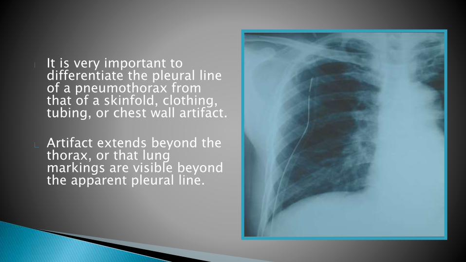
It is very important to differentiate the pleural line of a pneumothorax from that of a skinfold, clothing, tubing, or chest wall artifact.
Artifact extends beyond the thorax, or that lung markings are visible beyond the apparent pleural line.
CT scanning is done if accurate size estimates are required.It is only recommended to difficult cases such as patients in whom the lungs are obscured by overlying surgical emphysema.To differentiate a pneumothorax from suspected bulla in complex cystic lung disease.

Goals
◦ To promote lung expansion.
◦ To eliminate the pathogenesis.
◦ To decrease pneumothorax
recurrence.
Treatment options according
to
◦ Classification of pneumothorax.
◦ Pathogenesis.
◦ Pneumothorax frequency.
◦ The extension of lung collapse.
◦ Severity of disease.
◦ Complication and concomitant
underlying diseases.
24
Small, closed mildly symptomatic spontaneous
pneumothoraces.
Do not require hospital admission
It should be stressed to patient that they should be return
directly to hospital in the event of developing breathlessness.

25
Small SSP of less than 1 cm depth or isolated apical
pneumothoraces in asymptomatic patients.
Hospitalisation is recommended in these cases.
All other cases will require active intervention ( aspiration or
chest drain insertion)
26
Marked breathlessness in a patient with a small (<2 cm) PSP may herald tension pneumothorax.
Observation alone is inappropriate and active intervation is required.
If a patient is hospitalised for observation, supplemental high flow (10 l/min) oxygen should be given.

27
Inhalation of high concentration of oxygen may reduce the total
pressure of gases in pleural capillaries by reducing the partial
pressure of nitrogen.
This should increase the pressure gradient between the pleural
capillaries and the pleural cavity.
Thereby increasing absorption of air from the pleural cavity.
28
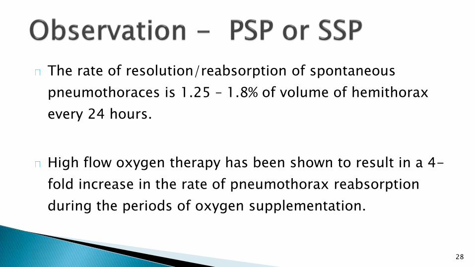
The rate of resolution/reabsorption of spontaneous
pneumothoraces is 1.25 – 1.8% of volume of hemithorax
every 24 hours.
High flow oxygen therapy has been shown to result in a 4-
fold increase in the rate of pneumothorax reabsorption
during the periods of oxygen supplementation.
29
It is recommended as first line treatment for all PSP requiring intervention.
It is less likely to succeed in secondary pneumothoraces and in this situation,it is
only recommended as an initial treatment in small (<2 cm) pneumothoraces in
minimally breathless patients under the age of 50 years.
Patients with secondary pneumothoraces treated successfully with simple
aspiration should be admitted to hospital and observed for at least 24 hours
before discharge.

Repeated aspiration is reasonable for primary pneumothorax
when the first aspiration has been unsuccessful.
A volume of < 2.5 L has been aspirated on the first attempt.
The aspiration can be used by needle or catheter.

31
Fix the catheter and cover with gauze
Making a small incisionUsing a forceps to extend the holeInserting a catheter into pleural cavity
32
INDICATIONS ◦ Unstable pneumothorax
◦ Severe dyspnea
◦ Large lung collapse
◦ Open or tension pneumothoraces
◦ Frequent recurrent pneumothoraces
◦ Simple aspiration or catheter aspiration drainage is unsuccessful in controlling symptoms

33
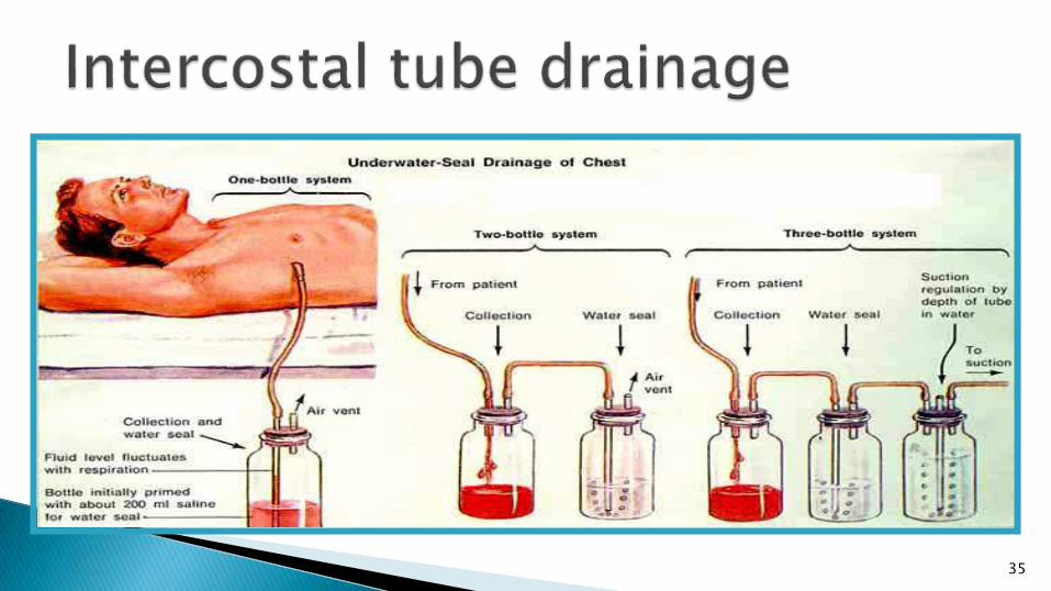
Position of intercostal tube
The chest tube should be positioned in the uppermost part of the pleural space, where residual air accumulates
This procedure permits the air in the pleural space to be evacuated rapidly
34
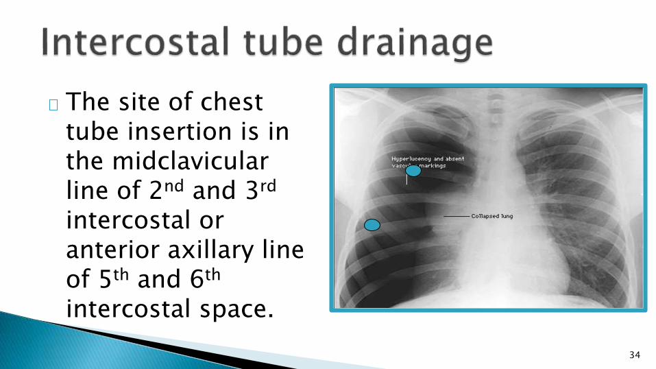
The site of chest tube insertion is in the midclavicularline of 2nd and 3rd
intercostal or anterior axillary line of 5th and 6th
intercostal space.
35
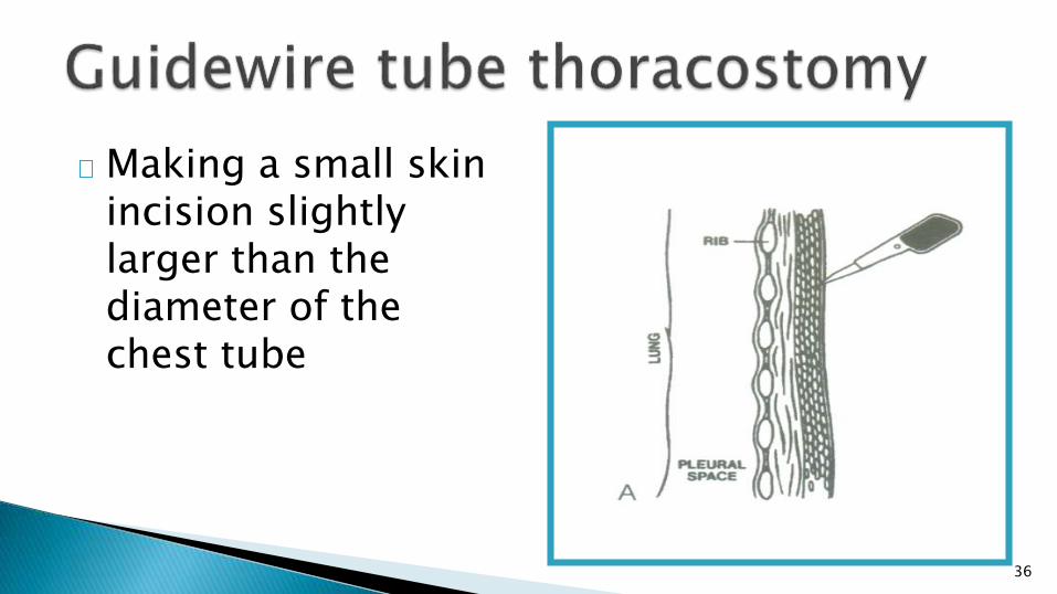
36
Making a small skin incision slightly larger than the diameter of the chest tube
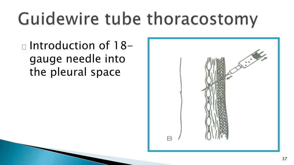
37
Introduction of 18-gauge needle into the pleural space

38
Insertion of wire with “J” end into the pleural space
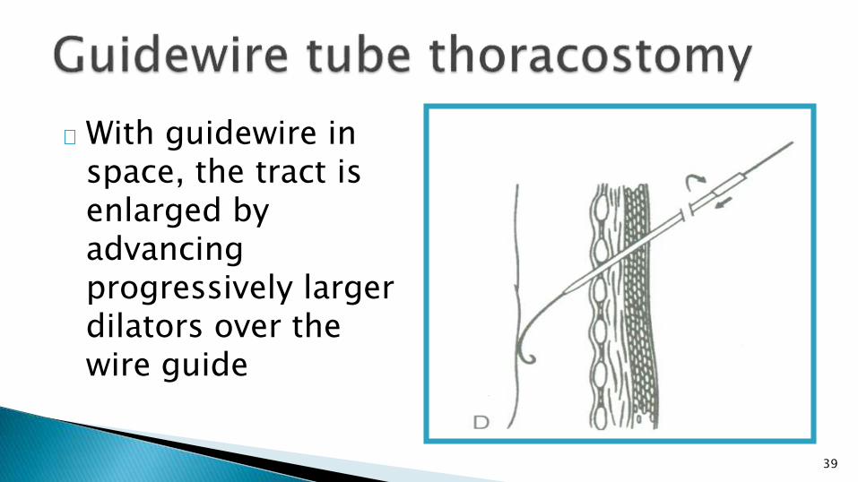
39
With guidewire in space, the tract is enlarged by advancing progressively larger dilators over the wire guide
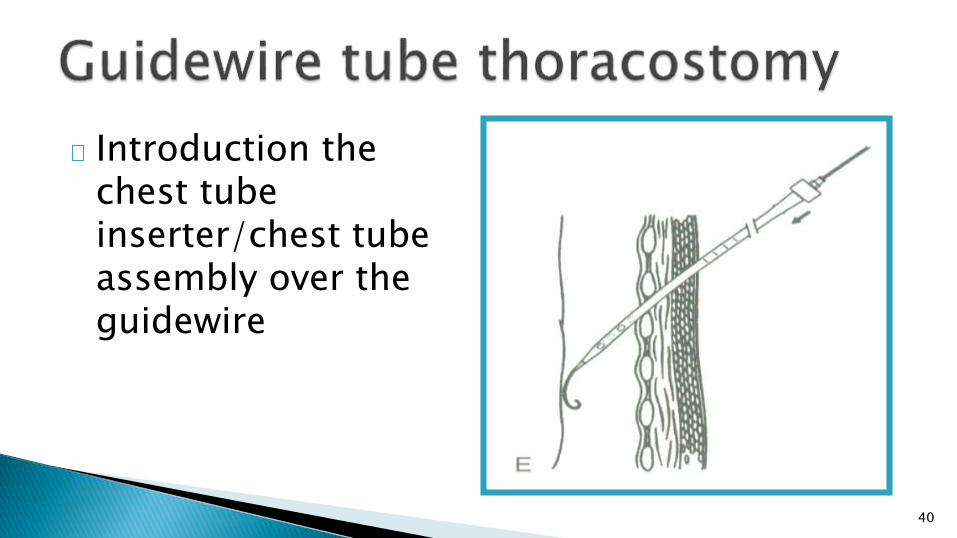
40
Introduction the chest tube inserter/chest tube assembly over the guidewire

41
The guidewire and chest tube inserter have been removed, leaving the chest tube positioned with the pleural space
42
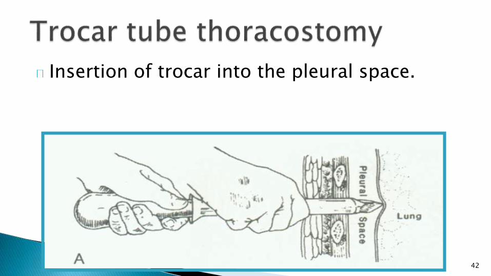
Insertion of trocar into the pleural space.

43
Insertion of the chest tube through the trocar
44
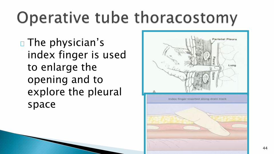
The physician’s index finger is used to enlarge the opening and to explore the pleural space

45
Placement of chest tube intrapleurally using large hemostat
46
No bubble released ◦ The lung reexpansion◦ The chest tube is obstructed by secretion or blood clot◦ The chest tube shift to chest wall, the hole of the chest tube is
located in the chest wall
If the lung reexpansion, removing the chest tube 24 hours after reexpansion.
Otherwise, the chest tube will be inserted again or regulated the position.
47
Penetration of major organs◦ Lung, stomach, spleen, liver, heart and great
vessels◦ It occurs more commonly when a sharp metal trocar
is inappropriately applied
Pleural infection◦ Empyema, the rate of 1%
Surgical emphysema ◦ Subcutaneous emphysema

48
Goals ◦ To prevent pneumothorax recurrence ◦ To produce inflammation of pleura and adhesions
Indications◦ Persist air leak and repeated pneumothorax◦ Bilateral pneumothoraces◦ Complicated with bullae◦ Lung dysfunction, not tolerate to operation
49
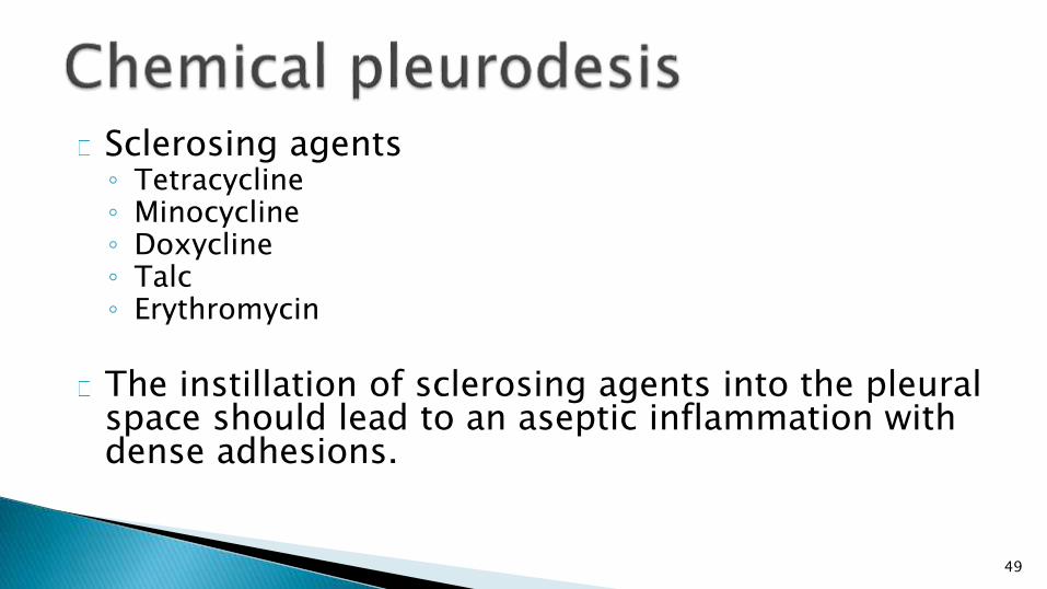
Sclerosing agents◦ Tetracycline◦ Minocycline◦ Doxycline◦ Talc ◦ Erythromycin
The instillation of sclerosing agents into the pleural space should lead to an aseptic inflammation with dense adhesions.

50
Methods
◦ Via chest tube or by surgical mean
◦ Administration of intrapleural local anaesthesia, 200 – 400 mg lidocaine intrapleurally
injection
◦ Agents diluted by 60 – 100 ml saline
◦ Injected to pleural space
◦ Clamp the tube 1 – 2 hours
◦ Drainage again
◦ Observed by chest X-ray film, if air of pleural space is absorption, remove the chest tube
◦ If pneumothorax still exist, repeated pleurodesis.

51
Side effct◦ Chest pain
◦ Fever
◦ Dyspnea
◦ Acute respiratory distress syndrome
◦ Acute respiratory failure
52
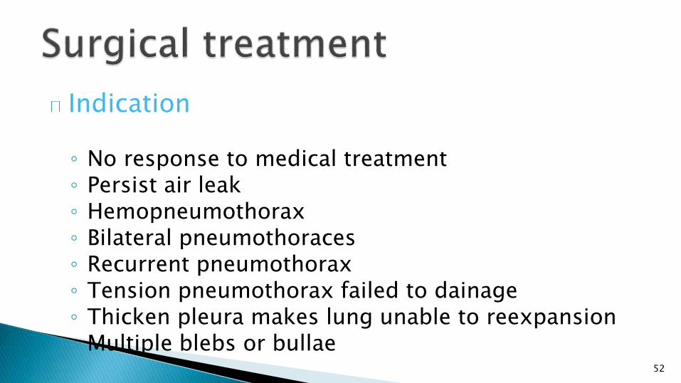
Indication
◦ No response to medical treatment◦ Persist air leak◦ Hemopneumothorax◦ Bilateral pneumothoraces◦ Recurrent pneumothorax◦ Tension pneumothorax failed to dainage◦ Thicken pleura makes lung unable to reexpansion◦ Multiple blebs or bullae
53
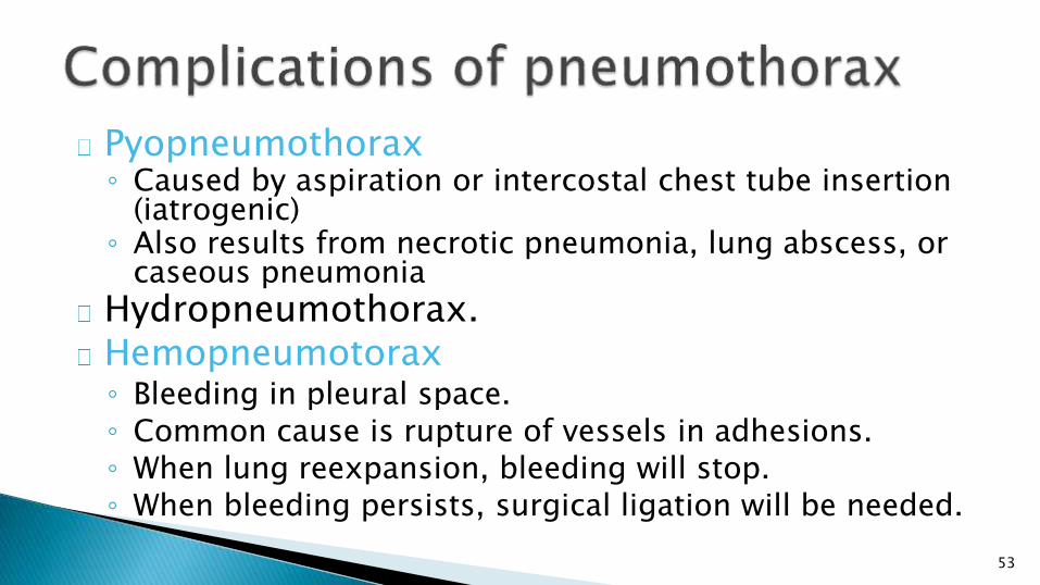
Pyopneumothorax◦ Caused by aspiration or intercostal chest tube insertion
(iatrogenic)◦ Also results from necrotic pneumonia, lung abscess, or
caseous pneumonia
Hydropneumothorax.Hemopneumotorax◦ Bleeding in pleural space.◦ Common cause is rupture of vessels in adhesions.◦ When lung reexpansion, bleeding will stop.◦ When bleeding persists, surgical ligation will be needed.
54
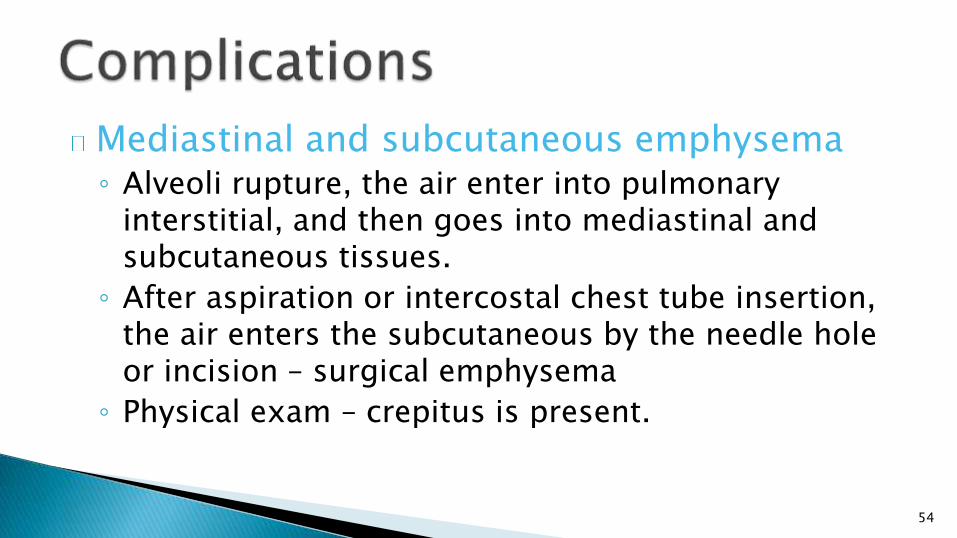
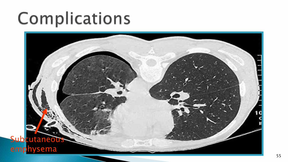
Mediastinal and subcutaneous emphysema◦ Alveoli rupture, the air enter into pulmonary
interstitial, and then goes into mediastinal and subcutaneous tissues.
◦ After aspiration or intercostal chest tube insertion, the air enters the subcutaneous by the needle hole or incision – surgical emphysema
◦ Physical exam – crepitus is present.
55
Subcutaneous emphysema

56
Treatment ◦ Automatic absorption when pneumothorax is gone
◦ Inhalation of high concentration of oxygen
◦ Making a small incision in suprasternal pit for draining the air from mediastinal and subcutaneous tissues.