plus - home | kcj
TRANSCRIPT

Official Journal of The Kidney Cancer Association
An Edu cational Service for Medical Oncologists, Hematologist-Oncologists, and Urologists
Volume 15, Number 3 2017
kidney-cancer-journal.com
Now in Its
15th Yea
r
Radiation and RCC: Will New Data Crush the Myth of Radioresistance? Plus: The Ultimate Guide to Sunitinib Dosing to Optimize VEGF Blockade, Efficacy & Tolerability


�������������������������
Connecting More Dots in Kidney Cancer: Updates on Radiation Therapy, Optimal Dosing Schedules for VEGF Blockade
t has always been the aim of the Kidney Cancer Journal to cover the broadest spectrum of topics related to all aspects of managing renal cell carcinoma (RCC). When
I was asked to serve as Guest Editor, I knew that this issue of the journal would be no exception. The assortment of articles attests to how our field is rapidly evolving—whether it is new information emerging from the 2017 meeting of the European Society of Medical Oncology (ESMO) or updates from our authors on changing stan-
dards in clinical practice or new results from the bench with translational impact.
This year’s ESMO scientific sessions presented some pivotal information on key issues with potential translational impact, including the combina-tion of immunotherapy and targeted therapy, and the sequence of sunitinib and nephrectomy. These findings, notably an update on the Checkmate 214 trial and the SURTIME trial, are reviewed in this issue of the Kidney Cancer Journal on Pages 65 and 82, in addition to other results from ESMO.
If you follow the medical literature as closely as I do, you may be surprised at how the latest studies have not only more pointedly addressed long-standing controversies in RCC but go further—approaching a consen-sus that tends to debunk some of the myths surrounding practice standards. A case in point—our content on the safety and efficacy of stereotactic body radiation therapy (SBRT) for RCC, both primary and metastatic.
In terms of our knowledge about and application of this technique, we are light years away from the 1950s when a Swedish neurosurgeon first described single-dose ablative radiotherapy delivered to brain lesions. The revolutionary development concerns the way this principle has been extrapolated to the stereotactic delivery of severely hypofractionated treat-ments to body targets, including kidney, either primary or metastatic, cranial and extracranial. And yet, the myth has lingered that such applica-tion in RCC is limited by perceived radioresistance to conventional frac-tionation. If this is still your perception (a misconception, I might add) then review the article by Raquib Hannan, MD, on how such resistance can be overcome in many clinical settings, thus sparing many patients from nephrectomy, especially those who are poor surgical candidates.
One of the areas that has long been a focus of my research is the optimal dosing schedule for sunitinib and efforts to
T A B L E O F C O N T E N T SEditorial Mission The purpose of Kidney Cancer Journal is to serve as a compre- hensive resource of information for physicians regarding advances in the diagnosis and treatment of renal cell carci-noma. Content of the journal focuses on the impact of transla-tional research in oncology and urology and also provides a forum for cancer patient advocacy. Kidney Cancer Journal is circulated to medical oncologists, hematologist-oncologists, and urologists. Editor-in-Chief Robert A. Figlin, MD, FACP Steven Spielberg Family Chair in Hematology Oncology Professor of Medicine and Biomedical Sciences Director, Division of Hematology Oncology Deputy Director, Samuel Oschin Comprehensive Cancer Institute Cedars-Sinai Medical Center
Medical Advisory Board Michael B. Atkins, MD Deputy Director Lombardi Comprehensive Cancer Center Professor of Oncology and Medicine, Georgetown University Medical Center Washington, DC
Ronald M. Bukowski, MD Emeritus Staff & Consultant CCF Taussig Cancer Center Professor of Medicine CCF Lerner College of Medicine of CWRU Cleveland, Ohio
Robert J. Motzer, MD Attending Physician Memorial Sloan-Kettering Cancer Center New York, NY
Christopher G. Wood, MD, FACS Douglas E. Johnson, MD Professorship Professor & Deputy Chairman Department of Urology M.D. Anderson Cancer Center Houston, Texas Nurse Advisory Board Nancy Moldawer, RN, MSN Nursing Director Cedars-Sinai Medical Center Samuel Oschin Comprehensive Cancer Institute Los Angeles, California
Laura Wood, RN, MSN, OCN Renal Cancer Research Coordinator Cleveland Clinic Taussig Cancer Center Cleveland, Ohio
Patient Advocate William P. Bro Chief Executive Officer Kidney Cancer Association
Publishing Staff Stu Chapman, Executive Editor Jenny Chapman, Director, Business Development Gloria Catalano, Production Director Michael McClain, Design Director Business and Administration Office 333 E. 75th Street, Suite 5D New York, NY 10021 Editorial Offices Genitourinary Publishing 2557 Tulip Street Sarasota, FL 34239 Tel: (516) 356-5006 © Copyright 2016 Genitourinary Publishing. All rights reserved. None of the contents may be reproduced in any form without the permission of the publisher. About the Cover Artist's depiction of a targeted radiation beam destroying a malignant tumor. Definitive, high-dose-per-fraction stereotac-tic radiation is having a sharp impact as an option for a grow-ing population of kidney cancer patients. (Copyright ©, Science Source)
64 Journal Club
65 Medical Intelligence
66 Optimizing Sunitinib Dosing: Balancing Efficacy and Tolerability
72 Stereotactic Ablative Radiation for RCC: Novel Paradigms Emerge as the Myth of Radioresistance Fades
G U E S T E D I T O R ’ S M E M O
(continued on page 80)
Bernard J. Escudier, MD
I

Michael B. Atkins, MD Lombardi Comprehensive Cancer Center Professor of Oncology and Medicine, Georgetown University Medical Center-Washington, DC
Arie Belldegrun, MD David Geffen School of Medicine at UCLA Los Angeles, California
Steven Campbell, MD Cleveland Clinic Foundation Cleveland, Ohio
Toni K. Choueiri, MD Dana-Farber Cancer Institute Harvard Medical School Boston, Massachusetts
Janice P. Dutcher, MD Associate Director, Cancer Research Foundation of New York Chappaqua, New York
Timothy Eisen, MD University of Cambridge Department of Oncology, Addenbrooke's Hospital Cambridge, UK
Paul Elson, PhD Cleveland Clinic Foundation Cleveland, Ohio
Bernard Escudier, MD Institut Gustave-Roussy Villejuif, France
James H. Finke, PhD Cleveland Clinic Lerner College of Medicine of Case Western Reserve University Cleveland, Ohio
Keith T. Flaherty, MD Lecturer, Department of Medicine, Harvard Medical School Director of Developmental Therapeutics, Cancer Center Massachusetts General Hospital Boston, Massachusetts
Daniel J. George, MD Duke Clinical Research Institute Durham, North Carolina
Inderbir S. Gill, MD USC Institute of Urology University of Southern California Los Angeles, California
Martin Gore, MD Royal Marsden Hospital London, UK
Gary Hudes, MD Fox Chase Cancer Center Philadelphia, Pennsylvania
Thomas Hutson, DO, PharmD Baylor University Medical Center Dallas, Texas
Eugene D. Kwon, MD Mayo Clinic Rochester, Minnesota
Bradley C. Leibovich, MD Mayo Clinic Rochester, Minnesota
David Nanus, MD New York Presbyterian Hospital- Weill Cornell Medical Center New York, New York
Leslie Oleksowicz, MD College of Medicine University of Cincinnati Medical Center Cincinnati, Ohio
Allan Pantuck, MD David Geffen School of Medicine at UCLA Los Angeles, California
W. Kimryn Rathmell, MD, PhD Director, Division of Hematology Oncology Professor, Department of Clinical Medicine and Cancer Biology Vanderbilt University Nashville, Tennessee
Brian Rini, MD Cleveland Clinic Foundation Cleveland, Ohio
Paul Russo, MD Memorial Sloan-Kettering Cancer Center New York, New York
Ihor S. Sawczuk, MD Hackensack University Medical Center Hackensack, New Jersey
Domenic A. Sica, MD Medical College of Virginia Richmond, Virginia
Jeffrey A. Sosman, MD Vanderbilt University Medical Center Vanderbilt-Ingram Cancer Center Nashville, Tennessee
Nicholas J. Vogelzang, MD Comprehensive Cancer Centers of Nevada Las Vegas, Nevada
�������������������������
EDITORIAL ADVISORY BOARD
Kidney Cancer Journal Author GuidelinesScope of Manuscripts The Kidney Cancer Journal considers the following types of manu-scripts for publication:
• Reviews that summarize and synthesize peer-reviewed literature to date on relevant topics in a scholarly fashion and format.
• Original contributions based on original, basic, clinical, translational, epidemiological, or prevention studies relating to kidney cancer that are well documented, novel, and significant.
• Letters to the Editor on timely and relevant subjects pertaining to the diagnosis and treatment of renal cell carcinoma.
• Clinical case studies. Manuscript Submission Authors are required to submit their manuscripts in an electronic for-mat, preferably by email to the Editor-in-Chief, Robert A. Figlin, MD, at [email protected]. Please provide in a word processing program. Images should be submitted electronically as well. All material reproduced from previously published, copyrighted material should contain a full credit line acknowledging the original source. The author is responsible for obtaining this permission. Contact information List all authors, including mailing address, titles and affiliations, phone, fax, and email. Please note corresponding author. Peer Review and Editing Manuscripts will be peer reviewed. Accepted manuscripts will be edited for clarity, spelling, punctuation, grammar, and consistency with American Medical Association (AMA) style. Authors whose manuscripts are not initially accepted may have the opportunity to revise the manuscript based on recommendations from peer reviewers and at the discretion of the Editor-in-Chief.
Conflict of Interest Kidney Cancer Journal policy requires that authors reveal to the Editor-in-Chief any relationships that they believe could be construed as resulting in an actual, potential, or apparent conflict of interest with regard to the manuscript submitted for review. Authors must disclose this information in the covering letter accompanying their submission. Manuscript Preparation Length: Full-length manuscripts should not exceed 4000 words, including references. Please limit the reference list to 50 citations. Manuscripts should be accompanied by figures and/or tables. Gener-ally 4-5 figures and 2-3 tables are preferred for each manuscript. Please include a brief description to accompany these items, as well as a legend for all abbreviations. Manuscripts should not contain an ab-stract but an introduction is recommended. Spacing: One space after periods. Manuscripts should be double spaced. References All submissions should have references that are referred to in the text by superscripted numbers and that conform to AMA style. Example: Lewczuk J, Piszko P, Jagas J, et al. Prognostic factors in medically treated patients with chronic pulmonary embolism. Chest. 2001;119:818-823. Copyright Manuscripts and accompanying material are accepted for exclusive publication in the Kidney Cancer Journal. None of the contents may be reproduced without permission of the Kidney Cancer Journal. To request permission, please contact Stu Chapman, Executive Editor, (516) 356-5006; email: [email protected].

Randomized Phase III Trial of Adjuvant Pazopanib Versus Placebo After Nephrectomy in Patients With Localized or Locally Advanced Renal Cell Carcinoma. Motzer RJ, Haas NB, Donskov, et al. J Clin Oncol. 2017 Sep 13; JCO2017735324. doi: 10.1200/JCO.2017.73. 5324. Summary: This phase III trial evaluated the efficacy and safety of pazopanib versus placebo in patients with locally advanced renal cell carcinoma (RCC) at high risk for relapse after nephrectomy. A total of 1,538 patients with resected pT2 (high grade) or ≥ pT3, including N1, clear cell RCC were randomly assigned to pazopanib or placebo for 1 year; 403 patients received a starting dose of 800 mg or placebo. To address toxicity attrition, the 800-mg starting dose was lowered to 600 mg, and the primary end point analysis was changed to disease-free survival (DFS) for pazopanib 600 mg versus placebo (n = 1,135). Primary analysis was performed after 350 DFS events in the intent-to- treat (ITT) pazopanib 600 mg group (ITT600mg), and DFS follow-up analysis was performed 12 months later. Secondary end point analyses included DFS with ITT pazopanib 800 mg (ITT800mg) and safety. The primary analysis results of DFS ITT600mg favored pazopanib but did not show a significant improvement over placebo (P = .165). The secondary analysis of DFS in ITT800mg (n = 403) yielded an HR of 0.69. Follow-up analysis in ITT600mg yielded an HR of 0.94 (95% CI, 0.77 to 1.14). Increased ALT and AST were common adverse events lead-ing to treatment discontinuation in the pazopanib 600 mg (ALT, 16%; AST, 5%) and 800 mg (ALT, 18%; AST, 7%) groups. Conclusion: The results of the primary DFS analysis of pazopanib 600 mg showed no benefit over placebo in the adjuvant setting. Long-Term Response to Sunitinib Treatment in Metastatic Renal Cell Carcinoma: A Pooled Analysis of Clinical Trials. Tannir NM, Figlin RA, Gore ME, et al. EClin Genitourin Cancer. 2017 Jun 20. S1558-7673(17) 30171-4. Summary: We characterized clinical outcomes of patients with metastatic renal cell carcinoma (mRCC) treated with sunitinib who were long-term responders (LTRs), defined as patients having progression-free survival (PFS) > 18 months. A retrospective analysis of data from 5714 patients with mRCC treated with sunitinib in 8 phase II/III clinical trials and the expanded access program. Duration on-study and objective response rate (ORR) were compared between LTRs and patients with PFS ≤ 18 months
(“others”). PFS and overall survival (OS) were summarized using Kaplan-Meier methodology. Overall, 898 (15.7%) patients achieved a long-term response and 4816 (84.3%) patients did not achieve long-term response. The median (range) duration on-study was 28.6 (16.8-70.7) months in LTRs and 5.5 (0-68.8) months in others. ORR was 51% in LTRs versus 14% in others (P < .0001). Median PFS in LTRs was 32.11 months and median OS was not reached. LTRs had higher percentage of early tumor shrinkage ≥ 10% at the first scan (67.1% vs. 51.2%; P = .0018) and greater median maximum on-study tumor shrinkage from base-line (-56.9 vs. -27.1; P < .0001) versus others. Conclusion: White race, Eastern Cooperative Oncology Group performance status 0, time from diagnosis to treat-ment ≥ 1 year, clear cell histology, no liver metastasis, lac-tate dehydrogenase ≤ 1.5 upper limit of normal (ULN), corrected calcium ≤ 10 mg/dL, hemoglobin greater than the lower limit of normal, platelets less than or equal to ULN, body mass index ≥ 25 kg/m2, and low neutrophil- to-lymphocyte ratio were associated with LTR. A subset of patients with mRCC treated with sunitinib achieved long-term response. LTRs had improved ORR, PFS, and OS. Biomarker-Based Phase II Trial of Savolitinib in Pa-tients With Advanced Papillary Renal Cell Cancer. Choueiri TK, Plimack E, Arkenau HT, et al. J Clin Oncol. 2017 Sep 10;35(26):2993-3001. doi: 10.1200/JCO.2017.72.2967 Summary: Patients with advanced papillary renal cell car-cinoma (PRCC) have limited therapeutic options. PRCC may involve activation of the MET pathway, for example, through gene amplification or mutations. Savolitinib (AZD6094, HMPL-504, volitinib) is a highly selective MET tyrosine kinase inhibitor. We report results of a single-arm, multicenter, phase II study evaluating the safety and effi-cacy of savolitinib in patients with PRCC according to MET status. Patients and Methods Patients with histologi-cally confirmed locally advanced or metastatic PRCC were enrolled and received savolitinib 600 mg orally once daily. MET-driven PRCC was defined as any of the following: chromosome 7 copy gain, focal MET or HGF gene amplifi-cation, or MET kinase domain mutations. Efficacy was assessed according to MET status. Safety, toxicity, and patient-reported health-related quality-of-life outcomes were assessed in all patients. Results Of 109 patients treated, PRCC was MET driven in 44 (40%) and MET independent in 46 (42%); MET status was unknown in 19 (17%). MET-driven PRCC was strongly associated with
64 Kidney Cancer Journal
Essential Peer-Reviewed Reading in Kidney Cancer
The peer-reviewed articles summarized in this section were selected by the Guest Editor, Bernard J. Escudier, MD, for their timeliness, importance, relevance, and potential impact on clinical practice or translational research.
J O U R N A L C L U B
(continued on page 81)

Kidney Cancer Journal 65
MEDICAL INTELLIGENCE
Newsworthy, late-breaking information from Web-based sources, professional societies, and government agencies
KCJ Medical Intelligence: ESMO Highlights
CABOSUN Update Affirms PFS Advantage With Cabozantinib Over Sunitinib in Advanced RCC MADRID—-Patients with untreated advanced renal cell carcinoma (RCC) lived significantly longer without disease progression when they received the multikinase inhibitor cabozantinib (Cabometyx) as initial therapy versus suni-tinib (Sutent), according to results of an independent review of the randomized CABOSUN trial reported at the 2017 European Society of Medical Oncology (ESMO) Congress.
Results showed that cabozantinib-treated patients had a median progression-free survival (PFS) of 8.6 months compared with 5.3 months for patients treated initially with sunitinib. The difference represented a 52% reduction in the hazard for progression or death.The independent review of the data confirmed the primary analysis of the CABOSUN randomized trial, which showed a median PFS of 8.2 months with cabozantinib and 5.6 months with sunitinib by investigator assessment (HR, 0.66; 95% CI, 46%-95%; 1-sided P = 0.012).
“Cabozantinib treatment resulted in clinically mean- ingful and statistically significant prolongation of progres-sion-free survival per independent review compared with sunitinib as initial targeted therapy in patients with advanced RCC,” Toni Choueiri, MD, director of the Kidney Cancer Center at Dana-Farber Cancer Institute, and collabo-rators concluded in a poster presentation. An updated review of overall survival (OS) showed a numerical advan-tage in favor of cabozantinib, but the difference did not achieve statistical significance, which was consistent with the initial investigator review of survival. The CABOSUN trial involved 157 poor- and intermediate-risk patients with advanced RCC, a subgroup of patients with worse progno-sis and survival compared with patients who advanced RCC and favorable risk characteristics. Intermediate-risk patients accounted for 81% of the study population.
The patient population had a median age of about 63. Key clinical features included bone metastases in about 36% of patients, prior nephrectomy in 75%, and 3 or more metastatic sites in about 35%. The most common sites of metastasis were lung (70%), lymph nodes (55%), and bone (38%). Patients were randomized 1:1 to receive oral cabozantinib at 60 mg once daily (n = 79) or oral sunitinib at 50 mg daily for 4 weeks on/2 weeks off (n = 78). Treat-ment was administered until disease progression or intolerable toxicity.
CABOSUN had a primary endpoint of PFS as assessed by trial investigators. A key secondary endpoint was assess-ment of PFS by an independent review committee. The review was conducted by means of retrospective blinded
review of radiographic images. The independent review of PFS confirmed the primary analysis, and the 3.3-month absolute difference in favor of cabozantinib remained statistically significant, consistent with the primary analysis (HR, 0.48; 95% CI, 0.31-0.74; P = 0.0008). A subgroup analysis favored cabozantinib for all prespecified patient groups, including intermediate or poor risk, presence or absence of bone metastases, and positive or negative MET status. The updated survival analy-sis occurred after a median follow-up of 30.8 months and showed a median OS of 26.6 months in the cabozantinib arm versus 21.2 months in the sunitinib arm. The difference represented a 20% reduction in the hazard ratio in favor of cabozantinib—a difference that did not achieve statistical significance (HR, 0.80; 95% CI, 0.53-1.21; P = 0.29). Twice as many patients had objective responses with cabozantinib compared with sunitinib (20% vs 9%). In the cabozantinib group, 16 patients had confirmed partial re-sponses and 43 had stable disease, resulting in a disease control rate (DCR) of 75%. That compared with 7 partial responses, stable disease in 30 patients, and a DCR of 47% in the sunitinib arm. The original report of investigator- assessed outcomes showed DCRs of 78% and 54% for cabozantinib and sunitinib, respectively. CheckMate-214: Nivolumab + Ipilimumab vs Sunitinib in First-LineTreatment for Advanced or Metastatic RCC MADRID—Combined immunotherapy with nivolumab plus ipilimumab resulted in a greater objective response rate (ORR) and prolonged progression-free survival (PFS) compared to sunitinib in intermediate- and poor-risk patients with previously untreated advanced or metastatic renal cell carcinoma (RCC). These results came from the CheckMate-214 study presented at ESMO 2017. Greater benefit was observed in these patients, especially those with higher levels of PD-L1 expression at baseline; however, ORR and PFS were improved with sunitinib in patients with favorable risk, advanced or metastatic disease. Bernard Escudier, MD Institut Gustave Roussy, Villejuif, France presented the results of the phase III, randomised, open-label CheckMate-214 study evaluating the combina-tion of nivolumab and ipilimumab compared to sunitinib in patients with previously untreated advanced or metastatic RCC. The 550 patients in the combination arm were treated with nivolumab at 3 mg/kg plus ipilimumab at 1 mg/kg every 3 weeks for 4 doses, followed by nivolumab at 3 mg/kg every 2 weeks and 546 patients received sunitinib at 50 mg once daily for 4 weeks and 2 weeks off in 6-week cycles. After approximately 17.5 months of follow-up, CheckMate-214 met the co-primary endpoint of ORR in
(continued on page 82)

66 Kidney Cancer Journal
T
In what might be described as “real-world” data meeting clin-ical trial results, guidelines for the optimal use of sunitinib re-flect the manner in which clinical practice has kept pace with and even supplanted some of the evidence-driven recommen-dations. More specifically, there has been a move from the 4-weeks-on / 2-weeks-off towards the 2-weeks-on / 1-week-off schedule. New studies, albeit mostly retrospective or single-arm prospective, provide key insights as to how exposure to the drug can be maximized while solving the riddle of adverse effects that often stand in the way of treatment compliance. If there is a textbook example of how oncologists modify the indicated dosing of oral anticancer drugs to coincide with what is happening in “real-world” clinical practice, then look no further than how the rules are changing for the scheduling of sunitinib in metastatic renal cell carci-noma (mRCC). And yet, there is still significant contro-versy surrounding the optimal approach to the use of sunitinib. The most commonly used sunitinib dosing reg-imens are the traditional 50 mg by mouth 4-weeks-on / 2-weeks-off (4/2) and the alternative schedule of 50 mg by mouth 2-weeks-on / 1-week-off (2/1). Until we obtain further prospective data from head-to-head trials of the safety and efficacy of these two widely used strategies, the conundrum will remain: what is the sunitinib dosing schedule that delivers the optimal benefit-risk balance for patients with mRCC?
Despite the growing evidence from numerous trials addressing sunitinib dosing, one of the most intriguing aspects of the analyses and meta-analyses is that real-
world experience from many centers around the globe has emerged as a driving force frequently determining, or at least influencing, the dosing choice.1-3 However, to ac-cept a strategy as the standard of care we need to examine a number of questions before reaching a consensus, be-fore a truly evidenced based approach is validated. Recent reports are elucidating much of this needed information and they point toward a time when we will be able to more accurately predict which patients are most likely to benefit from a specific dosing strategy while ensuring that more patients are started on the optimal dose of suni-tinib.
The use of sunitinib in the first-line setting is being challenged in numerous Phase III studies testing other ty-rosine kinase inhibitors (TKIs), the combination of im-mune checkpoint inhibitors with TKIs, or the combi- nation of two immune checkpoint inhibitors.4,5 It re-mains to be seen what the impact of these studies will be on first-line choices, and how the use of the 4/2 and 2/1 schedules of sunitinib will have relative merit. It is indeed important to delineate the optimal schedule for sunitinib before designing a fair comparison of this drug with newer agents.
Pharmacokinetics and Antitumor Activity of Sunitinib Initial preclinical and clinical data elucidated the phar-macokinetics and pharmacodynamics of sunitinib in pa-tients with advanced malignancies, and ultimately determined the recommended dose and tolerability that served as the basis for sunitinib’s approval. In their report more than 10 years ago, Faivre et al6 delineated the profile of sunitinib, a novel at that time, oral, multitargeted TKI with antitumor and antiangiogenic activities. Earlier re-ports identified the drug as a potent inhibitor of Vascular Endothelial Growth Factor Receptors -1 and -2 (VEGFR-1 and VEGFR-2), fetal liver tyrosine kinase receptor 3 (FLT3), KIT, Platelet Derived Growth Factor receptors α
Keywords: sunitinib, dosing schedules, pharmacokinetics, RESTORE trial, RAINBOW trial, dose modification, VEGF blockade, adverse effects. Corresponding Author: Pavlos Msaouel, MD, UT MD Anderson Cancer Center, 1400 Holcombe Blvd., Unit 463, Houston, TX 77030. Email: [email protected]
Optimizing Sunitinib Dosing: Balancing Efficacy and Tolerability
Pavlos Msaouel, MD, PhD Oncology Fellow, The University of Texas MD Anderson Cancer Center Houston, Texas
Nizar M. Tannir, MD Professor, Department of Genitourinary Medical Oncology Division of Cancer Medicine, The University of Texas MD Anderson Cancer Center and Deputy Department Chair Department of Genitourinary Medical Oncology Division of Cancer Medicine The University of Texas MD Anderson Cancer Center Houston, Texas


68 Kidney Cancer Journal
and β (PDGFRα and PDGFRβ). In vitro data showed that sunitinib was metabolized by cytochrome CYP3A4, thus forming a major pharmacologically active metabolite, SU12662.7 Subsequent pharmacokinetic studies in animal models extended this line of evidence: target plasma con-centrations of sunitinib plus SU12662 ranging from 50-100 ng/mL successfully inhibited the phosphorylation of PDGFR and VEGFR-2.7 These earlier studies led to the Phase I dose-escalation trial that determined the recom-mended dose, tolerability, basic pharmacokinetics and antitumor effects of sunitinib when given orally daily for a 4-week on, 2-week off schedule (4/2) in patients with advanced malignancies.6 The recommended dose of 50mg/day led to the desired plasma concentration of 50-100 ng/mL, the maximum plasma concentration oc-curred ~5 hours after administration, and half-life ranged from 41 to 86 hours. This 50 mg 4/2 schedule became the standard that was used in the subsequent pivotal phase III trial that led to the FDA approval of sunitinib.8 Alternative Sunitinib Schedules: Improving Outcomes and Increasing Tolerability Higher exposure to sunitinib results in improved overall response rate, progression-free survival and overall sur-vival.9 Thus, maintaining drug adherence and maximizing drug exposure can result in improved outcomes. The main obstacle, however, is treatment-related adverse effects (AEs) such as fatigue, hypertension, hand-foot syndrome, and diarrhea. Numerous completed and ongoing trials are exploring whether alternative sunitinib doses and schedules provide a better balance between efficacy and tolerability than the traditional 50 mg 4/2 schedule.10 AEs generally tend to increase throughout the active drug period of each treatment cycle, and improve during the “week-off” period.11 Indeed, patients on the 4/2 schedule report the lowest quality of life scores after the 4 weeks on treat-ment, and the highest scores following the 2-week break.12
Continuous daily administration of sunitinib at a lower dose is not preferred after a randomized prospective phase II trial showed that sunitinib 37.5 mg daily contin-uously with no “weeks off” does not provide any safety benefit and may produce slightly worse outcomes com-pared with the standard 50 mg 4/2 schedule.12 Phase II data in patients with gastrointestinal stromal tumors have shown no difference in morning vs evening administra-tion of sunitinib.13 On the other hand, preclinical data suggest that pulsatile high dose sunitinib at doses of 200 mg once weekly or higher may produce a potent direct antitumor effect,14 and this approach is now studied in a phase I trial (NCT02058901 at clinicaltrials.gov). Fur-thermore, given the data suggesting that patients who de-velop less toxicity to sunitinib can have inferior disease response,15,16 it is possible that the cause of disease pro-gression in at least some patients on the 50 mg 4/2 (or 2/1) regimens is underdosing. This hypothesis is being tested in a phase II trial of sunitinib dose escalation up to 75 mg 2/1 (NCT01499121 at clinicaltrials.gov). Comparisons Between the 4/2 and 2/1 Schedules
The theoretical advantages of the 2/1 schedule vs 4/2 in-clude the shorter duration of both the “on treatment” and “treatment break” periods while maintaining the same overall dose exposure over each 6-week period.17 Population pharmacokinetic and phamacodynamic mod-eling predicted that the 2/1 regimen produces comparable efficacy to 4/2 with a less severe toxicity profile.17 Suni-tinib plus SU12662 reach steady-state concentrations and optimally suppress vascular perfusion within 14 days, while additional days of therapy do not produce substan-tial changes in pharmacokinetics.10 Furthermore, longer treatment break durations provide more time for both tumor and vascular endothelial cells to recover and pro-liferate.10 These considerations, along with the poten-tially more favorable toxicity profile and emerging efficacy data, have prompted clinicians to often favor the 2/1 over the FDA-recommended 4/2 schedule.1
A number of single center retrospective studies have suggested a favorable toxicity profile for the 2/1 schedule.15, 18-21 These data were further corroborated by the RAINBOW analysis, a large multicenter, retrospective analysis of 3 separate patient groups: one group was switched to the 2/1 schedule after developing significant AEs with the 4/2 format; the second group used the 2/1 schedule from the beginning; the third group served as a 4/2 control.22 Switching from 4/2 to 2/1 reduced the over-all incidence of grade 3-4 AEs from 45.7% to 8.2%.22 These AEs, including fatigue, hypertension, hand-foot syndrome, and thrombocytopenia, are the greatest deter-rent to the continued use of the 4/2 regimen both in clin-ical trials and in real-world practice.2,8,23 As expected, patients who switched to the 2/1 regimen achieved a long treatment duration, at least in part explained by the lower incidence of unmanageable toxicities.22 There was also evidence of increased efficacy in the 4/2-to-2/1 group compared with the 4/2 control.22 However, this finding may have been influenced by selection bias.24 Despite such limitations, the RAINBOW analysis showed that a switch from 4/2 to 2/1 can ameliorate the toxicity of suni-tinib. It would be reasonable to speculate that such an improved safety profile may translate into a survival ben-efit, as it allows higher and more prolonged drug expo-sure. A retrospective analysis of patients from 2 hospitals in China corroborated the results of the RAINBOW analy-sis by finding that switching from 4/2 to 2/1 reduces tox-icity. Of note, however, median progression-free survival (PFS) was significantly longer for patients who were ini-tiated on 2/1 compared with those that started on 4/2 and then switched to 2/1.25 A retrospective review of the ‘real-world’ experience with 2/1 in four Australian cancer centers showed that almost 1/3 of patients starting on 50 mg 2/1 required subsequent dose reductions but very few (6%) discontinued sunitinib due to toxicity, and there were no treatment-related deaths or grade 4 toxicities.[3]
The RESTORE trial was the first multicenter, random-ized, phase II trial comparing 4/2 with 2/1 in mRCC, and used 6-month failure-free survival (FFS) rate as the pri-mary endpoint.26 It found that the 6-month FFS rates

Kidney Cancer Journal 69
Table. Ongoing trials of alternative sunitinib regimens Trial Trial Title Phase Control group Alternative sunitinib Tumor Type Primary Endpoint Results/Current identifier schedule status NCT02060370 A Phase II Study II None (single- 50 mg 2/1 Metastatic renal Rate of Toxicity defined Completed accrual.
of Alternative arm trial) cell carcinoma as percentage of Preliminary data Sunitinib patients who experi- analysis* showed that Scheduling in ence one or more ≥grade the 2/1 schedule did Patients With 3 fatigue, hand-foot syn- not result in a lower Metastatic Renal drome, or diarrhea that rate of toxicity Cell Carcinoma are possibly, probably, compared with (mRCC) or definitely related to historical data from
study therapy the 4/2 schedule
NCT02689167 Open Label, Random- II Start at 50 mg 4/2. Start at 50 mg 4/2. Metastatic renal Median duration of Ongoing study. ized Multi-centre Phase II When a dose When a dose cell carcinoma treatment calculated Study to Assess the adjustment of adjustment of from sunitinib initiation. Efficacy and Tolerability sunitinib is sunitinib is required of Sunitinib by Dose required switch switch to 50 mg 2/1. Administration Regimen to 37.5 mg 4/2 (Dose Modification or Dose Interruptions) in Patients With Advanced or Metastatic Renal Cell Carcinoma (SURF trial)
NCT02398552 A Randomized Phase II II 50 mg 4/2 50 mg 2/1 Metastatic renal Progression-free survival Ongoing study. Trial of Sunitinib Four- cell carcinoma weeks on/Two-weeks Off Versus Two-weeks on/One-week Off as First Line Therapy in Metastatic Renal Cell Carcinoma.
NCT01499121 A Phase II, Multi-Centre, II None (single-arm Sunitinib is started at Metastatic renal Progression-free survival Completed accrual. Study of the Efficacy and trial) 50 mg /day for 4 weeks. cell carcinoma Preliminary data Safety of Sunitinib Given If there is at least grade 2 analysis** that this on an Individualized toxicity then stay on 50 individualized dosing Schedule as First-Line mg with the number of strategy is feasible Therapy for Metastatic days on sunitinib indi- and safe. The null Renal Cell Cancer vidualized to goal ≤ grade- hypothesis of PFS 8.5
2 toxicity. Dose can be months was rejected reduced to 37.5 mg and at p < 0.001. then to 25 mg if 50 mg or 37.5 mg respectively can- not be tolerated for at least 7 days. If no grade ≥2 toxicity on 50 mg / day for 4 weeks then dose escalate to 62.5 mg 2/1 and up to 75 mg 2/1.
NCT02058901 A Phase I Study of High- I None (single-arm Initial dose of sunitinib Locally advanced Determine the maximum Ongoing study.
dose, Intermittent trial) 200 mg once weekly or metastatic solid tolerated dose (MTD), Sunitinib in Patients which can be escalated malignanies safety and tolerability With Solid Tumors. by 100 mg increments
until the maximum tolerated dose is reached
ISRCTN0647 A randomized multi- II/III Sunitinib 50 mg 4/2 Sunitinib 50 mg 4/2 or Locally advanced Stage A: Recruitment rate/ Ongoing study. 3203 stage, phase II/III trial of or pazopanib 800 pazopanib 800 mg or metastatic renal month Stage B: Time to
standard first-line therapy mg daily conven- daily drug-free cell carcinoma Strategy Failure (sunitinib or pazopanib) tional continuation interval strategy Stage C/Overall: 2 year comparing temporary strategy OS and averaged quality cessation with allowing adjusted life years (over continuation in the treat= recruitment and follow-up) ment of locally advanced and/or metastatic renal cancer (STAR trial)
*http://ascopubs.org/doi/abs/10.1200/JCO.2017.35.6_suppl.533 ** http://meetinglibrary.asco.org/record/144696/abstract

70 Kidney Cancer Journal
were higher in the 2/1 (63%) vs the 4/2 group (44%). In addition, patients in the 2/1 group achieved an objective response rate (ORR) of 47% with a median time to pro-gression (TTP) of 12.1 months compared with ORR 36% and median TTP 10.1 months in the 4/2 group. Further-more, AEs such as fatigue and neutropenia were more common in the 4/2 group.26 Due to its open-label design, it is possible that some patients in the 4/2 group may have crossed-over to 2/1 if they were aware of the poten-tially better tolerability of this schedule. FFS is a compos-ite outcome that can be difficult to interpret since the published results do not specify how many failures were due to disease progression, treatment toxicity, patient re-fusal, or death. Furthermore, the small sample size hin-dered the detection of differences in more established outcomes such as PFS and overall survival (OS). Never-theless, the RESTORE trial added to the growing number of data showing that the 2/1 schedule can reduce toxicity without a substantial compromise in efficacy.
The abundance of mainly retrospec-tive information published on alterna-tive dosing schedules for sunitinib may leave the clinician without a consensus. Despite the doubts cast on the tradi-tional 4/2 schedule, it still has the high-est level of evidence and is used in most clinical trial protocols. It is therefore still deeply rooted in many practices and at least some skepticism surrounds the de-cision to switch to the 2/1 schedule de-spite the clear trend in this direction. A group of European experts recently critically reviewed27 the data of the RE-STORE trial26 and of three retrospective published studies18,19,22 comparing 4/2 with 2/1. Although their analysis supported the large con-sensus that the 2/1 schedule improves tolerability com-pared with 4/2, the low level of evidence regarding the comparative efficacy of 2/1 vs 4/2 precluded the authors from endorsing the use of 2/1 in all patients with mRCC. They thus suggested a strategy incorporating both sched-ules, echoing the RAINBOW ana-lysis: all patients should be started on 4/2 but then be switched to 2/1 if they de-velop dose-limiting toxicities during weeks 3-4 of the 4/2 cycle. This reasonable recommendation, favoring a switch in schedules instead of a dose-reduction, will need to be readdressed as higher level data are published comparing 4/2 with 2/1. The best test will be a randomized clinical trial comparing the safety, tolerability, and efficacy of switching from 4/2 to 2/1 vs dose-reducing but maintain-ing the 4/2 schedule in patients who develop AEs. Such a phase II trial is currently underway (NCT02689167 at clinicaltrials.gov). Withholding and Re-initiating Sunitinib: The ‘Stop and Go’ Strategy One of the strategies receiving attention is called the ‘stop and go’ approach, so called because sunitinib is ‘stopped’
when a prespecified response endpoint has been reached and only reinitiated upon predefined disease progression.28 Initial retrospective data in small cohorts indicated that disease control can be achieved by the rein-troduction of sunitinib in cases where the drug was held after a complete response.29 A phase II single-arm study conducted at the Cleveland Clinic tested the efficacy of ‘stop-and-go’ sunitinib in 20 patients. Sunitinib was ini-tially dosed at the standard 50mg 4/2 schedule for 4 cy-cles (24 weeks total) and then the treatment was held if there was a ≥ 10% reduction in tumor burden. The pa-tients were closely monitored and sunitinib was restarted if there was a ≥ 10% increase in tumor burden. Treatment would then be held again if there was a ≥ 10% reduction in tumor burden. This intermittent dosing scheme was followed until there was disease progression or unaccept-able toxicity.30 The major limitations of this trial include its small size, lack of comparator arm, and use of feasibil-ity as the primary endpoint, defined as the proportion of
eligible patients who underwent the ‘stop and go’ strategy on trial. Thus, this small trial was not designed to test whether the ‘stop-and-go’ approach produced better efficacy or lower toxicity compared with the standard 4/2 approach. Nevertheless, its results indicated that the ‘stop-and-go’ strategy is feasible, tolerable, and low-cost as patients can spend prolonged time periods off therapy without major compromises in clinical efficacy.30 A vari-ation of this approach is currently being tested in the randomized multi-stage phase II/III STAR trial (ISRCTN06473203) which is investigating whether tempo-rary discontinuation of first-line suni-
tinib or pazopanib is non-inferior compared with conventional dosing in terms of 2-year overall survival (OS) and quality adjusted life year (QALY).31 Future Directions: Ongoing Trials Search for the Elusive Prospective Data While the 2/1 schedule is gaining more traction in the ‘real-world’ a number of ongoing trials, listed in the Table, are seeking the prospective evidence to guide op-timal sunitinib dosing. At the same time, however, up-coming results from studies of novel drug regimens may completely change the treatment landscape of mRCC within the next 1-2 years, and substantially reduce the role of sunitinib as a first-line agent. Nevertheless, suni-tinib will still be used as a salvage regimen or as part of combination strategies, and these decisions should be guided by data on how to best balance efficacy and toler-ability. References 1. Conter H. Practical first-line management of renal cell carcinoma in a community practice. Can Urol Assoc J. 2016;10:S239-S41. 2. Akaza H, Naito S, Ueno N, Aoki K, Houzawa H, Pitman Lowenthal S, et al. Real-world use of sunitinib in Japanese patients with advanced
“Despite the growing evidence from numerous trials address-ing sunitinib dosing, one of the most intriguing aspects of the analyses and meta-analyses is that 'real world' experience from many centers around the globe has emerged as a driving force frequently determining, or at least influencing, the dosing choice.”

Kidney Cancer Journal 71
renal cell carcinoma: efficacy, safety and biomarker analyses in 1689 consecutive patients. Jpn J Clin Oncol. 2015;45:576-83. 3. Crumbaker M, Guminski A, Gurney H, Sabanathan D, Wong S, Pavlakis N. Real-world experience of the feasibility and tolerability of the 2/1 dosing schedule with sunitinib in the treatment of patients with advanced renal cell carcinoma in Australia. Asia Pac J Clin Oncol. 2017. 4. Choueiri TK, Halabi S, Sanford BL, Hahn O, Michaelson MD, Walsh MK, et al. Cabozantinib Versus Sunitinib As Initial Targeted Therapy for Patients With Metastatic Renal Cell Carcinoma of Poor or Intermediate Risk: The Alliance A031203 CABOSUN Trial. J Clin Oncol. 2017;35:591-7. 5. Escudier B, Tannir NM, McDermott DF, Frontera OA, Melichar B, Pli-mack ER, et al. LAB5 - CheckMate 214: Efficacy and safety of nivolumab + ipilimumab (N+I) v sunitinib (S) for treatment-naïve advanced or metastatic renal cell carcinoma (mRCC), including IMDC risk and PD-L1 expression subgroups. . ESMO 2017. 6. Faivre S, Delbaldo C, Vera K, Robert C, Lozahic S, Lassau N, et al. Safety, pharmacokinetic, and antitumor activity of SU11248, a novel oral multitarget tyrosine kinase inhibitor, in patients with cancer. J Clin Oncol. 2006;24:25-35. 7. Faivre S, Demetri G, Sargent W, Raymond E. Molecular basis for suni-tinib efficacy and future clinical development. Nat Rev Drug Discov. 2007;6:734-45. 8. Motzer RJ, Hutson TE, Tomczak P, Michaelson MD, Bukowski RM, Rixe O, et al. Sunitinib versus interferon alfa in metastatic renal-cell car-cinoma. N Engl J Med. 2007;356:115-24. 9. Houk BE, Bello CL, Poland B, Rosen LS, Demetri GD, Motzer RJ. Re-lationship between exposure to sunitinib and efficacy and tolerability endpoints in patients with cancer: results of a pharmacokinetic/phar-macodynamic meta-analysis. Cancer Chemother Pharmacol. 2010;66:357-71. 10. Kalra S, Rini BI, Jonasch E. Alternate sunitinib schedules in patients with metastatic renal cell carcinoma. Ann Oncol. 2015;26:1300-4. 11. Schwandt A, Wood LS, Rini B, Dreicer R. Management of side effects associated with sunitinib therapy for patients with renal cell carcinoma. Onco Targets Ther. 2009;2:51-61. 12. Motzer RJ, Hutson TE, Olsen MR, Hudes GR, Burke JM, Edenfield WJ, et al. Randomized phase II trial of sunitinib on an intermittent ver-sus continuous dosing schedule as first-line therapy for advanced renal cell carcinoma. J Clin Oncol. 2012;30:1371-7. 13. George S, Blay JY, Casali PG, Le Cesne A, Stephenson P, Deprimo SE, et al. Clinical evaluation of continuous daily dosing of sunitinib malate in patients with advanced gastrointestinal stromal tumour after ima-tinib failure. Eur J Cancer. 2009;45:1959-68. 14. Rovithi M, de Haas RR, Honeywell RJ, Poel D, Peters GJ, Griffioen AW, et al. Alternative scheduling of pulsatile, high dose sunitinib effi-ciently suppresses tumor growth. J Exp Clin Cancer Res. 2016;35:138. 15. Bjarnason GA, Khalil B, Hudson JM, Williams R, Milot LM, Atri M, et al. Outcomes in patients with metastatic renal cell cancer treated with individualized sunitinib therapy: correlation with dynamic microbubble ultrasound data and review of the literature. Urol Oncol. 2014;32:480-7. 16. Bjarnason GA. Can individualized sunitinib dose and schedule changes optimize outcomes for kidney cancer patients? Can Urol Assoc J. 2016;10:S252-S5. 17. Khosravan R, Motzer RJ, Fumagalli E, Rini BI. Population Pharma-cokinetic/Pharmacodynamic Modeling of Sunitinib by Dosing Schedule in Patients with Advanced Renal Cell Carcinoma or Gastrointestinal
Stromal Tumor. Clin Pharmacokinet. 2016;55:1251-69. 18. Atkinson BJ, Kalra S, Wang X, Bathala T, Corn P, Tannir NM, et al. Clinical outcomes for patients with metastatic renal cell carcinoma treated with alternative sunitinib schedules. J Urol. 2014;191:611-8. 19. Najjar YG, Mittal K, Elson P, Wood L, Garcia JA, Dreicer R, et al. A 2 weeks on and 1 week off schedule of sunitinib is associated with de-creased toxicity in metastatic renal cell carcinoma. Eur J Cancer. 2014;50: 1084-9. 20. Kondo T, Takagi T, Kobayashi H, Iizuka J, Nozaki T, Hashimoto Y, et al. Superior tolerability of altered dosing schedule of sunitinib with 2-weeks-on and 1-week-off in patients with metastatic renal cell carci-noma—comparison to standard dosing schedule of 4-weeks-on and 2-weeks-off. Jpn J Clin Oncol. 2014;44:270-7. 21. Ezz El Din M. Sunitinib 4/2 Versus 2/1 Schedule for Patients With Metastatic Renal Cell Carcinoma: Tertiary Care Hospital Experience. Clin Genitourin Cancer. 2017;15:e455-e62. 22. Bracarda S, Iacovelli R, Boni L, Rizzo M, Derosa L, Rossi M, et al. Sunitinib administered on 2/1 schedule in patients with metastatic renal cell carcinoma: the RAINBOW analysis. Ann Oncol. 2015;26:2107-13. 23. Miyake H, Miyazaki A, Harada K, Fujisawa M. Assessment of efficacy, safety and quality of life of 110 patients treated with sunitinib as first-line therapy for metastatic renal cell carcinoma: experience in real-world clinical practice in Japan. Med Oncol. 2014;31:978. 24. Basso U, Maruzzo M. More on sunitinib 2 weeks on/1 week off schedule: the Rainbow analysis. Ann Oncol. 2016;27:202-3. 25. Pan X, Huang H, Huang Y, Liu B, Cui X, Gan S, et al. Sunitinib dos-ing schedule 2/1 improves tolerability, efficacy, and health-related qual-ity of life in Chinese patients with metastatic renal cell carcinoma. Urol Oncol. 2015;33:268 e9-15. 26. Lee JL, Kim MK, Park I, Ahn JH, Lee DH, Ryoo HM, et al. Random-izEd phase II trial of Sunitinib four weeks on and two weeks off versus Two weeks on and One week off in metastatic clear-cell type REnal cell carcinoma: RESTORE trial. Ann Oncol. 2015;26:2300-5. 27. Bracarda S, Negrier S, Casper J, Porta C, Schmidinger M, Larkin J, et al. How clinical practice is changing the rules: the sunitinib 2/1 schedule in metastatic renal cell carcinoma. Expert Rev Anticancer Ther. 2017; 17:227-33. 28. Guida FM, Santoni M, Conti A, Burattini L, Savini A, Zeppola T, et al. Alternative dosing schedules for sunitinib as a treatment of patients with metastatic renal cell carcinoma. Crit Rev Oncol Hematol. 2014;92: 208-17. 29. Johannsen M, Florcken A, Bex A, Roigas J, Cosentino M, Ficarra V, et al. Can tyrosine kinase inhibitors be discontinued in patients with metastatic renal cell carcinoma and a complete response to treatment? A multicentre, retrospective analysis. Eur Urol. 2009;55:1430-8. 30. Ornstein MC, Wood LS, Elson P, Allman KD, Beach J, Martin A, et al. A Phase II Study of Intermittent Sunitinib in Previously Untreated Patients With Metastatic Renal Cell Carcinoma. J Clin Oncol. 2017;35:1764-9. 31. Collinson FJ, Gregory WM, McCabe C, Howard H, Lowe C, Potrata D, et al. The STAR trial protocol: a randomised multi-stage phase II/III study of Sunitinib comparing temporary cessation with allowing con-tinuation, at the time of maximal radiological response, in the first-line treatment of locally advanced/metastatic renal cancer. BMC Cancer. 2012;12:598. ���

72 Kidney Cancer Journal
Are we at an inflection point for the use of radiotherapy in renal cell carcinoma? Perhaps. Definitive, high-dose-per-frac-tion stereotactic radiation is having a sharp impact as an op-tion for a growing population of patients. A review of the emerging data calls for guidelines for its use as an alternative to invasive approaches. A new consensus is emerging for the treatment of a broad spectrum of renal cell tumors at multiple stages, including primary renal cell carcinoma (RCC), locally advanced RCC, central nervous system (CNS) RCC metastases, and extra cranial oligo-metastases. It is part of a significant evolution in thinking emphasizing the use of stereotactic ablative radiotherapy (SAbR) to deliver high ablative doses of radiation focally to the tumor to achieve local control in cases previously thought to be a radioresistant histology. SAbR is an emerging treatment that delivers a very high and ablative dose of radiation very precisely to any site within the body. SAbR has been implemented successfully in the definitive management of several can-cers including primary lung and prostate,1-4 and is cur-rently under investigation in many other sites including breast, pancreas and liver.5-9 SAbR has also been success-fully implemented for the local control of metastatic le-sions in multiple sites.5,10-12
Several lines of evidence have converged in recent years to largely debunk the long-held belief that radio-therapy is not well suited to the paradigm of treatment in kidney cancer. First, for early stage RCC (T1a) as the standard of care has shifted away from radical nephrec-tomy and toward more nephron-sparing approaches, the goals of treatment have also pivoted toward less invasive
ablative treatments such as radiofrequency ablation or cryo-ablation. However, these modalities are also inva-sive, have attendant risks and limited to treatment on se-lective tumor locations within the kidney.1
RCC has traditionally been considered a radioresistant tumor.13 This conclusion was based entirely on a single study that examined the radiosensitivity of multiple human cancer cell lines in vitro14 and examined one human RCC cell line, which happened to be the most ra-dioresistant among all tested cell lines when conven-tional low dose per fraction radiation was used. Since then, multiple in vitro and in vivo studies have demon-strated that RCC is indeed radiosensitive, particularly at higher doses per fraction such as are used for SAbR.15,16 Clinical experience mimics this conclusion with SABR showing efficacy ranges of 90-100% and 82-95% for RCC CNS and extra-CNS metastases respectively.17-23
Nomenclature is not always the same in the literature. Different terminology is often used to refer to SAbR. In some cases, it has been referred to as “stereotactic radio-surgery” or stereotactic body radiotherapy (SBRT). The significant advantage of SAbR is its ability to deliver high-dose radiotherapy to the tumor while minimizing dose to adjacent normal tissues.24 In their review of SAbR with respect to local control and toxicity outcomes, Siva et al25 delineate the reasons for the technique’s effectiveness. The very large hypofractionated doses used in SAbR can be given safely because: 1) The treated volumes are small with tight margins (Fig-
ure 1) and 2) The technique employs a large number of beams (8 or
more) which individually contribute small dose along their path but together result in a much larger dosewhere they intersect and are summed at the locus of the cancer (Figure 2). The utilization of volumetric modulated arc (VMAT)
therapy, where the gantry delivering radiation rotates continuously around the tumor to deliver radiation and
Stereotactic Ablative Radiation for RCC: Novel Paradigms Emerge as the Myth of Radioresistance Fades
Raquibul Hannan, MD, PhD Associate Professor of Radiation Oncology UT Southwestern Medical Center Dallas, Texas
Keywords: Stereotactic ablative radiation, renal cell carcinoma, SBRT, primary RCC, metastatic, CNS metastases, oligometastases, spine. Corresponding Author: Raquibul Hannan, MD, UT Southwestern Medical Center, 5323 Harry Hines Blvd., Dallas, TX 75390. Email: [email protected]
Kidney Cancer Journal 73
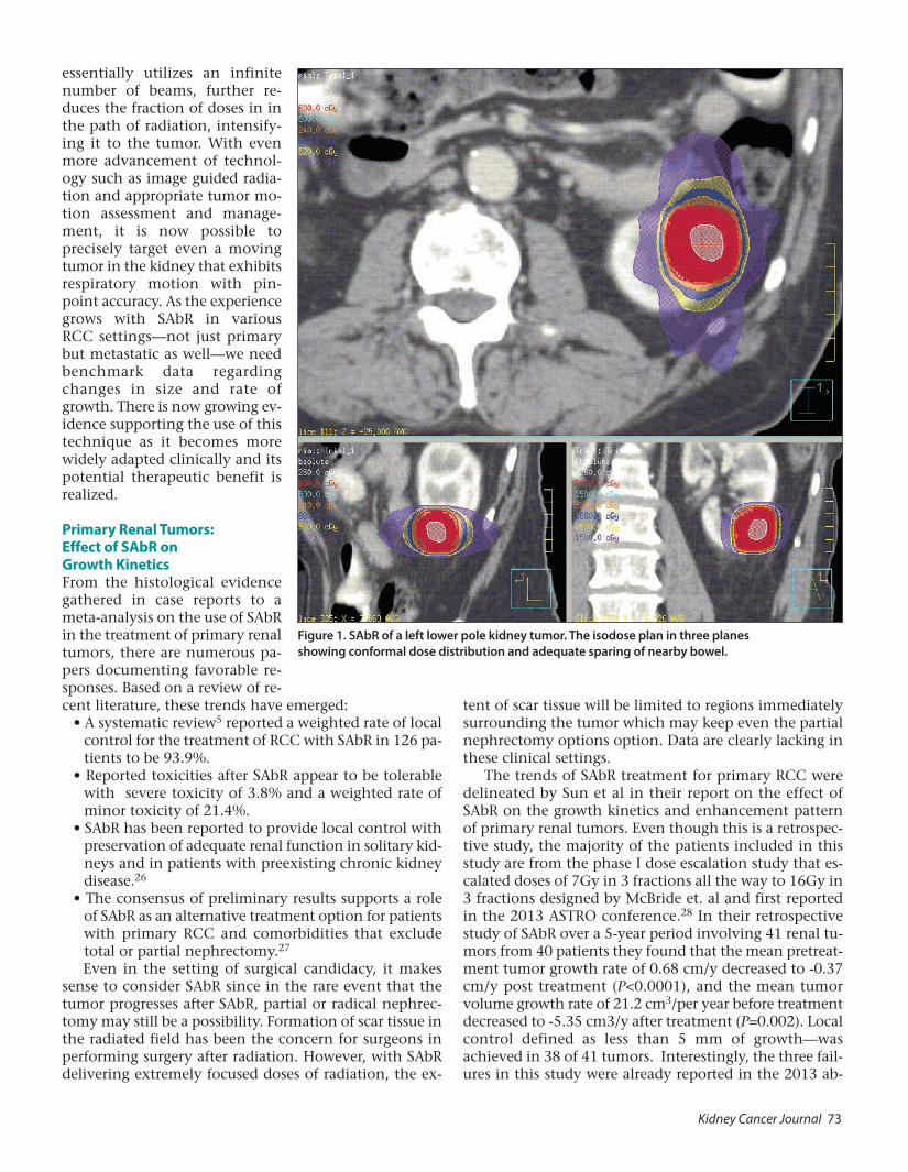
essentially utilizes an infinite number of beams, further re-duces the fraction of doses in in the path of radiation, intensify-ing it to the tumor. With even more advancement of technol-ogy such as image guided radia-tion and appropriate tumor mo- tion assessment and manage-ment, it is now possible to precisely target even a moving tumor in the kidney that exhibits respiratory motion with pin-point accuracy. As the experience grows with SAbR in various RCC settings—not just primary but metastatic as well—we need benchmark data regarding changes in size and rate of growth. There is now growing ev-idence supporting the use of this technique as it becomes more widely adapted clinically and its potential therapeutic benefit is realized. Primary Renal Tumors: Effect of SAbR on Growth Kinetics From the histological evidence gathered in case reports to a meta-analysis on the use of SAbR in the treatment of primary renal tumors, there are numerous pa-pers documenting favorable re-sponses. Based on a review of re- cent literature, these trends have emerged:
• A systematic review5 reported a weighted rate of local control for the treatment of RCC with SAbR in 126 pa-tients to be 93.9%.
• Reported toxicities after SAbR appear to be tolerable with severe toxicity of 3.8% and a weighted rate of minor toxicity of 21.4%.
• SAbR has been reported to provide local control with preservation of adequate renal function in solitary kid-neys and in patients with preexisting chronic kidney disease.26
• The consensus of preliminary results supports a role of SAbR as an alternative treatment option for patients with primary RCC and comorbidities that exclude total or partial nephrectomy.27 Even in the setting of surgical candidacy, it makes
sense to consider SAbR since in the rare event that the tumor progresses after SAbR, partial or radical nephrec-tomy may still be a possibility. Formation of scar tissue in the radiated field has been the concern for surgeons in performing surgery after radiation. However, with SAbR delivering extremely focused doses of radiation, the ex-
tent of scar tissue will be limited to regions immediately surrounding the tumor which may keep even the partial nephrectomy options option. Data are clearly lacking in these clinical settings.
The trends of SAbR treatment for primary RCC were delineated by Sun et al in their report on the effect of SAbR on the growth kinetics and enhancement pattern of primary renal tumors. Even though this is a retrospec-tive study, the majority of the patients included in this study are from the phase I dose escalation study that es-calated doses of 7Gy in 3 fractions all the way to 16Gy in 3 fractions designed by McBride et. al and first reported in the 2013 ASTRO conference.28 In their retrospective study of SAbR over a 5-year period involving 41 renal tu-mors from 40 patients they found that the mean pretreat-ment tumor growth rate of 0.68 cm/y decreased to -0.37 cm/y post treatment (P<0.0001), and the mean tumor volume growth rate of 21.2 cm3/per year before treatment decreased to -5.35 cm3/y after treatment (P=0.002). Local control defined as less than 5 mm of growth—was achieved in 38 of 41 tumors. Interestingly, the three fail-ures in this study were already reported in the 2013 ab-
Figure 1. SAbR of a left lower pole kidney tumor. The isodose plan in three planes showing conformal dose distribution and adequate sparing of nearby bowel.

74 Kidney Cancer Journal
stract to be on the 7Gy and 9Gy x3 dose fractionations and no failures were observed when >10Gy in three frac-tions were used.
Authors made an intriguing observation in this study. They noticed that even in the setting of good local con-trol, SAbR did not have an impact on the enhancement of the residual mass. Physicians treating renal tumors with ablative technique are reassured when enhancement is lost since the technique inherently disrupts the treated tissue leading to loss of tumor vasculature and lack of contrast dye uptake. However, SAbR primarily kills tumor cells by DNA damage leading to mitotic catastrophe or a loss of their proliferative ability with minimal damage to the vasculature. As a result, it is not surprising that local control is seen in the setting of continued contrast en-hancement.
Despite these promising results, there are unresolved issues concerning the tolerability of escalating doses of SAbR for primary treatment of localized RCC in poor sur-gical candidates.29 For example, the dose regimens used in earlier studies from 2005 to 2007 ranged from 16-48 Gy in 3-5 fractions but no consensus emerged regarding the optimal dose regimen for RCC.30 The phase 1 dose-escalation study by Ponsky et al offers insights as it ex-plores data on achieving the maximum tolerated dose for SAbR. It highlights concerns with the delivery of ablative doses of radiation. These concerns are related to tumor motion with respiration and the close proximity to vari-ous organs at risk, including the small bowel.
Ponsky et al used a robotic radiosurgery system with tumor tracking capability to deliver the radiation requir-ing a smaller margin around the gross tumor volume (GTV) to create a target planning volume (PTV). This en-abled the authors to treat a smaller amount of ipsilateral normal kidney and other organs at risk. A stepwise dose escalation regimen was followed and 48 Gy in 4 fractions was reached without causing dose-limiting toxicity. One patient experienced an acute and late grade 4 duodenal ulcer. Interestingly, while none of the 15 evaluable pa-tients developed progression at a median follow up of 13.67 months, 7 of the 11 tumors biopsied post-SAbR showed “viable” tumor. The efficacy of radiation (or chemotherapy for that matter) in controlling tumor cells in vitro comes from cell survival curves from clonogenic assays where after a certain dose of radiation the tumor cell’s ability to form colonies are measured and reported as surviving fraction. In reality, the surviving fraction is not reporting on whether the cells are alive or dead, they are merely reporting on whether the cells are able to di-vide and form colonies. In essence, a cancer cell that has lost its ability to divide, is perhaps not a cancer anymore.
In the context of no progression, a “viable” reading from H&E staining on a biopsy that did not perform any tests of proliferation is certainly inconclusive and the au-thors address this in the discussion and agrees to add pro-liferative indices in the future patients. Therefore, while the dose escalation design and primary endpoint of this study is robust, the secondary endpoint of local control
definition (loss of enhancement and biopsy) is flawed. As a result, with the goal of further improving local control, the investigators are enrolling patients for a starting dose of 48 Gy in 3 fractions. If the acute toxicity is acceptable, then the next 4 patients will be escalated to 54 Gy in 3 fractions. And then, if a dose limit has not been reached at that point, the last group of 4 patients will be treated to 60 Gy in 3 fractions. Based on all the other reported doses and local control rates for primary and metastatic RCC, one might argue that a dose escalation to this extent is likely unnecessary. Nonetheless, the reported study re-mains to be an important and the first prospective dose escalation study on SAbR for primary RCC.
Corroborative evidence of safety and efficacy for SAbR appeared in a European study by Staehler et al31 who re-ported on renal tumors treated with single fraction radio-surgery. This study demonstrated the short-term benefits of the technique in 40 patients who had an indication for nephrectomy and subsequent hemodilation. The phase 2 study devised an aggressive treatment approach delivering 25Gy in a single fraction with fiducial place-ment and respiratory motion tracking using the Cy-berKnife system. The study overcame the challenges seen with conventional radiation: even when lesions were close to or in the ureter, a measure that is not possible with ablative techniques, the authors achieved complete tumor control without functional impairment. A high dose of radiation could be applied precisely with 1 mm accuracy to the renal tumor, thus avoiding collateral dam-age to surrounding healthy tissue. The disadvantage of this set-up is the dependency on fiducial placement which is a (minimally) invasive procedure. Utilizing image guidance technology, it is now possible to admin-ister the same dose without the need for fiducials making SAbR completely non-invasive.
Figure 2. SAbR setup and beam arraignment for kidney lesion. The setup for SAbR abdominal treatment includes a vacuum bag for accurate reproducibility and a body frame that allows the stereotaxy. Multiple beams in non-coplanar arrangements are typically used to produce the focal dose distribution.

Kidney Cancer Journal 75
With a 98% local tumor control rate after a median followup of 28 months, according to this report, SAbR seems to be more effective and certainly less invasive than thermal ablation. There was a measurable size reduction in 38 lesions, including complete remission in 19 while the renal function remained stable. The authors can only speculate on what their results might look like when pa-tients are followed for the long term. However, even if renal function were to fail after a longer followup, the pa-tients still would have experienced a prolonged period free from hemodialysis. Ongoing Trials Seek to Extend Findings Two ongoing trials are recruiting patients to continue in-vestigations into the use of SAbR. One is being conducted at the University of Texas Southwestern Medical Center where patients with enlarging (>4mm growth within the past year) early renal cancers will undergo treatment of
SAbR of 36Gy in 3 or 40Gy in 5 fractions. The primary endpoint of this single arm phase II trial is local control at 1 year which will be evaluated with radiographic scans and a tumor biopsy one year after treatment carefully evaluating the proliferative capability of baseline com-pared to post-treatment bi-opsy to confirm tumor non-viability. The estimated study completion date is Decem- ber of 2018. The Clinical Trials.gov identifier is NCT021 41919.32
A second ongoing study, also phase II single arm, TROG 15.03 FASTRACKII,33 is being conduced in Aus-tralia, has an accrual target of 70 patients and is scheduled for completion in September of 2021. All participants will be assessed at regular intervals post treatment in order to estimate the activity and efficacy of the technique, toler-ability, survival, distant failure rate, and change of renal function after SAbR. SAbR dose fractionation in this study depends on tumor size: fraction schedule 1: 26Gy in 1
5 fractions showing optimized doses to critical organs as well as target volume (PTV). (E) Radiation dose constraints used for treatment planning.
Figure 3. SAbR plan for IVC tumor thrombus. (A-C) Representative axial, sagittal, and coronal images of the SAbR treatment plan with isodose lines showing dose distribution and coverage of the IVC-TT. (D) Radiation dose volume histogram from SAbR plan of 50 Gy in

76 Kidney Cancer Journal
fraction, for tumors of less than or equal to 4cm in size; fraction schedule 2: 42Gy in 3 fractions, for tumors of greater than 4cm in size (i.e. 14Gy per fraction, given in 3 fractions over a maximum of 3 weeks, each fraction given on non-consecutive days). SAbR in Locally Advanced RCC: A Bench to Bedside Case Report The application of SAbR may find a role in a subset of kid-ney cancer patients—those who present with inferior vena cava (IVC) tumor thrombus (IVC-TT) and comprise up to 10% of RCC patients. Surgery is the only treatment with proven efficacy for this setting, but such resection is difficult, and mortality and morbidity is high. Until re-cently, the difficulty with surgery has left clinicians with no treatment options for recurrent or unresectable RCC IVC-TT which left untreated can lead to Budd-Chiari syndrome.
Our experience34 treating 2 RCC patients with Level IV IVC-TT, one with recurrent disease and the other un-resectable—with SAbR —suggests how this modality may have utility in this difficult-to-treat setting. Our case re-port followed two elderly (75 years old and 83 years old) male patients both of whom had been refractory to sys-temic therapy. One received 50 Gy in 5 fractions and at
2 years of followup is doing well with a significant de-crease in the enhancement and size of the IVC-TT. (Fig-ures 3, 4) The second patient survived 18 months post SAbR. None of the patients that underwent SAbR to IVC-TT experienced any treatment-related toxicity. The sur-vival of 18 months and 24 months for these patients is comparable to the reported median survival of 20 months in similar groups of patients who underwent surgical re-section.20
Despite the high surgical mortality (10%) and morbid-ity (up to 30%), majority of the patients return with sys-temic metastasis perhaps from the tumor emboli shed from the IVC-TT itself.35 Therefore, an intriguing hypoth-esis raised by our study is whether SAbR could have fur-ther application in the neoadjuvant setting and whether it might lower the likelihood of systemic metastases by making the tumor emboli non-viable. A safety lead-in phase 2 clinical trial is addressing this issue where level II or higher IVC-TT is being radiated to 40Gy in 5 treat-ments immediately prior to surgery and looking at re-lapse-free survival at one year as the primary outcome measure. The target enrollment is 30 patients and the es-timated completion date is December of 2018 for this trial with a ClinicalTrials.gov identifier of NCT02 473536.36
Figure 4. MRI of IVC Tumor Thrombus in clear cell RCC before and after SAbR. Coronal (top) and axial (bottom) contrast enhanced MR images at different time points during the course of treatment. After nephrectomy and thrombectomy, the patient had an intralu-minal recurrence of tumor thrombus, which was adherent to the IVC wall (arrowheads, A). The superior extent of the thrombus is inferior to the diaphragm (Level III; arrow, A). Note the size of the thrombus at the level of the right hepatic vein (arrow, B). After systemic
targeted therapy (C) there was obvious disease progression with thrombus extending superior to the diaphragm (level IV, arrow) and increased enhancement (arrowhead, C). Note marked increased in transverse diameter (arrow, D). Two years after SABR therapy there is persistent thrombus extending above the diaphragm (arrow, E) although exhibiting clear decrease in enhancement (arrowhead, E) and marked reduction in transverse diameter (arrow, F).

Kidney Cancer Journal 77
SAbR for CNS RCC Metastases Gamma knife surgery (GKS) for metastatic brain tumors from RCC has a long history since stereotaxis initially was invented and designed for intracranial lesions, beginning with the first report published more than 20 years ago. Recent studies, however, are not only building on the pre-vious track record of successful results but elucidating ad-ditional benefits that may accrue from such radiosurgery, including improved tumor reduction and long-term sur-vival. These reports are exploring some of the underap-preciated nuances of GKS in this setting.
To what extent is GSK effective for growth control of metastatic tumors and what effect can it have on peritu-moral edema control? This question was addressed in a retrospective report by Shuto et al study-ing 280 metastatic brain tumors—80 from RCC and others involving breast and lung. In addition, the authors in-cluded 11 patients with metastatic brain tumors from RCC who had direct sur-gery. After compiling the data, Shuto et al17 present a treatment algorithm with a recommended strategy depending on tumor size, toleration of general anes-thesia, presence of symptomatic peritu-moral edema, and number of tumors.
The retrospective findings suggested a tumor growth control rate of 84.3%. The key findings: The primary site (renal or not renal) and the delivered marginal dose (25 Gy or more) were significantly correlated with control of peritumoral edema; although peritumoral edema was extensive, it disappeared within 1-3 months. All tumors treated with direct surgery were 2 cm in max-imum diameter.
Significant total tumor volume reduction at an early treatment seems to result in long-term survival, according to Kim et al, who proposed prognostic factors worth con-sidering in determining outcomes. The median survival time for 46 patients in a study spanning 12 years was 18 months in the good response group, significantly longer than that observed in the poor response group (9 months. P=0.025). After treatment, local tumor control was achieved in 84.7% of the 85 tumors assessed.
Classification in the “good-response” group was the only independent prognostic factor for longer survival. Although the study did not specifically address the effect of total tumor volume reduction on quality of life, the authors suggest that such reduction can lead to improved neurologic symptoms and patients may be better able to undergo systemic therapy. Spine Radiosurgery: Emerging Issues, Guidelines Spine radiosurgery is an effective tool in manag ing pa-tients with RCC. Although RT has little role in the treat-ment of primary disease, SRS does play an important role in the treatment of patients with spinal metastases, par-
ticularly those who received prior RT or instrumentation according to Taunk et al.37 It is known from multiple se-ries that spine SRS for RCC has extremely high rates of durable local control and palliation. However, it demands high quality control, precision guidance, and careful pa-tient selection in multi-modality consultations to be safely and effectively implemented.
SAbR requires several special techniques to de liver ab-lative RT safely and effectively, including (1) use of mul-tiple conformal beams with intensity-modulation, (2) accuracy within millimeters, (3) im age guidance with each treatment, and (4) custom immobilization. Multiple beams allow for shaping of highly conformal dose, par-ticularly sparing the spinal cord, which is usually within
millimeters of the target volume. Custom immobilization requires comfortable, re-producible patient positioning while se- curely immobilizing the shoulders, neck, abdomen, or pelvis, as needed. Image guidance uses daily on-board imaging, ideally with pretreatment cone-beam CT.38
Between 2004 and 2010, MSKCC treated 105 RCC metastases (59 spine le-sions) with single-dose SRS or hypofrac-tionated SRS. The overall 3-year ac tuarial local progression-free survival rate was 44%. In patients with disease treated in a single fraction and with a dose of 24 Gy or greater, the 3-year local progression-
free survival rate was 88%. In contrast, patients receiving hypofractionated treatment in 3 or 5 fractions had a 3-year local control rate of 17%. Treatment delivered in a single fraction and with a dose of 24 Gy or greater signif-icantly improved local control in multivariate analysis.20
The authors’ practice is to recommend SRS alone in patients with oligometastatic disease and mechanically stable spines. Operating in the NOMS (Neurologic, On-cologic, Mechanical instability, and Systemic disease) clinical framework, patients with spine lesions are as-sessed in a multidisciplinary clinic at MSKCC by a radia-tion oncologist, spine neurosurgeon, and neurointer- ventional radiologist. Careful patient selection is critical to identify those who may benefit the most from treat-ment, includ ing patients for whom prior radiation treat-ment failed. Indicated procedures are performed for stabilization using implanted hardware or kyphoplasty before ra diation. Patients with RCC who present with high-grade spinal cord compression often require surgical decompression and stabilization to separate the tu mor from the spinal cord and facilitate delivery of SRS while remaining within spinal cord tolerance. Stereotactic Radiotherapy for Extra-CNS Oligometastases Although the evidence is relatively sparse compared to other treatment settings, data are growing and suggest compelling results for the use of SAbR in RCC extracranial
“The application of SAbR is currently the standard of care for CNS and spine metastases from RCC where surgery is difficult and morbid. Additional indications currently under investigation include SAbR for oligometastatic and oligo- progressive mRCC and neo- adjuvant treatment for IVC tumor thrombus.”

78 Kidney Cancer Journal
metastases. Theoretical basis for this approach comes from the surgical metastasectomy data that showed over-all survival benefit for oligo-metastatic RCC patients when all site of metastases were resected.39 SAbR offers a non-invasive technique for metastasectomy that can be applied to multiple sites of metastases throughout the body. The earlier reports on extracranial applications highlight how patient selection may be a key factor in whether SAbR is successful.
Svedman et al,40 for example, suggest that SAbR can be considered as an option to surgery when there are a limited number of metastases, as local treatment in RCC with an indolent presentation or as a method of reducing tumor burden prior to medical treatment. One of the in-triguing suggestions from this study is whether high-dose radiotherapy triggers regression of untreated metastases. Support for this hypothesis comes from other authors who speculate that this effect could be due to radiation induced immune response.41 From the same institution, the report from Wersäll et al23 also explored the extent to which certain patients may benefit more than others from SAbR, thus highlighting how patient selection could be used to greater advantage. For example, retrospective results in 58 patients in this Swedish study indicated that patients with one to three metastases and patients with inoperable primary tumors or local recurrence benefited more from this treatment than those with four or more metastases. The majority of patients were treated for me-tastases in the lungs.
In a detailed analysis of our institutional experience for treatment of extracranial mRCC,24 we provide guide-lines with dosimetric data and new insights on clinical factors affecting local control. Until recently, little has been known definitively about these factors. In the largest published experience of 175 metastatic lesions from 84 patients, we observed no failures when SAbR regimens of 24 Gy, 12 Gy, or 8 Gy in 1, 3, or 5 fractions were used with at least 95% PTV coverage. Overall local control rates were 91.2% at a one year. The most critical factors affect-ing local control of mRCC were adequate radiation dose and appropriate target coverage. Late toxicities were low and less than 3% were high grade. Interestingly, previous use of >1 systemic therapy came out to be an independ-ent predictor of local failure in multi-variate analysis sug-gesting that higher radiation doses may be required to achieve the same local control in these patients.
In one of the subgroups analyzed in this retrospective study are patients that are showing progression on lim-ited sites of disease on systemic therapy that received SAbR to delay the switching of systemic therapy or essen-tially to extend the progression free survival (PFS) of the ongoing systemic therapy. The benefits of treating oligo-progressive RCC metastases with SAbR could be many folds: 1) owing to the tumor genetic heterogeneity be-tween primary and metastatic sites as described elegantly by Gerlinger et al.42 It is possible that a majority of metas-tases in a patient are responding and only 1-2 sites are progressing. Therefore, SAbR allows continuation of a
therapy that is otherwise effective and being tolerated by the patient. 2) By allowing effective continuation of the current therapy, SAbR may be preserving more lines of systemic therapy for a patient thereby possibly extending survival (OS). In a case report published by the same in-stitution. Straka et. al. demonstrated extending the PFS of sunitinib from 14 months to 22 months by the use of SAbR for oligo-progressive disease.43
While the reported local control of SAbR for RCC is high, the question remains as to who truly benefits from the local control. It may be intuitive to treat oligo-metastatic RCC patients with SAbR either with a curative intent in the setting of metachronous metastasis or with the intent of preserving quality of live by delaying the initiation of systemic therapy. However, the most impor-tant question is whether by delaying systemic therapy, is the OS is being compromised? Data is lacking in this arena as to the effect of SAbR in PFS and OS, a few intrigu-ing ongoing clinical trials are expected to provide insight to these settings. In one of these clinical trials begin con-ducted at UT Southwestern, oligo-metastatic RCC pa-tients are being randomized to receive SAbR or standard of care systemic therapy in a phase II randomized trial. The accrual goal is 18 patients in each arm. The primary endpoint is the PFS on first line systemic therapy and es-sentially the study is measuring how long SAbR is able to extend the PFS on first line therapy. In addition to com-paring OS, an important secondary endpoint is quality of life which is expected to be better in the SAbR arm that delayed the initiation of systemic therapy. The Clinical-Trials.gov. identifier is NCT02956798.
A second clinical trial being conducted at multiple lo-cations in Canada by Georg Bjernason is evaluating how well SAbR can destroy kidney cancer metastases no longer controlled by sunitinib. Only oligo-progressive patients on sunitinib with up to 5 sits of progression will be en-rolled. The primary endpoint is local control while the secondary endpoint is PFS. This study will seek an enroll-ment of 68 patients with an estimated completion date of December 2019.44 The ClinicalTrials.gov. identifier is NCT020195766. Conclusion SAbR is becoming a more widely accepted modality for the treatment of a broad spectrum of renal tumors, in-cluding primary RCC, CNS RCC metastases, and extra-CNS oligometastases. It may also have potential appli- cation as neoadjuvant therapy. Renal tumors consistently show a very high (>85%) rate of local control in these set-tings after treatment with SAbR. The pivotal factor in op-timizing the effectiveness of SAbR is appropriate dose selection. While additional clinical trials and longer-term follow ups are required to establish guidelines on the op-timal dose in controlling RCC, the focus now is on how and when to integrate an effective local control modality such as SAbR with the growing armament of approved systemic agents for RCC. Treatment of oligo-progressive RCC metastasis may be a strategy to improve PFS of sys-
Kidney Cancer Journal 79
temic agents for RCC. The now well-known immune modulatory and antigen presenting properties of SAbR, which has not been elaborated and is beyond the scope of this review, may synergize with the approved and up-coming immunotherapies for RCC. While much is known about the local control, the impact of SAbR on PFS and eventually on extending patient survival is not known yet and careful design of prospective trials is clearly needed in this setting. With greater application of SAbR, the focus needs to be maintained on careful patient selection for the technique to be safely and effectively im-plemented and outcomes optimized, including long-term survival. References 1. Boike TP, Lotan Y, Cho LC, Brindle J, DeRose P, Xie XJ, Yan J, Foster R, Pistenmaa D, Perkins A, Cooley S, Timmerman R. Phase I dose-esca-lation study of stereotactic body radiation therapy for low- and inter-mediate-risk prostate cancer. J Clin Oncol. 2011;29(15):2020-6. Epub 2011/04/06. doi: 10.1200/JCO.2010.31.4377. PubMed PMID: 21464418; PMCID: 3138546. 2. Timmerman R, Paulus R, Galvin J, Michalski J, Straube W, Bradley J, Fakiris A, Bezjak A, Videtic G, Johnstone D, Fowler J, Gore E, Choy H. Stereotactic body radiation therapy for inoperable early stage lung can-cer. JAMA. 2010;303(11):1070-6. 3. King CR, Freeman D, Kaplan I, Fuller D, Bolzicco G, Collins S, Meier R, Wang J, Kupelian P, Steinberg M, Katz A. Stereotactic body radiother-apy for localized prostate cancer: Pooled analysis from a multi-institu-tional consortium of prospective phase II trials. Radiotherapy and Oncology. 2013. doi: 10.1016/j.radonc.2013.08.030. PubMed PMID: 24060175. 4. Katz AJ, Santoro M, Diblasio F, Ashley R. Stereotactic body radiother-apy for localized prostate cancer: disease control and quality of life at 6 years. Radiat Oncol. 2013;8(1):118. doi: 10.1186/1748-717X-8-118. PubMed PMID: 23668632; PMCID: 3674983. 5. Timmerman RD, Kavanagh BD, Cho LC, Papiez L, Xing L. Stereotactic body radiation therapy in multiple organ sites. J Clin Oncol. 2007;25(8):947-52. 6. Berber B, Sanabria JR, Braun K, Yao M, Ellis RJ, Kunos CA, Sohn J, Machtay M, Teh BS, Huang Z, Mayr NA, Lo SS. Emerging role of stereo-tactic body radiotherapy in the treatment of pancreatic cancer. Expert Review of Anticancer Therapy. 2013;13(4):481-7. 7. Tao C, Yang LX. Improved radiotherapy for primary and secondary liver cancer: stereotactic body radiation therapy. Anticancer Research. 2012;32(2):649-55. 8. Kwon JH, Bae SH, Kim JY, Choi BO, Jang HS, Jang JW, Choi JY, Yoon SK, Chung KW. Long-term effect of stereotactic body radiation therapy for primary hepatocellular carcinoma ineligible for local ablation ther-apy or surgical resection. Stereotactic radiotherapy for liver cancer. BMC Cancer. 2010;10:475. doi: 10.1186/1471-2407-10-475. PubMed PMID: 20813065; PMCID: 2940809. 9. Bondiau PY, Courdi A, Bahadoran P, Chamorey E, Queille-Roussel C, Lallement M, Birtwisle-Peyrottes I, Chapellier C, Pacquelet-Cheli S, Fer-rero JM. Phase 1 clinical trial of stereotactic body radiation therapy con-comitant with neoadjuvant chemotherapy for breast cancer. Inter- national Journal of Radiation Oncology, Biology, Physics. 2013;85 (5):1193-9. 10. Chang BK, Timmerman RD. Stereotactic body radiation therapy: a comprehensive review. American Journal of Clinical Oncology. 2007;30 (6):637-44. 11. Lo SS, Fakiris AJ, Teh BS, Cardenes HR, Henderson MA, Forquer JA, Papiez L, McGarry RC, Wang JZ, Li K, Mayr NA, Timmerman RD. Stereo-tactic body radiation therapy for oligometastases. Expert Review of Anti-cancer Therapy. 2009;9(5):621-35. 12. Lo SS, Fakiris AJ, Chang EL, Mayr NA, Wang JZ, Papiez L, Teh BS, McGarry RC, Cardenes HR, Timmerman RD. Stereotactic body radiation therapy: a novel treatment modality. Nature Reviews Clinical Oncology. 2010;7(1):44-54.
13. Bianco AI, The BS, Amato RJ. Role of radiation therapy in the man-agement of renal cell carcinoma. Cancers. 2011;3:4010-4023. 14. Deschavanne PJ, Fertil B. A review of human cell radiosensitivity in vitro. Int J Radiat Oncol Biol Phys. 1996;34:251-266. 15.Ning S, Trisler K, Wessels BW, et al. Radiologic studies of radioim-munotherapy and external beam radiotherapy in vitro and in vivo in human renal cell carcinoma xenografts. Cancer. 1997;80:(Suppl) 2519-25288. 16.Walsh L, Stanfield JL, Cho LC, et al. Efficacy of ablative high-dose-per fraction radiation for implanted human renal cell cancer in a nude mouse model. Eur Urol. 2006;50:795-800. 17. Shuto T, Matsunaga S, Suenaga J, Inomori S, Fujino H. Treatment strategy for metastatic brain tumors from renal cell carcinoma: selection of gamma knife surgery or craniotomy for control of growth and peri-tumoral edema. Journal of Neuro-Oncology. 2010;98(2):169-75. 18. Gerszten PC, Burton SA, Ozhasoglu C, Welch WC. Radiosurgery for spinal metastases: clinical experience in 500 cases from a single institu-tion. Spine. 2007;32(2):193-9. 19. Gerszten PC, Burton SA, Ozhasoglu C, Vogel WJ, Welch WC, Baar J, Friedland DM. Stereotactic radiosurgery for spinal metastases from renal cell carcinoma. Journal of neurosurgery Spine. 2005;3(4):288-95. 20. Yamada Y, Bilsky MH, Lovelock DM, Venkatraman ES, Toner S, John-son J, Zatcky J, Zelefsky MJ, Fuks Z. High-dose, single-fraction image-guided intensity-modulated radiotherapy for metastatic spinal lesions. International Journal of Radiation Oncology, Biology, Physics. 2008; 71(2):484-90. 21. Nguyen QN, Shiu AS, Rhines LD, Wang H, Allen PK, Wang XS, Chang EL. Management of spinal metastases from renal cell carcinoma using stereotactic body radiotherapy. International Journal of Radiation Oncology, Biology, Physics. 2010;76(4):1185-92. 22. Ranck MC, Golden DW, Corbin KS, Hasselle MD, Liauw SL, Stadler WM, Hahn OM, Weichselbaum RR, Salama JK. Stereotactic Body Radio-therapy for the Treatment of Oligometastatic Renal Cell Carcinoma. American Journal of Clinical Oncology. 2012. Epub 2012/08/08. doi:10. 1097/COC.0b013e31825d52b2. PubMed PMID: 22868242. 23. Wersall PJ, Blomgren H, Lax I, Kalkner KM, Linder C, Lundell G, Nilsson B, Nilsson S, Naslund I, Pisa P, Svedman C. Extracranial stereo-tactic radiotherapy for primary and meitastatic renal cell carcinoma. Ra-diotherapy and Oncology. 2005;77(1):88-95. 24. Adler JR, Chang SD, Murphy MJ, et al. The Cyberknife: a frameless robotic system for radiosurgery. Stereotact Funct Neurosurg. 1997;69:124-128. 25. Siva S, Pham D, Gill S, et al. A systematic review of stereotactic ra-diotherapy ablation for primary renal cell carcinoma. BJU Int. 2012;110: E77377-E743. 26. Lo CH, Huang WY, Chao HL, et al. Novel application of stereotactic ablative radiotherapy using Cyberkife for early-stage renal cell carci-noma in patients with pre-existing chronic kidney disease: initial clin-ical experiences. Oncol Lett. 2014;8:355-360. 27. Sun MRM, Brook A, Powell MF, et al. Effect of stereotactic body ra-diotherapy on the growth kinetics and enhancement pattern of primary renal tumors. Am J Roent. 2016;544-553. 28. McBride SM, Warner AA, Kaplan ID. A phase 1 dose-escalation study of robotic radiosurgery in inoperable primary renal cell carcinoma. Int J Rad Oncol Biol Phys. 2013;87:S84. 29. Ponsky L, Lo SS, Zhang Y, et al. Phase I dose-escalation study of stereotactic body radiotherapy (SAbR) for poor surgical candidates with localized renal cell carcinoma. Radiother Oncol.2015;117:183-187. 30. Lo SS, Loblaw A, Chang EL, Mayr NA, Teh BS, Huang Z, et al. Emerg-ing applications of stereotactic body radiotherapy. Future Oncol. 2014;10: 1299–310. 31. Staehler M, Bader M, Schlenker B, et al. Single fraction radiosurgery for the treatment of renal tumors. J Urol. 2015;193:771-775. 32. ClinicalTrials.gov.NCT02141919 33. Focus ablative Stereotactic tadiosurgery for cancers of the kidney—a phase II clinical trial. TROG trial 15.03 on Australian Cancer Trials website. 34. Hannan R, Margulis V, Chun SG, et al. Stereotactic radiation therapy of renal cancer inferior vena cava tumor thrombus. Cancer Biology & Therapy. 2015;16:65-661.

80 Kidney Cancer Journal
35. Haddad AQ, Leibovich BC, Abel EJ, et al. Preoperative multivariable prognostic models for prediction of survival and major complications following survival resection of renal cell carcinoma with suprahepatic caval tumor thrombus. Urol Oncol. 2015;33;388:e1-9. 36.ClinicalTrials.gov. NCT02473536. 37. Taunk NK, Spratt DE, Bilsky M, et al. Spine radiosurgery in the man-agement of renal cell carcinoma metastases. J Nat Compreh Can Network. 2015;13:801-809. 38. Taunk NK, Spratt DE, Bilsky M, et al. Spine radiosurgery in the man-agement of renal cell carcinoma metastases. J Nat Compreh Can Network. 2015;13:801-809. 39. Alt AL1, Boorjian SA, Lohse CM, Survival after complete surgical re-section of multiple metastases from renal cell carcinoma. Cancer. 2011; 117:2873-2882. 40. Svedman C, Sandstrom P, Pia P, et al. A prospective Phase II trial of
using extracranial stereotactic radiotherapy in primary and metastatic renal cell carcinoma. Acta Oncologica. 2006;45:870-875. 41. Camphausen K, Moses MA, Menard C, Sproull M, Beecken WD, Folkman J, et al. Radiation abscopal antitumor effect is mediated through p53. Cancer Res. 2003;63(8):1990-1993. 42.Gerlinger M, Rowan AJ, Horswell S, et al. Intratumor heterogeneity and branched evolution revealed by multiregion sequecning. N Engl J Med. 2012;36:883-892. 43. Straka C1, Kim DW, Timmerman RD, et al. Ablation of a site of pro-gression with stereotactic body radiation therapy extends sunitinib treatment from 14 to 22 months. J Clin Oncol. 2013 Aug 10;31(23): e401-3. 44. CliicalTrials.gov. NCT020195766. ���
prolong exposure to the drug while limiting the adverse effects that often stand in the way of continuing with the 4 weeks on and 2 weeks off strategy. So here, too, an intriguing and significant evolution in the standard of care is taking place with regards to the relative merits of the 4/2 dosing schedule vs the 2/1 schedule. The particularly interesting aspect of this evolution is the apparent initiative on the part of many of our colleagues in community practice to stay ahead of the curve or integrate novel approaches that enable them to prolong exposure to sunitinib or at least mitigate the impact of adverse effects through various drug-free interval strategies.
There are skeptics, however, who may ask whether it is appropriate to expend more resources prospectively testing (data we need to verify appropriate dosing schedules) a new schedule of an older targeted agent in
lieu of the excitement over the latest generation of immune-oncologics in RCC. There are two reasons for continuing to investigate VEGF blockade in this con-text. First, not all RCCs respond to immunotherapy, as Lauren C. Harshman, MD reminds us in an Editorial in the Journal of Clinical Oncology (2017;35(16) 1755-1757. This is true of even the modern and more tolerable PD-1 pathway antibodies. Also, VEGF-pathway blockade remains the backbone of many ongoing, first-line PD-1 combination studies. These considerations remain essential in view of the new agents recently approved and the implications for the treatment paradigm. The article in this issue by Pavlos Msaouel, MD, and Nizar Tannir, MD, not only delineates these issues but suggests state-of-the-art thinking on tailoring sunitinib schedules to achieve maximum benefit.
Bernard J. Escudier, MD Guest Editor
EDITOR ’S MEMO (continued from page 62)

response; there were eight confirmed partial responders with MET-driven disease (18%), but none with MET-inde-pendent disease (P = .002). Median progression-free survival for patients with MET-driven and MET-indepen-dent PRCC was 6.2 months and 1.4 months, respectively (hazard ratio, 0.33; 95% CI, 0.20 to 0.52; log-rank P < .001). The most frequent adverse events associated with savolitinib were nausea, fatigue, vomiting, and peripheral edema. Conclusion: These data show activity and tolerability of savolitinib in the subgroup of patients with MET-driven PRCC. Furthermore, molecular characterization of MET status was more predictive of response to savolitinib than a classification based on pathology. These findings justify investigating savolitinib in MET-driven PRCC. Safety and Efficacy of Nivolumab in Combination With Ipilimumab in Metastatic Renal Cell Carcinoma: The CheckMate 016 Study. Hammers HJ, Plimack ER, Infante JR, et al. J Clin Oncol. 2017 Jul 5;JCO20167219 85. doi: 10.1200. Summary: Combination treatment with immune check-point inhibitors has shown enhanced antitumor activity compared with monotherapy in tumor types such as melanoma. The open-label, parallel-cohort, dose-escala-tion, phase I CheckMate 016 study evaluated the efficacy and safety of nivolumab plus ipilimumab in combination, and nivolumab plus a tyrosine kinase inhibitor in metasta-tic renal cell carcinoma (mRCC). Safety and efficacy results from the nivolumab plus ipilimumab arms of the study are presented. Patients with mRCC received intravenous nivolumab 3 mg/kg plus ipilimumab 1 mg/kg (N3I1), nivolumab 1 mg/kg plus ipilimumab 3 mg/kg (N1I3), or nivolumab 3 mg/kg plus ipilimumab 3 mg/kg (N3I3) every 3 weeks for four doses followed by nivolumab monother-apy 3 mg/kg every 2 weeks until progression or toxicity. End points included safety (primary), objective response rate, and overall survival (OS). All patients in the N3I3 arm (n = 6) were censored at the time of analysis as a result of dose-limiting toxicity or other reasons. Forty-seven patients were treated in both the N3I1 and the N1I3 arm, and baseline patient characteristics were balanced between arms. Grade 3 to 4 treatment-related adverse events were reported in 38.3% and 61.7% of the patients in the N3I1 and N1I3 arms, respectively. At a median follow-up of 22.3 months, the confirmed objective response rate was 40.4% in both arms, with ongoing responses in 42.1% and 36.8% of patients in the N3I1 and N1I3 arms, respectively. The 2-year OS was 67.3% and 69.6% in the N3I1 and N1I3 arms, respectively.
Conclusion: Nivolumab plus ipilimumab therapy demon-strated manageable safety, notable antitumor activity, and durable responses with promising OS in patients with mRCC. Correlation of c-MET Expression with PD-L1 Expression in Metastatic Clear Cell Renal Cell Carcinoma Treated by Sunitinib First-Line Therapy. Kammerer-Jacquet SF, Medane S, Bensalah K, et al. Target Oncol. 2017 Aug; 12(4):487-494. Summary: Cabozantinib, an anti-angiogenic tyrosine kinase inhibitor that targets c-MET, provided interesting results in metastatic ccRCC treatment. To understand bet-ter the role of c-MET in ccRCC, we assessed its status in a population of patients with metastatic ccRCC. For this purpose, tumor samples were analyzed for c-MET expres-sion by immunohistochemistry (IHC), for c-MET copy number alterations by fluorescence in situ hybridization (FISH), and for c-MET mutations by next generation sequencing (NGS) in a retrospective cohort of 90 primary ccRCC of patients with metastases treated by first-line sunitinib. The expression of c-MET was correlated with pathological, immunohistochemical (VEGFA, CAIX, PD-L1), clinical, and molecular criteria (VHL status) by univariate and multivariate analyses and to clinical outcome using Kaplan-Meier curves compared by log-rank test. Of ccRCC, 31.1% had low c-MET expression (absent to weak intensity by IHC) versus 68.9% with high expres-sion (moderate to strong intensity). High expression of c-MET was associated with a gain in FISH analysis (P = 0.0284) without amplification. No mutations were detected in NGS. Moreover, high c-MET expression was associated with lymph node metastases (P = 0.004), sarco-matoid component (P = 0.029), VEGFA (P = 0.037), and PD-L1 (P = 0.001) overexpression, the only factor that remained independently associated (P < 0.001) after logis-tic regression. No difference was observed in clinical outcomes. Conclusion: This study is the first to analyze c-MET status in metastatic ccRCC. The high expression of c-MET in the majority of ccRCC and its independent association with PD-L1 expression, may suggest a potential benefit from combining c-MET inhibitors and targeted immunotherapy. New Insights into Adjuvant Renal Cell Carcinoma Treatment with Vascular Endothelial Growth Factor Inhibitors: What Have We Learned So Far? Escudier B, Staehler M. Eur Urol. 2017 Sep 7; pii: S0302-2838(17) 30713-3. doi: 10.1016 Summary: Adjuvant treatment of renal cell carcinoma with vascular endothelial growth factor inhibitors is feasible and effective with careful patient selection and standard dosing levels. ���
Kidney Cancer Journal 81
JOURNAL CLUB (continued from page 64)

82 Kidney Cancer Journal
MEDICAL INTELLIGENCE (continued from page 65)
intermediate/poor risk patients, which was 41.6% for the nivolumab/ipilimumab combination compared to 26.5% for sunitinib (P < 0.0001) with 9.4% of patients receiving combination therapy achieving complete response (CR) compared to1.2% of patients on sunitinib. There was an improvement in median PFS with the combination in this cohort; median PFS was 11.6 months for the nivolumab and ipilimumab combination versus 8.4 months with sunitinib, hazard ratio [HR] 0.82 (P = 0.03).
The efficacy outcomes differed according to the levels of PD-L1 expression and IMDC risk group. Both the ORR per independent committee and PFS significantly favored nivolumab plus ipilimumab over sunitinib in intermedi-ate/poor risk patients having baseline PD-L1 expression ≥1% where the ORR was 58% versus 25%, and median PFS was 22.8 (95% CI 9.4, NR) months versus 5.9 (95% CI 4.4, 7,1) months, respectively, HR 0.48 (95% CI 0.28, 0.82; P = 0.0003). The investigators found that baseline tumor PD-L1 expres-sion was lower in the cohort of patients at favorable risk where 11% of patients on combination had PD-L1 levels ≥1% versus12% of patients on sunitinib compared to 26% versus 29% of patients at intermediate or poor risk in the respective treatment arms.
In patients at favorable risk, both the ORR and PFS were higher with sunitinib over combination; in this cohort, ORR was 29% with nivolumab/ipilimumab versus 52% with sunitinib (P = 0.0002) and median PFS was 15.3 (95% CI 9.7, 20.3) months versus 25.1 (95% CI 20.9, NR) months, respec-tively, HR 2.17 (95% CI 1.46, 3.22; p < 0.0001). In the overall composite of patients at any risk, no significant difference between treatments was demonstrated in ORR (P = 0.0191) or PFS (P = 0.819). Any grade drug-related adverse events (AEs) occurred in 509 (93%) of patients in the nivolumab/ ipilimumab cohort and in 521 (97%) of patients receiving sunitinib. With the combination, 54% of patients had a grade 3 to 4 AE, and with sunitinib 63% of patients had a grade 3 to 5 AE.
Progression-free Rate Unaffected by Sequence of Cytoreductive Nephrectomy and Sunitinib in Patients with Synchronous mRCC MADRID—Treating primary tumors by administering targeted therapy with sunitinib prior to cytoreductive nephrectomy (CN) did not improve the progression-free rate at 28 weeks over a sequence of immediate CN followed by sunitinib in patients with synchronous metastatic renal cell carcinoma (mRCC), according to findings from a randomized trial presented at ESMO 2017.
Axel Bex, Surgical Oncology-Urology, The Netherlands Cancer Institute in Amsterdam, Netherlands and colleagues investigated whether the outcome after sequential cyto- reductive nephrectomy (CN) followed by targeted therapy with sunitinib could be improved with the opposite sequence. They randomized 99 patients with mRCC to im-mediate CN followed by sunitinib (n=50) versus three cycles of sunitinib followed by CN plus sunitinib (n=49). The study (EORTC 30073 SURTIME NCT01099423) included patients with histologically confirmed clear-cell subtype, and a resectable asymptomatic primary tumor plus 3 or fewer surgical risk factors.
After 5.7 years the study included 99 patients from 19 institutions. The immediate CN arm had 50 patients and the deferred CN arm had 49 patients. The majority of patients were male in both arms with a median age of 60 compared to 58 years, and MSKCC intermediate risk was reported for 86% versus 87.7% of patients, respectively. In the respective arms, WHO performance status (PS) was 0 and 1 in 72% and 28% versus 63.3 % and 36.7%; 86% versus 93.9% of patients had ≥ 2 measurable metastatic sites and the mean (stan-dard deviation) size of the primary tumor was 93.1 (37.8) mm versus 96.8 (31.3) mm. Patients in each arm derived different benefit from each sequence. At a median follow-up of 3.3 years, 46 of 50 patients underwent CN in the immedi-ate CN arm and 40 of these patients had received post-CN sunitinib. In the deferred CN arm, 48 of 49 patients had been treated with sunitinib prior to CN; of these patients, 40 underwent CN and 26 also received post-CN sunitinib. ���

A Round Table Discussion featuring perspectives and insights from three thought leaders on: Reassessing the Paradigm in RCC to Meet the Challenges and Leverage the Advantages of Combination Therapy • analyzing how new approaches to combination
therapy can address multiple pathways in kidney cancer
• ‘take-home’ messages on how these strategies can optimize progression-free survival
Plus: • How targeted therapies are being applied in
VHL Disease • Highlights from the 2017 Kidney Cancer Association
Scientific Sessions in Miami, November 3-4.

333 E. 75th Street, Suite 5D New York, NY 10021.