plos medicine commercial serological antibody detection ... · commercial serological antibody...
TRANSCRIPT

Commercial Serological Antibody DetectionTests for the Diagnosis of PulmonaryTuberculosis: A Systematic ReviewKaren R. Steingart
1,2, Megan Henry
3, Suman Laal
4,5,6, Philip C. Hopewell
1,2, Andrew Ramsay
7, Dick Menzies
8,9,
Jane Cunningham7
, Karin Weldingh10
, Madhukar Pai8,9*
1 Division of Pulmonary and Critical Care Medicine, San Francisco General Hospital, University of California, San Francisco, California, United States of America, 2 Francis J.
Curry National Tuberculosis Center, San Francisco, California, United States of America, 3 County of Sacramento Department of Health and Human Services, Sacramento,
California, United States of America, 4 Department of Pathology, New York, New York, United States of America, 5 Department of Microbiology, New York University School
of Medicine, New York, New York, United States of America, 6 Veterans Affairs Medical Center, New York, United States of America, 7 UNICEF/UNDP/World Bank/WHO Special
Programme for Research and Training in Tropical Diseases, World Health Organization, Geneva, Switzerland, 8 Department of Epidemiology, Biostatistics and Occupational
Health, McGill University, Montreal, Canada, 9 Respiratory Epidemiology and Clinical Research Unit, Montreal Chest Institute, Montreal, Canada, 10 Statens Serum Institut,
Department of Infectious Disease Immunology, Copenhagen, Denmark
Funding: This work was supportedby the UNICEF/UNDP/World Bank/WHO Special Programme forResearch and Training in TropicalDiseases (TDR). AR and JC, both withTDR, contributed to conception anddesign of the systematic review andcritical revision of and decision topublish the manuscript. AR alsoparticipated in data interpretation.
Competing Interests: KW is co-inventor on a number of patentsrelating to mycobacterial antigenswhich may be used for serologicalassays. All rights have been assignedto Statens Serum Institut.
Academic Editor: Paul Garner,Liverpool School of TropicalMedicine, United Kingdom
Citation: Steingart KR, Henry M, LaalS, Hopewell PC, Ramsay A, et al.(2007) Commercial serologicalantibody detection tests for thediagnosis of pulmonary tuberculosis:A systematic review. PLoS Med 4(6):e202. doi:10.1371/journal.pmed.0040202
Received: November 17, 2006Accepted: April 20, 2007Published: June 12, 2007
Copyright: � 2007 Steingart et al.This is an open-access articledistributed under the terms of theCreative Commons AttributionLicense, which permits unrestricteduse, distribution, and reproductionin any medium, provided theoriginal author and source arecredited.
Abbreviations: AUC, area under thecurve; CI, confidence interval; ELISA,enzyme-linked immunosorbentassay; FPR, false positive rate; LAM,lipoarabinomannan; ROC, receiveroperating characteristic; SROC,summary receiver operatingcharacteristic; TB, tuberculosis; TPR,true positive rate
* To whom correspondence shouldbe addressed. E-mail: [email protected]
A B S T R A C T
Background
The global tuberculosis epidemic results in nearly 2 million deaths and 9 million new cases ofthe disease a year. The vast majority of tuberculosis patients live in developing countries, wherethe diagnosis of tuberculosis relies on the identification of acid-fast bacilli on unprocessedsputum smears using conventional light microscopy. Microscopy has high specificity intuberculosis-endemic countries, but modest sensitivity which varies among laboratories (range20% to 80%). Moreover, the sensitivity is poor for paucibacillary disease (e.g., pediatric and HIV-associated tuberculosis). Thus, the development of rapid and accurate new diagnostic tools isimperative. Immune-based tests are potentially suitable for use in low-income countries assome test formats can be performed at the point of care without laboratory equipment.Currently, dozens of distinct commercial antibody detection tests are sold in developingcountries. The question is ‘‘do they work?’’
Methods and Findings
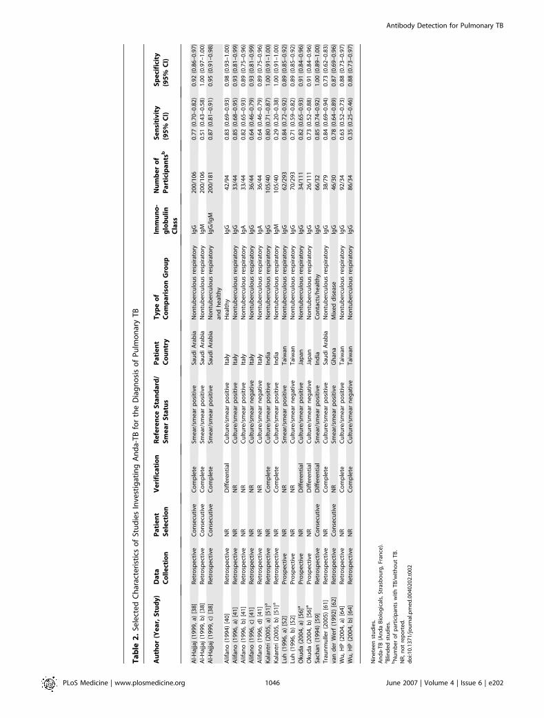
We conducted a systematic review to assess the accuracy of commercial antibody detectiontests for the diagnosis of pulmonary tuberculosis. Studies from all countries using culture and/or microscopy smear for confirmation of pulmonary tuberculosis were eligible. Studies withfewer than 50 participants (25 patients and 25 control participants) were excluded. In acomprehensive search, we identified 68 studies. The results demonstrate that (1) overall,commercial tests vary widely in performance; (2) sensitivity is higher in smear-positive thansmear-negative samples; (3) in studies of smear-positive patients, Anda-TB IgG by enzyme-linked immunosorbent assay shows limited sensitivity (range 63% to 85%) and inconsistentspecificity (range 73% to 100%); (4) specificity is higher in healthy volunteers than in patients inwhom tuberculosis disease is initially suspected and subsequently ruled out; and (5) there areinsufficient data to determine the accuracy of most commercial tests in smear microscopy–negative patients, as well as their performance in children or persons with HIV infection.
Conclusions
None of the commercial tests evaluated perform well enough to replace sputum smearmicroscopy. Thus, these tests have little or no role in the diagnosis of pulmonary tuberculosis.Lack of methodological rigor in these studies was identified as a concern. It will be important toreview the basic science literature evaluating serological tests for the diagnosis of pulmonarytuberculosis to determine whether useful antigens have been described but their potential hasnot been fully exploited. Activities leading to the discovery of new antigens withimmunodiagnostic potential need to be intensified.
The Editors’ Summary of this article follows the references.
PLoS Medicine | www.plosmedicine.org June 2007 | Volume 4 | Issue 6 | e2021041
PLoSMEDICINE
Introduction
The burden of disability and death due to tuberculosis (TB)is immense, with 8.8 million new cases of the disease and 1.6million deaths estimated to have occurred in 2005 alone [1].Although the incidence of TB is constant or falling in manyregions of the world, rates remain high in sub-Saharan Africaas a consequence of the HIV epidemic [1,2]. The expansion ofDOTS, the centerpiece of the international TB partnershipcontrol strategy, has resulted in improved case-detectionrates during the past several years; however, the majority ofDOTS programs in high-burden countries have fallen short ofthe 2005 global target of 70% case detection of the mostinfectious cases [1].
The vast majority of TB patients live in low- and middle-income countries [2], where the diagnosis of TB disease reliesprimarily on identification of acid-fast bacilli on unprocessedsputum smears using a conventional light microscope.Microscopy is highly specific for Mycobacterium tuberculosis inTB-endemic countries [3,4]. Although microscopy has beenreported to have greater than 80% sensitivity for identifyingcases of pulmonary TB in some settings [5,6], the sensitivity ofthe test has been lower and variable in other reports (range20% to 80%) [4,7]. Moreover, sensitivity is poor forpaucibacillary disease (e.g., pediatric and HIV-associatedTB) [8,9], a major concern on account of the strongassociation between HIV infection and smear negativity[10,11]. This lack of sensitivity of the sole diagnostic test inmany parts of the world results in delays in diagnosis,enabling the disease to progress and increasing the potentialfor transmission of M. tuberculosis [5]. To ensure appropriatecare for patients and to improve control of the global TBepidemic, simple, accurate, inexpensive, and, ideally, point-of-care diagnostic tools for TB are urgently needed.
The relative importance of the different characteristics of adiagnostic test depends upon the setting in which the test isto be performed and the intended use of the results.Technical simplicity, for example, is essential if a test is tobe used in a primary health-care clinic or basic healthlaboratory in low-income countries. If test results are to beused to exclude a diagnosis of TB in patients with respiratorysymptoms in TB-endemic countries, then tests with a highsensitivity (high negative predictive value) are required evenif the test is only moderately specific. Excluding TB patientsfrom this group would then allow a more rigorous diagnosticwork-up to be performed on a smaller group of patients. Onthe other hand, if a test is to be used to identify patients withrespiratory symptoms in endemic countries for anti-TBtreatment, a high specificity (high positive predictive value)is required. In the latter case, high sensitivity would also bedesirable.
Immune-based tests would seem to offer the potential toimprove case detection as currently performed, as some ofthe test formats (e.g., immunochromatographic test) aresuitable for resource-limited areas. The major advantages ofimmune-based tests are their speed (results may be availablewithin hours) and simplicity compared with microscopy[8,12]. The development of immune-based tests for thedetection of antibodies, antigens, and immune complexeshas been attempted for decades, and their performance hasbeen critically appraised in several descriptive reviews andtextbook chapters [13–22]. The most common of these tests
rely on detection of the humoral (serological) antibodyimmune response to M. tuberculosis (the subject of thissystematic review), as opposed to the T cell–based cellularimmune response (e.g., interferon-gamma release assays), ordirect detection of antigens in specimens other than serum(e.g., lipoarabinomannan [LAM] detection in urine [23,24]and pleural fluid [25]).A number of in-house antibody detection tests have been
developed but are not marketed. These tests use differentantigens and distinct protocols and techniques.Currently, in developing countries, where diagnostic tests
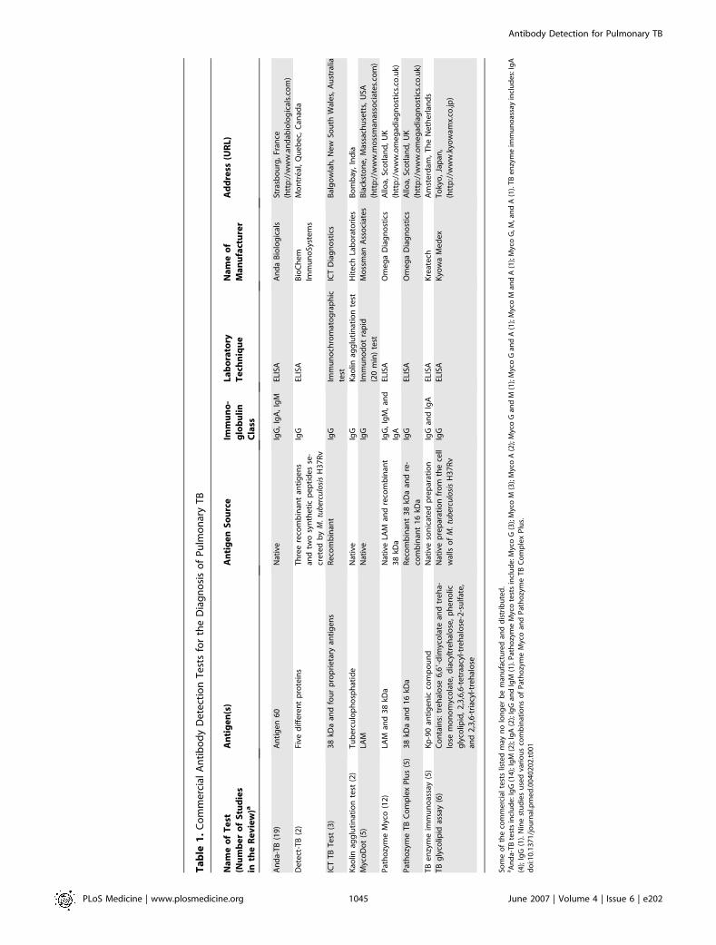
are rarely subjected to regulatory review or approval [26,27],test manufacturers and distributors are marketing dozens ofdifferent antibody detection diagnostic commercial kits. Theextent of their use is largely unknown; however, companiesreport sales volumes between 3,000 and 300,000 tests per year[28]. These tests differ in a number of their features,including antigen composition, antigen source (e.g., nativeor recombinant), chemical composition (e.g., protein, carbo-hydrate, or lipid), extent and manner of purification of theantigen(s), and class of immunoglobulin detected (e.g., IgG,IgM, or IgA). Performance data are often limited to thosefound in the package inserts and, being based on small sets ofpatients, are typically favorable (J. Cunningham, personalcommunication).An antibody detection test can be developed into a number
of formats depending on the membrane, antigen(s) coating,and incubation technique. Common designs include theenzyme-linked immunosorbent assay (ELISA) format and theimmunochromatographic test format. ELISA is a complexassay with several steps: coating of antigens onto the surfaceof plastic wells, the addition of serum samples to the wells,and several washing stages. Antigen–antibody reactions arevisualized using anti-human antibody linked to an enzymaticindicator system [29]. The assay can take hours to perform.The immunochromatographic test is simpler. In this techni-que, the antigens are pre-coated in lines across a membrane(e.g., nitrocellulose) to which samples are applied. Antigen–antibody reactions are visualized on the lines using anti-human antibody bound to substances such as colloidal gold.The test takes only a few minutes to perform [30].The ELISA format has the advantages that many serum
samples can be tested in parallel and the process can becompletely automated, making this technique attractive in fullyequipped laboratories that test a large number of samples. Fordeveloping countries with limited laboratory resources andaccess, an immunochromatographic test would be the pre-ferred method, as this format requires only visual inspection ofthe antigen-containing lines and can, therefore, be performedat the point of care without laboratory equipment.An initial survey of the literature found more than 200
studies that have evaluated commercial serological antibodydetection tests, hereafter referred to as commercial tests, forthe diagnosis of pulmonary TB. To our knowledge, this vastbody of literature has not been systematically reviewed andsynthesized. We therefore conducted a systematic review tosummarize the evidence on accuracy (sensitivity and specificity)of commercial tests, according to the guidelines and methodsproposed for diagnostic systematic reviews and meta-analyses[31]. We specifically addressed two questions. (1) How accurateare commercial tests for the diagnosis of pulmonary TB overalland for smear-positive and smear-negative disease? (2) What is
PLoS Medicine | www.plosmedicine.org June 2007 | Volume 4 | Issue 6 | e2021042
Antibody Detection for Pulmonary TB

the specificity of commercial tests in healthy control partic-ipants compared with the specificity in patients without TB, butin whom TB was initially suspected?
Methods
Search StrategyWe searched electronic databases for primary studies:
PubMed (http: / /www.ncbi.nlm.nih.gov/entrez/query.fcgi?DB¼pubmed) (1990 to May 2006), BIOSIS (http://scientific.thomson.com/biosis/) (1990 to October 2005), Embase(http://www.embase.com) (1990 to October 2005), and Web ofScience (http://scientific.thomson.com/products/wos/) (1990 toOctober 2005). The search terms used included the following:‘‘tuberculosis’’, ‘‘Mycobacterium tuberculosis’’, ‘‘immunologicaltests’’, ‘‘serological tests’’, ‘‘antibody detection’’, ‘‘antigendetection’’, ‘‘ELISA’’, ‘‘Western blot’’, and ‘‘sensitivity andspecificity’’. We identified additional studies by contactingexperts in the field and by searching reference lists fromprimary studies, review articles, and textbook chapters.
Study SelectionOur search strategy sought to identify all available articles
published in English that evaluated commercial tests for theserological diagnosis of pulmonary TB. We included onlythose studies in which patients had bacteriologically con-firmed pulmonary TB, and in which results were providedseparately for smear-positive and smear-negative patients. Inparticular, we defined the reference standard as eitherisolation of M. tuberculosis on culture, or, for studiesconducted without culture in endemic countries, the pres-ence of acid-fast bacilli detected by sputum smear micro-scopy. No restrictions were made with respect to study design(cross sectional or case control) or data collection (prospec-tive or retrospective).
We excluded studies that relied solely on clinical orradiological features or improvement while on anti-TBtherapy as the criteria for establishing the diagnosis of TB.In addition, the following studies were excluded: (1) studiespublished before 1990, for the reason that many studies usedcrude antigen extracts or obsolete immunological methods;(2) studies with fewer than 50 participants (at least 25 TBpatients and 25 control participants were required forinclusion); (3) studies of latent TB infection; (4) studies ofnontuberculous mycobacteria; (5) studies of antibody re-sponses during or after TB treatment; (6) investigationsconducted using non-immunologic methods for detection ofantibodies; (7) basic science literature that focuses on cloningof new antigens or their immunologic properties (e.g.,epitope mapping) or other new methods of antibodydetection; and (8) case reports and reviews.
Initially, two reviewers (KRS and MH) screened citationsretrieved from all sources. To identify relevant studiespertaining specifically to commercial tests, a second screen-ing was done (KRS and MH) of full texts from citations foundto be relevant in the first screen. A list of excluded studies,along with the reasons for exclusion, is available from theauthors on request.
Data ExtractionWe created and piloted a data-extraction form with a
subset of eligible studies. Based upon experience gained in
the pilot, the data-extraction form was finalized. Onereviewer (KRS) extracted data from all eligible studies onthe following qualities: study design and methodologicalquality (see assessment of study quality below), studypopulation, antigen and antibody characteristics, laboratorytechnique, reference standard, and outcome data (sensitivityand specificity). To verify reproducibility of data extraction, asecond reviewer (M. Henry) independently extracted datafrom 15% of the included studies. The inter-rater agreementbetween the two reviewers for sensitivity and specificityestimates was 100%. When data were not clearly reported, theinformation was coded as ‘‘not reported’’. When necessary,we attempted to contact authors for additional information.Although some authors compared performance of com-
mercial tests in several different groups without TB, wepreferentially selected only one comparison group (controlparticipants) for each study in the following order: (1)patients in whom pulmonary TB was initially suspected butwho were later found to have nontuberculous respiratorydisease; (2) patients diagnosed with a variety of diseases otherthan TB (mixed disease); (3) healthy persons from endemiccountries; (4) contacts of patients with TB; (5) mixed groupsfrom categories (1) to (4); and (6) healthy persons from non-endemic countries. In our view, this hierarchy gave priority tothe populations expected to be encountered in a routineclinical setting.
Assessment of Study QualityWe assessed the quality of studies using the following
criteria, which have been suggested as being important fordiagnostic studies [31]. (1) Was there a comparison of thecommercial test with an independent, appropriate referencestandard (i.e., the commercial test did not form part of thereference standard)? (2) Was the commercial test resultperformed and recorded by technicians who were unaware(blinded) of the results of the reference standard? (3) Did thewhole sample or a randomly selected subset of the samplereceive verification using the reference standard? (4) Did thestudy prospectively recruit consecutive patients suspected ofhaving pulmonary TB?
Data Collation and Meta-AnalysisWe used standard methods recommended for meta-
analyses of diagnostic test evaluations [31,32]. As studies wereheterogeneous, particularly with respect to the antigencomposition of the tests, antibody class (IgG, IgM, or IgA),comparison groups, and sputum status of the patients, wefirst grouped studies by type of commercial test and thenfurther stratified by immunoglobulin class and smear status.To calculate sensitivity and specificity values for thecommercial tests, we cross-tabulated each result against thereference standard. Sensitivity refers to the proportion ofpulmonary TB patients with a positive result on a givencommercial test; specificity refers to the proportion of TB-negative participants who had negative results on a givencommercial test. Whenever possible, we extracted raw datafrom primary studies to fill the four cell values of a diagnostic2 3 2 table: true positives, false positives, true negatives, andfalse negatives. In calculations of sensitivity, we includedstudies from endemic countries that used sputum smearpositivity as the reference standard along with studies usingculture as the reference standard. We recognized that some
PLoS Medicine | www.plosmedicine.org June 2007 | Volume 4 | Issue 6 | e2021043
Antibody Detection for Pulmonary TB
authors used the same comparison group for multiple studies,and thus derived identical specificity estimates. Therefore, indetermining specificity, we included the specificity value forthe specific comparison group only once when appropriate.For clarity of presentation, studies that reported resultsstratified by subgroups are shown more than once in tables orfigures.
Data were analyzed using SPSS (version 14.0.1.366) [33] andMeta-DiSc (version 1.4) software [34]. Sensitivity and specific-ity values were calculated for the commercial tests inves-tigated in each study, along with their 95% confidenceintervals (CIs). In addition to the sensitivity and specificityestimates and forest plots generated for this review, truepositive rates (TPR¼ sensitivity) and false positive rates (FPR¼ 1 � specificity) were summarized using an asymmetricsummary receiver operating characteristic (SROC) curve [35].TPR and FPR are not independent of each other as they varywith the thresholds (cut points for determining test positives)employed in the original studies. In addition, it is likely thatdifferent thresholds were used in various studies, eitherimplicitly or explicitly. Because of the inherent trade-offbetween TPR and FPR, it is imperative to plot the estimates ofthe two quantities in a receiver operating characteristic
(ROC) space and to use meta-analytic methods that take intoaccount the threshold effect. Thus, we did not pool thesensitivity and specificity estimates separately; instead weanalyzed TPR and FPR as pairs in an SROC analysis, andexplored the effect of variability in cut points on studyresults.Unlike a traditional ROC plot that explores the effect of
varying thresholds on sensitivity and specificity in a singlestudy, each data point in the SROC space represents anindividual study. As described by Littenberg and Moses [32],the SROC analysis involves three steps: (1) the pairs of TPRand FPR estimates from each study are transformed onto asuitable scale of log odds; (2) a linear regression equation isfitted using the transformed data; and (3) the coefficientsfrom the linear regression model are used to generate a curvein the original ROC space.The area under the curve (AUC) (in this case, being the area
under the SROC curve) presents an overall summary of testperformance and displays the trade-off between sensitivityand specificity. An AUC of 1.0 (100%) indicates perfectdiscriminatory ability of the diagnostic test. In addition, theQ* index is another useful global estimate of test accuracy forcomparing SROC curves. The Q* index, defined by the point
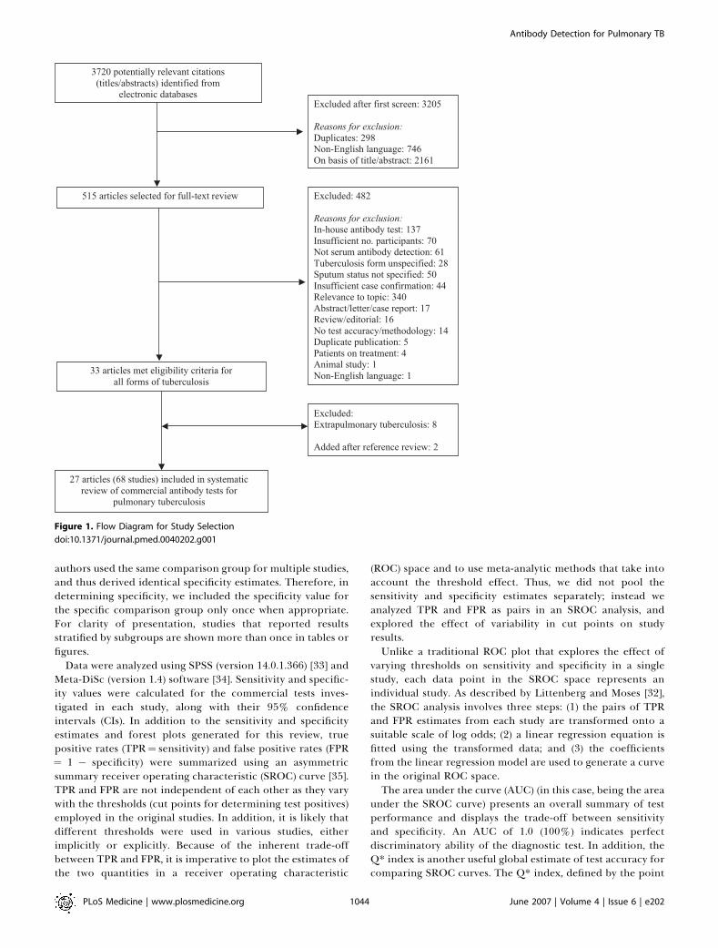
Figure 1. Flow Diagram for Study Selection
doi:10.1371/journal.pmed.0040202.g001
PLoS Medicine | www.plosmedicine.org June 2007 | Volume 4 | Issue 6 | e2021044
Antibody Detection for Pulmonary TB
Ta
ble
1.
Co
mm
erc
ial
An
tib
od
yD
ete
ctio
nT
est
sfo
rth
eD
iag
no
sis
of
Pu
lmo
nar
yT
B
Na
me
of
Te
st
(Nu
mb
er
of
Stu
die
s
inth
eR
ev
iew
)a
An
tig
en
(s)
An
tig
en
So
urc
eIm
mu
no
-
glo
bu
lin
Cla
ss
La
bo
rato
ry
Te
chn
iqu
e
Na
me
of
Ma
nu
fact
ure
r
Ad
dre
ss(U
RL
)
An
da-
TB
(19
)A
nti
ge
n6
0N
ativ
eIg
G,
IgA
,Ig
MEL
ISA
An
da
Bio
log
ical
sSt
rasb
ou
rg,
Fran
ce
(htt
p:/
/ww
w.a
nd
abio
log
ical
s.co
m)
De
tect
-TB
(2)
Five
dif
fere
nt
pro
tein
sT
hre
ere
com
bin
ant
anti
ge
ns
and
two
syn
the
tic
pe
pti
de
sse
-
cre
ted
by
M.
tub
ercu
losi
sH
37
Rv
IgG
ELIS
AB
ioC
he
m
Imm
un
oSy
ste
ms
Mo
ntr
eal
,Q
ue
be
c,C
anad
a
ICT
TB
Te
st(3
)3
8kD
aan
dfo
ur
pro
pri
eta
ryan
tig
en
sR
eco
mb
inan
tIg
GIm
mu
no
chro
mat
og
rap
hic
test
ICT
Dia
gn
ost
ics
Bal
go
wla
h,
Ne
wSo
uth
Wal
es,
Au
stra
lia
Kao
linag
glu
tin
atio
nte
st(2
)T
ub
erc
ulo
ph
osp
hat
ide
Nat
ive
IgG
Kao
linag
glu
tin
atio
nte
stH
ite
chLa
bo
rato
rie
sB
om
bay
,In
dia
Myc
oD
ot
(5)
LAM
Nat
ive
IgG
Imm
un
od
ot
rap
id
(20
min
)te
st
Mo
ssm
anA
sso
ciat
es
Bla
ckst
on
e,
Mas
sach
use
tts,
USA
(htt
p:/
/ww
w.m
oss
ma
nas
soci
ate
s.co
m)
Pat
ho
zym
eM
yco
(12
)LA
Man
d3
8kD
aN
ativ
eLA
Man
dre
com
bin
ant
38
kDa
IgG
,Ig
M,
and
IgA
ELIS
AO
me
ga
Dia
gn
ost
ics
Allo
a,Sc
otl
and
,U
K
(htt
p:/
/ww
w.o
me
gad
iag
no
stic
s.co
.uk)
Pat
ho
zym
eT
BC
om
ple
xP
lus
(5)
38
kDa
and
16
kDa
Re
com
bin
ant
38
kDa
and
re-
com
bin
ant
16
kDa
IgG
ELIS
AO
me
ga
Dia
gn
ost
ics
Allo
a,Sc
otl
and
,U
K
(htt
p:/
/ww
w.o
me
gad
iag
no
stic
s.co
.uk)
TB
en
zym
eim
mu
no
assa
y(5
)K
p-9
0an
tig
en
icco
mp
ou
nd
Nat
ive
son
icat
ed
pre
par
atio
nIg
Gan
dIg
AEL
ISA
Kre
ate
chA
mst
erd
am,
Th
eN
eth
erl
and
s
TB
gly
colip
idas
say
(6)
Co
nta
ins:
tre
hal
ose
6,6
9-d
imyc
ola
tean
dtr
eh
a-
lose
mo
no
myc
ola
te,
dia
cylt
reh
alo
se,
ph
en
olic
gly
colip
id,
2,3
,6,6
-te
traa
cyl-
tre
hal
ose
-2-s
ulf
ate
,
and
2,3
,6-t
riac
yl-t
reh
alo
se
Nat
ive
pre
par
atio
nfr
om
the
cell
wal
lso
fM
.tu
ber
culo
sis
H3
7R
v
IgG
ELIS
AK
yow
aM
ed
ex
To
kyo
,Ja
pan
,
(htt
p:/
/ww
w.k
yow
am
x.co
.jp)
Som
eo
fth
eco
mm
erc
ial
test
slis
ted
may
no
lon
ge
rb
em
anu
fact
ure
dan
dd
istr
ibu
ted
.aA
nd
a-T
Bte
sts
incl
ud
e:I
gG
(14
);Ig
M(2
);Ig
A(2
);Ig
Gan
dIg
M(1
).P
ath
ozy
me
Myc
ote
sts
incl
ud
e:M
yco
G(3
);M
yco
M(3
);M
yco
A(2
);M
yco
Gan
dM
(1);
Myc
oG
and
A(1
);M
yco
Man
dA
(1);
Myc
oG
,M,a
nd
A(1
).T
Be
nzy
me
imm
un
oas
say
incl
ud
es:I
gA
(4);
IgG
(1).
Nin
est
ud
ies
use
dva
rio
us
com
bin
atio
ns
of
Pat
ho
zym
eM
yco
and
Pat
ho
zym
eT
BC
om
ple
xP
lus.
do
i:10
.13
71
/jo
urn
al.p
me
d.0
04
02
02
.t0
01
PLoS Medicine | www.plosmedicine.org June 2007 | Volume 4 | Issue 6 | e2021045
Antibody Detection for Pulmonary TB
Ta
ble
2.
Sele
cte
dC
har
acte
rist
ics
of
Stu
die
sIn
vest
igat
ing
An
da-
TB
for
the
Dia
gn
osi
so
fP
ulm
on
ary
TB
Au
tho
r(Y
ea
r,S
tud
y)
Da
ta
Co
lle
ctio
n
Pa
tie
nt
Se
lect
ion
Ve
rifi
cati
on
Re
fere
nce
Sta
nd
ard
/
Sm
ea
rS
tatu
s
Pa
tie
nt
Co
un
try
Ty
pe
of
Co
mp
ari
son
Gro
up
Imm
un
o-
glo
bu
lin
Cla
ss
Nu
mb
er
of
Pa
rtic
ipa
nts
bS
en
siti
vit
y
(95
%C
I)
Sp
eci
fici
ty
(95
%C
I)
Al-
Haj
jaj
(19
99
,a)
[38
]R
etr
osp
ect
ive
Co
nse
cuti
veC
om
ple
teSm
ear
/sm
ear
po
siti
veSa
ud
iA
rab
iaN
on
tub
erc
ulo
us
resp
irat
ory
IgG
20
0/1
06
0.7
7(0
.70
–0
.82
)0
.92
(0.8
6–
0.9
7)
Al-
Haj
jaj
(19
99
,b
)[3
8]
Re
tro
spe
ctiv
eC
on
secu
tive
Co
mp
lete
Sme
ar/s
me
arp
osi
tive
Sau
di
Ara
bia
No
ntu
be
rcu
lou
sre
spir
ato
ryIg
M2
00
/10
60
.51
(0.4
3–
0.5
8)
1.0
0(0
.97
–1
.00
)
Al-
Haj
jaj
(19
99
,c)
[38
]R
etr
osp
ect
ive
Co
nse
cuti
veC
om
ple
teSm
ear
/sm
ear
po
siti
veSa
ud
iA
rab
iaN
on
tub
erc
ulo
us
resp
irat
ory
and
he
alth
y
IgG
/Ig
M2
00
/18
10
.87
(0.8
1–
0.9
1)
0.9
5(0
.91
–0
.98
)
Alif
ano
(19
94
)[4
0]
Re
tro
spe
ctiv
eN
RD
iffe
ren
tial
Cu
ltu
re/s
me
arp
osi
tive
Ital
yH
eal
thy
IgG
42
/94
0.8
3(0
.69
–0
.93
)0
.98
(0.9
3–
1.0
0)
Alif
ano
(19
96
,a)
[41
]R
etr
osp
ect
ive
NR
NR
Cu
ltu
re/s
me
arp
osi
tive
Ital
yN
on
tub
erc
ulo
us
resp
irat
ory
IgG
33
/44
0.8
5(0
.68
–0
.95
)0
.93
(0.8
1–
0.9
9)
Alif
ano
(19
96
,b
)[4
1]
Re
tro
spe
ctiv
eN
RN
RC
ult
ure
/sm
ear
po
siti
veIt
aly
No
ntu
be
rcu
lou
sre
spir
ato
ryIg
A3
3/4
40
.82
(0.6
5–
0.9
3)
0.8
9(0
.75
–0
.96
)
Alif
ano
(19
96
,c)
[41
]R
etr
osp
ect
ive
NR
NR
Cu
ltu
re/s
me
arn
eg
ativ
eIt
aly
No
ntu
be
rcu
lou
sre
spir
ato
ryIg
G3
6/4
40
.64
(0.4
6–
0.7
9)
0.9
3(0
.81
–0
.99
)
Alif
ano
(19
96
,d
)[4
1]
Re
tro
spe
ctiv
eN
RN
RC
ult
ure
/sm
ear
ne
gat
ive
Ital
yN
on
tub
erc
ulo
us
resp
irat
ory
IgA
36
/44
0.6
4(0
.46
–0
.79
)0
.89
(0.7
5–
0.9
6)
Kal
antr
i(2
00
5,
a)[5
1]a
Re
tro
spe
ctiv
eN
RC
om
ple
teC
ult
ure
/sm
ear
po
siti
veIn
dia
No
ntu
be
rcu
lou
sre
spir
ato
ryIg
G1
05
/40
0.8
0(0
.71
–0
.87
)1
.00
(0.9
1–
1.0
0)
Kal
antr
i(2
00
5,
b)
[51
]aR
etr
osp
ect
ive
NR
Co
mp
lete
Cu
ltu
re/s
me
arp
osi
tive
Ind
iaN
on
tub
erc
ulo
us
resp
irat
ory
IgM
10
5/4
00
.29
(0.2
0–
0.3
8)
1.0
0(0
.91
–1
.00
)
Luh
(19
96
,a)
[52
]P
rosp
ect
ive
NR
NR
Sme
ar/s
me
arp
osi
tive
Tai
wan
No
ntu
be
rcu
lou
sre
spir
ato
ryIg
G6
2/2
93
0.8
4(0
.72
–0
.92
)0
.89
(0.8
5–
0.9
2)
Luh
(19
96
,b
)[5
2]
Pro
spe
ctiv
eN
RN
RC
ult
ure
/sm
ear
ne
gat
ive
Tai
wan
No
ntu
be
rcu
lou
sre
spir
ato
ryIg
G7
0/2
93
0.7
1(0
.59
–0
.82
)0
.89
(0.8
5–
0.9
2)
Oku
da
(20
04
,a)
[56
]aP
rosp
ect
ive
NR
Dif
fere
nti
alC
ult
ure
/sm
ear
po
siti
veJa
pan
No
ntu
be
rcu
lou
sre
spir
ato
ryIg
G3
4/1
11
0.8
2(0
.65
–0
.93
)0
.91
(0.8
4–
0.9
6)
Oku
da
(20
04
,b
)[5
6]a
Pro
spe
ctiv
eN
RD
iffe
ren
tial
Cu
ltu
re/s
me
arn
eg
ativ
eJa
pan
No
ntu
be
rcu
lou
sre
spir
ato
ryIg
G2
6/1
11
0.7
3(0
.52
–0
.88
)0
.91
(0.8
4–
0.9
6)
Sach
an(1
99
4)
[59
]R
etr
osp
ect
ive
Co
nse
cuti
veD
iffe
ren
tial
Sme
ar/s
me
arp
osi
tive
Ind
iaC
on
tact
s/h
eal
thy
IgG
66
/32
0.8
5(0
.74
–0
.92
)1
.00
(0.8
9–
1.0
0)
Tra
un
mu
ller
(20
05
)[6
1]
Re
tro
spe
ctiv
eN
RC
om
ple
teC
ult
ure
/sm
ear
po
siti
veSa
ud
iA
rab
iaN
on
tub
erc
ulo
us
resp
irat
ory
IgG
38
/79
0.8
4(0
.69
–0
.94
)0
.73
(0.6
2–
0.8
3)
van
de
rW
erf
(19
92
)[6
2]
Re
tro
spe
ctiv
eC
on
secu
tive
NR
Sme
ar/s
me
arp
osi
tive
Gh
ana
Mix
ed
dis
eas
eIg
G4
6/3
00
.78
(0.6
4–
0.8
9)
0.8
7(0
.69
–0
.96
)
Wu
,H
P(2
00
4,
a)[6
4]
Re
tro
spe
ctiv
eN
RC
om
ple
teC
ult
ure
/sm
ear
po
siti
veT
aiw
anN
on
tub
erc
ulo
us
resp
irat
ory
IgG
92
/34
0.6
3(0
.52
–0
.73
)0
.88
(0.7
3–
0.9
7)
Wu
,H
P(2
00
4,
b)
[64
]R
etr
osp
ect
ive
NR
Co
mp
lete
Cu
ltu
re/s
me
arn
eg
ativ
eT
aiw
anN
on
tub
erc
ulo
us
resp
irat
ory
IgG
86
/34
0.3
5(0
.25
–0
.46
)0
.88
(0.7
3–
0.9
7)
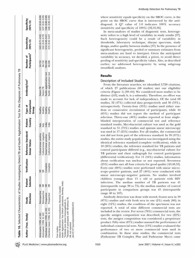
Nin
ete
en
stu
die
s.A
nd
a-T
B(A
nd
aB
iolo
gic
als,
Stra
sbo
urg
,Fr
ance
).aB
lind
ed
stu
die
s.b
Nu
mb
er
of
par
tici
pan
tsw
ith
TB
/wit
ho
ut
TB
.N
R,
no
tre
po
rte
d.
do
i:10
.13
71
/jo
urn
al.p
me
d.0
04
02
02
.t0
02
PLoS Medicine | www.plosmedicine.org June 2007 | Volume 4 | Issue 6 | e2021046
Antibody Detection for Pulmonary TB

Ta
ble
3.
Sele
cte
dC
har
acte
rist
ics
of
Stu
die
sIn
vest
igat
ing
TB
Enzy
me
Imm
un
oas
say
for
the
Dia
gn
osi
so
fP
ulm
on
ary
TB
Au
tho
r
(Ye
ar,
Stu
dy
)
Stu
dy
Dir
ect
ion
Pa
tie
nt
Se
lect
ion
Ve
rifi
cati
on
Re
fere
nce
Sta
nd
ard
/
Sm
ea
rS
tatu
s
Pa
tie
nt
Co
un
try
Ty
pe
of
Co
mp
ari
son
Gro
up
Imm
un
o-
glo
bu
lin
Cla
ss
Nu
mb
er
of
Pa
rtic
ipa
nts
bS
en
siti
vit
y
(95
%C
I)
Sp
eci
fici
ty
(95
%C
I)
Alif
ano
(19
97
,a)
[39
]R
etr
osp
ect
ive
Co
nse
cuti
veD
iffe
ren
tial
Cu
ltu
re/s
me
arp
osi
tive
Ital
yN
on
tub
erc
ulo
us
resp
irat
ory
IgA
32
/28
0.7
5(0
.57
–0
.89
)0
.86
(0.6
7–
0.9
6)
Alif
ano
(19
97
,b
)[3
9]
Re
tro
spe
ctiv
eC
on
secu
tive
Dif
fere
nti
alC
ult
ure
/sm
ear
ne
gat
ive
Ital
yN
on
tub
erc
ulo
us
resp
irat
ory
IgA
56
/28
0.6
8(0
.54
–0
.80
)0
.86
(0.6
7–
0.9
6)
Co
nd
e(2
00
4,
a)[4
6]a
Pro
spe
ctiv
eN
RC
om
ple
teC
ult
ure
/sm
ear
po
siti
veB
razi
lN
on
tub
erc
ulo
us
resp
irat
ory
IgG
40
/31
0.7
5(0
.59
–0
.87
)0
.77
(0.5
9–
0.9
0)
Co
nd
e(2
00
4,
b)
[46
]aP
rosp
ect
ive
NR
Co
mp
lete
Cu
ltu
re/s
me
arp
osi
tive
Bra
zil
No
ntu
be
rcu
lou
sre
spir
ato
ryIg
A4
0/3
10
.83
(0.6
7–
0.9
3)
0.4
8(0
.30
–0
.67
)
Julia
n(2
00
0)
[50
]P
rosp
ect
ive
NR
Co
mp
lete
Cu
ltu
re/m
ear
po
siti
veSp
ain
No
ntu
be
rcu
lou
sre
spir
ato
ryIg
A2
7/3
60
.74
(0.5
4–
0.8
9)
0.4
7(0
.30
–0
.65
)
Five
stu
die
s.T
BEn
zym
eIm
mu
no
assa
y(K
reat
ech
Dia
gn
ost
ics,
Am
ste
rdam
,T
he
Ne
the
rlan
ds)
.aB
lind
ed
stu
die
s.b
Nu
mb
er
of
par
tici
pan
tsw
ith
TB
/wit
ho
ut
TB
.N
R,
no
tre
po
rte
d.
do
i:10
.13
71
/jo
urn
al.p
me
d.0
04
02
02
.t0
03
Ta
ble
4.
Sele
cte
dC
har
acte
rist
ics
of
Stu
die
sIn
vest
igat
ing
Pat
ho
zym
eT
BC
om
ple
xP
lus
for
the
Dia
gn
osi
so
fP
ulm
on
ary
TB
Au
tho
r
(Ye
ar,
Stu
dy
)
Stu
dy
Dir
ect
ion
Pa
tie
nt
Se
lect
ion
Ve
rifi
cati
on
Re
fere
nce
Sta
nd
ard
/
Sm
ea
rS
tatu
s
Pa
tie
nt
Co
un
try
Ty
pe
of
Co
mp
ari
son
Gro
up
Nu
mb
er
of
Pa
rtic
ipa
nts
bS
en
siti
vit
y
(95
%C
I)
Sp
eci
fici
ty
(95
%C
I)
Bu
tt(2
00
4,
c)[4
4]
Re
tro
spe
ctiv
eN
RC
om
ple
teSm
ear
/sm
ear
po
siti
veP
akis
tan
He
alth
y9
4/9
00
.64
(0.5
3–
0.7
3)
0.9
7(0
.91
–0
.99
)
Imaz
(20
04
,h
)[4
8]a
Pro
spe
ctiv
eC
on
secu
tive
Co
mp
lete
Cu
ltu
re/s
me
arn
eg
ativ
eA
rge
nti
na
Mix
ed
dis
eas
e4
1/4
50
.29
(0.1
6–
0.4
6)
1.0
0(0
.92
–1
.00
)
Julia
n(2
00
4,
d)
[49
]R
etr
osp
ect
ive
NR
Dif
fere
nti
alC
ult
ure
/sm
ear
po
siti
veSp
ain
No
ntu
be
rcu
lou
sre
spir
ato
ry2
9/3
50
.31
(0.1
5–
0.5
1)
0.8
6(0
.70
–0
.95
)
Wilk
inso
n(1
99
7,
a)[6
3]
Re
tro
spe
ctiv
eN
RN
RC
ult
ure
/sm
ear
po
siti
veIn
dia
He
alth
y1
25
/34
0.7
8(0
.70
–0
.85
)0
.94
(0.8
0–
0.9
9)
Wilk
inso
n(1
99
7,
b)
[63
]R
etr
osp
ect
ive
NR
NR
Cu
ltu
re/s
me
arn
eg
ativ
eIn
dia
He
alth
y7
1/3
40
.76
(0.6
4–
0.8
5)
0.9
4(0
.80
–0
.99
)
Five
stu
die
s.P
ath
ozy
me
TB
Co
mp
lex
Plu
s(O
me
ga
Dia
gn
ost
ics,
Allo
a,Sc
otl
and
,U
K)
de
tect
sim
mu
no
glo
bu
linG
.aB
lind
ed
stu
die
s.b
Nu
mb
er
of
par
tici
pan
tsw
ith
TB
/wit
ho
ut
TB
.N
R,
no
tre
po
rte
d.
do
i:10
.13
71
/jo
urn
al.p
me
d.0
04
02
02
.t0
04
PLoS Medicine | www.plosmedicine.org June 2007 | Volume 4 | Issue 6 | e2021047
Antibody Detection for Pulmonary TB

Ta
ble
5.
Sele
cte
dC
har
acte
rist
ics
of
Stu
die
sIn
vest
igat
ing
TB
Gly
colip
idA
ssay
for
the
Dia
gn
osi
so
fP
ulm
on
ary
TB
Au
tho
r
(Ye
ar,
Stu
dy
)
Stu
dy
Dir
ect
ion
Pa
tie
nt
Se
lect
ion
Ve
rifi
cati
on
Re
fere
nce
Sta
nd
ard
/
Sm
ea
rS
tatu
s
Pa
tie
nt
Co
un
try
Ty
pe
of
Co
mp
ari
son
Gro
up
Nu
mb
er
of
Pa
rtic
ipa
nts
bS
en
siti
vit
y
(95
%C
I)
Sp
eci
fici
ty
(95
%C
I)
Iinu
ma
(20
02
)[4
7]
Re
tro
spe
ctiv
eN
RD
iffe
ren
tial
Sme
ar/s
me
arp
osi
tive
Jap
anN
on
tub
erc
ulo
us
resp
irat
ory
47
/54
0.8
9(0
.77
–0
.96
)0
.72
(0.5
8–
0.8
4)
Mae
kura
(20
01
,a)
[54
]aR
etr
osp
ect
ive
NR
Dif
fere
nti
alC
ult
ure
/sm
ear
po
siti
veJa
pan
No
ntu
be
rcu
lou
sre
spir
ato
ry1
64
/18
00
.90
(0.8
4–
0.9
4)
0.8
5(0
.79
–0
.90
)
Mae
kura
(20
01
,b
)[5
4]a
Re
tro
spe
ctiv
eN
RD
iffe
ren
tial
Cu
ltu
re/s
me
arn
eg
ativ
eJa
pan
No
ntu
be
rcu
lou
sre
spir
ato
ry5
2/1
80
0.6
9(0
.55
–0
.81
)0
.85
(0.7
9–
0.9
0)
Mae
kura
(20
03
)[5
3]a
Pro
spe
ctiv
eC
on
secu
tive
Co
mp
lete
Cu
ltu
re/s
me
arn
eg
ativ
eJa
pan
No
ntu
be
rcu
lou
sre
spir
ato
ry7
0/5
20
.60
(0.4
8–
0.7
2)
0.8
8(0
.77
–0
.96
)
Oku
da
(20
04
,e
)[5
6]a
Pro
spe
ctiv
eN
RD
iffe
ren
tial
Cu
ltu
re/s
me
arp
osi
tive
Jap
anN
on
tub
erc
ulo
us
resp
irat
ory
34
/11
10
.76
(0.5
9–
0.8
9)
0.8
9(0
.82
–0
.94
)
Oku
da
(20
04
,f)
[56
]aP
rosp
ect
ive
NR
Dif
fere
nti
alC
ult
ure
/sm
ear
ne
gat
ive
Jap
anN
on
tub
erc
ulo
us
resp
irat
ory
26
/11
10
.77
(0.5
6–
0.9
1)
0.8
9(0
.82
–0
.94
)
Six
stu
die
s.T
BG
lyco
lipid
Ass
ay(K
yow
aM
ed
ex,
To
kyo
,Ja
pan
)d
ete
cts
imm
un
og
lob
ulin
G.
aB
lind
ed
stu
die
s.b
Nu
mb
er
of
par
tici
pan
tsw
ith
TB
/wit
ho
ut
TB
.N
R,
no
tre
po
rte
d.
do
i:10
.13
71
/jo
urn
al.p
me
d.0
04
02
02
.t0
05
Ta
ble
6.
Sele
cte
dC
har
acte
rist
ics
of
Stu
die
sIn
vest
igat
ing
ICT
for
the
Dia
gn
osi
so
fP
ulm
on
ary
TB
Au
tho
r
(Ye
ar,
Stu
dy
)
Stu
dy
Dir
ect
ion
Pa
tie
nt
Se
lect
ion
Ve
rifi
cati
on
Re
fere
nce
Sta
nd
ard
/
Sm
ea
rS
tatu
s
Pa
tie
nt
Co
un
try
Ty
pe
of
Co
mp
ari
son
Gro
up
Nu
mb
er
of
Pa
rtic
ipa
nts
bS
en
siti
vit
y
(95
%C
I)
Sp
eci
fici
ty
(95
%C
I)
McC
on
key
(20
02
)[5
5]a
Pro
spe
ctiv
eN
RC
om
ple
teC
ult
ure
/sm
ear
po
siti
veEg
ypt
No
ntu
be
rcu
lou
sre
spir
ato
ry7
1/7
40
.87
(0.7
7–
0.9
4)
0.8
2(0
.72
–0
.90
)
On
gu
t(2
00
6)
[57
]R
etr
osp
ect
ive
NR
Dif
fere
nti
alC
ult
ure
/sm
ear
po
siti
veT
urk
ey
He
alth
y5
3/5
40
.40
(0.2
6–
0.5
4)
1.0
0(0
.93
–1
.00
)
Pe
rkin
s(2
00
3)
[58
]R
etr
osp
ect
ive
NR
Co
mp
lete
Sme
ar/s
me
arp
osi
tive
Bra
zil
No
ntu
be
rcu
lou
sre
spir
ato
ry1
20
/34
0.6
4(0
.55
–0
.73
)0
.85
(0.6
9–
0.9
5)
Th
ree
stu
die
s.IC
T(I
CT
Dia
gn
ost
ics,
Bal
go
wla
h,
Ne
wSo
uth
Wal
es,
Au
stra
lia)
de
tect
sim
mu
no
glo
bu
linG
.aB
lind
ed
stu
die
s.b
Nu
mb
er
of
par
tici
pan
tsw
ith
TB
/wit
ho
ut
TB
.N
R,
no
tre
po
rte
d.
do
i:10
.13
71
/jo
urn
al.p
me
d.0
04
02
02
.t0
06
Ta
ble
7.
Sele
cte
dC
har
acte
rist
ics
of
Stu
die
sIn
vest
igat
ing
Myc
oD
ot
for
the
Dia
gn
osi
so
fP
ulm
on
ary
TB
Au
tho
r
(Ye
ar,
Stu
dy
)
Stu
dy
Dir
ect
ion
Pa
tie
nt
Se
lect
ion
Ve
rifi
cati
on
Re
fere
nce
Sta
nd
ard
/
Sm
ea
rS
tatu
s
Pa
tie
nt
Co
un
try
Ty
pe
of
Co
mp
ari
son
Gro
up
Nu
mb
er
of
Pa
rtic
ipa
nts
bS
en
siti
vit
y
(95
%C
I)
Sp
eci
fici
ty
(95
%C
I)
An
tun
es
(20
02
,a)
[43
]aP
rosp
ect
ive
Ran
do
mC
om
ple
teSm
ear
/sm
ear
po
siti
veG
uin
ea-
Bis
sau
Mix
ed
dis
eas
e4
6/2
23
0.6
3(0
.48
–0
.77
)0
.92
(0.8
8–
0.9
5)
An
tun
es
(20
02
,b
)[4
3]a
,cP
rosp
ect
ive
Ran
do
mC
om
ple
teSm
ear
/sm
ear
po
siti
veG
uin
ea-
Bis
sau
Mix
ed
dis
eas
e3
4/1
98
0.6
8(0
.49
–0
.83
)0
.92
(0.8
7–
0.9
5)
Oku
da
(20
04
,c)
[56
]aP
rosp
ect
ive
NR
Dif
fere
nti
alC
ult
ure
/sm
ear
po
siti
veJa
pan
No
ntu
be
rcu
lou
sre
spir
ato
ry3
4/1
11
0.7
6(0
.59
–0
.89
)0
.97
(0.9
2–
0.9
9)
Oku
da
(20
04
,d
)[5
6]a
Pro
spe
ctiv
eN
RD
iffe
ren
tial
Cu
ltu
re/s
me
arn
eg
ativ
eJa
pan
No
ntu
be
rcu
lou
sre
spir
ato
ry2
6/1
11
0.5
8(0
.37
–0
.77
)0
.97
(0.9
2–
0.9
9)
Som
i(1
99
9)
[60
]a,d
Pro
spe
ctiv
eN
RC
om
ple
teC
ult
ure
/sm
ear
po
siti
veT
anza
nia
No
ntu
be
rcu
lou
sre
spir
ato
ry3
9/1
02
0.2
6(0
.13
–0
.42
)0
.84
(0.7
6–
0.9
1)
Five
stu
die
s.M
yco
Do
t(M
oss
man
Ass
oci
ate
s,B
lack
sto
ne
,M
assa
chu
sett
s,U
SA)
de
tect
sim
mu
no
glo
bu
linG
.aB
lind
ed
stu
die
s.b
Nu
mb
er
of
par
tici
pan
tsw
ith
TB
/wit
ho
ut
TB
.cH
IV-n
eg
ativ
ein
div
idu
als
on
ly,
sub
stu
dy
of
An
tun
es
stu
dy
(a)
wh
ich
incl
ud
esb
oth
HIV
-po
siti
vean
dH
IV-n
eg
ativ
ein
div
idu
als.
dC
om
me
rcia
lte
stp
erf
orm
ed
on
wh
ole
blo
od
.N
R,
no
tre
po
rte
d.
do
i:10
.13
71
/jo
urn
al.p
me
d.0
04
02
02
.t0
07
PLoS Medicine | www.plosmedicine.org June 2007 | Volume 4 | Issue 6 | e2021048
Antibody Detection for Pulmonary TB

Ta
ble
8.
Sele
cte
dC
har
acte
rist
ics
of
Stu
die
sIn
vest
igat
ing
Pat
ho
zym
eM
yco
for
the
Dia
gn
osi
so
fP
ulm
on
ary
TB
Au
tho
r
(Ye
ar,
Stu
dy
)
Stu
dy
Dir
ect
ion
Pa
tie
nt
Se
lect
ion
Ve
rifi
cati
on
Re
fere
nce
Sta
nd
ard
/
Sm
ea
rS
tatu
s
Pa
tie
nt
Co
un
try
Ty
pe
of
Co
mp
ari
son
Gro
up
Imm
un
og
lob
uli
n
Cla
ss
Nu
mb
er
of
Pa
rtic
ipa
nts
bS
en
siti
vit
y
(95
%C
I)
Sp
eci
fici
ty
(95
%C
I)
Bu
tt(2
00
4,
a)[4
4]
Re
tro
spe
ctiv
eN
RC
om
ple
teSm
ear
/sm
ear
po
siti
veP
akis
tan
He
alth
yIg
G9
4/9
00
.46
(0.3
5–
0.5
6)
0.9
3(0
.86
–0
.98
)
Bu
tt(2
00
4,
b)
[44
]R
etr
osp
ect
ive
NR
Co
mp
lete
Sme
ar/s
me
arp
osi
tive
Pak
ista
nH
eal
thy
IgM
94
/90
0.6
7(0
.57
–0
.76
)0
.98
(0.9
2–
1.0
0)
Imaz
(20
04
,a)
[48
]aP
rosp
ect
ive
Co
nse
cuti
veC
om
ple
teC
ult
ure
/sm
ear
ne
gat
ive
Arg
en
tin
aM
ixe
dd
ise
ase
IgG
41
/45
0.4
9(0
.33
–0
.65
)1
.00
(0.9
2–
1.0
0)
Imaz
(20
04
,b
)[4
8]a
Pro
spe
ctiv
eC
on
secu
tive
Co
mp
lete
Cu
ltu
re/s
me
arn
eg
ativ
eA
rge
nti
na
Mix
ed
dis
eas
eIg
M4
1/4
50
.32
(0.1
8–
0.4
8)
0.9
3(0
.82
–0
.99
)
Imaz
(20
04
,c)
[48
]aP
rosp
ect
ive
Co
nse
cuti
veC
om
ple
teC
ult
ure
/sm
ear
ne
gat
ive
Arg
en
tin
aM
ixe
dd
ise
ase
IgA
41
/45
0.3
4(0
.20
–0
.51
)0
.98
(0.8
8–
1.0
0)
Imaz
(20
04
,d
)[4
8]a
Pro
spe
ctiv
eC
on
secu
tive
Co
mp
lete
Cu
ltu
re/s
me
arn
eg
ativ
eA
rge
nti
na
Mix
ed
dis
eas
eIg
Gan
dIg
M4
1/4
50
.63
(0.4
7–
0.7
8)
0.9
3(0
.82
–0
.99
)
Imaz
(20
04
,e
)[4
8]a
Pro
spe
ctiv
eC
on
secu
tive
Co
mp
lete
Cu
ltu
re/s
me
arn
eg
ativ
eA
rge
nti
na
Mix
ed
dis
eas
eIg
Gan
dIg
A4
1/4
50
.59
(0.4
2–
0.7
4)
0.9
8(0
.88
–1
.00
)
Imaz
(20
04
,f)
[48
]aP
rosp
ect
ive
Co
nse
cuti
veC
om
ple
teC
ult
ure
/sm
ear
ne
gat
ive
Arg
en
tin
aM
ixe
dd
ise
ase
IgM
and
IgA
41
/45
0.4
9(0
.33
–0
.65
)0
.91
(0.7
9–
0.9
8)
Imaz
(20
04
,g
)[4
8]a
Pro
spe
ctiv
eC
on
secu
tive
Co
mp
lete
Cu
ltu
re/s
me
arn
eg
ativ
eA
rge
nti
na
Mix
ed
dis
eas
eIg
G,
IgM
,an
dIg
A4
1/4
50
.68
(0.5
2–
0.8
2)
0.9
1(0
.79
–0
.98
)
Julia
n(2
00
4,
a)[4
9]
Re
tro
spe
ctiv
eN
RD
iffe
ren
tial
Cu
ltu
re/s
me
arp
osi
tive
Spai
nN
on
tub
erc
ulo
us
resp
irat
ory
IgG
29
/35
0.4
1(0
.24
–0
.61
)0
.97
(0.8
5–
1.0
0)
Julia
n(2
00
4,
b)
[49
]R
etr
osp
ect
ive
NR
Dif
fere
nti
alC
ult
ure
/sm
ear
po
siti
veSp
ain
No
ntu
be
rcu
lou
sre
spir
ato
ryIg
M2
9/3
50
.10
(0.0
2–
0.2
7)
0.9
7(0
.85
–1
.00
)
Julia
n(2
00
4,
c)[4
9]
Re
tro
spe
ctiv
eN
RD
iffe
ren
tial
Cu
ltu
re/s
me
arp
osi
tive
Spai
nN
on
tub
erc
ulo
us
resp
irat
ory
IgA
29
/35
0.2
1(0
.08
–0
.40
)0
.97
(0.8
5–
1.0
0)
Tw
elv
est
ud
ies.
Pat
ho
zym
eM
yco
(Om
eg
aD
iag
no
stic
s,A
lloa,
Sco
tlan
d,
UK
).aB
lind
ed
stu
die
s.b
Nu
mb
er
of
par
tici
pan
tsw
ith
TB
/wit
ho
ut
TB
.N
R,
no
tre
po
rte
d.
do
i:10
.13
71
/jo
urn
al.p
me
d.0
04
02
02
.t0
08
Ta
ble
9.
Sele
cte
dC
har
acte
rist
ics
of
Stu
die
sIn
vest
igat
ing
Pat
ho
zym
eM
yco
and
Pat
ho
zym
eT
BC
om
ple
xP
lus
inV
ario
us
Co
mb
inat
ion
sfo
rth
eD
iag
no
sis
of
Pu
lmo
nar
yT
B
Au
tho
r
(Ye
ar,
Stu
dy
)
Stu
dy
Dir
ect
ion
Pa
tie
nt
Se
lect
ion
Ve
rifi
-
cati
on
Re
fere
nce
Sta
nd
ard
/
Sm
ea
rS
tatu
s
Pa
tie
nt
Co
un
try
Ty
pe
of
Co
mp
ari
son
Gro
up
An
tib
od
yT
est
bC
om
bin
ati
on
Nu
mb
er
of
Pa
rtic
i-
pa
nts
c
Se
nsi
tiv
ity
(95
%C
I)
Sp
eci
fici
ty
(95
%C
I)
Bu
tt(2
00
4,
d)
[44
]R
etr
osp
ect
ive
NR
Co
mp
lete
Sme
ar/s
me
arp
osi
tive
Pak
ista
nH
eal
thy
Pat
ho
zym
eM
yco
Gan
dP
ath
ozy
me
TB
Co
mp
lex
Plu
s9
4/9
00
.76
(0.6
6–
0.8
4)
0.9
3(0
.86
–0
.98
)
Bu
tt(2
00
4,
e)
[44
]R
etr
osp
ect
ive
NR
Co
mp
lete
Sme
ar/s
me
arp
osi
tive
Pak
ista
nH
eal
thy
Pat
ho
zym
eM
yco
Man
dP
ath
ozy
me
TB
Co
mp
lex
Plu
s9
4/9
00
.82
(0.7
3–
0.8
9)
0.9
7(0
.91
–0
.99
)
Bu
tt(2
00
4,
f)[4
4]
Re
tro
spe
ctiv
eN
RC
om
ple
teSm
ear
/sm
ear
po
siti
veP
akis
tan
He
alth
yP
ath
ozy
me
Myc
oG
,M
yco
M,
and
Pat
ho
zym
eT
B
Co
mp
lex
Plu
s
94
/90
0.8
7(0
.79
–0
.93
)0
.93
(0.8
6–
0.9
8)
Imaz
(20
04
,i)
[48
]aP
rosp
ect
ive
Co
nse
cuti
veC
om
ple
teC
ult
ure
/sm
ear
ne
gat
ive
Arg
en
tin
aM
ixe
dd
ise
ase
Pat
ho
zym
eM
yco
Gan
dP
ath
ozy
me
TB
Co
mp
lex
Plu
s4
1/4
50
.59
(0.4
2–
0.7
4)
1.0
0(0
.92
–1
.00
)
Imaz
(20
04
,j)
[48
]aP
rosp
ect
ive
Co
nse
cuti
veC
om
ple
teC
ult
ure
/sm
ear
ne
gat
ive
Arg
en
tin
aM
ixe
dd
ise
ase
Pat
ho
zym
eM
yco
Man
dP
ath
ozy
me
TB
Co
mp
lex
Plu
s4
1/4
50
.51
(0.3
5–
0.6
7)
0.9
3(0
.82
–0
.99
)
Imaz
(20
04
,k)
[48
]aP
rosp
ect
ive
Co
nse
cuti
veC
om
ple
teC
ult
ure
/sm
ear
ne
gat
ive
Arg
en
tin
aM
ixe
dd
ise
ase
Pat
ho
zym
eM
yco
Aan
dP
ath
ozy
me
TB
Co
mp
lex
Plu
s4
1/4
50
.49
(0.3
3–
0.6
5)
0.9
8(0
.88
–1
.00
)
Imaz
(20
04
,l)
[48
]aP
rosp
ect
ive
Co
nse
cuti
veC
om
ple
teC
ult
ure
/sm
ear
ne
gat
ive
Arg
en
tin
aM
ixe
dd
ise
ase
Pat
ho
zym
eM
yco
G,
Myc
oA
,an
dP
ath
ozy
me
TB
Co
mp
lex
Plu
s
41
/45
0.6
6(0
.49
–0
.80
)0
.98
(0.8
8–
1.0
0)
Imaz
(20
04
,m
)[4
8]a
Pro
spe
ctiv
eC
on
secu
tive
Co
mp
lete
Cu
ltu
re/s
me
arn
eg
ativ
eA
rge
nti
na
Mix
ed
dis
eas
eP
ath
ozy
me
Myc
oA
,M
yco
M,
and
Pat
ho
zym
eT
B
Co
mp
lex
Plu
s
41
/45
0.6
1(0
.45
–0
.76
)0
.91
(0.7
9–
0.9
8)
Imaz
(20
04
,n
)[4
8]a
Pro
spe
ctiv
eC
on
secu
tive
Co
mp
lete
Cu
ltu
re/s
me
arn
eg
ativ
eA
rge
nti
na
Mix
ed
dis
eas
eP
ath
ozy
me
Myc
oG
,M
yco
A,
Myc
oM
,an
dP
ath
ozy
me
TB
Co
mp
lex
Plu
s
41
/45
0.7
6(0
.60
–0
.88
)0
.91
(0.7
9–
0.9
8)
Nin
est
ud
ies.
Pat
ho
zym
eM
yco
and
Pat
ho
zym
eT
BC
om
ple
xP
lus
(bo
thfr
om
Om
eg
aD
iag
no
stic
s,A
lloa,
Sco
tlan
d,
UK
).P
ath
ozy
me
TB
Co
mp
lex
Plu
sd
ete
cts
IgG
anti
bo
dy.
aB
lind
ed
stu
die
s.b
Myc
oG
,M
,an
dA
de
tect
IgG
,Ig
M,
and
IgA
anti
bo
die
s,re
spe
ctiv
ely
.cN
um
be
ro
fp
arti
cip
ants
wit
hT
B/w
ith
ou
tT
B.
NR
,n
ot
rep
ort
ed
.d
oi:1
0.1
37
1/j
ou
rnal
.pm
ed
.00
40
20
2.t
00
9
PLoS Medicine | www.plosmedicine.org June 2007 | Volume 4 | Issue 6 | e2021049
Antibody Detection for Pulmonary TB
where sensitivity equals specificity on the SROC curve, is thepoint on the SROC curve that is intersected by the anti-diagonal. A Q* value of 1.0 indicates 100% accuracy(sensitivity and specificity of 100%) [32,35,36].In meta-analyses of studies of diagnostic tests, heteroge-
neity refers to a high level of variability in study results [37].Such heterogeneity could be a result of variability inthresholds, laboratory technique, disease spectrum, studydesign, and/or quality between studies [37]. In the presence ofsignificant heterogeneity, pooled or summary estimates frommeta-analyses are hard to interpret. Given the anticipatedvariability in accuracy, we decided, a priori, to avoid directpooling of sensitivity and specificity values. Also, as describedearlier, we addressed heterogeneity by using subgroup(stratified) analyses.
Results
Description of Included StudiesFrom the literature searches, we identified 3,720 citations,
of which 27 publications (68 studies) met our eligibilitycriteria (Figure 1) [38–64]. We considered most studies to bedistinct ([43], study b, is a substudy). Therefore, no effort wasmade to account for lack of independence. Of the total 68studies, 32 (47%) collected data prospectively and 36 (53%),retrospectively. Twenty-four (35%) studies used either ran-dom or consecutive recruitment of participants, while 44(65%) studies did not report the method of participantselection. Thirty-one (46%) studies reported at least single-blinded interpretation of commercial test and referencestandard results. Mycobacterial culture was used as the goldstandard in 51 (75%) studies and sputum smear microscopywas used in 17 (25%) studies. For all studies, the commercialtest did not form part of the reference standard. In 39 (57%)studies, the entire study population was investigated using theidentical reference standard (complete verification), while in18 (26%) studies, the reference standard for TB patients andcontrol participants differed (e.g., mycobacterial culture forTB patients and chest radiograph for control participants[differential verification]). For 11 (16%) studies, informationabout verification was unclear or not reported. Seventeen(25%) studies met all four criteria for good quality [43,48,53].Forty-one (60%) studies were performed with smear micro-scopy–positive patients, and 27 (40%) were conducted withsmear microscopy–negative patients. No studies involvedchildren younger than 15 y old or patients with HIVinfection. The median number of TB patients was 41(interquartile range 38 to 75); the median number of controlparticipants in comparison groups was 45 (interquartilerange 40 to 107).Antibody detection was done with stored, frozen sera in 59
(87%) studies and with fresh sera in one (2%) study [60]; ineight (12%) studies, the condition of the specimens was notreported. A total of nine different commercial tests areincluded in the review. For seven (78%) commercial tests, thespecific antigen composition was described; for two (22%)tests, the antigen composition was considered a proprietaryproduct. Fifty-nine (87%) studies assessed the performance ofindividual commercial tests. Nine (13%) studies evaluated theperformance of two or more commercial tests used incombination. In these nine studies, the commercial tests(Pathozyme TB Complex Plus and Pathozyme Myco) cameT
ab
le1
0.
Sele
cte
dC
har
acte
rist
ics
of
Stu
die
sIn
vest
igat
ing
De
tect
-TB
and
Kao
linA
gg
luti
nat
ion
Te
stfo
rth
eD
iag
no
sis
of
Pu
lmo
nar
yT
B
Au
tho
r(Y
ea
r,S
tud
y)
Stu
dy
Dir
ect
ion
Pa
tie
nt
Se
lect
ion
Ve
rifi
cati
on
Re
fere
nce
Sta
nd
ard
/
Sm
ea
rS
tatu
s
Pa
tie
nt
Co
un
try
Ty
pe
of
Co
mp
ari
son
Gro
up
An
tib
od
yT
est
aN
um
be
ro
f
Pa
rtic
ipa
nts
bS
en
siti
vit
y
(95
%C
I)
Sp
eci
fici
ty
(95
%C
I)
Am
ico
san
te(1
99
9,
a)[4
2]
Re
tro
spe
ctiv
eN
RN
RC
ult
ure
/sm
ear
po
siti
veIt
aly
He
alth
yD
ete
ct-T
B5
4/1
50
0.8
3(0
.71
–0
.92
)0
.97
(0.9
2–
0.9
9)
Am
ico
san
te(1
99
9,
b)
[42
]R
etr
osp
ect
ive
NR
NR
Cu
ltu
re/s
me
arn
eg
ativ
eIt
aly
He
alth
yD
ete
ct-T
B4
6/1
50
0.6
5(0
.50
–0
.79
)0
.97
(0.9
2–
0.9
9)
Ch
and
rase
kara
n(1
99
0,
a)[4
5]
Pro
spe
ctiv
eN
RC
om
ple
teSm
ear
/sm
ear
po
siti
veIn
dia
He
alth
yK
aolin
agg
luti
nat
ion
test
18
4/2
08
0.5
4(0
.46
–0
.61
)0
.86
(0.8
0–
0.9
0)
Ch
and
rase
kara
n(1
99
0,
b)
[45
]P
rosp
ect
ive
NR
Co
mp
lete
Cu
ltu
re/s
me
arn
eg
ativ
eIn
dia
He
alth
yK
aolin
agg
luti
nat
ion
test
66
/20
80
.55
(0.4
2–
0.6
7)
0.8
6(0
.80
–0
.90
)
Fou
rst
ud
ies.
De
tect
-TB
(Bio
Ch
em
Imm
un
oSy
ste
ms,
Mo
ntr
eal,
Can
ada)
.K
aolin
Ag
glu
tin
atio
nT
est
(Hit
ech
Lab
ora
tori
es,
Bo
mb
ay,
Ind
ia).
aA
nti
bo
dy
test
sd
ete
ctim
mu
no
glo
bu
linG
.b
Nu
mb
er
of
par
tici
pan
tsw
ith
TB
/wit
ho
ut
TB
.N
R,
no
tre
po
rte
d.
do
i:10
.13
71
/jo
urn
al.p
me
d.0
04
02
02
.t0
10
PLoS Medicine | www.plosmedicine.org June 2007 | Volume 4 | Issue 6 | e2021050
Antibody Detection for Pulmonary TB

Figure 2. Sensitivity Estimates of Commercial Tests for the Diagnosis of Pulmonary TB
The circles and lines represent the point estimates and 95% CIs, respectively. The size of the circle indicates the study size.doi:10.1371/journal.pmed.0040202.g002
PLoS Medicine | www.plosmedicine.org June 2007 | Volume 4 | Issue 6 | e2021051
Antibody Detection for Pulmonary TB

from the same manufacturer, but differed in their antigencomposition and/or immunoglobulin class. Table 1 listsselected characteristics of the commercial tests employed inthe review, together with the names and addresses of theirrespective manufacturers.
How Accurate are Commercial Tests for the Diagnosis ofPulmonary TB Overall and for Smear-Positive and Smear-Negative Disease?
Tables 2–10 show performance and other selected charac-teristics for the commercial tests in the review. When all 68studies were considered together, the sensitivity estimatesranged from 10% to 90% (Figure 2), and the specificityestimates ranged from 47% to 100% (Figure 3). On the whole,both measures varied widely among studies of a givencommercial test and in studies across different commercialtests. Figure 4 shows performance for all commercial testscombined in a SROC curve. The AUC was 0.89 (95% CI 0.86–0.92), and the Q* index was 0.82, indicating modest accuracy.
The accuracy of commercial tests in patients with smear-positive pulmonary TB was higher (AUC¼ 0.90, 95% CI 0.86–0.94; 41 studies [Figure 5A]) than in patients with smear-negative disease (AUC ¼ 0.84, 95% CI 0.77–0.91; 27 studies[Figure 5B]). These differences were, however, not statisticallysignificant, based on overlapping CIs for the AUCs. Corre-sponding forest plots of sensitivity and specificity are shownin Figures S1–S4.
Anda-TB IgG was the test most frequently evaluated in thereview. Figure 6 shows the forest plots for studies using Anda-TB IgG for the diagnosis of pulmonary TB in smear micro-scopy–positive patients (ten studies) [38,40,41,51,52,56,59,61,62,64]. Figure S5 shows the corresponding SROC curve.Sensitivity values ranged from 63% to 85%, with sensitivity �80% in four (40%) studies [38,51,62,64]; specificity valuesranged from 73% to 100%, with specificity , 90% in four(40%) studies [52,61,62,64]. The AUC was 0.86 (95% CI 0.77–0.95), which was lower than the value noted in studies of smearmicroscopy–positive patients for all commercial tests com-bined. In smear microscopy–negative patients (four studies)[41,52,56,64], sensitivity estimates for Anda-TB IgG were lowand variable (64%, 71%, 73%, and 35%); correspondingspecificity estimates were similar (93%, 89%, 91%, and 88%)(Table 2). After stratifying by smear status, there were fewerthan four studies for the other commercial tests. For each ofthese studies, sensitivity and specificity values are shown inTables 2–10.
What is the Specificity of Commercial Tests in HealthyControl Participants Compared with the Specificity inPatients without TB, but in Whom TB was InitiallySuspected?
We determined specificity estimates for a subgroup ofstudies involving healthy participants [40,42,44,45,57,63]compared to patients in whom TB disease was initiallyconsidered and subsequently ruled out (nontuberculousrespiratory disease) [38,39,41,46,47,49–56,58,60,61,64]. Inhealthy volunteers, specificity values ranged from 86% to100%, with seven (88%) studies [40,42,44,57,63] demonstrat-ing specificity values higher than 90%. In comparison, instudies of patients with nontuberculous respiratory disease(23 studies), specificity values ranged from 47% to 100%;specificity values were higher than 90% in only seven (30%)
studies [38,41,49,51,56] (Figure 7). Compared with studiesinvolving patients with nontuberculous respiratory disease,studies in healthy volunteers showed a higher value for theAUC (0.91 [95% CI 0.73–1.10] versus 0.87 [95% CI 0.82–0.92]),although this difference was not statistically significant, basedon overlapping CIs (Figure S6A and S6B).
Discussion
Principal FindingsOur systematic review of 68 studies examining the
performance of commercial tests for the diagnosis ofpulmonary TB suggests that (1) overall, commercial tests varywidely in performance; (2) sensitivity is higher in smear-positive than smear-negative samples; (3) in studies of smear-positive patients, Anda-TB IgG by ELISA shows limitedsensitivity (range 63% to 85%) and inconsistent specificity(range 73% to 100%); (4) specificity is higher in healthyvolunteers than in patients in whom TB disease is initiallysuspected and subsequently ruled out; and (5) there areinsufficient data to determine the accuracy of most commer-cial tests in smear-negative patients, as well as their perform-ance in children or persons with HIV infection.Our systematic review had several strengths. First, the
comprehensive search strategy with various overlappingapproaches enabled us to retrieve relevant studies publishedsince 1990. Moreover, two reviewers independently com-pleted screening and study selection. To verify reproduci-bility of data extraction, a second reviewer independentlyextracted data on sensitivity, specificity and study quality in15% of the included studies. We contacted authors formissing data. None of the studies in the review used the resultfrom the antibody test as a reference to confirm TB(incorporation bias). When possible, we selected studies usingpatients with disease, in preference to healthy controlparticipants, to evaluate how well antibody tests performedin patients with and without pulmonary TB among personssuspected of having TB. Finally, we analyzed data withinspecific subgroups to lessen the effect of heterogeneity.This review also had limitations. Except for Anda-TB IgG,
there were not sufficient relevant studies for specificcommercial tests to provide meaningful summary measuresof performance, particularly in smear-negative samples. Inaddition, there were no studies that met our inclusion criteriafor the diagnosis of pulmonary TB in children or in patientswith HIV infection. Another problem concerned limitedinformation on clinical status and disease severity in thepopulations tested. Differing criteria for patient selectionand greater duration and severity of illness of the studypopulations may have introduced significant variability infindings among studies (selection bias). Moreover, differentsettings (e.g., areas of high HIV prevalence, hospitals, and out-patient clinics) may have accounted for some of the observedvariation in test performance. The condition of specimensmay have affected the sensitivity results. Only one study [60]used fresh serum; the vast majority (87%) of studies usedfrozen sera; thus samples were subjected to an unknownfreeze–thaw cycle history, which may decrease sensitivity [58].A majority (75%) of studies used mycobacterial culture as thereference standard. Although in TB diagnostic trials, myco-bacterial culture is considered the gold standard, culture doesnot detect all cases of TB; therefore, some degree of
PLoS Medicine | www.plosmedicine.org June 2007 | Volume 4 | Issue 6 | e2021052
Antibody Detection for Pulmonary TB

Figure 3. Specificity Estimates of Commercial Tests for the Diagnosis of Pulmonary TB
The circles and lines represent the point estimates and 95% CIs, respectively. The size of the circle indicates the study size.doi:10.1371/journal.pmed.0040202.g003
PLoS Medicine | www.plosmedicine.org June 2007 | Volume 4 | Issue 6 | e2021053
Antibody Detection for Pulmonary TB

misclassification of disease for study participants was possi-ble. We included studies from endemic countries that usedsputum smear microscopy, a test with modest sensitivity (upto 70%–80%), as the reference standard; use of an insensitivereference standard may have led to biased estimates ofcommercial test accuracy [65]. The choice of the referencestandard (culture and/or smear) for this review was a problemfor pediatric TB. In this group, it is difficult to diagnosepulmonary TB on the basis of bacteriologic confirmation [9].
Another set of problems involved shortcomings in studydesign and quality. Only 24 (35%) studies recruited partic-ipants in a random or consecutive manner. Therefore, moststudies lacked the sound probabilistic sampling frameworkpossible in consecutive or random sampling designs. Onlyapproximately half of the studies (31 [46%]) reported blindedinterpretation of the results of the commercial test and thereference standard, a major limitation of the currentlyavailable literature. Lack of blinding may have resulted inan overestimation of the sensitivity of the commercial testresult [66]. In 29 (43%) studies, different diagnostic tests wereperformed in TB patients and control participants (myco-bacterial culture for patients and chest radiographs forcontrol participants [verification bias]), or information aboutthe tests to rule out TB, if performed, was not reported. Only17 (25%) studies met all four criteria for good quality.Variability in study design and study quality might accountfor some of the observed heterogeneity evident in the results.Although statistical tests and graphical methods are availableto detect potential publication bias in meta-analyses ofrandomized trials, such techniques have not been adequatelyevaluated for diagnostic data [67]. Thus, it is difficult to rule
out publication bias in our review. In addition, our searchstrategy may have missed some relevant studies by excludingnon-English publications. This represented approximately20% of all excluded studies.Finally, because the antigen content of commercial tests is
sometimes considered proprietary information, we could notdetermine the antigen composition for all tests in the review.Test manufacturers may change their product withoutforewarning and, in some countries, such as Brazil, productsare registered in the distributor’s name; a change indistributor will therefore result in a change in product name,making it difficult to ascertain which tests are in current use.These points argue for independent evaluations of newimmune-based diagnostic tests and, by extension, greaterregulatory oversight of diagnostic tests in general, partic-ularly for diseases that have a significant public healthimpact.Despite the long history of failed attempts to develop an
accurate serodiagnostic test for TB and the inadequateperformance of the current commercial tests, it is importantto emphasize that studies during the past decade have notonly permitted an understanding of the lacunae in the effortsso far [19,20,68,69], but have also identified several promisingcandidate antigens [19,20,68–73]. A systematic analysis of thehumoral immune responses of TB patients has shown that theprofile of antigenic proteins of M. tuberculosis recognized byantibodies differs at different stages of infection and diseaseprogression [68,69,74,75] and thus, an accurate diagnostic testfor TB will need to be based on a combination of antigens[68,69,75]. In HIV-infected TB patients, several antigens thatare recognized by antibodies have been delineated, however,
Figure 4. SROC Curve of Commercial Tests for the Diagnosis of Pulmonary TB
Each solid circle represents an individual study in the meta-analysis. The curve is the regression line that summarizes the overall diagnostic accuracy. SE(AUC), standard error of AUC; Q*, an index defined by the point on the SROC curve where the sensitivity and specificity are equal; SE (Q*), standard errorof Q* index.doi:10.1371/journal.pmed.0040202.g004
PLoS Medicine | www.plosmedicine.org June 2007 | Volume 4 | Issue 6 | e2021054
Antibody Detection for Pulmonary TB

Figure 5. SROC Curve of Commercial Tests for the Diagnosis of Pulmonary TB
(A) Smear microscopy–positive patients; (B) smear microscopy–negative patients. Each solid circle represents an individual study in the meta-analysis.The curve is the regression line that summarizes the overall diagnostic accuracy. SE (AUC), standard error of AUC; Q*, an index defined by the point onthe SROC curve where the sensitivity and specificity are equal; SE (Q*), standard error of Q* index.doi:10.1371/journal.pmed.0040202.g005
PLoS Medicine | www.plosmedicine.org June 2007 | Volume 4 | Issue 6 | e2021055
Antibody Detection for Pulmonary TB

only the 81-kDa malate synthase protein has been evaluatedin patients from different countries [19,20,70–73]. It isencouraging that this antigen provided equivalent sensitivityin patients from India, the United States, and Uganda[19,20,70] since it has been suggested that differences ingenetic make-up might account for the observed variations inantibody responses to specific M. tuberculosis antigens [13,76].
Antigens that are immunodominant during extrapulmo-nary and pediatric TB also need to be identified. A systematicreview of 21 studies evaluating the performance of commer-cial tests for the diagnosis of extrapulmonary TB showedhighly variable estimates of sensitivity and specificity, and nostudies were of sufficient quality to enable their evaluation inpatients with HIV infection or in children—the two groupsfor which the test would be most useful [77].
Conclusions and Policy ImplicationsThe evidence provided in this systematic review suggests
that, at this point in time, published data on commercialantibody detection tests produce inconsistent estimates ofaccuracy, and none of the assays perform well enough toreplace sputum smear microscopy. These tests thus have littleor no role to play in the diagnosis of pulmonary TB at the
present time. Given these findings, we express concern thatcommercial tests in present use may divert resources indeveloping countries away from smear microscopy. Ourfindings underscore the need for greater regulatory oversightof in vitro diagnostics and improved capacity in countries todesign, conduct, and report diagnostic test evaluations which,in turn, can guide procurement and clinical practice. Lack ofmethodological rigor in these studies was a cause for concern.Recent articles have attested to the mediocre quality ofdiagnostic studies for TB [26,78]. Use of guidelines such as theStandards for Reporting of Diagnostic Accuracy (STARD)[79] and the tool for quality assessment of diagnostic accuracystudies (the QUADAS tool) [80] may lead to improvements inthe quality of future studies. Guidelines specifically for theevaluation of diagnostic tests for infectious diseases haverecently been published [81].It is important that the literature from research laborato-
ries that have evaluated the immunodiagnostic potential ofdifferent antigens be reviewed to determine whether thereare useful antigens which have been described but whosepotential has not been fully exploited. The increased under-standing of the humoral immune responses in TB patientsand the new tools of genomics and proteomics could well lead
Figure 6. Sensitivity and Specificity Estimates of Anda-TB IgG for the Diagnosis of Pulmonary TB, Smear Microscopy–Positive Patients
(A) Sensitivity; (B) specificity. The circles and lines represent the point estimates and 95% CIs, respectively. The size of the circle indicates the study size.doi:10.1371/journal.pmed.0040202.g006
PLoS Medicine | www.plosmedicine.org June 2007 | Volume 4 | Issue 6 | e2021056
Antibody Detection for Pulmonary TB
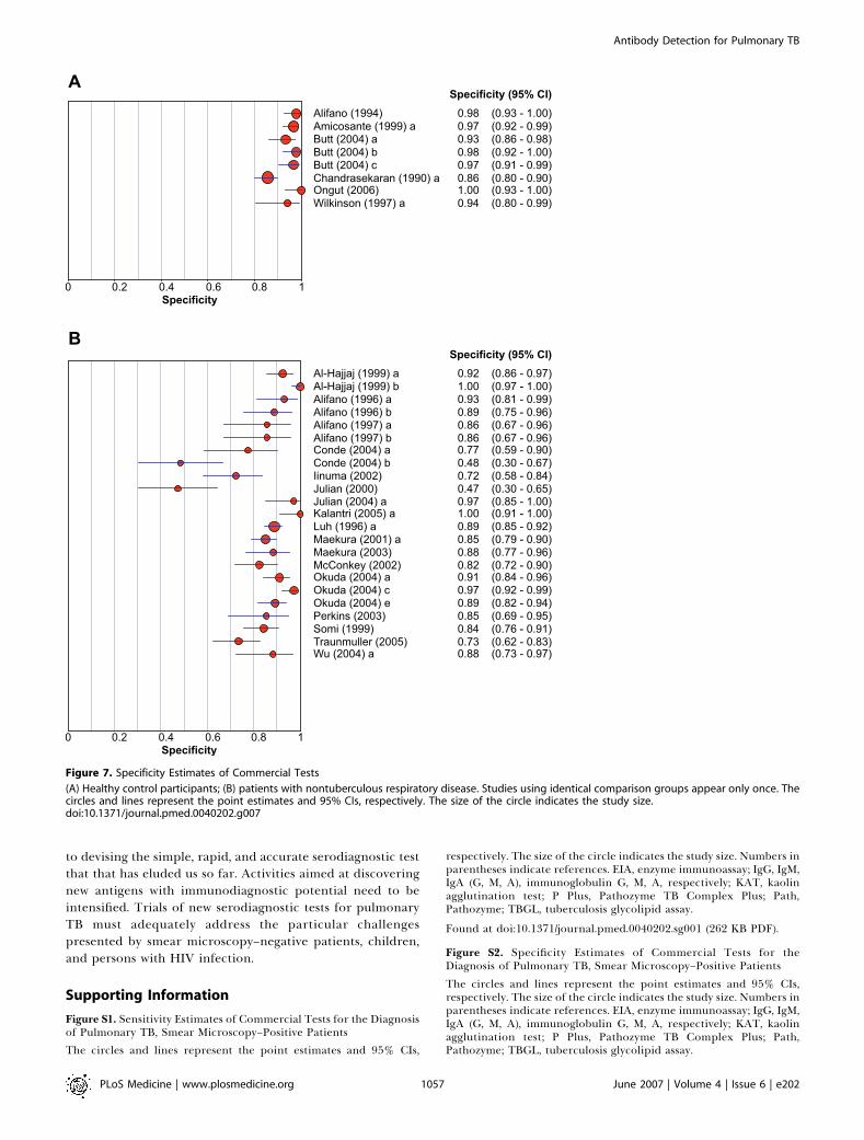
to devising the simple, rapid, and accurate serodiagnostic testthat that has eluded us so far. Activities aimed at discoveringnew antigens with immunodiagnostic potential need to beintensified. Trials of new serodiagnostic tests for pulmonaryTB must adequately address the particular challengespresented by smear microscopy–negative patients, children,and persons with HIV infection.
Supporting Information
Figure S1. Sensitivity Estimates of Commercial Tests for the Diagnosisof Pulmonary TB, Smear Microscopy–Positive Patients
The circles and lines represent the point estimates and 95% CIs,
respectively. The size of the circle indicates the study size. Numbers inparentheses indicate references. EIA, enzyme immunoassay; IgG, IgM,IgA (G, M, A), immunoglobulin G, M, A, respectively; KAT, kaolinagglutination test; P Plus, Pathozyme TB Complex Plus; Path,Pathozyme; TBGL, tuberculosis glycolipid assay.
Found at doi:10.1371/journal.pmed.0040202.sg001 (262 KB PDF).
Figure S2. Specificity Estimates of Commercial Tests for theDiagnosis of Pulmonary TB, Smear Microscopy–Positive Patients
The circles and lines represent the point estimates and 95% CIs,respectively. The size of the circle indicates the study size. Numbers inparentheses indicate references. EIA, enzyme immunoassay; IgG, IgM,IgA (G, M, A), immunoglobulin G, M, A, respectively; KAT, kaolinagglutination test; P Plus, Pathozyme TB Complex Plus; Path,Pathozyme; TBGL, tuberculosis glycolipid assay.
Figure 7. Specificity Estimates of Commercial Tests
(A) Healthy control participants; (B) patients with nontuberculous respiratory disease. Studies using identical comparison groups appear only once. Thecircles and lines represent the point estimates and 95% CIs, respectively. The size of the circle indicates the study size.doi:10.1371/journal.pmed.0040202.g007
PLoS Medicine | www.plosmedicine.org June 2007 | Volume 4 | Issue 6 | e2021057
Antibody Detection for Pulmonary TB

Found at doi:10.1371/journal.pmed.0040202.sg002 (257 KB PDF).
Figure S3. Sensitivity Estimates of Commercial Tests for the Diagnosisof Pulmonary TB, Smear Microscopy–Negative Patients
The circles and lines represent the point estimates and 95% CIs,respectively. The size of the circle indicates the study size. Numbers inparentheses indicate references. EIA, enzyme immunoassay; IgG, IgM,IgA (G, M, A), immunoglobulin G, M, A, respectively; KAT, kaolinagglutination test; P Plus, Pathozyme TB Complex Plus; Path,Pathozyme; TBGL, tuberculosis glycolipid assay.
Found at doi:10.1371/journal.pmed.0040202.sg003 (259 KB PDF).
Figure S4. Specificity Estimates of Commercial Tests for theDiagnosis of Pulmonary TB, Smear Microscopy–Negative Patients
The circles and lines represent the point estimates and 95% CIs,respectively. The size of the circle indicates the study size. Numbers inparentheses indicate references. EIA, enzyme immunoassay; IgG, IgM,IgA (G, M, A), immunoglobulin G, M, A, respectively; KAT, kaolinagglutination test; P Plus, Pathozyme TB Complex Plus; Path,Pathozyme; TBGL, tuberculosis glycolipid assay.
Found at doi:10.1371/journal.pmed.0040202.sg004 (256 KB PDF).
Figure S5. SROC Curve of Anda-TB IgG for the Diagnosis ofPulmonary TB, Smear Microscopy–Positive Patients
Each solid circle represents an individual study in the meta-analysis.The curve is the regression line that summarizes the overalldiagnostic accuracy. SE (AUC), standard error of AUC; Q*, an indexdefined by the point on the SROC curve where the sensitivity andspecificity are equal; SE (Q*), standard error of Q* index.
Found at doi:10.1371/journal.pmed.0040202.sg005 (234 KB PDF).
Figure S6. SROC Curve of Commercial Tests for the Diagnosis ofPulmonary TB
(A) Healthy control participants; (B) patients with nontuberculousrespiratory disease. Each solid circle represents an individual study inthe meta-analysis. The curve is the regression line that summarizesthe overall diagnostic accuracy. SE (AUC), standard error of AUC; Q*,an index defined by the point on the SROC curve where thesensitivity and specificity are equal; SE (Q*), standard error of Q*index.
Found at doi:10.1371/journal.pmed.0040202.sg006 (266 KB PDF).
Acknowledgments
We wish to thank Nandini Dendukuri, Maya Bhat-Gregerson, andAnna Meddaugh for technical assistance, Madelyn Hall for help inacquiring publications in the review, and Izabela Suder-Dayao andMelissa Anthony of WHO/TDR for administrative assistance. We alsothank the reviewers for their helpful comments.
Author contributions. KRS, SL, PCH, AR, DM, JC, KW, and MPconceived and designed the study. KRS and MH collected andanalyzed the data. KRS, MH, SL, AR, DM, KW, and MP interpreted thedata. KRS, SL, and MP drafted the manuscript. KRS, SL, PCH, AR,DM, JC, KW, and MP performed critical revision of the manuscript.
References1. World Health Organization (2007) Global tuberculosis control: Surveil-
lance, planning, financing. WHO report. Geneva: World Health Organ-ization. WHO/HTM/TB/2007.376.
2. Dye C, Watt CJ, Bleed DM, Hosseini SM, Raviglione MC (2005) Evolution oftuberculosis control and prospects for reducing tuberculosis incidence,prevalence, and deaths globally. JAMA 293: 2767–2775.
3. Toman K (2004) What are the advantages and disadvantages of fluorescencemicroscopy? In: Frieden T, editor. Toman’s tuberculosis: Case detection,treatment, and monitoring—questions and answers. 2nd edition. Geneva:World Health Organization. pp. 31–34.
4. Steingart KR, Ng V, Henry M, Hopewell PC, Ramsay A, et al. (2006) Sputumprocessing methods to improve the sensitivity of smear microscopy fortuberculosis: A systematic review. Lancet Infect Dis 6: 664–674.
5. Behr MA, Warren SA, Salamon H, Hopewell PC, Ponce de Leon A, et al.(1999) Transmission of Mycobacterium tuberculosis from patients smear-negative for acid-fast bacilli. Lancet 353: 444–449.
6. Grzybowski S, Barnett GD, Styblo K (1975) Contacts of cases of activepulmonary tuberculosis. Bull Int Union Tuberc 50: 90–106.
7. Urbanczik R (1985) Present position of microscopy and of culture indiagnostic mycobacteriology. Zentralbl Bakteriol Mikrobiol Hyg [A] 260:81–87.
8. Perkins MD, Roscigno G, Zumla A (2006) Progress towards improvedtuberculosis diagnostics for developing countries. Lancet 367: 942–943.
9. Shingadia D, Novelli V (2003) Diagnosis and treatment of tuberculosis inchildren. Lancet Infect Dis 3: 624–632.
10. Elliott AM, Namaambo K, Allen BW, Luo N, Hayes RJ, et al. (1993) Negativesputum smear results in HIV-positive patients with pulmonary tuberculosisin Lusaka, Zambia. Tuberc Lung Dis 74: 191–194.
11. Johnson JL, Vjecha MJ, Okwera A, Hatanga E, Byekwaso F, et al. (1998)Impact of human immunodeficiency virus type-1 infection on the initialbacteriologic and radiographic manifestations of pulmonary tuberculosisin Uganda. Makerere University-Case Western Reserve University ResearchCollaboration. Int J Tuberc Lung Dis 2: 397–404.
12. Menzies D (2004) What is the current and potential role of diagnostic testsother than sputum microscopy and culture? In: Frieden T, editor. Toman’stuberculosis: Case detection, treatment, and monitoring—Questions andanswers. 2nd edition. Geneva: World Health Organization. pp. 87–91.
13. Bothamley GH (1995) Serological diagnosis of tuberculosis. Eur Respir JSuppl 20: 676S–688S.
14. Chan ED, Heifets L, Iseman MD (2000) Immunologic diagnosis oftuberculosis: A review. Tuberc Lung Dis 80: 131–140.
15. Daniel TM (1989) Rapid diagnosis of tuberculosis: Laboratory techniquesapplicable in developing countries. Rev Infect Dis 11: S471–S478.
16. Daniel TM (1990) The rapid diagnosis of tuberculosis: A selective review. JLab Clin Med 116: 277–282.
17. Gennaro ML (2000) Immunologic diagnosis of tuberculosis. Clin Infect Dis30: S243–S246.
18. Iseman MD (2000) Immunity and pathogenesis. In: Iseman MD, editor. Aclinician’s guide to tuberculosis. Philadelphia: Lippincott Williams andWilkins. pp. 63–96.
19. Laal S (2004) Immunodiagnosis. In: Rom WN, Garay SM, editors. Tuber-culosis. Philadelphia: Lippincott Williams and Wilkins. pp. 185–191.
20. Laal S, Skeiky YA (2005) Immune-based methods. In: Cole ST, editor.Tuberculosis and the tubercle bacillus. Washington (D. C.): ASM Press. pp.71–83.
21. Palomino JC (2005) Nonconventional and new methods in the diagnosis oftuberculosis: Feasibility and applicability in the field. Eur Respir J 26: 339–350.
22. Pai M, Kalantri S, Dheda K (2006) New tools and emerging technologies forthe diagnosis of tuberculosis: Part II. Active tuberculosis and drugresistance. Expert Rev Mol Diagn 6: 423–432.
23. Boehme C, Molokova E, Minja F, Geis S, Loscher T, et al. (2005) Detectionof mycobacterial lipoarabinomannan with an antigen-capture ELISA inunprocessed urine of Tanzanian patients with suspected tuberculosis.Trans R Soc Trop Med Hyg 99: 893–900.
24. Tessema TA, Hamasur B, Bjun G, Svenson S, Bjorvatn B (2001) Diagnosticevaluation of urinary lipoarabinomannan at an Ethiopian tuberculosiscentre. Scand J Infect Dis 33: 279–284.
25. Kunter E, Cerrahoglu K, Ilvan A, Isitmangil T, Turken O, et al. (2003) Thevalue of pleural fluid anti-A60 IgM in BCG-vaccinated tuberculous pleurisypatients. Clin Microbiol Infect 9: 212–220.
26. Small PM, Perkins MD (2000) More rigour needed in trials of newdiagnostic agents for tuberculosis. Lancet 356: 1048–1049.
27. Walsh A, McNerney R (2004) Guidelines for establishing trials of new teststo diagnose tuberculosis in endemic countries. Int J Tuberc Lung Dis 8:609–613.
28. Special Programme for Research and Training in Tropical Diseases,Foundation for Innovative New Diagnostics (2006) Current TB diagnosticsmarket. Diagnostics for tuberculosis: Global demand and market potential.Geneva: World Health Organization. pp. 49–72.
29. Janeway CA, Travers P, Walport M, Shlomchik M (2005) Appendix I,Immunologists’ toolbox. Immunobiology, the immune system in health anddisease. 6th edition. New York: Garland Science Publishing. pp. 683–729.
30. Bangs Laboratories Immunochromatographic, lateral flow or strip testsdevelopment ideas. Available: http://www.pall.com/34445_4154.asp#4166.Accessed 10 May 2007.
31. Pai M, McCulloch M, Enanoria W, Colford JM Jr (2004) Systematic reviewsof diagnostic test evaluations: What’s behind the scenes? ACP J Club 141:A11–A13.
32. Littenberg B, Moses LE (1993) Estimating diagnostic accuracy frommultiple conflicting reports: A new meta-analytic method. Med DecisMaking 13: 313–321.
33. SPSS (2006) SPSS for Windows. 14.0.1.366 edition. Chicago: SPSS.34. Zamora J, Abraira V, Muriel A, Khan KS, Coomarasamy A (2006) Meta-
DiSc: A software for meta-analysis of test accuracy data. BMC Med ResMethodol 6: 31.
35. Walter SD (2002) Properties of the summary receiver operating character-istic (SROC) curve for diagnostic test data. Stat Med 21: 1237–1256.
36. Jones CM, Athanasiou T (2005) Summary receiver operating characteristiccurve analysis techniques in the evaluation of diagnostic tests. Ann ThoracSurg 79: 16–20.
37. Lijmer JG, Bossuyt PM, Heisterkamp SH (2002) Exploring sources ofheterogeneity in systematic reviews of diagnostic tests. Stat Med 21: 1525–1537.
38. Al-Hajjaj MS, Gad-el-Rab MO, Al-Orainey IO, Al-Kassimi FA (1999)
PLoS Medicine | www.plosmedicine.org June 2007 | Volume 4 | Issue 6 | e2021058
Antibody Detection for Pulmonary TB

Improved sensitivity for detection of tuberculosis cases by a modifiedAnda-TB ELISA test. Tuberc Lung Dis 79: 181–185.
39. Alifano M, De Pascalis R, Sofia M, Faraone S, Del Pezzo M, et al. (1997)Evaluation of IgA-mediated humoral immune response against themycobacterial antigen P-90 in diagnosis of pulmonary tuberculosis. Chest111: 601–605.
40. Alifano M, Del Pezzo M, Lamberti C, Faraone S, Covelli I (1994) ELISAmethod for evaluation of anti-A60 IgG in patients with pulmonary andextrapulmonary tuberculosis. New Microbiol 17: 37–43.
41. Alifano M, Sofia M, Mormile M, Micco A, Mormile AF, et al. (1996) IgAimmune response against the mycobacterial antigen A60 in patients withactive pulmonary tuberculosis. Respiration 63: 292–297.
42. Amicosante M, Houde M, Guaraldi G, Saltini C (1999) Sensitivity andspecificity of a multi-antigen ELISA test for the serological diagnosis oftuberculosis. Int J Tuberc Lung Dis 3: 736–740.
43. Antunes A, Nina J, David S (2002) Serological screening for tuberculosis inthe community: An evaluation of the Mycodot procedure in an Africanpopulation with high HIV-2 prevalence (Republic of Guinea-Bissau). ResMicrobiol 153: 301–305.
44. Butt T, Malik HS, Abbassi SA, Ahmad RN, Mahmood A, et al. (2004) Genusand species-specific IgG and IgM antibodies for pulmonary tuberculosis. JColl Physicians Surg Pak 14: 105–107.
45. Chandrasekaran S, Gupta EVV, Chauhan MM, Baily GVJ, Chaudhuri K(1990) Serodiagnosis of pulmonary tuberculosis by kaolin agglutinationtest. Indian J Tuberc 37: 11–15.
46. Conde MB, Suffys P, Silva J, Kritski AL, Dorman SE (2004) ImmunoglobulinA (IgA) and IgG immune responses against P-90 antigen for diagnosis ofpulmonary tuberculosis and screening for Mycobacterium tuberculosis in-fection. Clin Diagn Lab Immunol 11: 94–97.
47. Iinuma Y, Senda K, Takakura S, Ichiyama S, Tano M, et al. (2002) Evaluationof a commercially available serologic assay for antibodies against tuber-culosis-associated glycolipid antigen. Clin Chem Lab Med 40: 832–836.
48. Imaz MS, Comini MA, Zerbini E, Sequeira MD, Latini O, et al. (2004)Evaluation of commercial enzyme-linked immunosorbent assay kits fordetection of tuberculosis in Argentinean population. J Clin Microbiol 42:884–887.
49. Julian E, Matas L, Alcaide J, Luquin M (2004) Comparison of antibodyresponses to a potential combination of specific glycolipids and proteinsfor test sensitivity improvement in tuberculosis serodiagnosis. Clin DiagnLab Immunol 11: 70–76.
50. Julian E, Matas L, Hernandez A, Alcaide J, Luquin M (2000) Evaluation of anew serodiagnostic tuberculosis test based on immunoglobulin A detectionagainst Kp-90 antigen. Int J Tuberc Lung Dis 4: 1082–1085.
51. Kalantri Y, Hemvani N, Bhatia GC, Chitnis DS (2005) ELISA kit evaluationfor IgG and IgM antibodies to A-60 tubercular protein antigen. Indian JMed Sci 59: 337–346.
52. Luh KT, Yu CJ, Yang PC, Lee LN (1996) Tuberculosis antigen A60serodiagnosis in tuberculous infection: Application in extrapulmonary andsmear-negative pulmonary tuberculosis. Respirology 1: 145–151.
53. Maekura R, Kohno H, Hirotani A, Okuda Y, Ito M, et al. (2003) Prospectiveclinical evaluation of the serologic tuberculous glycolipid test incombination with the nucleic acid amplification test. J Clin Microbiol 41:1322–1325.
54. Maekura R, Okuda Y, Nakagawa M, Hiraga T, Yokota S, et al. (2001) Clinicalevaluation of anti-tuberculous glycolipid immunoglobulin G antibody assayfor rapid serodiagnosis of pulmonary tuberculosis. J Clin Microbiol 39:3603–3608.
55. McConkey SJ, Youssef FG, Azem E, Frenck RW, Weil GJ (2002) Evaluation ofa rapid-format antibody test and the tuberculin skin test for diagnosis oftuberculosis in two contrasting endemic settings. Int J Tuberc Lung Dis 6:246–252.
56. Okuda Y, Maekura R, Hirotani A, Kitada S, Yoshimura K, et al. (2004)Rapid serodiagnosis of active pulmonary Mycobacterium tuberculosis byanalysis of results from multiple antigen-specific tests. J Clin Microbiol42: 1136–1141.
57. Ongut G, Ogunc D, Gunseren F, Ogus C, Donmez L, et al. (2006) Evaluationof the ICT tuberculosis test for the routine diagnosis of tuberculosis. BMCInfect Dis 6: 37.
58. Perkins MD, Conde MB, Martins A, Kritski AL (2003) Serologic diagnosis oftuberculosis using a simple commercial multiantigen assay. Chest 123: 107–112.
59. Sachan A, Gupta R, Gupta D, Gautam K, Gupta D (1994) Diagnostic
significance of antigen A-60 by ELISA test in pulmonary tuberculosis.Indian J Tuberc 41: 239–243.
60. Somi GR, O’Brien RJ, Mfinanga GS, Ipuge YA (1999) Evaluation of theMycoDot (TM) test in patients with suspected tuberculosis in a field settingin Tanzania. Int J Tuberc Lung Dis 3: 231–238.
61. Traunmuller F, Haslinger I, Lagler H, Wolfgang G, Zeitlinger MA, et al.(2005) Influence of the washing buffer composition on the sensitivity of anenzyme-linked immunosorbent assay using mycobacterial glycolipids ascapture antigens. J Immunoassay Immunochem 26: 179–188.
62. van der Werf TS, Das PK, van Soolingen D, Yong S, van der Mark TW, et al.(1992) Sero-diagnosis of tuberculosis with A60 antigen enzyme-linkedimmunosorbent assay: Failure in HIV-infected individuals in Ghana. MedMicrobiol Immunol (Berl) 181: 71–76.
63. Wilkinson RJ, Haslov K, Rappuoli R, Giovannoni F, Narayanan PR, et al.(1997) Evaluation of the recombinant 38-kilodalton antigen of Mycobacte-rium tuberculosis as a potential immunodiagnostic reagent. J Clin Microbiol35: 553–557.
64. Wu HP, Shieh WB, Hsien FK, Hua CC (2004) The significance ofMycobacterium tuberculosis antibody, antigen 60 IgG in patients withabnormal chest radiography. Chang Gung Med J 27: 869–876.
65. Whiting P, Rutjes AW, Reitsma JB, Glas AS, Bossuyt PM, et al. (2004)Sources of variation and bias in studies of diagnostic accuracy: A systematicreview. Ann Intern Med 140: 189–202.
66. Lijmer JG, Mol BW, Heisterkamp S, Bonsel GJ, Prins MH, et al. (1999)Empirical evidence of design-related bias in studies of diagnostic tests.JAMA 282: 1061–1066.
67. Tatsioni A, Zarin DA, Aronson N, Samson DJ, Flamm CR, et al. (2005)Challenges in systematic reviews of diagnostic technologies. Ann InternMed 142: 1048–1055.
68. Samanich K, Belisle JT, Laal S (2001) Homogeneity of antibody responses intuberculosis patients. Infect Immun 69: 4600–4609.
69. Sartain MJ, Slayden RA, Singh KK, Laal S, Belisle JT (2006) Disease statedifferentiation and identification of tuberculosis biomarkers via nativeantigen array profiling. Mol Cell Proteomics 5: 2102–2113.
70. Hendrickson RC, Douglass JF, Reynolds LD, McNeill PD, Carter D, et al.(2000) Mass spectrometric identification of Mtb81, a novel serologicalmarker for tuberculosis. J Clin Microbiol 38: 2354–2361.
71. Houghton RL, Lodes MJ, Dillon DC, Reynolds LD, Day CH, et al. (2002) Useof multiepitope polyproteins in serodiagnosis of active tuberculosis. ClinDiagn Lab Immunol 9: 883–891.
72. Weldingh K, Rosenkrands I, Okkels LM, Doherty TM, Andersen P (2005)Assessing the serodiagnostic potential of 35 Mycobacterium tuberculosisproteins and identification of four novel serological antigens. J ClinMicrobiol 43: 57–65.
73. Mukherjee S, Daifalla N, Zhang Y, Douglass J, Brooks L, et al. (2004)Potential serological use of a recombinant protein that is a replica of aMycobacterium tuberculosis protein found in the urine of infected mice. ClinDiagn Lab Immunol 11: 280–286.
74. Samanich KM, Belisle JT, Sonnenberg MG, Keen MA, Zolla-Pazner S, et al.(1998) Delineation of human antibody responses to culture filtrate antigensof Mycobacterium tuberculosis. J Infect Dis 178: 1534–1538.
75. Singh KK, Zhang X, Patibandla AS, Chien P Jr, Laal S (2001) Antigens ofMycobacterium tuberculosis expressed during preclinical tuberculosis: Sero-logical immunodominance of proteins with repetitive amino acidsequences. Infect Immun 69: 4185–4191.
76. Lyashchenko K, Colangeli R, Houde M, Al Jahdali H, Menzies D, et al. (1998)Heterogeneous antibody responses in tuberculosis. Infect Immun 66: 3936–3940.
77. Steingart KR, Henry M, Laal S, Hopewell PC, Ramsay A, et al. (2007) Asystematic review of commercial serological antibody detection tests forthe diagnosis of extrapulmonary tuberculosis. Thorax. In press.
78. Pai M, O’Brien R (2006) Tuberculosis diagnostics trials: Do they lackmethodological rigor? Expert Rev Mol Diagn 6: 509–514.
79. Bossuyt PM, Reitsma JB, Bruns DE, Gatsonis CA, Glasziou PP, et al. (2003)Towards complete and accurate reporting of studies of diagnosticaccuracy: The STARD Initiative. Ann Intern Med 138: 40–44.
80. Whiting P, Rutjes AW, Reitsma JB, Bossuyt PM, Kleijnen J (2003) Thedevelopment of QUADAS: A tool for the quality assessment of studies ofdiagnostic accuracy included in systematic reviews. BMC Med ResMethodol 3: 25.
81. Banoo S, Bell D, Bossuyt P, Herring A, Mabey D, et al. (2006) Evaluation ofdiagnostic tests for infectious diseases: General principles. Nat RevMicrobiol 4: S21–S31.
PLoS Medicine | www.plosmedicine.org June 2007 | Volume 4 | Issue 6 | e2021059
Antibody Detection for Pulmonary TB
Editors’ Summary
Background. Tuberculosis (TB) is, globally, one of the most importantinfectious diseases. It is thought that in 2005 around 1.6 million peopledied as a result of TB. Controlling TB requires that the disease is correctlydiagnosed so that it can then be promptly treated, which will reduce therisk of infection being passed on to other individuals. The methodnormally used for diagnosing TB disease in poor countries (where mostpeople with TB disease live) involves taking a sample of mucus coughedup from the lungs; this mucus is then spread thinly onto a glass slide,dyed, and viewed under the microscope. The bacteria responsible for TBtake up the dye in a particular pattern and can be clearly seen under themicroscope. Although this test (sputum smear) is relatively straightfor-ward to carry out even where facilities are basic, it is not particularlygood at identifying TB disease in children or amongst individuals whoare HIV-positive. Finally, the sputum smear test is also not very sensitive;that is, many people who have TB disease may not give a positivereading. Therefore, there is an urgent need to develop and evaluate newtests that are suitable for use in poor countries, which will accuratelydiagnose TB disease, especially amongst children and people who areHIV-positive.
Why Was This Study Done? New tests for TB have become availablewhich detect whether an individual has raised antibodies againstparticular proteins and other substances present on the surface of theTB bacterium. These tests are carried out on blood samples, once bloodcells and other factors have been taken out. These antibody tests areoften quite simple to carry out, so in principle they could be suitable foruse in developing countries. Since the tests are available on the marketand can be freely used in some developing countries without any needfor government regulatory bodies to approve them, it is important toknow how good these tests are at diagnosing TB disease. Theresearchers here wanted, therefore, to evaluate all of the available datarelating to the accuracy of antibody detection tests for diagnosis of TBdisease.
What Did the Researchers Do and Find? In order to evaluate all of theinformation available on commercial antibody detection tests fordiagnosis of TB disease of the lungs, the researchers carried out asystematic review. First, they searched biomedical literature databasesusing specific terms to identify studies for inclusion. A study wasincluded in their analysis if the commercial test was compared againstone of two other standard tests (sputum smear microscopy, or growth ofTB bacteria in culture). One researcher from the team then pulled outspecific pieces of information from each published study: these includedthe type of study design; information on study participants; the type oftest; what the test was compared against; and finally the results ofevaluation of the test. A second researcher pulled out pieces of
information from several of the same studies. The researchers thencompared the information to ensure that it was recorded correctly. Eachstudy was also assigned a quality rating, based on four distinct criteria.For each type of test, the researchers used the data in the publishedstudies to work out the test’s accuracy, both in terms of its ability to givea positive reading for people who have TB disease as well as its ability togive a negative reading for people who do not have TB disease.
The researchers found 27 papers meeting their criteria. These papersreported the results of 68 original studies. Nine different commercialtests were examined in the studies. Overall, the studies seemed to be ofrelatively poor quality, with only 25% of them meeting all four of theresearchers’ criteria for a good-quality study. The different studiesappeared to produce varying results for the accuracy of thesecommercial tests. In particular, the tests seemed to be less accurate atdetecting TB disease amongst people who had a negative sputum smearthan amongst people with a positive sputum smear. When all the datafor these different studies were combined, the statistics indicated thatthe commercial tests, overall, were only modestly accurate for diagnosisof TB disease. None of the studies had been carried out in children or inHIV-positive people.
What Do These Findings Mean? The results of this systematic reviewsuggest that the commercial antibody detection tests considered hereare not particularly useful in diagnosis of TB disease as compared toother tests, such as sputum smear and bacterial culture. Some people areconcerned that there is pressure in certain developing countries to startusing these tests, but the current data do not support greater use. Thissystematic review also highlights the fact that many studies evaluatingcommercial TB tests are of poor quality, and that further research needsto be done to evaluate the accuracy of different TB tests amongstchildren and HIV-positive patients.
Additional Information. Please access these Web sites via the onlineversion of this summary at http://dx.doi.org/10.1371/journal.pmed.0040202.
� World Health Organization Stop TB Department website. Informationabout the current Stop TB strategy, data and factsheets about TB, andother resources are available� Questions and Answers about Tuberculosis provided by the US
Centers for Disease Control and Prevention� Information about TB tests from Medicins sans Frontieres (MSF). Links
to MSF reports on new diagnostic tests are also available� Wikipedia entry on Systematic Reviews (Note: Wikipedia is an internet
encyclopedia anyone can edit)
PLoS Medicine | www.plosmedicine.org June 2007 | Volume 4 | Issue 6 | e2021060
Antibody Detection for Pulmonary TB