plos mechanism for multiple ligand recognition by the...
TRANSCRIPT

Mechanism for Multiple Ligand Recognitionby the Human Transferrin ReceptorAnthony M. Giannetti
1, Peter M. Snow
2, Olga Zak
3, Pamela J. Bjorkman
4*
1 Graduate Option in Biochemistry and Molecular Biophysics, California Institute of Technology, Pasadena, California, United States of America, 2 Caltech Protein Expression
Center, Division of Biology, California Institute of Technology, Pasadena, California, United States of America, 3 Department of Physiology and Biophysics, Albert Einstein
College of Medicine, Bronx, New York, United States of America, 4 Division of Biology and Howard Hughes Medical Institute, California Institute of Technology, Pasadena,
California, United States of America
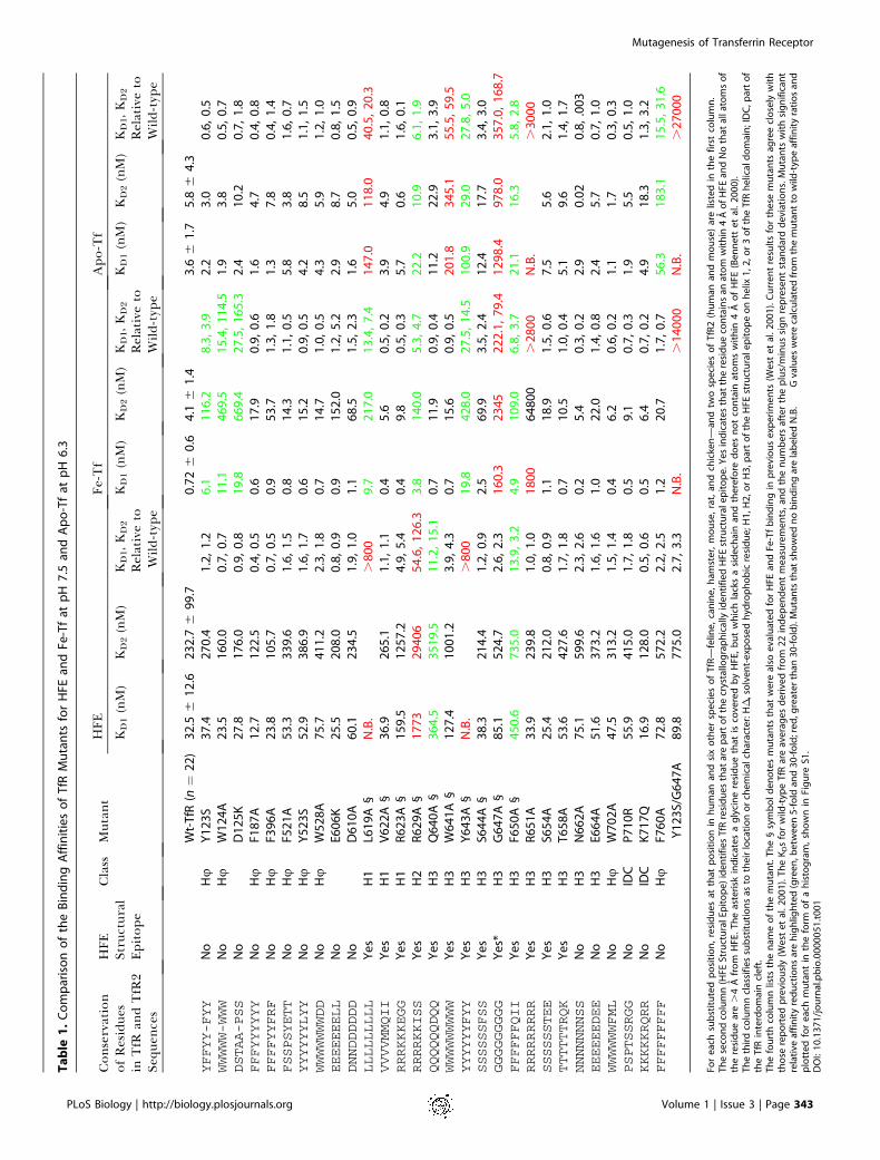
Transferrin receptor 1 (TfR) plays a critical role in cellular iron import for most higher organisms. Cell surface TfR bindsto circulating iron-loaded transferrin (Fe-Tf) and transports it to acidic endosomes, where low pH promotes iron todissociate from transferrin (Tf) in a TfR-assisted process. The iron-free form of Tf (apo-Tf) remains bound to TfR and isrecycled to the cell surface, where the complex dissociates upon exposure to the slightly basic pH of the blood. Fe-Tfcompetes for binding to TfR with HFE, the protein mutated in the iron-overload disease hereditary hemochromatosis.We used a quantitative surface plasmon resonance assay to determine the binding affinities of an extensive set of site-directed TfR mutants to HFE and Fe-Tf at pH 7.4 and to apo-Tf at pH 6.3. These results confirm the previous finding thatFe-Tf and HFE compete for the receptor by binding to an overlapping site on the TfR helical domain. Spatially distantmutations in the TfR protease-like domain affect binding of Fe-Tf, but not iron-loaded Tf C-lobe, apo-Tf, or HFE, andmutations at the edge of the TfR helical domain affect binding of apo-Tf, but not Fe-Tf or HFE. The binding datapresented here reveal the binding footprints on TfR for Fe-Tf and apo-Tf. These data support a model in which the Tf C-lobe contacts the TfR helical domain and the Tf N-lobe contacts the base of the TfR protease-like domain. Thedifferential effects of some TfR mutations on binding to Fe-Tf and apo-Tf suggest differences in the contact pointsbetween TfR and the two forms of Tf that could be caused by pH-dependent conformational changes in Tf, TfR, or both.From these data, we propose a structure-based model for the mechanism of TfR-assisted iron release from Fe-Tf.
Introduction
Transferrin receptor 1 (TfR) is a homodimeric type IImembrane protein that plays a critical role in the primaryiron acquisition mechanism for all iron-requiring cell typesin vertebrates (Enns 2002). TfR binds the serum iron-carrierprotein transferrin (Fe-Tf) and imports it to acidic endo-somes, where iron is released and transported to the cytosol.The complex between TfR and iron-free transferrin (apo-Tf)is then recycled to the cell surface where apo-Tf dissociatesand returns to circulation (reviewed in Enns et al. 1996). TfRalso binds the hereditary hemochromatosis protein HFE(Parkkila et al. 1997; Feder et al. 1998). HFE is a class I majorhistocompatibility complex (MHC)-related protein that ismutated in patients with hereditary hemochromatosis (Federet al. 1996), an iron-storage disease characterized by excessiveiron absorption leading to an accumulation of iron princi-pally in the liver, heart, pancreas, parathyroid, and pituitarygland, leading to tissue damage (Cullen et al. 1999).
The X-ray crystal structures of the human TfR ectodomain,both alone (Lawrence et al. 1999) and in complex with HFE(Bennett et al. 2000), have been reported. The homodimericTfR ectodomain contains three domains on each polypeptidechain: a protease-like domain resembling amino- andcarboxypeptidases (residues 121–188 and 384–606), an apicaldomain (residues 189–383), and a helical domain involved inTfR homodimerization (residues 607–760). Intact TfR alsoincludes a glycosylated stalk region (residues 90–120), atransmembrane domain (residues 62–89), and an N-terminalcytoplasmic domain (residues 1–61) that includes a tyrosine-based endosomal sorting sequence (YTRF) (Enns 2002). Thestructure of a 2:1 HFE/TfR complex (two HFEs bound to ahomodimeric TfR) shows that each HFE interacts with helices
1 and 3 of the TfR helical domain (Bennett et al. 2000) (Figure1A and 1B). The central portion of the interface includes ahydrophobic core consisting of TfR residues Leu619, Val622,and Tyr643 packed against hydrophobic residues from the a1domain helix of HFE.The structures of various transferrins (Tfs) and related
proteins such as lactoferrin have been studied extensively byX-ray crystallography (Bailey et al. 1988; Anderson et al. 1989;Gerstein et al. 1993; Zuccola 1993; Kurokawa et al. 1995, 1999;Baker et al. 1998; Karthikeyan et al. 1999). Tf and its relativesare single-chain molecules consisting of two similarly foldedlobes (the N- and C-lobes), each of which contains twodomains (NI and NII in the N-lobe; CI and CII in the C-lobe).Diferric Tf (Fe-Tf) contains two iron atoms, each held in acleft between the domains of each lobe. Transition betweenthe ferric and iron-free states of Tf involves significantconformational changes (Grossmann et al. 1992, 1993).Specifically, loss of iron results in a 548–638 rotation betweenthe two domains that comprise each lobe (Gerstein et al.
Received July 15, 2003; Accepted September 10, 2003; Published December 22,2003DOI: 10.1371/journal.pbio.0000051
Copyright: � 2003 Giannetti et al. This is an open-access article distributedunder the terms of the Creative Commons Attribution License, which permitsunrestricted use, distribution, and reproduction in any medium, provided theoriginal work is properly cited.
Abbreviations: apo-Tf, iron-free transferrin; Fe-C-lobe, iron-loaded transferrin C-lobe; Fe-Tf, diferric transferrin; HFE, hereditary hemochromatosis protein; KD,equilibrium dissociation constant; MHC, major histocompatibility complex; PIPES,piperazine-1,4-bis(2-ethanesulphonic) acid; RU, resonance unit; Tf, transferrin; TfR,transferrin receptor
Academic Editor: Janet Thornton, European Bioinformatics Institute
*To whom correspondence should be addressed. E-mail: [email protected]
PLoS Biology | http://biology.plosjournals.org Volume 1 | Issue 3 | Page 341
PLoS BIOLOGY

1993). Additionally, the interface between the lobes repacks,exposing previously buried residues and burying previouslyexposed residues (Kurokawa et al. 1999). In vivo, theseconformational changes presumably take place while Tf isbound to TfR, as the two proteins remain complexedthroughout endocytosis and recycling (Dautry-Varsat et al.1983).
Free Fe-Tf releases iron at acidic pH, but binding to TfRaffects the iron release at both basic and acidic pH (Bali andAisen 1991, 1992; Bali et al. 1991). At pH 7.4, iron releasefrom Fe-Tf bound to TfR is slower than from free Fe-Tf. Atlow pH, the opposite effect is observed, such that binding toTfR significantly increases the iron-release rate (Bali andAisen 1991, 1992; Bali et al. 1991; Sipe and Murphy 1991).Attempts to determine the mechanism by which TfR mediatesthese effects on the iron release rate have been hampered bya lack of detailed knowledge of the binding footprints of Fe-Tf and apo-Tf on TfR and by the unavailability of crystalstructures of Fe-Tf or apo-Tf bound to TfR.
Although the structural details of the interaction betweenTf and TfR remain unknown, early studies established thattwo Tf molecules bind to each TfR homodimer (Enns andSussman 1981) by primarily interacting with what is nowstructurally defined as the TfR helical domain (Buchegger etal. 1996). A subsequent mutagenesis study further localizedthe binding site to include a conserved RGD sequence(residues 646–648) within the TfR helical domain (Dubljevicet al. 1999). The HFE/TfR co-crystal structure revealed thatHFE directly contacts TfR residues 646 and 648 (Bennett et al.2000), which is consistent with biochemical inhibition studiesthat suggested that HFE and Tf bind to the same or anoverlapping site on TfR (Lebron et al. 1999). As Fe-Tf is alarge protein (approximately 90A 3 50A 3 40 A, measuredusing the structure of iron-bound ovo-Tf [Kurokawa et al.1995]), the remainder of the Tf contact site on TfR couldinclude other TfR domains, the TfR interdomain cleft, orboth (Figure 1B), as previously suggested (Lawrence et al.
1999). A subsequent mutagenesis study sought to identifyother Tf-contacting residues on TfR (West et al. 2001). In thatstudy, residues identified from the HFE/TfR co-crystalstructure as involved in contacting HFE were mutated, andtheir effects on binding to HFE and Fe-Tf were quantitativelyevaluated. These experiments identified several residueswithin the TfR helical domain that are involved in bindingto each protein (defined as a substitution producing a greaterthan or equal to 5-fold reduction in binding affinity) andconfirmed that the Fe-Tf- and HFE-binding sites on TfRoverlap. However, the larger size of Tf relative to the HFEectodomain (679 amino acids in Tf compared with 374 for theHFE/b2-microglobulin ectodomain) suggested that Fe-Tfcould contact residues outside of the TfR helical domain.Also, the effects of the TfR substitutions on binding to apo-Tfwere not evaluated; thus, the question of whether Fe-Tf andapo-Tf bind differently to TfR was not addressed.We therefore sought to expand the library of TfR mutants
to more extensively map the Fe-Tf interface and to comparethe effects of TfR mutants for binding to Fe-Tf versus apo-Tf.Here we report the affinities of 30 mutants of human TfR forbinding to HFE and Fe-Tf at pH 7.5 and to apo-Tf at pH 6.3.As expected, the most important residues for Tf binding arelocated in the center of the TfR helical domain in the vicinityof critical residues for HFE binding. However, we alsoidentified residues within the TfR protease-like domain thatmake significant contributions to binding of Fe-Tf, but notapo-Tf, to TfR. Conversely, substitution of residues at theedge of the TfR helical domain affects binding of apo-Tf, butnot Fe-Tf. This information, together with the identificationof common Fe-Tf- and apo-Tf-contacting residues within thehelical domain, constrains the possible positions of Fe-Tf andapo-Tf on TfR, allowing for construction of structural modelsfor the placement of the two forms of Tf on TfR. Our dataalso suggest a structural mechanism to explain TfR’s role inthe pH-dependent modulation of iron release rates from Fe-Tf.
Figure 1. TfR Structure
(A) Ribbon diagram of TfR homodimerderived from the 3.2 A structure of TfR(Lawrence et al. 1999). The HFE-bindingsite (deduced from an analysis using theHFE/TfR co-crystal structure [Bennett etal. 2000]) on the TfR helical domainclosest to the viewer is highlighted incyan.(B) Space-filling representation of onechain from the TfR homodimer, with theHFE structural epitope residues high-lighted as in (A). The location of theinterdomain cleft is indicated by anorange asterisk.(C–E) Summary of effects of TfR sub-stitutions for binding HFE (C), Fe-Tf (D),and apo-Tf (E). Color-coding of the TfRsidechains designates the effects of thesubstitutions on binding affinities asindicated.Figures were made with Molscript (Krau-lis 1991) or GRASP (Nicholls et al. 1993)and rendered with Raster3D (Merritt andBacon 1997).DOI: 10.1371/journal.pbio.0000051.g001
PLoS Biology | http://biology.plosjournals.org Volume 1 | Issue 3 | Page 342
Mutagenesis of Transferrin Receptor
Table
1.
Co
mp
aris
on
of
the
Bin
din
gA
ffin
itie
so
fT
fRM
uta
nts
for
HFE
and
Fe-T
fat
pH
7.5
and
Ap
o-T
fat
pH
6.3
Con
servation
HFE
Class
Mutan
tHFE
Fe-T
fApo-Tf
ofResidue
sin
TfR
andTfR
2Se
quen
ces
Structural
Epitop
eKD1(nM)
KD2(nM)
KD1,K
D2
Relativeto
Wild-type
KD1(nM)
KD2(nM)
KD1,K
D2
Relativeto
Wild-type
KD1(nM)
KD2(nM)
KD1,K
D2
Relativeto
Wild-type
Wt-
TfR
(n¼
22
)3
2.5
61
2.6
23
2.7
69
9.7
0.7
26
0.6
4.1
61
.43
.66
1.7
5.8
64
.3YFFYY-FYY
No
Hu
Y1
23
S3
7.4
27
0.4
1.2
,1
.26
.11
16
.28
.3,
3.9
2.2
3.0
0.6
,0
.5WWWWW-WWW
No
Hu
W1
24
A2
3.5
16
0.0
0.7
,0
.71
1.1
46
9.5
15
.4,
11
4.5
1.9
3.8
0.5
,0
.7DSTAA-PSS
No
D1
25
K2
7.8
17
6.0
0.9
,0
.81
9.8
66
9.4
27
.5,
16
5.3
2.4
10
.20
.7,
1.8
FFFYYYYYY
No
Hu
F18
7A
12
.71
22
.50
.4,
0.5
0.6
17
.90
.9,
0.6
1.6
4.7
0.4
,0
.8FFFFYYFRF
No
Hu
F39
6A
23
.81
05
.70
.7,
0.5
0.9
53
.71
.3,
1.8
1.3
7.8
0.4
,1
.4FSSPSYETT
No
Hu
F52
1A
53
.33
39
.61
.6,
1.5
0.8
14
.31
.1,
0.5
5.8
3.8
1.6
,0
.7YYYYYYLYY
No
Hu
Y5
23
S5
2.9
38
6.9
1.6
,1
.70
.61
5.2
0.9
,0
.54
.28
.51
.1,
1.5
WWWWWWWDD
No
Hu
W5
28
A7
5.7
41
1.2
2.3
,1
.80
.71
4.7
1.0
,0
.54
.35
.91
.2,
1.0
EEEEEEELL
No
E60
6K
25
.52
08
.00
.8,
0.9
0.9
15
2.0
1.2
,5
.22
.98
.70
.8,
1.5
DNNDDDDDD
No
D6
10
A6
0.1
23
4.5
1.9
,1
.01
.16
8.5
1.5
,2
.31
.65
.00
.5,
0.9
LLLLLLLLL
Ye
sH
1L6
19
A§
N.B
..
80
09
.72
17
.01
3.4
,7
.41
47
.01
18
.04
0.5
,2
0.3
VVVVMMQII
Ye
sH
1V
62
2A
§3
6.9
26
5.1
1.1
,1
.10
.45
.60
.5,
0.2
3.9
4.9
1.1
,0
.8RRRKKKEGG
Ye
sH
1R
62
3A
§1
59
.51
25
7.2
4.9
,5
.40
.49
.80
.5,
0.3
5.7
0.6
1.6
,0
.1RRRRKKISS
Ye
sH
2R
62
9A
§1
77
32
94
06
54
.6,
12
6.3
3.8
14
0.0
5.3
,4
.72
2.2
10
.96
.1,
1.9
QQQQQQDQQ
Ye
sH
3Q
64
0A
§3
64
.53
51
9.5
11
.2,
15
.10
.71
1.9
0.9
,0
.41
1.2
22
.93
.1,
3.9
WWWWWWWWW
Ye
sH
3W
64
1A
§1
27
.41
00
1.2
3.9
,4
.30
.71
5.6
0.9
,0
.52
01
.83
45
.15
5.5
,5
9.5
YYYYYYFYY
Ye
sH
3Y
64
3A
§N
.B.
.8
00
19
.84
28
.02
7.5
,1
4.5
10
0.9
29
.02
7.8
,5
.0SSSSSSFSS
Ye
sH
3S6
44
A§
38
.32
14
.41
.2,
0.9
2.5
69
.93
.5,
2.4
12
.41
7.7
3.4
,3
.0GGGGGGGGG
Ye
s*H
3G
64
7A
§8
5.1
52
4.7
2.6
,2
.31
60
.32
34
52
22
.1,
79
.41
29
8.4
97
8.0
35
7.0
,1
68
.7FFFFFFQII
Ye
sH
3F6
50
A§
45
0.6
73
5.0
13
.9,
3.2
4.9
10
9.0
6.8
,3
.72
1.1
16
.35
.8,
2.8
RRRRRRRRR
Ye
sH
3R
65
1A
33
.92
39
.81
.0,
1.0
18
00
64
80
0.
28
00
N.B
..
30
00
SSSSSSTEE
Ye
sH
3S6
54
A2
5.4
21
2.0
0.8
,0
.91
.11
8.9
1.5
,0
.67
.55
.62
.1,
1.0
TTTTTTRQK
Ye
sH
3T
65
8A
53
.64
27
.61
.7,
1.8
0.7
10
.51
.0,
0.4
5.1
9.6
1.4
,1
.7NNNNNNNSS
No
H3
N6
62
A7
5.1
59
9.6
2.3
,2
.60
.25
.40
.3,
0.2
2.9
0.0
20
.8,
.00
3EEEEEEDEE
No
H3
E66
4A
51
.63
73
.21
.6,
1.6
1.0
22
.01
.4,
0.8
2.4
5.7
0.7
,1
.0WWWWWWFML
No
Hu
W7
02
A4
7.5
31
3.2
1.5
,1
.40
.46
.20
.6,
0.2
1.1
1.7
0.3
,0
.3PSPTSSRGG
No
IDC
P7
10
R5
5.9
41
5.0
1.7
,1
.80
.59
.10
.7,
0.3
1.9
5.5
0.5
,1
.0KKKKKRQRR
No
IDC
K7
17
Q1
6.9
12
8.0
0.5
,0
.60
.56
.40
.7,
0.2
4.9
18
.31
.3,
3.2
FFFFFFFFF
No
Hu
F76
0A
72
.85
72
.22
.2,
2.5
1.2
20
.71
.7,
0.7
56
.31
83
.11
5.5
,3
1.6
Y1
23
S/G
64
7A
89
.87
75
.02
.7,
3.3
N.B
..
14
00
0N
.B.
.2
70
00
For
eac
hsu
bst
itu
ted
po
siti
on
,re
sid
ue
sat
that
po
siti
on
inh
um
anan
dsi
xo
the
rsp
eci
es
of
TfR
—fe
line
,ca
nin
e,
ham
ste
r,m
ou
se,
rat,
and
chic
ken
—an
dtw
osp
eci
es
of
TfR
2(h
um
anan
dm
ou
se)
are
liste
din
the
firs
tco
lum
n.
Th
ese
con
dco
lum
n(H
FESt
ruct
ura
lEp
ito
pe
)id
en
tifi
es
TfR
resi
du
es
that
are
par
to
fth
ecr
ysta
llog
rap
hic
ally
ide
nti
fie
dH
FEst
ruct
ura
le
pit
op
e.Y
es
ind
icat
es
that
the
resi
du
eco
nta
ins
anat
om
wit
hin
4A
of
HFE
and
No
that
alla
tom
so
fth
ere
sid
ue
are.
4A
fro
mH
FE.
Th
eas
teri
skin
dic
ate
sa
gly
cin
ere
sid
ue
that
isco
vere
db
yH
FE,
bu
tw
hic
hla
cks
asi
de
chai
nan
dth
ere
fore
do
es
no
tco
nta
inat
om
sw
ith
in4
Ao
fH
FE(B
en
ne
tte
tal
.2
00
0).
Th
eth
ird
colu
mn
clas
sifi
es
sub
stit
uti
on
sas
toth
eir
loca
tio
no
rch
em
ical
char
acte
r:H�
,so
lve
nt-
exp
ose
dh
ydro
ph
ob
icre
sid
ue
;H1
,H2
,or
H3
,par
to
fth
eH
FEst
ruct
ura
le
pit
op
eo
nh
elix
1,2
,or
3o
fth
eT
fRh
elic
ald
om
ain
;ID
C,p
art
of
the
TfR
inte
rdo
mai
ncl
eft
.T
he
fou
rth
colu
mn
lists
the
nam
eo
fth
em
uta
nt.
Th
e§
sym
bo
ld
en
ote
sm
uta
nts
that
we
real
soe
valu
ate
dfo
rH
FEan
dFe
-Tf
bin
din
gin
pre
vio
us
exp
eri
me
nts
(We
ste
tal
.2
00
1).
Cu
rre
nt
resu
lts
for
the
sem
uta
nts
agre
ecl
ose
lyw
ith
tho
sere
po
rte
dp
revi
ou
sly
(We
ste
tal
.2
00
1).
Th
eK
Ds
for
wild
-typ
eT
fRar
eav
era
ge
sd
eri
ved
fro
m2
2in
de
pe
nd
en
tm
eas
ure
me
nts
,an
dth
en
um
be
rsaf
ter
the
plu
s/m
inu
ssi
gn
rep
rese
nt
stan
dar
dd
evi
atio
ns.
Mu
tan
tsw
ith
sig
nif
ican
tre
lati
veaf
fin
ity
red
uct
ion
sar
eh
igh
ligh
ted
(gre
en
,be
twe
en
5-f
old
and
30
-fo
ld;r
ed
,gre
ate
rth
an3
0-f
old
).M
uta
nts
that
sho
we
dn
ob
ind
ing
are
lab
ele
dN
.B.�
�G
valu
es
we
reca
lcu
late
dfr
om
the
mu
tan
tto
wild
-typ
eaf
fin
ity
rati
os
and
plo
tte
dfo
re
ach
mu
tan
tin
the
form
of
ah
isto
gra
m,
sho
wn
inFi
gu
reS1
.D
OI:
10
.13
71
/jo
urn
al.p
bio
.00
00
05
1.t
00
1
PLoS Biology | http://biology.plosjournals.org Volume 1 | Issue 3 | Page 343
Mutagenesis of Transferrin Receptor
Results
Design of TfR MutantsOur choice of TfR residues to substitute was guided by the
crystal structures of TfR alone and bound to HFE (Lawrenceet al. 1999; Bennett et al. 2000) and by a previous bindingstudy involving ten TfR point mutants (West et al. 2001) (§symbol in Table 1). Four substitutions at the HFE-binding siteon the TfR helical domain (L619A, R629A, Y643A, andF650A) were found to significantly reduce (greater than orequal to 5-fold) the binding affinity for both HFE and Fe-Tf atpH 7.5, giving a first-order map of the Fe-Tf-binding site onTfR (West et al. 2001). In order to identify additional TfRresidues critical for Fe-Tf binding and to evaluate theireffects on apo-Tf binding, we extended our TfR mutantlibrary to include an additional 20 mutants. The new set ofmutations were chosen using three different strategies: (1) analanine scan involving solvent-exposed residues on helix 3 ofthe helical domain (R651A, S654A, T658A, N662A, E664A)(classified as H3 in Table 1); (2) substitution of residues in theTfR interdomain cleft (see Figure 1B), suggested to be part ofthe Fe-Tf-binding site (Lawrence et al. 1999), for the residuesof chicken TfR, which does not bind human Tf (Buchegger etal. 1996) (P710R, K717Q) (classified as IDC in Table 1); (3)mutation of large solvent-exposed hydrophobic residues,which often provide much of the free energy of binding inprotein–protein interactions (Jones and Thornton 1996; Tsaiet al. 1997; Lo Conte et al. 1999), throughout the remainingTfR surface area (Y123S, F187A, F396A, F521A, Y523S,W528A, W702A, F760A) (classified as Hu in Table 1). Asecond generation of mutants was subsequently made tofurther define newly identified binding sites (W124A, D125K,E606K, D610A) and to test the effect of combining sub-stitutions (Y123S/G647A). Mutants involving TfR residuesknown from the HFE/TfR crystal structure (Bennett et al.2000) to contact HFE are denoted as part of the HFEstructural epitope in Table 1.
TfR mutants were expressed as N-terminally 6x-His-taggedsoluble ectodomains in baculovirus-infected cells, as pre-viously described (Lebron et al. 1998; West et al. 2001). In aprevious TfR mutagenesis study, it was shown that mutantsthat had a strong effect on binding were properly folded asdetermined by comparison of their far-UV circular dichroismspectra and gel filtration profiles to that of wild-type TfR(West et al. 2001). In this study, we note that all of the newlymade mutants retain wild-type or near wild-type bindingaffinities for at least one of the three TfR ligands tested (HFE,Fe-Tf, or apo-Tf) (Table 1), confirming their structuralintegrity.
Affinity Measurements and AnalysesEach of the TfR mutants designed in the current screen,
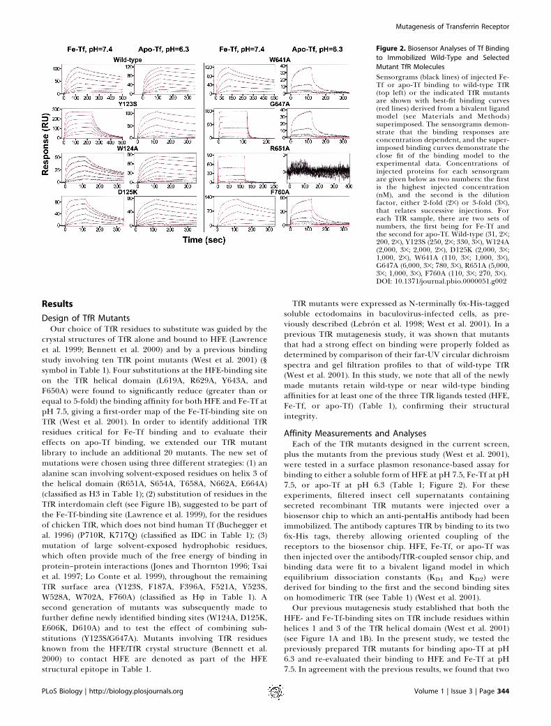
plus the mutants from the previous study (West et al. 2001),were tested in a surface plasmon resonance-based assay forbinding to either a soluble form of HFE at pH 7.5, Fe-Tf at pH7.5, or apo-Tf at pH 6.3 (Table 1; Figure 2). For theseexperiments, filtered insect cell supernatants containingsecreted recombinant TfR mutants were injected over abiosensor chip to which an anti-pentaHis antibody had beenimmobilized. The antibody captures TfR by binding to its two6x-His tags, thereby allowing oriented coupling of thereceptors to the biosensor chip. HFE, Fe-Tf, or apo-Tf wasthen injected over the antibody/TfR-coupled sensor chip, andbinding data were fit to a bivalent ligand model in whichequilibrium dissociation constants (KD1 and KD2) werederived for binding to the first and the second binding siteson homodimeric TfR (see Table 1) (West et al. 2001).Our previous mutagenesis study established that both the
HFE- and Fe-Tf-binding sites on TfR include residues withinhelices 1 and 3 of the TfR helical domain (West et al. 2001)(see Figure 1A and 1B). In the present study, we tested thepreviously prepared TfR mutants for binding apo-Tf at pH6.3 and re-evaluated their binding to HFE and Fe-Tf at pH7.5. In agreement with the previous results, we found that two
Figure 2. Biosensor Analyses of Tf Binding
to Immobilized Wild-Type and Selected
Mutant TfR Molecules
Sensorgrams (black lines) of injected Fe-Tf or apo-Tf binding to wild-type TfR(top left) or the indicated TfR mutantsare shown with best-fit binding curves(red lines) derived from a bivalent ligandmodel (see Materials and Methods)superimposed. The sensorgrams demon-strate that the binding responses areconcentration dependent, and the super-imposed binding curves demonstrate theclose fit of the binding model to theexperimental data. Concentrations ofinjected proteins for each sensorgramare given below as two numbers: the firstis the highest injected concentration(nM), and the second is the dilutionfactor, either 2-fold (23) or 3-fold (33),that relates successive injections. Foreach TfR sample, there are two sets ofnumbers, the first being for Fe-Tf andthe second for apo-Tf. Wild-type (31, 23;200, 23), Y123S (250, 23; 330, 33), W124A(2,000, 33; 2,000, 23), D125K (2,000, 33;1,000, 23), W641A (110, 33; 1,000, 33),G647A (6,000, 33; 780, 33), R651A (5,000,33; 1,000, 33), F760A (110, 33; 270, 33).DOI: 10.1371/journal.pbio.0000051.g002
PLoS Biology | http://biology.plosjournals.org Volume 1 | Issue 3 | Page 344
Mutagenesis of Transferrin Receptor
mutants, L619A and Y643A, showed no detectable HFEbinding and a significant (greater than or equal to 5-fold)decrease in Fe-Tf binding. These substitutions also signifi-cantly reduced apo-Tf binding at acidic pH. Two othermutants, R629A and Q640A, were again found to significantlyreduce HFE binding and to have a relatively minor effect onFe-Tf binding (R629A) or no significant effect (Q640A). Theeffects of these substitutions on apo-Tf binding correlatedwith their effects on Fe-Tf binding. Likewise, the F650Amutant, which shows a moderate reduction in binding affinityfor both HFE and Fe-Tf, also shows a reduced affinity forbinding apo-Tf. Only one of the previously analyzed mutants,G647A, exhibited a major (greater than 100-fold) reduction inFe-Tf binding affinity, and the present analysis reveals that ithas a similar effect on apo-Tf binding. Interestingly, one ofthe previously analyzed mutants, W641A, which does notsignificantly affect HFE or Fe-Tf binding at pH 7.5, exerted asignificant reduction in the binding affinity for apo-Tf at pH6.3 (see Table 1; Figure 1E; Figure 2), suggesting that it mightbe possible to find additional substitutions with differentialeffects on binding of the two forms of Tf.
Our first strategy for finding additional residues critical forTf binding involved substitution of solvent-exposed residuesC-terminal to the Tf-binding epitope residues Gly647 andPhe650 on helix 3 of the TfR helical domain. Of the five newTfR mutants constructed (R651A, S654A, T658A, N662A,E664A), only one (R651A) affected Tf binding, resulting in agreater than 2,800-fold reduction in binding of Fe-Tf andapo-Tf. Having identified a ‘‘hot spot’’ for Tf bindinginvolving TfR helical domain residues Gly647 and Arg651,we then searched for residues affecting Tf binding that weredistant from this site, which would allow approximatepositioning of the bi-lobed Tf structure on TfR. Two residueswithin the cleft formed by portions of the three TfR domainswere changed to their chicken TfR counterparts to test theprediction that Tf binds to the TfR interdomain cleft(Lawrence et al. 1999). There were no significant differencesin Tf binding affinity for either the P710R or the K717Qmutants, suggesting that at least this region of the interdo-main cleft is not critical for binding to either form of Tf.Consistent with this interpretation, we found a secondbinding site at the base of the TfR protease-like domain thatis distant from the interdomain cleft (approximately 46 A).The Y123S mutant, which was constructed as part of a screento test the effects of changing large solvent-exposed hydro-phobic residues, shows a significantly reduced affinity for Fe-Tf, but not to apo-Tf or HFE. To confirm that Tyr123 formspart of the Fe-Tf-binding site, three additional mutants wereconstructed: the double mutant Y123S/G647A and the twosingle mutants W124A and D125K. The double mutantshowed an increased effect on Fe-Tf binding compared tothe G647A alone, consistent with the involvement of Tyr123in Fe-Tf binding. In addition, the W124A and D125K singlemutants, which change residues adjacent to Tyr123, alsoreduced TfR’s affinity for Fe-Tf, but not apo-Tf. Thus, thebase of the protease-like domain in the vicinity of Tyr123 isinvolved in differential binding to the iron-loaded form of Tf,but not apo-Tf. None of the other substitutions constructedin the screen of solvent-exposed hydrophobic residuessignificantly affected binding to either form of Tf or to HFE.
As Tf is a bi-lobed structure, it should be possible toevaluate the binding of isolated lobes to wild-type and mutant
TfRs to gain information regarding the positions of the twoTf lobes on TfR. Isolated iron-loaded Tf C-lobe (Fe-C-lobe)binds to TfR with an affinity of approximately 650 nM (Zak etal. 1994; Zak and Aisen 2002). Assuming independent bindingof the two Tf lobes without effects of cooperativity, theaffinity increase to a KD of approximately 1 nM for intact Fe-Tf binding to TfR suggests the KD for binding isolated N-lobewould be approximately 1.5 mM. This affinity is too weak tobe detected by most binding assays. Consistent with thisassumption, isolated Tf N-lobe neither binds detectably toTfR nor donates iron to TfR-expressing cells (Zak et al. 1994;Mason et al. 1997). We therefore tested purified Fe-C-lobe(Zak and Aisen 2002) for binding to wild-type TfR andselected TfR mutants.Fe-C-lobe was injected over wild-type TfR and TfR mutants
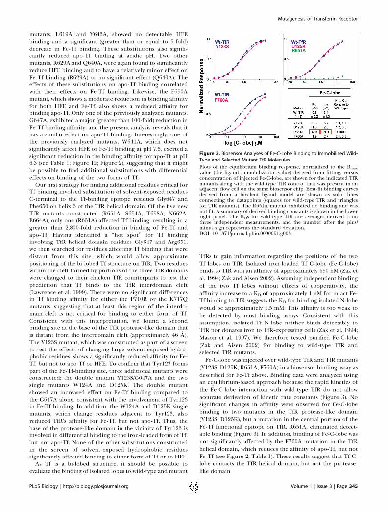
(Y123S, D125K, R651A, F760A) in a biosensor binding assay asdescribed for Fe-Tf above. Binding data were analyzed usingan equilibrium-based approach because the rapid kinetics ofthe Fe-C-lobe interaction with wild-type TfR do not allowaccurate derivation of kinetic rate constants (Figure 3). Nosignificant changes in affinity were observed for Fe-C-lobebinding to two mutants in the TfR protease-like domain(Y123S, D125K), but a mutation in the central portion of theFe-Tf functional epitope on TfR, R651A, eliminated detect-able binding (Figure 3). In addition, binding of Fe-C-lobe wasnot significantly affected by the F760A mutation in the TfRhelical domain, which reduces the affinity of apo-Tf, but notFe-Tf (see Figure 2; Table 1). These results suggest that Tf C-lobe contacts the TfR helical domain, but not the protease-like domain.
Figure 3. Biosensor Analyses of Fe-C-Lobe Binding to Immobilized Wild-
Type and Selected Mutant TfR Molecules
Plots of the equilibrium binding response, normalized to the Rmaxvalue (the ligand immobilization value) derived from fitting, versusconcentration of injected Fe-C-lobe, are shown for the indicated TfRmutants along with the wild-type TfR control that was present in anadjacent flow cell on the same biosensor chip. Best-fit binding curvesderived from a bivalent ligand model are shown as solid linesconnecting the datapoints (squares for wild-type TfR and trianglesfor TfR mutants). The R651A mutant exhibited no binding and wasnot fit. A summary of derived binding constants is shown in the lowerright panel. The KDs for wild-type TfR are averages derived fromthree independent measurements, and the number after the plus/minus sign represents the standard deviation.DOI: 10.1371/journal.pbio.0000051.g003
PLoS Biology | http://biology.plosjournals.org Volume 1 | Issue 3 | Page 345
Mutagenesis of Transferrin Receptor
Discussion
Despite many years of investigation of the Tf/TfR pathwayfor iron uptake, molecular details about the interactionbetween TfR and Tf have been limited largely due to a lack ofstructural information for a Tf/TfR complex. In the absenceof a three-dimensional structure, site-directed mutagenesiscan be used to map out a protein–protein interaction. Tonarrow down a subset of residues for mutageneis from the639 residues in a soluble TfR monomer, we used the crystalstructures of TfR alone (Lawrence et al. 1999) and TfR boundto HFE (Bennett et al. 2000) to locate solvent-exposedresidues in the vicinity of the HFE-binding site, which wassuggested from competition studies to overlap with the Tf-binding site on TfR (Lebron et al. 1999). We identifiedresidues within the TfR helical domain whose substitutionaffected binding of both HFE and Fe-Tf at pH 7.5 in aprevious mutagenesis study involving ten human TfR mutants(West et al. 2001). These results established that HFE and Fe-Tf bind to the same or an overlapping site on TfR. In thepresent study, we have expanded the library of TfR mutantsto more precisely map the Tf-binding site on TfR andcompared binding of Fe-Tf and apo-Tf to TfR. From a surveyof 29 point mutants of human TfR, we identified 11 residues,which, when substituted, reduce the affinity of TfR for eitherhuman Fe-Tf, apo-Tf, or both (see Table 1). Six of the 11residues are completely conserved in different species of TfRand in a more recently identified Tf-binding receptor, TfR2,which shares 45% sequence identity with TfR (Kawabata et al.1999). Most notably, four of the residues exerting the largesteffects on Fe-Tf binding, apo-Tf binding, or both (Leu619,Trp641, Gly647, and Arg651) are completely conserved acrossall currently known TfR and TfR2 sequences (see Table 1).Others, such as the tyrosines at positions 123 and 643, areeither conserved or conservatively substituted for phenyl-alanine in some TfR species. By contrast, of the 18 positionsat which substitutions did not significantly affect Tf binding,16 are not conserved, and two (Phe187 and Glu664) areconservatively substituted (see Table 1). These results suggestthat our conclusions about the mode of binding betweenhuman Tf and human TfR can be generalized to include Tf/TfR complexes from other species and the interactionbetween TfR2 and Tf.
From a quantitative analysis of the affinities of thedifferent TfR mutants for Fe-Tf and apo-Tf, we can classifythe residues we mutated using the criteria of Wells andcolleagues (Cunningham and Wells 1993), which categorizethe structural and functional epitope residues in a protein–protein interaction. The functional epitope is defined asresidues exerting a major effect on the binding affinity (a��G value, �2 kcal/mol after substitution of a single residue,corresponding to an affinity reduction of at least 30-fold atroom temperature). The structural epitope on a protein is allresidues at the contact interface with the binding partner,which can be deduced from a co-crystal structure (Cunning-ham and Wells 1993). Substitution of some, but not all, of theresidues at the structural epitope of a protein–proteininterface will result in affinity changes (Cunningham andWells 1993). This is illustrated in our study by comparing thecrystallographically-defined structural epitope on TfR forbinding HFE (Bennett et al. 2000) (see Figure 1A and 1B;Table 1; Figure S1) with the results of mutagenic mapping of
residues affecting HFE binding (see Figure 1C). In the absenceof a Tf/TfR co-crystal structure, we can use our mutagenesisresults to predict the functional and structural epitoperesidues (affinity reductions of greater than or equal to 30-fold or between 5- and 30-fold, respectively) on TfR forbinding to Fe-Tf and apo-Tf. From the comparison of wild-type and mutant TfR binding affinities, Arg651 was identifiedas a functional epitope residue for binding both Fe-Tf at pH7.5 and binding apo-Tf at pH 6.3, as substitution of this singleresidue to alanine greatly reduces binding to either form ofTf (see Table 1). In combination with the previously studiedG647A mutant (Dubljevic et al. 1999; West et al. 2001), whichreduces affinity for both Fe-Tf and apo-Tf by over 100-fold,these residues define a functional epitope for Fe-Tf and apo-Tf binding located in the bottom central portion of the TfRhelical domain (see Figure 1D and 1E). Two other nearbyresidues, Leu619 and Trp641, can be considered part of thefunctional epitope for binding apo-Tf. The HFE/TfR crystalstructure shows that these residues are at the contactinterface with HFE (see Figure 1A and 1B) (Bennett et al.2000), but with the exception of Leu619, their substitutionsdo not significantly affect HFE binding (see Table 1). Instead,the functional epitope for HFE binding is shifted slightlyupwards on the TfR helical domain from the Fe-Tf functionalepitope to include residues Leu619 and Tyr643 (see Figure1C). Thus, although most of the functional epitope residuesfor binding of HFE and Tf are physically separated, they areclose enough that binding of either HFE or Fe-Tf to TfRwould sterically preclude binding of the other species (seeFigure 1C and 1D). In addition, some substitutions in TfRsignificantly lower the affinity for both HFE and Tf (L619A,R629A, Y643A, and F650A) (see Table 1).Since Tf is a larger molecule than HFE, we reasoned that Tf
could also interact with residues not contained in the HFEbinding footprint on TfR. We therefore tested substitutionsof residues outside of the TfR helical domain for their effectson binding to Tf. To narrow down the search, we chose tosubstitute solvent-exposed hydrophobic residues, which areoften found in protein–protein interfaces (Jones and Thorn-ton 1996; Lo Conte et al. 1999). We also restricted the searchto residues within approximately 90 A (the longest dimensionof Fe-Tf) of the Fe-Tf functional-binding epitope forsubstitution. Using this strategy, we identified a region atthe base of the protease-like domain involving residuesTyr123, Trp124, and Asp125, where substitutions showedsignificant effects on binding to Fe-Tf at pH 7.5, but not toHFE at pH 7.5 or to apo-Tf at pH 6.3 (see Figure 1D and 1E;Table 1). Having defined two predicted Fe-Tf contact areason TfR that are separated by approximately 33 A (measuredbetween TfR residues Arg651 and Tyr123) constrains the waysin which Tf can interact with TfR. In particular, computermodeling suggests that a single Tf lobe cannot makeproductive contacts with both regions of TfR (A. M.Giannetti, unpublished data); thus both lobes of Fe-Tf arelikely to be involved in the interface with TfR. Previousstudies of the binding of isolated Fe-N- and Fe-C-lobes of Tfsuggested that the majority of the binding energy in the Tf/TfR interaction comes from the C-lobe (Zak et al. 1994; Zakand Aisen 2002). It has also been observed that mixingpurified N- and C-lobes results in a significant enhancementof TfR binding over that of C-lobe alone (Mason et al. 1997;Zak and Aisen 2002). These observations are consistent with a
PLoS Biology | http://biology.plosjournals.org Volume 1 | Issue 3 | Page 346
Mutagenesis of Transferrin Receptor
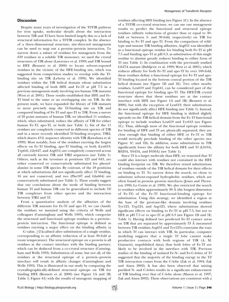
Tf orientation on TfR in which the C-lobe contacts the Tffunctional epitope on the TfR helical domain and the N-lobecontacts the Tyr123 area at the base of the TfR protease-likedomain (Figure 4). In this model, allosteric effects need not beinvoked to explain the increased affinity of the N-lobe/C-lobemixture over C-lobe alone (Zak and Aisen 2002). Instead, theobserved increase in affinity is predicted to arise from directcontacts between the N-lobe and TfR. To test the predictedorientation of Tf on TfR (Figure 4), we compared theaffinities of isolated Fe-C-lobe (Zak and Aisen 2002) to wild-type TfR and to TfR mutants with substitutions in the helicaldomain (R651A, F760A) and the protease-like domain (Y123S,D125K) (see Figure 3). As predicted, substitutions in theprotease-like domain do not affect binding of Fe-C-lobe,whereas a functional epitope substitution (R651A) in the TfRhelical domain eliminates detectable binding of Fe-C-lobe toTfR.
Our binding data also allow us to assess potential differ-ences in the binding of Fe-Tf versus apo-Tf to TfR. Two priorobservations are consistent with differences in the bindingfootprints of Fe-Tf and apo-Tf on TfR. First, Fe-Tf undergoesa large conformational change upon acidification and releaseof iron, as deduced by comparison of crystal structures offerric and iron-free forms of Tf and Tf-related moleculessuch as the lactoferrins (Bailey et al. 1988; Anderson et al.1989; Gerstein et al. 1993; Zuccola 1993; Kurokawa et al. 1995,1999; Baker et al. 1998; Karthikeyan et al. 1999) (see Figure 4).Second, TfR has been suggested to undergo a pH-dependentconformational change resulting in aggregation at pH ,6 inthe absence of Tf (Turkewitz et al. 1988). Our finding ofdifferential effects of TfR substitutions for binding Fe-Tf atpH 7.5 versus apo-Tf at pH 6.3 is consistent with conforma-tional changes in Tf,TfR, or both at acidic pH. We find oneTfR region that affects binding of Fe-Tf, but not apo-Tf (theregion near Tyr123 involving TfR residues 123–125 at thebase of the protease-like domain), and another region thataffects binding of apo-Tf, but not Fe-Tf or Fe-C-lobe (theregion defined by Trp641 and Phe760, two spatially proximalresidues [10.2 A apart] at the edge of the TfR helical domain)(see Figure 1E and 1F). The apo-Tf-specific binding site maybe important for TfR’s ability to significantly accelerate ironrelease from receptor-bound Fe-Tf (Bali and Aisen 1991).
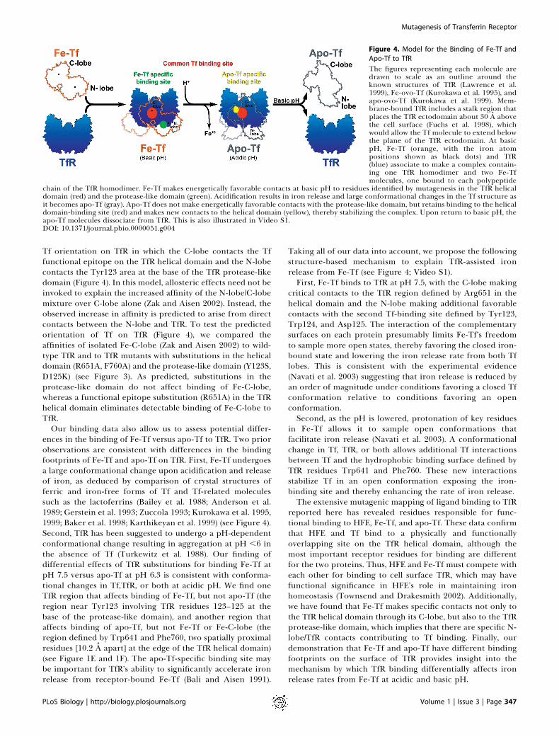
Taking all of our data into account, we propose the followingstructure-based mechanism to explain TfR-assisted ironrelease from Fe-Tf (see Figure 4; Video S1).First, Fe-Tf binds to TfR at pH 7.5, with the C-lobe making
critical contacts to the TfR region defined by Arg651 in thehelical domain and the N-lobe making additional favorablecontacts with the second Tf-binding site defined by Tyr123,Trp124, and Asp125. The interaction of the complementarysurfaces on each protein presumably limits Fe-Tf’s freedomto sample more open states, thereby favoring the closed iron-bound state and lowering the iron release rate from both Tflobes. This is consistent with the experimental evidence(Navati et al. 2003) suggesting that iron release is reduced byan order of magnitude under conditions favoring a closed Tfconformation relative to conditions favoring an openconformation.Second, as the pH is lowered, protonation of key residues
in Fe-Tf allows it to sample open conformations thatfacilitate iron release (Navati et al. 2003). A conformationalchange in Tf, TfR, or both allows additional Tf interactionsbetween Tf and the hydrophobic binding surface defined byTfR residues Trp641 and Phe760. These new interactionsstabilize Tf in an open conformation exposing the iron-binding site and thereby enhancing the rate of iron release.The extensive mutagenic mapping of ligand binding to TfR
reported here has revealed residues responsible for func-tional binding to HFE, Fe-Tf, and apo-Tf. These data confirmthat HFE and Tf bind to a physically and functionallyoverlapping site on the TfR helical domain, although themost important receptor residues for binding are differentfor the two proteins. Thus, HFE and Fe-Tf must compete witheach other for binding to cell surface TfR, which may havefunctional significance in HFE’s role in maintaining ironhomeostasis (Townsend and Drakesmith 2002). Additionally,we have found that Fe-Tf makes specific contacts not only tothe TfR helical domain through its C-lobe, but also to the TfRprotease-like domain, which implies that there are specific N-lobe/TfR contacts contributing to Tf binding. Finally, ourdemonstration that Fe-Tf and apo-Tf have different bindingfootprints on the surface of TfR provides insight into themechanism by which TfR binding differentially affects ironrelease rates from Fe-Tf at acidic and basic pH.
Figure 4. Model for the Binding of Fe-Tf and
Apo-Tf to TfR
The figures representing each molecule aredrawn to scale as an outline around theknown structures of TfR (Lawrence et al.1999), Fe-ovo-Tf (Kurokawa et al. 1995), andapo-ovo-Tf (Kurokawa et al. 1999). Mem-brane-bound TfR includes a stalk region thatplaces the TfR ectodomain about 30 A abovethe cell surface (Fuchs et al. 1998), whichwould allow the Tf molecule to extend belowthe plane of the TfR ectodomain. At basicpH, Fe-Tf (orange, with the iron atompositions shown as black dots) and TfR(blue) associate to make a complex contain-ing one TfR homodimer and two Fe-Tfmolecules, one bound to each polypeptide
chain of the TfR homodimer. Fe-Tf makes energetically favorable contacts at basic pH to residues identified by mutagenesis in the TfR helicaldomain (red) and the protease-like domain (green). Acidification results in iron release and large conformational changes in the Tf structure asit becomes apo-Tf (gray). Apo-Tf does not make energetically favorable contacts with the protease-like domain, but retains binding to the helicaldomain-binding site (red) and makes new contacts to the helical domain (yellow), thereby stabilizing the complex. Upon return to basic pH, theapo-Tf molecules dissociate from TfR. This is also illustrated in Video S1.DOI: 10.1371/journal.pbio.0000051.g004
PLoS Biology | http://biology.plosjournals.org Volume 1 | Issue 3 | Page 347
Mutagenesis of Transferrin Receptor
Materials and Methods
Preparation of TfR ligands. A soluble form of human HFE(residues 1–275 of the mature protein noncovalently associated withthe light chain b2-microglobulin) was expressed and purified aspreviously described (Lebron et al. 1998). Human Fe-Tf was preparedfrom apo-Tf (Sigma, St. Louis, Missouri, United States) by incubationwith bicarbonate and excess ferric ammonium sulfate. Free iron wasremoved by dialysis, and the protein was further purified by gel-filtration chromatography. Iron saturation was 100% as determinedspectrophotometrically (A465/A280, ;0.05) (He and Mason 2002).Purified recombinant Fe-C-lobe was cleaved from a full-length Fe-Tf in which the loop that connects the N- and C-lobes was replacedwith a Factor Xa site (Zak and Aisen 2002). Concanavalin Achromatography was used to separate the glycosylated C-lobe fromunglycosylated N-lobe (Zak and Aisen 2002). Protein concentrationswere determined from the A280- value using extinction coefficients of52,200 M�1 cm�1 (Fe-C-lobe) (O. Zak, personal communication),83,360 M�1 cm�1 (Tf), and 96,570 M�1 cm�1 (HFE/b2-microglobulin)(Lebron et al. 1998).
Production of wild-type TfR and TfR mutants. Soluble human TfRand TfR mutants were expressed in a lytic baculovirus/insect cellexpression system as previously described (Lebron et al. 1998).Mutations were introduced through PCR mutagenesis (Quickchange,Strategene, La Jolla, California, United States) into a baculovirusexpression vector (pACGP67A; Pharmingen, San Diego, California,United States) containing a hydrophobic leader sequence, 6x-His tag,Factor Xa site, and residues 121–760 of human TfR. All mutationswere confirmed by DNA sequencing of the protein-coding region ofthe vector. The Y123S mutation was further confirmed by N-terminalsequencing of the purified mutant protein, yielding the sequenceADPHHHHHHSSGIEGRGEFRLSWDD (the serine substitution fortyrosine is underlined), corresponding to residual leader sequenceresidues (A), vector-encoded sequence (DP), the 6x-His tag, spacerresidues (SSG), a Factor Xa site (IEGR), a spacer segment (GEF), andresidues 121–126 of the mutant TfR (RLSWDD). The double mutantY123S/G647A was constructed by introducing the Y123S substitutioninto the G647A–TfR expression construct, after which the protein-coding region of the expression plasmid was again sequenced.Recombinant viruses were generated by cotransfection of a transfervector with linearized viral DNA (Baculogold, Pharmingen). Super-natants of baculovirus-infected High 5 cells were used as the source ofwild-type TfR and TfR mutants for surface plasmon resonance-basedaffinity measurements.
Affinity measurements. We used a BIACORE 2000 biosensorsystem (Pharmacia, LKB Biotechnology, Uppsalla, Sweden) to assaythe interaction between TfR and HFE, Fe-Tf, and apo-Tf as described(West et al. 2001). Binding of injected proteins (the analytes wereHFE, Fe-Tf, or apo-Tf) to a protein immobilized on the sensor chip(the ligand was TfR) results in changes in surface plasmon resonancethat are read out in real time as resonance units (RUs) (Fagerstam etal. 1992; Malmqvist 1993).
For each experiment, the four flow cells of a CM5 biosensor chip(Pharmacia) were prepared by covalently attaching an anti-His-tagantibody (anti-PentaHis; Qiagen, Valencia, California, United States)to a coupling density of 2,000–4,000 RUs through standard aminecoupling chemistry (BIACORE manual). Insect cell supernatants (50–300 ll) containing secreted 6x-His-tagged wild-type or mutant TfRwere passed through a 0.2 lm filter and injected over one of the fourflow cells of a biosensor chip at a flow rate of 30 ll/min, resulting instable binding of TfR to density of 200–400 RUs. In a typicalexperiment, a small amount of TfR immediately dissociates from theanti-His antibody, but most TfR protein (.85%) remains boundduring the course of the injection of the TfR ligands, resulting in anegligible baseline drift. On each biosensor chip, one flow cellcontaining only the immobilized antibody was used as the referencecell, one cell containing wild-type TfR served as an internal controlfor binding of the three TfR binding partners, and TfR mutants werecoupled to the other two flow cells. HFE or Fe-Tf was injected overthe flow cells at 50 ll/min or 70 ll /min, respectively, at 258 C in 50mM PIPES (pH 7.5), 150 mM NaCl, and 0.005% surfactant P20 (v/v).All analyte injections were made as serial 2- or 3-fold dilutions. TheHFE concentration series ranged from 30 nM to 10 lM, and the Fe-Tfand apo-Tf injections typically spanned from 1 nM to 200 nM, exceptfor experiments involving low-affinity mutants requiring higherconcentrations to properly derive affinities (see legend to Figure 2).In test experiments, the sensorgrams from duplicate injections couldbe overlaid to within the experimental noise; thus, single injectionswere done for each concentration of injected protein in a bindingexperiment. Between successive injections of analytes, the chips were
regenerated to preinjection response levels by either flowing withrunning buffer until baseline was achieved (in the case of HFE) or bya 12-second injection of the injection buffer containing 0.5 M MgCl2(in the case of Fe-Tf). This treatment did not cause dissociation ofTfR from the anti-His-tag antibody. Apo-Tf was injected in 50 mMPIPES (pH 6.3), 150 mM NaCl, 0.005% surfactant P20 (v/v), including50 lM desferrioxamine as an iron chelator, and chip regenerationwas achieved with an injection of the same buffer at pH 7.5.
Raw sensorgram data were preprocessed using the Scrubbersoftware package (BioLogic Software, Campbell, Australia; www.bio-logic.com.au). The response from the reference flow cell wassubtracted from the experimental flow cells to eliminate bulkrefractive index changes. The response from the average of at leastthree buffer-only injections was then subtracted to correct forpotential systematic instrument artifacts. Kinetic constants wereobtained by simultaneous fitting of the association and dissociationphases of all curves in the working set using the program Clamp99(Morton and Myszka 1998). The data were fit to a bivalent ligandmodel, which describes the two sequential binding events for eitherTf or HFE binding to homodimeric TfR. A simple 1:1 binding modeldid not account for the observed data as judged from large residualsin the fits (data not shown). Equilibrium dissociation constants (KDs)were calculated from the ratio of the dissociation and association rateconstants, koff (s
�1) and kon (M�1�s�1), respectively, yielding KDs for the
first and second binding events (KD1 and KD2) in the followingreaction mechanism:
Aþ TfR $ A:TfR ðrate constants: kon;1 and koff;1ÞKD1 ¼ koff;1=kon;1
Aþ A: TfR $ A2 :TfR ðrate constants: kon;2 and koff;2ÞKD2 ¼ koff;2=kon;2
where A is either HFE, Fe-Tf, or apo-Tf. For independent bindingsites, the apparent stepwise equilibrium dissociation constants (KD1and KD2) are related to the intrinsic binding constants for the firstand second binding events to TfR (KD,intrinsic and KD,intrinsic), asfollows:
KD1;intrinsic ¼ KD1=2
KD2;intrinsic ¼ 2KD2
Hence, if the binding of a TfR ligand is independent of whether aligand is bound on the other face of the TfR homodimer, KD2¼ 4KD1.
For each mutant, the relative effect on HFE, Fe-Tf, or apo-Tfbinding was calculated as a ratio between the mutant KD and theaverage of 22 independent determinations of the wild-type KD (seeTable 1) and as a ratio between the mutant KD and the wild-type KDderived from wild-type protein coupled to a flow cell on the samesensor chip as the mutant (data not shown). No significant differenceswere found for the two methods of calculating the ratios. All mutantswere evaluated for HFE and Fe-Tf binding in at least twoindependent experiments. For apo-Tf binding, those mutants thatshowed a significant difference in binding compared to wild-type TfRwere reevaluated in a separate, independent experiment. Nosignificant differences in KDs were observed in independentdeterminations of mutant affinities. When accurate affinities couldnot be derived in a duplicate experiment due to problems withbaseline drift, visual inspection of the sensorgrams demonstrated thateach mutant exerted the same relative effects compared with wild-type TfR in independent binding experiments. Table 1 presentsaffinities derived from one binding experiment per mutant/ligandpair. The reproducibility of the binding experiments can be assessedby the standard deviation of the wild-type TfR affinity for each of theligands (derived from 22 independent binding experiments) and fromthe fact that the affinities of many of the mutants are not significantlychanged compared to wild-type TfR.
For binding interactions involving the Fe-C-lobe, which reachequilibrium quickly, we derived KDs using an equilibrium-basedapproach. In these experiments, KDs were derived by non-linearregression analysis of plots of Req (the equilibrium binding response)versus the log of the analyte concentration. The data were fit to abinding model assuming a bivalent ligand in BIAevaluation 3.0(BIACORE). We were unable to detect significant amounts of bindingbetween apo-C-lobe and wild-type TfR at pH 6.3, presumably due toan intrinsically weak binding affinity.
PLoS Biology | http://biology.plosjournals.org Volume 1 | Issue 3 | Page 348
Mutagenesis of Transferrin Receptor
Supporting Information
Figure S1. ��G for Mutant TfR Binding to HFE, Fe-Tf, and Apo-TfHistogram of ��G values for the change relative to wild-type TfR inTfR mutant affinities for HFE (blue), Fe-Tf (pink), and apo-Tf (gray).��G values (the difference in binding energy for a mutant TfRcompared to wild-type TfR) were calculated using the KD1 valuesfrom Table 1 as ��G¼�RTln(KD1,mut/KD1,wild-type), where R is the gasconstant (1.99 3 10�3 kcal mol�1 K�1), and T is the temperature indegrees Kelvin (298 K). The dashed green line represents the cutofffor TfR mutants with a greater than or equal to 5-fold affinityreduction in ligand binding, and the dashed red line indicates agreater than or equal to 30-fold affinity reduction. An orange starindicates non-binding mutants and mutants with a greater than 160-fold affinity reduction whose ��G values exceed the y-axis limit ofthe histogram (L619A and Y643A, �4 kcal/mol; G647A¼ 3.2 kcal/mol;and R651A, �4.6 kcal/mol).
View online at DOI: 10.1371/journal.pbio.0000051.sg001 (1.86 MBTIFF).
Video S1. Model of TfR-Assisted Iron Release from Fe-TfView online at DOI: 10.1371/journal.pbio.0000051.sv001 (12 MBMOV).
Accession Numbers
The SwissProt accessions numbers for the proteins discussed inthis paper are b2-microglobulin (P01884), Fe-Tf (P02787), HFE
(Q30201), TfR canine (Q9GLD3), TfR chicken (Q90997), TfR feline(Q9MYZ3), TfR hamster (Q07891), TfR human (P02786), TfR mouse(Q62351), TfR rat (Q99376), TfR2 human (Q9UP52), and TfR2 mouse(Q62351).
Acknowledgments
This work was supported by grants from the National Institutes ofHealth (1-R01-DK60770 to PJB and DK-15056 to Dr. Philip Aisen) andan National Research Service Award predoctoral training grant(5T32-GM-7616 to AMG). We are grateful to Inderjit Nangiana andCynthia Jones (Caltech Protein Expression Facility) for assistance inexpressing TfR proteins and to the Caltech Protein/Peptide Micro-Analytical Laboratory for protein sequencing. We thank Drs.Anthony West, Andy Herr, Caroline Enns, and Anne B. Mason forhelpful discussions and members of the Bjorkman lab for criticalreading of the manuscript. We give a special thanks to D. G. Myszkafor beta versions of Clamp and Scrubber and discussions ofBIACORE experimental details.
Conflicts of interest. The authors have declared that no conflicts ofinterest exist.
Author contributions. AMG and PJB conceived and designed theexperiments. AMG performed the experiments. AMG and PJBanalyzed the data. AMG, PMS, and OZ contributed reagents/materials/analysis tools. AMG and PJB wrote the paper. AMGdeveloped and produced the animated figure. &
ReferencesAnderson BF, Baker HM, Norries GE, Rice DW, Baker EN (1989) Structure of
human lactoferrin: Crystallographic structure analysis and refinement at 2.8A resolution. J Mol Biol 209: 711–734.
Bailey S, Evans RW, Garratt RC, Gorinsky B, Hasnain S, et al. (1988) Molecularstructure of serum transferrin at 3.3 A resolution. Biochemistry 27: 5804–5812.
Baker EN, Anderson BF, Baker HM, MacGillivray RTA, Moore SA, et al. (1998)Three-dimensional structure of lactoferrin: Implications for function,including comparisons with transferrin. Adv Exp Med Biol 443: 1–14.
Bali PK, Aisen P (1991) Receptor-modulated iron release from transferrin:Differential effects on N- and C-terminal sites. Biochemistry 30: 9947–9952.
Bali PK, Aisen P (1992) Receptor-induced switch in site-site cooperativityduring iron release by transferrin. Biochemistry 31: 3963–3967.
Bali PK, Zak O, Aisen P (1991) A new role for the transferrin receptor in therelease of iron from transferrin. Biochemistry 30: 324–328.
Bennett MJ, Lebron JA, Bjorkman PJ (2000) Crystal structure of the hereditaryhaemochromatosis protein HFE complexed with transferrin receptor.Nature 403: 46–53.
Buchegger F, Trowbridge IS, Liu L-FS, White S, Collawn JF (1996) Functionalanalysis of human/chicken transferrin receptor chimeras indicates that thecarboxy-terminal region is important for ligand binding. Eur J Biochem 235:9–17.
Cullen LM, Anderson GJ, Ramm GA, Jazwinska EC, Powell LW (1999) Geneticsof hemochromatosis. Annu Rev Med 50: 87–98.
Cunningham BC, Wells JA (1993) Comparison of a structural and a functionalepitope. J Mol Biol 234: 554–563.
Dautry-Varsat A, Ciechanover A, Lodish HF (1983) pH and the recycling oftransferrin during receptor-mediated endocytosis. Proc Natl Acad Sci U S A80: 2258–2262.
Dubljevic V, Sali A, Goding A (1999) A conserved RGD (Arg–Gly–Asp) motif inthe transferrin receptor is required for binding to transferrin. Biochem J341: 11–14.
Enns CA (2002) The transferrin receptor. In: Templeton DM, editor. Molecularand cellular iron transport. New York: Marcel Dekker. pp. 71–94.
Enns CA, Sussman HH (1981) Physical characterization of the transferrinreceptor in human placentae. J Biol Chem 256: 9820–9823.
Enns CA, Rutledge EA, Williams AM (1996) The transferrin receptor. In:Biomembranes. New York: JAI Press. pp. 255–287.
Fagerstam LG, Frostell-Karlsson A, Karlsson R, Persson B, Ronnber I (1992)Biospecific interaction analysis using surface plasmon resonance detectionapplied to kinetic, binding site and concentration analysis. J Chromatogr597: 397–410.
Feder JN, Gnirke A, Thomas W, Zsuchihashi Z, Ruddy DA, et al. (1996) A novelMHC class I-like gene is mutated in patients with hereditary haemochro-matosis. Nat Genet 13: 399–408.
Feder JN, Penny DM, Irrinki A, Lee VK, Lebron JA, et al. (1998) Thehemochromatosis gene product complexes with the transferrin receptor,and lowers its affinity for ligand binding. Proc Natl Acad Sci U S A 95: 1472–1477.
Fuchs H, Luchken U, Tauber R, Engel A, Gessner R (1998) Structural model ofphospholipid-reconstituted human transferrin receptor derived by electronmicroscopy. Structure 6: 1235–1243.
Gerstein M, Anderson BF, Norris GE, Baker EN, Lesk AM, et al. (1993) Domainclosure in lactoferrin: Two hinges produce a see-saw motion betweenalternative close-packed interfaces. J Mol Biol 234: 357–372.
Grossmann JG, Neu M, Pantos E, Schwab FJ, Evans RW, et al. (1992) X-raysolution scattering reveals conformational changes upon iron uptake inlactoferrin, serum and ovo-transferrins. J Mol Biol 225: 811–819.
Grossmann JG, Neu M, Evans RW, Lindley PF, Appel H, et al. (1993) Metal-induced conformational changes in transferrins. J Mol Biol 229: 585–590.
He Q-Y, Mason AB (2002) Molecular aspects of release of iron from transferrin.In: Templeton DM, editor. Molecular and cellular iron transport. New York:Marcel Dekker. pp. 95–124.
Jones S, Thornton JM (1996) Principles of protein–protein interaction. ProcNatl Acad Sci U S A 93: 13–20.
Karthikeyan S, Paramasivam M, Yadav S, Srinivasan A, Singh TP (1999)Structure of buffalo lactoferrin at 2.5 A resolution using crystals grown at303 K shows different orientations of the N and C lobes. Acta Crystallogr DBiol Crystallogr 55: 1805–1813.
Kawabata H, Yang R, Hirama T, Vuong PT, Kawno S, et al. (1999) Molecularcloning of transferrin receptor 2. J Biol Chem 274: 20826–20832.
Kraulis PJ (1991) MolScript: A program to produce both detailed and schematicplots of protein structures. J Appl Crystallogr 24: 946–950.
Kurokawa H, Mikami B, Hirose M (1995) Crystal structure of diferric henovotransferrin at 2.4 A resolution. J Mol Biol 254: 196–207.
Kurokawa H, Dewan JC, Mikami B, Sacchettini JC, Hirose M (1999) Crystalstructure of hen apo-ovotransferrin: Both lobes adopt an open conforma-tion upon loss of iron. J Biol Chem 274: 28445–28452.
Lawrence CM, Ray S, Babyonyshev M, Galluser R, Borhani DW, et al. (1999)Structure of the ectodomain of human transferrin receptor. Science 286:779–782.
Lebron JA, Bennett MJ, Vaughn DE, Chirino AJ, Snow PM, et al. (1998) Crystalstructure of the hemochromatosis protein HFE and characterization of itsinteraction with transferrin receptor. Cell 93: 111–123.
Lebron JA, West AP, Bjorkman PJ (1999) The hemochromatosis protein HFEcompetes with transferrin for binding to the transferrin receptor. J Mol Biol294: 239–245.
Lo Conte L, Chothia C, Janin J (1999) The atomic structure of protein–proteinrecognition sites. J Mol Biol 285: 2177–2198.
Malmqvist M (1993) Biospecific interaction analysis using biosensor technology.Nature 361: 186–187.
Mason AB, Tam BM, Woodworth RC, Oliver RW, Green BN, et al. (1997)Receptor recognition sites reside in both lobes of human serum transferrin.Biochem J 326: 77–85.
Merritt EA, Bacon DJ (1997) Raster3D: Photorealistic molecular graphics.Methods Enzymol 277: 505–524.
Morton TA, Myszka DG (1998) Kinetic analysis of macromolecular interactionsusing surface plasmon resonance. Methods Enzymol 295: 268–294.
Navati MS, Samuni U, Aisen P, Friedman JM (2003) Binding and release of ironby gel-encapsulated human transferrin: Evidence for a conformationalsearch. Proc Natl Acad Sci U S A 100: 3832–3837.
Nicholls A, Bharadwaj R, Honig B (1993) GRASP: Graphical representation andanalysis of surface properties. Biophys J 64: A166–A166.
Parkkila S, Waheed A, Britton RS, Bacon BR, Zhou XY, et al. (1997) Associationof the transferrin receptor in human placenta with HFE, the protein
PLoS Biology | http://biology.plosjournals.org Volume 1 | Issue 3 | Page 349
Mutagenesis of Transferrin Receptor

defective in hereditary hemochromatosis. Proc Natl Acad Sci U S A 94:13198–13202.
Sipe DM, Murphy RF (1991) Binding to cellular receptor results in increasediron release from transferrin at mildly acidic pH. J Biol Chem 266: 8002–8007.
Townsend A, Drakesmith H (2002) Role of HFE in iron metabolism, hereditaryhaemochromatosis, anaemia of chronic disease, and secondary iron over-load. Lancet 359: 786–790.
Tsai CJ, Lin SL, Wolfson HJ, Nussinov R (1997) Studies of protein–proteininterfaces: A statistical analysis of the hydrophobic effect. Protein Sci 6: 53–64.
Turkewitz AP, Schwartz AL, Harrison SC (1988) A pH-dependent reversible
conformational transition of the human transferrin receptor leads to self-association. J Biol Chem 263: 16309–16315.
West AP Jr, Giannetti AM, Herr AB, Bennett MJ, Nangiana JS, et al. (2001)Mutational analysis of the transferrin receptor reveals overlapping HFE andtransferrin binding sites. J Mol Biol 313: 385–397.
Zak O, Aisen P (2002) A new method for obtaining human transferrin C-lobe inthe native conformation: Preparation and properties. Biochemistry 41:1647–1653.
Zak O, Trinder D, Aisen P (1994) Primary receptor-recognition site of humantransferrin is in the C-terminal lobe. J Biol Chem 269: 7110–7114.
Zuccola HJ (1993) The crystal structure of monoferric human serumtransferrin [dissertation]. Atlanta, Georgia: Georgia Institute of Technology.
PLoS Biology | http://biology.plosjournals.org Volume 1 | Issue 3 | Page 350
Mutagenesis of Transferrin Receptor