both protein dynamics and ligand concentration can … protein dynamics and ligand concentration can...
TRANSCRIPT

Both protein dynamics and ligand concentration canshift the binding mechanism between conformationalselection and induced fitNicholas Greives and Huan-Xiang Zhou1
Department of Physics and Institute of Molecular Biophysics, Florida State University, Tallahassee, FL 32306
Edited by J. Andrew McCammon, University of California, San Diego, La Jolla, CA, and approved June 9, 2014 (received for review April 25, 2014)
This study aimed to shed light on the long debate over whetherconformational selection (CS) or induced fit (IF) is the governingmechanism for protein–ligand binding. The main difference be-tween the two scenarios is whether the conformational transitionof the protein from the unbound form to the bound form occursbefore or after encountering the ligand. Here we introduce the IFfraction (i.e., the fraction of binding events achieved via IF), toquantify the binding mechanism. Using simulations of a model pro-tein–ligand system, we demonstrate that both the rate of the con-formational transition and the concentration of ligand moleculescan affect the IF fraction. CS dominates at slow conformationaltransition and low ligand concentration. An increase in either quan-tity results in a higher IF fraction. Despite the many-body nature ofthe system and the involvement of multiple, disparate types ofdynamics (i.e., ligand diffusion, protein conformational transition,and binding reaction), the overall binding kinetics over wide rangesof parameters can be fit to a single exponential, with the apparentrate constant exhibiting a linear dependence on ligand concentra-tion. The present study may guide future kinetics experiments anddynamics simulations in determining the IF fraction.
protein–ligand complex | conformational dynamics | diffusion-influencedbimolecular reaction | induced-fit fraction
The binding of proteins to small molecules (i.e., ligands) iscentral to many essential biological functions, including en-
zyme catalysis, receptor activation, and drug action. Generally,significant differences in protein conformation exist between theunbound and bound states, as exemplified by hemoglobin uponbinding oxygen (1–4) and HIV-1 protease upon binding a substrateor a drug molecule (5). In the latter as well as some other cases(6–10), loops and other groups collapse around the bound ligand,leading to a closed binding pocket. The conformational redis-tribution and dynamics of the protein molecule exhibited during thebinding process can potentially play a critical role in determining themagnitude of the rate constant as well as the mechanism of ligandbinding (11, 12). Two mechanistic models have emerged as arche-types. In the induced-fit (IF) model, one assumes that, owing tointeractions with the incoming ligand, the protein transitions froman “inactive” conformation to an “active” conformation (13). In theconformational-selection (CS) model, one assumes that the proteincan preexist in the active conformation with a low probability, and itis when the protein is in this conformation that the ligand comesinto contact, leading to productive binding (14). Both models havegarnered defenders and detractors (15–19). This study aimed toshed light on the long debate over whether CS or IF is the gov-erning mechanism for protein–ligand binding.It has been suggested that observation of the active confor-
mation without the ligand, akin to constitutive activity of re-ceptors, is direct evidence of CS (17, 19). However, detractors ofCS have noted that, at least for cases with a closed bindingpocket in the active conformation, direct binding to the latterconformation cannot proceed (9, 15). In some cases, a partiallyclosed conformation has been observed by a sensitive probe suchas paramagnetic relaxation enhancement (20) or in molecular
dynamics simulations. Accordingly, a revised model known asextended CS has been put forward (21–28), whereby the ligandbinds to the partially closed conformation and then the protein–ligand system evolves to the bound state with the closed bindingpocket. Although the divide between CS and IF is somewhatblurred by extended CS, strictly speaking the latter is an IFmodel, in the sense that the ligand binds to an inactive confor-mation (i.e., the partially closed conformation) before the pro-tein adopts the final active conformation with the closed bindingpocket. Indeed, a strict CS mechanism is not possible for a pro-tein whose active conformation features a closed binding pocket.In any event, mere observation of the active conformation in theunbound state cannot be taken as proof of the CS mechanism.According to the Boltzmann distribution, every conformation, in-cluding the active conformation, has a certain equilibrium proba-bility. Whether the active conformation can be observed dependson the magnitude of its equilibrium probability as well as thesensitivity of the probe. The binding mechanism should notchange just because the probe has become more sensitive.It thus seems that neither CS nor IF should be the sole dominant
mechanism governing protein–ligand binding. What, then, are thedeterminants of binding mechanism? Hammes et al. (29) andDaniels et al. (30) have suggested that an increase in ligand con-centration can shift the binding mechanism from CS to IF, becausea higher ligand concentration would make binding more likely. Theassumption is that that would increase the chance for the binding tooccur before the conformational transition, but one cannot becertain without additional information about the dynamics andinteractions of the protein and ligand molecules. Others have sug-gested that the timescale of the protein conformational transition,relative to the timescale of the ligand diffusional approach to thebinding pocket, controls the binding mechanism (12), but the effectof ligand concentration was not studied.
Significance
Mechanisms of protein–ligand binding have long generatedinterest in wide circles, including enzyme catalysis, receptorfunction, and drug design. This study reconciles some of theconflicting views. Using simulations of a model protein–ligandsystem, we demonstrate that neither conformational selection(CS) nor induced fit (IF) is the sole mechanism governing pro-tein–ligand binding. Instead, both intrinsic and extrinsic factorscan shift the binding mechanism between CS and IF. CS domi-nates when both protein conformational transition rate andligand concentration are low. With the increase of either fac-tor, the binding mechanism shifts to IF. The rigorous theoreticalframework established here can guide experiments and simu-lations in determining the fraction of binding events achievedvia either CS or IF.
Author contributions: H.-X.Z. designed research; N.G. performed research; N.G. and H.-X.Z.analyzed data; and N.G. and H.-X.Z. wrote the paper.
The authors declare no conflict of interest.
This article is a PNAS Direct Submission.1To whom correspondence should be addressed. E-mail: [email protected].
www.pnas.org/cgi/doi/10.1073/pnas.1407545111 PNAS | July 15, 2014 | vol. 111 | no. 28 | 10197–10202
BIOPH
YSICSAND
COMPU
TATIONALBIOLO
GY

To unequivocally determine the binding mechanism, one hasto follow the protein–ligand relative translation and the proteininternal motion, from the unbound state until two reactantmolecules form the bound product. This process involves dis-parate types of dynamics, including ligand diffusion, proteinconformational transition, and the final binding reaction. As thesimplest model, protein conformational transition has been treatedas gating, that is, the transitions between two conformational statesare approximated as rate processes (31–33). The transition rateswere initially assumed to be unaffected by protein–ligand inter-actions. More recently it was recognized that protein–ligand in-teractions necessarily influence the conformational transitionrates and such influence is an essential ingredient of molecularrecognition (12, 34, 35). Accordingly, the transition rates wereassigned different values depending on whether the ligand isinside or outside the binding pocket, resulting in the dual-transition-rates model.Here we studied the binding mechanism and kinetics of a sys-
tem consisting of a concentration of ligand molecules surroundinga protein molecule whose conformational dynamics follows thedual-transition-rates model (Fig. 1A). From dynamics simulations,we calculate the IF fraction (i.e., the fraction of binding eventsachieved via IF) and show that the binding mechanism is shiftedby both the rate of protein conformational transition and theconcentration of ligand molecules. CS dominates at slow confor-mational transition and low ligand concentration. With the in-crease of either quantity, the binding mechanism shifts from CS toIF. The overall binding kinetics over wide ranges of parameterscan be fit to a single exponential, with the apparent binding rateconstant exhibiting a linear dependence on ligand concentration.The concentration dependence of the binding kinetics thus yieldslittle information on the binding mechanism, but kinetics experi-ments and dynamics simulations can be designed to determine theIF fraction.
Results and DiscussionThe Protein–Ligand System. Our model system, illustrated in Fig.1A, has the essential ingredients of ligand-binding proteins thatundergo conformational transitions. Its simplicity allowed us tocomprehensively explore the determinants of binding mecha-nism. The protein is modeled as an immobile sphere (radius R)with a thin-shell binding pocket (inner and outer radii R and R1 =1.1R) that fluctuates between an inactive and active state. Theligand molecules, totaling N and each treated as a point-likeparticle, are confined between the protein surface (at radial dis-tance r = R) and an outer wall (at r = Rw = 11R), mimicking thetypical experimental condition with ligand excess. The resultingligand concentration is C=N=½4πðR3
w −R3Þ=3�. The basic setup ofthe system is similar to one in a previous study (33), but with thekey difference that the transition rates between the inactive and
active conformations depend on the positions of the ligand mol-ecules, as further described below.Each ligand molecule undergoes diffusion (with diffusion con-
stantD) and interacts with the protein, giving rise to an interactionenergy UsðrÞ, where s = a or i for the active or inactive state and rdenotes the position of the ligand molecule. UiðrÞ is assumed to beuniformly 0, whereas UaðrÞ is assumed to switch smoothly butsharply from 0 when r > R1 to U0 when R < r < R1:
UaðrÞ=−U0ftanh½ðr−R1Þ=L�− 1g=2:
The parameter L (set to 0.005R) measures the sharpness of theswitch. We will refer to a ligand molecule with R < r < R1 asloosely bound. The ligand molecules do not have direct interac-tions with each other. Hereafter length and time will be reportedin units of R and R2/D, respectively.As already pointed out, we model the transitions of the protein
between the inactive and active conformations as rate processes,with transition rates ω+ and ω–:
Pi �ω+
ω−Pa:
We use ω∞± to denote the values of the transition rates when all ofthe ligand molecules are far away from the protein. In that situa-tion, the equilibrium constant between the active and inactive con-formations is ω∞+/ω∞–. When the ligand molecules are at positionsfrig, the new equilibrium constant, given by the ratio of the tran-sition rates, should satisfy the following detailed-balance relation:
ω+ðfrigÞω−ðfrigÞ=
ω∞+e−βP
iUaðriÞ
ω∞−e−βP
iUiðriÞ
;
where β is the inverse of the product of the Boltzmann constantand the absolute temperature. This generalizes a similar relationwhen only a single ligand molecule is present (12, 34). For simplic-ity, we set ω−ðfrigÞ=ω∞−. Correspondingly, the inactive-to-activetransition rate carries all of the influence of the protein–ligandinteractions. We chose ω∞+/ω∞– = 0.1 and exp(–βU0) = 100, suchthat the equilibrium constant favors the inactive conformationwhen all of the ligands are far away but favors the active confor-mation when a ligand molecule is loosely bound. Such a shift inthe more stable conformation from the inactive to the active istypical of ligand-binding proteins.When the protein molecule is in the active conformation,
a loosely bound ligand molecule has a chance to react with theprotein to form the final complex. We model this last step also asa rate process (with rate constant γ), as done previously (33, 36,37). We chose γ = 10; for a single protein–ligand pair with such
R
R1
Rw
A B
t
c
c tr
t te tr
inac�ve
ac�ve
Fig. 1. The model protein–ligand system and itsbinding mechanism. (A) A spherical protein issurrounded by point-like ligand molecules insidea spherical container (with radius Rw). The proteincan transition between an inactive conformationand active conformation, and the transition ratesdepend on whether a ligand molecule is in thebinding pocket (with inner and outer radii Rand R1, respectively). (B) A binding event ach-ieved through either the conformational selection(Upper) or the induced fit (Lower) mechanism.The crucial difference is whether the last inactive-to-active transition (at time tc) before the bindingreaction (at tr) occurs with or without a looselybound ligand molecule.
10198 | www.pnas.org/cgi/doi/10.1073/pnas.1407545111 Greives and Zhou

a parameter value the binding process can be described as largelydiffusion-controlled (as opposed to reaction-controlled) (34).Overall the binding process involves three different types of
dynamics: ligand diffusion, protein conformational transition, andthe final binding reaction. Even though the ligand molecules donot directly interact with each other, they experience any confor-mational transition of the protein at the same time. The latterevent introduces coupling between the ligand molecules (33), andas a result our protein–ligand system is many-body in nature. Wecarried out dynamics simulations of the system for ω– from 2 × 10⁻³
to 2 × 10² and for N from 5 to 100. In real units, the corre-sponding half-lives of the active conformation would be roughly0.01 ms to 0.1 ns, and the corresponding ligand concentrationswould be 0.2–4 mM. Each simulation was followed until eithera binding reaction or a cutoff time. In the former case the time ofits occurrence and whether it occurred via CS or IF were recorded.
Both Protein Dynamics and Ligand Concentration Can Shift the BindingMechanism. We use a strict definition of CS in this study (Fig. 1B,Upper). That is, a binding event is classified as CS only if no ligandmolecule was inside the binding pocket when the protein transi-tioned to the active conformation for the last time (at tc) beforereaction (at tr). The binding reaction would have to be between theprotein and a ligand molecule that entered the binding pocket atsome time (te) between tc and tr. Before tc, the protein could havemade multiple transitions between the inactive and active con-formations; between tc and te, one or more ligand molecules couldhave entered and left the binding pocket.The alternative is that a ligand molecule first entered the
binding pocket and the protein then made the last transition tothe active conformation (Fig. 1B, Lower). The subsequent bindingevent is classified as IF. Between tc and tr, the loosely bound ligand
molecule could have left and reentered the binding pocket, orcould even be replaced by another ligand molecule. Irrespective ofthese nuances, the fact remains that the final conformational tran-sition was caused by a loosely bound ligand molecule (thereby pre-paring the protein for the binding reaction), and hence an IFclassification is appropriate.From simulations for the transition rate ω– from 2 × 10⁻³ to 2 ×
102 and for the number N of ligand molecules from 5 to 100, wedetermined the fraction, ΦIF, of binding reactions classified as IF.The results are shown as symbols in Fig. 2A. At a given N (or,equivalently, ligand concentration), ΦIF increases with increas-ing ω−. For example, at N = 5, ΦIF increases from 0.10 at ω– = 2 ×10⁻³ to 0.99 at ω– = 2 × 10². As N increases, the IF fractionincreases rapidly at the lowest ω– and less so at intermediate ω–,and shows little variation at ω– ≥ 1. Correspondingly, the shift fromCS to IF upon increasing ω– becomes more and more gradual.We were able to fit the dependence of ΦIF on ω– to the fol-
lowing function:
ΦIFðω−Þ= 1=½1+ ðωtr=ω−Þm�ν; [1]
where ωtr would be the midpoint of the shift from CS to IF if ν = 1,m directly controls the sharpness of the shift, and a ν that is lessthan 1 serves to raise ΦIF at ω– << ωtr and thus make the transitionto IF less gradual (Fig. 2A, curves). All of the three parametershave strong dependences on N and can be fit to a power law (Fig.2A, Inset). Applying the latter fits in Eq. 1, we can plot the de-pendence of ΦIF on ω– and N as a two-dimensional surface (Fig.2B). This surface shows that CS dominates only when both thetransition rate and the ligand concentration are small. The IF
A B
Fig. 2. Dependence of the IF fraction (ΦIF) on con-formational transition rate (ω–) and ligand concen-tration. (A) ΦIF values from the simulations at N = 5,10, 20, 40, and 100, displayed as symbols from bottomto top. Error bars represent SDs of ΦIF values obtainedfrom five batches of 1,000 simulations each. Thecurves show fits to Eq. 1. (Inset) Dependences of thefitting parameters ωtr, m, and ν on N; on the log-logscale shown, power-law dependences appear as lin-ear. (B) Two-dimensional surface showing the simul-taneous dependence of ΦIF on ω– and N.
0.01
0.1
1
0 200 400 600 800 1000
0.05
0.5
1 10 1000.01
0.1
1
0 200 400 600 800 1000
20210.20.10.020.002
A B
P(t)
P(t)
tt
Fig. 3. The time dependence of the probability ofproduct formation, fit to a single exponential. Sim-ulation data and fits are displayed as thick and thincurves, respectively. (A) N = 5. The ω– values areshown as legend. (B) N = 40. (Inset) Short-time por-tions of the curves (except for the lowest ω– value)displayed on the log-log scale.
Greives and Zhou PNAS | July 15, 2014 | vol. 111 | no. 28 | 10199
BIOPH
YSICSAND
COMPU
TATIONALBIOLO
GY
fraction increases when either the transition rate or the ligandconcentration increases.The effects of transition rate and ligand concentration on the
shifting binding mechanism can be easily understood from ourmodel. As described above, for a binding reaction to be CS, tworequirements have to be satisfied. The protein must first transi-tion to the active conformation while free of any ligand moleculeand then stay in the active conformation until a ligand moleculeenters the binding pocket and completes the binding reaction. Ata low ligand concentration, the protein is free of any ligandmolecule for most of the time, including some occasions whenthe protein transitions to the active conformation, thus allowingthe first requirement of CS to be satisfied. If after such an oc-casion a ligand molecule enters the binding pocket and confor-mational transition is slow, then a binding reaction will occurbefore the protein has a chance to make another transition, thussatisfying the second requirement of CS.An increase in ligand concentration makes it more likely to vio-
late the first requirement of CS, whereas an increase in transitionrate makes it more likely to violate the second requirement. Eitherway, the binding mechanism will shift toward IF, as further ex-plained next. At a high ligand concentration, a ligand molecule willbe inside the binding pocket most of the time, including occasionswhen the protein makes an inactive-to-active transition. After suchan apparently ligand-induced transition, a low transition rate willlikely keep the protein in the active conformation until the bindingreaction. However, regardless of the level of ligand concentration,after a ligand molecule enters the binding pocket, a high transitionrate will produce many transitions between the inactive and activeconformations before the ligand molecule escapes. If one such in-active-to-active transition is followed by a binding reaction, then IFis again the binding mechanism.
Binding Kinetics Can Be Fit to a Single Exponential. The ligandbinding kinetics can be monitored by the probability, P(t), ofproduct formation, which equals the fraction of reacted simu-lations at time t and was calculated from the recorded lifetimes.For each ω− and each N in the aforementioned ranges, P(t) canbe well fit to a single exponential:
PðtÞ= 1− ð1−P0Þexp�−kappt
�; [2]
where P0 is the nominal initial value of P(t) and kapp is theapparent rate constant for protein–ligand binding. In Fig. 3 wedisplay the P(t) data and their fits for a range of ω– and N = 5 and40. The correct initial value of P(t) for our system is 0. The fittingvalues of P0 are overall very close to 0 but rise to ∼0.1 at thelowest transition rate (i.e., ω– = 2 × 10⁻³) or the highest concen-trations (N = 40 and 100). The deviation of P0 from 0 indicates
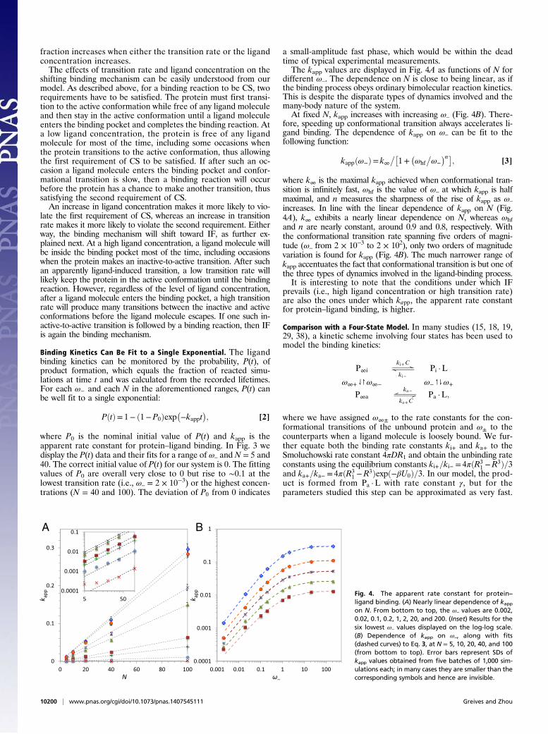
a small-amplitude fast phase, which would be within the deadtime of typical experimental measurements.The kapp values are displayed in Fig. 4A as functions of N for
different ω–. The dependence on N is close to being linear, as ifthe binding process obeys ordinary bimolecular reaction kinetics.This is despite the disparate types of dynamics involved and themany-body nature of the system.At fixed N, kapp increases with increasing ω– (Fig. 4B). There-
fore, speeding up conformational transition always accelerates li-gand binding. The dependence of kapp on ω– can be fit to thefollowing function:
kappðω−Þ= k∞��1+
�ωhf
�ω−
�n�; [3]
where k∞ is the maximal kapp achieved when conformational tran-sition is infinitely fast, ωhf is the value of ω– at which kapp is halfmaximal, and n measures the sharpness of the rise of kapp as ω–
increases. In line with the linear dependence of kapp on N (Fig.4A), k∞ exhibits a nearly linear dependence on N, whereas ωhfand n are nearly constant, around 0.9 and 0.8, respectively. Withthe conformational transition rate spanning five orders of magni-tude (ω– from 2 × 10⁻³ to 2 × 10²), only two orders of magnitudevariation is found for kapp (Fig. 4B). The much narrower range ofkapp accentuates the fact that conformational transition is but one ofthe three types of dynamics involved in the ligand-binding process.It is interesting to note that the conditions under which IF
prevails (i.e., high ligand concentration or high transition rate)are also the ones under which kapp, the apparent rate constantfor protein–ligand binding, is higher.
Comparison with a Four-State Model. In many studies (15, 18, 19,29, 38), a kinetic scheme involving four states has been used tomodel the binding kinetics:
P∞i �ki+C
ki−Pi ·L
ω∞+⇃↾ω∞− ω−↿⇂ω+
P∞a �ka−
ka+CPa ·L;
where we have assigned ω∞± to the rate constants for the con-formational transitions of the unbound protein and ω± to thecounterparts when a ligand molecule is loosely bound. We fur-ther equate both the binding rate constants ki+ and ka+ to theSmoluchowski rate constant 4πDR1 and obtain the unbinding rateconstants using the equilibrium constants ki+=ki− = 4πðR3
1 −R3Þ=3and ka+=ka− = 4πðR3
1 −R3Þexpð−βU0Þ=3. In our model, the prod-uct is formed from Pa ·L with rate constant γ, but for theparameters studied this step can be approximated as very fast.
A B
Fig. 4. The apparent rate constant for protein–ligand binding. (A) Nearly linear dependence of kappon N. From bottom to top, the ω– values are 0.002,0.02, 0.1, 0.2, 1, 2, 20, and 200. (Inset) Results for thesix lowest ω– values displayed on the log-log scale.(B) Dependence of kapp on ω–, along with fits(dashed curves) to Eq. 3, at N = 5, 10, 20, 40, and 100(from bottom to top). Error bars represent SDs ofkapp values obtained from five batches of 1,000 sim-ulations each; in many cases they are smaller than thecorresponding symbols and hence are invisible.
10200 | www.pnas.org/cgi/doi/10.1073/pnas.1407545111 Greives and Zhou

To go from P∞i to Pa ·L, there are two pathways. The CS path-way has P∞a as the intermediate, whereas the IF pathway hasPi ·L as the intermediate.By making the steady-state approximation for P∞a and Pi ·L,
we obtain the apparent rate constant for protein–ligand binding:
kapp =ω∞+ka+C=ðω∞− + ka+CÞ+ ki+Cω+=ðki− +ω+Þ: [4]
It can be easily seen that the first term comes from the reactive fluxalong the CS pathway, whereas the second term is the counterpartalong the IF pathway. The IF fraction can thus be obtained as
ΦIF =ki+ω+=ðki− +ω+Þ
ω∞+ka+=ðω∞− + ka+CÞ+ ki+ω+=ðki− +ω+Þ:
In Fig. 5 we compare the kapp and ΦIF predictions of the ki-netic scheme with our simulation results. kapp is well predicted,except for the highest ligand concentration along with the lowesttransition rate.* However, ΦIF is only semiquantitatively pre-dicted. The plateauing at both the low and high ω– ends seems tobe overly abrupt.
Concluding RemarksMechanisms of protein–ligand binding have long generated interestin wide circles, including enzyme catalysis, receptor function, anddrug design. We have presented a comprehensive study on thebinding mechanism and kinetics of a model protein–ligand system.Our results reconcile some of the conflicting views. In particular, therate of protein conformational transition has been emphasized bysome (12) as an intrinsic factor for dictating the binding mechanism,whereas the concentration of ligand molecules has been empha-sized by others (29, 30) as an extrinsic factor. We show here thatboth factors can shift the binding mechanism between CS and IFand propose to use the IF fraction (i.e., the fraction of bindingevents achieved via IF) to quantify the binding mechanism. CSdominates (i.e., IF fraction is low) only when both conformationaltransition is slow and ligand concentration is low. With the increaseof either conformational transition rate or ligand concentration thebinding mechanism shifts to IF (i.e., toward high IF fraction).Whereas it is very easy to change ligand concentrations, it may
also be possible to perturb conformational transition rates bymutations. If conformations are separated by an energy barrier,mutations targeting the barrier are expected to directly affect the
transition rates (while perhaps minimally affecting the confor-mational equilibrium). Examples include mutations in the hingeregion of a protein that undergoes domain opening/closure (38–40). It will be interesting to test whether such mutations canproduce a shift in binding mechanism.Despite the many-body nature of the system and the involvement
of multiple types of dynamics, and in contrast to the shifting bindingmechanism, the binding kinetics is surprisingly simple. The time-dependent probability of product formation over wide ranges ofmodel parameters can be fit to a single-exponential, and the ap-parent rate constant for protein–ligand binding has a linear de-pendence on ligand concentration. The concentration dependenceof the binding kinetics thus provides little information on thebinding mechanism of our system, even though such data havebeen suggested as a means for mechanism determination (19, 38).However, we have found an apparent correlation between kapp
and ΦIF (i.e., higher kapp corresponds to higher ΦIF, and viceversa). The correlation comes about because the same factorscontrol both kapp and ΦIF. The increase (decrease) of eitherconformational transition rate or ligand concentration leads tothe increase (decrease) of both kapp and ΦIF.Our study does not support the idea (17, 19, 41) that sampling of
the active conformation in the unbound state is evidence for CS.This finding in part reflects the strict definition of CS adoptedhere, which involves only the final inactive-to-active transition be-fore the binding reaction (Fig. 1B, Upper). The mechanism is CS ifthis transition occurs when no ligand molecule is inside the bindingpocket. Sampling of the active conformation in the unbound stateis a necessary condition for CS but does not guarantee it. In fact, ifthe protein rapidly transitions between the inactive and activeconformations (when the energy barrier separating them is low) inthe unbound state, it may do so also while a ligand molecule isloosely bound. The latter situation, as our study indicates, will pushthe mechanism toward IF. Fundamentally, binding mechanism isdictated by the interplay of the various types of dynamics involvedin the binding process, rather than by equilibrium properties alone.The kind of dynamics simulations with a concentration of ligand
molecules will be difficult to perform beyond the model systemstudied here. It is thus very helpful that the often-invoked four-statekinetic scheme is not bad for modeling the binding process. Itmakes good predictions for the apparent rate constant for protein–ligand binding and at least semiquantitative predictions even forthe IF fraction.† The problem is then reduced to the determinationof six elemental rate constants. Experimentally, single-molecule
A B
Fig. 5. Predictions of kapp and ΦIF by the four-statekinetic scheme. (A) Comparison of predicted kapp(solid curves) against the simulation data, shown bythe same dashed curves as in Fig. 4B. (B) Comparisonof predicted ΦIF (solid curves) against the simulationdata, shown by the same dashed curves as in Fig. 2A.
*With a high ligand concentration and a low transition rate, there is some chance formore than one ligand molecule to be loosely bound. By allowing for at most one ligandmolecule to be loosely bound (via moving the reflecting boundary from R to R1 whenone ligand molecule is loosely bound), kapp is slightly reduced and is in better agreementwith the prediction of the kinetic scheme. The restriction to single occupancy alsoslightly lowers the IF fraction (8% at N = 100 and ω– = 0.1).
†A simple application of the kinetic scheme is to predict how the strength of the inter-action energy (i.e., jU0j) affects kapp and ΦIF. An increase in exp(–βU0) will lead to anincrease in ω+ and correspondingly an increase in the reactive flux along the IF pathway(without affecting the CS pathway; see Eq. 4). As a result, both kapp and ΦIF wouldincrease. This prediction was confirmed by simulations.
Greives and Zhou PNAS | July 15, 2014 | vol. 111 | no. 28 | 10201
BIOPH
YSICSAND
COMPU
TATIONALBIOLO
GY

fluorescence resonance energy transfer measurements are nowmaking such determination possible (38), and NMR spectroscopy iscapable of determining at least the four conformational transitionrates (10). It is also possible to compute these intra- and inter-molecular rate constants by dynamics simulations. The resulting IFfraction will provide a full picture of the binding mechanism.
Computational MethodsThe Simulations. Three different events were simulated: ligand diffusion,protein conformational transition, and binding reaction. Ligand positionswere updated according to the Ermak–McCammon algorithm (42):
r= r0 + βF0DΔt + ð2DΔtÞ1=2ℜ;
where r0 is the old position, r is the new position after the timestep Δt (set to5× 10−6), F0 is the force calculated from the interaction potential UsðrÞ, andℜ is a Gaussian random vector with zero mean and unit variance. Both theprotein surface and the outer wall were treated as reflecting, with any newattempted position across a boundary placed back to the old position (36).
Both the transitions between conformations and the binding reactionwere modeled as rate processes and implemented in similar ways. If the rateconstant of a rate process has values ω0 and ω, respectively, before and afterthe ligand position update, then the chance for the reaction to occur in thepresent timestep is 1−exp½− ðω0 +ωÞΔt=2�. The latter quantity was com-pared with a random number uniformly distributed between 0 and 1 todetermine whether the reaction indeed took place. Any loosely bound li-gand molecule could react with the protein. In the unlikely event (for thelow ligand concentrations studied here) that more than one ligand moleculewas loosely bound, they each could independently react with the protein.
Determination of Binding Mechanism. To determine whether a binding reactionevent was preceded by the CS or IF mechanism, we tracked the trajectories of theprotein conformation and the ligand positions. For each set ofmodel parameters,the simulations were repeated 5,000 times with different random number seeds.Initiallytheproteinwasassignedtotheinactiveandactiveconformationsaccordingto their equilibrium probabilities [ω∞−=ðω∞+ +ω∞−Þ and ω∞+=ðω∞+ +ω∞−Þ, re-spectively] in the unbound state, and the ligand molecules were uniformly dis-tributed outside the binding pocket.
For a binding event to be counted as CS, the protein would have to makean inactive-to-active transition while all of the ligand molecules were outsidethe binding pocket and stay in the active conformation until a ligand mol-ecule loosely binds to and react with the protein. Any binding event notcounted as CS was counted as IF. Each simulation was followed until eithera binding reaction occurred or the time reached a preset cutoff time (tcut),which was 103 for all but the lowest ω– (i.e., 2 × 10⁻³) and 104 in the lattercase. The binding reaction was assumed to be irreversible. Among the sim-ulations that were terminated by binding reactions, the IF fraction wascalculated. Alternatively, the growth of the IF count over time was fit toa single exponential similar to Eq. 2, and the extrapolated value at infinitetime divided by the total number of simulations was taken as the final ΦIF.These methods yielded results that differ by less than 2%.
Monitoring of Binding Kinetics. Tomonitor the binding kinetics of the protein–ligand system, the lifetimes of the simulations were recorded. From theselifetimes, the probability of product formation (i.e., the fraction of reactedsimulations) at times up to tcut was calculated.
ACKNOWLEDGMENTS. This work was supported by National Institutes ofHealth Grant GM58187.
1. Monod J, Wyman J, Changeux J-P (1965) On the nature of allosteric transitions:A plausible model. J Mol Biol 12(1):88–118.
2. Perutz MF (1970) Stereochemistry of cooperative effects in haemoglobin. Nature228(5273):726–739.
3. Szabo A (1978) Kinetics of hemoglobin and transition state theory. Proc Natl Acad SciUSA 75(5):2108–2111.
4. Eaton WA, Henry ER, Hofrichter J (1991) Application of linear free energy relations toprotein conformational changes: The quaternary structural change of hemoglobin.Proc Natl Acad Sci USA 88(10):4472–4475.
5. Miller M, et al. (1989) Structure of complex of synthetic HIV-1 protease with a sub-strate-based inhibitor at 2.3 A resolution. Science 246(4934):1149–1152.
6. Spurlino JC, Lu GY, Quiocho FA (1991) The 2.3-A resolution structure of the maltose-or maltodextrin-binding protein, a primary receptor of bacterial active transport andchemotaxis. J Biol Chem 266(8):5202–5219.
7. Müller CW, Schulz GE (1992) Structure of the complex between adenylate kinase fromEscherichia coli and the inhibitor Ap5A refined at 1.9 A resolution. A model fora catalytic transition state. J Mol Biol 224(1):159–177.
8. Ikura M, et al. (1992) Solution structure of a calmodulin-target peptide complex bymultidimensional NMR. Science 256(5057):632–638.
9. Rivas-Pardo JA, et al. (2013) Crystal structure, SAXS and kinetic mechanism of hy-perthermophilic ADP-dependent glucokinase from Thermococcus litoralis reveala conserved mechanism for catalysis. PLoS ONE 8(6):e66687.
10. Whittier SK, Hengge AC, Loria JP (2013) Conformational motions regulate phosphoryltransfer in related protein tyrosine phosphatases. Science 341(6148):899–903.
11. Burgen AS, Roberts GC, Feeney J (1975) Binding of flexible ligands to macromolecules.Nature 253(5494):753–755.
12. Zhou H-X (2010) From induced fit to conformational selection: A continuum ofbinding mechanism controlled by the timescale of conformational transitions. Bio-phys J 98(6):L15–L17.
13. Koshland DE (1958) Application of a theory of enzyme specificity to protein synthesis.Proc Natl Acad Sci USA 44(2):98–104.
14. Burgen AS (1981) Conformational changes and drug action. Fed Proc 40(13):2723–2728.15. Sullivan SM, Holyoak T (2008) Enzymes with lid-gated active sites must operate by an
induced fit mechanism instead of conformational selection. Proc Natl Acad Sci USA105(37):13829–13834.
16. Boehr DD, Nussinov R, Wright PE (2009) The role of dynamic conformational en-sembles in biomolecular recognition. Nat Chem Biol 5(11):789–796.
17. Changeux JP, Edelstein S (2011) Conformational selection or induced fit? 50 years ofdebate resolved. F1000 Biol Rep 3:19.
18. Kiefhaber T, Bachmann A, Jensen KS (2012) Dynamics and mechanisms of coupledprotein folding and binding reactions. Curr Opin Struct Biol 22(1):21–29.
19. Vogt AD, Di Cera E (2013) Conformational selection is a dominant mechanism of li-gand binding. Biochemistry 52(34):5723–5729.
20. Tang C, Schwieters CD, Clore GM (2007) Open-to-closed transition in apo maltose-binding protein observed by paramagnetic NMR. Nature 449(7165):1078–1082.
21. Arora K, Brooks CL, 3rd (2007) Large-scale allosteric conformational transitions ofadenylate kinase appear to involve a population-shift mechanism. Proc Natl Acad SciUSA 104(47):18496–18501.
22. Csermely P, Palotai R, Nussinov R (2010) Induced fit, conformational selection andindependent dynamic segments: an extended view of binding events. Trends Bio-chem Sci 35(10):539–546.
23. Daily MD, Phillips GN, Jr, Cui Q (2010) Many local motions cooperate to produce theadenylate kinase conformational transition. J Mol Biol 400(3):618–631.
24. Grant BJ, McCammon JA, Gorfe AA (2010) Conformational selection in G-proteins:Lessons from Ras and Rho. Biophys J 99(11):L87–L89.
25. Bucher D, Grant BJ, McCammon JA (2011) Induced fit or conformational selection? The roleof the semi-closed state in the maltose binding protein. Biochemistry 50(48):10530–10539.
26. Silva D-A, Bowman GR, Sosa-Peinado A, Huang X (2011) A role for both conforma-tional selection and induced fit in ligand binding by the LAO protein. PLOS ComputBiol 7(5):e1002054.
27. Nussinov R, Ma B (2012) Protein dynamics and conformational selection in bi-directional signal transduction. BMC Biol 10(1):2.
28. Clore GM (2014) Interplay between conformational selection and induced fit inmultidomain protein-ligand binding probed by paramagnetic relaxation enhance-ment. Biophys Chem 186:3–12.
29. Hammes GG, Chang YC, Oas TG (2009) Conformational selection or induced fit: a fluxdescription of reaction mechanism. Proc Natl Acad Sci USA 106(33):13737–13741.
30. Daniels KG, et al. (2014) Ligand concentration regulates the pathways of coupledprotein folding and binding. J Am Chem Soc 136(3):822–825.
31. McCammon JA, Northrup SH (1981) Gated binding of ligands to proteins. Nature293(5830):316–317.
32. Szabo A, Shoup D, Northrup SH, McCammon JA (1982) Stochastically gated diffusion-influenced reactions. J Chem Phys 77(9):4484–4493.
33. Zhou H-X, Szabo A (1996) Theory and simulation of stochastically-gated diffusion-influenced reactions. J Phys Chem 100(7):2597–2604.
34. Cai L, Zhou H-X (2011) Theory and simulation on the kinetics of protein-ligandbinding coupled to conformational change. J Chem Phys 134(10):105101.
35. Zhou HX (2011) Rapid search for specific sites on DNA through conformational switchof nonspecifically bound proteins. Proc Natl Acad Sci USA 108(21):8651–8656.
36. Zhou HX (1990) Kinetics of diffusion-influenced reactions studied by Brownian dy-namics. J Phys Chem 94(25):8794–8800.
37. Zhou HX, Szabo A (1996) Theory and simulation of the time-dependent rate co-efficients of diffusion-influenced reactions. Biophys J 71(5):2440–2457.
38. Kim E, et al. (2013) A single-molecule dissection of ligand binding to a protein withintrinsic dynamics. Nat Chem Biol 9(5):313–318.
39. Marvin JS, Hellinga HW (2001) Manipulation of ligand binding affinity by exploitationof conformational coupling. Nat Struct Biol 8(9):795–798.
40. Millet O, Hudson RP, Kay LE (2003) The energetic cost of domain reorientation inmaltose-binding protein as studied by NMR and fluorescence spectroscopy. Proc NatlAcad Sci USA 100(22):12700–12705.
41. Chen J, Dima RI, Thirumalai D (2007) Allosteric communication in dihydrofolate re-ductase: signaling network and pathways for closed to occluded transition and back.J Mol Biol 374(1):250–266.
42. Ermak DL, McCammon JA (1978) Brownian dynamics with hydrodynamic interactions.J Chem Phys 69(4):1352–1360.
10202 | www.pnas.org/cgi/doi/10.1073/pnas.1407545111 Greives and Zhou