pleurostomophora ochracea, a novel agent of human eumycetoma
TRANSCRIPT

Pleurostomophora ochracea, a Novel Agent of Human Eumycetomawith Yellow Grains
Najwa A. Mhmoud,a Sarah Abdalla Ahmed,b,c,d Ahmed H. Fahal,a G. Sybren de Hoog,b,c,e A. H. G. Gerrits van den Ende,b
and Wendy W. J. van de Sandef
Mycetoma Research Centre, University of Khartoum, Khartoum, Sudana; Centraalbureau voor Schimmelcultures KNAW Fungal Biodiversity Centre, Utrecht, Netherlandsb;Institute of Biodiversity and Ecosystem Dynamics, University of Amsterdam, Amsterdam, Netherlandsc; Faculty of Medical Laboratory Sciences, University of Khartoum,Khartoum, Sudand; Peking University Health Center, Beijing, China, and Sun Yat-Sen Memorial Hospital, Sun Yat-Sen University, Guangzhou, Chinae; and Erasmus MC,University Medical Centre Rotterdam, Department of Medical Microbiology and Infectious Diseases, Rotterdam, Netherlandsf
The first yellow-grain fungal mycetoma, in a 60-year-old man from Central Sudan, is reported. Morphological and phylogeneticanalysis of the ribosomal small subunit (SSU), large subunit (LSU), internal transcribed spacer (ITS), �-tubulin (BT2), actin(ACT1), and elongation factor (TEF1) genes revealed that the isolate deviated from any known agent of mycetoma; it clustered inthe genus Pleurostoma (anamorph genus, Pleurostomophora) in the order Calosphaeriales. The novel species, here named Pleu-rostomophora ochracea, is characterized by phenotypic features. The species proved to be highly susceptible to itraconazole,ketoconazole, posaconazole, and voriconazole, but not to fluconazole. The fungus was inhibited by caspofungin at 8 �g/ml,while no inhibition was found with 5-flucytosine (MIC > 64 �g/ml). Compared to other members of the genus Pleurosto-mophora, P. ochracea is slow growing, with a relatively high optimum growth temperature (36 to 37°C). This is the first case of ayellow-grain fungal mycetoma; yellow grains are otherwise of bacterial nature. Our case emphasizes that identification of myce-toma agents by the color of the grain only is not sufficient and may lead to inappropriate therapy.
Mycetoma is a chronic granulomatous, progressive, subcuta-neous inflammatory infection (12). It is caused by true fungi
or by bacteria, and hence, it is classified as eumycetoma or actino-mycetoma, respectively (1, 3). The triad of a painless subcutane-ous mass, multiple sinuses, and seropurulent discharge contain-ing grains is pathognomic of mycetoma (5, 14). Eumycetoma ismore common than actinomycetoma throughout the world, andin Sudan, it accounts for 70% of all mycetoma cases (5). A varietyof fungi are implicated in causing eumycetoma (Table 1). In Su-dan, eumycetoma is usually caused by the fungus Madurella my-cetomatis, which forms black grains in human tissue (1, 15). Themost common fungal pathogens causing eumycetoma produceeither black grains or white grains (5, 10). While yellow grainshave been encountered in cases of actinomycetoma, they havenever been reported before in eumycetoma. The available diag-nostic tools for mycetoma are limited and are mostly of a pheno-typic nature (1). A first indication is usually obtained from thecolor of the grain: black grains are produced by fungi and yellowgrains by bacteria, while white grains can be of bacterial or fungalnature. Culture media used for diagnosis are based upon this firstindication. Here, we present a case of yellow-grain mycetoma,which was initially misdiagnosed as actinomycetoma based on thecolor of the grain and on cytology. In this report, we show thatcultural and molecular techniques, in combination with an up-dated taxonomy, can be successfully used to identify the yellow-grain-producing fungus Pleurostomophora ochracea as a new caus-ative agent of human mycetoma.
CASE REPORT
The patient was a 60-year-old male farmer from central Sudan. In2006, he presented to the Mycetoma Clinic at the Mycetoma Re-search Centre of the University of Khartoum, Khartoum, Sudan,with a massive, recurrent right-foot swelling. His conditionstarted 20 years prior to presentation with a small, painless right-
foot swelling, which had gradual onset and progress and was pain-ful. He developed multiple sinuses, and the seropurulent dis-charge contained yellow grains. The patient underwent foursurgical excisions under general and spinal anesthesia done else-where. He had neither a history of local trauma nor a family his-tory of mycetoma. The social and geographic history and drug usewere not contributory to his problem. On examination, he lookedwell, not pale and neither jaundiced nor cyanosed. He had a nor-mal pulse rate (75/min), respiratory rate (16/min), blood pressure(120/80), and temperature (37°C). Also, his renal and hepaticfunction tests, as well as his full blood count, were within normallimits. Local examination revealed a painless massive mass involv-ing the dorsal and plantar parts of the right foot. There were mul-tiple active and healed sinuses, and the seropurulent dischargecontained yellow grains (Fig. 1). The foot X-ray confirmed a mas-sive soft tissue swelling and revealed a periosteal reaction and bonedestruction (Fig. 1). Fine-needle aspiration for cytology was takenfrom the lesion and showed an intense inflammatory infiltratewith grains typical of actinomycetes. The patient was started onstreptomycin (1 g per day) and cotrimoxazole (945 mg twice aday) and was followed up regularly in the outpatient clinic. How-ever, there was no clinical response to this treatment. He was thenswitched to amikacin sulfate (15 mg/kg of body weight twice a
Received 4 June 2012 Returned for modification 20 June 2012Accepted 25 June 2012
Published ahead of print 3 July 2012
Address correspondence to Wendy W. J. van de Sande, [email protected].
N.A.M. and S.A.A. contributed equally to this article.
Copyright © 2012, American Society for Microbiology. All Rights Reserved.
doi:10.1128/JCM.01470-12
September 2012 Volume 50 Number 9 Journal of Clinical Microbiology p. 2987–2994 jcm.asm.org 2987
Dow
nloa
ded
from
http
s://j
ourn
als.
asm
.org
/jour
nal/j
cm o
n 22
Nov
embe
r 20
21 b
y 20
7.65
.150
.237
.

day) and cotrimaxozole (945 mg twice a day) with no clinicalimprovement. He underwent surgical debulking to improve theresponse to medical treatment. Numerous yellow grains were col-lected from discharging sinuses, which were hard in consistencyand irregular in size. Culturing of the grains revealed fungal
growth, indicating that the patient was suffering from a eumyce-toma rather than an actinomycetoma. Unfortunately, changingthe treatment accordingly was not possible; the patient was lost atfollow-up and was never seen again.
MATERIALS AND METHODSClinical specimen. A clinical specimen was collected from the patientwhen he presented with yellow-grain mycetoma at the Mycetoma Re-search Centre, University of Khartoum, Khartoum, Sudan, on 27 October2008. After obtaining written consent from the patient, an aspirate withvisible yellow grains was taken from the sinuses. The yellow grains werecollected for further identification.
Fungal isolation. Grains collected from the sinus were washed twice inphysiological saline and inoculated into Lowenstein-Jensen (LJ) medium(Hi Media Laboratories, Mumbai, India). The grain was cultured for 3 to4 weeks at 37°C. This initial inoculation on LJ medium was guided by thecolor of the grain: yellow grains were considered to be of bacterial origin.After a week, the color of the grains converted from yellow to black, andthe grain was subcultured on Sabouraud’s dextrose agar (SDA) (Hi Me-dia). The culture was identified morphologically as a fungal species. Foridentification, the isolate was shipped to the Department of Medical Mi-crobiology and Infectious Diseases, Erasmus MC, Rotterdam, Nether-lands, and to the Centraalbureau voor Schimmelcultures (CBS) KNAWFungal Biodiversity Centre, Utrecht, Netherlands, and deposited in theCBS collection under number CBS 131321.
Morphology and physiology. Colony characteristics and growthmorphology were studied by inoculating the isolate onto plates of maltextract agar (MEA) (Oxoid, United Kingdom), oat meal agar (OA)(homemade at CBS, Netherlands), corn meal agar (CMA) (homemade atCBS, Netherlands), and potato dextrose agar (PDA) (Oxoid, United King-dom) and incubating the plates in the dark at 30°C for up to 4 weeks.Microscopic mounts in lactic acid with cotton blue were made from cul-tures grown on a PDA plate and slide cultures after 2 weeks of incubationat 30°C. The slides were examined and measured with a light microscope(Nikon Eclipse 80i), and pictures were taken using a camera attached tothe microscope (Nikon; digital-sight DS-5 M). A minimum of 10 mea-surements per structure were taken after processing in Adobe PhotoshopCS3, and the average was calculated. Cardinal growth temperatures weredetermined on PDA, and the cultures were incubated in the dark for 4weeks at temperatures ranging from 15°C to 40°C at 3°C intervals. Theability of the isolate to assimilate carbohydrate sources was determinedwith the API 20C AUX system (bioMérieux, Marcy l’Etoile, France). Priorto the carbohydrate assimilation test, the homogenized fungal suspensionwas prepared by ultrasonic treatment as published previously (4, 36) andthen was adjusted to a 2 McFarland standard with the medium provided.
TABLE 1 Agents of mycetoma in mammals and their phylogeneticpositionsa
Name Order Grain
Acremonium kiliense Hypocreales WhiteAcremonium potronii Hypocreales WhiteAcremonium recifei Hypocreales WhiteCylindrocarpon cyanescens Hypocreales WhiteCylindrocarpon destructans Hypocreales WhiteFusarium falciforme Hypocreales WhiteFusarium verticillioides Hypocreales WhiteFusarium solani Hypocreales WhitePhialemonium obovatum Hypocreales WhiteAspergillus flavus Eurotiales WhiteAspergillus hollandicus Eurotiales WhiteAspergillus nidulans Eurotiales WhiteBipolaris spicifera Pleosporales BlackCorynespora cassiicola Pleosporales BlackCurvularia geniculata Pleosporales BlackCurvularia lunata Pleosporales BlackLeptosphaeria senegalensis Pleosporales BlackLeptosphaeria thompkinsii Pleosporales BlackMadurella grisea Pleosporales BlackPseudochaetosphaeronema larense Pleosporales BlackPyrenochaeta mackinnonii Pleosporales BlackPyrenochaeta romeroi Pleosporales BlackNeotestudina rosatii Pleosporales White/blackMadurella mycetomatis Sordariales BlackMadurella pseudomycetomatis Sordariales BlackMadurella fahalii Sordariales BlackMadurella tropicana Sordariales BlackPhaeoacremonium parasiticum Diaporthales WhitePhaeoacremonium krajdenii Diaporthales WhiteExophiala jeanselmei Chaetothyriales BlackPseudallescheria boydii Microascales WhiteTrichophyton sp. Onygenales WhiteMicrosporum canis Onygenales Whitea Adapted from de Hoog et al. (10).
FIG 1 Clinical presentation of the patient. (A) The patient had a mycetoma on the right foot, with massive, recurrent foot swelling. (B) On the foot X-ray, amassive soft tissue swelling, a periosteal reaction, and bone destruction are seen.
Mhmoud et al.
2988 jcm.asm.org Journal of Clinical Microbiology
Dow
nloa
ded
from
http
s://j
ourn
als.
asm
.org
/jour
nal/j
cm o
n 22
Nov
embe
r 20
21 b
y 20
7.65
.150
.237
.

Finally, 100 �l of this inoculum was used to fill the cupules of the test stripsas directed by the manufacturer. The ability of the isolate to hydrolyzecasein, gelatin, and starch was determined by culturing the strains onselective plates according to the manufacturer’s instructions. The abilityto produce urease was determined by culturing on urea agar (urea baseconcentrate [Difco] with 1.5% [wt/vol] agar). Sodium chloride tolerancewas determined by comparing the growth after 2 weeks on SDA slantscontaining 0, 0.5, 5, 10, and 30% NaCl.
DNA extraction, amplification, and sequencing. Genomic DNA wasextracted by scraping material off Sabouraud plates, freezing it in liquidnitrogen, and grinding it with a mortar and pestle. DNA was extractedfrom the resulting pulp with the Promega Wizard Kit (Promega) accord-ing to the manufacturer’s instructions. The ribosomal DNA (rDNA) in-ternal transcribed spacer (ITS) gene and partial sequences of the actin(ACT1), �-tubulin (BT2), and elongation factor 1� (TEF1) genes, as wellas the 18S rDNA gene (small subunit [SSU]) and 28S rDNA gene (largesubunit [LSU]), were amplified and sequenced. The primer pairs for thegenes were ITS4 and ITS5 (39), ACT-512F and ACT-783R (9), T1 andBT2b (18, 28), EF2 and EF728F (9, 23), NS1 and NS24 (17, 39), and LRoRand LR5 (38), respectively. Additional primers used for sequencing of theSSU included NS2, NS3, NS6, and NS7 (39).
Alignment and phylogenetic analysis. A consensus sequence wascomputed from the forward and reverse sequences with SeqMan from theLasergene package (DNAstar, Madison, WI). Sequences retrieved fromGenBank are listed in Fig. 2 and in Table 2. Sequences were aligned withMultiple Sequence Comparison by Log-Expectation (MUSCLE) using theEMBL-EBI Web server (http://www.ebi.ac.uk/Tools/msa/muscle/). Analignment was constructed for the complete ITS (ITS1-5.8S-ITS2), in-
cluding 30 strains representing 12 species. A combined 28S LSU and 18SSSU alignment for 39 strains from 36 fungal species was constructed. Toinvestigate the phylogenetic relationship of the newly isolated fungus andrelated taxa, maximum-likelihood, maximum-parsimony, and Bayesiananalyses were used for both alignments. Maximum likelihood with 500bootstraps using the Tamura-Nei model and maximum-parsimony anal-ysis were conducted in MEGA v. 5.05 (33, 34). Bayesian analysis was doneusing MrBayes v. 3.1.2 software. All trees were constructed by the out-group method and edited in MEGA v. 5.05.
Antifungal susceptibility. Antifungal susceptibilities for eight anti-fungal drugs were determined in triplicate by using the colorimetric Sen-sititre YeastOne method (Trek Diagnostic Systems, East Grinstead,United Kingdom) as described previously (36). In short, the isolate wascultured for 10 days in RPMI 1640 medium supplemented with L-glu-tamine (0.3 g/liter) and 20 mM morpholinepropanesulfonic acid at 37°C.Mycelia were harvested by 5 min of centrifugation at 2,158 � g andwashed once with sterile saline. After sonication (20 s at 28-�m maximumpower; Soniprep, Beun de Ronde, Netherlands) of the hyphal suspension,Tween 60 was added at 0.05% (vol/vol), and the transmissions were ad-justed to 70% at 660 nm (Novaspec II; Pharmacia Biotech). The inocu-lated plates were incubated at 37°C for 7 days. The drug concentrationsused ranged from 0.016 �g/ml to 8 �g/ml for amphotericin B, itracona-zole, ketoconazole, and voriconazole; from 0.25 �g/ml to 128 �g/ml forfluconazole; and from 0.125 �g/ml to 64 �g/ml for 5-flucytosine.
Nucleotide sequence accession numbers. The GenBank accessionnumbers for isolate CBS 131321 amplified genes are as follows: 28S rRNA,JX073274; 18S rRNA, JX073269; �-tubulin, JX073271; actin, JX073275;and elongation factor, JX097097.
MycoBank accession number. The MycoBank accession number forP. ochracea is MB800514.
RESULTSPhylogeny. A BLAST search with the ITS, 18S SSU, and 28S LSUsequences did not yield identity with any known fungus. There-fore, the sequences were used to determine the higher-order phy-logeny for our isolate. The alignment of LSU and SSU consisted of1,909 characters, of which 1,520 were constant, 62 were parsi-mony uninformative, and 327 were parsimony informative. In-trons were found in SSUs of three strains from the data set, andthey were deleted from the alignment. Maximum-parsimonyanalysis of the combined data set resulted in 8 most parsimonioustrees (length of the tree � 1,109; consistency index [CI] �0.500451; retention index [RI] � 0.759444; rescaled consistencyindex [RCI] � 0.380065). The SSU and LSU phylogenetic analysesshowed that the reported pathogenic isolate was closely related tothe family Pleurostomataceae, with high bootstrap support with allalgorithms used (1.00, 100, and 100) (Fig. 2) in the orderCalosphaeriales.
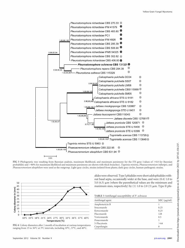
The ITS alignment consisted of 600 characters, of which 342 wereconstant, 242 were parsimony informative, and 16 were parsimonyuninformative. Maximum-parsimony analysis resulted in 217 mostparsimonious trees (length � 356; CI � 0.688202; RI � 0.904475;RCI � 0.622462). ITS analysis confirmed that the species belonged tothe Pleurostomataceae, in which it formed a well-supported clade withthe genus Pleurostoma (0.99, 93, and 94) (Fig. 3). Moreover, analysisof the actin, �-tubulin, and elongation factor of the newly isolatedstrain and a representative isolate from each species currently attrib-uted to the Pleurostomataceae confirmed that the isolate was appro-priately placed in this family (data not shown) as a member of theanamorph genus Pleurostomophora.
Physiology. The minimum growth temperature for the isolatewas 15°C, and the maximum was above 40°C, with an optimum at36 to 37°C (Fig. 4). Measurements were also taken after 8 days of
TABLE 2 Calosphaeriales strains
Strain no. Name ITS Source
STE-U 6181 Calosphaeria africana EU367445 PlantCBS 120870 Calosphaeria africana EU367444 PlantCBS 115999 Calosphaeria pulchella EU367451 PlantSM05 Calosphaeria pulchella HM237300 PlantLM06 Calosphaeria pulchella HM237298 PlantDC04 Calosphaeria pulchella HM237299 PlantSS07 Calosphaeria pulchella HM237297 PlantCBS 127681 Jattaea discreta HQ878587 PlantCBS 119343 Jattaea leucospermi EU552127 PlantSTE-U 6401 Jattaea mookgoponga EU367450 PlantCBS 120867 Jattaea mookgoponga EU367449 PlantCBS 120871 Jattaea prunicola EU367446 PlantSTE-U 6400 Jattaea prunicola EU367448 PlantSTE-U 6399 Jattaea prunicola EU367447 PlantCBS 294.39 Pleurostomophora repens AF083195 PlantCBS 131321 Pleurostomophora ochracea (JX073270) HumanCBS 115329 Pleurostoma ootheca HQ878590 PlantIFM5 54325 Pleurostomophora richardsiae AB364704 HumanCBS 302.62 Pleurostomophora richardsiae AB364698 PlantCBS 506.90 Pleurostomophora richardsiae AB364702 HumanCBS 295.39 Pleurostomophora richardsiae AB364697 PlantIFM 4926 Pleurostomophora richardsiae AB364694 HumanPC1 Pleurostomophora richardsiae AB364693 HumanCBS 483.80 Pleurostomophora richardsiae AB364701 HumanIFM 41579 Pleurostomophora richardsiae AB364695 HumanCBS 270.33 Pleurostomophora richardsiae AY729811 HumanCBS 406.93 Pleurostomophora richardsiae AB364703 PlantCBS 631.94 Phaeoacremonium aleophilum AF266647 PlantCBS 222.95 Phaeoacremonium inflatipes AF266655 PlantSTE-U 5963 Togninia minima EU128019 PlantCBS 113648 Togniniella acerosa EU367453 PlantCBS 113726 Togniniella acerosa EU367452 Plant
Yellow-Grain Fungal Mycetoma
September 2012 Volume 50 Number 9 jcm.asm.org 2989
Dow
nloa
ded
from
http
s://j
ourn
als.
asm
.org
/jour
nal/j
cm o
n 22
Nov
embe
r 20
21 b
y 20
7.65
.150
.237
.

incubation on PDA to compare the growth pattern of the isolatewith those of other Pleurostomophora species. The colony diame-ter reached only 1 to 2 mm when grown at 20°C. The isolatedecomposed casein in the first week. Starch was hydrolyzed; someplates showed a narrow clear zone around the colony, and in oth-ers, a zone extending up to 2.4 cm was observed. Gelatin hydroly-sis was positive using both the tube and plate techniques. Ureasewas strongly positive within 3 days. The isolate showed some sen-sitivity to cycloheximide and was sensitive to salt, exhibiting someinhibition with 0.5%, poor or no growth with 5%, and no growthwith 10% and 30% NaCl.
Antifungal susceptibility. The MICs of the strain tested areshown in Table 3. Our strain was highly susceptible to the azolesitraconazole, ketoconazole, posaconazole, and voriconazole. LowMICs were also found for amphotericin B. The only azole forwhich a high MIC was found (128 �g/ml) was fluconazole. The
fungus was inhibited by caspofungin at 8 �g/ml, but not by 5-flu-cytosine (MIC � 64 �g/ml).
Taxonomy. Pleurostomophora ochracea Mhmoud, AbdallaAhmed, Fahal, de Hoog, van de Sande, sp. nov. MycoBank acces-sion number MB800514. Etymology: named after its formation ofyellow grains in human tissue.
After 1 week of growth on all media tested (PDA, MEA, OA, andCMA), the colony surface and reverse were typically creamy white topale yellow and felty, initially with some mycelial tufts. On OA andCMA, colonies turned dark olivaceous brown to nearly black after 1month of incubation. No diffusible pigment was produced in anymedium (Fig. 5). Vegetative hyphae were branched, septate, and hy-aline to pale brown, turning dark brown after longer incubation. Hy-phae were 1.2 to 3.2 �m wide. Hyphal walls were smooth or verrucu-lose. Conidiophores were absent. Conidiogenous cells were variableand poorly differentiated, and phialidic. Three distinct types of phi-
FIG 2 Phylogram of two loci (SSU and LSU) obtained by Bayesian analysis, maximum likelihood, and maximum parsimony (values of �0.8 for Bayesianprobability and �80% for maximum likelihood and maximum parsimony are shown with thick branches). Leotia lubrica and Crinula caliciiformis wereused as the outgroup. The portion of the phylogram relating to Calosphaeriales is shaded. The names of families within the order and the new isolate areshown in bold.
Mhmoud et al.
2990 jcm.asm.org Journal of Clinical Microbiology
Dow
nloa
ded
from
http
s://j
ourn
als.
asm
.org
/jour
nal/j
cm o
n 22
Nov
embe
r 20
21 b
y 20
7.65
.150
.237
.
alides were observed. Type I phialides were short adelophialides with-out basal septa, occasionally wider at the base, and were (0.4) 1.0 to5.0 (6.5) �m (where the parenthetical values are the minimum andmaximum sizes, respectively) by (1) 1.0 to 2.0 (3) �m. Type II phi-
FIG 4 Colony diameters after 1 month of incubation at various temperaturesranging from 15 to 36°C at 3°C intervals, including 10°C, 37°C, and 40°C.
FIG 3 Phylogenetic tree resulting from Bayesian analysis, maximum likelihood, and maximum parsimony for the ITS gene (values of �0.8 for Bayesianprobability and �80% for maximum likelihood and maximum parsimony are shown with thick branches). Togninia minima, Phaeoacremonium inflatipes, andPhaeoacremonium aleophilum were used as the outgroup. Light-gray circles, strains isolated from plants; dark-gray circles, human-pathogenic strains.
TABLE 3 Antifungal susceptibility of P. ochracea
Antifungal agent MIC (�g/ml)
Amphotericin B 1Itraconazole 0.25Ketoconazole 0.25Fluconazole 128Voriconazole 0.5Posaconazole 15-Flucytosin �64Caspofungin 8
Yellow-Grain Fungal Mycetoma
September 2012 Volume 50 Number 9 jcm.asm.org 2991
Dow
nloa
ded
from
http
s://j
ourn
als.
asm
.org
/jour
nal/j
cm o
n 22
Nov
embe
r 20
21 b
y 20
7.65
.150
.237
.

alides were elongate-ampulliform, swollen at the base and taperedtoward the apex, (6.2) 7 to 8.6 (8.7) by (1.3) 1.0 to 2.0 (2.5) �m. TypeIII phialides were (10.7) 11.6 to 17.9 (20.0) by (1.3) 1.7 to 2.4 (2.6)�m, (sub)cylindrical to elongate-ampulliform (Fig. 5). Conidia weresmooth-walled and aggregate at the tip of the phialides; most conidiawere hyaline, and some were brown. Two types were observed: eithersmall and subspherical to ellipsoidal, (2.0) 2.2 to 3.0 (3.5) by (1.3) 1.6to 2.0 (2.3) �m, or larger and allantoid, (3.5) 4 to 6 (6.2) by (1.2) 1.7to 2.5 (2.6) �m (Fig. 5). Teleomorph unknown. Holotype: dried cul-ture in CBS Herbarium H-20972; ex-type strain CBS 131321, isolated
by N. A. Mhmoud and A. H. Fahal from human yellow-grain myce-toma, Khartoum, Sudan.
DISCUSSION
Mycetoma is a chronic progressive disease characterized by sup-purative, swollen lesions and sinuses and can be caused by bothbacteria and fungi (2, 15). Based on the color of the grain, fungalmycetomata can be divided into two large groups: those causingblack or white grains. Black-grain mycetoma is mainly associatedwith M. mycetomatis, Madurella grisea, Leptosphaeria senegalensis,
FIG. 5. P. ochracea growth and morphology. Shown are colonies after 2 weeks of incubation at 37°C. (A) PDA. (B) MEA. (C) OA. (D, E, and F) Different-shapeconidia. (G, H, and I) Type I phialides. (J, K, and L) Type II phialides. (M, N, and O) Type III phialides. All scale bars, 10 �m.
Mhmoud et al.
2992 jcm.asm.org Journal of Clinical Microbiology
Dow
nloa
ded
from
http
s://j
ourn
als.
asm
.org
/jour
nal/j
cm o
n 22
Nov
embe
r 20
21 b
y 20
7.65
.150
.237
.

Pyrenochaeta romeroi, and Exophiala jeanselmei; white-grain my-cetoma is usually caused by Pseudallescheria boydii, Acremoniumkiliense, and other, occasional agents (5) (Table 1). Yellow-graineumycetoma, to our knowledge, has never been reported. In areasof high endemicity, like the Sudan, 70% of all mycetoma cases areeumycetoma, caused by M. mycetomatis, a species producingblack grains (5, 15). The remaining 30% of mycetoma cases are ofbacterial origin, and many of these are characterized by the pres-ence of yellow grains. With the limited availability of diagnostictechniques in routine laboratories in areas of high endemicity, thecolor of grains has decisive diagnostic value (15). In this commu-nication, we report on the first authenticated case of human eu-mycetoma infection caused by a yellow-grain-producing fungus.
We identified the causative agent by multigene phylogeneticanalysis. The novel pathogenic isolate clustered in the ascomyceteorder Calosphaeriales. The small order Calosphaeriales comprisesonly four genera separated into two families: the family Calospha-eriaceae contains three genera, viz., Calosphaeria, Jattaea, and Tog-niniella, while our fungus clustered in the Pleurostomataceae, con-taining only a single teleomorph genus, Pleurostoma, and a singleanamorph genus, Pleurostomophora (30). Our fungus was para-phyletic to Pleurostoma ootheca and Pleurostomophora repens (Fig.3). Because of the absence of sexual sporulation, we prefer to clas-sify the new species in the anamorph genus Pleurostomophora.
The majority of causative agents of mycetoma belong to theorders Sordariales (particularly the genus Madurella) and Pleospo-rales (particularly coelomycetes) (Table 1). The order Calospha-eriales is a new addition to the list of orders harboring potentialagents. The order was described by Barr in 1983 (6, 7); recenttaxonomic revision showed that the Togniniaceae telemorph ofPhaeoacremonium had some affinity to Diaporthales (27), but inour study, the family was found as a separate cluster (Fig. 2), whichis in agreement with the results of Réblová (29). Based on SSU andLSU data, and unlike previous publications (21, 29), the Pleuros-tomataceae showed affinity to the Diaporthales, whereas theCalosphaeriaceae formed a separate, well-supported clade (Fig. 2).The order Calosphaeriales, comprising only 10 species, partly atlarge mutual phylogenetic distances, is still poorly defined. Sup-posedly related fungi, such as Phaeoacremonium, were found to beunalignable for the species-level genes used in this study, i.e., withdegrees of variability equal to or larger than that of the ITS.
Most members of the cluster Pleurostoma-Pleurostomophoraare wood-inhabiting fungi (8, 37). Two cases of phaeohyphomy-cosis caused by P. repens have been reported (20, 26). However,reexamination of both pathogenic strains revealed misidentifica-tions, with the correct name being Phaeoacremonium krajdenii(26). Pleurostomophora richardsiae is the only confirmed human-pathogenic species in the order Calosphaeriales. It is known as anenvironmental fungus but has been reported as the causativeagent of subcutaneous granuloma, phaeohyphomycosis, a chro-moblastomycosis-like infection, and a bone infection (19, 22, 24,32, 35).
Pleurostomophora ochracea and P. richardsiae both producedifferent types of phialides and conidia, but in P. richardsiae it isnoticeably more pronounced (37). In P. ochracea, all three types ofphialides are able to produce differently shaped conidia. All spe-cies of Pleurostomophora and the teleomorph genus Pleurostomagrow at temperatures ranging between 10 and 40°C, but P. ochra-cea has a relatively high optimum growth temperature (36°C ver-sus 30°C) and a low growth rate. This is expressed after 8 days at
20°C, where colony diameters of P. ochracea are only 1 to 2 mm,while P. richardsiae reaches 18 to 19 mm, P. ootheca 30 to 32 mm,and P. repens 59 to 61 mm (37).
The clinical isolate of P. ochracea in this study appeared to behighly susceptible, in vitro, to ketoconazole and itraconazole,compounds that are commonly used in the treatment of subcuta-neous infections, including mycetoma by pigmented fungi (16,21, 25, 31). Of all azole antifungals tested, fluconazole was the leasteffective. Once it was established that the causative agent was afungus, our patient was advised to take ketoconazole at 400 mg/day, according to the guidelines of the Mycetoma Research Centrein Khartoum, Sudan. However, the patient refused treatment anddid not return to the center, and follow-up was impossible.
As noted in most cases of mycetoma, it is difficult to explainhow our patient acquired his infection. He did not report any typeof injury. Since the patient is a farmer by occupation, he is in dailycontact with soil, thorns, wood, and other trauma-causing ob-jects; even minor trauma might have facilitated the introductionof the fungus into his foot. The present case report shows thatproper species identification in the diagnosis of mycetoma re-mains troublesome and that identification based exclusively onthe color of the grains may be erratic (11, 13). Molecular identifi-cation is recommended to ascertain appropriate therapy.
ACKNOWLEDGMENT
Wendy W. J. van de Sande is supported by a postdoctoral fellowship fromthe Netherlands Organization for Scientific Research (NWO, VENI-Grant 916.11.178).
REFERENCES1. Ahmed A, et al. 2002. Environmental occurrence of Madurella mycet-
omatis, the major agent of human eumycetoma in Sudan. J. Clin. Micro-biol. 40:1031–1036.
2. Ahmed A, van de Sande W, Verbrugh H, Fahal A, van Belkum A. 2003.Madurella mycetomatis strains from mycetoma lesions in Sudanese pa-tients are clonal. J. Clin. Microbiol. 41:4537– 4541.
3. Ahmed A, et al. 2007. Management of mycetoma: major challenge intropical mycoses with limited international recognition. Curr. Opin. In-fect. Dis. 20:146 –151.
4. Ahmed AO, et al. 2004. In vitro susceptibilities of Madurella mycetomatisto itraconazole and amphotericin B assessed by a modified NCCLSmethod and a viability-based 2,3-bis(2-methoxy-4-nitro-5-sulfophenyl)-5-[(phenylamino)carbonyl]-2H-tetrazolium hydroxide (XTT) assay. An-timicrob. Agents Chemother. 48:2742–2746.
5. Ahmed AO, et al. 2004. Mycetoma caused by Madurella mycetomatis: aneglected infectious burden. Lancet Infect. Dis. 4:566 –574.
6. Barr ME. 1983. The ascomycete connection. Mycologia 75:1–13.7. Barr ME. 1985. Notes on the Calosphaeriales. Mycologia 77:509 –565.8. Barr ME. 1990. Prodromus to nonlichenized pyrenomycetous members
of the class Hymenoascomycetes. Mycotaxon 39:43–184.9. Carbone I, Kohn LM. 1999. A method for designing primer sets for
speciation studies in filamentous ascomycetes. Mycologia 91:553–556.10. de Hoog GS, Adelmann D, Ahmed AO, van Belkum A. 2004. Phylogeny
and typification of Madurella mycetomatis, with a comparison of otheragents of eumycetoma. Mycoses 47:121–130.
11. de Hoog GS, et al. 1993. Diagnostic problems with imported cases ofmycetoma in The Netherlands. Mycoses 36:81– 87.
12. Fahal AH. 2010. Management of mycetoma. Expert Rev. Dermatol. 5:87–93.13. Fahal AH. 2006. Mycetoma, clinicopathological monograph, 1st ed.
Khartoum University Press, Khartoum, Sudan.14. Fahal AH. 2004. Mycetoma: a thorn in the flesh. Trans. R. Soc. Trop. Med.
Hyg. 98:3–11.15. Fahal AH, Hassan MA. 1992. Mycetoma. Br. J. Surg. 79:1138 –1141.16. Fahal AH, Rahman IA, El-Hassan AM, Rahman ME, Zijlstra EE. 2011.
The safety and efficacy of itraconazole for the treatment of patients witheumycetoma due to Madurella mycetomatis. Trans. R. Soc. Trop. Med.Hyg. 105:127–132.
Yellow-Grain Fungal Mycetoma
September 2012 Volume 50 Number 9 jcm.asm.org 2993
Dow
nloa
ded
from
http
s://j
ourn
als.
asm
.org
/jour
nal/j
cm o
n 22
Nov
embe
r 20
21 b
y 20
7.65
.150
.237
.

17. Gargas A, Taylor JW. 1992. Polymerase chain reaction (PCR) primers foramplifying and sequencing 18S rDNA from lichenized fungi. Mycologia84:589 –592.
18. Glass NL, Donaldson GC. 1995. Development of primer sets designed foruse with the PCR to amplify conserved genes from filamentous ascomy-cetes. Appl. Environ. Microbiol. 61:1323–1330.
19. Gueho E, Bonnefoy A, Luboinski J, Petit JC, de Hoog GS. 1989.Subcutaneous granuloma caused by Phialophora richardsiae: case reportand review of the literature. Mycoses 32:219 –223.
20. Hironaga M, Nakano K, Yokoyama I, Kitajima J. 1989. Phialophorarepens, an emerging agent of subcutaneous phaeohyphomycosis in hu-mans. J. Clin. Microbiol. 27:394 –399.
21. Hood SV, Moore CB, Cheesbrough JS, Mene A, Denning DW. 1997.Atypical eumycetoma caused by Phialophora parasitica successfullytreated with itraconazole and flucytosine. Br. J. Dermatol. 136:953–956.
22. Ikai K, Tomono H, Watanabe S. 1988. Phaeohyphomycosis caused byPhialophora richardsiae. J. Am. Acad. Dermatol. 19:478 – 481.
23. Jacobs K, et al. 2004. Leptographium wingfieldii introduced into NorthAmerica and found associated with exotic Tomicus piniperda and nativebark beetles. Mycol. Res. 108:411– 418.
24. Lieb DF, Smiddy WE, Miller D, Cooperman EW. 2003. Case report:fungal endophthalmitis caused by Phialophora richardsiae. Retina 23:406 –407.
25. Mahgoub ES, Gumaa SA. 1984. Ketoconazole in the treatment of eumy-cetoma due to Madurella mycetomii. Trans. R. Soc. Trop. Med. Hyg. 78:376 –379.
26. Meyers WM, Dooley JR, Kwon-Chung KJ. 1975. Mycotic granulomacaused by Phialophora repens. Am. J. Clin. Pathol. 64:549 –555.
27. Mostert L, Groenewald JZ, Summerbell RC, Gams W, Crous PW. 2006.Taxonomy and pathology of Togninia (Diaporthales) and its Phaeoacre-monium anamorphs. Studies Mycol. 54:1–113.
28. O’Donnell K, Cigelnik E. 1997. Two divergent intragenomic rDNA ITS2types within a monophyletic lineage of the fungus Fusarium are nonor-thologous. Mol. Phylogenet Evol. 7:103–116.
29. Réblová M. 2011. New insights into the systematics and phylogeny of thegenus Jattaea and similar fungi of the Calosphaeriales. Fungal Diversity49:167–198.
30. Réblová M, Mostert L, Gams W, Crous PW. 2004. New genera inCalosphaeriales: Togniniella and its anamorph Phaeocrella, and Calospha-eriophora as anamorph of Calosphaeria. Studies Mycol. 50:533–550.
31. Richardson MD, Kokki M. 2001. Therapeutic guidelines in systemicfungal infections. Current Medical Literature Ltd, London, United King-dom.
32. Son YM, et al. 2010. Chromoblastomycosis caused by Phialophora rich-ardsiae. Ann. Dermatol. 22:362–366.
33. Tamura K, Nei M. 1993. Estimation of the number of nucleotide substi-tutions in the control region of mitochondrial DNA in humans and chim-panzees. Mol. Biol. Evol. 10:512–526.
34. Tamura K, et al. 2011. MEGA5: molecular evolutionary genetics analysisusing maximum likelihood, evolutionary distance, and maximum parsi-mony methods. Mol. Biol. Evol. 28:2731–2739.
35. Torstrick RF, Harrison K, Heckman JD, Johnson JE. 1979. Chronicbursitis caused by Phialophora richardsiae. A case report. J. Bone JointSurg. Am. 61:772–774.
36. van de Sande WWJ, Luijendijk A, Ahmed AO, Bakker-Woudenberg IA,van Belkum A. 2005. Testing of the in vitro susceptibilities of Madurellamycetomatis to six antifungal agents by using the Sensititre system in com-parison with a viability-based 2,3-bis(2-methoxy-4-nitro-5-sulfophenyl)-5-[(phenylamino)carbonyl]-2H-tetrazolium hydroxide (XTT) assay anda modified NCCLS method. Antimicrob. Agents Chemother. 49:1364 –1368.
37. Vijaykrishna D, et al. 2004. Pleurostomophora, an anamorph of Pleuros-toma (Calosphaeriales), a new anamorph genus morphologically similarto Phialophora. Studies Mycol. 50:387–395.
38. Vilgalys R, Hester M. 1990. Rapid genetic identification and mapping ofenzymatically amplified ribosomal DNA from several Cryptococcus spe-cies. J. Bacteriol. 172:4238 – 4246.
39. White TJ, Bruns T, Lee S, Taylor JW. 1990. Amplification and directsequencing of fungal ribosomal RNA genes for phylogenetics, p 315–322. In Innis MA, Gelfand DH, Sninsky JJ, White TJ (ed), PCR proto-cols: a guide to methods and applications. Academic Press, Inc., NewYork, NY.
Mhmoud et al.
2994 jcm.asm.org Journal of Clinical Microbiology
Dow
nloa
ded
from
http
s://j
ourn
als.
asm
.org
/jour
nal/j
cm o
n 22
Nov
embe
r 20
21 b
y 20
7.65
.150
.237
.