1 pleurostomophora ochracea - journal of clinical...
TRANSCRIPT
Pleurostomophora ochracea, a novel agent of human eumycetoma with yellow grains 1
2
3 Najwa A Mhmoud¹*, Sarah Abdalla Ahmed2,3,4*, Ahmed H Fahal¹, G. Sybren de Hoog2,3,5 , 4 A.H.G. Gerrits van den Ende2, and Wendy W. J. van de Sande6# 5
6
1 Mycetoma Research Centre, University of Khartoum, Khartoum, Sudan 7
2 Centraalbureau voor Schimmelcultures KNAW Fungal Biodiversity Centre, Utrecht, 8 Netherlands 9
3 Institute of Biodiversity and Ecosystem Dynamics, University of Amsterdam, Amsterdam, 10 Netherlands 11
4 Faculty of Medical Laboratory Sciences, University of Khartoum, Khartoum, Sudan 12
5Peking University Health Center, Beijing, China and Sun Yat-sen Memorial Hospital, Sun Yat-13 sen University, Guangzhou, China 14
6Erasmus MC, University Medical Centre Rotterdam, Department of Medical Microbiology and 15 Infectious Diseases, Rotterdam, Netherlands 16
17
*These authors contributed equally 18
# corresponding author [email protected] 19
20
21
Number of words in abstract: 172 22
Number of words in text: 3148 23
24
25
26
Copyright © 2012, American Society for Microbiology. All Rights Reserved.J. Clin. Microbiol. doi:10.1128/JCM.01470-12 JCM Accepts, published online ahead of print on 3 July 2012
on August 20, 2018 by guest
http://jcm.asm
.org/D
ownloaded from
Abstract 27
The first yellow-grain fungal mycetoma, in a 60-year-old man from Central Sudan, is reported. 28 Morphological and phylogenetic analysis of ribosomal small subunit (SSU), large subunit (LSU), 29 internal transcribed spacer (ITS), ß-tubulin (BT2), actin (ACT1), and elongation factor (TEF1) 30 revealed that the isolate deviated from any known agent of mycetoma; it clustered in the genus 31 Pleurostoma (anamorph genus: Pleurostomophora) in the order Calosphaeriales. The novel 32 species, here named Pleurostomophora ochracea is characterized with phenotypic features. The 33 species proved to be highly susceptible to itraconazole, ketoconazole, posaconazole and 34 voriconazole, but not to fluconazole. The fungus was inhibited by caspofungin at 8 μg/ml, while 35 no inhibition was found with 5-flucytosine (MIC > 64 μg/ml). Compared to other members of the 36 genus Pleurostomophora, P. ochracea is slow growing with relatively high optimum growth 37 temperature (36-37°C). This is the first case of a yellow-grain fungal mycetoma; yellow grains 38 otherwise being of bacterial nature. Our case emphasizes that identification of mycetoma agents 39 by the color of the grain only is not sufficient and may lead to inappropriate therapy. 40
41
42
43
44
on August 20, 2018 by guest
http://jcm.asm
.org/D
ownloaded from

Introduction 45
Mycetoma is a chronic granulomatous, progressive, subcutaneous inflammatory infection 46
(12). It is caused by true fungi or by bacteria and hence it is classified as eumycetoma or 47
actinomycetoma, respectively (1, 3). The triad of painless subcutaneous mass, multiple 48
sinuses and sero-purulent discharge containing grains is pathognomic of mycetoma (5, 14). 49
Eumycetoma is more common than actinomycetoma throughout the world and in Sudan it 50
accounts for 70% of all mycetoma cases (5). A variety of fungi are incriminated in the cause 51
of eumycetoma (Table 1). In Sudan eumycetoma is usually caused by the fungus Madurella 52
mycetomatis which forms black grains in human tissue (1, 15). The most common fungal 53
pathogens causing eumycetoma produce either black grains or white grains (5, 10). While 54
yellow grains have been encountered in cases of actinomycetoma, they have never been 55
reported before in eumycetoma. The available diagnostic tools for mycetoma are limited, and 56
are mostly of a phenotypic nature (1). A first indication is usually obtained by the color of the 57
grain: black grains are produced by fungi, yellow grains by bacteria, while white grains can 58
be of bacterial or fungal nature. Culture media used for diagnosis are based upon this first 59
indication. Here we present a case of yellow-grain mycetoma, which was initially 60
misdiagnosed as actinomycetoma based on the color of the grain and on cytology. In this 61
report, we show that the cultural and molecular techniques in combination with an updated 62
taxonomy are successfully used to identify the yellow grain-producing fungus 63
Pleurostomophora ochracea as a new causative agent of human mycetoma. 64
65
Case Report 66
The patient was a 60 year-old male farmer from Central Sudan. In 2006, he presented to the 67
Mycetoma Clinic at the Mycetoma Research Centre of the University of Khartoum, 68
Khartoum, Sudan with a massive, recurrent right foot swelling. His condition started 20 years 69
prior to presentation with a small painless right foot swelling which was of gradual onset and 70
on August 20, 2018 by guest
http://jcm.asm
.org/D
ownloaded from

progress and was painful. He developed multiple sinuses and the sero-purulent discharge 71
contained yellow grains. The patient underwent four surgical excisions under general and 72
spinal anaesthesia done elsewhere. He had neither a past history of local trauma nor a family 73
history of mycetoma. The social, geographic history or drug use were not contributory to his 74
present problem. On examination he looked well, not pale, neither jaundiced nor cyanosed. 75
He had a normal pulse rate (75/min), respiratory rate (16/min), blood pressure (120/80) and 76
temperature (37°C). Also, his renal and hepatic function tests as well as his full blood count 77
were within normal limits. Local examination revealed a painless massive right foot mass 78
involving the dorsal and planter parts of the right foot. There were multiple active and healed 79
sinuses and the sero-purulent discharge contained yellow grains (Fig. 1). The foot X-ray 80
confirmed a massive soft tissue swelling, and revealed a periosteal reaction and bone 81
destruction (Fig. 1). Fine Needle Aspiration for Cytology from the lesion was taken and 82
showed an intense inflammatory infiltrate with grains typical of Actinomycetes. The patient 83
was started on Streptomycin (one gram per day) and Cotrimaxozole (945 mg twice a day) and 84
was followed up regularly in the outpatient clinic. However, there was no clinical response to 85
this treatment. He was then switched to amikacin sulphate (15 mg/kg twice a day) and 86
Cotrimaxozole (945 mg twice a day) with no clinical improvement. He underwent surgical 87
debulking to improve the response to medical treatment. Numerous yellow grains were 88
collected from discharging sinuses, which were hard in consistency and irregular in size. 89
Culturing of the grains revealed fungal growth indicating that the patient was suffering from a 90
eumycetoma rather than an actinomycetoma. Unfortunately, changing the treatment 91
accordingly was not possible; the patient was lost at follow up and never seen again. 92
93
Materials and Methods 94
Clinical specimen 95
on August 20, 2018 by guest
http://jcm.asm
.org/D
ownloaded from

A clinical specimen was collected from the patient when he presented with yellow grains 96
mycetoma at Mycetoma Research Centre, University of Khartoum, Sudan, on 27 October 97
2008. After obtaining written consent from the patient, an aspirate was taken from the sinuses 98
with visible yellow grains. The yellow grains were collected for further identification. 99
100
Fungal isolation 101
Grains collected from the sinus were washed twice in physiological saline and inoculated into 102
Lowenstein Jensen medium (LJ medium, Hi Media Laboratories, Mumbai, India). The grain 103
was cultured for 3-4 weeks at 37°C. This initial inoculation on LJ medium was guided by the 104
color of the grain, yellow grains being considered to be of bacterial origin. After a week the 105
color of the grains converted from yellow to black and the grain was subcultured on 106
Sabouraud’s dextrose agar (SDA, Hi Media). The culture was identified morphologically as a 107
fungal species. For identification the isolate was shipped to the Department of Medical 108
Microbiology and Infectious Diseases, Erasmus MC, Rotterdam, Netherlands and to the 109
Centraalbureau voor Schimmelcultures KNAW Fungal Biodiversity Centre, Utrecht, 110
Netherlands, and deposited in the CBS collection under number CBS 131321. 111
112
Morphology and physiology 113
Colony characteristics and growth morphology were studied by inoculating the isolate onto 114
plates of Malt Extract Agar (MEA, Oxoid, U.K.), Oat Meal agar (OA, homemade at CBS, 115
Netherlands), Corn Meal Agar (CMA, homemade at CBS, Netherlands), Potato-dextrose agar 116
(PDA, Oxoid, U.K.) and incubating the plates in the dark at 30°C for up to 4 weeks. 117
Microscopic mounts in Lactic acid with cotton blue were made from cultures grown on PDA 118
plate and slide cultures after 2 weeks incubation at 30°C. Slides were examined and measured 119
with a light microscope (Nikon ECLIPSE 80i) and pictures were taken using a camera 120
attached to the microscope (Nikon, digital sight, DS-5M). A minimum of 10 measurements 121
on August 20, 2018 by guest
http://jcm.asm
.org/D
ownloaded from

per structure were taken after processing in Adobe Photoshop CS3, and the average was 122
calculated. Cardinal growth temperatures were determined on PDA and incubated in the dark 123
for 4 weeks at temperatures ranging from 15°C to 40°C in 3°C intervals. The ability of the 124
isolate to assimilate carbohydrate sources was determined with the API 20C AUX system 125
(bioMérieux, Marcy l'Etoile, France). Prior to the carbohydrate assimilation test, the 126
homogenized fungal suspension was prepared by ultrasonic treatment as published previously 127
(4, 36) and then was adjusted to a 2 McFarland standard with the medium provided. Finally, 128
100 µl of this inoculum was used to fill the cupules of the test strips as directed by the 129
manufacturer. The ability of the isolate to hydrolyse casein, gelatin and starch was determined 130
by culturing the strains on selective plates according to the manufacturer’s instructions. The 131
ability to produce urease was determined by culturing on urea agar [urea base concentrate 132
(Difco) with 1.5% (w:v) agar]. Sodium chloride tolerance was determined by comparing the 133
growth after 2 weeks on SDA slants containing 0, 0.5, 5, 10, and 30% NaCl. 134
135
DNA extraction, amplification and sequencing 136
Genomic DNA was extracted by scraping off material from Sabouraud plates, freezing it in 137
liquid nitrogen and grinding it with a mortar and pestle. DNA was extracted from the 138
resulting pulp with the Promega Wizard Kit (Promega) according to the manufacturer’s 139
instructions. The ribosomal DNA Internal Transcribed Spacer gene (ITS), and partial 140
sequences of actin (ACT1), and ß-tubulin (BT2), elongation factor 1-α (TEF1) as well as 18S 141
rDNA gene (SSU) and 28S rDNA (LSU) were amplified and sequenced. Primer pairs for 142
each gene were: ITS4 and ITS5 (39), ACT-512F and ACT-783R (9), T1and BT2b (18, 28), 143
EF2 and EF728F (9, 23), NS1 and NS24 (17, 39), LRoR and LR5 (38) respectively. 144
Additional primers were used for sequencing of SSU include NS2, NS3, NS6, and NS7 (39). 145
146
Alignment and phylogenetic analysis: 147
on August 20, 2018 by guest
http://jcm.asm
.org/D
ownloaded from
A consensus sequence was computed from the forward and reverse sequences with SeqMan 148
from the Lasergene package (DNAstar, Madison, WI). Sequences retrieved from Genbank 149
were listed in the tree and in Table 2. Sequences were aligned with Multiple Sequence 150
Comparison by Log-Expectation (MUSCLE) using EMBL-EBI web server 151
(http://www.ebi.ac.uk/Tools/msa/muscle/). Alignment was constructed for complete ITS 152
(ITS1-5.8S-ITS2) including 30 strains representing 12 species. Combined 28S LSU and 18S 153
SSU alignment for 39 strains from 36 fungal species was constructed. To investigate the 154
phylogenetic relationship of newly isolated fungus and related taxa Maximum likelihood, 155
Maximum parsimony and Bayesian analysis were used for both alignments. Maximum 156
likelihood with 500 bootstrap using Tamura-Nei model and Maximum parsimony analysis 157
were conducted in MEGA v. 5.05 (33-34). Bayesian analysis was done using MrBayes v. 158
3.1.2 software. All trees were constructed with outgroup method and edited in MEGA v. 5.05. 159
160
Antifungal susceptibility 161
Antifungal susceptibilities for eight antifungal drugs were determined in triplicate by using 162
the colorimetric Sensititre® YeastOne® method (Trek Diagnostic Systems, East Grinstead, 163
U.K.) as described before (36). In short, the isolate was cultured for 10 days in RPMI 1640 164
medium supplemented with L-glutamine (0.3 g/Litre) and 20 mM morpholinepropanesulfon 165
acid at 37oC. Mycelia were harvested by 5 min of centrifugation at 2158 g and washed once 166
with sterile saline. After sonication (20 s at 28 µm maximum power; Soniprep, Beun de 167
Ronde, Netherlands) of the hyphal suspension, Tween 60 was added at 0.05% (v/v) and the 168
transmissions were adjusted to 70% at 660 nm (Novaspec II; Pharmacia Biotech). The 169
inoculated plates were incubated at 37°C for 7 days. Drug concentrations used ranged from 170
0.016 µg/ml to 8 µg/ml for amphotericin B, itraconazole, ketoconazole, and voriconazole, 171
from 0.25 µg/ml to 128 µg/ml for fluconazole, and from 0.125 µg/ml to 64 µg/ml for 5-172
flucytosine. 173
on August 20, 2018 by guest
http://jcm.asm
.org/D
ownloaded from

174
Results 175
176
Phylogeny 177
A BLAST search with the ITS, 18S SSU and 28S LSU sequences did not yield identity with any 178
known fungus. Therefore, the sequences were used to determine the higher order phylogeny for 179
our isolate. The alignment of LSU and SSU consisted of 1909 characters in which 1520 were 180
constant, 62 were parsimony-uninformative and 327 were parsimony-informative. Introns were 181
found in SSU of three strains from the data set and they were deleted from the alignment. 182
Maximum Parsimony analysis for the combined data set resulted in 8 most parsimonious trees 183
(length of the tree = 1109, CI = 0.500451, RI = 0.759444, RCI = 0.380065). The SSU and LSU 184
phylogenetic analyses showed that the reported pathogenic isolate was closely related to the 185
family Pleurostomataceae with high bootstrap support with all algorithms used (1.00, 100, 100) 186
(Fig. 2) in the order Calosphaeriales. 187
The ITS alignment consisted of 600 characters of which 342 were constant, 242 were 188
parsimony-informative and 16 were parsimony-uninformative. Maximum Parsimony analysis 189
resulted in 217 most parsimonious trees (length = 356, CI = 0.688202, RI = 0.904475, RCI = 190
0.622462). ITS analysis confirmed that the species belonged to the Pleurostomataceae in which it 191
formed a well-supported clade with the genus Pleurostoma (0.99, 93, 94) (Fig. 3). Moreover, 192
analysis of Actin, ß-tubulin and Elongation factor of the newly isolated strain and representative 193
isolate from each species currently attributed to the Pleurostomataceae confirmed that the isolate 194
was appropriately placed in this family (data not shown) as a member of the anamorph genus 195
Pleurostomophora. Genbank accession numbers for isolate CBS 131321 amplified genes were; 196
28S rRNA (JX073274), 18S rRNA (JX073269), ß-tubulin (JX073271), actin (JX073275), and 197
elongation factor (JX097097). 198
199
on August 20, 2018 by guest
http://jcm.asm
.org/D
ownloaded from
Physiology 200
The minimum growth temperature for the isolate was 15°C and the maximum was above 40°C, 201
with an optimum at 36–37°C (Fig. 4). Measurements were also taken after 8 days of incubation 202
on PDA to compare the growth pattern of the isolate with other Pleurostomophora species. The 203
colony diameter reached only 1–2 mm when grown at 20°C. The isolate decomposed casein in the 204
first week. Starch was hydrolyzed, some plates showed a narrow clear zone around the colony 205
and in others a zone extending up to 2.4 cm was observed. Gelatin hydrolysis was positive using 206
both the tube and plate techniques. Urease was strongly positive within three days. The isolate 207
showed some sensitivity to cycloheximide and was sensitive to salt, exhibiting some inhibition 208
with 0.5%, poor or no growth with 5%, and no growth with 10% and 30% NaCl. 209
210
Antifungal susceptibility 211
MICs of strain tested are shown in Table 3. Our strain was highly susceptible to the azoles 212
itraconazole, ketoconazole, posaconazole and voriconazole. Low MICs were also found for 213
amphotericin B. The only azole for which a high MIC was found (128 µg/ml) was fluconazole. 214
The fungus was inhibited by caspofungin at 8 μg/ml, but not by 5-flucytosine (MIC > 64 μg/ml). 215
216
Taxonomy 217
218
Pleurostomophora ochracea Mhmoud, Abdalla Ahmed, Fahal, de Hoog, van de Sande, sp. nov., 219
Mycobank MB800514 220
221
Etymology: Named after its formation of yellow grains in human tissue. 222
223
on August 20, 2018 by guest
http://jcm.asm
.org/D
ownloaded from

After one week growth on all media tested (PDA, MEA, OA, and CMA) colony surface and 224
reverse were typically creamy white to pale yellow, felty, initially with some mycelial tufts. On 225
OA and CMA colonies turned dark olivaceous brown to nearly black after one month incubation. 226
No diffusible pigment was produced in any medium (Fig. 5). Vegetative hyphae were branched, 227
septate, hyaline to pale brown, turning dark brown after longer incubation. Hyphae were 1.2–3.2 228
µm wide. Hyphal walls were smooth-walled or verruculose. Conidiophores were absent. 229
Conidiogenous cells were variable and poorly differentiated, phialidic. Three distinct types of 230
phialides were observed. Type I phialides were short adelophialides without basal septa, 231
occasionally wider at the base and were (0.4) 1.0–5.0 (6.5) × (1) 1.0–2.0 (3) µm, Type II 232
phialides were elongate-ampulliform, swollen at the base and tapered towards the apex, (6.2) 7–233
8.6 (8.7) × (1.3) 1.0–2.0 (2.5) µm. Type III phialides were (10.7) 11.6–17.9 (20.0) × (1.3) 1.7–2.4 234
(2.6) µm, (sub)cylindrical to elongate-ampulliform (Fig. 5). Conidia were smooth-walled, 235
aggregate at the tip of the phialides; most conidia were hyaline, some were brown. Two types 236
were observed: either small, subspherical to ellipsoidal (2.0) 2.2–3.0 (3.5) × (1.3) 1.6–2.0 (2.3) 237
µm, or larger, allantoid (3.5) 4–6 (6.2) × (1.2) 1.7–2.5 (2.6) µm (Fig. 5). Teleomorph unknown. 238
Holotype: dried culture in CBS Herbarium H-20972; ex-type strain CBS 131321, isolated by 239
N.A. Mhmoud and A. Fahal from human yellow-grain mycetoma, Khartoum, Sudan. 240
241
242
Discussion 243
Mycetoma is a chronic progressive disease characterized by suppurative, swollen lesions and 244
sinuses, and can be caused by both bacteria and fungi (2, 15). Based on the color of the grain, 245
fungal mycetomata can be divided into two large groups: those causing black or white grains, 246
respectively. Black grain mycetoma is mainly associated with Madurella mycetomatis, M. grisea, 247
Leptosphaeria senegalensis, Pyrenochaeta romeroi, and Exophiala jeanselmei; white-grain 248
mycetoma is usually caused by Pseudallescheria boydii, Acremonium kiliense and other, 249
on August 20, 2018 by guest
http://jcm.asm
.org/D
ownloaded from

occasional agents (5; Table 1). Yellow-grain eumycetoma has to our knowledge never been 250
reported. In highly endemic areas as the Sudan, 70% of all mycetoma cases are eumycetoma 251
caused by M. mycetomatis, a species producing black grains (5, 15). The remaining 30% of 252
mycetoma cases is of bacterial origin, and many of these are characterized by the presence of 253
yellow grains. With the limited availability of diagnostic techniques in routines laboratories in 254
endemic areas, the color of grains has decisive diagnostic value (15). In this communication we 255
report on the first authenticated case of human eumycetoma infection caused by a yellow grain 256
producing fungus. 257
We identified the causative agent with multi-gene phylogenetic analysis. The novel 258
pathogenic isolate clustered in the ascomycete order Calosphaeriales. The small order 259
Calosphaeriales comprises only four genera separated in two families: the Calosphaeriaceae 260
contain three genera, viz. Calosphaeria, Jattaea, and Togniniella, while our fungus clustered in 261
the Pleurostomataceae containing only a single teleomorph genus, Pleurostoma, and a single 262
anamorph genus, Pleurostomophora (30). Our fungus was paraphyletic to Pleurostoma ootheca 263
and Pleurostomophora repens (Fig. 3). Because of absence of sexual sporulation we prefer to 264
classify the new species in the anamorph genus Pleurostomophora. 265
The majority of causative agents of mycetoma belong to the orders Sordariales 266
(particularly the genus Madurella) and Pleosporales (particularly coelomycetes; Table 1). The 267
order Calosphaeriales is a new addition to the list harbouring potential agents. The order was 268
described by Barr 1983 (6-7), recent taxonomic revision showed that Togniniaceae telemorph of 269
Phaeoacremonium had some affinity to Diaporthales (27), but in our study the family was found 270
as a separate cluster (Fig. 2), which is in agreement with the results of Reblova (29). Based on 271
SSU and LSU data, and unlike previous publications (21, 29), the Pleurostomataceae showed 272
affinity to the Diaporthales, whereas the Calosphaeriaceae formed a separate, well-supported 273
clade (Fig. 2). The order Calosphaeriales, comprising only 10 species partly at large mutual 274
phylogenetic distances, is still poorly defined. Supposedly related fungi, such as 275
on August 20, 2018 by guest
http://jcm.asm
.org/D
ownloaded from
Phaeoacremonium, were found to be unalignable for the species-level genes used in this study, 276
i.e. with degrees of variability equal to or larger than that of ITS. 277
Most members of the cluster Pleurostoma / Pleurostomophora are wood-inhabiting fungi 278
(8, 37). Two cases of phaeohyphomycosis by Pleurostomophora repens have been reported (20, 279
26). However, re-examination of both pathogenic strains revealed misidentifications, the correct 280
name being Phaeoacremonium krajdenii (26). Pleurostomophora richardsae is the only 281
confirmed human pathogenic species in the order Calosphaeriales. It is known as an 282
environmental fungus, but has been reported as causative agent of subcutaneous granuloma, of 283
phaeohyphomycosis, of a chromoblastomycosis-like infection, and of a bone infection (19, 22, 284
24, 32, 35). 285
Pleurostomophora ochracea and P. richardsiae both produce differing types of phialides 286
and conidia, but in P. richardsiae it is noticeably more pronounced (37). In P. ochracea all three 287
types of phialides are able to produce differently shaped conidia. All species of 288
Pleurostomophora and the teleomorph genus Pleurostoma grow at temperatures ranging between 289
10 and 40°C, but P. ochracea has a relatively high optimum growth temperature (36°C versus 290
30°C) and a slow growth rate. This is expressed after 8 days at 20°C where colony diameters of 291
P. ochracea are only 1–2 mm, while P. richardsiae reaches 18–19 mm, P. ootheca 30–32 mm, 292
and P. repens 59–61 mm (37). 293
The clinical isolate of Pleurostomophora ochracea in this study appeared to be highly 294
susceptible, in vitro, to ketoconazole and itraconazole, compounds that are commonly used in the 295
treatment of subcutaneous infections including mycetoma by pigmented fungi (16, 21, 25, 31). Of 296
all azole antifungal tested fluconazole was the least effective. Once it was established that the 297
causative agent was a fungus, our patient was advised to take ketoconazole 400 mg/day, 298
according to the guidelines of the Mycetoma Research Centre in Khartoum. However, the patient 299
refused treatment and did not return to the centre, follow-up being impossible. 300
on August 20, 2018 by guest
http://jcm.asm
.org/D
ownloaded from
As noted in most cases of mycetoma, it is difficult to explain how our patient acquired his 301
infection. He did not report any type of injury. Since the patient is a farmer by occupation he is in 302
daily contact with soil, thorns, wood, and other trauma-causing objects; even minor trauma might 303
have facilitated the introduction of this fungus in his foot. The current case report shows, that 304
proper species identification in the diagnosis of mycetoma remains troublesome, and that 305
identification based exclusively on color of the grains may be erratic (11, 13). Molecular 306
identification is recommended to ascertain appropriate therapy. 307
308
Acknowledgements 309
Wendy van de Sande is supported by a postdoctoral fellowship from the Netherlands 310
Organization for Scientific Research (NWO, VENI-Grant 916.11.178) 311
312
References 313
1. Ahmed, A., D. Adelmann, A. Fahal, H. Verbrugh, A. van Belkum, and S. de Hoog. 2002. 314 Environmental occurrence of Madurella mycetomatis, the major agent of human 315 eumycetoma in Sudan. J Clin Microbiol 40:1031-1036. 316
2. Ahmed, A., W. van de Sande, H. Verbrugh, A. Fahal, and A. van Belkum. 2003. 317 Madurella mycetomatis strains from mycetoma lesions in Sudanese patients are clonal. J 318 Clin Microbiol 41:4537-4541. 319
3. Ahmed, A., W. W. J. van de Sande, A. Fahal, I. A. Bakker-Woudenberg, H. Verbrugh, 320 and A. van Belkum. 2007. Management of mycetoma: major challenge in tropical 321 mycoses with limited international recognition. Curr. Opin. Infect. Dis. 20:146-151. 322
4. Ahmed, A. O., W. W. J. van de Sande, W. van Vianen, A. van Belkum, A. H. Fahal, H. A. 323 Verbrugh, and I. A. Bakker-Woudenberg. 2004. In vitro susceptibilities of Madurella 324 mycetomatis to itraconazole and amphotericin B assessed by a modified NCCLS method 325 and a viability-based 2,3-Bis(2-methoxy-4-nitro-5- sulfophenyl)-5-326 [(phenylamino)carbonyl]-2H-tetrazolium hydroxide (XTT) assay. Antimicrob. Agents. 327 Chemother. 48:2742-2746. 328
5. Ahmed, A. O., W. van Leeuwen, A. Fahal, W. W. J. van de Sande, H. Verbrugh, and A. 329 van Belkum. 2004. Mycetoma caused by Madurella mycetomatis: a neglected infectious 330 burden. Lancet Infect. Dis. 4:566-574. 331
6. Barr, M. E. 1983. The ascomycete connection. Mycologia 75:1-13. 332 7. Barr, M. E. 1985. Notes on the Calosphaeriales. Mycologia 77:509-565. 333
on August 20, 2018 by guest
http://jcm.asm
.org/D
ownloaded from
8. Barr, M. E. 1990. Prodromus to nonlichenized Pyrenomycetous members of the class 334 Hymenoascomycetes. Mycotaxon 39:43-184. 335
9. Carbone, I., and L. M. Kohn. 1999. A method for designing primer sets for speciation 336 studies in filamentous ascomycetes. Mycologia 91:553-556. 337
10. de Hoog, G. S., D. Adelmann, A. O. Ahmed, and A. van Belkum. 2004. Phylogeny and 338 typification of Madurella mycetomatis, with a comparison of other agents of 339 eumycetoma. Mycoses 47:121-130. 340
11. de Hoog, G. S., A. Buiting, C. S. Tan, A. B. Stroebel, C. Ketterings, E. J. de Boer, B. Naafs, 341 R. Brimicombe, M. K. Nohlmans-Paulssen, G. T. Fabius, and et al. 1993. Diagnostic 342 problems with imported cases of mycetoma in The Netherlands. Mycoses 36:81-87. 343
12. Fahal, A. H. 2010. Management of mycetoma. Expert Rev Dermatol 5:87-93. 344 13. Fahal, A. H. 2006. Mycetoma, Clinicopathological Monograph, 1st ed. Khartoum 345
University Press, Khartoum. 346 14. Fahal, A. H. 2004. Mycetoma: a thorn in the flesh. Trans R Soc Trop Med Hyg 98:3-11. 347 15. Fahal, A. H., and M. A. Hassan. 1992. Mycetoma. Br J Surg 79:1138-1141. 348 16. Fahal, A. H., I. A. Rahman, A. M. El-Hassan, M. E. Rahman, and E. E. Zijlstra. 2011. The 349
safety and efficacy of itraconazole for the treatment of patients with eumycetoma due 350 to Madurella mycetomatis. Trans. R. Soc. Trop. Med. Hyg. 105:127-132. 351
17. Gargas, A., and J. W. Taylor. 1992. Polymerase chain reaction (PCR) primers for 352 amplifying and sequencing 18S rDNA from lichenized fungi. Mycologia 84:589-592. 353
18. Glass, N. L., and G. C. Donaldson. 1995. Development of primer sets designed for use 354 with the PCR to amplify conserved genes from filamentous ascomycetes. Appl Environ 355 Microbiol 61:1323-1330. 356
19. Gueho, E., A. Bonnefoy, J. Luboinski, J. C. Petit, and G. S. de Hoog. 1989. Subcutaneous 357 granuloma caused by Phialophora richardsiae: case report and review of the literature. 358 Mycoses 32:219-223. 359
20. Hironaga, M., K. Nakano, I. Yokoyama, and J. Kitajima. 1989. Phialophora repens, an 360 emerging agent of subcutaneous phaeohyphomycosis in humans. J Clin Microbiol 361 27:394-399. 362
21. Hood, S. V., C. B. Moore, J. S. Cheesbrough, A. Mene, and D. W. Denning. 1997. 363 Atypical eumycetoma caused by Phialophora parasitica successfully treated with 364 itraconazole and flucytosine. Br J Dermatol 136:953-956. 365
22. Ikai, K., H. Tomono, and S. Watanabe. 1988. Phaeohyphomycosis caused by 366 Phialophora richardsiae. J Am Acad Dermatol 19:478-481. 367
23. Jacobs, K., D. R. Bergdahl, M. J. Wingfield, S. Halik, K. A. Seifert, D. E. Bright, and B. D. 368 Wingfield. 2004. Leptographium wingfieldii introduced into North America and found 369 associated with exotic Tomicus piniperda and native bark beetles. Mycol Res 108:411-370 418. 371
24. Lieb, D. F., W. E. Smiddy, D. Miller, and E. W. Cooperman. 2003. Case report: Fungal 372 endophthalmitis caused by Phialophora richardsiae. Retina 23:406-407. 373
25. Mahgoub, E. S., and S. A. Gumaa. 1984. Ketoconazole in the treatment of eumycetoma 374 due to Madurella mycetomii. Trans R Soc Trop Med Hyg 78:376-379. 375
26. Meyers, W. M., J. R. Dooley, and K. J. Kwon-Chung. 1975. Mycotic granuloma caused by 376 Phialophora repens. Am J Clin Pathol 64:549-555. 377
27. Mostert, L., J. Z. Groenewald, R. C. Summerbell, W. Gams, and P. W. Crous. 2006. 378 Taxonomy and pathology of Togninia (Diaporthales) and its Phaeoacremonium 379 anamorphs. Studies in Mycology 54:1-113. 380
on August 20, 2018 by guest
http://jcm.asm
.org/D
ownloaded from
28. O'Donnell, K., and E. Cigelnik. 1997. Two divergent intragenomic rDNA ITS2 types within 381 a monophyletic lineage of the fungus Fusarium are nonorthologous. Mol Phylogenet 382 Evol 7:103-116. 383
29. Réblová, M. 2011. New insights into the systematics and phylogeny of the genus Jattaea 384 and similar fungi of the Calosphaeriales. Fungal Diversity 49:167-198. 385
30. Réblová, M., L. Mostert, W. Gams, and P. W. Crous. 2004. New genera in 386 Calosphaeriales: Togniniella and its anamorph Phaeocrella, and Calosphaeriophora as 387 anamorph of Calosphaeria. Studies in Mycology 50:533-550. 388
31. Richardson, M. D., and M. Kokki. 2001. Therapeutic Guidelines in Systemic Fungal 389 Infections. Current Medical Literature Ltd, London, UK. 390
32. Son, Y. M., H. K. Kang, S. Y. Na, H. Y. Lee, J. O. Baek, J. R. Lee, J. Y. Roh, and Y. H. Seo. 391 2010. Chromoblastomycosis Caused by Phialophora richardsiae. Ann Dermatol 22:362-392 366. 393
33. Tamura, K., and M. Nei. 1993. Estimation of the number of nucleotide substitutions in 394 the control region of mitochondrial DNA in humans and chimpanzees. Mol Biol Evol 395 10:512-526. 396
34. Tamura, K., D. Peterson, N. Peterson, G. Stecher, M. Nei, and S. Kumar. 2011. MEGA5: 397 Molecular Evolutionary Genetics Analysis using maximum likelihood, evolutionary 398 distance, and maximum parsimony methods. Mol. Biol. Evol. 28:2731-2739. 399
35. Torstrick, R. F., K. Harrison, J. D. Heckman, and J. E. Johnson. 1979. Chronic bursitis 400 caused by Phialophora richardsiae. A case report. J Bone Joint Surg Am 61:772-774. 401
36. van de Sande, W. W. J., A. Luijendijk, A. O. Ahmed, I. A. Bakker-Woudenberg, and A. 402 van Belkum. 2005. Testing of the in vitro susceptibilities of Madurella mycetomatis to 403 six antifungal agents by using the sensititre system in comparison with a viability-based 404 2,3-bis(2-methoxy-4-nitro-5-sulfophenyl)-5- [(phenylamino)carbonyl]-2H-tetrazolium 405 hydroxide (XTT) assay and a modified NCCLS method. Antimicrob. Agents Chemother. 406 49:1364-1368. 407
37. Vijaykrishna, D., L. Mostert, R. Jeewon, W. Gams, K. D. Hyde, and P. W. Crous. 2004. 408 Pleurostomophora, an anamorph of Pleurostoma (Calosphaeriales), a new anamorph 409 genus morphologically similar to Phialophora. Studies in Mycology 50:387-395. 410
38. Vilgalys, R., and M. Hester. 1990. Rapid genetic identification and mapping of 411 enzymatically amplified ribosomal DNA from several Cryptococcus species. J Bacteriol 412 172:4238-4246. 413
39. White, T. J., T. Bruns, S. Lee, and J. W. Taylor. 1990. Amplification and direct 414 sequencing of fungal ribosomal RNA genes for phylogenetics, p. 315-322. In M. A. Innis, 415 D. H. Gelfand, J. J. Sninsky, and T. J. White (ed.), PCR protocols: a guide to methods and 416 applications. New York Academic Press, Inc., New York, USA. 417
418 419 420
on August 20, 2018 by guest
http://jcm.asm
.org/D
ownloaded from

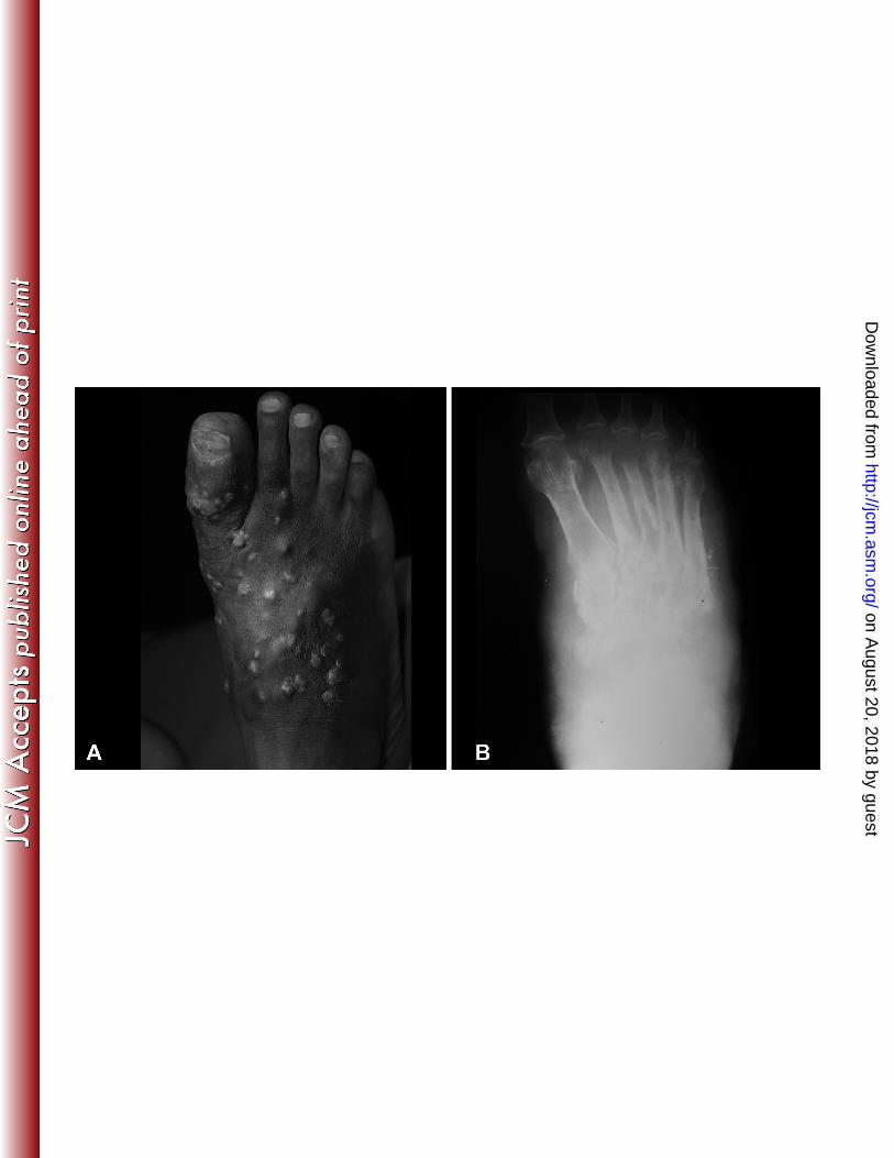
Figure 1: Clinical presentation of the patient. A: The patient had a mycetoma on the right foot 421
with a massive, recurrent foot swelling. B: On the foot X-ray a massive soft tissue swelling, a 422
periosteal reaction and bone destruction are seen. 423
424
425
426
427
428
429
430
431
432
433
434
435
436
437
438
439
on August 20, 2018 by guest
http://jcm.asm
.org/D
ownloaded from

440
Figure 2: Phylogram of two loci (SSU, LSU) obtained by Bayesian analysis, Maximum 441 likelihood, and Maximum parsimony (values > 0.8 Bayesian probability, > 80% Maximum 442 likelihood and Maximum parsimony are shown with bolded branches). Leotia lubrica and 443 Crinula caliciiformis were used as outgroup. 444
445
446
447
448
449
450
451
452
453
454
455
456
457
458
459
460
on August 20, 2018 by guest
http://jcm.asm
.org/D
ownloaded from

Figure 3: Phylogenetic tree resulting from a Bayesian analysis, Maximum likelihood, and 461
Maximum parsimony for the ITS gene.(values > 0.8 Bayesian probability, > 80% Maximum 462
likelihood and Maximum parsimony are shown with bolded branches) Togninia minima, 463
Phaeoacremonium inflatipes, and Phaeoacremonium aleophilum used as outgroup. Strain 464
isolated from plant (light gray circles), human pathogenic strain (dark gray circles). 465
on August 20, 2018 by guest
http://jcm.asm
.org/D
ownloaded from
Figure 4: Colony diameters after 1 month incubation at various temperature ranging from 15-466
36°C with 3°C intervals including 10°C, 37°C, and 40°C. 467
468
469
470
471
472
473
474
475
476
477
478
on August 20, 2018 by guest
http://jcm.asm
.org/D
ownloaded from
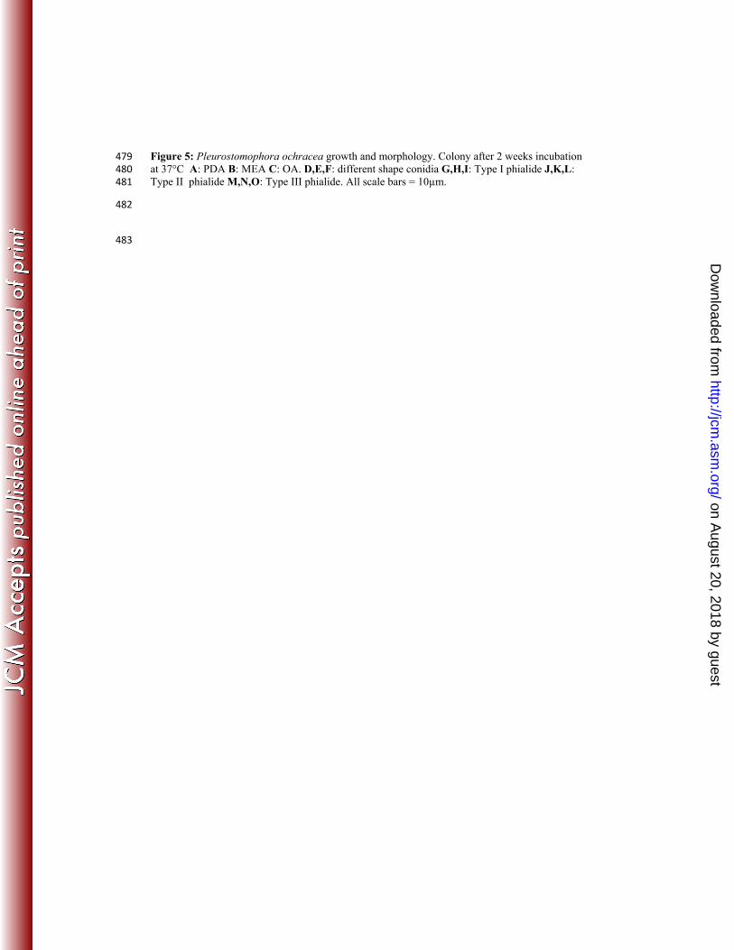
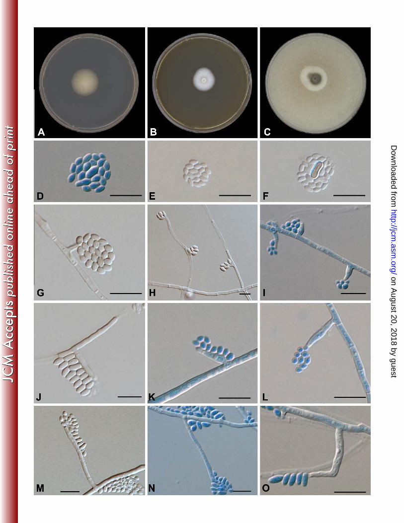
Figure 5: Pleurostomophora ochracea growth and morphology. Colony after 2 weeks incubation 479 at 37°C A: PDA B: MEA C: OA. D,E,F: different shape conidia G,H,I: Type I phialide J,K,L: 480 Type II phialide M,N,O: Type III phialide. All scale bars = 10µm. 481
482
483
on August 20, 2018 by guest
http://jcm.asm
.org/D
ownloaded from
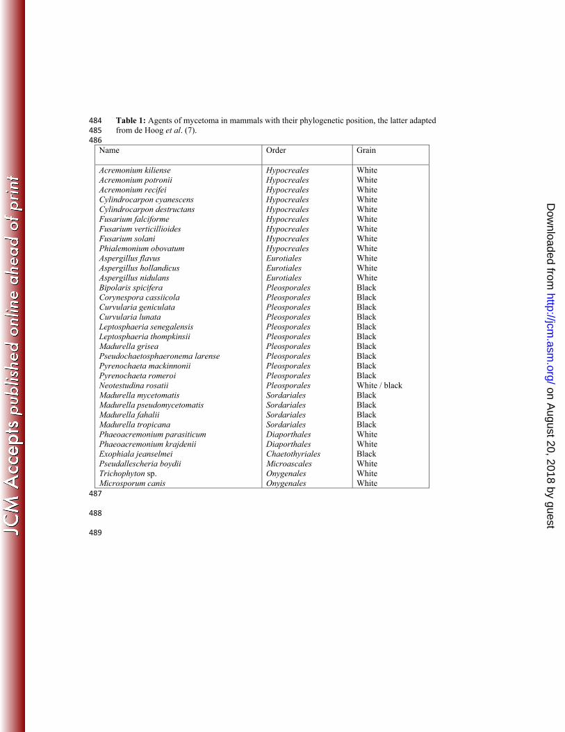
Table 1: Agents of mycetoma in mammals with their phylogenetic position, the latter adapted 484 from de Hoog et al. (7). 485 486
Name Order Grain
Acremonium kiliense Acremonium potronii Acremonium recifei Cylindrocarpon cyanescens Cylindrocarpon destructans Fusarium falciforme Fusarium verticillioides Fusarium solani Phialemonium obovatum Aspergillus flavus Aspergillus hollandicus Aspergillus nidulans Bipolaris spicifera Corynespora cassiicola Curvularia geniculata Curvularia lunata Leptosphaeria senegalensis Leptosphaeria thompkinsii Madurella grisea Pseudochaetosphaeronema larense Pyrenochaeta mackinnonii Pyrenochaeta romeroi Neotestudina rosatii Madurella mycetomatis Madurella pseudomycetomatis Madurella fahalii Madurella tropicana Phaeoacremonium parasiticum Phaeoacremonium krajdenii Exophiala jeanselmei Pseudallescheria boydii Trichophyton sp. Microsporum canis
Hypocreales Hypocreales Hypocreales Hypocreales Hypocreales Hypocreales Hypocreales Hypocreales Hypocreales Eurotiales Eurotiales Eurotiales Pleosporales Pleosporales Pleosporales Pleosporales Pleosporales Pleosporales Pleosporales Pleosporales Pleosporales Pleosporales Pleosporales Sordariales Sordariales Sordariales Sordariales Diaporthales Diaporthales Chaetothyriales Microascales Onygenales Onygenales
White White White White White White White White White White White White Black Black Black Black Black Black Black Black Black Black White / black Black Black Black Black White White Black White White White
487
488
489
on August 20, 2018 by guest
http://jcm.asm
.org/D
ownloaded from
490
Table 2: Calosphaeriales strains. 491
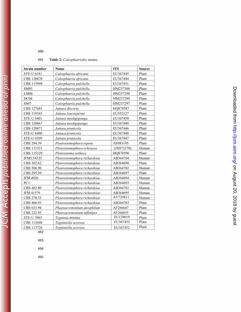
Strain number Name ITS Source STE-U 6181 Calosphaeria africana EU367445 Plant CBS 120870 Calosphaeria africana EU367444 Plant CBS 115999 Calosphaeria pulchella EU367451 Plant SM05 Calosphaeria pulchella HM237300 Plant LM06 Calosphaeria pulchella HM237298 Plant DC04 Calosphaeria pulchella HM237299 Plant SS07 Calosphaeria pulchella HM237297 Plant CBS 127681 Jattaea discreta HQ878587 Plant CBS 119343 Jattaea leucospermi EU552127 Plant STE-U 6401 Jattaea mookgoponga EU367450 Plant CBS 120867 Jattaea mookgoponga EU367449 Plant CBS 120871 Jattaea prunicola EU367446 Plant STE-U 6400 Jattaea prunicola EU367448 Plant STE-U 6399 Jattaea prunicola EU367447 Plant CBS 294.39 Pleurostomophora repens AF083195 Plant CBS 131321 Pleurostomophora ochracea (JX073270) Human CBS 115329 Pleurostoma ootheca HQ878590 Plant IFM5 54325 Pleurostomophora richardsiae AB364704 Human CBS 302.62 Pleurostomophora richardsiae AB364698 Plant CBS 506.90 Pleurostomophora richardsiae AB364702 Human CBS 295.39 Pleurostomophora richardsiae AB364697 Plant IFM 4926 Pleurostomophora richardsiae AB364694 Human PC1 Pleurostomophora richardsiae AB364693 Human CBS 483.80 Pleurostomophora richardsiae AB364701 Human IFM 41579 Pleurostomophora richardsiae AB364695 Human CBS 270.33 Pleurostomophora richardsiae AY729811 Human CBS 406.93 Pleurostomophora richardsiae AB364703 Plant CBS 631.94 Phaeoacremonium aleophilum AF266647 Plant CBS 222.95 Phaeoacremonium inflatipes AF266655 Plant STE-U 5963 Togninia minima EU128019 Plant CBS 113648 Togniniella acerosa EU367453 Plant
CBS 113726 Togniniella acerosa EU367452 Plant 492
493
494
495
on August 20, 2018 by guest
http://jcm.asm
.org/D
ownloaded from
496
Table 3: Antifungal susceptibility of Pleurostomophora ochracea. 497
Antifungal agent MICs
(µg/ml)
Amphotericin B 1
Itraconazole 0.25
Ketoconazole 0.25
Fluconazole 128
Voriconazole 0.5
Posaconazole 1
5-Flucytosin >64
Caspofungin 8
498
499
on August 20, 2018 by guest
http://jcm.asm
.org/D
ownloaded from