plasticity and stability of somatosensory maps in thalamus and cortex
TRANSCRIPT

494
Work on elucidating the mechanisms of plasticity insomatosensory maps continues apace. Recent work hasfocused on both the nature of the thalamocortical interactionsthat determine plasticity, and on the differences betweenplasticity induced by nerve block or damage versus thatinduced by experience. Recordings from awake behavinganimals have thrown light on the thalamocortical circuitmechanisms that underlie map plasticity; meanwhile,intracellular recordings from cortical slices have thrown light onthe precise synaptic mechanisms underlying plasticity.
AddressesCardiff School of Biosciences, Cardiff University, Museum Avenue,Cardiff CF10 3US, Wales, UK*e-mail: [email protected]
Current Opinion in Neurobiology 2000, 10:494–497
0959-4388/00/$ — see front matter© 2000 Elsevier Science Ltd. All rights reserved.
IntroductionSomatosensory maps are often studied both as a means ofunderstanding how information is processed between cor-tical columns and as a means of measuring plasticity. Themonkey hand representation (in the primary somatosenso-ry cortex) and the rat whisker representation (in the barrelcortex) have received much attention in recent years. Bothcontain unitary representations of body locations: digits inthe first case and whiskers in the second. The mapping ofadjacent body areas presumably confers some computa-tional advantage on the system, and yet the map is plasticenough to alter these relationships in response to changesin sensory experience or to nerve damage.
In this review, we focus on a number of recent papers con-cerned with the somatosensory map and plasticity of thesomatosensory system. Although lesion-induced plasticitydiffers in many respects from experience-dependent plastic-ity, and neonatal plasticity may differ from that found in theadult, we have tended to be inclusive in this review in orderto cover work published during the past year. We have splitthe reports into three sections. The first section concernsplasticity of a single whisker’s cortical representation,assessed by intrinsic signal imaging. The second section cov-ers recent results on the location of plasticity within thesomatosensory neuraxis, as assessed by single- and multi-electrode recording. In the third section, we review evidenceon some of the cellular mechanisms that might underlie mapplasticity in the cortex, such as coincidence detection ofpresynaptic and postsynaptic action potentials (or spikes).
Functional imaging of whisker representationIt has previously been found that stimulation of a singlewhisker elicits a response within the posterior medial barrel
subfield that covers an area of approximately 2 mm2 asassessed with optical imaging of the intrinsic signal [1,2].An area of 2 mm2 is roughly equivalent to the principal bar-rel-column for the stimulated whisker plus all of thesurrounding barrels (8 or 9, depending on the location).This finding is consistent with the fact that neurones in theprincipal barrel-column plus all of the surrounding barrelsshow responses to stimulation of a single whisker, althoughthe response magnitude in the principal barrel is greaterthan that in the surrounds [3].
In a recent study, Polley et al. [4•] applied optical imagingbased on intrinsic signals to measure the functional repre-sentation of a single whisker at different time-pointsduring sensory deprivation in the same animals. Data wereobtained before sensory deprivation, after 28 days ofdepriving all but one whisker and, for a few cases, after28 days of recovery. For animals that remained in theirfamiliar environment during the period of deprivation, thecortical representation of the spared whisker showed alarge-scale expansion. This result is qualitatively similar tofindings using single-cell spike responses [5] or metabolicactivity [6] to map the cortical representation. However,the scale of the enlargement reported by Polley et al. (fromabout 2 to 6 mm2) is very different to that observed in aquantitative 2-deoxyglucose study [6], where the activearea increased from about 0.33 to 0.65 mm2. It also differsfrom results found with single-cell recordings, where spikeresponses increased only in the neighbouring column [5].In fact, the enlarged representation found by Polley et al.covers almost the entire posterior medial barrel field.
In a further series of experiments, the same authors wenton to correlate the magnitude of the cortical change withthe frequency with which the spared whiskers were used.Within the 28 days of sensory deprivation, animals wereexposed for 2 min every other day to an enriched environ-ment that encouraged whisker-guided exploration.Surprisingly, this total of 14 min exposure time resulted ina substantial shrinkage of the functional representation ofthe spared whisker from nearly 4 to 1 mm2. Decreases indeprived whisker representation have been describedbefore [7], but this is the first description of a decrease inthe spared whisker representation. Finally, for a few ani-mals, the authors have shown that the functionalrepresentation can return to its original size from either acontracted or an expanded initial condition [4•].
The locus of sensory map plasticityWhen changes in cortical somatosensory map representa-tion were described in 1983 by Merzenich et al. [8] foradult animals, it was natural to assume that the changesdescribed originated in the cortex. Subsequent work has
Plasticity and stability of somatosensory maps in thalamus and cortexKevin Fox*, Stanislaw Glazewski and Silke Schulze
Somatosensory maps in thalamus and cortex Fox, Glazewski and Schulze 495
shown that this certainly can be the case if plasticity isinduced by changes in sensory experience. For example, aperiod of single-whisker experience leads to corticalexpression of plasticity that depends on intracortical con-nections [9] but that is not accompanied by changes in thesubcortical receptive fields [7,10•,11•]. However, in caseswhere peripheral nerve conduction is prevented, eitherpermanently by direct lesion or reversibly by application oflocal anaesthetic, subcortical changes are the norm ratherthan the exception. For example, peripheral nerve blockcauses an expansion of thalamic receptive fields [12••],damage to nucleus gracilis causes changes in thalamic rep-resentation [13•], and damage to the hand innervationcauses changes in the dorsal column nuclei [14].
Several laboratories have recently examined the causes ofsubcortical plasticity in the somatosensory system. Krupaet al. [12••] have studied a form of rapid plasticity inducedby peripheral nerve block. They show that if the cortex isinactivated by muscimol before the peripheral nerve blockoccurs, the expansion of receptive field size, or ‘unmask-ing’, in the thalamus is reduced though not completelyprevented. This implies a role for the cortex in the induc-tion of thalamic plasticity. A similar conclusion has beenreached by Parker and Dostrovsky [13•], who show thatthalamic plasticity can be induced by ablating nucleus gra-cilis, but only if the cortex is intact initially. Once plasticityhas been induced, it makes no difference to the expressionof plasticity whether the cortex is present or not.
What is the nature of the cortical influence on the thala-mus? Some insight is provided by Ergenzinger et al. [15]who show, in the monkey, that inactivating the cortex foran extended period causes thalamic receptive field expan-sion. This suggests that the cortex might exert a tonicinhibitory effect on the thalamus that is relieved by corti-cal inactivation. However, this point is far from resolution.Parker and Dostrovsky [13•] find no effect of corticallesions on thalamic responses in monkeys, and Diamondet al. [16] find cortical inactivation has no effect on ventralposterior medial thalamic responses in rats. It is possiblethat different thalamic cells are controlled in opposingways by cortex, so that blocking the cortical influence hasno overall net effect. In support of this idea, Krupa et al.[12••] have shown that 38% of neurones in the thalamus ofrats show unmasked receptive field components after cor-tical activity is blocked with muscimol. In other words,whiskers that did not drive the cell were able to after cor-tical inactivation. In contrast, 52% show a decrease inlong-latency responses representing a decrease in excita-tion. Further studies will be needed to resolve this issue.
Other recent studies have investigated changes caused bysensory deprivation rather than nerve damage or nerveblock. One of the issues raised by previous work on inter-actions between whisker responses was whethercross-whisker inhibition or cross-whisker excitation domi-nated in undeprived animals. Kelly et al. [17••] were ableto record responses to natural whisker stimulation using
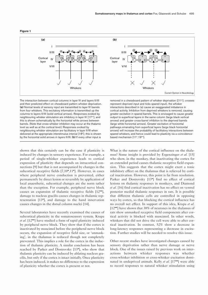
Figure 1
Current Opinion in Neurobiology
+ + ++ + +
I
II/III
IV
V/VI
I
II/III
IV
V/VI
Whiskers Whiskers
+ ++
– – – – – – – – –Corticallayer
(a)
Corticallayer
(b)
The interaction between cortical columns in layer IV and layers II/IIIand their predicted effect on chessboard pattern whisker deprivation.(a) Normal levels of sensory input are transmitted to layer IV barrelsfrom four whiskers. This excitatory information is transmitted up thecolumns to layers II/III (solid vertical arrows). Responses evoked byneighbouring whisker stimulation are inhibitory in layer IV [17••], andthis is shown schematically by the horizontal white arrows betweenbarrels. (Note that cross-whisker inhibition may occur at the thalamiclevel as well as at the cortical level.) Responses evoked byneighbouring whisker stimulation are facilitatory in layer II/III whendelivered at the appropriate interstimulus interval [18•]; this is shownby the horizontal solid arrows in layers II/III. (b) If every other input is
removed in a chessboard pattern of whisker deprivation ([11•]; crossesrepresent deprived input and ticks spared input), the whiskerinteractions described in (a) cause an exaggerated imbalance incortical activity. Inhibition from deprived whiskers is removed, causinggreater excitation in spared barrels. This is envisaged to cause greateroutput to superficial layers in the same column (large black verticalarrows) and greater cross-barrel inhibition to the deprived barrels(large white horizontal arrows). Greater excitation of horizontalpathways emanating from superficial layers (large black horizontalarrows) will increase the probability of facilitatory interactions betweenspared whiskers, and hence could lead to plasticity via a coincidence-based mechanism [11•,19••].
implanted multi-electrode arrays. They found that thewhiskers immediately surrounding a given whisker exert anet inhibitory effect on the whisker at the centre.Following removal of whiskers immediately neighbouringthe centre whisker, the response in the spared whisker’sbarrel (when the animal performed the same whisking taskagain) was greater than before deprivation. This is pre-sumably attributable to the fact that, during whisking,surrounding whiskers hit the target object at about thesame time as the whisker under study. Hence, any cross-barrel inhibition that is transmitted rapidly enough willtend to decrease the response from the centre whisker.
This result has implications for how plasticity might beinduced in the cortex by whisker deprivation. In the single-spared-whisker paradigm, the loss of phasic inhibition fromsurrounding barrels would produce unusually large respons-es in layer IV of the spared whisker’s principal barrel (seeFigure 1). The ‘disinhibited’ responses of the layer IV cellswould cause a greater output into the spared whisker’s col-umn and this would in turn activate horizontal connectionsbetween barrels more powerfully than before. This would bepredicted to lead to strengthening of the intracortical excita-tory connections between barrels over a period of time.
In the case of the single spared whisker, excitation fromthe spared whisker radiates out into a surrounding cortexthat is relatively quiet because all the other whiskers havebeen deprived. There is no cross-whisker excitation inthese cases and yet plasticity does occur; so does this ruleout a role for associative excitation in plasticity? Recentstudies suggest not. If multiple whiskers are spared, poten-tiation occurs more rapidly than if only a single whisker isspared (plasticity can take place within 7 days rather than18 days; [10•,11•]). Removing every other whisker in achessboard pattern would still be expected to cause thedisinhibition seen in layer IV [17••], but would also allowassociative excitatory interactions between barrel-columnsin extragranular layers (Figure 1). In this vein, it is inter-esting that Shimegi et al. [18•] have found that layer II/IIIcell responses are facilitated if two whiskers are activatedsequentially within about 2–4 ms. This effect is rarely seenin layer IV and may represent a real difference betweenthe cortical layers (see below). So, the recent evidence sug-gests that cross-whisker interactions in layer IV areinhibitory [17••], whereas in extragranular layers they areexcitatory when stimulation is delivered at the appropriateinterval [18•]. As described in the following section, thereis also reason to believe that cross-whisker excitation couldactivate a specific plasticity mechanism in the cortex.
Coincidence detection as a mechanism formap plasticitySubstantial progress has been made recently toward uncov-ering cellular mechanisms for experience-dependentplasticity in the barrel cortex [19••]. Experiments in two-week-old rat brain slices, using dual recordings frominterconnected intrabarrel spiny stellate neurones, show
that transmission between these neurones can be downreg-ulated by pairing presynaptic and postsynaptic spikeactivity. Moreover, this form of LTD can be evoked only ifpresynaptic and postsynaptic spiking occur within a 10 mstime window. The order of presynaptic and postsynapticspikes has no effect on the outcome. In contrast, pairedstimulation applied to interconnected pyramidal cells insupragranular layers results in long-term potentiation. Asmentioned above, facilitatory responses are also found inlayer II/III cells as a result of pairing adjacent whisker stim-ulation [18•], which suggests that this mechanism could alsobe employed in vivo.
Evidence that whisker deprivation causes synaptic poten-tiation in horizontal pathways between barrel-columns hasrecently been found by recording from layer II cells in cor-tical slices. The short-term dynamics of pathways betweencolumns have been studied, and greater steady-statedepression of synaptic responses has been found betweenspared and deprived columns than between undeprivedcolumns [20••]. There are theoretical and experimentalreasons to believe that these changes in short-term dynam-ics are the signature of a potentiated pathway (see [20••]).
Plasticity not only occurs between columns in the barrelcortex, but also within columns. In this regard, it is of inter-est that associative changes in synaptic strength can also beinduced in vertical pathways running between layer IV andsupragranular layers [20••,21]. Changes can be induced bypairing excitatory postsynaptic potentials (EPSPs) evokedby stimulation of layer IV with postsynaptic spikes in thelayer II/III cells. Pairing presynaptic and postsynapticresponses such that the EPSP precedes the spikes by nomore than 20 ms generates potentiation. However, pairingpresynaptic and postsynaptic responses such that the EPSPfollows the action potential by 20–100 ms results in depres-sion. The latter result suggests a mechanism by whichprincipal whisker responses might become depressed indeprived barrel-columns [7]. If the spontaneous activitythat remains in the deprived whisker pathway is relativelyrandom and does not correlate with pyramidal cell spikingdriven by the spared whisker, one would expect depressionof the response to the deprived input.
ConclusionsThe multiplicity of techniques being brought to bear onsomatosensory cortex, from multi-electrode recording andintrinsic imaging through to dual intracellular recording inslices, is yielding a greater wealth of information on plastic-ity in this area than ever before. Recent studies on plasticityinduced by nerve damage or nerve block show that the cor-tex is involved in changing subcortical representations. Theexact nature of the thalamocortical interaction will need tobe studied further in order to understand this phenomenon.Recent studies on cross-whisker interactions in layer IV andlayer II/III could help explain how imbalances in sensoryinput are amplified to induce cortical plasticity (seeFigure 1). Basic spike coincidence-detection mechanisms
496 Sensory systems

may be able to explain plasticity at the system level, butmodelling studies will probably be required to test thesetheories in more detail in the future.
References and recommended readingPapers of particular interest, published within the annual period of review,have been highlighted as:
• of special interest••of outstanding interest
1. Masino SA, Frostig RD: Quantitative long-term imaging of thefunctional representation of a whisker in the rat barrel cortex.Proc Natl Acad Sci USA 1996, 93:4942-4947.
2. Bhavin R, Moore CI, Sur M: Temporal modulation of spatial bordersin rat barrel cortex. J Neurophysiol 1998, 79:464-470.
3. Armstrong-James M, Fox, K: Spatiotemporal convergence anddivergence in rat SI ‘barrel’ cortex. J Comp Neurol 1987,263:265-281.
4. Polley DB, Chen-Bee CH, Frostig RD: Two directions of plasticity in• the sensory-deprived adult cortex. Neuron 1999, 24:623-637.Optical imaging of instrinsic signals is used to measure the area of barrelcortex activated by stimulating a single whisker. This area increases for thespared whisker if all others are removed, but contracts if the animal isexposed to a novel environment for a few minutes per session. Both con-traction and expansion are reversed by whisker regrowth.
5. Glazewski S, Fox K: The time course of experience-dependentsynaptic potentiation and depression in barrel cortex ofadolescent rats. J Neurophysiol 1996, 75:1714-1729.
6. Kossut M, Hand PJ, Greenberg J, Hand CL: Single vibrissal corticalcolumn in SI cortex of rats and its alterations in neonatal andadult vibrissa-deafferented animals: a quantitative 2DG study.J Neurosci 1988, 60:829-851.
7. Glazewski S, McKenna M, Jacquin M, Fox K: Experience-dependentdepression of vibrissae responses in rat barrel cortex. Eur JNeurosci 1998,10:2107-2116.
8. Merzenich MM, Kaas JH, Wall J, Nelson RJ, Sur M, Felleman D:Topographic reorganization of somatosensory cortical areas 3band 1 in adult monkeys following restricted deafferentation.Neuroscience 1983, 8:33-55.
9. Fox K: The cortical component of experience-dependent synapticplasticity in the barrel cortex. J Neurosci 1994, 14:7665-7679.
10. Wallace H, Fox K: Local cortical interactions determine the form of• cortical plasticity. J Neurobiol 1999, 41:58-63.The effects of different patterns of whisker deprivation are compared.Potentiation of responses to stimulation of spared whiskers occurred, anddepression of responses to deprived (regrown) whiskers occurred.Potentiation occurs faster in animals with multiple spared whiskers than inanimals with a single spared whisker. Depression is greater for deprivedwhisker responses the more whiskers are spared. Depression and potentia-tion occur in cortex but not in thalamus, indicating that both are cortical inorigin and depend on cortical circuits and synapses.
11. Wallace H, Fox K: The effect of chessboard deprivation pattern on• potentiation and depression of vibrissae responses in rat barrel
cortex. Somatosens Mot Res 1999, 16:122-138.The effect of a new pattern of sensory deprivation, where every other whiskeris removed in a chessboard pattern, is described. Each deprived cortical col-umn is surrounded by a spared column and vice versa. Potentiation anddepression of whisker responses are shown to occur in the cortex but notthe thalamus.
12. Krupa DJ, Ghazanfar AA, Nicolelis MA: Immediate thalamic sensory•• plasticity depends on corticothalamic feedback. Proc Natl Acad
Sci USA 1999, 96:8200-8205.Plasticity is induced in the thalamus by injecting local anaesthetic into thewhisker pad. The degree of rapid plasticity measured with multi-electrodearrays in the thalamus is decreased if the cortex is inactivated with muscimol.The results imply that thalamic plasticity depends on the cortex.
13. Parker JL, Dostrovsky JO: Cortical involvement in the induction, but• not expression, of thalamic plasticity. J Neurosci 1999, 19:8623-8629.In the monkey, plasticity is induced in the thalamus by making a lesion in nucle-us gracilis of the dorsal column system. If the somatosensory cortex is ablatedbefore inducing plasticity, plasticity is prevented. However, if the cortex is ablat-ed after plasticity has occurred, plasticity is still expressed in the thalamus.
14. Xu J, Wall JT: Evidence for brainstem and supra-brainstemcontributions to rapid cortical plasticity in adult monkeys.J Neurosci 1999, 19:7578-7590.
15. Ergenzinger ER, Glasier MM, Hahm JO, Pons TP: Cortically inducedthalamic plasticity in the primate somatosensory system. NatNeurosci 1998, 1:226-229.
16. Diamond ME, Armstrong-James M, Budway MJ, Ebner FF: Somaticsensory responses in the rostral sector of the posterior group(POm) and in the ventral posterior medial nucleus (VPM) of therat thalamus: dependence on the barrel field cortex. J CompNeurol 1992, 319:66-84.
17. Kelly MK, Carvell GE, Kodger JM, Simons DJ: Sensory loss by•• selected whisker removal produces immediate disinhibition in the
somatosensory cortex of behaving rats. J Neurosci 1999,19:9117-9125.
An implanted electrode array is used to record from cortex or thalamus dur-ing a standard whisking task. The responses of cortical neurones increase ina principal barrel if the whiskers surrounding that barrel’s principal whiskerare removed. This shows that surround receptive field whiskers produce lat-eral inhibition in the cortex. Conversely, thalamic responses to the sparedprincipal whisker decrease, suggesting that surround receptive fieldwhiskers provide a net lateral excitation in thalamus.
18. Shimegi S, Ichikawa T, Akasaki T, Sato H: Temporal characteristics• of response integration evoked by multiple whisker stimulations
in the barrel cortex of rats. J Neurosci 1999, 19:10164-10175.Extracellular recordings are used to measure responses of neurones to paireddeflections of adjacent whiskers. Facilitatory responses occur in layer II/IIIcells when deflections are spaced up to 3 ms apart. Facilitation is more com-mon in layers II/III (65% of neurons) than in layers IV or V (15% of neurons).
19. Egger V, Feldmeyer D, Sakman B: Coincidence detection and•• changes of synaptic efficacy in spiny stellate neurons in rat barrel
cortex. Nat Neurosci 1999, 12:1098-1105.Dual intracellular recordings are made in interconnected intrabarrel layer IVstellate cells from thalamocortical slices. Pairing spikes in the two cells withina 10 ms time-window leads to depression of responses to presynaptic input.
20. Finnerty GT, Langdon SE, Roberts, Connors BW: Sensory•• experience modifies the short term dynamic of neocortical
synapses. Nature 1999, 400:367-371.Slices of barrel cortex are taken from animals that have had the A,B,C or D,Erow whiskers removed for 10 days. Recordings are made from layer II cellsin either the deprived or the spared barrel-columns and responses evokedby electrical stimulation from vertical or horizontal pathways. Steady-statedepression to repeated stimuli is greater in horizontal pathways from sparedcolumns to deprived columns than between columns in undeprived cortex.The authors use modelling to show how this indicates that these pathwayshave been potentiated by deprivation. Similarly, vertical pathways betweenlayer IV and layer II show potentiation in spared columns.
21. Feldman DE: LTP and LTD induced by action potential–EPSPpairing at vertical inputs to layer II/III pyramids in ratsomatosensory cortex. Soc Neurosci Abstr 1999, 28:223.
Somatosensory maps in thalamus and cortex Fox, Glazewski and Schulze 497