development/plasticity/repair ... ·...
TRANSCRIPT

Development/Plasticity/Repair
Barrel Map Development Relies on Protein Kinase ARegulatory Subunit II�-Mediated cAMP Signaling
Melis Inan,1 Hui-Chen Lu,1,3 Michael J. Albright,2 Wei-Chi She,1,3 and Michael C. Crair1,2
1Program in Developmental Biology, 2Department of Neuroscience, and 3The Cain Foundation Laboratories, Department of Pediatrics, Division ofNeurology/Developmental Neuroscience, Baylor College of Medicine, Houston, Texas 77030
The cellular and molecular mechanisms mediating the activity-dependent development of brain circuitry are still incompletely under-stood. Here, we examine the role of cAMP-dependent protein kinase [protein kinase A (PKA)] signaling in cortical development andplasticity, focusing on its role in thalamocortical synapse and barrel map development. We provide direct evidence that PKA activitymediates barrel map formation using knock-out mice that lack type II� regulatory subunits of PKA (PKARII�). We show that PKARII�-mediated PKA function is required for proper dendritogenesis and the organization of cortical layer IV neurons into barrels, but not forthe development and plasticity of thalamocortical afferent clustering into a barrel pattern. We localize PKARII� function to postsynapticprocesses in barrel cortex and show that postsynaptic PKA targets, but not presynaptic PKA targets, have decreased phosphorylation inpkar2b knock-out (PKARII��/�) mice. We also show that long-term potentiation at TC synapses and the associated developmentalincrease in AMPA receptor function at these synapses, which normally occurs as barrels form, is absent in PKARII��/� mice. Together,these experiments support an activity-dependent model for barrel map development in which the selective addition and elimination ofthalamocortical synapses based on Hebbian mechanisms for synapse formation is mediated by a cAMP/PKA-dependent pathway thatrelies on PKARII� function.
Key words: somatosensory cortex; activity dependent; glutamate; PKA; barrel; NMDA receptor
IntroductionA significant remaining challenge in developmental neuroscienceis to understand the cellular and molecular mechanisms respon-sible for the organization of the intricate pattern of neural con-nections typical of the mammalian forebrain. Topographic mapsof the sensory periphery are useful for this purpose, because eventheir gross form reflects the organization of precise underlyingcircuitry. Evidence suggests that the development of crude topog-raphy in these maps requires molecular cues, and map refine-ment depends, at least in part, on neural activity (Erzurumlu andKind, 2001; Kind and Neumann, 2001; Cline, 2003; Lopez-Bendito and Molnar, 2003). A powerful model to study this pro-cess is the representation of facial whiskers in the primary so-matosensory cortex of mice, known as “barrel” cortex. Eachcortical barrel is composed of a barrel “wall” of densely packedlayer IV cortical neurons that orient their dendrites into a cell-sparse barrel “hollow” occupied by clustered thalamocortical(TC) axons (TCAs) that relay input from a single whiskerthrough the ventroposteromedial (VPM) thalamus. Evidencefrom a variety of sources, including studies of activity-dependent
thalamocortical synapse development (Crair and Malenka, 1995;Feldman and Knudsen, 1998) and examination of various mu-tant mice with barrel map deficits (Erzurumlu and Kind, 2001;Kind and Neumann, 2001), suggests that barrel formation relieson an activity dependent process.
The cellular signaling events triggered by neuronal activitythat influence barrel development are just now being examined indetail. Studies with “barrelless” (brl) mice, a loss of functionmutant in the gene coding for adenylyl cyclase type I (Adcy1)(Welker et al., 1996; Abdel-Majid et al., 1998), have focused at-tention on the role of protein kinase A (PKA) signaling in barrelmap development. Adcy1 is a Ca 2�/calmodulin-stimulated ad-enylyl cyclase that catalyzes the formation of cAMP when Ca 2�
concentrations increase, which occurs as a result of activation ofNMDA receptors (NMDARs) (Wang and Storm, 2003). Themain target of cAMP is PKA, a kinase strongly implicated inactivity dependent changes in synaptic function related to learn-ing and memory (Sweatt, 2001; Bauman et al., 2004) and devel-opmental plasticity (Beaver et al., 2001, 2002; Yasuda et al., 2003;Fischer et al., 2004; Rao et al., 2004). We previously describeddeficits in thalamocortical long-term potentiation (LTP) andPKA-dependent phosphorylation of AMPA receptors(AMPARs) in brl mice, which are likely to play an important rolein activity-dependent barrel map formation (Lu et al., 2003).
The PKA holoenzyme is a tetramer composed of a ho-modimer of two regulatory subunits (RI�, RI�, RII�, or RII�),each of which binds to one of three catalytic subunits (C�, C�, orC�). Binding of cAMP to the regulatory subunit triggers its dis-
Received Sept. 4, 2005; revised Dec. 31, 2005; accepted Jan. 21, 2006.This work was supported by National Institutes of Health Grant R01 MH62639, the Mental Retardation and
Developmental Disabilities Research Center at Baylor College of Medicine, and a National Alliance for Research onSchizophrenia and Depression Sidney R. Baer Jr Foundation Investigator award to M.C.C. We thank Dr. G. Lonart forphospho-specific antibodies and Bobby Antalffy for help with histology.
Correspondence should be addressed to Michael C. Crair, Department of Neuroscience, Baylor College of Medi-cine, One Baylor Plaza, S-603, Houston, Texas 77030. E-mail: [email protected].
DOI:10.1523/JNEUROSCI.3745-05.2006Copyright © 2006 Society for Neuroscience 0270-6474/06/264338-12$15.00/0
4338 • The Journal of Neuroscience, April 19, 2006 • 26(16):4338 – 4349
sociation from the catalytic subunit, which becomes enzymati-cally active. In the last decade, mice with null mutations of thegenes encoding the various PKA subunits have been generated inan effort to distinguish the role of different subunits in PKAactivity (Brandon et al., 1997). Although the loss of a subunit isoften compensated for by an increase in expression of other sub-units, specific functions are perturbed in various PKA subunitknock-outs (Huang et al., 1995; Qi et al., 1996; Amieux et al.,1997; Brandon et al., 1998; Thiele et al., 2000; Howe et al., 2002;Fischer et al., 2004; Rao et al., 2004). Histological studies showthat all PKA subunits are expressed in neurons, but PKARII� is agood candidate for providing specificity for PKA activity in neu-rons because of its low cAMP affinity and restricted subcellularexpression pattern (Cadd and McKnight, 1989; Brandon et al.,1997).
Using pkar2b knock-out (PKARII��/�) mice, we provide ev-idence for a direct role of PKA activity in the morphological andcytoarchitectonic development of cortical layer IV neurons dur-ing barrel map formation. Our findings also reveal thatPKARII�-mediated PKA activity is not required for the grosspatterning of TCA development and plasticity. Finally, we showthat long-term potentiation and the associated development ofAMPAR-mediated function at thalamocortical synapses is im-paired in PKARII��/� mice, lending further evidence in supportof the hypothesis that activity-dependent regulation of thalamo-cortical synapse development by PKA plays an important role inbarrel map formation.
Materials and MethodsAnimals and PCR analysisPKARII� homozygous knock-out mice of the sixth backcross generation ofthe incipient C57BL/6-pkar2b�/� 98% congenic inbred strain were origi-nally generated by Dr. Stanley McKnight (University of Washington, Seattle,WA) (Brandon et al., 1998) and obtained commercially from Taconic Farms(Germantown, NY). PKARII��/� mice were generated by a neomycin cas-sette insertion into the first exon of the pkar2b gene. Experiments wereperformed blind to genotype, with littermate controls. With the exception ofthe Golgi data, n in the text refers to the number of animals, because only onecell or section per animal was used for analysis. DNA for genotyping waspurified from tails using the DNeasy kit from Qiagen (Valencia, CA). Wild-type PCR primers (5�-GCAGGATGAGCATCGAG-3�, 5�-TTCGAGAGTGAGGCGGA-3�) produced a 330 base pair (bp) band, and mutant prim-ers (5�-GCAGGATGAGCATCGAG-3�, 5�-TTCGAGAGTGAGGCGGA-3�) produced a 244 bp band. Sixteen to 20 �l volumes were used for PCRprepared using the PCR kit from Qiagen. The PCR was performed with aninitial denaturation step at 95°C for 5 min and 40 cycles at 94°C for 1 min,57°C for 1 min, 72°C for 45 s, and a single final cycle at 72°C for 10 min. PCRproducts were resolved on a 2% agarose gel and detected with ethidiumbromide.
HistologyCytochrome oxidase staining. Cytochrome oxidase (CO) is an enzyme ofthe mitochondrial electron transport chain that is responsible for most ofthe ATP production in cells. Its relative activity in tissue sections, whichcan be easily demonstrated by following a simple histochemical proce-dure (Seligman et al., 1968; Wong-Riley et al., 1978), reveals the patternof thalamic afferent clustering into barrels in S1 cortex of rodents(Wong-Riley and Welt, 1980). It can also be used to detect changes in thepattern of thalamic afferent clustering into cortical barrels after manip-ulation of the sensory periphery (Wong-Riley et al., 1980; Lu et al., 2001).Briefly, barrel cortex was removed following the methods described byStrominger and Woolsey (1987), fixed and flattened for 2– 4 h in 4%paraformaldehyde (PFA) at room temperature, and cut tangentially(parallel to layer IV) on a vibratome (VT1000S; Leica, Nussloch, Ger-many) into 100 �m sections and subject to CO staining. Sections wereincubated with CO reaction solution free-floating for 2 h at room tem-perature or 10 –14 h at 4°C. After visual detection of stain, sections were
washed with PBS three times and mounted with Fluoromount-G (Elec-tron Microscopy Sciences, Hatfield, PA).
Nissl staining and analysis of barrel cytoarchitecture. Nissl stain, whichmarks neuronal nuclei, was used to reveal cortical barrel cytoarchitec-ture. Briefly, animals were perfused transcardially with ice-cold PBS fol-lowed by 4% PFA in PBS. Barrel cortex was then removed and flattenedfor 4 h in 4% PFA at room temperature and cut tangentially (parallel tolayer IV) on a vibratome (VT1000S; Leica) into 50-�m-thick tangentialsections. Sections were then mounted and dried for 1 d on a slide warmerat 37°C. Slides were dehydrated and rehydrated in graded alcohol, thenfixed in 10% formalin (Sigma, St. Louis, MO) and stained with 0.2%cresyl violet solution for 15 min. After dehydration with graded alcoholand xylene, slides were mounted with Cytoseal (Richard-Allen Scientific,Kalamazoo, MI). The density of neurons in barrel walls and hollows wasdetermined in three representative barrels (b2, c2, and d2) by markingthe cells within a closed fixed contour drawn using Neurolucida software(MicroBrightField, Colchester, VT). The average (Avg) cell density ofthese three barrels was calculated for each animal and compared betweengenotypes. All counts were done blind to the genotype.
Hematoxylin and eosin staining. Whisker pads of the lesioned (right)and control (left) side were removed from the snout and fixed with 4%PFA overnight at 4°C. PFA (4%) was exchanged with 30% sucrose forcryoprotection for 2 d at 4°C. Serial tangential sections of 50 �m werecollected using a cryostat and subjected to standard hematoxylin andeosin (H&E) staining to examine hair follicle cells.
Golgi staining and analysis of dendrite asymmetry. Golgi stained neu-rons were obtained using the FD Rapid Golgi Stain kit (FD Neurotech-nologies, Ellicott City, MD). Tissue was prepared according to the usermanual, and 50 �m serial coronal sections were cut using a cryostat.Golgi staining was performed following kit instructions, and Nissl coun-terstaining, used to determine the boundaries of layer IV, was performedright after Golgi staining and before dehydration, as explained in the kit.Randomly selected layer IV spiny stellate neurons were examined undera light microscope at 10�, and dendrites were reconstructed in threedimensions at 100� with an oil immersion lens using Neurolucida soft-ware (MicroBrightField) blind to the genotype. Polar graphs that repre-sent the length of dendritic processes binned every 10° (Fig. 3 E, F ) wereplotted for each neuron using Neuroexplorer (MicroBrightField). Den-dritic asymmetry was evaluated from the polar plots by calculating thedendritic length in the hemisphere (180°) with the greatest density ofdendrites relative to the total dendritic length in a given neuron. Spinystellate neurons with a dendritic asymmetry of 0.75 and above wereaccepted to have a dendritic orientation bias. Other parameters such astotal dendritic length, spine density, and dendritic field span were alsodetermined using Neuroexplorer (MicroBrightField).
Sensory manipulationsC row of whiskers and whisker follicles from postnatal day 1 (P1) and P5animals were cauterized with a surgical cautery device (Malis BipolarCoagulator & Cutter; model CMC II; Codman, Piscataway, NJ). Pupswere anesthetized on ice, and whiskers on the right side were visualizedunder a light microscope. After cauterization, the pups were revived in awarmed, oxygenated chamber and returned to their mother. The animalswere then killed at P12–P14 for CO and H&E staining (see above). Ani-mals with lesions that spread beyond row C, or did not include at least thefirst four whiskers in row C, as revealed by the H&E staining, were ex-cluded from analysis.
For quantification of barrel map plasticity, the width of CO-stainedwhisker barrels corresponding to the b1, b2, b3, c1, c2, c3, d1, d2, and d3whiskers were measured using ImageJ Software (W. S. Rasband, NationalInstitutes of Health, Bethesda, MD). The ratio of the average width ofC-row whisker barrels (c1– c3) relative to the B- and D-row whiskerbarrels (b1– b3 and d1– d3) was used to quantify a map plasticity index(MPI) defined as follows: MPI � 2 � [Avg(c1, c2, c3)]/[Avg(b1, b2,b3) � Avg(d1 � d2 � d3)].
PKA activity assayPKA activity was measured in total tissue homogenates as a ratio in thepresence and absence of cAMP using the SignaTECT cAMP-dependent
Inan et al. • Barrel Development Relies on Postsynaptic PKARII� J. Neurosci., April 19, 2006 • 26(16):4338 – 4349 • 4339
protein kinase assay kit (Promega, Madison, WI). Briefly, somatosensorycortex was removed at P11 and homogenized in ice-cold homogeniza-tion buffer (0.32 M sucrose, 10 mM HEPES, pH 7.4) with protease andphosphatase inhibitor mixtures (Sigma) on the same day as the activitytest. One gram of tissue was homogenized in 5 ml of homogenizationbuffer. Appropriate dilutions were prepared in 0.1 mg/ml BSA, and thereaction was performed following the steps clearly explained in the kit. Acontrol reaction without substrate was also run for each sample to deter-mine the background radioactivity, which was counted using a scintilla-tion counter.
Protein analysesAntibodies. The following primary antibodies were used: PKARII� (at1:1000 dilution for immunoblotting and 1:500 for immunohistochemistry)and PKAC (1:1000) from PharMingen (San Diego, CA); serotonin trans-porter (5-HTT; 1:250) from Immunostar (Hudson, WI); microtubule-associated protein (MAP2; 1:1000) from Sternberger (Lutherville, MD); ac-tin (1:750) from Sigma; phospho-synapsin (Ser9) (1:1000) from CellSignaling Technology (Beverly, MA); phospho-MAPK (mitogen-activatedprotein kinase; 1:1000) from Chemicon (Temecula, CA); phospho-Rim (1:1000) from Dr. G. Lonart (Eastern Virginia Medical School, Norfolk, VA);phospho-glutamate receptor 1 (phospho-GluR1; 1:1000), phospho-NMDAR subunit 1 (phospho-NR1; 1:1000), and GluR1 (1:1000) from Up-state Biotechnology (Lake Placid, NY); Rim (1:1000) and NR1 (1:5000) fromSynaptic Systems (Gottingen, Germany). The following secondary antibod-ies were used: mouse IgG–HRP (1:20,000) and rabbit IgG–HRP (1:12,500)from Pierce (Rockford, IL); mouse IgG–Alexa 488 (1:500), mouse IgG–cyanine 3 (Cy3) (1:500), rabbit IgG–Alexa 488 (1:500) from Invitrogen (SanDiego, CA).
Immunoblotting. Barrel cortex was isolated from P11 mice and ho-mogenized in homogenization buffer with protease and phosphataseinhibitor mixtures (Sigma). Subcellular fractions were acquired as de-scribed previously (Lu et al., 2003). Briefly, the homogenates were cen-trifuged at 800 � g for 10 min and the pellet was saved as P1, which isenriched with cell bodies. The supernatant was then centrifuged at7100 � g for 15 min to yield pellet P2, the synaptosome-enriched frac-tion. Synaptosome enrichment was confirmed by probing different frac-tions with antibodies for synaptic marker proteins [e.g., PSD-95(postsynaptic density-95)]. Protein concentrations were measured withBradford assay using Bio-Rad (Hercules, CA) protein assay kit II. Twentymicrograms of total protein were loaded into 5–20% gradient SDS-PAGEgels and electrophoretically transferred to nitrocellulose membranes (Crite-rion system; Bio-Rad). Primary antibodies against the protein in questionwere applied and the signal detected with HRP-conjugated secondary anti-bodies. Immunoreactivity was quantified with ECL pico (Pierce), and den-sitometric quantification was performed with Optiquant software (PackardBioscience, Meriden, CT) by background subtracting and normalizing bandintensities to the wild-type band in each set. When using PKA site-specificantibodies, quantification was performed by comparing the level of phos-pho-specific antibody to the activity of antibodies against the total protein.Otherwise, quantification was performed relative to actin, which is expressedat consistent high levels throughout barrel development.
Immunofluorescence and colocalization analysis. Mice were perfused asexplained above, and the brain was removed and fixed overnight at 4°C.One hundred micrometer sections in the thalamocortical plane were cutwith a vibratome, and sections remained free-floating for all incubationsand washes. Briefly, tissues were permeabilized with 0.7% Triton X-100in PBS for 20 min, incubated with 0.1 M glycine for 30 min, and thenblocked with 1% normal goat serum (NGS) and 2 mg/ml BSA in PBScontaining 0.01% Triton X-100 (PBST). Sections were then transferredto the primary antibody for incubation overnight at 4°C with appropriatedilution in PBST with 1% NGS. After washing six times in PBST, sectionswere incubated in the secondary antibody for 2 h at room temperaturethen washed again six times. 4�,6�-Diamidino-2-phenylindole (300 nM)was often applied to label nuclei, followed by a wash and postfix of thesections with PFA, and then mounting with Fluoromount-G (ElectronMicroscopy Sciences). Immunolabeling was visualized with a fluores-cence microscope, and images were collected with a Leica DM confocalscanning microscope using 10, 20, and 63� water immersion lenses with
the channels for Cy3 (red) and Alexa488 (green) used sequentially. Eachimage was acquired with the laser intensity adjusted to prevent over-saturation. For double-labeling with two antibodies made in differentspecies, a mixture of the antibodies against the two proteins was appliedat once. If the two antibodies were made in the same species, the primaryantibodies were applied sequentially. Colocalization of antibodies thatmark either presynaptic (5-HTT) or postsynaptic (MAP2) processes withan antibody for PKARII� was performed on 63� images using the colo-calization finder function in ImageJ.
Slice electrophysiologyAcute TC slices were prepared as described previously (Lu et al., 2001).The artificial CSF (ACSF; in mM: 124 NaCl, 5 KCl, 1.25 NaH2PO4, 1.3MgSO4, 2 CaCl2, 26 NaHCO3, and 11 glucose, pH 7.2, 290 –300 mOsm)was saturated with 95% O2 and 5% CO2. The whole-cell recording solu-tion contained the following (in mM): 99 cesium gluconate, 17.5 CsCl, 8NaCl, 10 HEPES, 0.2 EGTA, 4 Mg-ATP, 0.3 GTP, 7 phosphocreatine, and10 BAPTA. BAPTA was included in the pipette to prevent inadvertentpotentiation of the postsynaptic neuron.
Stimuli were applied to the ventrobasal thalamus through bipolarsharpened and insulated stainless-steel microelectrodes (Frederick HaerCompany, Bowdoinham, ME). Data were collected and analyzed on-lineusing a computer-driven acquisition system (National Instruments,Austin, TX) and software that was written under the Igor (WaveMetrics,Lake Oswego, OR) programming environment.
Input– output analysis of synaptic transmission using field potentialrecordings was conducted with a range of stimulus intensities (0 – 850 �Awith 50 �A intervals). The nonspecific glutamate receptor antagonistkynurenic acid (10 mM) (Sigma) was routinely added to the perfusate atthe end of the recordings to ensure the identification of fiber volley andsynaptic response was correct. Fiber volley amplitudes and field EPSPslopes were averaged for 20 sweeps at each stimulus intensity.
EPSCs were measured in voltage-clamp mode using in vitro whole-cellvoltage-clamp recording techniques following published protocols (Luet al., 2001). To evaluate and monitor the health of the cell, input andseries resistances were continuously monitored, with cells that had �300M� input resistance or drifted �20% discarded. Only responses thatexhibited short and constant latencies that did not change with increas-ing stimulus intensity were considered monosynaptic. Because the re-sponses in young cells tend to drift down if the stimulation intensity orthe stimulation frequency is high, half-saturating stimulation strengthwith a relatively low stimulation frequency (10 –15 ms interval) was usedto evoke stable EPSCs. Before any experimental manipulation, 10 –15min of stable baseline response was acquired. EPSC amplitudes werecalculated by subtracting the mean current during a fixed 3– 4 ms win-dow before the stimulus artifact from the mean current during a similarwindow at the peak of the EPSC.
To measure the AMPAR/NMDAR current ratio, which is a gross mea-sure of the relative contribution of AMPA and NMDA receptor-mediated currents across a population of synapses, we first isolated theAMPA response by voltage clamping the cell at hyperpolarized mem-brane potentials (�70 mV) while stimulating the thalamus. We thendepolarized the cell to �40 mV to relieve the Mg 2� block of the NMDARand added 2,3-dihydroxy-6-nitro-7-sulfonyl-benzo[f]quinoxaline (10�M) (Tocris Cookson, Ballwin, MO) to the perfusate to block AMPA andkainate receptors, leaving a pure NMDAR response. These experimentswere done at normal (half-saturating) stimulus strength as a gross mea-sure of the relative amplitude of the AMPAR and NMDAR currents(Crair and Malenka, 1995; Lu et al., 2001, 2003) in the presence ofGABAergic antagonists (0.1 mM picrotoxin, cesium ions in the whole-cellsolution block GABAB responses).
For analysis of AMPA “evoked miniature” events, stable whole-cellvoltage-clamp recordings were established at �70 mV holding potential,and the Ca 2� in the ACSF was exchanged for Sr 2�. Sr 2�-based ACSFdesynchronizes release, allowing isolated evoked miniature currents tobe analyzed (Xu-Friedman and Regehr, 1999). Evoked miniature eventswere recorded in 1 s epochs every 2 s in Igor Pro using ACSF (2 mM Sr 2�)containing 100 �M picrotoxin and 50 �M D-AP-5 (Tocris Cookson) toeliminate inhibitory currents and possible NMDAR current contamina-
4340 • J. Neurosci., April 19, 2006 • 26(16):4338 – 4349 Inan et al. • Barrel Development Relies on Postsynaptic PKARII�
tion, respectively. Data was imported into Mini Analysis (Synaptosoft,Decatur, GA), amplitude thresholds were set at 2.5 times root meansquare (RMS) noise, and at least 80 events were identified and used foranalysis in each cell. Temporal windows were chosen to represent boththe fast and slow components of miniature event decay times. A signifi-cant advantage of measuring “evoked minis” is that the population isdominated by TC synapses, as opposed to measuring spontaneous minisin TTX, which is a random sample of all synapses on the cell.
LTP was induced using a pairing protocol, in which postsynaptic de-polarization is “paired” with presynaptic stimulation in whole-cellvoltage-clamp mode (Lu et al., 2001). For these experiments, BAPTA wasexcluded from the pipette solution, and EPSCs from somatosensory cor-tex layer IV neurons elicited with thalamic stimulation were recorded at�70 mV until a stable baseline of 6 –10 min was obtained. Pairing wasthen initiated by switching to a holding potential of �10 mV and stim-ulating at 1 Hz for 100 s. The holding potential was then switched back to�70 mV after pairing. The percentage of EPSC change was calculated asthe mean EPSC amplitude of 20 sweeps at 20 min after pairing minus themean EPSC amplitude of 20 sweeps right before pairing (baseline ampli-tude) divided by the baseline amplitude.
ResultsPKA activity and catalytic subunit protein levels are reducedin PKARII��/� micePKARII��/� mice lack a regulatory subunit that controls PKAsignaling activity. Because binding of PKA regulatory subunits toPKA catalytic subunits renders the PKA holoenzyme inactive,one would naively expect an increase in PKA activity inPKARII��/� mice. However, previous studies show that PKAactivity is actually reduced in PKARII��/� mice (Brandon et al.,1997) because of the rapid degradation of catalytic subunits in theabsence of a regulatory subunit (Amieux et al., 1997; Brandon etal., 1998).
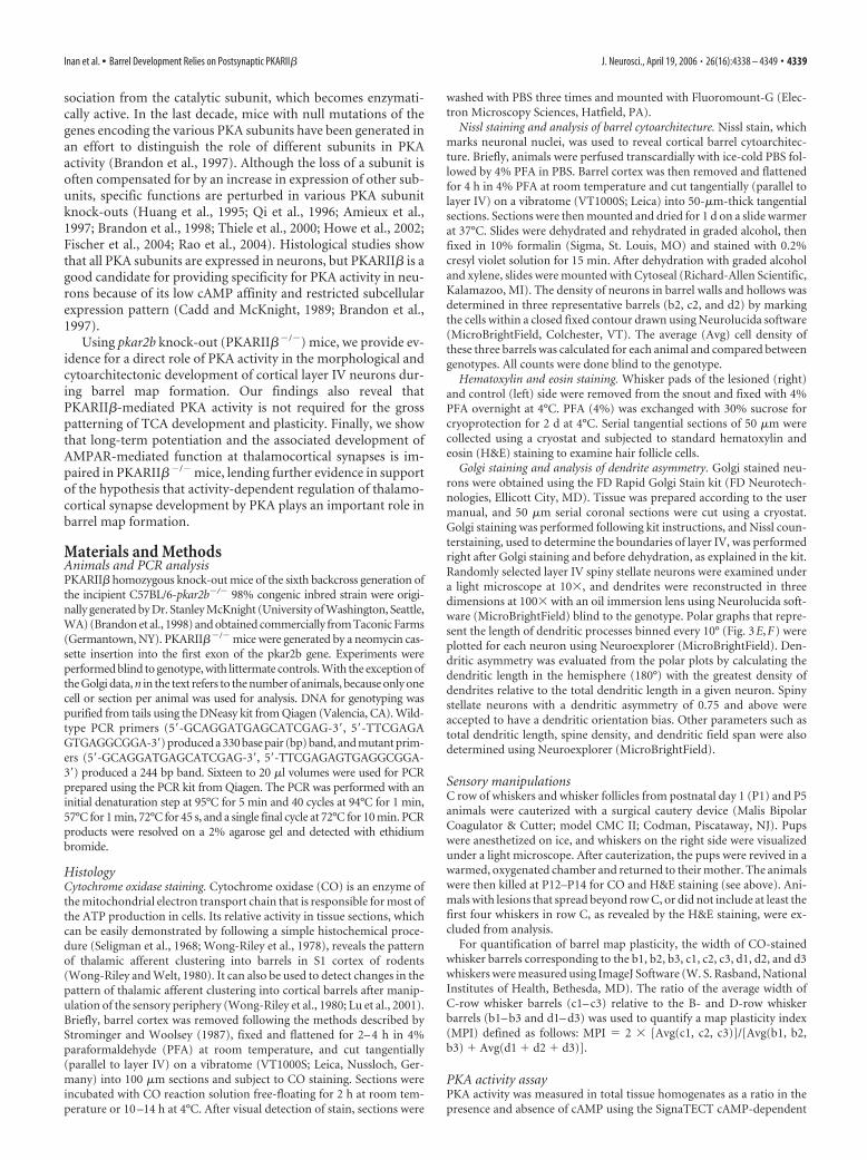
We examined how PKA activity was affected in the developingsomatosensory cortex of PKARII��/� mice using tissue homog-enates from P11 somatosensory cortex of PKARII��/� mice(n � 3) and wild-type controls (n � 3). When compared withwild types, knock-outs had a significant reduction in stimulatedPKA activity (ratio of PKA activity in the presence and absence ofcAMP) (2.55 0.07 vs 1.32 0.03; p � 0.005; t test) (Fig. 1A).We also measured PKAC protein levels in synaptosomes pre-pared from P11 somatosensory cortex of PKARII��/� mice andwild-type littermate controls using Western blot analysis (Fig.1A,B). Quantification demonstrated that PKAC subunit protein
levels are lower in PKARII��/� somatosensory cortex comparedwith wild-type controls (0.49 0.06; n � 5 pairs; p �� 0.01; ttest). These data show that a decrease in the basal level of catalyticsubunit protein likely causes a reduction in PKA activity in thedeveloping somatosensory cortex of PKARII��/� mice.
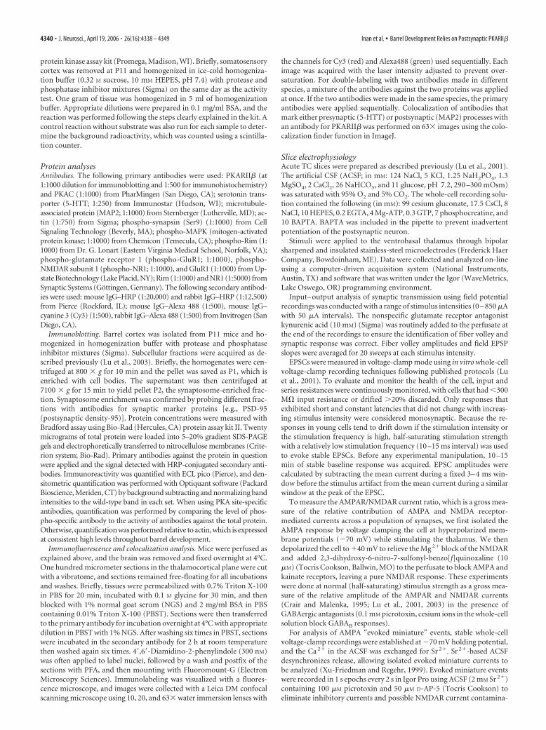
Layer IV barrel cytoarchitecture is disturbed inPKARII��/� miceBarrels are composed of the clustered arbors of thalamocorticalafferents and a surrounding ring of neurons in layer IV of thesomatosensory cortex. We examined the clustering of presynap-tic TCAs and the organization of postsynaptic layer IV neurons intangential sections from flattened somatosensory cortex of P14 –P21 PKARII��/� mice and wild-type littermate controls (Fig. 2).CO staining, a standard method for visualizing barrels (Seligmanet al., 1968; Wong-Riley and Welt, 1980; Wong-Riley et al., 1978),showed that clustering of PKARII��/� TCAs (n � 6) is compa-rable with that of wild-type littermate controls (n � 6) (Fig.2A,B). TCAs transiently express 5-HTT during barrel develop-ment (Lebrand et al., 1996, 1998; Hansson et al., 1998; Datwani etal., 2002). No difference in the TCA barrel pattern was observedusing an antibody against 5-HTT in tangential sections through
Figure 1. Reduced PKA activity in somatosensory cortex of PKARII��/� mice. A, The ratioof PKA activity measured in the presence (�cAMP) and absence (basal) of cAMP is significantlyreduced in P11 somatosensory cortex of PKARII� �/� (KO) mice compared with wild-type (WT)controls (histograms on left; *p � 0.005, t test). PKA activity was measured in tissue homoge-nates from somatosensory cortex using a protein kinase assay kit (Promega). PKAC proteinlevels are also significantly reduced in PKARII� �/� compared with wild-type littermate con-trol mice (histograms on right; *p �� 0.01, t test). B, Example Western blots of synaptosomesprepared from P11 somatosensory cortex of PKARII� �/� mice and their wild-type littermatecontrols using anti-PKARII� and anti-PKAC antibodies. Actin is used to normalize the proteinamount. As expected, PKARII� �/� mice have no PKARII� expression. Error bars show SEM.
Figure 2. Organization of layer IV neurons but not thalamocortical axon clustering is dis-rupted in PKARII��/� mice. A, B, Tangential sections from P16 wild-type (A) andPKARII� �/� (B) mice stained with CO (n � 6 for both genotypes). Posteromedial whiskerbarrel patterns (white box in A) are indistinguishable between genotypes. C, D, Tangentialsections from P7 wild-type (C) and PKARII� �/� (D) mice immunostained with anti-5-HTTantibody (n � 4 for both genotypes), which selectively labels thalamocortical afferents. Thebarrel pattern is again indistinguishable between genotypes, showing that the gross patterningof thalamocortical afferents in PKARII� �/� mice is similar to that of wild types. E, F, Nisslstained tangential sections from P16 wild-type (E) and PKARII� �/� (F ) mice. In wild-typemice (E), layer IV neurons organize into a barrel wall and hollow pattern characteristic of rodentsomatosensory cortex. In PKARII� �/� mice (F ), only a very rudimentary barrel pattern isvisible in posteromedial barrel field. Quantification confirms that the density of neurons in thebarrel wall relative to the barrel hollow is significantly higher in wild-type mice than inPKARII� �/� littermate mice (1.76 0.05 in n � 4 wild-type mice; 0.93 0.05 in n � 4PKARII� �/� mice; p � 0.001, t test). Scale bars, 500 �m.
Inan et al. • Barrel Development Relies on Postsynaptic PKARII� J. Neurosci., April 19, 2006 • 26(16):4338 – 4349 • 4341

somatosensory cortex of younger animals (P5–P7) either (n � 4for both genotypes) (Fig. 2C,D). In contrast, Nissl staining oftangential sections through barrel cortex, which reveals the pat-tern of postsynaptic neurons, showed that the organization oflayer IV neurons into barrels is disturbed in PKARII��/� mice(Fig. 2E,F). We quantified the barrel cytoarchitecture defect bymeasuring the relative density of neurons in barrel walls andhollows. As reported previously in wild-type mice (Pasternak andWoolsey, 1975), the cell density in barrel walls is much higherthan in barrel hollows (ratio of 1.76 0.05; n � 4), whereas inPKARII��/� mice, there is no difference in cell density betweenthe wall and hollow (0.93 0.05; n � 4; p � 0.001 for differencebetween PKARII��/� and wild type, t test).
In addition to forming cytoarchitectonic barrel walls in so-matosensory cortex, layer IV neurons also preferentially orienttheir dendrites toward a barrel hollow, presumably to make syn-apses with thalamocortical afferents relaying information fromone facial whisker (Woolsey et al., 1975; Datwani et al., 2002). Weexamined whether dendritic asymmetry in spiny stellate cells, alayer IV cell type known to have oriented dendrites (Harris andWoolsey, 1979, 1983), was impaired by the loss of PKARII� func-tion in the PKARII��/� mice (Fig. 3). Dendrites of Golgi stainedneurons in wild-type mice typically show substantial dendriticasymmetry (Fig. 3A,B,G), with only 16% of neurons lacking ori-entation bias (Fig. 3I) (arbitrarily defined as dendritic asymmetry�75%; see Materials and Methods). In contrast, 65% of layer IVneurons in PKARII��/� mice lacked orientation bias (Fig. 3J),resulting in significantly less dendritic asymmetry inPKARII��/� (0.71 0.02; n � 20 cells from four animals) (Fig.3G) compared with wild-type neurons (0.81 0.02; n � 19 cellsfrom four animals; p � 0.01; t test). The span of dendrites inPKARII��/� mice (249.43 31.77 �m; n � 20) was also signif-icantly broader than in wild-type neurons (174.24 11.96 �m;n � 19; p � 0.05; t test) (Fig. 3H), which is consistent with thebarrel cytoarchitectonic deficits in these mice. Other dendriticparameters, including total dendritic length (PKARII��/� mice,746.2 77.6 �m, n � 19; PKARII��/� mice, 988.7 113.1 �m,n � 20) and spine density (PKARII��/� mice, 0.22 0.02 per�m, n � 19; PKARII��/� mice, 0.19 0.02 per �m, n � 20),were normal in PKARII��/� mice. In total, these data show thatin PKARII��/� mice, presynaptic thalamic afferents form a nor-mal barrel pattern, but postsynaptic layer IV neurons organizeinto rudimentary barrel patches with dramatically disturbed neu-ronal morphology and cytoarchitecture.
PKARII� has a preferred postsynaptic expression inbarrel cortexTo gain some insight into the locus of action of PKARII�-mediated PKA signaling in barrel development, we examined thepattern of PKARII� expression in barrel cortex during the timebarrels are forming. Previous studies using in situ hybridizationand immunohistochemistry on adult mice showed that PKARII�is expressed strongly in neocortex but is expressed relativelyweakly in the thalamus (Cadd and McKnight, 1989; Glantz et al.,1992). In wild-type thalamocortical slices stained with an anti-PKARII� antibody, we found that PKARII� is present in layer IVat P3 (data not shown) and is enriched in a barrel pattern whenthe barrels emerge at around P5 (Fig. 4B,C).
To examine the subcellular localization of PKARII�, wedouble-labeled wild-type thalamocortical slices with antibodiesfor PKARII� and presynaptic or postsynaptic markers. Coimmu-nostaining of P5 wild-type thalamocortical slices with antibodiesfor PKARII� and 5-HTT, which mark presynaptic terminals on
thalamocortical synapses, showed little colocalization (7.1 2.2%; n � 4) (Fig. 4D–F). Similar results were obtained withantibodies to other presynaptic molecules, such as synapsin andGAP43 (data not shown). However, MAP2, a commonly used
Figure 3. Layer IV spiny stellate neurons in PKARII��/� mice have less orientation biasthan wild-type littermate controls. A–D, Examples of Golgi-stained layer IV spiny stellate neurons atlow magnification (left) and their computer-aided reconstructions (right) in wild-type (A, B) andPKARII��/� (C, D) mice at P14. E, F, Polar graphs of the dendrites of neurons illustrated in B and D(see Materials and Methods). G, PKARII��/� (KO) mice (0.71 0.02; 20 neurons from 4 animals)have significantly lower (*p � 0.01; t test) dendritic asymmetry compared with wild-type (WT)littermate controls (0.81 0.02; 19 neurons from 4 animals). Dendritic asymmetry, which must be�0.5, is defined as the ratio of the dendritic length in the hemisphere with the greatest density ofdendrites relative to the total dendritic length. H, Dendritic field span, the greatest distance betweenthe most distal dendrite tips of a particular layer IV spiny stellate neuron, is significantly higher (*p�0.05; t test) in PKARII��/� (KO) mice (249.43 31.77 �m) compared with wild-type (WT) litter-mate controls (174.2411.96�m). I, J, Pie charts showing the percentage of cells with and withoutorientation bias in PKARII��/� (J ) and wild-type littermate control mice (I ). Layer IV spiny stellateneurons are defined to have an orientation bias if their dendritic asymmetry is �0.75 (see Materialsand Methods). Scale bar, 50 �m. Error bars indicate SEM.
4342 • J. Neurosci., April 19, 2006 • 26(16):4338 – 4349 Inan et al. • Barrel Development Relies on Postsynaptic PKARII�

dendritic marker (Bernhardt and Matus, 1984; De Camilli et al.,1984), was extensively colocalized with PKARII� (28.8 7.5%;n � 4; p � 0.05 for the difference between colocalization ofPKARII� and 5-HTT and MAP2; t test) (Figs. 4G–I). These re-sults indicate that PKARII� is preferentially localized in thepostsynaptic terminal of layer IV neurons, which is consistentwith the layer IV barrel phenotype in PKARII��/� mice.
AMPAR/NMDAR current ratio does not increase withdevelopment in PKARII��/� miceThalamocortical synaptic transmission assayed using field poten-tial recordings in a thalamocortical brain slice was grossly normal
in PKARII��/� mice (Fig. 5A–C). In particular, the input– out-put curve, which measures synaptic response from a large popu-lation of neurons in layer IV of barrel cortex as a function ofthalamocortical fiber volley amplitude caused by thalamic(VPM) stimulation, was indistinguishable in wild-type controlsand PKARII��/� mice at P6 –P7 (n � 8 and n � 4, respectively)(Fig. 5C). This shows that there is no gross defect with functionalthalamocortical synaptic innervation of layer IV in barrel cortexof PKARII��/� mice.
We used whole-cell voltage-clamp techniques to examine glu-tamatergic synaptic transmission at TC synapses. In wild-typemice, the ratio of AMPAR-mediated to NMDAR-mediated cur-
Figure 4. PKARII� is extensively colocalized with postsynaptic but not presynaptic markers. A, Control immunostaining of a P5 PKARII� �/� thalamocortical slice with anti-PKARII� antibodyreveals that this antibody is specific to PKARII�. B, C, Cortical expression of PKARII� was examined by labeling wild-type thalamocortical (B) and tangential (C) slices with anti-PKARII� antibody.PKARII� is expressed in layer IV as it delaminates from layer II/III at P3 (data not shown) and forms a barrel pattern when barrels emerge at around P5 (B, C). D–I, Presynaptic and postsynapticlocalization of PKARII� was examined by coimmunostaining P5 wild-type thalamocortical slices using anti-5-HTT (D–F ) and anti-MAP2 (G–I ) antibodies, respectively, together with an anti-PKARII� antibody. D, Low-magnification image of a single barrel stained with a 5-HTT antibody (green) to label presynaptic thalamocortical terminals and a PKARII� (red) antibody and theirmerged image. At low magnification, both show a clear barrel pattern. E, Merged image of the barrel in D (white box) at 63� magnification. F, A 4� zoom of white-boxed area in E. G,Low-magnification image of a single barrel labeled with a MAP2 antibody (green) to mark postsynaptic dendrites and a PKARII� antibody (red) and their merged image. H, Merged image of thebarrel in G (white box) at 63� magnification. I, 4� zoom of white-boxed area in H. Colocalization analysis of high-magnification images using ImageJ shows that PKARII� expression colocalizessignificantly more ( p � 0.05; t test) with MAP2 (28.8 7.5%; n � 4) than with 5-HTT (7.1 2.2%; n � 4). LIV, Layer IV; LV, layer V. Scale bars: A–C, 500 �m; D, G, 100 �m; E, H, 40 �m;F, I, 10 �m.
Inan et al. • Barrel Development Relies on Postsynaptic PKARII� J. Neurosci., April 19, 2006 • 26(16):4338 – 4349 • 4343

rents (AMPAR/NMDAR current ratio) increases during the firstweek after birth (0.47 0.04, n � 5 for P4 –P7; 2.12 0.5, n � 6for P9 –P11; p � 0.05; t test) (Fig. 5G), which is typical of gluta-matergic synaptic development at many central synapses (Crairand Malenka, 1995; Wu et al., 1996; Lu et al., 2001). We previ-ously showed in brl mice that the AMPAR/NMDAR current ratioremained small during TC synapse development, but it was un-clear whether this was specifically attributable to the absence ofcAMP–PKA signaling in postsynaptic layer IV neurons (Lu et al.,2003). Because of the exclusive postsynaptic barrel map defect ofPKARII��/� mice and the preferable postsynaptic expressionpattern of PKARII�, PKARII��/� mice provided us an opportu-nity to analyze the effect of lowered PKA activity in layer IVneurons on TC synapse maturation. In PKARII��/� mice, thenormal developmental increase in AMPAR/NMDAR current ra-tio failed to occur (0.68 0.18, n � 9 for P4 –P7; 0.91 0.05, n �8 for P9 –P11; p � 0.27; t test), and the AMPAR/NMDAR currentratio remained small ( p � 0.05 for wild type vs PKARII��/� atP9 –P11; t test) (Fig. 5D–G), which is similar to what was ob-
served in brl (Lu et al., 2003). These results indicate that theincrease in AMPAR/NMDAR current ratio during TC synapsematuration is mediated by postsynaptic PKA function and inter-fering with this function impairs postsynaptic barrel mapformation.
AMPAR-evoked miniature currents are small at PKARII��/�
TC synapsesTheoretically, the small AMPAR/NMDAR current ratio inPKARII��/� mice could be attributable to small AMPAR-mediated currents or large NMDAR-mediated currents or both.We examined miniature AMPAR-mediated synaptic currents(“AMPA minis”) in P9 –P11 mice to determine whether AMPARcurrents are specifically affected by the loss of PKARII� function.Substitution of Ca 2� with Sr 2� in the extracellular ACSF leads toa reduction in synchronous evoked response and the appearanceof delayed miniature responses as a result of the persistence ofasynchronous quantal release (Xu-Friedman and Regehr, 1999)(Fig. 6A,B). This makes Sr 2� a useful tool to analyze evokedminiature EPSCs (evoked mini-EPSCs). Evoked mini-EPSCs inPKARII��/� neurons (n � 6) were on average smaller than com-parable wild-type neurons (n � 7; 7.3 0.59 vs 10.6 1.3 pA;p � 0.05; t test) (Fig. 6C, inset). Comparison of the evoked mini-EPSC amplitude histograms shows a shift in the peak of the dis-tribution, with the histogram in PKARII��/� mice peaking at asmaller amplitude than wild-type littermate controls (Fig. 6C). Inaddition, wild-type controls often had responses with very largeamplitudes (Fig. 6A,C). The mean cumulative probability distri-butions also revealed a shift in evoked mini EPSCs inPKARII��/� mice toward smaller amplitudes compared withwild-type littermate controls (Fig. 6D). The difference betweengenotypes was not caused by an intrinsic difference in RMS noiselevels (1.98 0.06 vs 2.06 0.09 pA; p � 0.45; t test). Theseresults indicate that in the absence of PKARII�-mediated PKAsignaling, the number of functional AMPARs at TC synapses islower on average, leading to smaller AMPAR mediated currents.
LTP is impaired at developing thalamocortical synapses inPKARII��/� miceThe lack of a developmental increase in AMPAR currents couldbe attributable to deficits in thalamocortical LTP at developingthalamocortical synapses in PKARII��/� mice (Lu et al., 2003).In this scenario, the cellular mechanisms responsible for LTPmediate the increase in AMPAR currents during thalamocorticalsynapse development via PKA-mediated phosphorylation ofAMPA receptor subunits (Ehlers, 2000; Lee et al., 2000, 2003;Esteban et al., 2003). We examined this possibility using a stan-dard LTP pairing protocol in thalamocortical slices from P4 –P6PKARII��/� and wild-type littermate control mice (Fig. 7). Inwild-type mice, pairing induced a robust potentiation ofthalamocortical response (Fig. 7A,C,D) (122.96 41.04%; n �7). In contrast, neurons in littermate PKARII��/� mice showedsignificantly less potentiation (1.59 6.92%; n � 5; p � 0.05; ttest) (Fig. 7B–D). This suggests that an activity-dependent pro-cess that is mediated by PKARII� is responsible for the normalfunctional development of thalamocortical synapses.
Phosphorylation of postsynaptic but not presynaptic PKAtargets is reduced in PKARII��/� miceThere are a number of PKA targets in the presynaptic andpostsynaptic terminal of thalamocortical synapses that are poten-tially affected by decreased PKA activity in PKARII��/� mice.We examined the phosphorylation levels of several of the most
Figure 5. Developmental increase in AMPAR/NMDAR current ratio is absent in PKARII��/�
mice. A–C, Input– output analyses of extracellular field potential recordings indicate no differ-ence in gross synaptic transmission between wild-type and PKARII� �/� thalamocortical syn-apses. A, B, Sample average responses at different stimulation strengths of P6 wild-type andPKARII� �/� mice. The arrowhead shows the fiber volley, and the arrow shows the postsyn-aptic response. C, Input– output curves in wild-type (black; n � 8) and PKARII� �/� (red; n �4) mice at P6 –P7 are similar. Regression analysis for wild types (green) and PKARII� �/�
(blue) show no significant difference at 95% confidence interval (dashed lines overlap). D–F,Sample whole-cell voltage-clamp measurements of AMPAR-mediated and NMDAR-mediatedthalamocortical EPSCs in a P10 wild-type littermate control (D) and P10 PKARII� �/� (E)mouse. F, Overlay of the responses in D and E scaled so that the NMDAR currents are the sameamplitude. Note the AMPAR-mediated EPSC in PKARII� �/� animal is small in comparison tothe littermate control. G, Summary quantification of AMPAR/NMDAR current ratios for differentage groups and different genotypes. The AMPAR/NMDAR current ratio of wild-type animalsincreases significantly with age (*p � 0.05; t test). However, this increase is absent inPKARII� �/� (KO) mice (p � 0.27; t test). The AMPAR/NMDAR current ratio of P9 –P11PKARII� �/� mice is also significantly lower than that of P9 –P11 wild-type (WT) littermatecontrols (*p � 0.05; t test). Error bars indicate SEM.
4344 • J. Neurosci., April 19, 2006 • 26(16):4338 – 4349 Inan et al. • Barrel Development Relies on Postsynaptic PKARII�

prominent targets, some of which have demonstrably reducedphosphorylation in brl mice (Lu et al., 2003, 2006). Western blotanalysis on synaptosomes prepared from P11 somatosensory cor-tex of major presynaptic PKA targets, including phospho-synapsin (at Ser 9) and phospho-Rim, shows no difference be-tween PKARII��/� mice and wild-type littermate controls(0.91 1.66, p � 0.26 and 1.03 0.07, p � 0.56, respectively; ttest; n � 5 for both). In contrast, phosphorylation of two postsyn-aptic PKA targets, GluR1 (at Ser 845) and NR1 (at Ser 897), had asignificant reduction in PKARII��/� mice compared with wild-type littermate controls (0.45 0.1 and 0.61 0.08, respectively;n � 4; p � 0.005 for both; t test) (Fig. 8A,B). PKA also regulatesMAPK signaling in neurons (Morozov et al., 2003; Waltereit andWeller, 2003). However, analysis of phosphorylation levels withan anti-phospho MAPK [extracellular signal-regulated kinase(ERK)] antibody that recognizes both p42ERK and p44ERKshowed no significant difference between genotypes (1.23 0.34;n � 5; p � 0.51; t test) (Fig. 8A). These data are consistent withthe small AMPAR-mediated currents we observe physiologically,because PKA phosphorylation of GluR1 through an LTP-likeprocess is known to regulate AMPAR trafficking at the synapse(Ehlers, 2000; Lee et al., 2000, 2003; Esteban et al., 2003). Thereduced PKA phosphorylation of postsynaptic but not presynap-tic targets is also consistent with the postsynaptic localization ofPKARII� and the postsynaptic barrel phenotype observed in
PKARII��/� mice, reinforcing a postsyn-aptic function for PKARII� during barrelmap formation.
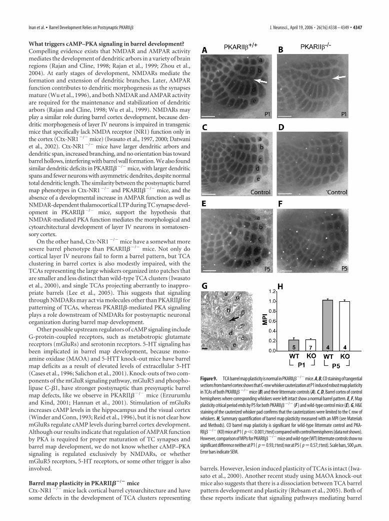
PKARII��/� mice have normal TCAmap plasticityAnother important feature of corticalmaps is their developmental plasticity, inwhich the size of cortex devoted to a par-ticular peripheral sense organ changes ifthe pattern of sensory input changes. Forexample, if one row of whiskers is re-moved during the first week after birth,the area of cortex devoted to this row willshrink and the neighboring rows will ex-pand at the expense of the deprived row.In mammals, cortical maps have a criticalperiod for this type of plasticity. In mousebarrel cortex, the critical period (P0–P5) co-incides with barrel map formation. There-fore, it is intriguing to investigate whetherbarrel map development and plasticity aremediated by common molecular pathways.
Recently, map plasticity in visual cor-tex (ocular dominance plasticity) wasshown to be defective in PKARII��/�
mice (Fischer et al., 2004). Because PKA-RII��/� mice have poorly formed barrel(Nissl) maps, we were unable to quantita-tively examine cytoarchitectural plasticityin PKARII��/� mice. However, TCAclustering is grossly normal in PKA-RII��/� mice, so we examined map plas-ticity of TCA clustering using cytochromeoxidase stain in PKARII��/� mice by re-moving a row of whiskers at P1 (Fig. 9A–D,H) or P5 (Fig. 9E,F,H). We found thatPKARII��/� mice have a similar degree
of thalamocortical map plasticity (MPI) as wild-type littermatecontrol mice at P1 (MPI for wild type, 0.22 0.04, n � 5; MPI forPKARII��/� mice, 0.23 0.02, n � 7; p � 0.93; t test) (Fig. 9H).Thalamocortical map plasticity is significantly reduced in bothPKARII��/� (1.00 0.03; n � 4) and their wild-type littermatecontrol (1.03 0.03; n � 4; p � 0.57 for difference betweenPKARII��/� and PKARII��/�; t test) mice by P5. This suggeststhat loss of PKARII� does not affect the developmental plasticityof thalamocortical afferents.
DiscussionWe examined the cellular mechanisms mediating neural circuitformation, focusing on the role of PKA signaling in barrel mapdevelopment. We showed that PKARII��/� mice have decreasedPKA activity in somatosensory cortex and associated barrel mapdeficits in the morphology and cytoarchitectural organization oflayer IV cortical neurons but not thalamocortical afferents. Usingimmunohistochemical and biochemical techniques, we localizedPKARII� expression to postsynaptic terminals in barrel cortexand showed that postsynaptic, but not presynaptic, PKA targetshave decreased phosphorylation in PKARII��/� synapses. Thesefindings are consistent with previous reports in adult mice thatanalyzed different brain regions using immunohistochemicaland electron microscopy techniques to show that PKARII� islocalized to dendrites, perikaya, and postsynaptic densities but is
Figure 6. Small AMPAR mini-EPSCs at PKARII��/� thalamocortical synapses. A, B, Sample traces of evoked AMPAR-mediatedcurrent responses in Ca 2�–ACSF (gray) and Sr 2�–ACSF (black) from recordings in P9 –P11 PKARII� �/� (B) and wild-typelittermate control (A) neurons. In the presence of Sr 2�–ACSF, responses to evoked synchronous release is lower and quantalevents (evoked mini-EPSCs) in response to asynchronous release appear. The amplitudes of these quantal events are smaller onaverage in PKARII� �/� mice. C, Average frequency histogram of evoked mini-EPSCs. The PKARII� �/� histogram (white bars)peaks at a smaller amplitude than wild-type littermate controls (gray bars), which have a longer large amplitude tail. Inset, Meanevoked mini-EPSC amplitude is significantly lower (*p � 0.05; t test) for PKARII� �/� mice (white) with respect to wild-typelittermate controls (gray). D, The cumulative probability distribution in PKARII� �/� (KO) animals (black) also shows a shifttoward smaller amplitudes compared with wild-type (WT) littermate controls (gray). Comparison of root mean square noise showsno difference (p � 0.45; t test) between genotypes (data not shown). Error bars indicate SEM.
Inan et al. • Barrel Development Relies on Postsynaptic PKARII� J. Neurosci., April 19, 2006 • 26(16):4338 – 4349 • 4345

rarely present in axon terminals (Ludvig et al., 1990; Glantz et al.,1992). In addition, we examined thalamic barrelloids using COstaining at P5 and found that organization of barrelloids is intact
in PKARII��/� mice (supplemental Fig. 1, available at www.jneurosci.org as supplemental material). Moreover, we did notdetect any difference in anatomical plasticity of TCAs inPKARII��/� and wild-type mice. These findings suggest thatthe PKARII��/� barrel phenotype is specific to the cortex, andPKARII�-mediated PKA signaling is not necessary for the grossdevelopment and plasticity of TCA clusters, although it is re-quired for postsynaptic barrel map development.
We also examined the physiological development of thalamo-cortical synapses in PKARII��/� mice and found that thalamo-cortical LTP and AMPAR function were significantly impaired.The smaller AMPAR currents we observed in older animals areconsistent with the absence of LTP and the decreased PKA phos-phorylation of GluR1 detected biochemically in PKARII��/�
mice. In total, these experiments support an activity-dependentmodel for barrel map formation in which the selective addition ofthalamocortical synapses based on Hebbian mechanisms for syn-apse formation act through a PKA-dependent pathway thatstrengthens connections by increasing the number of functionalAMPARs at the synapse.
cAMP–PKA signaling in barrel developmentBrl (Adcy1�/�) mice, which have been studied in great detail,have a barrel map defect that is more severe than PKARII��/�
mice, because they lack organization of both presynaptic andpostsynaptic elements of the barrel map (Welker et al., 1996;Abdel-Majid et al., 1998), whereas TCA clustering is intact inPKARII��/� mice. It is possible that the less severe barrel phe-notype in PKARII��/� mice stems from the remaining 50% ofPKA activity in the somatosensory cortex of these mutants. Thephenotypic difference between PKARII��/� and brl mice alsosuggests that different signaling components downstream ofAdcy1 play a role in the formation of presynaptic and postsynap-tic barrel patterns. Data presented here and in our previous re-port (Lu et al., 2003) suggest that Adcy1 acts via PKARII� toregulate postsynaptic barrel map development. However, in pre-synaptic TCA terminals, Adcy1 may act through signaling mole-cules other than PKA or through a PKAR subunit distinct fromPKARII�. Unlike in brl mice, the phosphorylation of presynapticPKA targets is not reduced in PKARII��/� mice, strengtheningthe suggestion that a different PKAR subunit is responsible forTCA clustering in barrel cortex (Lu et al., 2003, 2006).
Our previous studies with brl mice provided indirect evidencefor the role of PKA activity in TC synapse maturation. Similar tothe results we report here, we found that the AMPAR/NMDARcurrent ratio, which normally increases during TC synapse mat-uration, remained unchanged in brl mice (Lu et al., 2003). Thesmaller AMPAR/NMDAR current ratio was attributable, at leastin part, to smaller AMPAR currents (Lu et al., 2003). As in brlmice, the reduced number of functional AMPARs at TC synapsesof PKARII��/� mice may be because of a change in AMPARproperties and/or because of an impairment in AMPAR insertioninto the synapse, both of which are regulated by PKA phosphor-ylation of the GluR1 subunit through an LTP-like process (Bankeet al., 2000; Ehlers, 2000; Lee et al., 2000, 2003; Esteban et al., 2003).Consistent with the LTP deficit we observe in PKARII��/� mice, wepreviously showed that LTP was a PKA-dependent process that isimpaired in brl mice (Lu et al., 2003). Our results here provide directevidence for the requirement of postsynaptic PKA activity in theregulation of AMPAR function via synaptic plasticity mechanisms,such as LTP, and the activity-dependent maturation of TC synapsesduring barrel map development.
Figure 7. LTP deficits in PKARII��/� thalamocortical synapses. A, B, Example whole-cellvoltage-clamp recordings from P6 wild-type control (A) and PKARII� �/� (B) neurons showthat thalamocortical response in wild-type but not PKARII� �/� layer IV neurons can be po-tentiated using an LTP pairing protocol. Traces on the right (average of 20 sweeps) from before(1) and 30 min after pairing (2) show significant potentiation in wild-type but not PKARII� �/�
neurons. C, Summary graph of pairing experiments showing the EPSC percentage change forPKARII� �/� mice (black circles) and their wild-type littermate controls (gray circles).PKARII� �/� thalamocortical synapses show no sign of potentiation. D, Summary histogramof EPSC percentage change from the average of 20 sweeps starting at 20 min after pairing for allneurons. PKARII� �/� (KO) thalamocortical synapses show almost no potentiation (1.59 6.92%; n � 5), and their EPSC percentage change is significantly lower (*p � 0.05; t test) thanthat of wild-type (WT) littermate controls (122.96 41.04%; n � 7). Error bars indicate SEM.
Figure 8. PKARII��/� mice have a significant reduction in phosphorylation of postsynapticbut not presynaptic PKA targets. A, Western blot examples of synaptosomes prepared from P11somatosensory cortex of PKARII� �/� mice and their littermate controls using antibodiesagainst PKA phosphorylation targets. B, Quantification of normalized ratio of anti-phosphoGluR1 (PGluR1, Ser845) to total GluR1 and anti-phospho NR1 (PNR1, Ser 897) to total NR1reveals a significant difference between PKARII� �/� (KO) and wild-type (WT) littermate con-trol mice (*p � 0.005 for both; t test). None of the presynaptic PKA phosphorylation targetssuch as phospho-synapsin (p � 0.26; t test) and phospho-Rim (p � 0.56; t test) reveal adifference between PKARII� �/� mice and their wild-type littermate controls (data notshown). Quantification of the band labeled with an anti-phospho-MAPK (PMAPK) antibody,which recognizes both p44 (top band in A) and p42 (bottom band in A) phosphorylation ofMAPK (or ERK), also shows no difference (data not shown; p � 0.51; t test). Error bars indicateSEM.
4346 • J. Neurosci., April 19, 2006 • 26(16):4338 – 4349 Inan et al. • Barrel Development Relies on Postsynaptic PKARII�
What triggers cAMP–PKA signaling in barrel development?Compelling evidence exists that NMDAR and AMPAR activitymediates the development of dendritic arbors in a variety of brainregions (Rajan and Cline, 1998; Rajan et al., 1999; Zhou et al.,2004). At early stages of development, NMDARs mediate theformation and extension of dendritic branches. Later, AMPARfunction contributes to dendritic morphogenesis as the synapsesmature (Wu et al., 1996), and both NMDAR and AMPAR activityare required for the maintenance and stabilization of dendriticarbors (Rajan and Cline, 1998; Wu et al., 1999). NMDARs mayplay a similar role during barrel cortex development, because den-dritic morphogenesis of layer IV neurons is impaired in transgenicmice that specifically lack NMDA receptor (NR1) function only inthe cortex (Ctx-NR1�/� mice) (Iwasato et al., 1997, 2000; Datwaniet al., 2002). Ctx-NR1�/� mice have larger dendritic arbors anddendritic span, increased branching, and no orientation bias towardbarrel hollows, interfering with barrel wall formation. We also foundsimilar dendritic deficits in PKARII��/� mice, with larger dendriticspans and fewer neurons with asymmetric dendrites, despite normaltotal dendritic length. The similarity between the postsynaptic barrelmap phenotypes in Ctx-NR1�/� and PKARII��/� mice, and theabsence of a developmental increase in AMPAR function as well asNMDAR-dependent thalamocortical LTP during TC synapse devel-opment in PKARII��/� mice, support the hypothesis thatNMDAR-mediated PKA function mediates the morphological andcytoarchitectural development of layer IV neurons in somatosen-sory cortex.
On the other hand, Ctx-NR1�/� mice have a somewhat moresevere barrel phenotype than PKARII��/� mice. Not only docortical layer IV neurons fail to form a barrel pattern, but TCAclustering in barrel cortex is also modestly impaired, with theTCAs representing the large whiskers organized into patches thatare smaller and less distinct than wild-type TCA clusters (Iwasatoet al., 2000), and single TCAs projecting aberrantly to inappro-priate barrels (Lee et al., 2005). This suggests that signalingthrough NMDARs may act via molecules other than PKARII� forpatterning of TCAs, whereas PKARII�-mediated PKA signalingplays a role downstream of NMDARs for postsynaptic neuronalorganization during barrel map development.
Other possible upstream regulators of cAMP signaling includeG-protein-coupled receptors, such as metabotropic glutamatereceptors (mGluRs) and serotonin receptors. 5-HT signaling hasbeen implicated in barrel map development, because mono-amine oxidase (MAOA) and 5-HTT knock-out mice have barrelmap deficits as a result of elevated levels of extracellular 5-HT(Cases et al., 1996; Salichon et al., 2001). Knock-outs of two com-ponents of the mGluR signaling pathway, mGluR5 and phospho-lipase C-�1, have stronger postsynaptic than presynaptic barrelmap defects, like we observe in PKARII��/� mice (Erzurumluand Kind, 2001; Hannan et al., 2001). Stimulation of mGluRsincreases cAMP levels in the hippocampus and the visual cortex(Winder and Conn, 1993; Reid et al., 1996), but it is not clear howmGluRs regulate cAMP levels during barrel cortex development.Although our results indicate that regulation of AMPAR functionby PKA is required for proper maturation of TC synapses andbarrel map development, we do not know whether cAMP–PKAsignaling is regulated exclusively by NMDARs, or whethermGluR5 receptors, 5-HT receptors, or some other trigger is alsoinvolved.
Barrel map plasticity in PKARII��/� miceCtx-NR1�/� mice lack cortical barrel cytoarchitecture and havesome defects in the development of TCA clusters representing
barrels. However, lesion induced plasticity of TCAs is intact (Iwa-sato et al., 2000). Another recent study using MAOA knock-outmice also suggests that there is a dissociation between TCA barrelpattern development and plasticity (Rebsam et al., 2005). Both ofthese reports indicate that signaling pathways mediating barrel
Figure 9. TCA barrel map plasticity is normal in PKARII��/�mice. A, B, CO staining of tangentialsectionsfrombarrelcortexshowsthatC-rowwhiskercauterizationatP1inducedrobustmapplasticityin TCAs of both PKARII��/� mice (B) and their littermate controls (A). C, D, Barrel cortex of controlhemispheres where corresponding whiskers were left intact show a normal barrel pattern. E, F, Mapplasticity critical period ends by P5 for both PKARII��/� (F ) and wild-type control mice (E). G, H&Estaining of the cauterized whisker pad confirms that the cauterizations were limited to the C row ofwhiskers. H, Summary quantification of barrel map plasticity measured with an MPI (see Materialsand Methods). CO barrel map plasticity is significant for wild-type littermate control and PKA-RII��/�(KO)miceatP1( p��0.001;ttest)comparedwithcontrolhemispheres(datanotshown).However, comparison of MPIs for PKARII��/�mice and wild-type (WT) littermate controls show nosignificant difference neither at P1 ( p�0.93; t test) nor at P5 ( p�0.57; t test). Scale bars, 500�m.Error bars indicate SEM.
Inan et al. • Barrel Development Relies on Postsynaptic PKARII� J. Neurosci., April 19, 2006 • 26(16):4338 – 4349 • 4347

map plasticity and the initial development of TCAs are distinct,or at a minimum a defect in one pathway that disrupts patterndevelopment is insufficient to disrupt map plasticity. Because thebarrel map phenotype of PKARII��/� mice is exclusivelypostsynaptic, we were able to analyze TCA map plasticity. We sawno difference in TCA map plasticity in PKARII��/� mice andwild-type controls, indicating that reducing PKA activity in layerIV neurons is not sufficient to disrupt map plasticity in TCAs.
Role of PKARII� in PKA functioncAMP and PKA mediate a variety of signaling cascades, evenwithin the same cell (Colledge and Scott, 1999; Carnegie andScott, 2003; Bauman et al., 2004). Specificity of action for eachsignaling pathway is achieved by localizing PKA to distinct sub-cellular compartments via interaction of PKA regulatory sub-units with A kinase anchoring proteins (AKAPs) (Coghlan et al.,1995; Westphal et al., 1999; Colledge et al., 2000). AKAPs thatinteract with type II PKAR, such as AKAP79/150, bind to scaf-folding proteins that associate with glutamate receptors at thepostsynaptic density and regulate AMPAR currents (Rosenmundet al., 1994; Colledge et al., 2000). This interaction is specificallyrequired for the phosphorylation of GluR1, so the anchoring ofPKA in close proximity to its substrates allows for tight regulationand signal integration in response to neuronal activity (Colledgeet al., 2000; Tavalin et al., 2002). Endogenous PKARII� is likelycompartmentalized near AMPARs at thalamocortical synapsesand may be responsible for their activity-dependent traffickingduring barrel formation. Therefore, the functional and anatom-ical deficits observed in barrel cortex of PKARII��/� mice maynot only be caused by lower overall PKA activity, but may bespecifically caused by the improper localization of PKA and theconsequent misregulation of local PKA activity in postsynapticlayer IV neurons.
ReferencesAbdel-Majid RM, Leong WL, Schalkwyk LC, Smallman DS, Wong ST, Storm
DR, Fine A, Dobson MJ, Guernsey DL, Neumann PE (1998) Loss ofadenylyl cyclase I activity disrupts patterning of mouse somatosensorycortex. Nat Genet 19:289 –291.
Amieux PS, Cummings DE, Motamed K, Brandon EP, Wailes LA, Le K,Idzerda RL, McKnight GS (1997) Compensatory regulation of RIalphaprotein levels in protein kinase A mutant mice. J Biol Chem272:3993–3998.
Banke TG, Bowie D, Lee H, Huganir RL, Schousboe A, Traynelis SF (2000)Control of GluR1 AMPA receptor function by cAMP-dependent proteinkinase. J Neurosci 20:89 –102.
Bauman AL, Goehring AS, Scott JD (2004) Orchestration of synaptic plas-ticity through AKAP signaling complexes. Neuropharmacology46:299 –310.
Beaver CJ, Ji Q, Fischer QS, Daw NW (2001) Cyclic AMP-dependent pro-tein kinase mediates ocular dominance shifts in cat visual cortex. NatNeurosci 4:159 –163.
Beaver CJ, Fischer QS, Ji Q, Daw NW (2002) Orientation selectivity is re-duced by monocular deprivation in combination with PKA inhibitors.J Neurophysiol 88:1933–1940.
Bernhardt R, Matus A (1984) Light and electron microscopic studies of thedistribution of microtubule-associated protein 2 in rat brain: a differencebetween dendritic and axonal cytoskeletons. J Comp Neurol226:203–221.
Brandon EP, Idzerda RL, McKnight GS (1997) PKA isoforms, neural path-ways, and behaviour: making the connection. Curr Opin Neurobiol7:397– 403.
Brandon EP, Logue SF, Adams MR, Qi M, Sullivan SP, Matsumoto AM,Dorsa DM, Wehner JM, McKnight GS, Idzerda RL (1998) Defectivemotor behavior and neural gene expression in RII�-protein kinase Amutant mice. J Neurosci 18:3639 –3649.
Cadd G, McKnight GS (1989) Distinct patterns of cAMP-dependent pro-tein kinase gene expression in mouse brain. Neuron 3:71–79.
Carnegie GK, Scott JD (2003) A-kinase anchoring proteins and neuronalsignaling mechanisms. Genes Dev 17:1557–1568.
Cases O, Vitalis T, Seif I, De Maeyer E, Sotelo C, Gaspar P (1996) Lack ofbarrels in the somatosensory cortex of monoamine oxidase A-deficientmice: role of a serotonin excess during the critical period. Neuron16:297–307.
Cline H (2003) Sperry and Hebb: oil and vinegar? Trends Neurosci26:655– 661.
Coghlan VM, Perrino BA, Howard M, Langeberg LK, Hicks JB, Gallatin WM,Scott JD (1995) Association of protein kinase A and protein phospha-tase 2B with a common anchoring protein. Science 267:108 –111.
Colledge M, Scott JD (1999) AKAPs: from structure to function. Trends CellBiol 9:216 –221.
Colledge M, Dean RA, Scott GK, Langeberg LK, Huganir RL, Scott JD (2000)Targeting of PKA to glutamate receptors through a MAGUK-AKAP com-plex. Neuron 27:107–119.
Crair MC, Malenka RC (1995) A critical period for long-term potentiationat thalamocortical synapses. Nature 375:325–328.
Datwani A, Iwasato T, Itohara S, Erzurumlu RS (2002) NMDA receptor-dependent pattern transfer from afferents to postsynaptic cells and den-dritic differentiation in the barrel cortex. Mol Cell Neurosci 21:477– 492.
De Camilli P, Miller PE, Navone F, Theurkauf WE, Vallee RB (1984) Distri-bution of microtubule-associated protein 2 in the nervous system of therat studied by immunofluorescence. Neuroscience 11:817– 846.
Ehlers MD (2000) Reinsertion or degradation of AMPA receptors deter-mined by activity-dependent endocytic sorting. Neuron 28:511–525.
Erzurumlu RS, Kind PC (2001) Neural activity: sculptor of ‘barrels’ in theneocortex. Trends Neurosci 24:589 –595.
Esteban JA, Shi SH, Wilson C, Nuriya M, Huganir RL, Malinow R (2003)PKA phosphorylation of AMPA receptor subunits controls synaptic traf-ficking underlying plasticity. Nat Neurosci 6:136 –143.
Feldman DE, Knudsen EI (1998) Experience-dependent plasticity and thematuration of glutamatergic synapses. Neuron 20:1067–1071.
Fischer QS, Beaver CJ, Yang Y, Rao Y, Jakobsdottir KB, Storm DR, McKnightGS, Daw NW (2004) Requirement for the RII� isoform of PKA, but notcalcium-stimulated adenylyl cyclase, in visual cortical plasticity. J Neuro-sci 24:9049 –9058.
Glantz SB, Amat JA, Rubin CS (1992) cAMP signaling in neurons: patternsof neuronal expression and intracellular localization for a novel protein,AKAP 150, that anchors the regulatory subunit of cAMP-dependent pro-tein kinase II beta. Mol Biol Cell 3:1215–1228.
Hannan AJ, Blakemore C, Katsnelson A, Vitalis T, Huber KM, Bear M, RoderJ, Kim D, Shin HS, Kind PC (2001) PLC-beta1, activated via mGluRs,mediates activity-dependent differentiation in cerebral cortex. Nat Neu-rosci 4:282–288.
Hansson SR, Mezey E, Hoffman BJ (1998) Serotonin transporter messengerRNA in the developing rat brain: early expression in serotonergic neuronsand transient expression in non-serotonergic neurons. Neuroscience83:1185–1201.
Harris RM, Woolsey TA (1979) Morphology of Golgi-impregnated neuronsin mouse cortical barrels forming vibrissae damage at different post-natalages. Brain Res 161:143–149.
Harris RM, Woolsey TA (1983) Computer assisted analyses of barrel neu-ron axons and their putative synaptic contacts. J Comp Neurol 220:63–79.
Howe DG, Wiley JC, McKnight GS (2002) Molecular and behavioral effectsof a null mutation in all PKA C beta isoforms. Mol Cell Neurosci20:515–524.
Huang YY, Kandel ER, Varshavsky L, Brandon EP, Qi M, Idzerda RL,McKnight GS, Bourtchouladze R (1995) A genetic test of the effects ofmutations in PKA on mossy fiber LTP and its relation to spatial andcontextual learning. Cell 83:1211–1222.
Iwasato T, Erzurumlu RS, Huerta PT, Chen DF, Sasaoka T, Ulupinar E,Tonegawa S (1997) NMDA receptor-dependent refinement of somato-topic maps. Neuron 19:1201–1210.
Iwasato T, Datwani A, Wolf AM, Nishiyama H, Taguchi Y, Tonegawa S,Knopfel T, Erzurumlu RS, Itohara S (2000) Cortex-restricted disruptionof NMDAR1 impairs neuronal patterns in the barrel cortex. Nature406:726 –731.
Kind PC, Neumann PE (2001) Plasticity: downstream of glutamate. TrendsNeurosci 24:553–555.
Lebrand C, Cases O, Adelbrecht C, Doye A, Alvarez C, El Mestikawy S, Seif I,
4348 • J. Neurosci., April 19, 2006 • 26(16):4338 – 4349 Inan et al. • Barrel Development Relies on Postsynaptic PKARII�

Gaspar P (1996) Transient uptake and storage of serotonin in develop-ing thalamic neurons. Neuron 17:823– 835.
Lebrand C, Cases O, Wehrle R, Blakely RD, Edwards RH, Gaspar P (1998)Transient developmental expression of monoamine transporters in therodent forebrain. J Comp Neurol 401:506 –524.
Lee HK, Barbarosie M, Kameyama K, Bear MF, Huganir RL (2000) Regula-tion of distinct AMPA receptor phosphorylation sites during bidirec-tional synaptic plasticity. Nature 405:955–959.
Lee HK, Takamiya K, Han JS, Man H, Kim CH, Rumbaugh G, Yu S, Ding L,He C, Petralia RS, Wenthold RJ, Gallagher M, Huganir RL (2003) Phos-phorylation of the AMPA receptor GluR1 subunit is required for synapticplasticity and retention of spatial memory. Cell 112:631– 643.
Lee LJ, Iwasato T, Itohara S, Erzurumlu RS (2005) Exuberant thalamocor-tical axon arborization in cortex-specific NMDAR1 knockout mice.J Comp Neurol 485:280 –292.
Lopez-Bendito G, Molnar Z (2003) Thalamocortical development: how arewe going to get there? Nat Rev Neurosci 4:276 –289.
Lu HC, Gonzalez E, Crair MC (2001) Barrel cortex critical period plasticityis independent of changes in NMDA receptor subunit composition. Neu-ron 32:619 – 634.
Lu HC, She WC, Plas DT, Neumann PE, Janz R, Crair MC (2003) Adenylylcyclase I regulates AMPA receptor trafficking during mouse cortical “bar-rel” map development. Nat Neurosci 6:939 –947.
Lu HC, Butts BA, Kaeser PS, She WC, Janz R, Crair MC (2006) Barrel mapdevelopment relies on efficient neurotransmitter release. J Neurosci26:2692–2703.
Ludvig N, Ribak CE, Scott JD, Rubin CS (1990) Immunocytochemical lo-calization of the neural-specific regulatory subunit of the type II cyclicAMP-dependent protein kinase to postsynaptic structures in the ratbrain. Brain Res 520:90 –102.
Morozov A, Muzzio IA, Bourtchouladze R, Van-Strien N, Lapidus K, Yin D,Winder DG, Adams JP, Sweatt JD, Kandel ER (2003) Rap1 couplescAMP signaling to a distinct pool of p42/44MAPK regulating excitability,synaptic plasticity, learning, and memory. Neuron 39:309 –325.
Pasternak JR, Woolsey TA (1975) The number, size and spatial distributionof neurons in lamina IV of the mouse SmI neocortex. J Comp Neurol160:291–306.
Qi M, Zhuo M, Skalhegg BS, Brandon EP, Kandel ER, McKnight GS, IdzerdaRL (1996) Impaired hippocampal plasticity in mice lacking the Cbeta1catalytic subunit of cAMP-dependent protein kinase. Proc Natl Acad SciUSA 93:1571–1576.
Rajan I, Cline HT (1998) Glutamate receptor activity is required for normaldevelopment of tectal cell dendrites in vivo. J Neurosci 18:7836 –7846.
Rajan I, Witte S, Cline HT (1999) NMDA receptor activity stabilizes presyn-aptic retinotectal axons and postsynaptic optic tectal cell dendrites invivo. J Neurobiol 38:357–368.
Rao Y, Fischer QS, Yang Y, McKnight GS, LaRue A, Daw NW (2004) Re-duced ocular dominance plasticity and long-term potentiation in the de-veloping visual cortex of protein kinase A RII alpha mutant mice. EurJ Neurosci 20:837– 842.
Rebsam A, Seif I, Gaspar P (2005) Dissociating barrel development andlesion-induced plasticity in the mouse somatosensory cortex. J Neurosci25:706 –710.
Reid SN, Daw NW, Gregory DS, Flavin H (1996) cAMP levels increased byactivation of metabotropic glutamate receptors correlate with visual plas-ticity. J Neurosci 16:7619 –7626.
Rosenmund C, Carr DW, Bergeson SE, Nilaver G, Scott JD, Westbrook GL
(1994) Anchoring of protein kinase A is required for modulation ofAMPA/kainate receptors on hippocampal neurons. Nature 368:853– 856.
Salichon N, Gaspar P, Upton AL, Picaud S, Hanoun N, Hamon M, De MaeyerE, Murphy DL, Mossner DL, Lesch KP, Hen R, Seif I (2001) Excessiveactivation of serotonin (5-HT) 1B receptors disrupts the formation ofsensory maps in monoamine oxidase A and 5-HT transporter knock-outmice. J Neurosci 21:884 – 896.
Seligman AM, Karnovsky MJ, Wasserkrug HL, Hanker JS (1968) Nondro-plet ultrastructural demonstration of cytochrome oxidase activity with apolymerizing osmiophilic reagent, diaminobenzidine (DAB). J Cell Biol38:1–14.
Strominger RN, Woolsey TA (1987) Templates for locating the whisker areain fresh flattened mouse and rat cortex. J Neurosci Methods 22:113–118.
Sweatt JD (2001) Memory mechanisms: the yin and yang of protein phos-phorylation. Curr Biol 11:R391–R394.
Tavalin SJ, Colledge M, Hell JW, Langeberg LK, Huganir RL, Scott JD (2002)Regulation of GluR1 by the A-kinase anchoring protein 79 (AKAP79)signaling complex shares properties with long-term depression. J Neuro-sci 22:3044 –3051.
Thiele TE, Willis B, Stadler J, Reynolds JG, Bernstein IL, McKnight GS (2000)High ethanol consumption and low sensitivity to ethanol-induced seda-tion in protein kinase A-mutant mice. J Neurosci 20:RC75(1– 6).
Waltereit R, Weller M (2003) Signaling from cAMP/PKA to MAPK andsynaptic plasticity. Mol Neurobiol 27:99 –106.
Wang H, Storm DR (2003) Calmodulin-regulated adenylyl cyclases: cross-talk and plasticity in the central nervous system. Mol Pharmacol63:463– 468.
Welker E, Armstrong-James M, Bronchti G, Ourednik W, Gheorghita-Baechler F, Dubois R, Guernsey DL, Van der Loos H, Neumann PE(1996) Altered sensory processing in the somatosensory cortex of themouse mutant barrelless. Science 271:1864 –1867.
Westphal RS, Tavalin SJ, Lin JW, Alto NM, Fraser ID, Langeberg LK, ShengM, Scott JD (1999) Regulation of NMDA receptors by an associatedphosphatase-kinase signaling complex. Science 285:93–96.
Winder DG, Conn PJ (1993) Activation of metabotropic glutamate recep-tors increases cAMP accumulation in hippocampus by potentiating re-sponses to endogenous adenosine. J Neurosci 13:38 – 44.
Wong-Riley MT, Welt C (1980) Histochemical changes in cytochrome ox-idase of cortical barrels after vibrissal removal in neonatal and adult mice.Proc Natl Acad Sci USA 77:2333–2337.
Wong-Riley MT, Merzenich MM, Leake PA (1978) Changes in endogenousenzymatic reactivity to DAB induced by neuronal inactivity. Brain Res141:185–192.
Woolsey TA, Dierker ML, Wann DF (1975) Mouse SmI cortex: qualitativeand quantitative classification of Golgi-impregnated barrel neurons. ProcNatl Acad Sci USA 72:2165–2169.
Wu G, Malinow R, Cline HT (1996) Maturation of a central glutamatergicsynapse. Science 274:972–976.
Wu GY, Zou DJ, Rajan I, Cline H (1999) Dendritic dynamics in vivo changeduring neuronal maturation. J Neurosci 19:4472– 4483.
Xu-Friedman MA, Regehr WG (1999) Presynaptic strontium dynamics andsynaptic transmission. Biophys J 76:2029 –2042.
Yasuda H, Barth AL, Stellwagen D, Malenka RC (2003) A developmentalswitch in the signaling cascades for LTP induction. Nat Neurosci 6:15–16.
Zhou Q, Homma KJ, Poo MM (2004) Shrinkage of dendritic spines associ-ated with long-term depression of hippocampal synapses. Neuron 44:749 –757.
Inan et al. • Barrel Development Relies on Postsynaptic PKARII� J. Neurosci., April 19, 2006 • 26(16):4338 – 4349 • 4349