placenta artificial: avanços recentes e potenciais ... · tipologia: monografia trabalho efetuado...
TRANSCRIPT

2017/2018
Catarina Metelo Coimbra dos Santos Ferreira
Placenta Artificial: Avanços Recentes e
Potenciais Aplicações Clínicas
Artificial Placenta: Recent Advances
and Potential Clinical Applications
março, 2018

Mestrado Integrado em Medicina
Área: Fisiologia
Tipologia: Monografia
Trabalho efetuado sob a Orientação de:
Professor Doutor Roberto Roncon-Albuquerque
Trabalho organizado de acordo com as normas da revista:
Pediatric Pulmonology
Catarina Metelo Coimbra dos Santos Ferreira
Placenta Artificial: Avanços Recentes e
Potenciais Aplicações Clínicas
Artificial Placenta: Recent Advances
and Potential Clinical Applications
março, 2018


1
ARTIFICIAL PLACENTA: RECENT ADVANCES AND POTENTIAL CLINICAL
APPLICATIONS
Catarina Metelo-Coimbra and Roberto Roncon-Albuquerque Jr1,2
1. Department of Physiology and Cardiothoracic Surgery, Faculty of Medicine of Porto, PORTUGAL
2. Department of Emergency and Intensive Care Medicine, Hospital de S. João, Porto, PORTUGAL
Abbreviated Title: Recent Advances in the Artificial Placenta.
Correspondence address:
Roberto Roncon-Albuquerque Jr, MD PhD
Department of Physiology and Cardiothoracic Surgery, Faculty of Medicine of Porto and
Department of Emergency and Intensive Care Medicine Hospital de S. João
Al. Prof. Hernâni Monteiro, Porto 4200-319, PORTUGAL
Tel.: +351 916 454 074
Fax: +351 225 025 766
E-mail: [email protected]

2
ABSTRACT
Lung immaturity remains a major cause of morbidity and mortality in extremely
premature infants. Positive-pressure mechanical ventilation, the method of choice for
respiratory support in premature infants, frequently promotes by itself lung injury and a
negative impact in the circulatory function. Extracorporeal lung support has been
proposed for more than 50 years as a potential alternative to mechanical ventilation in
the treatment of severe respiratory failure of extremely premature infants. Recent
advances in this field included the development of miniaturized centrifugal pumps and
polymethylpentene oxygenators, as well as the successful use of pump-assisted veno-
venous extracorporeal gas exchange systems in experimental artificial placenta models.
This review, which includes studies published from 1958 to 2015, presents an update on
the artificial placenta concept and its potential clinical applications. Special focus will
be devoted to the milestones achieved so far and to the limitations that must be
overcome before its clinical application. Notwithstanding, the artificial placenta stands
as a promising alternative to mechanical ventilation in extremely premature infants.
Keywords: Artificial placenta; bronchopulmonary dysplasia; extracorporeal membrane
oxygenation; extremely premature infant; respiratory distress syndrome.

3
INTRODUCTION
According to the World Health Organization, 15 million premature infants are
born every year, with almost 1 million deaths directly attributed to prematurity1.
Although all premature infants are at risk for complications, extremely premature (EPT)
infants, born at or before 28 weeks of gestation, suffer the greatest morbidity and
mortality1. Respiratory distress syndrome and bronchopulmonary dysplasia remain a
major cause of morbidity and mortality in EPT infants2. This relates with pulmonary
immaturity, given that EPT infants are born during the canalicular period of lung
development, characterized by capillarization and pulmonary acini morphogenesis, with
insufficient surfactant production3.
Positive-pressure mechanical ventilation with high oxygen concentrations
remains the method of choice to provide lung support in preterm infants with severe
respiratory failure4. However, mechanical ventilation in preterm infants promotes, by
itself, lung injury that negatively impacts survival. The known direct mechanisms of
ventilator-induced lung injury are barotrauma, volutrauma and atelectrauma5. These
mechanisms promote biotrauma with capillary endothelium, alveolar epithelium, and
basal membrane damage, which results in fluid, protein and blood extravasation into the
airways, alveoli, and pulmonary interstitium, with consequent surfactant inhibition and
activation of local and systemic inflammatory responses6. Mechanical ventilation also
adversely affects the circulatory function of preterm infants, reducing pulmonary blood
flow and left ventricular output7,8. In fact, positive-pressure mechanical ventilation
decreases the alveolar/capillary transmural pressure gradient, causing compression of
the intra-alveolar capillaries, which increases pulmonary vascular resistance, therefore
decreasing pulmonary blood flow9. Further increases in airway pressure and pulmonary
vascular resistance may sustain pulmonary arterial pressures above systemic arterial

4
pressures, potentiating continued right-to-left shunt through the ductus arteriosus. The
effect of positive-pressure ventilation is not limited to the pulmonary vasculature, with
direct compressive effects observed on the newborn heart, resulting in reduced cardiac
performance and ventricular output10. This could be particularly relevant in the
immature myocardium of the preterm heart that presents low contractility with an
inability to cope with increasing afterload in the days after birth11.
In light of these limitations, extracorporeal lung support has been proposed more
than 50 years ago as a potential alternative to mechanical ventilation in the treatment of
severe respiratory failure of EPT infants12. Its main benefits reside in the fact that, by
bypassing the lungs completely, the AP avoids potential barotrauma resulting from
mechanical ventilation. However, by then, artificial organ technology was still in its
infancy and the understanding of EPT pathophysiology was very limited. More recently,
important developments in extracorporeal lung assist technology were observed,
fostering a renewed interest in artificial placenta (AP) research.
In this review, an update on the AP concept and its potential applications is
presented. Special focus will be devoted to the milestones achieved so far and to the
major limitations that must be overcome before its clinical application.

5
METHODS
Eligible studies were identified by an electronic search of PubMed and Scopus,
involving studies published from 1958 to 2015. The sensitive search strategy combined
the following keywords: artificial placenta; bronchopulmonary dysplasia;
extracorporeal lung assist; extracorporeal membrane oxygenation; extremely
premature infants; polymethylpentene oxygenator; and respiratory distress syndrome.
All articles and cross-referenced studies from retrieved articles were screened for
pertinent information and reviewed by both Authors.
Inclusion criteria consisted in experimental and systematic review articles,
published as original studies, with available abstract. Publications not written in English
or not related to the neonatal period were excluded.

6
ARTIFICIAL PLACENTA: THE CONCEPT
The terminology describing extracorporeal life support (ECLS) in premature
infants is divided in three categories, established according to the gestational age13: i)
neonatal ECMO: for infants of at least 34 weeks of gestation (moderate to late preterm
newborns1); this technique is successfully used in clinical practice for more than 30
years; ii) preemie ECMO: for premature infants between 29 and 33 weeks of gestation
(very preterm newborns); although technically feasible, reduced survival and increased
rates of intra-ventricular hemorrhage have been reported14; and iii) artificial placenta:
for EPT infants, born before 28 weeks; the AP currently remains under experimental
research.
Therefore, the AP concept consists in extracorporeal membrane gas exchange
(blood oxygenation and extracorporeal CO2 removal) for EPT infants with immature
pulmonary system and severe respiratory failure, as a bridge to the development of
native lung function8. The AP does not include provision of other placental functions
such as nutrient or metabolic product exchange.
AP models are generally defined by the following characteristics 8,13,15,16: i)
extracorporeal lung support with preservation of fetal circulation: either the umbilical
artery (pumpless arterio-venous ECLS, AV-ECLS) or a central vein (pump-assisted
veno-venous ECLS, VV-ECLS) are used for blood outflow from the patient to the
extracorporeal circuit; the umbilical vein is always used for inflow to the patient from
the extracorporeal circuit17 (Fig. 1); ii) low partial pressure of oxygen, given that
oxygen-binding capacity of fetal hemoglobin and hematocrit are increased; iii) absence
of positive-pressure mechanical ventilation; iv) simulated fetal breathing with fluid-
filled lungs; v) biocompatibility of the extracorporeal circuit inner surfaces in direct

7
contact with blood; high levels of systemic anticoagulation associated with an
unacceptable risk of intracranial hemorrhage in EPT infants18.
Pump-assisted VV-ECLS systems presents several advantages when compared
to pumpless AV-ECLS17: i) arterial vessel cannulation is not required; cannulation of
umbilical arteries in EPT infants is technically challenging and frequently complicates
with vessel spasm; ii) an external blood reservoir is not required given that the EPT
infant’s own venous system is used as blood reservoir; iii) it operates in parallel with
systemic circulation, therefore not increasing afterload of the fetal heart; differently,
pumpless AV-ECLS operates in series with systemic circulation, increasing the fetal
heart workload that can complicate with high-output heart failure.
Membrane lung permeability to O2 and CO2 is a critical factor influencing AP
performance. Silicone rubber membranes were initially used, composed of a permeable
non-porous polymeric material with loosely packed polymeric chains19. Another type of
oxygenator is the hollow-fiber membrane, which has woven capillary tubes composed
of microporous polypropylene20. Despite higher gas permeability, polypropylene
membranes are less frequently used given the high plasma leakage risk with increased
extracorporeal circuit blood pressures20. Moreover, silicone rubber membranes present
higher biocompatibility when compared with polypropylene fibers for long-term
ECLS21.
Polymethylpentene (PMP) oxygenators have been used in clinical practice for a
decade22. PMP fibers have an asymmetric pore structure with a very thin dense outer
skin that allows gas transference while suppressing the direct contact of blood and gas
across micropores23. These features enhance durability and greatly reduce plasma
leakage23. Moreover, PMP oxygenators present more efficient priming, reduced
hemodynamic resistance and better preservation of coagulation proteins21.

8
EARLY MILESTONES
Following the development and implementation of the heart-lung machine, it
was soon recognized that this concept could be similarly applied to the treatment of
severe respiratory failure of the premature infant24. In 1958, Westin et al. prolonged the
life of previable human fetuses by cannulating the umbilical vessels and circulating the
fetal blood through a rotating oxygenator25. When injecting regular doses of glucose
solution, the fetal heart continued to beat for a period up to 12 hours25. The fetuses were
maintained in a warm artificial amniotic fluid bath25.
Callaghan et al. were the first to develop the AP concept in animal experiments,
back in 1961. A pump-assisted VV-ECLS circuit with a rotating disc oxygenator was
used in eight sheep26. Blood outflow from the animal to the extracorporeal circuit was
performed via both femoral and jugular veins, while inflow to the animal from the
extracorporeal circuit was made either through the right atrium or the right ventricle26.
In 1962, a period of up to 2.5 hours survival of mongrel dogs using this procedure was
reported27.
These achievements have been overcome by Lawn and McCance28, who
conceived a pumpless AV-ECLS circuit with a dialyzer that was tested in previable pig
fetuses. Blood drained from the umbilical arteries circulated through the oxygenator and
then through cellophane tubing immersed in a suitable rinse. Blood returned to the
umbilical vein without requiring external pump assistance28. A similar perfusion
apparatus was constructed in 1964 by Alexander et al., but the dialyzer was excluded29.
It was concluded from their experiments that a perfusion system with constant volume
would be necessary, due to changes in venous pressure29.
9
Meanwhile, SenGupta et al. described a portable, self-contained and self-
powered AP consisting of a flexible, inert silicone elastomer membrane oxygenator and
a pump30. Their first experiment had eleven out of sixteen survivors during up to 5
hours of connection to the AP30. Two years later, there were sixteen survivors out of
twenty dogs, and most perfusions took more than 2 hours. Though satisfactory
oxygenation was obtained, the short period of survival was considered unsafe31.
Significant progress was achieved in 1969 by Zapol et al.32, when a premature
lamb fetus was totally sustained by extracorporeal perfusion using a silicone-membrane
blood oxygenator32. The animal received parenteral nutritional support and remained
metabolically stable for up to 55 hours. A study with ten lamb fetuses using
angiocardiographic techniques was presented the following year33. Zapol et al. also
described the modulation of ductus arteriosus and pulmonary blood flow by blood
oxygen tension in their AP model34.
Efforts on the development of an AP were almost entirely abandoned by 1979,
when a completely different approach to treat severe respiratory failure in premature
infants was implemented: positive-pressure mechanical ventilation35,36. This led to a
dramatic improvement of premature infants’ survival, although many problems
remained37. Compared with positive-pressure mechanical ventilation, the AP was at that
time too complex and unsafe for clinical use leading to a gap of research in the next
decade.
In 1987, Kuwabara et al.38 developed a novel AP system. They compared two
types of circuits, with and without a blood reservoir, using goat fetuses. In the first
group, the duration of incubation was increased to 165 hours, in contrast to the 8 hours
achieved by the control group38. The oxygenator was made of silicone, and blood was
drained from the umbilical arteries and returned to the umbilical vein38. This was the

10
first report on animal experiments of successful long-term (up to 7 days) AP support.
Several improvements were made to this novel AP system, including alterations in fetal
catheterization and addition of a dialyzing system to the extracorporeal circuit, which
allowed the survival of goat fetuses up to 236 hours39.
In 1993, continuing this work, Unno et al. tried a new protocol to study the
influence of body movement on goat fetuses’ survival40. It was demonstrated that AV-
ECLS with umbilical blood access could support premature goat fetuses for up to 3
weeks40. The following studies focused on fetal hemodynamics, such as goat fetal
ductal blood velocity through Doppler echocardiography41 and the effect of
prostaglandin E142. This last experiment suggested that the administration of
prostaglandin E1 prevented the constriction of ductus arteriosus, a phenomenon that
was found to disturb fetal circulation to the AP43. This research group also compared
four different methods to control blood flow in an AV-ECLS circuit, concluding that the
control of extracorporeal circulation flow by altering the circuit resistance was one of
the main contributing factors to the success of long-term incubation44.
In 1998, Sakata et al. reported the successful use of a centrifugal pump, which
allowed higher extracorporeal flow rates45. This group used polyolefin hollow fiber
membrane oxygenators, which contributed to low circuit resistance45. In the same year,
Yasufuku et al. refined the AP concept by performing upper tracheal ligation, which
maintained lung expansion and protected from meconium aspiration46,47.

11
RECENT MILESTONES
In 2012 Gray et al.48, from the University of Michigan ECLS Laboratory,
hypothesized that a pump-assisted VV-ECLS circuit would preserve systemic fetal
circulation while providing adequate extracorporeal gas exchange48. The right jugular
vein was cannulated for outflow from the animal to the extracorporeal circuit, whereas
an umbilical vein was used for blood inflow to the animal from the extracorporeal
circuit48. Blood cavitation was reduced, since blood was passively drained from the
right atrium48. A miniaturized polypropylene hollow fiber oxygenator was used. The
experiment was successful, being the first report of a 24-hour survival of five lamb
fetuses using a pump-assisted VV-ECLS circuit. Continuing this work, seven lambs
were incubated on a dry heated waterbed and maintained on VV-ECLS for up to 70
hours49. This AP model was able to provide hemodynamic stability and efficient
extracorporeal gas exchange, with preservation of cerebral perfusion for an extended
period of time49. No signs of gross or microscopic intra-ventricular hemorrhage were
found despite systemic anticoagulation with heparin49.
In 2015, Bryner et al. compared a pump-assisted VV-ECLS system with
positive-pressure mechanical ventilation in EPT lamb fetuses16. Four lambs were
successfully supported for 1 week using a polypropylene oxygenator and a rotary
pump16. Differently, lambs treated with positive-pressure mechanical ventilation
survived on average less than 4 hours, despite the use of exogenous surfactant and
steroids16. No evidence of intracranial hemorrhage was observed. The main issues faced
by researchers were directly related to cannulation, with one case of pericardial
tamponade and arrhythmias16.
Besides the improvements in extracorporeal circuit configuration, important
advances were achieved in oxygenator technology. Arens et al.50 developed a
12
miniaturized oxygenator to be used in AP models. This research group catheterized
lambs through two umbilical arteries and two umbilical veins, and the fetuses were kept
in a warming bed. Their oxygenator, NeonatOx, was placed as close as possible to the
lambs, allowing short tubing and low circuit resistance50. Furthermore, the oxygenator
was miniaturized to a priming volume of only 20 ml. This reduced device surface area,
decreasing thrombogenesis and inflammation50. NeonatOx allowed successful
extracorporeal gas exchange for 6 hours in six out of seven animals51. One limitation
related to the durability of umbilical vascular accesses, since artificial amniotic medium
submersion was not performed 51.
Meanwhile, Canadian investigators designed a microfluidic oxygenator with
efficient gas exchange20. Four different gas permeable membranes were tested using
human blood. The porous polydimethylsiloxane membrane had the highest gas
exchange rate20. Recently, further improvements were performed to this oxygenator52,
with the novel device being modifiable according to the EPT infants’ body weight. This
microfluidic oxygenator was tested in piglets during 4 hours52.

13
FUTURE DIRECTIONS
Although much progress has been made in the AP field and despite the different
models studied so far (Table 1 and Table 2), several limitations still preclude its clinical
application.
Regarding the extracorporeal circuit itself, pump-assisted VV-ECLS models
have shown many advantages over pumpless AV-ECLS circuits. Although its simplicity
is appealing, the use of pumpless AV-ECLS in EPT infants seems technically
impracticable due to the small size and tortuosity of the umbilical arteries, as well as to
the need of prolonged extracorporeal lung support and hemodynamic stability.
Concerning anticoagulation, the development of novel biomaterials will
presumably improve surface biocompatibility of the extracorporeal circuit, reducing (or
even eliminating) the need for systemic anticoagulation, importantly decreasing
intracranial hemorrhage risk18. In this regard, research is underway towards the
development of non-thrombogenic surface extracorporeal circuit coating53,54.
Artificial placenta miniaturization will also be improved, decreasing
extracorporeal surface area and circuit resistance51. The use of PMP oxygenators, which
present high durability and reduced plasma leakage, is a predictable next step, given its
successful use in adult ECMO 23.
Further studies are required to show that the lung is protected and continues to
mature during AP support. This implies that lung development between the stages of
birth, AP support and progression to air breathing needs to be demonstrated and
documented.
Concerning the brain, studies need to show that there is adequate brain perfusion
and that this organ is protected without bleeding or white matter injury during AP

14
support. This is essential given that, regarding neurological complications, the majority
of sequelae appear to be related to hypoxemia and hemodynamic instability that occurs
before the onset of ECLS55.
The impact of the AP in the cardiovascular, gastrointestinal and renal systems
also deserves further investigation. Cardiovascular stability during pump-assisted VV-
ECLS in EPT infants also needs to be confirmed, before establishing this configuration
as the preferred AP circuit.
The ability of EPT infants to wean from AP support without major lung sequelae
is a central issue that needs further demonstration. This will impact on clinical criteria
for AP use, which remain to be established.

15
CONCLUSION
Lung immaturity still associates with high morbidity and mortality in EPT
infants. Extracorporeal lung support has been proposed for more than 50 years as a
potential treatment of severe respiratory failure of EPT infants. Recent progresses in
extracorporeal circuit biotechnology renewed the interest in experimental and clinical
AP research. Notwithstanding the several challenges remaining, the AP remains an
attractive potential alternative for EPT infants failing positive-pressure mechanical
ventilation. The successful application of the AP into clinical practice would definitely
be a milestone in neonatal medicine.
56
57
58
59
60
61

16
REFERENCES
1. Howson CP, Kinney MV, McDougall L, Lawn JE, Born Too Soon Preterm Birth
Action G. 2013. Born too soon: Preterm birth matters. Reprod Health. 10 Suppl
1:S1.
2. Stoll BJ, Hansen NI, Bell EF, Shankaran S, Laptook AR, Walsh MC, Hale EC,
Newman NS, Schibler K, Carlo WA et al. 2010. Neonatal outcomes of
extremely preterm infants from the nichd neonatal research network. Pediatrics.
126(3):443-456.
3. Davis RP, Mychaliska GB. 2013. Neonatal pulmonary physiology. Semin Pediatr
Surg. 22(4):179-184.
4. Committee on F, Newborn, American Academy of P. 2014. Respiratory support in
preterm infants at birth. Pediatrics. 133(1):171-174.
5. Carvalho CG, Silveira RC, Procianoy RS. 2013. Ventilator-induced lung injury in
preterm infants. Rev Bras Ter Intensiva. 25(4):319-326.
6. Dreyfuss D, Saumon G. 1998. Ventilator-induced lung injury: Lessons from
experimental studies. Am J Respir Crit Care Med. 157(1):294-323.
7. Kluckow M, Evans N. 1996. Relationship between blood pressure and cardiac output
in preterm infants requiring mechanical ventilation. The Journal of pediatrics.
129(4):506-512.

17
8. Rochow N, Chan EC, Wu WI, Selvaganapathy PR, Fusch G, Berry L, Brash J, Chan
AK, Fusch C. 2013. Artificial placenta--lung assist devices for term and preterm
newborns with respiratory failure. Int J Artif Organs. 36(6):377-391.
9. Lang JA, Pearson JT, Binder-Heschl C, Wallace MJ, Siew ML, Kitchen MJ, Te Pas
AB, Fouras A, Lewis RA, Polglase GR et al. 2015. Increase in pulmonary blood
flow at birth; role of oxygen and lung aeration. J Physiol.
10. Biondi JW, Schulman DS, Soufer R, Matthay RA, Hines RL, Kay HR, Barash PG.
1988. The effect of incremental positive end-expiratory pressure on right
ventricular hemodynamics and ejection fraction. Anesth Analg. 67(2):144-151.
11. Takahashi Y, Harada K, Kishkurno S, Arai H, Ishida A, Takada G. 1997. Postnatal
left ventricular contractility in very low birth weight infants. Pediatr Cardiol.
18(2):112-117.
12. Callaghan JC, Maynes EA, Hug HR. 1965. Studies on lambs of the development of
an artificial placenta. Review of nine long-term survivors of extracorporeal
circulation maintained in a fluid medium. Canadian journal of surgery Journal
canadien de chirurgie. 8:208-213.
13. Davis RP, Bryner B, Mychaliska GB. 2014. A paradigm shift in the treatment of
extreme prematurity: The artificial placenta. Curr Opin Pediatr. 26(3):370-376.
14. Kim A MK, Rana A, Drongowski R, Bartlett R, Hirschl R, Mychaliska G. 2009.
Pushing the boundaries of ecls: Outcomes in < 34 week ega neonates. Paper

18
presented at: American Pediatric Surgical Association Fortieth Annual Meeting.
American Pediatric Surgical Association; Puerto Rico
15. Bryner BS, Mychaliska GB. 2014. Ecls for preemies: The artificial placenta. Semin
Perinatol. 38(2):122-129.
16. Bryner B, Gray B, Perkins E, Davis R, Hoffman H, Barks J, Owens G, Bocks M,
Rojas-Pena A, Hirschl R et al. 2015. An extracorporeal artificial placenta
supports extremely premature lambs for 1 week. Journal of pediatric surgery.
50(1):44-49.
17. Gray BW, Shaffer AW, Mychaliska GB. 2012. Advances in neonatal extracorporeal
support: The role of extracorporeal membrane oxygenation and the artificial
placenta. Clin Perinatol. 39(2):311-329.
18. Awad JA, Cloutier R, Fournier L, Major D, Martin L, Masson M, Guidoin R. 1995.
Pumpless respiratory assistance using a membrane oxygenator as an artificial
placenta: A preliminary study in newborn and preterm lambs. J Invest Surg.
8(1):21-30.
19. Robb WL. 1968. Thin silicone membranes--their permeation properties and some
applications. Ann N Y Acad Sci. 146(1):119-137.
20. Wu WI, Rochow N, Chan E, Fusch G, Manan A, Nagpal D, Selvaganapathy PR,
Fusch C. 2013. Lung assist device: Development of microfluidic oxygenators
for preterm infants with respiratory failure. Lab Chip. 13(13):2641-2650.

19
21. Khoshbin E, Westrope C, Pooboni S, Machin D, Killer H, Peek GJ, Sosnowski AW,
Firmin RK. 2005. Performance of polymethyl pentene oxygenators for neonatal
extracorporeal membrane oxygenation: A comparison with silicone membrane
oxygenators. Perfusion. 20(3):129-134.
22. Peek GJ, Killer HM, Reeves R, Sosnowski AW, Firmin RK. 2002. Early experience
with a polymethyl pentene oxygenator for adult extracorporeal life support.
ASAIO J. 48(5):480-482.
23. Toomasian JM, Schreiner RJ, Meyer DE, Schmidt ME, Hagan SE, Griffith GW,
Bartlett RH, Cook KE. 2005. A polymethylpentene fiber gas exchanger for long-
term extracorporeal life support. ASAIO J. 51(4):390-397.
24. Schoberer M, Arens J, Lohr A, Seehase M, Jellema RK, Collins JJ, Kramer BW,
Schmitz-Rode T, Steinseifer U, Orlikowsky T. 2012. Fifty years of work on the
artificial placenta: Milestones in the history of extracorporeal support of the
premature newborn. Artif Organs. 36(6):512-516.
25. Westin B, Nyberg R, Enhorning G. 1958. A technique for perfusion of the previable
human fetus. Acta Paediatr. 47(4):339-349.
26. Callaghan JC, Angeles JD. 1961. Long-term extracorporeal circulation in the
development of an artificial placenta for respiratory distress of the newborn.
Surg Forum. 12:215-217.

20
27. Callaghan JC, Cardozo D, Boracchia B, Aleksiuka. 1962. Study of prepulmonary
bypass in the development of an artificial placenta for prematurity and
respiratory distress syndrome of the newborn. The Journal of thoracic and
cardiovascular surgery. 44:600-607.
28. Lawn L, McCance RA. 1962. Ventures with an artificial placenta. I. Principles and
preliminary results.
29. Alexander DP, Britton HG, Nixon DA. 1964. Survival of the foetal sheep at term
following short periods of perfusion through the umbilical vessels. J Physiol.
175:113-124.
30. SenGupta A, Taylor HP, Kolff WJ. 1964. An artificial placenta designed to maintain
life during cardiorespiratory distress. Trans Am Soc Artif Intern Organs. 10:63-
65.
31. Sarin CL, SenGupta A, Taylor HP, Kolff WJ. 1966. Further development of an
artificial placenta with the use of membrane oxygenator and venovenous
perfusion. Surgery. 60(3):754-760.
32. Zapol WM, Kolobow T, Pierce JG, Bowman RL. 1969. Artificial placenta: Two
days of total extrauterine support of the isolated premature lamb fetus. Science.
166(3905):617-618.
33. Doppman JL, Zapol W, Kolobow T, Pierce J. 1970. Angiocardiography of fetal
lambs on artificial placenta. Invest Radiol. 5(3):181-186.

21
34. Zapol WM, Kolobow T, Doppman J, Pierce JE. 1971. Response of ductus arteriosus
and pulmonary blood flow to blood oxygen tension in immersed lamb fetuses
perfused through an artificial placenta. The Journal of thoracic and
cardiovascular surgery. 61(6):891-903.
35. Speidel BD, Dunn PM. 1976. Use of nasal continuous positive airway pressure to
treat severe recurrent apnoea in very preterm infants. Lancet. 2(7987):658-660.
36. Kirby RR. 1977. Intermittent mandatory ventilation in the neonate. Crit Care Med.
5(1):18-22.
37. Gerhardt T, Reifenberg L, Goldberg RN, Bancalari E. 1989. Pulmonary function in
preterm infants whose lungs were ventilated conventionally or by high-
frequency oscillation. The Journal of pediatrics. 115(1):121-126.
38. Kuwabara Y, Okai T, Imanishi Y, Muronosono E, Kozuma S, Takeda S, Baba K,
Mizuno M. 1987. Development of extrauterine fetal incubation system using
extracorporeal membrane oxygenator. Artif Organs. 11(3):224-227.
39. Kuwabara Y, Okai T, Kozuma S, Unno N, Akiba K, Shinozuka N, Maeda T,
Mizuno M. 1989. Artificial placenta: Long-term extrauterine incubation of
isolated goat fetuses. Artif Organs. 13(6):527-531.
40. Unno N, Kuwabara Y, Okai T, Kido K, Nakayama H, Kikuchi A, Narumiya Y,
Kozuma S, Taketani Y, Tamura M. 1993. Development of an artificial placenta:
22
Survival of isolated goat fetuses for three weeks with umbilical arteriovenous
extracorporeal membrane oxygenation. Artif Organs. 17(12):996-1003.
41. Sakata M, Hisano K, Okada M, Yasufuku M, Yokoyama N, Uetani Y, Noma O,
Nakamura H. 1994. Experimental study of artificial placenta; doppler
echocardiographic evaluation of goat fetal ductal blood velocity and waveforms
in artificial placenta. Japanese Journal of Artificial Organs. 23(3):921-924.
42. Sakata M, Hisano K, Okada M, Yasufuku M, Yokoyama N, Uetani Y, Nakamura H.
1995. Effect of prostaglandin e1 for fetal circulation in artificial placenta.
Japanese Journal of Artificial Organs. 24(2):604-607.
43. Sakata M, Yasufuku M, Hisano K, Okada M. 1996. Fetal circulation under the
condition of artificial placenta with av-ecmo. Japanese Journal of Artificial
Organs. 25(2):290-293.
44. Unno N, Baba K, Kozuma S, Nishina H, Okai T, Kuwabara Y, Taketani Y. 1997.
An evaluation of the system to control blood flow in maintaining goat fetuses on
arterio-venous extracorporeal membrane oxygenation: A novel approach to the
development of an artificial placenta. Artif Organs. 21(12):1239-1246.
45. Sakata M, Hisano K, Okada M, Yasufuku M. 1998. A new artificial placenta with a
centrifugal pump: Long-term total extrauterine support of goat fetuses. The
Journal of thoracic and cardiovascular surgery. 115(5):1023-1031.

23
46. Yasufuku M, Hisano K, Sakata M, Okada M. 1998. Arterio-venous extracorporeal
membrane oxygenation of fetal goat incubated in artificial amniotic fluid
(artificial placenta): Influence on lung growth and maturation. Journal of
pediatric surgery. 33(3):442-448.
47. Carmel JA, Friedman F, Adams FH. 1965. Fetal tracheal ligation and lung
development. Am J Dis Child. 109:452-456.
48. Gray BW, El-Sabbagh A, Rojas-Pena A, Kim AC, Gadepali S, Koch KL, Capizzani
TR, Bartlet RH, Mychaliska GB. 2012. Development of an artificial placenta iv:
24 hour venovenous extracorporeal life support in premature lambs. ASAIO J.
58(2):148-154.
49. Gray BW, El-Sabbagh A, Zakem SJ, Koch KL, Rojas-Pena A, Owens GE, Bocks
ML, Rabah R, Bartlett RH, Mychaliska GB. 2013. Development of an artificial
placenta v: 70 h veno-venous extracorporeal life support after ventilatory failure
in premature lambs. Journal of pediatric surgery. 48(1):145-153.
50. Arens J, Schoberer M, Lohr A, Orlikowsky T, Seehase M, Jellema RK, Collins JJ,
Kramer BW, Schmitz-Rode T, Steinseifer U. 2011. Neonatox: A pumpless
extracorporeal lung support for premature neonates. Artif Organs. 35(11):997-
1001.
51. Schoberer M, Arens J, Erben A, Ophelders D, Jellema RK, Kramer BW, Bruse JL,
Brouwer P, Schmitz-Rode T, Steinseifer U et al. 2014. Miniaturization: The clue
to clinical application of the artificial placenta. Artif Organs. 38(3):208-214.

24
52. Rochow N, Manan A, Wu WI, Fusch G, Monkman S, Leung J, Chan E, Nagpal D,
Predescu D, Brash J et al. 2014. An integrated array of microfluidic oxygenators
as a neonatal lung assist device: In vitro characterization and in vivo
demonstration. Artif Organs. 38(10):856-866.
53. Major TC, Handa H, Annich GM, Bartlett RH. 2014. Development and
hemocompatibility testing of nitric oxide releasing polymers using a rabbit
model of thrombogenicity. Journal of biomaterials applications. 29(4):479-501.
54. Reynolds MM, Annich GM. 2011. The artificial endothelium. Organogenesis.
7(1):42-49.
55. Walsh-Sukys MC, Bauer RE, Cornell DJ, Friedman HG, Stork EK, Hack M. 1994.
Severe respiratory failure in neonates: Mortality and morbidity rates and
neurodevelopmental outcomes. The Journal of pediatrics. 125(1):104-110.
56. Callaghan JC, Angeles J, Boracchia B, Fisk L, Hallgren R. 1963. Studies of the first
successful delivery of an unborn lamb after 40 minutes in the artificial placenta.
Canadian journal of surgery Journal canadien de chirurgie. 6:199-206.
57. Chamberlain G. 1968. An artificial placenta: The development of an extracorporeal
system for maintenance of immature infants with respiratory problems.
American journal of obstetrics and gynecology. 100(5):615-626.
25
58. Alexander DP, Britton HG, Nixon DA. 1968. Maintenance of sheep fetuses by an
extracorporeal circuit for periods up to 24 hours. American journal of obstetrics
and gynecology. 102(7):969-975.
59. Griffith BP, Borovetz HS, Hardesty RL, Hung TK, Bahnson HT. 1979.
Arteriovenous ecmo for neonatal respiratory support. A study in perigestational
lambs. The Journal of thoracic and cardiovascular surgery. 77(4):595-601.
60. Reoma JL, Rojas A, Kim AC, Khouri JS, Boothman E, Brown K, Grotberg J, Cook
KE, Bartlett RH, Hirschl RB et al. 2009. Development of an artificial placenta i:
Pumpless arterio-venous extracorporeal life support in a neonatal sheep model.
Journal of pediatric surgery. 44(1):53-59.
61. Miura Y, Matsuda T, Funakubo A, Watanabe S, Kitanishi R, Saito M, Hanita T.
2012. Novel modification of an artificial placenta: Pumpless arteriovenous
extracorporeal life support in a premature lamb model. Pediatric research.
72(5):490-494.

FIGURE 1

FIGURE LEGEND
Figure 1. Artificial placenta circuit configurations. (A) Pumpless arterio-venous
extracorporeal lung support (AV-ECLS): blood inflow to the extracorporeal circuit is
performed by cannulation of umbilical arteries; the oxygenated blood is then returned to
EPT infant through the umbilical vein. (B) Pump-assisted veno-venous extracorporeal
lung support (VV-ECLS): blood is drained from central vein(s) (e.g. internal jugular
vein); the oxygenated blood is then returned to EPT infant through the umbilical vein.

TABLE 1 - Overview of early experiments performed with several AP models
Reference Year Model Circuit Oxygenator Pump Submersion Survival
25 1958 Human AV-ECLS Rotating film Yes Yes 5-12 hours
26 1961 Lamb VV-ECLS Rotating disc Yes Yes 8-19 hours
27 1962 Dog VV-ECLS Rotating disc Yes Yes 2.5 hours
28 1962 Piglet AV-ECLS Rotating disc film Yes Yes 8 hours
56 1963 Lamb AV-ECLS Rotating disc film Yes Yes 40 minutes
29 1964 Lamb AV-ECLS Rotating disc film Yes No 1 hour
30 1964 Dog AV-ECLS Membrane Yes - 2-5 hours
12 1965 Lamb AV-ECLS Rotating disc film No Yes 0.3-3 hours
57 1968 Human - Coiled membrane Yes - 1.5-5 hours
58 1968 Lamb AV-ECLS Rotating disc film Yes Yes 24 hours
32 1969 Lamb AV-ECLS Silicone coiled Yes Yes 4-55 hours

TABLE 1 (continued)
Reference Year Model Circuit Oxygenator Pump Submersion Survival
59 1979 Lamb AV-ECLS Microchannel membrane Yes No -
38 1987 Goat AV-ECLS Silicone hollow fiber Yes Yes Up to 165 hours
39 1989 Goat AV-ECLS Silicone hollow fiber Yes Yes Up to 236 hours
40 1993 Goat AV-ECLS Silicone hollow fiber Yes Yes Up to 542 hours
46 1998 Goat AV-ECLS Polyolefin hollow fiber Yes Yes Up to 237 hours
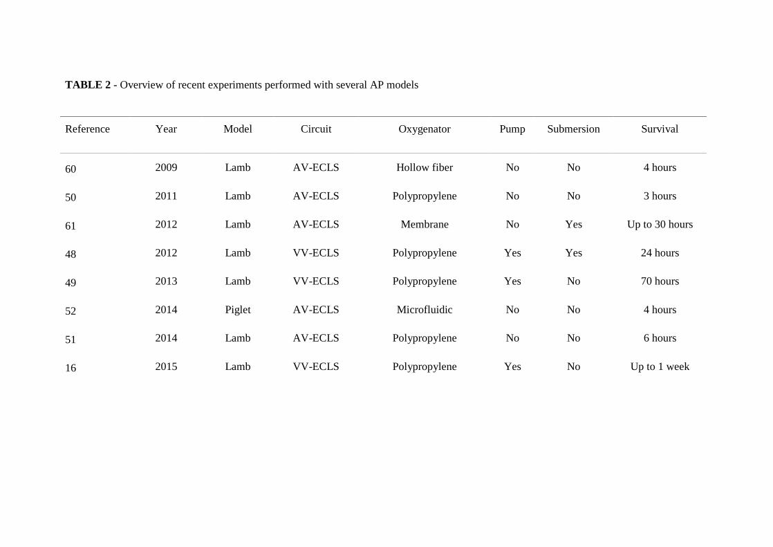
TABLE 2 - Overview of recent experiments performed with several AP models
Reference Year Model Circuit Oxygenator Pump Submersion Survival
60 2009 Lamb AV-ECLS Hollow fiber No No 4 hours
50 2011 Lamb AV-ECLS Polypropylene No No 3 hours
61 2012 Lamb AV-ECLS Membrane No Yes Up to 30 hours
48 2012 Lamb VV-ECLS Polypropylene Yes Yes 24 hours
49 2013 Lamb VV-ECLS Polypropylene Yes No 70 hours
52 2014 Piglet AV-ECLS Microfluidic No No 4 hours
51 2014 Lamb AV-ECLS Polypropylene No No 6 hours
16 2015 Lamb VV-ECLS Polypropylene Yes No Up to 1 week

ANEXOS
I - 2018 Update on “Artificial Placenta: Recent Advances and Potential Clinical
Applications”
II - Editorial “The Artificial Placenta: is Clinical Translation Next?”
III - Citações do artigo “Artificial Placenta: Recent Advances and Potential Clinical
Applications”
IV - Normas de publicação da revista científica Pediatric Pulmonology
V - Agradecimentos
1
2018 UPDATE ON “ARTIFICIAL PLACENTA: RECENT ADVANCES AND
POTENTIAL CLINICAL APPLICATIONS”
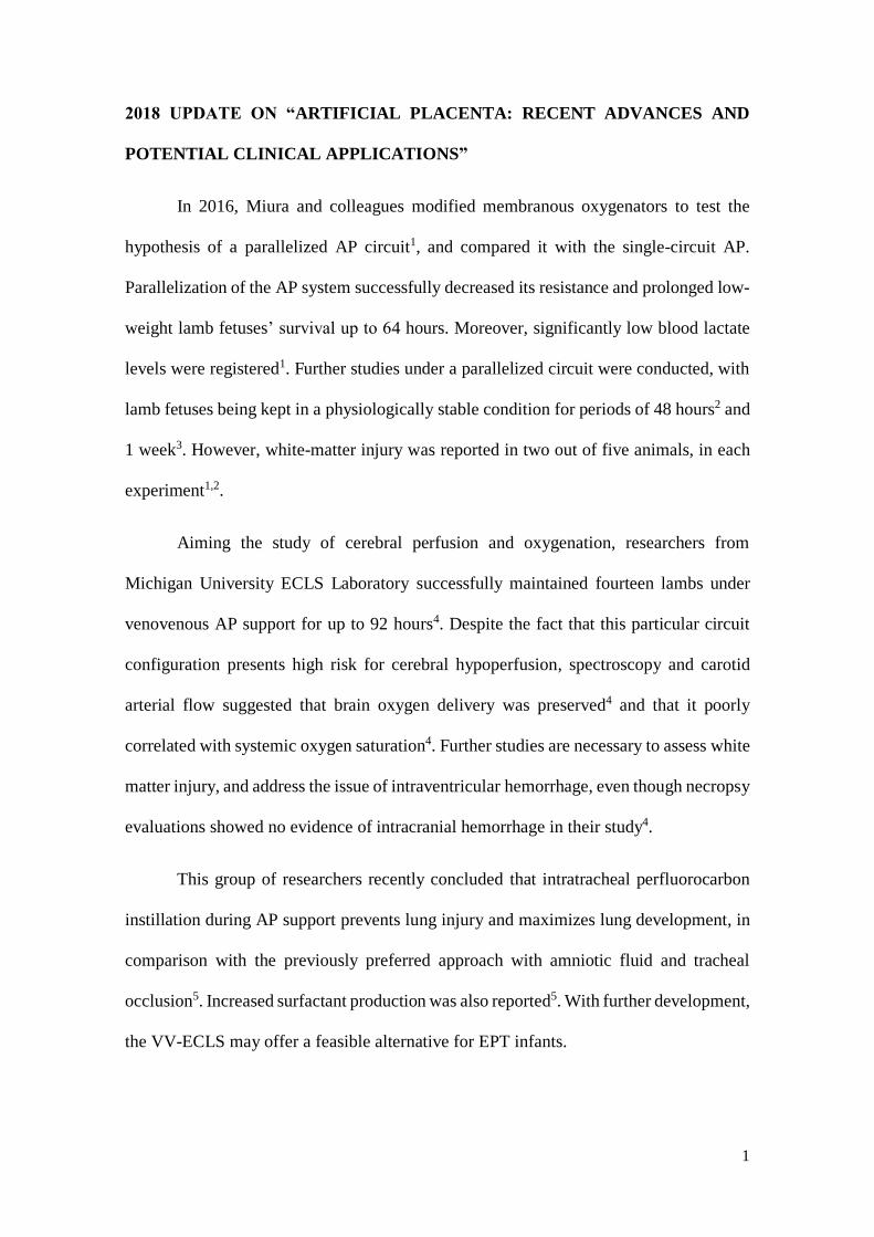
In 2016, Miura and colleagues modified membranous oxygenators to test the
hypothesis of a parallelized AP circuit1, and compared it with the single-circuit AP.
Parallelization of the AP system successfully decreased its resistance and prolonged low-
weight lamb fetuses’ survival up to 64 hours. Moreover, significantly low blood lactate
levels were registered1. Further studies under a parallelized circuit were conducted, with
lamb fetuses being kept in a physiologically stable condition for periods of 48 hours2 and
1 week3. However, white-matter injury was reported in two out of five animals, in each
experiment1,2.
Aiming the study of cerebral perfusion and oxygenation, researchers from
Michigan University ECLS Laboratory successfully maintained fourteen lambs under
venovenous AP support for up to 92 hours4. Despite the fact that this particular circuit
configuration presents high risk for cerebral hypoperfusion, spectroscopy and carotid
arterial flow suggested that brain oxygen delivery was preserved4 and that it poorly
correlated with systemic oxygen saturation4. Further studies are necessary to assess white
matter injury, and address the issue of intraventricular hemorrhage, even though necropsy
evaluations showed no evidence of intracranial hemorrhage in their study4.
This group of researchers recently concluded that intratracheal perfluorocarbon
instillation during AP support prevents lung injury and maximizes lung development, in
comparison with the previously preferred approach with amniotic fluid and tracheal
occlusion5. Increased surfactant production was also reported5. With further development,
the VV-ECLS may offer a feasible alternative for EPT infants.

2
In 2017, Flake and colleagues presented a unique AP system that incorporates a
pumpless oxygenator through an umbilical vascular interface kept within a closed fluid
circuit, the polyethylene Biobag6. This model, which resembles the womb environment
in shape and size, allowed eight fetal lambs to grow in a temperature-controlled, near-
sterile environment, breathing an amniotic-like fluid6. Swallowing an electrolyte solution
improved fetal fluid homeostasis and provided an additional route for nutrition.
Furthermore, since there was continuous fluid exchange, this configuration solved the
problem of gross fluid contamination and infection6.
In this study, double umbilical artery and single umbilical vein cannulation were
preferred over carotid use, preserving a length of native umbilical cord between the
cannula tips and the abdominal wall. This approach allowed vascular adaptation to
pressure changes across the AV shunt7 and was proven to optimize circuit flow dynamics
and stability, since it closely relates to the placental physiology in comparison to other
cannulation methods7.
Remarkably, Flake et al. reported successful transition to air breathing after AP
support in two lambs that were connected to the circuit for 21 and 29 days, respectively8.
The animals presented satisfying responses to feeding and normal somatic growth8. One
did not survive a pyelonephritis after 12 days of independent life, whereas the other
reportedly surpassed three months survival with apparently normal neurological
outcomes8.
These results are historical and superior to all previous attempts of EPT lambs’
ECLS in both duration and physiologic well-being. Researchers will carry on evaluating
and refining this AP system, since it needs to be downsized for human infants, who are
one-third the size of the experimental fetus lambs. The first clinical trial is expected to
occur within five years.

3
REFERENCES
1. Miura Y, Matsuda T, Usuda H, Watanabe S, Kitanishi R, Saito M, Hanita T, Kobayashi
Y. 2016. A parallelized pumpless artificial placenta system significantly
prolonged survival time in a preterm lamb model. Artif Organs. 40(5):E61-68.
2. Usuda H, Watanabe S, Miura Y, Saito M, Musk GC, Rittenschober-Bohm J, Ikeda H,
Sato S, Hanita T, Matsuda T et al. 2017. Successful maintenance of key
physiological parameters in preterm lambs treated with ex vivo uterine
environment therapy for a period of 1 week. Am J Obstet Gynecol.
217(4):457.e451-457.e413.
3. Miura Y, Usuda H, Watanabe S, Woodward E, Saito M, Musk GC, Kallapur SG, Sato
S, Kitanishi R, Matsuda T et al. 2017. Stable control of physiological parameters,
but not infection, in preterm lambs maintained on ex vivo uterine environment
therapy. Artif Organs. 41(10):959-968.
4. El-Sabbagh AM, Gray BW, Shaffer AW, Bryner BS, Church JT, McLeod JS, Zakem
S, Perkins EM, Shellhaas RA, Barks JDE et al. 2017. Cerebral oxygenation of
premature lambs supported by an artificial placenta. Asaio j.
5. Church JT, Perkins EM, Coughlin MA, McLeod JS, Boss K, Bentley JK, Hershenson
MB, Rabah R, Bartlett RH, Mychaliska GB. 2018. Perfluorocarbons prevent lung
injury and promote development during artificial placenta support in extremely
premature lambs. Neonatology. 113(4):313-321.

4
6. Partridge EA, Davey MG, Hornick MA, McGovern PE, Mejaddam AY, Vrecenak JD,
Mesas-Burgos C, Olive A, Caskey RC, Weiland TR et al. 2017. An extra-uterine
system to physiologically support the extreme premature lamb. Nature
Communications. 8:15112.
7. Hornick MA, Davey MG, Partridge EA, Mejaddam AY, McGovern PE, Olive AM,
Hwang G, Kim J, Castillo O, Young K et al. 2018. Umbilical cannulation
optimizes circuit flows in premature lambs supported by the extra-uterine
environment for neonatal development (extend). J Physiol.
8. Partridge EA, Davey MG, Hornick MA, Flake AW. 2017. An extrauterine environment
for neonatal development: Extending fetal physiology beyond the womb. Semin
Fetal Neonatal Med. 22(6):404-409.

Pediatric Pulmonology 51:557–559 (2016)
Editorial
The Artificial Placenta: Is Clinical Translation Next?
George B. Mychaliska, MD*
Despite significant advances in the treatment ofprematurity including antenatal steroids, advanced me-chanical ventilation strategies, and exogenous surfactant,the mortality and morbidity remain high for thesevulnerable infants. In particular, the mortality andmorbidity of extremely low gestational age newborns(ELGANs) defined as <28 weeks estimated gestationalage (EGA), is extremely high.1 A radical paradigm shift inthe treatment of extreme prematurity would be to re-create the intra-uterine environment using an extracorpo-real artificial placenta (AP).In this issue,Metelo-Coimbra andRoncon-Albuquerque2
review recent advances in the field and assess barriers toclinical translation. From the outset, it should be acknowl-edged that although there is a large number of prematurebirths worldwide (defined as <37 weeks EGA), outcomeshave substantially improved for infants >28 weeks EGA.Apart from some specific congenital anomalies likecongenital diaphragmatic hernia, the AP is intendedfor the treatment of ELGANS who experience themost complications of prematurity and whose outcomeremains poor.The authors focus on lung immaturity and provide
substantial evidence of the iatrogenic effects of mechani-cal ventilation on both lung injury and the deleteriouscardiovascular effects.2 It is worth noting that althoughELGANS are predisposed to interventricular hemorrhage(IVH) given their immature germinal matrix, mechanicalventilation has been implicated in the pathogenesis ofIVH by increasing intrathoracic and intracranial pressurewith every breath.3 The ELGANswho are never subjectedto positive airway pressure have fewer complications ofprematurity. An appreciation of the deleterious effects ofmechanical ventilation and high oxygen concentrationson premature lungs has led to a dramatic shift toward lessinvasive ventilator strategies for premature infants.Although this strategy appears promising for somepatients,4 there is still a subset of ELGANS that cannotmaintain adequate gas exchange with the most invasiveventilator strategies.
Although the pulmonary system is critical to initialsurvival and long-term pulmonary morbidity is highamong survivors, there are other significant complicationsof ELGANs that warrant consideration. Predictable andunsolved complications associated with prematurityinclude neurologic injury (IVH, white matter injury),necrotizing enterocolitis (NEC), retinopathy of prematu-rity (ROP), and sepsis. Our current inability to preventthese complications relates both to organ immaturity andconventional treatment strategies such as positivepressure ventilation, that have historically been developedfor full term infants. To potentially solve these problems,the AP must not only recreate the fetal milieu and providelife-sustaining functions such as adequate gas exchangeand fetal hemodynamic stability, but it should also protectagainst organ trauma and allow the normal developmentalpathways to occur.An AP may appear far beyond the reach of modern
science, but the idea of creating a life support system tomaintain growing fetuses in a womb-like environmentwith extracorporeal support was first investigated 60 years
Section of Pediatric Surgery, Department of Surgery, Fetal Diagnosis and
Treatment Center, University of Michigan Medical School, C.S. Mott
Children’s Hospital,, 1540 E. Medical Center Drive, SPC 4211, Ann Arbor,
Michigan 48109.
Conflict of Interest: None.
Funding source: NIH, Number 1R01HD073475-01A1.
�Correspondence to: George B. Mychaliska, MD, Section of Pediatric
Surgery, Department of Surgery, Fetal Diagnosis and Treatment Center,
University of Michigan Medical School, C.S. Mott Children’s Hospital,
1540 E. Medical Center Drive, SPC 4211, Ann Arbor, MI 48109.
E-mail: [email protected]
Received 20 February 2016; Accepted 6 March 2016.
DOI 10.1002/ppul.23412
Published online 19 April 2016 in Wiley Online Library
(wileyonlinelibrary.com).
� 2016 Wiley Periodicals, Inc.

ago! Metelo-Coimbra and Roncon-Albuquerque providea succinct review of AP terminology and history ofmilestones.2 Since fetuses normally develop with extra-corporeal support, it should perhaps not surprise us thatresearchers were drawn to this concept shortly after thesuccessful introduction of cardio-pulmonary bypass. Forhistorians of science, it is noteworthy that researcherswere on the right path, but got derailed many times due tothe state of biomedical technology and insufficientknowledge of the physiology of premature infants. Inmy view, the history of the development of the AP ismarked by many experimental failures with episodicsuccesses. Despite incremental success, many researchgroups abandoned this work as progress was being madewith antenatal steroids, exogenous surfactant, advancedmechanical ventilation strategies, and ECMO for termand near-term infants.An appreciation of the unsolved problems of extreme
prematurity coupled with recent advances detailed by theauthors2 has led to a resurgence of work on theAP. The fetallamb is the best model, and the lamb gestational age whichcorresponds to ELGAN lungs is 118 days gestation(term¼ 145 days). Although the AP is promising and hasthe potential to radically change the treatment ofprematurity, several obstacles remain. As the authors pointout,2 the first issue is themost effective ECLS configuration.A simple pumpless AV-ECLS circuit utilizing the umbilicalvessels is appealing, but our experience demonstrated onlyshort-term survival and declining cardiac function.5 Despitecannulation of the umbilical arteries to the sheep aorta (toobviate vessel spasm) and adding a pump, matchingextracorporeal flow to systemic flow is very difficult (thenative placenta does this automatically). In addition, giventhe tortuosity, size, and spasm associated with humanumbilical arteries, we transitioned to a pump-driven VV-ECLSmodel with inflow via the umbilical vein and outflowvia the jugular vein. This approach provides 7 days ofsupport with excellent gas exchange and hemodynamicstability.6 With current technology, we believe this strategyis clinically translatable to extremely premature infants.As mentioned previously, the AP strategy will require
long-term support (2–4 weeks in humans) and demon-stration that organs are maturing and protected fromtrauma. This corresponds to 10–14 days in the 118 daylamb model. As such, an in-depth study of lungdevelopment, long-term support, and weaning to aventilator and air breathing will be required. A crucialaspect of lung development will be the airway strategyduring AP support. In our early work, the fetal lambs weresubmerged in a warmed “amniotic bath” effectively re-creating the intrauterine environment.5,7 While appealingin some regards, there are infection and patient accessissues with this approach. More recently, we have beenintubating the fetal lambs, filling themwith amniotic fluidor Perflubron and either capping the endotracheal tube or
maintaining 5–8 cmH2O pressure. It is possible to harnessthe power of mechanotransduction with this approach andpossibly accelerate lung growth.8,9
Apart from lung development which is crucial duringAP support, other vulnerable premature organs need to beexamined. Brain perfusion, function, and developmentare critical to understand. Although the sheep is not agood model for IVH, brain physiology and evidence ofwhitematter injury can be assessed.With a high incidenceof NEC in premature infants, optimal nutrition andperfusion of the gastrointestinal system warrants investi-gation. Long term survivors of the AP should be examinedfor evidence of retinopathy of prematurity. Lastly, renaland hepatic function should be addressed.As a general rule, extracorporeal support is reserved for
infants � 34 weeks EGA due to a higher rate of IVH inextremely premature infants. The authors point out thefeasibility of “preemie ECMO” in infants from 29 to33 weeks,2 but ELGANs would have prohibitively highrates of IVH. As such, a critical barrier to clinicalapplication will be the development of non-thrombogenicsurfaces that will obviate the need for anti-coagula-tion.10,11 Lastly, clinical application will require a clinicalprognostication tool to select premature infants at thehighest risk for mortality on the first day of life.12
Given recent advances and ongoing work, we believethat the AP will be used in ELGANs in the next 5 years.
REFERENCES
1. Stoll BJ, Hansen NI, Bell EF, Shankaran S, Laptook AR, Walsh
MC, Hale EC, Newman NS, Schibler K, CarloWA, et al. Neonatal
outcomes of extremely preterm infants from the NICHD Neonatal
Research Network. Pediatrics 2010;126:443–456.
2. Metelo-Coimbra C, Roncon-Albuquerque R, Jr. Artificial
placenta: recent advances and potential clinical applications.
Pediatr Pulmonol. doi: 10.1002/ppul.23401.
3. Aly H, Hammad TA, Essers J, Wung JT. Is mechanical ventilation
associated with intraventricular hemorrhage in preterm infants?
Brain Dev 2012;34:201–205.
4. Schmolzer GM,KumarM, Pichler G, Aziz K, O’ReillyM,Cheung
PY. Non-invasive versus invasive respiratory support in preterm
infants at birth: systematic review and meta-analysis. BMJ
2013;347:f5980.
5. Reoma J, Rojas A, Kim A, Khouri J, Boothman E, Brown K,
Grotberg J, CookK, Bartlett R, Hirschl R, et al. Development of an
artificial placenta I: pumpless arterio-venous extracorporeal life
support in a neonatal sheep model. J Pediatr Surg 2009;44:53–59.
6. Bryner B, Gray B, Perkins E, Davis R, HoffmanH, Barks J, Owens
G, Bocks M, Rojas-Pena A, Hirschl R, et al. An extracorporeal
artificial placenta supports extremely premature lambs for 1 week.
J Pediatr Surg 2015;50:44–49.
7. Gray BW, El-Sabbagh A, Rojas-Pena A, Kim AC, Gadepali S,
Koch KL, Capizzani TR, Bartlett RH, Mychaliska GB. Develop-
ment of an artificial placenta IV: 24 hour venovenous extracorpo-
real life support in premature lambs. ASAIO J 2012;58:148–154.
8. Mychaliska G, Bryner B, Dechert R, Kreutzman J, Becker M,
Hirschl R. Safety and efficacy of perflubron-induced lung growth
in neonates with congenital diaphragmatic hernia: results of a
prospective randomized trial. J Pediatr Surg 2015;50:1083–1087.
558 Mychaliska
Pediatric Pulmonology

9. Shue EH, Miniati D, Lee H. Advances in prenatal diagnosis and
treatment of congenital diaphragmatic hernia. Clin Perinatol
2012;39:289–300.
10. Major TC, Brant DO, Burney CP, Amoako KA, Annich GM,
Meyerhoff ME, Handa H, Bartlett RH. The hemocompatibility of
a nitric oxide generating polymer that catalyzes S-nitrosothiol
decomposition in an extracorporeal circulation model. Biomate-
rials 2011;32:5957–5969.
11. Major TC, Brant DO, Reynolds MM, Bartlett RH, Meyerhoff
ME, Handa H, Annich GM. The attenuation of platelet and
monocyte activation in a rabbit model of extracorporeal
circulation by a nitric oxide releasing polymer. Biomaterials
2010;31:2736–2745.
12. Reid S, Bajuk B, Lui K, Sullivan EA. Comparing CRIB-II and
SNAPPE-II as mortality predictors for very preterm infants.
J Paediatr Child Health 2015;51:524–528.
The Artificial Placenta 559
Pediatric Pulmonology

CITAÇÕES DO ARTIGO “ARTIFICIAL PLACENTA: RECENT ADVANCES
AND POTENTIAL CLINICAL APPLICATIONS”
1. Mychaliska G. 2016. The artificial placenta: Is clinical translation next? Pediatric
pulmonology. 51(6):557-559.
2. Bird SD. 2017. Artificial placenta: Analysis of recent progress. Eur J Obstet Gynecol
Reprod Biol. 208:61-70.
3. Parga JJ, Garg M. 2017. Extracorporeal membrane oxygenation in neonates: History
and future directions. NeoReviews. 18(3):e166.
4. te Pas AB. 2017. Improving neonatal care with technology. Frontiers in Pediatrics.
5:110.
5. Mazumdar Bolanos M. 2017. Electrolysis-based system for generation and delivery of
oxygen to microfluidic oxygenator unit for preterm neonates with respiratory
distress syndrome. [MacSphere - McMaster University Libraries Institutional
Repository]: McMaster University.
6. Surate Solaligue DE, Rodriguez-Castillo JA, Ahlbrecht K, Morty RE. 2017. Recent
advances in our understanding of the mechanisms of late lung development and
bronchopulmonary dysplasia. Am J Physiol Lung Cell Mol Physiol.
313(6):L1101-l1153.
A1; B2; C3; D4; E5; F6.

Pediatric Pulmonology© Wiley Periodicals, Inc.
Edited By: Thomas Murphy
Impact Factor: 2.758
ISI Journal Citation Reports © Ranking: 2016: 19/121 (Pediatrics); 26/59 (Respiratory System)
Online ISSN: 1099-0496
Author Guidelines
SCOPE OF JOURNALPERMISSIONSAUTHOR RESOURCESENGLISH LANGUAGE SERVICESELECTRONIC SUBMISSION OF MANUSCRIPTSMANUSCRIPT GUIDELINES
Original Research ArticlesReviews/State of the Art PapersCase ReportsEditorials (Commentaries)Letters to the Editor
PRIOR TO SUBMITTINGCOMPONENTS OF ARTICLES/FILE PREPARATION
Main DocumentTitle PageSummary/AbstractAcknowledgementsInformed ConsentReferences

KeywordsAbbrevia�onsDrug NamesEponymsForma�ng Specific to Original Research Ar�cles
TablesImagesOnline Supporting Information
POLICIES/DISCLOSURE STATEMENTSConflict of InterestExperimental and Publication EthicsPlagiarismPrior PublicationClinical Trials
PEER REVIEW PROCESSFAST TRACK REVIEWSUBMISSIONS FROM EDITORS AND EDITORIAL BOARD MEMBERSAUTHOR CHARGESMANUSCRIPTS ACCEPTED FOR PUBLICATION
Online OpenCopyright Transfer Agreement
PROOFSREPRINTSAPPEALS PROCESSPRODUCTION QUESTIONSQUESTIONS ABOUT YOUR SUBMISSIONCONTACT THE EDITOR-IN-CHIEF
SCOPE OF JOURNALPediatric Pulmonology publishes the results of original clinical or laboratory research,state of the art reviews, exceptionally instructive or unique case reports, and letters tothe Editor (and responses), pertaining to the specialty.
Reports on meetings, conferences and symposia may be published after consultationwith the Publisher and the Editor-in-Chief.
Preliminary brief communications will be considered if the articles contain informationwhich would be considered a major breakthrough in the field.
We do not publish research funded by tobacco companies.

As the field is continually evolving, our Journal has seen an increase in the number ofsubmissions over the past few years, and, as a result, our rejection rate is climbing.
PERMISSIONSNo material published in Pediatric Pulmonology may be reproduced or publishedelsewhere without the written permission of the publisher and the author. To requestpermission to reproduce an article, in part, or in whole, click here to for the PermissionsPage (http://onlinelibrary.wiley.com/journal/10.1002/(ISSN)1099-0496/homepage/Permissions.html)
AUTHOR RESOURCESFor additional tools visit Author Services(http://authorservices.wiley.com/bauthor/default.asp) - an enhanced suite of online toolsfor WileyOnlineLibary journal authors, featuring Article Tracking, E-mail PublicationAlerts and Customized Research Tools.
ENGLISH LANGUAGE SERVICESThe Editors reserve the right to return any manuscript that is not in acceptable English.Translations from another language will not be provided by the Editorial Office. Authorsfrom countries in which English is not the primary language should have theirmanuscript reviewed and corrected by an English language service before submission.To read more about our policy, and to view a list of editing services, visit:http://authorservices.wiley.com/bauthor/english_language.asp(http://authorservices.wiley.com/bauthor/english_language.asp)
GUIDELINES FOR COVER SUBMISSIONS If you would like to send suggestions for artwork related to your manuscript to beconsidered to appear on the cover of the journal, please follow these general guidelines.(http://olabout.wiley.com/WileyCDA/Section/id-828302.html)
ELECTRONIC SUBMISSION OF MANUSCRIPTSIf you are familiar with our guidelines, click here(http://mc.manuscriptcentral.com/ppul) to login to your ScholarOne account to submityour manuscript. If you do not have an account, click on “Register Here” to establishone.
MANUSCRIPT GUIDELINESWe accept submissions of the following types of articles. Please note the specificguidelines for each type:
Original Research ArticlesOriginal Research Articles should follow the standard structure of abstract, introduction,methods, results, discussion, and references, and may include up to six tables and/orimages when appropriate. Original Research Articles should be limited to 3,500 words(not including the abstract or references). The abstract should not exceed 250 words, andreferences should be limited to forty (40).
Reviews/State of the Art PapersEditors generally commission Reviews and State of the Art papers, but uninvitedsubmissions are also welcome, particularly if the submission outlines an important andtopical subject with a focus on recent advances. Reviews should be limited to 4,000
words, while State of the Art papers should be limited to 5,000 words (not including theabstract or references). We ask that the abstracts for these manuscript types do notexceed 250 words. There is no set limit on images, tables, or references for these typesof manuscripts.
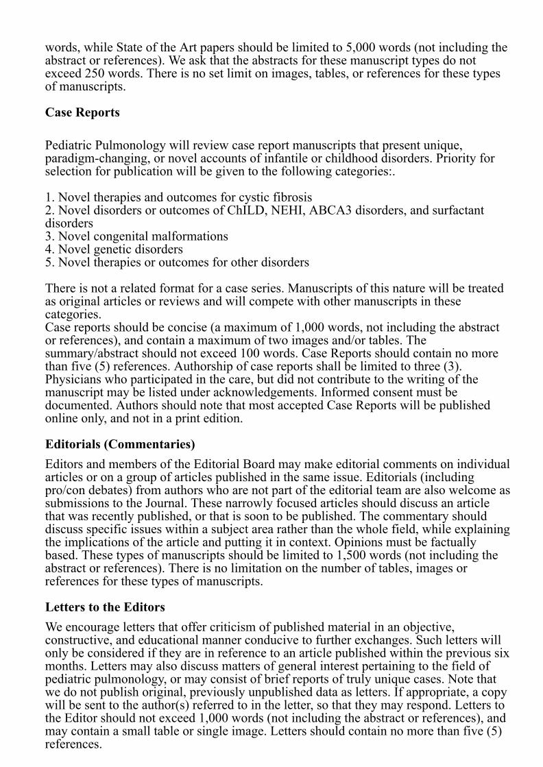
Case Reports Pediatric Pulmonology will review case report manuscripts that present unique,paradigm-changing, or novel accounts of infantile or childhood disorders. Priority forselection for publication will be given to the following categories:.
1. Novel therapies and outcomes for cystic fibrosis 2. Novel disorders or outcomes of ChILD, NEHI, ABCA3 disorders, and surfactantdisorders 3. Novel congenital malformations 4. Novel genetic disorders 5. Novel therapies or outcomes for other disorders
There is not a related format for a case series. Manuscripts of this nature will be treatedas original articles or reviews and will compete with other manuscripts in thesecategories. Case reports should be concise (a maximum of 1,000 words, not including the abstractor references), and contain a maximum of two images and/or tables. Thesummary/abstract should not exceed 100 words. Case Reports should contain no morethan five (5) references. Authorship of case reports shall be limited to three (3).Physicians who participated in the care, but did not contribute to the writing of themanuscript may be listed under acknowledgements. Informed consent must bedocumented. Authors should note that most accepted Case Reports will be publishedonline only, and not in a print edition.
Editorials (Commentaries)Editors and members of the Editorial Board may make editorial comments on individualarticles or on a group of articles published in the same issue. Editorials (includingpro/con debates) from authors who are not part of the editorial team are also welcome assubmissions to the Journal. These narrowly focused articles should discuss an articlethat was recently published, or that is soon to be published. The commentary shoulddiscuss specific issues within a subject area rather than the whole field, while explainingthe implications of the article and putting it in context. Opinions must be factuallybased. These types of manuscripts should be limited to 1,500 words (not including theabstract or references). There is no limitation on the number of tables, images orreferences for these types of manuscripts.
Letters to the EditorsWe encourage letters that offer criticism of published material in an objective,constructive, and educational manner conducive to further exchanges. Such letters willonly be considered if they are in reference to an article published within the previous sixmonths. Letters may also discuss matters of general interest pertaining to the field ofpediatric pulmonology, or may consist of brief reports of truly unique cases. Note thatwe do not publish original, previously unpublished data as letters. If appropriate, a copywill be sent to the author(s) referred to in the letter, so that they may respond. Letters tothe Editor should not exceed 1,000 words (not including the abstract or references), andmay contain a small table or single image. Letters should contain no more than five (5)references.

Top of Page
PRIOR TO SUBMITTINGPrior to submitting a manuscript through ScholarOne(http://mc.manuscriptcentral.com/ppul), prepare the text and images according to theinstructions found below. You may enter and exit the manuscript submission process atthe completion of each step, and you may save an unfinished draft in the system to workon later. However, once you submit your manuscript though the system, you will not beable to access it for editing. If you have any questions about this process please contactus at [email protected] (mailto:[email protected])
We recommend all authors familiarize themselves with the International Committee ofMedical Journal Editors: Uniform Requirements for Manuscripts submitted toBiomedical Journals. Ann Intern Med 1997;126:36-47. The complete text of thedocument be found online at www.icmje.org (http://www.icmje.org)
COMPONENTS OF ARTICLES/FILE PREPARATIONPlease make note of the following when preparing your submission:
Main DocumentAll manuscript types must include a title page, abstract, text and references in the MainDocument. Standard, double-spaced manuscript format, in 12 point font is requested.Number all pages consecutively.
Title page: The title should be brief (no more than 100 characters in length includingspaces) and useful for indexing. All authors’ names with highest academic degree,affiliation of each, but no position or rank, should be listed. For cooperative studies, theinstitution where research was primarily done should be indicated. In a separateparagraph, specify grants, other financial support received, and the granting institutions(grant number(s) and contact name(s) should be indicated on the title page). If supportfrom manufacturers of products used is listed, assurances about the absence of bias bythe sponsor and principal author must be given. Identify meetings, if any, at which thepaper was presented. The name, complete mailing address, telephone number, faxnumber, and e-mail address of the person to whom correspondence and reprint requestsare to be sent must be included. Keywords should also be noted on the title page. Forusage as a running head, provide an abbreviated title (maximum 50 characters) on thebottom of the title page.
Summary/Abstract: In accordance with the structure of the article, with or withoutseparate headings, outline the objectives, working hypothesis, study design, patient-subject selection, methodology, results (including numerical findings) and conclusions.The Summary should not exceed the word counts outlined above. If abbreviations areused several times, spell out the words followed by the abbreviations in parentheses.
Acknowledgements: Technical assistance, advice, referral of patients, etc. may bebriefly acknowledged at the end of the text under “Acknowledgements.”
Informed Consent: Informed consent statements, if applicable, should be included inthe Methods section.
References/citations: References may be included at the end of your text, or uploadedas a separate file. Ensure your references are up to date, and include a critical selectionfrom the world literature. References should be prepared according to CSE (Council of

Science Editors) citation-sequence style. Refer to the Scientific Style and Format: TheCSE Manual for Authors, Editors, and Publishers, 8th edition (University of ChicagoPress). Start the listing on a new page, double-spaced throughout.
Number the references in the sequence in which they first appear in the text, listing eachonly once even though it may be cited repeatedly.
When citing a reference in the text, the style advocated by CSE suggests numbersappear in superscript, and appear before punctuation marks (commas or periods). In thecitation-sequence system, sources are numbered by order of reference so that the firstreference cited in the paper is 1, the second 2, and so on. If the numbers are not in acontinuous sequence, use commas (with no spaces) between numbers. If you have morethan two numbers in a continuous sequence, use the first and last number of thesequence joined by a hyphen, for example 2,4,6-10.
In the references, list the first ten authors of the cited paper. If there are more than tenauthors, list the first 10 authors followed by 'et al'.
Journals’ names should be shown by their abbreviated title in Index Medicus.
Manuscripts in preparation or submitted for publication are not acceptable references. Ifa manuscript “in press” is used as a reference, a copy of it must be provided with yoursubmission.
Sample references:
Standard journal article Landau IL, Morgan W, McCoy KS, Taussig LM. Gender related differences in airwaytone in children. Pediatr Pulmonol 1993;16:31-35.
Book with authors Voet D, Voet JG. 1990. Biochemistry. New York: John Wiley & Sons. 1223 p.
Book with editors Coutinho A, Kazatch Kine MD, editors. Autoimmunity physiology and disease. NewYork. Wiley-Liss; 1994. 459 p.
Chapter from a book Hausdorf G. Late effects of anthracycline therapy in childhood: evaluation and currenttherapy. In: Bricker JT, Green DM, D'Angio GJ, editors. Cardiac toxicology aftertreatment for childhood cancer. New York: Wiley-Liss; 1993. p 73-86.
For a book reference only include the page numbers that have direct bearing on the workdescribed.
Keywords: On the title page, supply a minimum of 3 to 5 keywords, exclusive of wordsin the title of the manuscript. A guide to medical subject heading terms used by PubMedis available at http://www.nlm.nih.gov/mesh/MBrowser.html(http://www.nlm.nih.gov/mesh/MBrowser.html)
Abbreviations: Define abbreviations when they first occur in the manuscript and fromthere on use only the abbreviation. Whenever standardized abbreviations are availableuse those. Use standard symbols with subscripts and superscripts in their proper place.

Drug names: Use generic names. If identification of a brand name is required, insert itin parentheses together with the manufacturer’s name and address after the first mentionof the generic name.
Eponyms: Eponyms (diseases or biologic entities named for persons) should not beused when standard descriptive terminology is available. Examples include club cells(formerly known as Clara cells); and granulomatosis with polyangiitis (formerly knownas Wegener’s granulomatosis). It is permissible to use the eponym in parenthesis at thefirst mention of the term in cases in which the eponym is still in common use.
Formatting Specific to Original Research Articles: Divide article into: Title Page,Summary/Abstract, Introduction, Materials and Methods, Results, Discussion, andReferences, starting each section on a new page. All methodology and description ofexperimental subjects should be under Materials and Methods; results should not beincluded in the Introduction. Please ensure the following appears in the appropriatesection of your manuscript:
a concise introductory statement outlining the specific aims of the study andproviding a discussion of how each aim was fulfilled;a succinct description of the working hypothesis;a detailed explanation of assumptions and choices made regarding study design andmethodology;a description of the reasons for choosing the type and number of experimentalsubjects (patients, animals, controls) and individual measurements; if applicable,information about how and why the numbers may differ from an ideal design (e.g.,the number required for achieving 90% confidence in eliminating Type II error);specifics about statistical principles, techniques and calculations employed and, ifapplicable, methods for rejecting the null hypothesis;a concise comparison of the results with those of conflicting or confirmatory studiesin the literature;a brief summary of the limitations of the scientific methods and results; anda brief discussion of the implications of the findings for the field and for futurestudies.
Tables
Tables should not be included in the Main Document, but submitted as a separate DOCor RTF file. Number tables with Arabic numbers consecutively and in order ofappearance. Type each table double-spaced on a separate page, captions typed above thetabular material. Symbols for units should be used only in column headings. Do not useinternal horizontal or vertical lines; place horizontal lines between table caption andcolumn heading, under column headings, and at the bottom of the table (above thefootnotes if any). Use footnote letters (a, b, c, etc.) in consistent order in each table. Alltables should be referred to in the text. Do not submit tables as photographs and do notseparate legends from tables.
Images

Image files must be submitted in TIF or EPS (with preview) formats. Do not embedimages in the Main Document. Number images with Arabic numbers and refer to eachimage in the text. The preferred form is 5 X 7 inches (12.5 X 17.5 cm). Printreproduction requires files for full color images to be in a CMYK color space.
Please note authors are encouraged to supply color images regardless of whether or notthey are amenable to paying the color reproduction fees. Color images will be publishedonline, while greyscale versions will appear in print at no charge to the author. SeeAuthor Charges below.
Journal quality reproduction requires grey scale and color files at resolutions yieldingapproximately 300 ppi. Bitmapped line art should be submitted at resolutions yielding600-1200 ppi. These resolutions refer to the output size of the file; if you anticipate thatyour images will be enlarged or reduced, resolutions should be adjusted accordingly.
Lettering on images should be of a size and weight appropriate to the content and theclarity of printing must allow for legibility after reduction to final size. Labeling andarrows on images must be done professionally. Spelling, abbreviations, and symbolsshould precisely correspond to those used in the text. Indicate the stain andmagnification of each photomicrograph. Photographs of recognizable subjects must beaccompanied by signed consent of the subject of publication. Images previouslypublished must be accompanied by the author’s and publisher’s permission.
Image legends should be brief, and included as a separate DOC file under the heading:“Image Legends.” When borrowed material is used, the source of the image should beshown in parentheses after its legend, either by a reference number or in full if not listedunder References.
Online Supporting InformationAdditional non-essential material such as text, appendices, tables, images, video, andsoundtrack files may be submitted for posting as supporting information to an article.The scientific value of such material should be evident. The material should besubmitted simultaneously with the manuscript so that it may undergo peer review. Innaming these files, please note the file names should be preceded by the letter “E.” Forexample “E-table 1,“E-image 1,” “E-text,” etc.
Note that supporting online material is not typeset, nor proofread following the reviewprocess, so please ensure the material is accurate and free of typographical errors.Supporting material should be prepared in the same manner as the print material.
While supporting information does not appear in the print version, a notation is madethat supporting material is available online.
Top of PagePOLICIES/DISCLOSURE STATEMENTSWe recognize the importance of developing the highest ethical standards and we arecommitted to ethical publication practice. For more information on the publisher’spolicies, please see Wiley-Blackwell Guidelines on Publication Ethics and BestPractices www.wiley.com/bw/publicationethics (http://exchanges.wiley.com/publishing-ethics_252.html). Of particular importance is the section on Research Misconduct,which includes data fabrication, falsification, plagiarism, and inappropriate imagemanipulation.
Authors who submit to Pediatric Pulmonology should take heed of the following:
Conflict of Interest: Authors must indicate at the time of submission any potentialconflict of interest (particularly of a fiscal nature) that may have a perceived influenceon the results of the research. The existence of such does not automatically precludepublication. A conflict of interest statement should appear in the Acknowledgmentsection. For further information on Conflict of Interest please visit www.icjme.org(http://www.icmje.org/conflicts-of-interest/)
Experimental and Publication Ethics: Studies involving human subjects mustconform to the guiding principles of the World Medical Association Declaration ofHelsinki (http://www.wma.net/en/30publications/10policies/b3/). Human subjects musthave given informed consent and the study must have been approved by the Committeeon Human Research at the author(s) institution(s) and a statement to this effect mustappear in the Methods section of the submitted article. It is also important to documentin the Methods section that consent has been obtained from older children andadolescents. Similarly, animal studies must be approved by an Institutional AnimalResearch Review Board and a statement to this effect must appear in the Methodssection. In addition, details of anesthesia and euthanasia must appear in the Methodssection.
Plagiarism: It should be noted that Pediatric Pulmonology employs a plagiarismdetection system. By submitting your manuscript to this journal you accept that yourmanuscript may be screened for plagiarism against previously published works.CrossCheck is a multi-publisher initiative to screen published and submitted content fororiginality. Pediatric Pulmonology uses iThenticate software to detect instances ofoverlapping and similar text in submitted manuscripts. To find out more aboutCrossCheck visit http://www.crossref.org/crosscheck.html(http://www.crossref.org/crosscheck.html).
Prior Publication: Manuscripts submitted to Pediatric Pulmonology may not have beenpublished in any part or form in another publication of any type, professional or lay,including electronic publications, the exception being abstracts of no more than 400words. Any material available via PubMed or other electronic sources is considered tohave been published. When a question arises, the Editor-in-Chief will determine whatconstitutes duplicate publication. If duplicate publication is confirmed, the Editor-in-Chief will initiate a discussion with the sponsoring institution and the authors.
It is the responsibility of submitting authors to inform the Editor-in-Chief of potentiallyoverlapping or related data either in submitted manuscripts or papers in press, and suchmanuscripts should be appended to the submission. If there is significant overlap in datawith previously published articles this should be addressed by the author in the “AuthorsComments” section during the submission process. In particular, giving reasons why thenew submission should be published. The editors reserve the right to determine whetheror not publication is warranted.
For further information on redundant or duplicate publication, please visithttp://www.icmje.org (http://www.icmje.org/recommendations/browse/publishing-and-editorial-issues/overlapping-publications.html)
Clinical Trials: We endorse the Consolidated Statement of Reporting Trials(CONSORT Statement) Lancet 2002;357:1191-1194 which may be accessed atwww.consort-statement.org (http://www.consort-statement.org/). In accordance withICMJE standards, all clinical trials must be registered with a database that is publicly

accessible such as http://clinicaltrials.gov/ However, other free of charge publicregistries are acceptable. For further information, please visithttp://www.icmje.org/about-icmje/faqs/clinical-trials-registration/(http://www.icmje.org/about-icmje/faqs/clinical-trials-registration/)
PEER REVIEW PROCESSUpon submission, authors are encouraged to submit names of experts who they deemappropriate to review their paper. Authors may also indicate persons to whom they donot wish the manuscript sent for review. In most cases, articles will be reviewed by atleast two authorities as well as the editorial staff to determine validity, significance,novelty, and potential impact on the field of contents and conclusions. The reviewerswill be selected by the Editor-in-Chief, Associate Editors, and/or Editorial Boardmembers. The selection will be made on the basis of expertise, impartiality, and equaldistribution among the available experts, regardless of geographic origin of themanuscript or locations of the reviewers. Authors will be advised within the shortestpossible time whether their paper is accepted, requires major or minor revisions, or isrejected. All necessary efforts will be made to ensure a timely review process.
Authors should note the time from submission to final decision can be shortened by atimely return of a revised manuscript when revision has been requested.
The Editor-in-Chief reserves the right to reject any submission deemed not suitable forthe journal after an in-house review.
FAST TRACK REVIEWIf circumstances so warrant, a fast-track review of a paper may be requested in the coverletter. At the Editor-in-Chief’s discretion, a fast-track review will be undertaken toexpedite manuscripts that deserve rapid review and publication. Expedited peer reviewand publication is rare and is reserved for timely presentation of significant data. If fast-track review is requested in the cover letter, the corresponding author will be informed ifexpedited review has been granted or not.
SUBMISSIONS FROM EDITORS AND EDITORIAL BOARD MEMBERSPediatric Pulmonology strives to ensure that any submission from the Editor-in-Chief,Deputy Editor, the Associate Editors, or from a member of the journal’s Editorial Boardreceives an objective and unbiased evaluation. This is achieved by assigning anyresearch article submitted by the Editor-in-Chief, Deputy or Associate Editors to animpartial referee who can maintain the integrity of the review process. Whenappropriate, Pediatric Pulmonology may also utilize the services of Guest Editors whoare familiar with the peer review processes and policies of the journal. Articlessubmitted by Editorial Board members undergo a blinded peer review process that is asstringent as for those authors who are not on the Board. All submitting authors areautomatically blinded to all aspects of the review process.
AUTHOR CHARGES (AuthorCharges)Should authors wish for manuscript images appear in color in the print edition, colorreproduction fees will be charged to the authors. Current color reproduction fees are$800 per figure. Authors do, however, have the option of submitting color images foronline publication, and greyscale images for the print edition at no charge, and weencourage authors to do so. For information on color charges, please contact ProductionEditor, at ppulprod @wwiley.com

MANUSCRIPTS ACCEPTED FOR PUBLICATIONThe author identified as the formal corresponding author for the paper will receive anemail prompting them to login into Author Services where via the Wiley AuthorLicensing Service (WALS) they will be able to complete a license agreement on behalfof all authors on the paper:
Online OpenOnlineOpen (http://olabout.wiley.com/WileyCDA/Section/id-406241.html) fulfillsRCUK, Wellcome Trust, NIH, and other funder mandates. Authors can use OnlineOpento make their article open access and freely available to all on Wiley Online Library.Wiley also immediately deposit OnlineOpen articles in PubMed Central and PMCmirror sites. In addition, authors of OnlineOpen articles are permitted to post the final,published PDF of their article on a website, institutional repository or other free publicserver, immediately on publication.
With OnlineOpen, the author, the author's funding agency, or the author's institutionpays a fee to ensure that the article is made open access.
If the OnlineOpen option is selected the corresponding author will have a choice of thefollowing Creative Commons License Open Access Agreements (OAA):
(http://creativecommons.org/licenses/by-nc-nd/2.0/uk/)Creative Commons AttributionLicense OAA (http://creativecommons.org/licenses/by-nc/3.0/)(http://creativecommons.org/licenses/by-nc-nd/2.0/uk/)
- Creative Commons Attribution Non-Commercial -NoDerivs License OAA(http://creativecommons.org/licenses/by-nc-nd/2.0/uk/)
If you select the OnlineOpen option and your research is funded by The Wellcome Trustand members of the Research Councils UK (RCUK) you will be given the opportunityto publish your article under a CC-BY license supporting you in complying withWellcome Trust and Research Councils UK requirements. For more information on thispolicy and the Journal’s compliant self-archiving policy pleasevisit: http://www.wiley.com/go/funderstatement(http://www.wiley.com/go/funderstatement)
For RCUK and Wellcome Trust authors click on the link below to preview the terms andconditions of this license:
Creative Commons Attribution License OAA (http://creativecommons.org/licenses/by-nc/3.0/)
Note to NIH Grantees: Pursuant to the NIH mandate, Wiley Blackwell will post theaccepted version of contributions authored by NIH grant-holders to PubMed Centralupon acceptance. This accepted version will be made publicly available 12 months afterpublication. For further information, see Wiley Blackwell’s NIH Policy Statement.(http://www.wiley.com/WileyCDA/Section/id-321171.html)
Copyright Transfer AgreementIf the OnlineOpen option is not selected, the corresponding author will be presentedwith the copyright transfer agreement (CTA) to sign on behalf of all authors.

To preview the terms and conditions of the open access agreements, or the copyrighttransfer agreement, please visit the FAQs hosted on Wiley Author Services. Seehttp://authorservices.wiley.com/bauthor/faqs_copyright.asp(http://authorservices.wiley.com/bauthor/faqs_copyright.asp) andhttp://www.wileyopenaccess.com/details/content/12f25db4c87/Copyright--License.html(http://www.wiley.com/WileyCDA/Section/id-321171.html).
PROOFSFollowing acceptance, the corresponding author will be alerted by e-mail to accessgalley proofs in web-based proofing system. Corrections should be returned within 48hours of receipt, as delays in returning galley proofs cause delays in publication.Alterations should be kept to a minimum. Costs of extensive alterations to the galleyproof will be billed to the authors. All statements in (or omissions from) publishedmanuscripts are the responsibility of the authors who are asked to carefully reviewproofs prior to publication.
REPRINTSReprints may be ordered at: https://caesar.sheridan.com/reprints/eorder/order(https://caesar.sheridan.com/reprints/eorder/order.php?DBS=A&PUID=10089&IUID=4440&UNDO=1&SERVICE=2&ACCTUID=75)
APPEAL REQUESTSAuthors who wish to request reconsideration of a rejected manuscript should direct theirquery to the Editor in Chief, Tom Murphy, [email protected](mailto:[email protected]) or the PPUL editorial office,[email protected] ([email protected]). Requests must include themanuscript ID and a detailed description of why the authors believe the paper should bereconsidered.
Appeal requests will be evaluated by the Editor-in-Chief to determine if an appeal willbe permitted. These appeals will be approved on a very limited basis. If the appeal isallowed, instructions will be provided on how to resubmit your paper. Authors shouldnot resubmit their rejected paper without prior approval from the Editors and theEditorial Office. Requests that are sent elsewhere will not be considered.
PRODUCTION QUESTIONS?Contact Production Editor at [email protected](mailto:[email protected])
QUESTIONS ABOUT YOUR SUBMISSION?Contact the PPUL editorial office at [email protected]([email protected])
CONTACT THE EDITOR-IN-CHIEFThomas Murphy, [email protected](mailto:[email protected])
Top of Page

AGRADECIMENTOS
O trabalho refletido nesta monografia não teria sido possível sem a colaboração,
estímulo e empenho de diversas pessoas que, direta ou indiretamente, contribuíram para
a sua realização, e às quais não poderia deixar de agradecer.
Ao Professor Doutor Roberto Roncon-Albuquerque, orientador do projeto, por toda a
disponibilidade, pela confiança depositada e pelo incentivo constante, assim como por
me ter aberto as portas à atividade científica.
Aos Docentes com quem tive o privilégio de contactar durante o Mestrado Integrado em
Medicina, que tanto me inspiraram, nomeadamente ao Professor Doutor Tiago
Henriques-Coelho, que comigo partilhou o entusiasmo pelos resultados divulgados em
abril de 2017, relativos à mais promissora Placenta Artificial apresentada até à data.
Aos Tutores Clínicos que diariamente conjugam a atividade laboral com a vontade de
transmitir a sua experiência aos jovens estudantes, por influenciarem e ajudarem a
moldar a profissional que espero vir a ser no futuro.
A toda a minha família, em especial aos meus Avós, Alberto e Maria de Lurdes, pelo
apoio incondicional, pelo carinho e pela tranquilidade que me transmitem a cada dia.
À minha Mãe, esperando que esta etapa, que agora termino, possa, de alguma forma,
retribuir e compensar toda a dedicação que sempre manifestou para com o meu percurso
académico.
Ao meu irmão Diogo, companheiro de todas as horas, que celebrou comigo a plenos
pulmões cada vitória neste curso e na vida.
Aos meus padrinhos, Paula e Luís Miguel, pelo exemplo, e por acompanharem de perto
esta longa caminhada.

À Tatiana e à Inês, por todos os momentos memoráveis dos últimos seis anos, por me
terem ajudado a ultrapassar os mais variados desafios e por serem semper fi.
Aos colegas do YES Meeting, em particular aos “meus vinte”, pela honra de poder
trabalhar e aprender tanto com eles nessa grande aventura de organizar um congresso,
que recordo sempre com o maior dos sorrisos.
À Margarida e à Sofia, por todo o apoio e amizade, que ultrapassaram largamente a
convivência proporcionada pelo YES.
A todos os demais colegas e amigos da Faculdade de Medicina e da Faculdade de
Engenharia da Universidade do Porto, por terem enriquecido esta jornada.
Ao Gonçalo, por acreditar em mim por inteiro e por estar sempre presente.
