physiology of the skeletal muscle - uniba.sk · physiology of the skeletal muscle objectives ......
TRANSCRIPT

Physiology of the skeletal muscle
Objectives
• Organization of the skeletal muscle
• Mechanism of muscle contraction and relaxation
• Tetanus
• The all or nothing law in skeletal muscle
• Types of muscle fibres
• Mechanisms of skeletal muscle strength
•The smooth muscle
Practical tasks
Determination of work and fatigue in human
Determination of skeletal muscle strength in a human
Sim Muscle – PC experiment
© Katarína Babinská, MD, PhD. 2016

- muscle- fascicles
- fibres (cells)- myofibrils
- myofilaments(actin, myosin)
• myofibrils – a sequence of sarcomeres
• a sarcomere – a basic longitudinal
contractile unit of the striated muscle
• demarcated by two successive Z lines
• main components (myofilaments)
– thin filaments (actin)
– thick filaments (myosin)
(fixed by titin to the Z lines)
• cross striae formed by:
– I band - actin
– A band
• actin + myosin overlapped
• H band – myosin only
Organization of the skeletal muscle
A I I
H
http://www.sport-fitness-advisor.com/images/actin_myosin.jpg

Myofilaments
Thin filament
• actin globules arranged into fibres - helix of 2 filaments
• tropomyosin spreads along actin and covers the binding sites for myosin
• troponin (C, I, T) complex is present on each tropomyosin dimer
Thick filament
• myosin molecules
– in shape of golf clubs
– long tails bundled together,
– „heads“ sticking out

Neuro-muscular junction
The mechanism of AP transmission from a nerve to a muscle
3. this triggers release of
acetylcholine into synaptic cleft
4. acetylcholine binds to
receptors on the motor endplate
5. this causes opening of Na+
channels in motor endplate
6. motor endplate
potential is generated
7. action potential is generated
and travels along the
sarcolemma
- motor end-plate - the point of junction of a motor nerve fibre and a muscle fibre
- modified area of the muscle fibre membrane at which a synapse occurs
1. nerve impulse reaches the end of a motor neuron
2. Ca2+ voltage gated canals in the axon terminal open, Ca2+ influx into the terminal
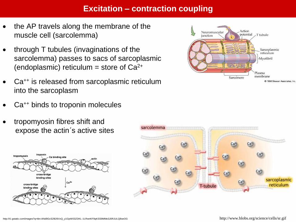
• the AP travels along the membrane of the
muscle cell (sarcolemma)
• through T tubules (invaginations of the
sarcolemma) passes to sacs of sarcoplasmic
(endoplasmic) reticulum = store of Ca2+
• Ca++ is released from sarcoplasmic reticulum
into the sarcoplasm
• Ca++ binds to troponin molecules
• tropomyosin fibres shift and
expose the actin´s active sites
http://t1.gstatic.com/images?q=tbn:ANd9GcS2B26VzQ_y1GpW03ZDKL-1LRwnKF9qKSS9MMe3J0RJULQ8xeOG
Excitation – contraction coupling
http://www.blobs.org/science/cells/sr.gif

Formation of actin – myosin cross-bridges
https://classconnection.s3.amazonaws.com/216/flashcards/1
042216/jpg/power_stroke1327356421108.jpg
The mechanism of muscle relaxation
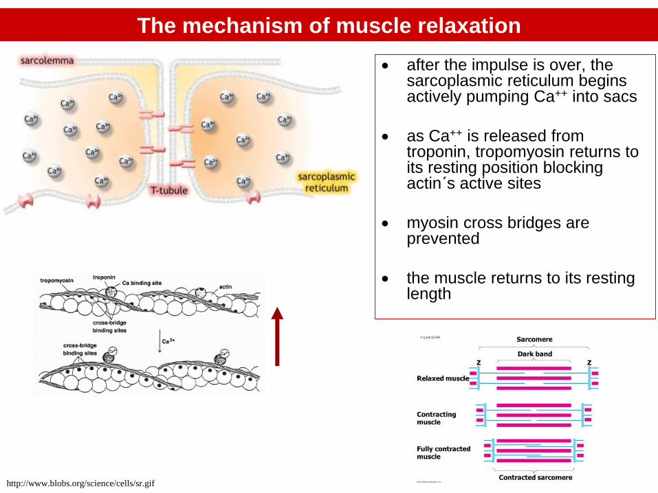
• after the impulse is over, the sarcoplasmic reticulum begins actively pumping Ca++ into sacs
• as Ca++ is released from troponin, tropomyosin returns to its resting position blocking actin´s active sites
• myosin cross bridges are prevented
• the muscle returns to its resting length
http://www.blobs.org/science/cells/sr.gif
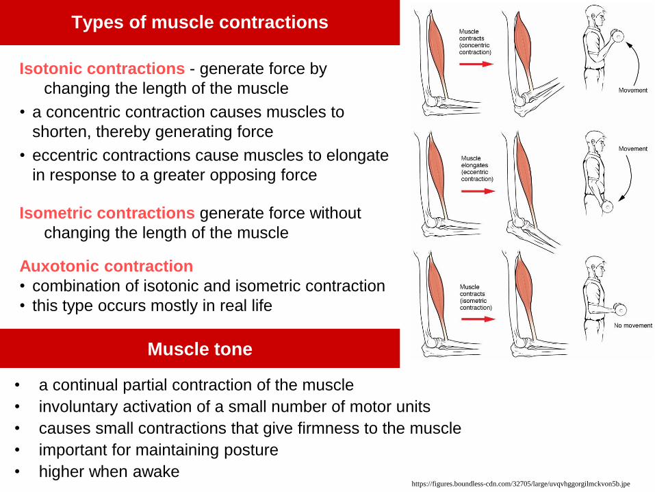
Types of muscle contractions
Isotonic contractions - generate force by
changing the length of the muscle
• a concentric contraction causes muscles to
shorten, thereby generating force
• eccentric contractions cause muscles to elongate
in response to a greater opposing force
Isometric contractions generate force without
changing the length of the muscle
Auxotonic contraction
• combination of isotonic and isometric contraction
• this type occurs mostly in real life
• a continual partial contraction of the muscle
• involuntary activation of a small number of motor units
• causes small contractions that give firmness to the muscle
• important for maintaining posture
• higher when awake
Muscle tone
https://figures.boundless-cdn.com/32705/large/uvqvhggorgilmckvon5b.jpe

The force of contraction depends on:
1. Motor unit recruitment and the graded strength principle
2. Increase in firing frequency
3. Muscle length - tension relationship
4. Type of muscle fibres
Muscle contraction strength mechanisms
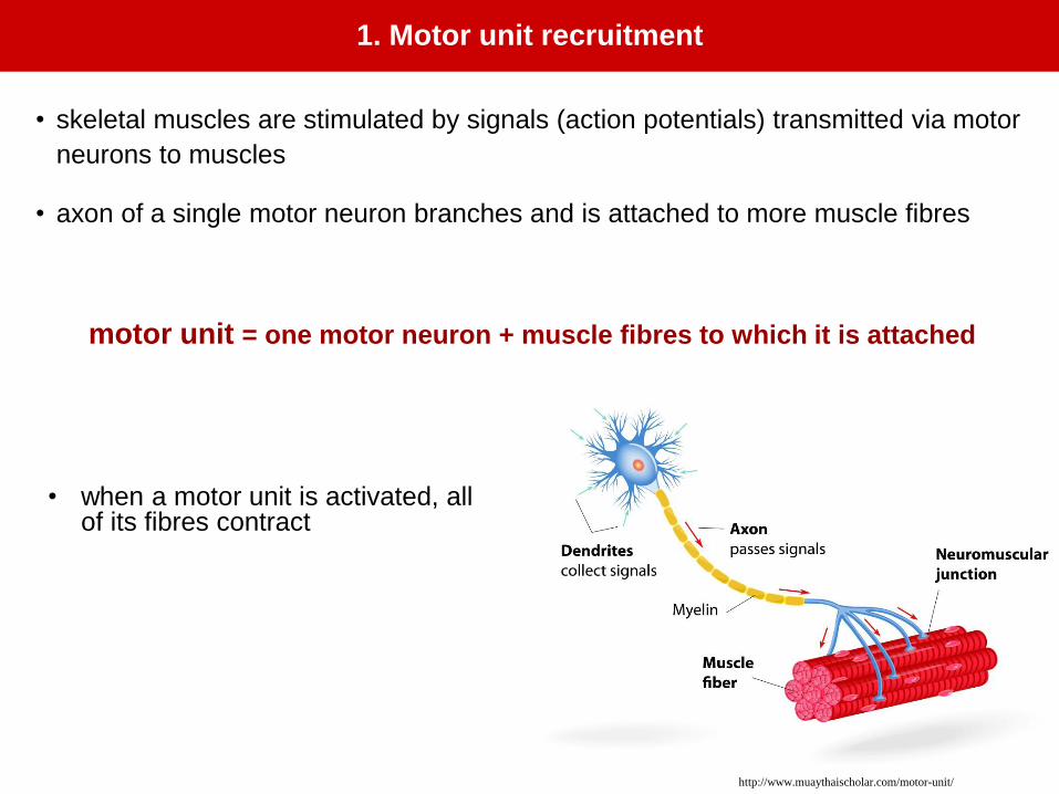
1. Motor unit recruitment
• skeletal muscles are stimulated by signals (action potentials) transmitted via motor
neurons to muscles
• axon of a single motor neuron branches and is attached to more muscle fibres
motor unit = one motor neuron + muscle fibres to which it is attached
http://www.muaythaischolar.com/motor-unit/
• when a motor unit is activated, all of its fibres contract

The „all or nothing“ principle in skeletal muscle
• when a motor unit receives a stimulus of sufficient intensity all the
muscle fibres within the unit will contract at the same time, and to the
maximum possible extent
• if the stimulus is not of sufficient intensity, the muscle fibres will not
respond, and contraction will not take place.
subtheshold threshold suprathreshold
stimulus stimulus stimulus
no response maximum contraction maximum contraction

• If more motor units are recruited to contract - muscle strength increases

• small motor units
– motor neuron is attached to fewermuscle fibres (eye, face, fingers)
– allow for fine and precise movements
– they produce little force
• large motor units
– involve – several hundreds of muscles
– e.g. in postural muscles
– produce large force
– allow for less precisemovements

The twitch contraction
• is a quick jerk (contraction) of the muscle fibre that occurs
after stimulation (e.g. electric stimulation)
• can be recorded by myograph (see picture)
• the curve of a twitch contraction includes 3 phases:
– latent period – time between stimulation and beginning
of contraction
– contraction phase
– relaxation phase
http://t2.gstatic.com/images?q=tbn:ANd9GcQ0QYA15DFQeIetqELpVYgRUnlzt_iXRYtCfXUxrcI_41B3rUVA
2. Increase in firing frequency

• if a series of stimuli come in longer intervals, the muscle has enough time to relax completely before next contraction
• a series of individual twitch contractions can be observed

Tetanus
- sustained contraction of a skeletal muscle, result of stimulation with high frequency
of stimuli
Incomplete tetanus
• the next stimulus arrives before the relaxation phase has ended
• muscle gets only partially relaxed
• summation of twitches occurs and the force of contraction increases
Complete tetanus
• the next stimulus comes at the peak of the previous contraction
• the muscle is instantly contracted – strength of contraction increases even more
Incomplete tetanus Complete tetanus

Increase in firing frequency – increases the strength of contraction
• single stimulus → release of Ca++ from sarcoplasmic reticulum → twitch
• twitch is terminated by reuptake of Ca++ into sarcoplasmic reticulum
• repeated stimulation in high frequency
– insufficient time to reaccumulate Ca++ into sarcoplasmic reticulum
– remains in sarcoplasm – sustained contraction = tetanus
• incomplete – next stimulus occurs in relaxation period of a twitch
• complete – next stimulus comes on the top of the twitch
Incomplete tetanus Complete tetanus

• the strength or maximum force produced by a muscle depends on the number
of cross bridges per unit area
• to increase the maximum force, increase the number of cross bridges
• the number of cross bridges depends on the starting position of actin and
myosin
http://ffden-
2.phys.uaf.edu/211_fall2004.web.dir/Katherine_
Van_Duine/actin%20and%20myosin.jpg
3. Muscle Length – Length tension relationship

Optimum muscle lenght – greatest force
- when the muscle is at an optimal length = indicated by the greatest possible
overlap of thick and thin filaments, maximal strength is produced.
-CNS maintains optimum length producing adequate muscle tone
Overly contracted
- thick filaments too close
to Z discs – cannot slide
more
Too stretched
-little overlap of thin and
thick filaments does not
allow for very many cross-
bridges to form

4. Muscle fibre type
Fast twith fibres
(type II, white)
Slow twitch fibres
(type I)
Contraction velocity High Low
Capillarization Low high
Myoglobin content Low High
Mitochondrial content Low High
Aerobic energy production Low High
Anaerobic energy production High Low
Glycogen stores High Low
Fatigue Fast Slow
Generation of speed and power High Low
Suited for Explosive sports Endurance sports
- Muscle fibre type is determined genetically + by training
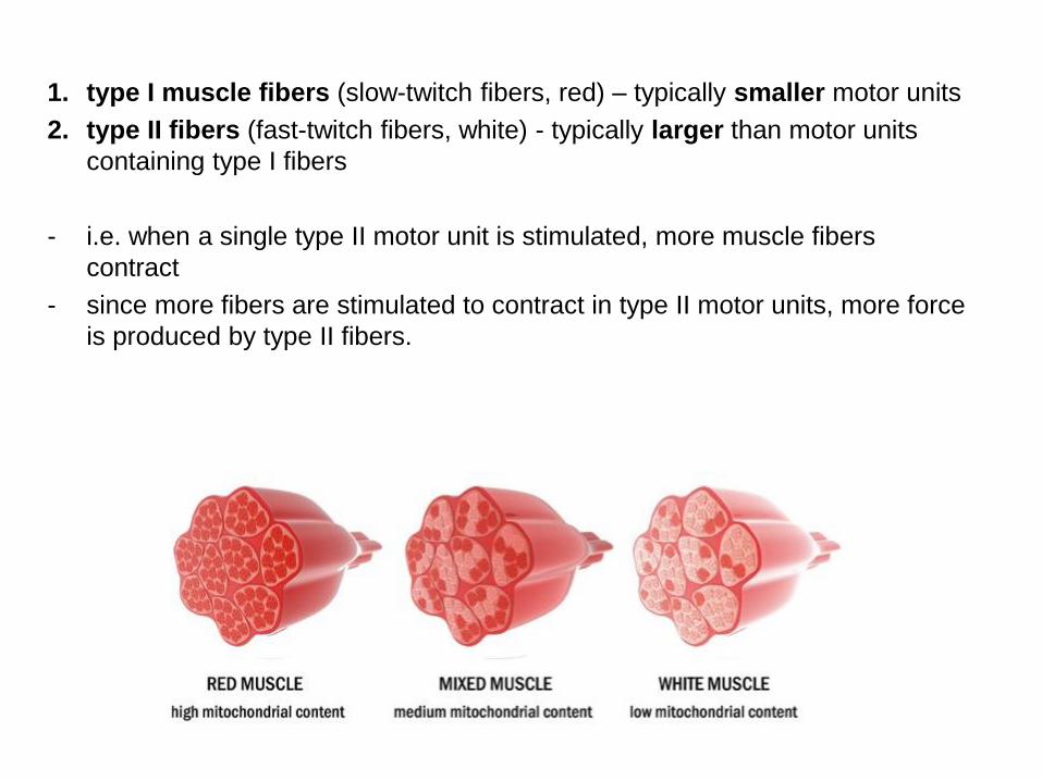
1. type I muscle fibers (slow-twitch fibers, red) – typically smaller motor units
2. type II fibers (fast-twitch fibers, white) - typically larger than motor units
containing type I fibers
- i.e. when a single type II motor unit is stimulated, more muscle fibers
contract
- since more fibers are stimulated to contract in type II motor units, more force
is produced by type II fibers.

Determination of skeletal muscle strength in a human
Muscle strength
• can be expressed as a maximum weight that can be kept by
contracted muscle (or group of muscles) in balance against gravity
• can be measured by hand dynamometer
• average (normal) value of the dominant hand
– 50 – 55 kg in an adult male
– 31 – 36 kg in an adult female
– value of the non-dominant hand – approx. 10 % less
http://www.getprice.com.au/images/uploadimg/2434/
Hydraulic-Hand-Dynamometer-Left-Side-
View_545_320x320.jpg

Report
A. Which of your hands is dominant?
B. Write down the measured and average values:
Right hand Left hand
measurement-1: measurement-1:
measurement-2: measurement-2:
measurement-3: measurement-3:
average value: average value:
C. Compare your average value for dominant hand with normal values
Procedure
1. Rotate the peak-hold needle counter to 0
2. Let the right upper extremity with dynamometer hang freely along the body (in
the standing position).
3. Compress the dynamometer by the right hand maximally
4. Record the value in kg (the peak-hold needle records the max force)
5. Repeat the measurement 3 times and calculate average value.
6. Repeat for the left hand.

Muscle fatigue
- the transient/reversible decrease in performance capacity of muscles induced
by exercise
- evidenced by a failure to maintain or develop a certain expected force or power, or
to sustain the task
- depends on
▪ the intensity/duration of the performance
▪ aerobic/anaerobic metabolism
▪ types of muscle fibres
▪ personal fitness
- experienced mainly in sustained and/or close to maximum activities
Sites, causes and mechanisms of fatigue:
• Neuromuscular depression (fatigue of synapses)
- synapse – most prone to fatigue
- every successive stimulation of a motor nerve causes weaker response in the post-
synaptic muscle fibre
- acetylcholine synthesis slower than required by fast firing rate

• Central fatigue
- subjective feeling of tiredness and a desire to stop the activity
- lack of motivation due to failure of cerebral cortex to send excitatory signals to
the motor neurons
- low pH seems to play role (in close to maximum physical activities)
• Cellular fatigue
- accumulation of extracellular K+ - due to repeated action potentials and the Na+-
K+ pump can not rapidly transport K+ back to the muscle - failure to reestablish the
resting membrane potential on a synapse
- rise in lactic acid concentration = lowering of pH – inhibits the cross-bridge
formation
- depletion of glycogen/glucose
- decrease in availability of Ca2+ ions - results in decreased Ca2+ release from
sarcoplasmic reticulum
- excessive accumulation of inorganic phosphate (ATP breakdown in cross-bridge
formation) in cytoplasm
- glycolytic fibres more prone to fatigue (slower Ca uptake)

Moss ergograph
serves for
• fixing the forearm and hand
• fixing the cable with load
• recording the contractions
Principle
• the volunteer lifts 2 kg load with m. flexor digitorum superficialis
• signs of fatigue are observed
Procedure
• the forearm of examinee is fixed in the ergograph holder
• the examinee holds the handle with his hand
• a leather ring is put on the second finger
• the examinee lifts a 2 kg load in pace given by a metronome
• the series of contractions are registered and evaluated
Task: Determination of work and fatigue in a human

1. ask the subject for any feelings of fatigue (pain, weakness of the finger…)
2. observe signs of fatigue on the record – the curve is flattened, the
contractions are irregular (the examinee continues to lift the load for
another 30-60 s)
3. start to encourage the volunteer – observe the effect of motivation on the
performance
4. calculate the work done per unit of time
- at the beginning of performance
- the period when sifns of of fatigue are visible
- in the period when the subject is encouraged
- unit of time = a segment, e.g. 15 cm
- select 3 segments from the beginning of the performance and the end - when
signs of fatigue are seen)

Positive dynamic work
- work done during muscle contraction
- e.g. load ligting
Negative dynamic work
-prohibits falling down (e.g. load releasing, going downstairs)
–this is not taken into account in this task
Work done (J) = load (kg) . gravitational acceleration (m.s-2) . trajectory (m)
trajectory = count of contractions . size of 1 contraction
count of contractions
size of
contractions
beginning fatigue encoura-
ging
frequency 12 6 13
size 0,035 0,027 0,027
trajectory 0,42 0,163 0,351
gravitational
acceleration
10 10 10
load 2 2 2
work done 8,4 J 3,26 J 7,02 J

The smooth muscle
http://faculty.ccri.edu/kamontgomery/muscle%20tissues.jpg

Types of muscles
Skeletal muscle Smooth muscle
Bigger cells, long and thin Smaller cells, spindle shaped
Syncytium (multinuclear cells) Single nucleus
Stiated muscle, sarcomere – basic unit No striations, no sarcomeres
Voluntary control Involuntary control
• Skeletal muscle
• Smooth muscle
• Cardiac muscle
http://faculty.ccri.edu/kamontgomery/muscle%20tissues.jpg
Skeletal muscle and smooth muscle – basic comparison

Types of the smooth muscles
Multi unit smooth muscle
• composed of separate smooth muscle fibers(separated by a glycoprotein/collagen layer)
• only a few fibres innervated by a single nerve ending
• small units of fibers can contract independently of the others
• their control is exerted mainly by nerve signals.
e.g. iris, ciliary muscle, erectores pili
Single unit (unitary) smooth muscle
• mass of hundreds/ thousands of smooth muscle
cells that contract together as a single unit.
• syncytium - cell membranes joined by gap
junctions - ions can flow freely from one cell to
another and cause depolarisation /contraction
• Often controled by non-nervous stimuli (e.g.hormones)
e.g. in the viscera, vessels

Innervation of the smooth muscle
Skeletal Smooth
Motor neurons Autonomic nerves
Synapse - motor end plate Synapse – varicosities
1muscle cell – 1 synapse 1 muscle cell – may have several
synapses
Smooth muscle contraction
Stimuli for a smooth muscle cells:
• Nervous
• Humoral – norepinephrine, epinephrine, acetylcholine, oxytocin, etc.
• Passive stretch
• Local tissue factors – excess of H+, CO2, lactate, deficit of O2

• Spike potentials
• Slow wave potentials - GIT
• Potentials with plateau – ureters,
uterus, some vessels
- Calcium channels play the role
(instead of Na) – slow channels
– therefore slow AP
- Ca ions only from ECT, not form
sarcoplasmic reticulum
Types of potentials in the smooth muscle

Contractile mechanism in the smooth muscle
• contraction – interaction of actin and myosin
filaments (different arrangement of the filaments
than in the skeletal muscle)
• actin filaments attached to the dense bodies (in
the cell membrane, inside the cell)
• the smooth muscle – does not contain troponin
• calmodulin is the regulatory protein - initiates
contraction in a different manner
This activation and contraction occur in the following sequence:
1. The calcium ions bind with calmodulin
2. The calmodulin-calcium combination joins with and activates myosin kinase, a
phosphorylating enzyme
3. Myosin heads become phosphorylated and are capable of interaction with actin

http://www.interactive-biology.com/wp-content/uploads/2012/04/Muscle-cells-
1024x1024.jpg
Skeletal muscle contraction Smooth muscle contraction
by 30% of their lenght by 80% of their lenght
Fast (10 – 300x) Slow (cycling of the cross bridges, long
lasting attachment of A-M, less ATP-ase
activity)
More energy required Less energy required to sustain the
contraction
Lower force of contraction Greater force of contraction
Other differences in the smooth muscle contraction

Muscular dystrophies
• a group of genetic diseases characterized by muscle weakness.
• the muscle force is diminished
• much of muscle dysfunction in later-stage muscle dystrophy arises
from an absence of functional myofibers.
• diaphragm may be affected – problems with respiration
Disorders of neuromuscular transmission
• Myasthenia gravis, the end plate potential (EPP) fails to effectively
activate the muscle fiber due to an autoimmune reaction against
acetylcholine receptors, resulting in muscle weakness and fatigue.
• Lambert-Eaton myasthenic syndrome, is usually associated with
presynaptic antibodies to the voltage-dependent calcium channel.
• Botulism
Neurotoxin may act on the neuromuscular junction either post
synaptically or presynaptically as there are several different forms of
toxins that the NMJ is sensitive to. Common mechanisms of action
include blockage of acetylcholine release at the synapse thus causing
the NMJ to become abnormal in function.

Physiology of striated muscle:
minimal and maximal muscle contraction
• grab the muscle and hang it on the hook,
• add 1 load on the muscle
• stimulate the muscle via ON button on the stimulator with increasing intensity
• when you find the minimum intensity, switch the „Store“ on
• increase the intensity of stimuli until you find maximal muscle contraction (max
intensity of stimulation)
• settings:
time base: 200 ms/div
channel 1: 200 mV/div
channel 2: 50 mV/div
STORE off
mode SINGLE
switch Zero Adj. On (blue)
black lever to FREE position
CH1 move with a mouse to about 1/3 of the screen
CH2 move to about 2/3 of the screen
1 23
• turn on the machines (Power on) of the simulation software – stimulator (1),
oscilloscope (2) and measuring device (3)

Physiology of striated muscle:
incomplete and complete tetanus
• settings:
time base: 100 ms/div
channel 1: 200 mV/div
channel 2: 50 mV/div
STORE off
mode TWIN
interval 350 ms
amplitude 300 mV
switch Zero Adj. On (blue)
black lever to FREE position
23
• turn on the machines (Power on) of the simulation software – stimulator (1),
oscilloscope (2) and measuring device (3)
CH1 move with a mouse to about 1/3 of the screen
CH2 move to about 2/3 of the screen
- Grab the muscle and hang it on the hook, add 1 load on the muscle
- Stimulate the nerve via ON button on stimulator
- Decrease the time interval between two impulses (always by 50 ms) and find the
interval which would cause:
a) superposition of muscle contractions
b) summation of muscle contractions
- if you change mode to Train (5-10 impulses), you may observe a longer series of
twitches