physiology of the skeletal muscle - dr. danazaha · 03/02/2014 · the effectors physiology of the...
TRANSCRIPT
THE EFFECTORS
Physiology of the skeletal muscle
About 40 percent of the body is skeletal muscle, and perhaps another 10 percent is smooth and cardiac muscle.
Anatomy of skeletal muscleAll skeletal muscles are composed of numerous fibers ranging from 10 to 80
micrometers in diameter, each of these fibers been made up of successively smaller subunits. In most skeletal muscles, each fiber extends the entire length of the muscle. Except for about 2 percent of the fibers, each fiber is usually innervated by only one nerve ending, located near the middle of the fiber.
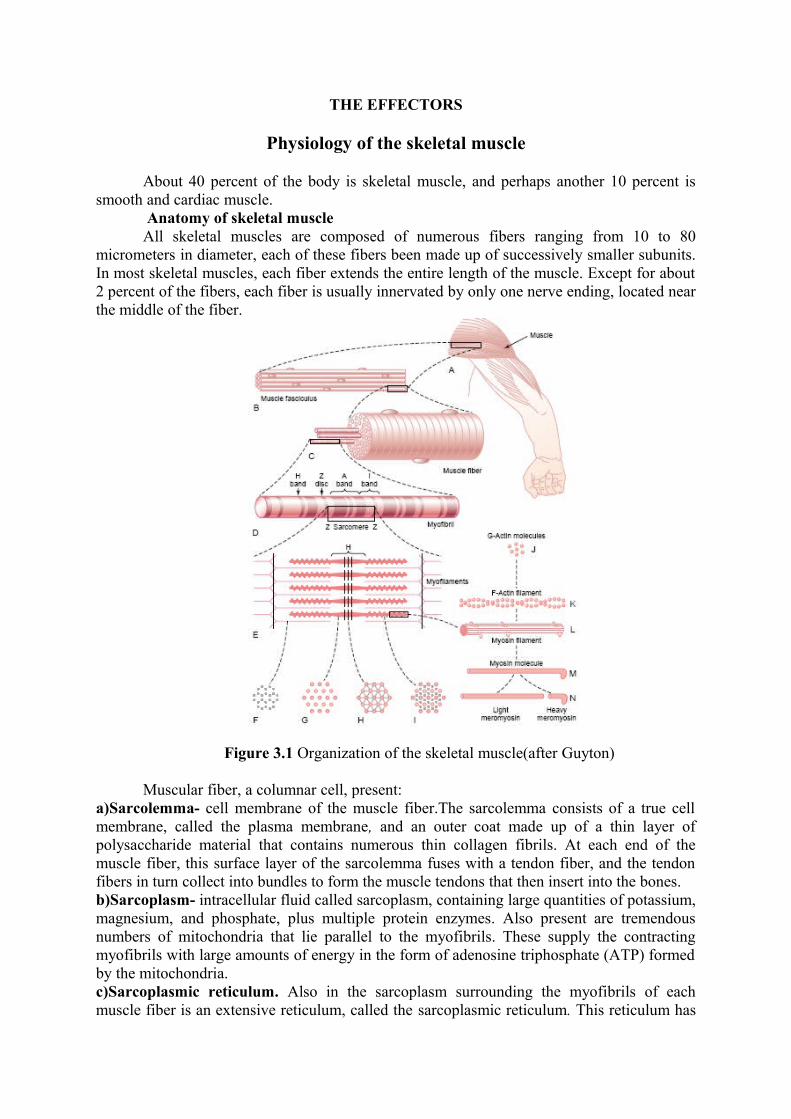
Figure 3.1 Organization of the skeletal muscle(after Guyton)
Muscular fiber, a columnar cell, present:a)Sarcolemma- cell membrane of the muscle fiber.The sarcolemma consists of a true cell membrane, called the plasma membrane, and an outer coat made up of a thin layer of polysaccharide material that contains numerous thin collagen fibrils. At each end of the muscle fiber, this surface layer of the sarcolemma fuses with a tendon fiber, and the tendon fibers in turn collect into bundles to form the muscle tendons that then insert into the bones.b)Sarcoplasm- intracellular fluid called sarcoplasm, containing large quantities of potassium, magnesium, and phosphate, plus multiple protein enzymes. Also present are tremendous numbers of mitochondria that lie parallel to the myofibrils. These supply the contracting myofibrils with large amounts of energy in the form of adenosine triphosphate (ATP) formed by the mitochondria.c)Sarcoplasmic reticulum. Also in the sarcoplasm surrounding the myofibrils of each muscle fiber is an extensive reticulum, called the sarcoplasmic reticulum. This reticulum has
a special organization that is extremely important in controlling muscle contraction.The very rapidly contracting types of muscle fibers have especially extensive sarcoplasmic reticula.d)Myofibrils. Each muscle fiber contains several hundred to several thousand myofibrils, which are demonstrated by the many small open dots in the cross-sectional view of Figure C. Each myofibril is composed of about 1500 adjacent myosin filaments and 3000 actin filaments, which are large polymerized protein molecules that are responsible for the actual muscle contraction. The thick filaments in the diagrams are myosin, and the thin filaments are actin. The myosin and actin filaments partially interdigitate and thus cause the myofibrils to have alternate light and dark bands. The light bands contain only actin filaments and are called I bands because they are isotropic to polarized light. The dark bands contain myosin filaments, as well as the ends of the actin filaments where they overlap the myosin, and are called A bands because they are anisotropic to polarized light. Note also the small projections from the sides of the myosin filaments. These are cross-bridges. It is the interaction between these cross-bridges and the actin filaments that causes contraction. The ends of the actin filaments are attached to a so-called Z disc. From this disc, these filaments extend in both directions to interdigitate with the myosin filaments. The Z disc, which itself is composed of filamentous proteins different from the actin and myosin filaments, passes crosswise across the myofibril and also crosswise from myofibril to myofibril, attaching the myofibrils to one another all the way across the muscle fiber. Therefore, the entire muscle fiber has light and dark bands, as do the individual myofibrils.These bands give skeletal and cardiac muscle their striated appearance. The portion of the myofibril (or of the whole muscle fiber) that lies between two successive Z discs is called a sarcomere. When the muscle fiber is contracted, the length of the sarcomere is about 2 micrometers. At this length, the actin filaments completely overlap the myosin filaments, and the tips of the actin filaments are just beginning to overlap one another.
The side-by-side relationship between the myosin and actin filaments is difficult to maintain. This is achieved by a large number of filamentous molecules of a protein called titin. Each titin molecule has a molecular weight of about 3 million, which makes it one of the largest protein molecules in the body. Also, because it is filamentous, it is very springy. These springy titin molecules act as a framework that holds the myosin and actin filaments in place so that the contractile machinery of the sarcomere will work. There is reason to believe that the titin molecule itself acts as template for initial formation of portions of the contractile filaments of the sarcomere, especially the myosin filaments.
General mechanism of muscle contraction
The initiation and execution of muscle contraction occur in the following sequential steps.1. An action potential travels along a motor nerve to its endings on muscle fibers.2. At each ending, the nerve secretes a small amount of the neurotransmitter substance acetylcholine.3. The acetylcholine acts on a local area of the muscle fiber membrane to open multiple “acetylcholinegated” channels through protein molecules floating in the membrane.4. Opening of the acetylcholine-gated channels allows large quantities of sodium ions to diffuse to the interior of the muscle fiber membrane. This initiates an action potential at the membrane. 5. The action potential travels along the muscle fiber membrane in the same way that action potentials travel along nerve fiber membranes.6. The action potential depolarizes the muscle membrane, and much of the action potential electricity flows through the center of the muscle fiber. Here it causes the sarcoplasmic
reticulum to release large quantities of calcium ions that have been stored within this reticulum.7. The calcium ions initiate attractive forces between the actin and myosin filaments, causing them to slide alongside each other, which is the contractile process.8. After a fraction of a second, the calcium ions are pumped back into the sarcoplasmic reticulum by a Ca++ membrane pump, and they remain stored in the reticulum until a new muscle action potential comes along; this removal of calcium ions from the myofibrils causes the muscle contraction to cease.
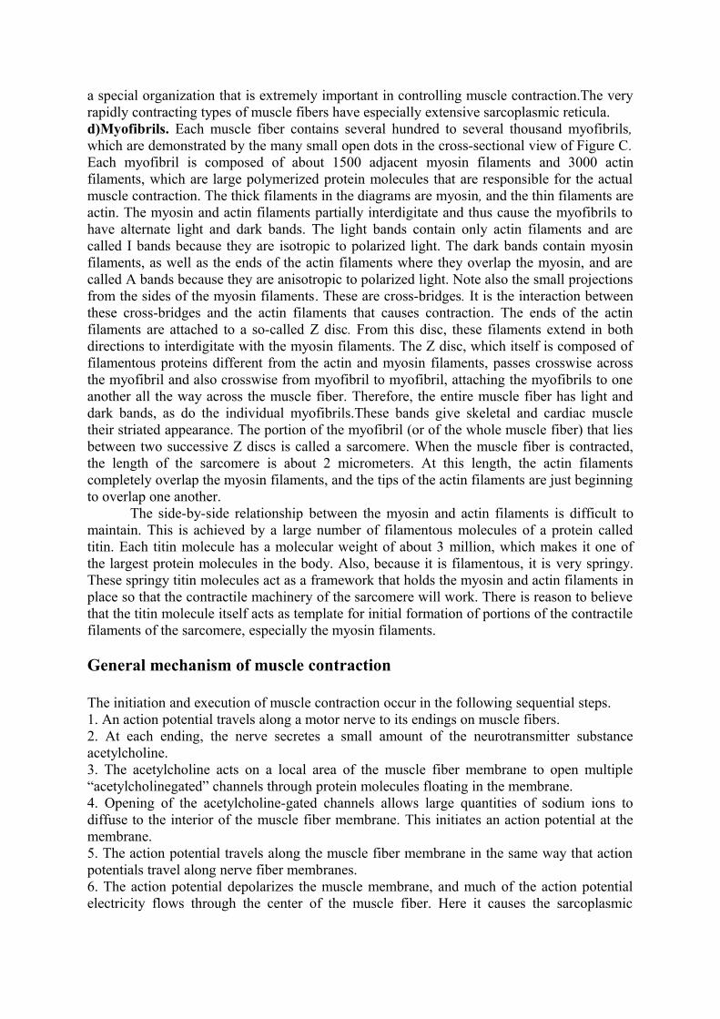
Molecular mechanism of muscle contraction- sliding filament mechanism of muscle contraction. It shows(figure 3.2 a) the relaxed state of a sarcomere (top) and the contracted state (bottom). In the relaxed state, the ends of the actin filaments extending from two successive Z discs barely begin to overlap one another. Conversely, in the contracted state, these actin filaments have been pulled inward among the myosin filaments, so that their ends overlap one another to their maximum extent. Also, the Z discs have been pulled by the actin filaments up to the ends of the myosin filaments. Thus, muscle contraction occurs by a sliding filament mechanism.The calcium ions in turn activate the forces between the myosin and actin filaments, and contraction begins. Energy is needed for the contractile process to proceed. This energy comes from high energy bonds in the ATP molecule, which is degraded to adenosine diphosphate (ADP) to liberate the energy.
Figure 3.2 a, b a) Relaxed and contracted states of a myofibril; b) Myosin molecule
The myosin molecule is composed of six polypeptide chains—two heavy chains, each with a molecular weight of about 200,000, and four light chains with molecular weights of about 20,000 each. The two heavy chains wrap spirally around each other to form a double helix, which is called the tail of the myosin molecule. One end of each of these chains is folded bilaterally into a globular polypeptide structure called a myosin head. Thus, there are two free heads at one end of the double-helix myosin molecule. The four light chains are also part of the myosin head, two to each head. These light chains help control the function of the head during muscle contraction. The myosin filament is made up of 200 or more individual myosin molecules.The central portion of one of these filaments, displaying the tails of the myosin molecules bundled together to form the body of the filament, while many heads of the molecules hang outward to the sides of the body. Also, part of the body of each myosin molecule hangs to the side along with the head, thus providing an arm that extends the head outward from the body, as shown in the figure. The protruding arms and heads together are called cross-bridges. Each cross-bridge is flexible at two points called hinges—one where the arm leaves the body of the myosin filament, and the other where the head attaches to the arm.
The hinged arms allow the heads either to be extended far outward from the body of the myosin filament or to be brought close to the body. The hinged heads in turn participate in the actual contraction process, as discussed in the following sections.
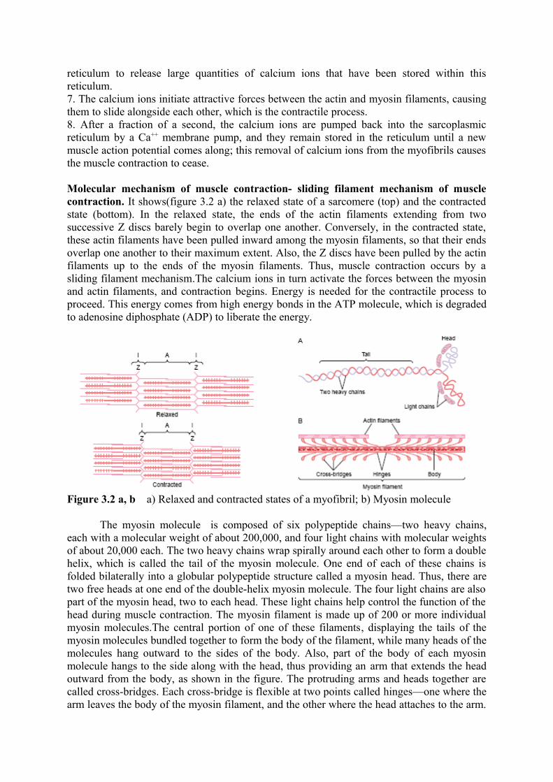
Figure 3.3 Walk along mechanism for contraction of the muscle
The total length of each myosin filament is uniform, almost exactly 1.6 micrometers. Note, however, that there are no cross-bridge heads in the very center of the myosin filament for a distance of about 0.2 micrometer because the hinged arms extend away from the center. The myosin filament itself is twisted so that each successive pair of crossbridges is axially displaced from the previous pair by 120 degrees.This ensures that the cross-bridges extend in all directions around the filament. Another feature of the myosin head that is essential for muscle contraction is that it functions as an ATP-ase enzyme. The actin filament is also complex. It is composed of three protein components: actin, tropomyosin, and troponin. The backbone of the actin filament is a doublestranded F-actin protein molecule. The two strands are wound in a helix in the same manner as the myosin molecule. Each strand of the double F-actin helix is composed of polymerized G-actin molecules, each having a molecular weight of about 42,000. Attached to each one of the G-actin molecules is one molecule of ADP. It is believed that these ADP molecules are the active sites on the actin filaments with which the crossbridges of the myosin filaments interact to cause muscle contraction.The active sites on the two F-actin strands of the double helix are staggered, giving one active site on the overall actin filament about every 2.7 nanometers. Each actin filament is about 1 micrometer long. The bases of the actin filaments are inserted strongly into the Z discs; the ends of the filaments protrude in both directions to lie in the spaces between the myosin molecules.
The actin filament also contains another protein, tropomyosin. Each molecule of tropomyosin has a molecular weight of 70,000 and a length of 40 nanometers. These molecules are wrapped spirally around the sides of the F-actin helix. In the resting state, the tropomyosin molecules lie on top of the active sites of the actin strands, so that attraction cannot occur between the actin and myosin filaments to cause contraction.
Attached intermittently along the sides of the tropomyosin molecules are still other protein molecules called troponin. These are actually complexes of three loosely bound protein subunits, each of which plays a specific role in controlling muscle contraction. One of the subunits (troponin I) has a strong affinity for actin, another (troponin T) for tropomyosin, and a third (troponin C) for calcium ions. This complex is believed to attach the tropomyosin to the actin. The strong affinity of the troponin for calcium ions is believed to initiate the contraction process.
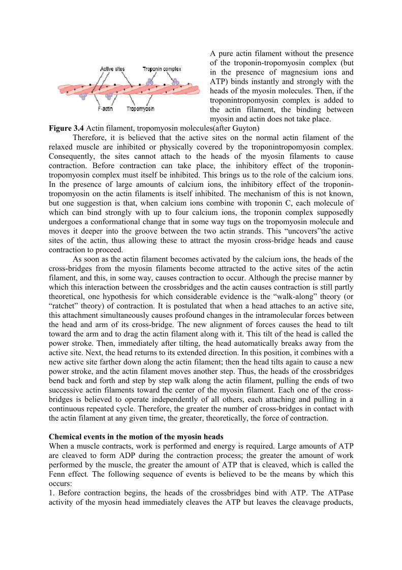
A pure actin filament without the presence of the troponin-tropomyosin complex (but in the presence of magnesium ions and ATP) binds instantly and strongly with the heads of the myosin molecules. Then, if the troponintropomyosin complex is added to the actin filament, the binding between myosin and actin does not take place.
Figure 3.4 Actin filament, tropomyosin molecules(after Guyton)Therefore, it is believed that the active sites on the normal actin filament of the
relaxed muscle are inhibited or physically covered by the troponintropomyosin complex. Consequently, the sites cannot attach to the heads of the myosin filaments to cause contraction. Before contraction can take place, the inhibitory effect of the troponin-tropomyosin complex must itself be inhibited. This brings us to the role of the calcium ions. In the presence of large amounts of calcium ions, the inhibitory effect of the troponin-tropomyosin on the actin filaments is itself inhibited. The mechanism of this is not known, but one suggestion is that, when calcium ions combine with troponin C, each molecule of which can bind strongly with up to four calcium ions, the troponin complex supposedly undergoes a conformational change that in some way tugs on the tropomyosin molecule and moves it deeper into the groove between the two actin strands. This “uncovers”the active sites of the actin, thus allowing these to attract the myosin cross-bridge heads and cause contraction to proceed.
As soon as the actin filament becomes activated by the calcium ions, the heads of the cross-bridges from the myosin filaments become attracted to the active sites of the actin filament, and this, in some way, causes contraction to occur. Although the precise manner by which this interaction between the crossbridges and the actin causes contraction is still partly theoretical, one hypothesis for which considerable evidence is the “walk-along” theory (or “ratchet” theory) of contraction. It is postulated that when a head attaches to an active site, this attachment simultaneously causes profound changes in the intramolecular forces between the head and arm of its cross-bridge. The new alignment of forces causes the head to tilt toward the arm and to drag the actin filament along with it. This tilt of the head is called the power stroke. Then, immediately after tilting, the head automatically breaks away from the active site. Next, the head returns to its extended direction. In this position, it combines with a new active site farther down along the actin filament; then the head tilts again to cause a new power stroke, and the actin filament moves another step. Thus, the heads of the crossbridges bend back and forth and step by step walk along the actin filament, pulling the ends of two successive actin filaments toward the center of the myosin filament. Each one of the cross-bridges is believed to operate independently of all others, each attaching and pulling in a continuous repeated cycle. Therefore, the greater the number of cross-bridges in contact with the actin filament at any given time, the greater, theoretically, the force of contraction.
Chemical events in the motion of the myosin headsWhen a muscle contracts, work is performed and energy is required. Large amounts of ATP are cleaved to form ADP during the contraction process; the greater the amount of work performed by the muscle, the greater the amount of ATP that is cleaved, which is called the Fenn effect. The following sequence of events is believed to be the means by which this occurs:1. Before contraction begins, the heads of the crossbridges bind with ATP. The ATPase activity of the myosin head immediately cleaves the ATP but leaves the cleavage products,
ADP plus phosphate ion, bound to the head. In this state, the conformation of the head is such that it extends perpendicularly toward the actin filament but is not yet attached to the actin.2. When the troponin-tropomyosin complex binds with calcium ions, active sites on the actin filament are uncovered, and the myosin heads then bind with these.3. The bond between the head of the cross-bridge and the active site of the actin filament causes a conformational change in the head, prompting the head to tilt toward the arm of the cross-bridge. This provides the power stroke for pulling the actin filament. The energy that activates the power stroke is the energy already stored, like a “cocked” spring, by the conformational change that occurred in the head when the ATP molecule was cleaved earlier.4. Once the head of the cross-bridge tilts, this allows release of the ADP and phosphate ion that were previously attached to the head. At the site of release of the ADP, a new molecule of ATP binds. This binding of new ATP causes detachment of the head from the actin.5. After the head has detached from the actin, the new molecule of ATP is cleaved to begin the next cycle, leading to a new power stroke. That is, the energy again “cocks” the head back to its perpendicular condition, ready to begin the new power stroke cycle.6. When the cocked head (with its stored energy derived from the cleaved ATP) binds with a new active site on the actin filament, it becomes uncocked and once again provides a new power stroke. Thus, the process proceeds again and again until the actin filaments pull the Z membrane up against the ends of the myosin filaments or until the load on the muscle becomes too great for further pulling to occur.
Relation of velocity of contraction to loadA skeletal muscle contracts extremely rapidly when it contracts against no load—to a
state of full contraction in about 0.1 second for the average muscle.When loads are applied, the velocity of contraction becomes progressively less as the load increases.That is, when the load has been increased to equal the maximum force that the muscle can exert, the velocity of contraction becomes zero and no contraction results, despite activation of the muscle fiber. This decreasing velocity of contraction with load is caused by the fact that a load on a contracting muscle is a reverse force that opposes the contractile force caused by muscle contraction. Therefore, the net force that is available to cause velocity of shortening is correspondingly reduced.
Energetics of muscle contraction work output during muscle contractionWhen a muscle contracts against a load, it performs work. This means that energy is
transferred from the muscle to the external load to lift an object to a greater height or to overcome resistance to movement. In mathematical terms, work is defined by the following equation: W = L x D in which W is the work output, L is the load, and D is the distance of movement against the load.The energy required to perform the work is derived from the chemical reactions in the muscle cells during contraction, as described in the following sections.
Muscle contraction depends on energy supplied by ATP. Most of this energy is required to actuate the walk-along mechanism by which the cross-bridges pull the actin filaments, but small amounts are required for (1) pumping calcium ions from the sarcoplasm into the sarcoplasmic reticulum after the contraction is over, and (2) pumping sodium and potassium ions through the muscle fiber membrane to maintain appropriate ionic environment for propagation of muscle fiber action potentials. The concentration of ATP in the muscle fiber, about 4 millimolar, is sufficient to maintain full contraction for only 1 to 2 seconds at most. The ATP is split to form ADP, which transfers energy from the ATP molecule to the contracting machinery of the muscle fiber. Then, the ADP is
rephosphorylated to form new ATP within another fraction of a second, which allows the muscle to continue its contraction.
There are several sources of the energy for this rephosphorylation.-The first source of energy that is used to reconstitute the ATP is the substance phosphocreatine, which carries a high-energy phosphate bond similar to the bonds of ATP. -The second important source of energy, which is used to reconstitute both ATP and phosphocreatine, is “glycolysis” of glycogen previously stored in the muscle cells. Rapid enzymatic breakdown of the glycogen to pyruvic acid and lactic acid liberates energy that is used to convert ADP to ATP; the ATP can then be used directly to energize additional muscle contraction and also to re-form the stores of phosphocreatine. The importance of this glycolysis mechanism is twofold. First, the glycolytic reactions can occur even in the absence of oxygen, so that muscle contraction can be sustained for many seconds and sometimes up to more than a minute, even when oxygen delivery from the blood is not available. Second, the rate of formation of ATP by the glycolytic process is about 2.5 times as rapid as ATP formation in response to cellular foodstuffs reacting with oxygen. However, so many end products of glycolysis accumulate in the muscle cells that glycolysis also loses its capability to sustain maximum muscle contraction after about 1 minute.-The third and final source of energy is oxidative metabolism. This means combining oxygen with the end products of glycolysis and with various other cellular foodstuffs to liberate ATP. More than 95 per cent of all energy used by the muscles for sustained, longterm contraction is derived from this source. The foodstuffs that are consumed are carbohydrates, fats, and protein. For extremely long-term maximal muscle activity—over a period of many hours—by far the greatest proportion of energy comes from fats, but for periods of 2 to 4 hours, as much as one half of the energy can come from stored carbohydrates.
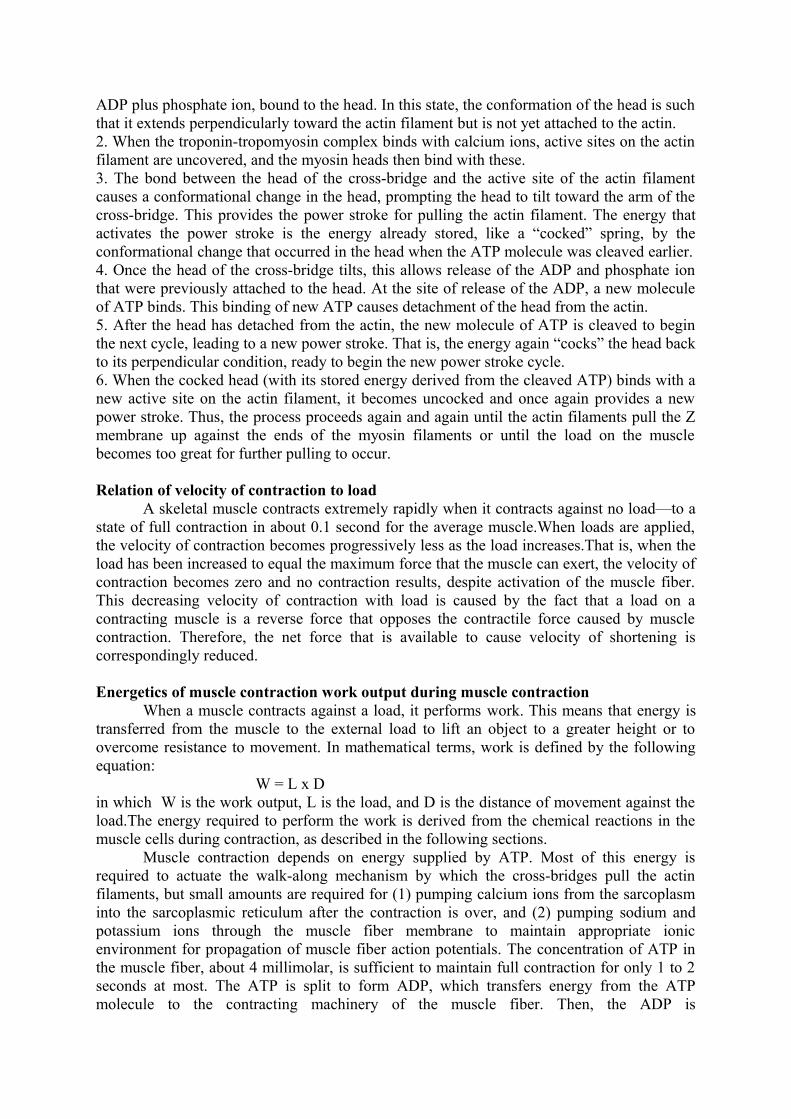
Isometric Versus Isotonic Contraction. Muscle contraction is said to be isometric when the muscle does not shorten during contraction and isotonic when it does shorten but the tension on the muscle remains constant throughout the contraction.
In the isometric system, the muscle contracts against a force transducer without decreasing the muscle length, as shown on the right in Figure. In the isotonic system, the muscle shortens against a fixed load; this is illustrated on the left in the figure, showing a muscle lifting a pan of weights. The characteristics of isotonic contraction depend on the load against which the muscle contracts, as well as the inertia of the load.
Figure 3.5 Isotonic and isometric systems for recording muscle contractions
However, the isometric system records strictly changes in force of muscle contraction itself. Therefore, the isometric system is most often used when comparing the functional characteristics of different muscle types.
Every muscle of the body is composed of a mixture of so-called fast and slow muscle fibers, with still other fibers gradated between these two extremes. The muscles that react rapidly are composed mainly of “fast” fibers with only small numbers of the slow variety. Conversely, the muscles that respond slowly but with prolonged contraction are composed mainly of “slow” fibers. The differences between these two types of fibers are as follows.
Each motoneuron that leaves the spinal cord innervates multiple muscle fibers, the number depending on the type of muscle. All the muscle fibers innervated by a single nerve fiber are called a motor unit. In general, small muscles that react rapidly and whose control must be exact have more nerve fibers for fewer muscle fibers (for instance, as few as two or three muscle fibers per motor unit in some of the laryngeal muscles). Conversely, large muscles that do not require fine control, such as the soleus muscle, may have several hundred muscle fibers in a motor unit. An average figure for all the muscles of the body is questionable, but a good guess would be about 80 to 100 muscle fibers to a motor unit.
Muscle contractions of different force—Force summation.Summation means the adding together of individual twitch contractions to increase
the intensity of overall muscle contraction. Summation occurs in two ways: (1) by increasing the number of motor units contracting simultaneously, which is called multiple fiber summation, and (2) by increasing the frequency of contraction, which is called frequency summation and can lead to tetanization.
When the central nervous system sends a weak signal to contract a muscle, the smaller motor units of the muscle may be stimulated in preference to the larger motor units. Then, as the strength of the signal increases, larger and larger motor units begin to be excited as well, with the largest motor units often having as much as 50 times the contractile force of the smallest units. This is called the size principle. It is important, because it allows the gradations of muscle force during weak contraction to occur in small steps, whereas the steps become progressively greater when large amounts of force are required. Another important feature of multiple fiber summation is that the different motor units are driven asynchronously by the spinal cord, so that contraction alternates among motor units one after the other, thus providing smooth contraction even at low frequencies of nerve signals.
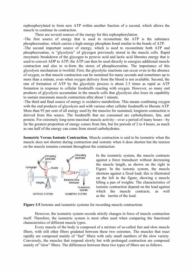
Figure 3.6 shows the principles of frequency summation and tetanization. To the left are displayed individual twitch contractions occurring one after another at low frequency of stimulation.Then, as the frequency increases, there comes a point where each new contraction occurs before the preceding one is over.
Figure 3.6 Summation and tetanization
As a result, the second contraction is added partially to the first, so that the total strength of contraction rises progressively with increasing frequency. When the frequency reaches a critical level, the successive contractions eventually become so rapid that they fuse together, and the whole muscle contraction appears to be completely smooth and continuous, as shown in the figure. This is called tetanization. At a slightly higher frequency, the strength of contraction reaches its maximum, so that any additional increase in frequency beyond that point has no further effect in increasing contractile force. This occurs because enough calcium ions are maintained in the muscle sarcoplasm, even between action potentials, so that full contractile state is sustained without allowing any relaxation between the action potentials.
Muscle action potential
Almost everything discussed in neurons regarding initiation and conduction of action potentials in nerve fibers applies equally to skeletal muscle fibers, except for quantitative differences. Some of the quantitative aspects of muscle potentials are the following:1. Resting membrane potential: about –80 to –90 millivolts in skeletal fibers—the same as in large myelinated nerve fibers.2. Duration of action potential: 1 to 5 milliseconds in skeletal muscle—about five times as long as in large myelinated nerves.3. Velocity of conduction: 3 to 5 m/sec—about 1/13 the velocity of conduction in the large myelinated nerve fibers that excite skeletal muscle.
The skeletal muscle fiber is so large that action potentials spreading along its surface membrane cause almost no current flow deep within the fiber. Yet, to cause maximum muscle contraction, current must penetrate deeply into the muscle fiber to the vicinity of the separate myofibrils.This is achieved by transmission of action potentials along transverse tubules (T tubules) that penetrate all the way through the muscle fiber from one side of the fiber to the other. The T tubule action potentials cause release of calcium ions inside the muscle fiber in the immediate vicinity of the myofibrils, and these calcium ions then cause contraction. This overall process is called excitation-contraction coupling.
The T tubules are very small and run transverse to the myofibrils. They begin at the cell membrane and penetrate all the way from one side of the muscle fiber to the opposite side. Not shown in the figure is the fact that these tubules branch among themselves so that they form entire planes of T tubules interlacing among all the separate myofibrils. Also, where the T tubules originate from the cell membrane, they are open to the exterior of the muscle fiber. Therefore, they communicate with the extracellular fluid surrounding the muscle fiber, and they themselves contain extracellular fluid in their lumens. In other words, the T tubules are actually internal extensions of the cell membrane. Therefore, when an action potential spreads over a muscle fiber membrane, a potential change also spreads along the T tubules to the deep interior of the muscle fiber. The electrical currents surrounding these T tubules then elicit the muscle contraction. Figure also shows a sarcoplasmic reticulum, in yellow. This is composed of two major parts: (1) large chambers called terminal cisternae that abut the T tubules, and (2) long longitudinal tubules that surround all surfaces of the actual contracting myofibrils.
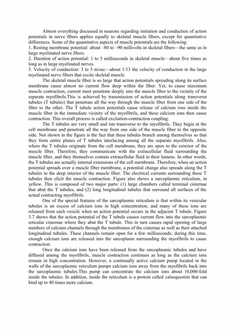
One of the special features of the sarcoplasmic reticulum is that within its vesicular tubules is an excess of calcium ions in high concentration, and many of these ions are released from each vesicle when an action potential occurs in the adjacent T tubule. Figure 3.7 shows that the action potential of the T tubule causes current flow into the sarcoplasmic reticular cisternae where they abut the T tubule. This in turn causes rapid opening of large numbers of calcium channels through the membranes of the cisternae as well as their attached longitudinal tubules. These channels remain open for a few milliseconds; during this time, enough calcium ions are released into the sarcoplasm surrounding the myofibrils to cause contraction.
Once the calcium ions have been released from the sarcoplasmic tubules and have diffused among the myofibrils, muscle contraction continues as long as the calcium ions remain in high concentration. However, a continually active calcium pump located in the walls of the sarcoplasmic reticulum pumps calcium ions away from the myofibrils back into the sarcoplasmic tubules.This pump can concentrate the calcium ions about 10,000-fold inside the tubules. In addition, inside the reticulum is a protein called calsequestrin that can bind up to 40 times more calcium.
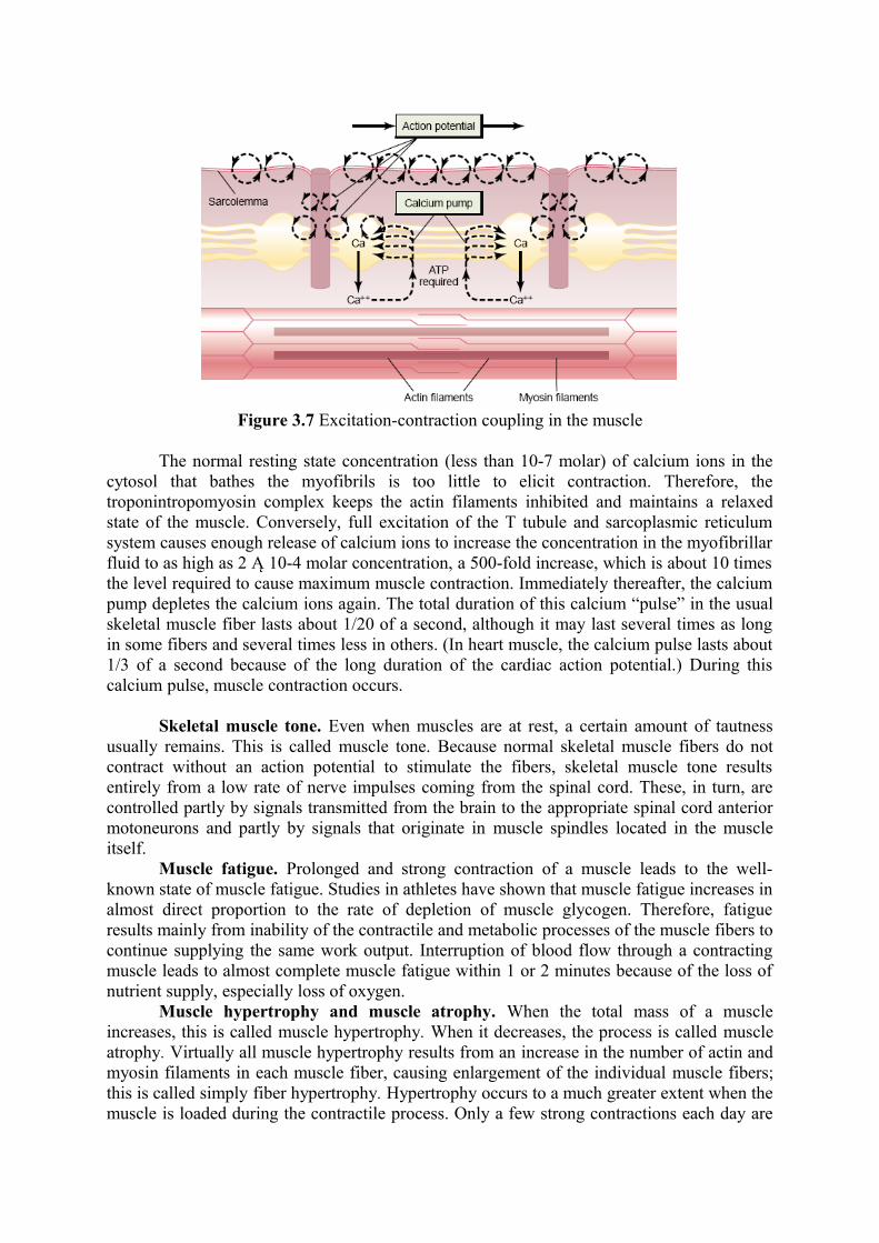
Figure 3.7 Excitation-contraction coupling in the muscle
The normal resting state concentration (less than 10-7 molar) of calcium ions in the cytosol that bathes the myofibrils is too little to elicit contraction. Therefore, the troponintropomyosin complex keeps the actin filaments inhibited and maintains a relaxed state of the muscle. Conversely, full excitation of the T tubule and sarcoplasmic reticulum system causes enough release of calcium ions to increase the concentration in the myofibrillar fluid to as high as 2 Ą 10-4 molar concentration, a 500-fold increase, which is about 10 times the level required to cause maximum muscle contraction. Immediately thereafter, the calcium pump depletes the calcium ions again. The total duration of this calcium “pulse” in the usual skeletal muscle fiber lasts about 1/20 of a second, although it may last several times as long in some fibers and several times less in others. (In heart muscle, the calcium pulse lasts about 1/3 of a second because of the long duration of the cardiac action potential.) During this calcium pulse, muscle contraction occurs.
Skeletal muscle tone. Even when muscles are at rest, a certain amount of tautness usually remains. This is called muscle tone. Because normal skeletal muscle fibers do not contract without an action potential to stimulate the fibers, skeletal muscle tone results entirely from a low rate of nerve impulses coming from the spinal cord. These, in turn, are controlled partly by signals transmitted from the brain to the appropriate spinal cord anterior motoneurons and partly by signals that originate in muscle spindles located in the muscle itself.
Muscle fatigue. Prolonged and strong contraction of a muscle leads to the well-known state of muscle fatigue. Studies in athletes have shown that muscle fatigue increases in almost direct proportion to the rate of depletion of muscle glycogen. Therefore, fatigue results mainly from inability of the contractile and metabolic processes of the muscle fibers to continue supplying the same work output. Interruption of blood flow through a contracting muscle leads to almost complete muscle fatigue within 1 or 2 minutes because of the loss of nutrient supply, especially loss of oxygen.
Muscle hypertrophy and muscle atrophy. When the total mass of a muscle increases, this is called muscle hypertrophy. When it decreases, the process is called muscle atrophy. Virtually all muscle hypertrophy results from an increase in the number of actin and myosin filaments in each muscle fiber, causing enlargement of the individual muscle fibers; this is called simply fiber hypertrophy. Hypertrophy occurs to a much greater extent when the muscle is loaded during the contractile process. Only a few strong contractions each day are
required to cause significant hypertrophy within 6 to 10 weeks. When a muscle remains unused for many weeks, the rate of decay of the contractile proteins is more rapid than the rate of replacement.Therefore, muscle atrophy occurs.
Under rare conditions of extreme muscle force generation, the actual number of muscle fibers has been observed to increase (but only by a few percentage points), in addition to the fiber hypertrophy process. This increase in fiber number is called fiber hyperplasia.
Effects of muscle denervation. When a muscle loses its nerve supply, it no longer receives the contractile signals that are required to maintain normal muscle size. Therefore, atrophy begins almost immediately. After about 2 months, degenerative changes also begin to appear in the muscle fibers themselves. If the nerve supply to the muscle grows back rapidly, full return of function can occur in as little as 3 months, but from that time onward, the capability of functional return becomes less and less, with no further return of function after 1 to 2 years. In the final stage of denervation atrophy, most of the muscle fibers are destroyed and replaced by fibrous and fatty tissue. The fibers that do remain are composed of a long cell membrane with a lineup of muscle cell nuclei but with few or no contractile properties and little or no capability of regenerating myofibrils if a nerve does regrow. The fibrous tissue that replaces the muscle fibers during denervation atrophy also has a tendency to continue shortening for many months, which is called contracture.
Rigor Mortis. Several hours after death, all the muscles of the body go into a state of contracture called “rigor mortis”; that is, the muscles contract and become rigid, even without action potentials. This rigidity results from loss of all the ATP, which is required to cause separation of the crossbridges from the actin filaments during the relaxation process. The muscles remain in rigor until the muscle proteins deteriorate about 15 to 25 hours later, which presumably results from autolysis caused by enzymes released from lysosomes. All these events occur more rapidly at higher temperatures.
Neuromuscular junction—The motor end plate
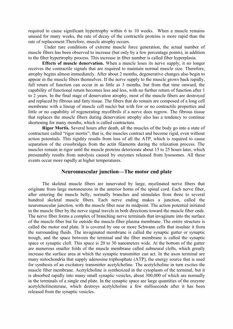
The skeletal muscle fibers are innervated by large, myelinated nerve fibers that originate from large motoneurons in the anterior horns of the spinal cord. Each nerve fiber, after entering the muscle belly, normally branches and stimulates from three to several hundred skeletal muscle fibers. Each nerve ending makes a junction, called the neuromuscular junction, with the muscle fiber near its midpoint. The action potential initiated in the muscle fiber by the nerve signal travels in both directions toward the muscle fiber ends. The nerve fiber forms a complex of branching nerve terminals that invaginate into the surface of the muscle fiber but lie outside the muscle fiber plasma membrane. The entire structure is called the motor end plate. It is covered by one or more Schwann cells that insulate it from the surrounding fluids. The invaginated membrane is called the synaptic gutter or synaptic trough, and the space between the terminal and the fiber membrane is called the synaptic space or synaptic cleft. This space is 20 to 30 nanometers wide. At the bottom of the gutter are numerous smaller folds of the muscle membrane called subneural clefts, which greatly increase the surface area at which the synaptic transmitter can act. In the axon terminal are many mitochondria that supply adenosine triphosphate (ATP), the energy source that is used for synthesis of an excitatory transmitter acetylcholine. The acetylcholine in turn excites the muscle fiber membrane. Acetylcholine is synthesized in the cytoplasm of the terminal, but it is absorbed rapidly into many small synaptic vesicles, about 300,000 of which are normally in the terminals of a single end plate. In the synaptic space are large quantities of the enzyme acetylcholinesterase, which destroys acetylcholine a few milliseconds after it has been released from the synaptic vesicles.
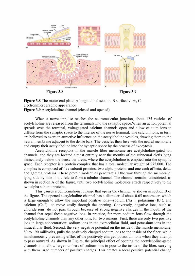
Figure 3.8 Figure 3.9
Figure 3.8 The motor end plate: A longitudinal section, B surface view, C electronmicrographic appearanceFigure 3.9 Acetylcholine channel (closed and opened)
When a nerve impulse reaches the neuromuscular junction, about 125 vesicles of acetylcholine are released from the terminals into the synaptic space.When an action potential spreads over the terminal, voltagegated calcium channels open and allow calcium ions to diffuse from the synaptic space to the interior of the nerve terminal. The calcium ions, in turn, are believed to exert an attractive influence on the acetylcholine vesicles, drawing them to the neural membrane adjacent to the dense bars. The vesicles then fuse with the neural membrane and empty their acetylcholine into the synaptic space by the process of exocytosis.
Acetylcholine receptors in the muscle fiber membrane are acetylcholine-gated ion channels, and they are located almost entirely near the mouths of the subneural clefts lying immediately below the dense bar areas, where the acetylcholine is emptied into the synaptic space. Each receptor is a protein complex that has a total molecular weight of 275,000. The complex is composed of five subunit proteins, two alpha proteins and one each of beta, delta, and gamma proteins. These protein molecules penetrate all the way through the membrane, lying side by side in a circle to form a tubular channel. The channel remains constricted, as shown in section A of the figure, until two acetylcholine molecules attach respectively to the two alpha subunit proteins. This causes a conformational change that opens the channel, as shown in section B of the figure. The opened acetylcholine channel has a diameter of about 0.65 nanometer, which is large enough to allow the important positive ions—sodium (Na+), potassium (K+), and calcium (Ca++)—to move easily through the opening. Conversely, negative ions, such as chloride ions, do not pass through because of strong negative charges in the mouth of the channel that repel these negative ions. In practice, far more sodium ions flow through the acetylcholine channels than any other ions, for two reasons. First, there are only two positive ions in large concentration: sodium ions in the extracellular fluid, and potassium ions in the intracellular fluid. Second, the very negative potential on the inside of the muscle membrane, 80 to –90 millivolts, pulls the positively charged sodium ions to the inside of the fiber, while simultaneously preventing efflux of the positively charged potassium ions when they attempt to pass outward. As shown in Figure, the principal effect of opening the acetylcholine-gated channels is to allow large numbers of sodium ions to pour to the inside of the fiber, carrying with them large numbers of positive charges. This creates a local positive potential change
inside the muscle fiber membrane, called the end plate potential. In turn, this end plate potential initiates an action potential that spreads along the muscle membrane and thus causes muscle contraction.
The acetylcholine, once released into the synaptic space, continues to activate the acetylcholine receptors as long as the acetylcholine persists in the space. However, it is removed rapidly by two means: (1) Most of the acetylcholine is destroyed by the enzyme acetylcholinesterase, which is attached mainly to the spongy layer of fine connective tissue that fills the synaptic space between the presynaptic nerve terminal and the postsynaptic muscle membrane. (2) A small amount of acetylcholine diffuses out of the synaptic space and is then no longer available to act on the muscle fiber membrane. The short time that the acetylcholine remains in the synaptic space—a few milliseconds at most—normally is sufficient to excite the muscle fiber. Then the rapid removal of the acetylcholine prevents continued muscle re-excitation after the muscle fiber has recovered from its initial action potential.
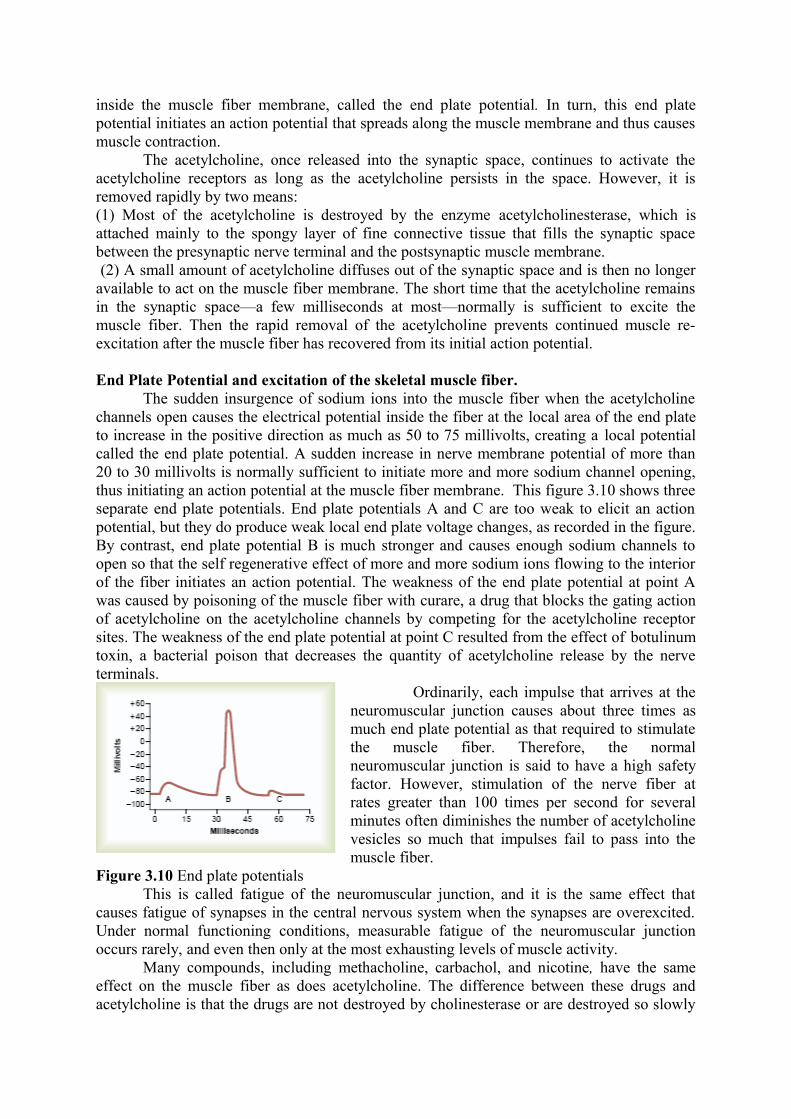
End Plate Potential and excitation of the skeletal muscle fiber.The sudden insurgence of sodium ions into the muscle fiber when the acetylcholine
channels open causes the electrical potential inside the fiber at the local area of the end plate to increase in the positive direction as much as 50 to 75 millivolts, creating a local potential called the end plate potential. A sudden increase in nerve membrane potential of more than 20 to 30 millivolts is normally sufficient to initiate more and more sodium channel opening, thus initiating an action potential at the muscle fiber membrane. This figure 3.10 shows three separate end plate potentials. End plate potentials A and C are too weak to elicit an action potential, but they do produce weak local end plate voltage changes, as recorded in the figure. By contrast, end plate potential B is much stronger and causes enough sodium channels to open so that the self regenerative effect of more and more sodium ions flowing to the interior of the fiber initiates an action potential. The weakness of the end plate potential at point A was caused by poisoning of the muscle fiber with curare, a drug that blocks the gating action of acetylcholine on the acetylcholine channels by competing for the acetylcholine receptor sites. The weakness of the end plate potential at point C resulted from the effect of botulinum toxin, a bacterial poison that decreases the quantity of acetylcholine release by the nerve terminals.
Ordinarily, each impulse that arrives at the neuromuscular junction causes about three times as much end plate potential as that required to stimulate the muscle fiber. Therefore, the normal neuromuscular junction is said to have a high safety factor. However, stimulation of the nerve fiber at rates greater than 100 times per second for several minutes often diminishes the number of acetylcholine vesicles so much that impulses fail to pass into the muscle fiber.
Figure 3.10 End plate potentialsThis is called fatigue of the neuromuscular junction, and it is the same effect that
causes fatigue of synapses in the central nervous system when the synapses are overexcited. Under normal functioning conditions, measurable fatigue of the neuromuscular junction occurs rarely, and even then only at the most exhausting levels of muscle activity.
Many compounds, including methacholine, carbachol, and nicotine, have the same effect on the muscle fiber as does acetylcholine. The difference between these drugs and acetylcholine is that the drugs are not destroyed by cholinesterase or are destroyed so slowly
that their action often persists for many minutes to several hours.The drugs work by causing localized areas of depolarization of the muscle fiber membrane at the motor end plate where the acetylcholine receptors are located. Then, every time the muscle fiber recovers from a previous contraction, these depolarized areas, by virtue of leaking ions, initiate a new action potential, thereby causing a state of muscle spasm.
Three particularly wellknown drugs, neostigmine, physostigmine, and diisopropyl fluorophosphate, inactivate the acetylcholinesterase in the synapses so that it no longer hydrolyzes acetylcholine. Therefore, with each successive nerve impulse, additional acetylcholine accumulates and stimulates the muscle fiber repetitively. This causes muscle spasm when even a few nerve impulses reach the muscle. Unfortunately, it also can cause death due to laryngeal spasm, which smothers the person. Neostigmine and physostigmine combine with acetylcholinesterase to inactivate the acetylcholinesterase for up to several hours, after which these drugs are displaced from the acetylcholinesterase so that the esterase once again becomes active.
A group of drugs known as curariform drugs can prevent passage of impulses from the nerve ending into the muscle. For instance, D-tubocurarine blocks the action of acetylcholine on the muscle fiber acetylcholine receptors, thus preventing sufficient increase in permeability of the muscle membrane channels to initiate an action potential.
Physiology of Smooth Muscle
Smooth muscle, which is composed of far smaller fibers— usually 1 to 5 micrometers in diameter and only 20 to 500 micrometers in length. In contrast, skeletal muscle fibers are as much as 30 times greater in diameter and hundreds of times as long. Many of the same principles of contraction apply to smooth muscle as to skeletal muscle. Most important, essentially the same attractive forces between myosin and actin filaments cause contraction in smooth muscle as in skeletal muscle, but the internal physical arrangement of smooth muscle fibers is very different.
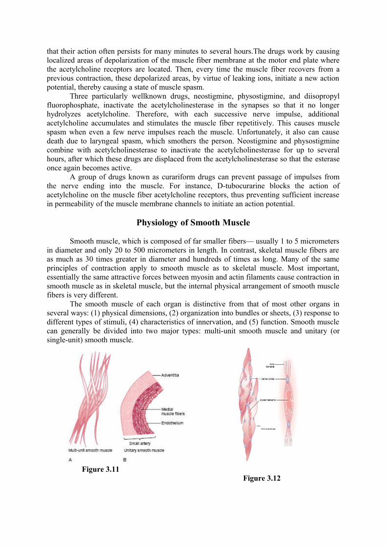
The smooth muscle of each organ is distinctive from that of most other organs in several ways: (1) physical dimensions, (2) organization into bundles or sheets, (3) response to different types of stimuli, (4) characteristics of innervation, and (5) function. Smooth muscle can generally be divided into two major types: multi-unit smooth muscle and unitary (or single-unit) smooth muscle.
Figure 3.11
Figure 3.12
Multi-Unit Smooth Muscle. This type of smooth muscle is composed of discrete, separate smooth muscle fibers. The outer surfaces of these fibers, like those of skeletal muscle fibers, are covered by a thin layer of basement membrane–like substance, a mixture of fine collagen and glycoprotein that helps insulate the separate fibers from one another. The most important characteristic of multi-unit smooth muscle fibers is that each fiber can contract independently of the others, and their control is exerted mainly by nerve signals. In contrast, a major share of control of unitary smooth muscle is exerted by non-nervous stimuli. Some examples of multi-unit smooth muscle are the ciliary muscle of the eye, the iris muscle of the eye, and the piloerector muscles that cause erection of the hairs when stimulated by the sympathetic nervous system.
Unitary Smooth Muscle. It means a mass of hundreds to thousands of smooth muscle fibers that contract together as a single unit. The fibers usually are arranged in sheets or bundles, and their cell membranes are adherent to one another at multiple points so that force generated in one muscle fiber can be transmitted to the next. In addition, the cell membranes are joined by many gap junctions through which ions can flow freely from one muscle cell to the next so that action potentials or simple ion flow without action potentials can travel from one fiber to the next and cause the muscle fibers to contract together. This type of smooth muscle is also known as syncytial smooth muscle because of its syncytial interconnections among fibers. It is also called visceral smooth muscle because it is found in the walls of most viscera of the body, including the gut, bile ducts, ureters, uterus, and many blood vessels.
Contractile mechanism in smooth muscleSmooth muscle contains both actin and myosin filaments, having chemical
characteristics similar to those of the actin and myosin filaments in skeletal muscle. It does not contain the normal troponin complex that is required in the control of skeletal muscle contraction, so the mechanism for control of contraction is different. Chemical studies have shown that actin and myosin filaments derived from smooth muscle interact with each other in much the same way that they do in skeletal muscle. Further, the contractile process is activated by calcium ions, and adenosine triphosphate (ATP) is degraded to adenosine diphosphate (ADP) to provide the energy for contraction. There are, however, major differences between the physical organization of smooth muscle and that of skeletal muscle, as well as differences in excitation contraction coupling, control of the contractile process by calcium ions, duration of contraction, and amount of energy required for contraction.
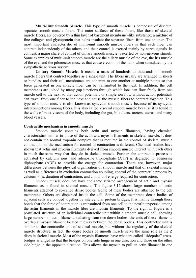
Smooth muscle does not have the same striated arrangement of actin and myosin filaments as is found in skeletal muscle. The figure 3.12 shows large numbers of actin filaments attached to so-called dense bodies. Some of these bodies are attached to the cell membrane. Others are dispersed inside the cell. Some of the membrane dense bodies of adjacent cells are bonded together by intercellular protein bridges. It is mainly through these bonds that the force of contraction is transmitted from one cell to the nextInterspersed among the actin filaments in the muscle fiber are myosin filaments. To the right in Figure is a postulated structure of an individual contractile unit within a smooth muscle cell, showing large numbers of actin filaments radiating from two dense bodies; the ends of these filaments overlap a myosin filament located midway between the dense bodies. This contractile unit is similar to the contractile unit of skeletal muscle, but without the regularity of the skeletal muscle structure; in fact, the dense bodies of smooth muscle serve the same role as the Z discs in skeletal muscle. Most of the myosin filaments have what are called “sidepolar” cross-bridges arranged so that the bridges on one side hinge in one direction and those on the other side hinge in the opposite direction. This allows the myosin to pull an actin filament in one
direction on one side while simultaneously pulling another actin filament in the opposite direction on the other side.
Although most skeletal muscles contract and relax rapidly, most smooth muscle contraction is prolonged tonic contraction, sometimes lasting hours or even days. Therefore, it is to be expected that both the physical and the chemical characteristics of smooth muscle versus skeletal muscle contraction would differ. Following are some of the differences.-Slow cycling of the myosin cross-bridges. The rapidity of cycling of the myosin cross-bridges in smooth muscle—that is, their attachment to actin, then release from the actin, and reattachment for the next cycle— is much, much slower in smooth muscle than in skeletal muscle; in fact, the frequency is as little as 1/10 to 1/300 that in skeletal muscle. Yet the fraction of time that the cross-bridges remain attached to the actin filaments, which is a major factor that determines the force of contraction, is believed to be greatly increased in smooth muscle. A possible reason for the slow cycling is that the cross-bridge heads have far less ATPase activity than in skeletal muscle, so that degradation of the ATP that energizes the movements of the cross-bridge heads is greatly reduced, with corresponding slowing of the rate of cycling.-Energy required to sustain smooth muscle contraction. Only 1/10 to 1/300 as much energy is required to sustain the same tension of contraction in smooth muscle as in skeletal muscle.This, too, is believed to result from the slow attachment and detachment cycling of the crossbridges and because only one molecule of ATP is required for each cycle, regardless of its duration. This sparsity of energy utilization by smooth muscle is exceedingly important to the overall energy economy of the body, because organs such as the intestines, urinary bladder, gallbladder, and other viscera often maintain tonic muscle contraction almost indefinitely.-Slowness of onset of contraction and relaxation of the total smooth muscle tissue. A typical smooth muscle tissue begins to contract 50 to 100 milliseconds after it is excited, reaches full contraction about 0.5 second later, and then declines in contractile force in another 1 to 2 seconds, giving a total contraction time of 1 to 3 seconds. This is about 30 times as long as a single contraction of an average skeletal muscle fiber. But because there are so many types of smooth muscle, contraction of some types can be as short as 0.2 second or as long as 30 seconds. The slow onset of contraction of smooth muscle, as well as its prolonged contraction, is caused by the slowness of attachment and detachment of the cross-bridges with the actin filaments. In addition, the initiation of contraction in response to calcium ions is much slower than in skeletal muscle.-Force of muscle contraction. Despite the relatively few myosin filaments in smooth muscle, and despite the slow cycling time of the cross-bridges, the maximum force of contraction of smooth muscle is often greater than that of skeletal muscle—as great as 4 to 6 kg/cm2 cross-sectional area for smooth muscle, in comparison with 3 to 4 kilograms for skeletal muscle. This great force of smooth muscle contraction results from the prolonged period of attachment of the myosin crossbridges to the actin filaments.-“Latch” mechanism for prolonged holding of contractions of smooth muscle. Once smooth muscle has developed full contraction, the amount of continuing excitation usually can be reduced to far less than the initial level, yet the muscle maintains its full force of contraction. Further, the energy consumed to maintain contraction is often minuscule, sometimes as little as 1/300 the energy required for comparable sustained skeletal muscle contraction. This is called the “latch” mechanism. The importance of the latch mechanism is that it can maintain prolonged tonic contraction in smooth muscle for hours with little use of energy. Little continued excitatory signal is required from nerve fibers or hormonal sources.-Stress-relaxation of smooth muscle. Another important characteristic of smooth muscle, especially the visceral unitary type of smooth muscle of many hollow organs, is its ability to
return to nearly its original force of contraction seconds or minutes after it has been elongated or shortened. For example, a sudden increase in fluid volume in the urinary bladder, thusstretching the smooth muscle in the bladder wall, causes an immediate large increase in pressure in the bladder. However, during the next 15 seconds to a minute or so, despite continued stretch of the bladder wall, the pressure returns almost exactly back to the original level. Then, when the volume is increased by another step, the same effect occurs again. Conversely, when the volume is suddenly decreased, the pressure falls very low at first but then rises back in another few seconds or minutes to or near to the original level. These phenomena are called stressrelaxation and reverse stress-relaxation. Their importance is that, except for short periods of time, they allow a hollow organ to maintain about the same amount of pressure inside its lumen despite long-term, large changes in volume.
Regulation of contraction by calcium ionsAs is true for skeletal muscle, the initiating stimulus for most smooth muscle
contraction is an increase in intracellular calcium ions. This increase can be caused in different types of smooth muscle by nerve stimulation of the smooth muscle fiber, hormonal stimulation, stretch of the fiber, or even change in the chemical environment of the fiber. Yet smooth muscle does not contain troponin, the regulatory protein that is activated by calcium ions to cause skeletal muscle contraction. Instead, smooth muscle contraction is activated by an entirely different mechanism, as follows. In place of troponin, smooth muscle cells contain a large amount of another regulatory protein called calmodulin. Although this protein is similar to troponin, it is different in the manner in which it initiates contraction. Calmodulin does this by activating the myosin cross-bridges. This activation and subsequent contraction occur in the following sequence:1. The calcium ions bind with calmodulin.2.The calmodulin-calcium combination joins with and activates myosin kinase, a phosphorylating enzyme.3. One of the light chains of each myosin head, called the regulatory chain, becomes phosphorylated in response to this myosin kinase. When this chain is not phosphorylated, theattachment-detachment cycling of the myosin head with the actin filament does not occur. But when the regulatory chain is phosphorylated, the head has the capability of binding repetitively with the actin filament and proceeding through the entire cycling process of intermittent “pulls,” the same as occurs for skeletal muscle, thus causing muscle contraction.Nervous and hormonal control of smooth muscle contraction
Although skeletal muscle fibers are stimulated exclusively by the nervous system, smooth muscle can be stimulated to contract by multiple types of signals: by nervous signals, by hormonal stimulation, by stretch of the muscle, and in several other ways. The principal reason for the difference is that the smooth muscle membrane contains many types of receptor proteins that can initiate the contractile process. Still other receptor proteins inhibit smooth muscle contraction, which is another difference from skeletal muscle.
Neuromuscular junctions of smooth muscleNeuromuscular junctions of the highly structured type found on skeletal muscle fibers
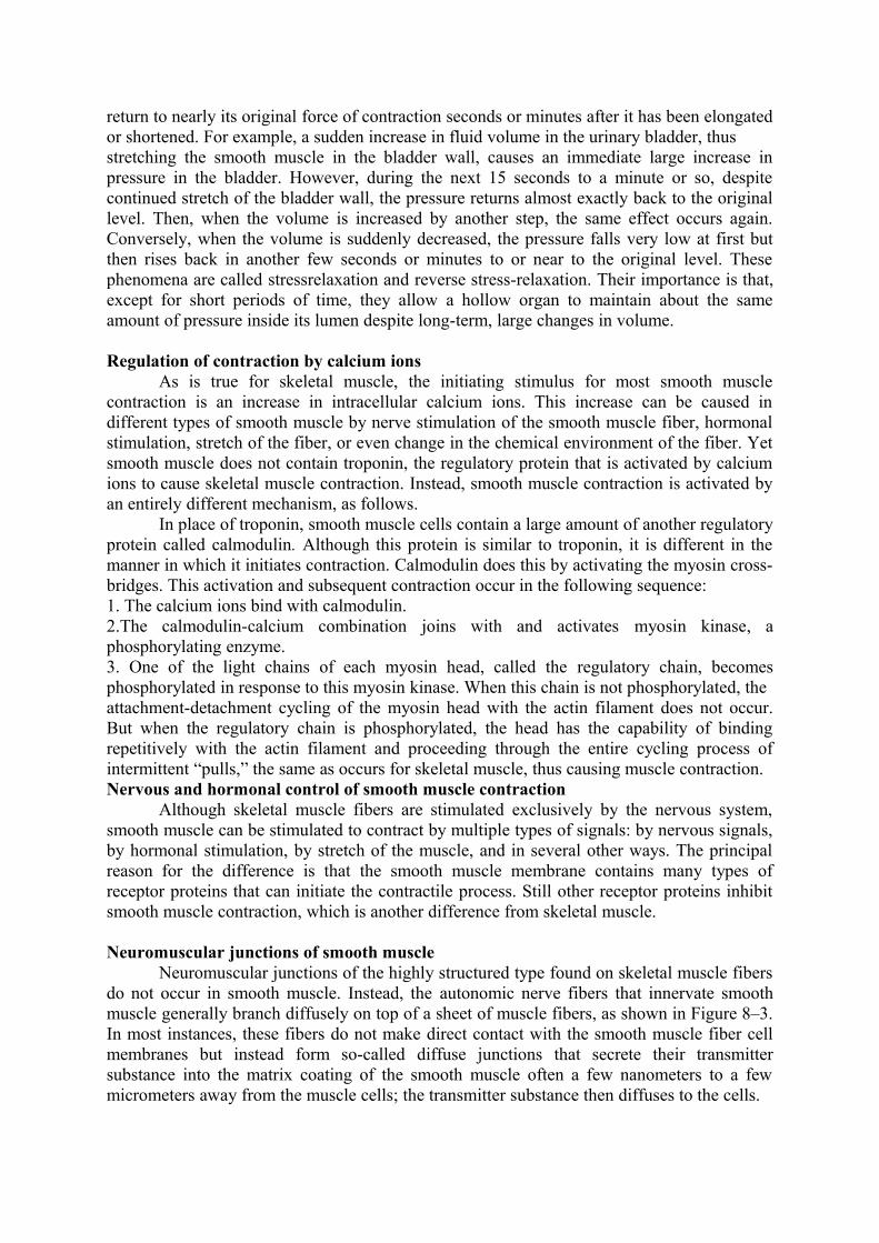
do not occur in smooth muscle. Instead, the autonomic nerve fibers that innervate smooth muscle generally branch diffusely on top of a sheet of muscle fibers, as shown in Figure 8–3. In most instances, these fibers do not make direct contact with the smooth muscle fiber cell membranes but instead form so-called diffuse junctions that secrete their transmitter substance into the matrix coating of the smooth muscle often a few nanometers to a few micrometers away from the muscle cells; the transmitter substance then diffuses to the cells.
Furthermore, where there are many layers of muscle cells, the nerve fibers often innervate only the outer layer, and muscle excitation travels from this outer layer to the inner layers by action potential conduction in the muscle mass or by additional diffusion of the transmitter substance.
Figure 3.13 Innervation of smooth muscle
The axons that innervate smooth muscle fibers do not have typical branching end feet of the type in the motor end plate on skeletal muscle fibers. Instead, most of the fine terminal axons have multiple varicosities distributed along their axes. At these points the Schwann cells that envelop the axons are interrupted so that transmitter substance can be secreted through the walls of the varicosities. In the varicosities are vesicles similar to those in the skeletal muscle end plate that contain transmitter substance. But, in contrast to the vesicles of skeletal muscle junctions, which always contain acetylcholine, the vesicles of the autonomic nerve fiber endings contain acetylcholine in some fibers and norepinephrine in others—and occasionally other substances as well. In a few instances, particularly in the multi-unit type of smooth muscle, the varicosities are separated from the muscle cell membrane by as little as 20 to 30 nanometers—the same width as the synaptic cleft that occurs in the skeletal muscle junction.These are called contact junctions, and they function in much the same way as the skeletal muscle neuromuscular junction; the rapidity of contraction of these smooth muscle fibers is considerably faster than that of fibers stimulated by the diffuse junctions.
Excitatory and inhibitory transmitter substances secreted at the smooth muscle neuromuscular junction
The most important transmitter substances secreted by the autonomic nerves innervating smooth muscle are acetylcholine and norepinephrine, but they are never secreted by the same nerve fibers. Acetylcholine is an excitatory transmitter substance for smooth muscle fibers in some organs but an inhibitory transmitter for smooth muscle in other organs. When acetylcholine excites a muscle fiber, norepinephrine ordinarily inhibits it. Conversely, when acetylcholine inhibits a fiber, norepinephrine usually excites it. The answer is that both acetylcholine and norepinephrine excite or inhibit smooth muscle by first binding with a receptor protein on the surface of the muscle cell membrane. Some of the receptor proteins are excitatory receptors, whereas others are inhibitory receptors. Thus, the type of receptor determines whether the smooth muscle is inhibited or excited and also determines which of the two transmitters, acetylcholine or norepinephrine, is effective in causing the excitation or inhibition. Membrane potentials and action potentials in smooth muscle membrane potentials in smooth muscle. The quantitative voltage of the membrane potential of smooth muscle depends on the momentary condition of the muscle. In the normal resting state, the intracellular potential is usually about -50 to -60 millivolts, which is about 30 millivolts less negative than in skeletal muscle. Action potentials in unitary smooth muscle. Action potentials occur in unitary smooth muscle (such as visceral muscle) in the same way that they occur in skeletal muscle. The action potentials of visceral smooth muscle occur in one of two forms: (1) spike potentials or (2) action potentials with plateaus.
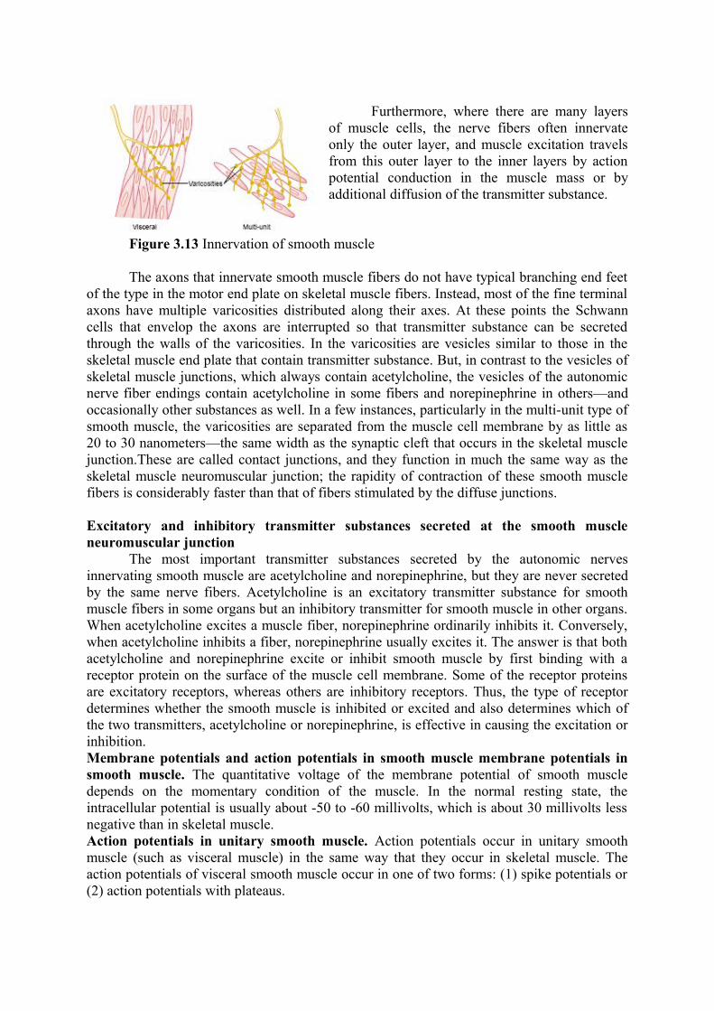
Typical spike action potentials, such as those seen in skeletal muscle, occur in most types of unitary smooth muscle. The duration of this type of action potential is 10 to 50 milliseconds, as shown in Figure 3.14 A. Such action potentials can be elicited in many ways, for example, by electrical stimulation, by the action of hormones on the smooth muscle, by the action of transmitter substances from nerve fibers, by stretch, or as a result of spontaneous generation in the muscle fiber itself, as discussed subsequently.
Figure 3.14 A Typical smooth muscle action potential B Repetitive spike potentials C Action potential with a plateau
Figure 3.14C shows a smooth muscle action potential with a plateau. The onset of this action potential is similar to that of the typical spike potential. However, instead of rapid repolarization of the muscle fiber membrane, the repolarization is delayed for several hundred to as much as 1000 milliseconds (1 second). The importance of the plateau is that it can account for the prolonged contraction that occurs in some types of smooth muscle, such as the ureter, the uterus under some conditions, and certain types of vascular smooth muscle.
The smooth muscle cell membrane has far more voltage-gated calcium channels than does skeletal muscle but few voltagegated sodium channels. Therefore, sodium participates little in the generation of the action potential in most smooth muscle. Instead, flow of calcium ions to the interior of the fiber is mainly responsible for the action potential. This occurs in the same self-regenerative way as occurs for the sodium channels in nerve fibers and in skeletal muscle fibers. However, the calcium channels open many times more slowly than do sodium channels, and they also remain open much longer. This accounts in large measure for the prolonged plateau action potentials of some smooth muscle fibers. Another important feature of calcium ion entry into the cells during the action potential is that the calcium ions act directly on the smooth muscle contractile mechanism to cause contraction. Thus, the calcium performs two tasks at once.
Some smooth muscle is self-excitatory.That is, action potentials arise within the smooth muscle cells themselves without an extrinsic stimulus. This often is associated with a basic slow wave rhythm of the membrane potential. A typical slow wave in a visceral smooth muscle of the gut is shown in Figure 3.14 B. The slow wave itself is not the action potential. That is, it is not a selfregenerative process that spreads progressively over the membranes of the muscle fibers. Instead, it is a local property of the smooth muscle fibers that make up the
muscle mass. The cause of the slow wave rhythm is unknown. One suggestion is that the slow waves are caused by waxing and waning of the pumping of positive ions (presumably sodium ions) outward through the muscle fiber membrane; that is, the membrane potential becomes more negative when sodium is pumped rapidly and less negative when the sodium pump becomes less active. Another suggestion is that the conductances of the ion channels increase and decrease rhythmically. The importance of the slow waves is that, when they are strong enough, they can initiate action potentials. The slow waves themselves cannot cause muscle contraction, but when the peak of the negative slow wave potential inside the cell membrane rises in the positive direction from -60 to about -35 millivolts (the approximate threshold for eliciting action potentials in most visceral smooth muscle), an action potential develops and spreads over the muscle mass.Then contraction does occur. Figure 8–4B demonstrates this effect, showing that at each peak of the slow wave, one or more action potentials occur. These repetitive sequences of action potentials elicit rhythmical contraction of the smooth muscle mass. Therefore, the slow waves are called pacemaker waves. This type of pacemaker activity controls the rhythmical contractions of the gut.
Some hormone receptors in the smooth muscle membrane open sodium or calcium ion channels and depolarize the membrane, the same as after nerve stimulation. Sometimes action potentials result, or action potentials that are already occurring may be enhanced. In other cases, depolarization occurs without action potentials, and this depolarization allows calcium ion entry into the cell, which promotes the contraction. Inhibition, in contrast, occurs when the hormone (or other tissue factor) closes the sodium and calcium channels to prevent entry of these positive ions; inhibition also occurs if the normally closed potassium channels are opened, allowing positive potassium ions to diffuse out of the cell. Both of these actions increase the degree of negativity inside the muscle cell, a state called hyperpolarization, which strongly inhibits muscle contraction. Sometimes smooth muscle contraction or inhibition is initiated by hormones without directly causing any change in the membrane potential. In these instances, the hormone may activate a membrane receptor that does not open any ion channels but instead causes an internal change in the muscle fiber, such as release of calcium ions from the intracellular sarcoplasmic reticulum; the calcium then induces contraction. To inhibit contraction, other receptor mechanisms are known to activate the enzyme adenylate cyclase or guanylate cyclase in the cell membrane; the portions of the receptors that protrude to the interior of the cells are coupled to these enzymes, causing the formation of cyclic adenosine monophosphate (cAMP) or cyclic guanosine monophosphate (cGMP), so-called second messengers.The cAMP or cGMP has many effects, one of which is to change the degree of phosphorylation of several enzymes that indirectly inhibit contraction. The pump that moves calcium ions from the sarcoplasm into the sarcoplasmic reticulum is activated, as well as the cell membrane pump that moves calcium ions out of the cell itself; these effects reduce the calcium ion concentration in the sarcoplasm, thereby inhibiting contraction. Smooth muscles have considerable diversity in how they initiate contraction or relaxation in response to different hormones, neurotransmitters, and other substances. In some instances, the same substance may cause either relaxation or contraction of smooth muscles in different locations. For example, norepinephrine inhibits contraction of smooth muscle in the intestine but stimulates contraction of smooth muscle in blood vessels.
Although the contractile process in smooth muscle, as in skeletal muscle, is activated by calcium ions, the source of the calcium ions differs; the difference is that the sarcoplasmic reticulum, which provides virtually all the calcium ions for skeletal muscle contraction, is only slightly developed in most smooth muscle. Instead, almost all the calcium ions that cause contraction enter the muscle cell from the extracellular fluid at the time of the action potential or other stimulus. That is, the concentration of calcium ions in the extracellular fluid is greater than 10-3 molar, in comparison with less than 10-7 molar inside the smooth muscle
cell; this causes rapid diffusion of the calcium ions into the cell from the extracellular fluid when the calcium pores open. The time required for this diffusion to occur averages 200 to 300 milliseconds and is called the latent period before contraction begins. This latent period is about 50 times as great for smooth muscle as for skeletal muscle contraction.
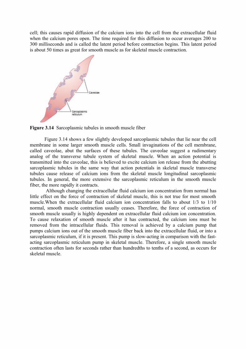
Figure 3.14 Sarcoplasmic tubules in smooth muscle fiber
Figure 3.14 shows a few slightly developed sarcoplasmic tubules that lie near the cell membrane in some larger smooth muscle cells. Small invaginations of the cell membrane, called caveolae, abut the surfaces of these tubules. The caveolae suggest a rudimentary analog of the transverse tubule system of skeletal muscle. When an action potential is transmitted into the caveolae, this is believed to excite calcium ion release from the abutting sarcoplasmic tubules in the same way that action potentials in skeletal muscle transverse tubules cause release of calcium ions from the skeletal muscle longitudinal sarcoplasmic tubules. In general, the more extensive the sarcoplasmic reticulum in the smooth muscle fiber, the more rapidly it contracts.
Although changing the extracellular fluid calcium ion concentration from normal has little effect on the force of contraction of skeletal muscle, this is not true for most smooth muscle.When the extracellular fluid calcium ion concentration falls to about 1/3 to 1/10 normal, smooth muscle contraction usually ceases. Therefore, the force of contraction of smooth muscle usually is highly dependent on extracellular fluid calcium ion concentration. To cause relaxation of smooth muscle after it has contracted, the calcium ions must be removed from the intracellular fluids. This removal is achieved by a calcium pump that pumps calcium ions out of the smooth muscle fiber back into the extracellular fluid, or into a sarcoplasmic reticulum, if it is present. This pump is slow-acting in comparison with the fast-acting sarcoplasmic reticulum pump in skeletal muscle. Therefore, a single smooth muscle contraction often lasts for seconds rather than hundredths to tenths of a second, as occurs for skeletal muscle.