physiology of renal system dr. hayder alhindy
TRANSCRIPT

Physiology of Renal System Dr. Hayder Alhindy
1
Organ system that produces, stores & carries urine. Includes two kidneys, two
ureters, the urinary bladder, two sphincter muscles & the urethra. Humans produce about
1.5 liters of urine over 24 hours, although this amount may vary according to the
circumstances. More fluid intake generally rises urine production. Raised perspiration
& respiration may drop the amount of fluid excreted through the kidneys. Medications
(as diuretics) interfere with urine production.
Kidney is paired organ (weight about 300 g), Composed of two parts cortex (isotonic
urine) and medulla (hypertonic urine). Each kidney consists of 8-10 conical pyramids
with bases in the cortex & apices project toward the pelvis. Each pyramid has outer
cortex & inner medulla. Medulla in turn subdivided into outer & inner zones. The outer
zone splits into outer & inner stripes. Each pyramid pours its urine into a minor calyx,
and then every 2-3 calyces unite to form a major calyx. Major calyces unites to form the
renal pelvis that leads urine through the ureter. Renal pelvis emerges through the hilum
of kidney from which renal artery, enters & renal vein leaves. Two ureters pour into
single urinary bladder from which urethra emerges.
Renal blood supply
About 25% of the resting cardiac output ﴾1.25 L\min﴿ supplies the 300 grams of renal
tissue with much greater flow to the cortex ﴾97%﴿ than to the medulla.
Arterial tree: Renal, segmental, interlobar, arcuate & interlobular arteries. Afferent
(then glomerular capillaries), efferent arterioles & peritubular capillaries (vasa recta).
Hence, Systemic blood is carried out of the glomerulus by an efferent arteriole
instead of a venule, as is observed in most other capillary.
Venous tree: Interlobular, arcuate, interlobar, segmental and renal veins.

Physiology of Renal System Dr. Hayder Alhindy
2
Characteristics of the renal blood flow:
1. Large blood flow: 200 ml/min (20% of
cardiac output) & (94%) to the cortex
2. 1ry & 2ry capillary networks:
- Primary network: glomerular capillary
network, between afferent and efferent
arterioles, high hydrostatic pressure
(60 mmHg), favor glomerular filtration.
- Secondary network: peritubular capillary
network, made by branch of efferent
arterioles, low pressure (13 mmHg),
favor tubular reabsorption.
3. Autoregulation of renal blood flow.
4. Tubuloglomerular feedback.
5. Nervous and humoral regulation.
6. High RBF not reflect high O2 intake as
kidney use only 8% of all O2 intake.
N.B. - There are 2 sets of capillaries & 2 sets of arterioles!! - The only circulation where there are capillaries, which are drained by arterioles

Physiology of Renal System Dr. Hayder Alhindy
3
Renal Major Capillaries))
Autoregulation of RBF determines GFR:
- RBF is about 20% of the cardiac OP: very large flow relative to the weight of the
kidneys (≈ 350 g).
- RBF also modifies solute & water reabsorption and delivers nutrients to nephron cells.
- RBF kept constant in BP of 80-180 mmHg by varying renal vascular resistance. I.e.
the resistances of the interlobular artery, afferent arteriole and efferent arteriole.
- RBF autoregulation is vital to prevent large changes in GFR that would greatly affect
urinary output & to allow normal renal excretion of water & solute.
- RBF autoregulation occurs in denervated & isolated kidney. I.e. its ''intrinsic property''.
Impact of autoregulation:
- With autoregulation: GFR=125m/min (180L/day) but tubular
reabsorption178.5L/day results in 1.5L/day of urine.
- Without autoregulation: Small ↑ in BP 100 to 125mm Hg, ↑GFR by 25% (180 to
225L/day). If tubular reabsorption constant, urine flow of 46.5 L/day!!
Glomerular capillary bed Peritubular capillary bed
1. Receives bl from afferent art. Receives bl from efferent art.
2. High pressure bed 45- 55 mmHg Low pressure bed 10- 13 mmHg
3. Represents arterial end of cap. Represents venous end of cap.
4. Allows fluid filtration. Allows fluid reabsorption.
Physiology of Renal System Dr. Hayder Alhindy
4
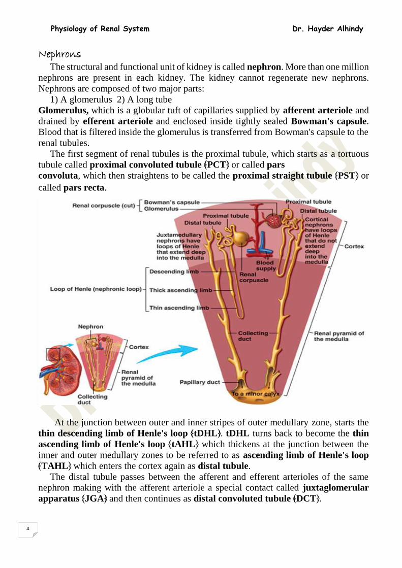
Nephrons The structural and functional unit of kidney is called nephron. More than one million
nephrons are present in each kidney. The kidney cannot regenerate new nephrons.
Nephrons are composed of two major parts:
1) A glomerulus 2) A long tube
Glomerulus, which is a globular tuft of capillaries supplied by afferent arteriole and
drained by efferent arteriole and enclosed inside tightly sealed Bowman's capsule.
Blood that is filtered inside the glomerulus is transferred from Bowman's capsule to the
renal tubules.
The first segment of renal tubules is the proximal tubule, which starts as a tortuous
tubule called proximal convoluted tubule ﴾PCT﴿ or called pars
convoluta, which then straightens to be called the proximal straight tubule ﴾PST﴿ or
called pars recta.
At the junction between outer and inner stripes of outer medullary zone, starts the
thin descending limb of Henle's loop ﴾tDHL﴿. tDHL turns back to become the thin
ascending limb of Henle's loop ﴾tAHL﴿ which thickens at the junction between the
inner and outer medullary zones to be referred to as ascending limb of Henle's loop
﴾TAHL﴿ which enters the cortex again as distal tubule.
The distal tubule passes between the afferent and efferent arterioles of the same
nephron making with the afferent arteriole a special contact called juxtaglomerular
apparatus ﴾JGA﴿ and then continues as distal convoluted tubule ﴾DCT﴿.

Physiology of Renal System Dr. Hayder Alhindy
5
Distal convoluted tubules unite as collecting tubules, which pour in larger collecting
tubes, & then cortical and medullary collecting ducts to end in main duct of renal
pyramid supplying the minor calyx.
Types of nephrons:
There are two types of nephrons:
Cortical nephrons:
More numerous) 70%-80% (lying in the outer layer
of cortex. The tubular system is relatively short, no
tAHL & efferent narrower than afferent arterioles.
Has peritubular capillaries, but no vasa recta . Na
reabsorption occurs mainly here, & JG apparatus is
well developed hence, share in the process of an
autoregulation.
Juxtamedullary nephrons:
Less numerous (20-30%), lie in the inner third of
cortex, long tubules with long tAHL dip deeply
down into the medulla toward the pyramids &
efferent wider than afferent arteriole ﴾because
efferent arteriole here supplies a much extensive
peritubular capillary network﴿ so vasa recta is
active. Urine concentration occurs mainly here, but
no autoregulation as the JG apparatus is absent.
So, why is the loop of Henle useful?
The longer the loop, the more concentrated the filtrate in the medulla become.
Importance: the collecting tubule runs through the hyperosmotic medulla more ability
to reabsorb H2O.
Renal pelvis
The major function of the renal pelvis is to
act as a funnel for urine flowing to the ureter.
It is the point of convergence of two or three
major calices .A branch of the renal pelvis
called a calyx surrounds each renal papilla.
Ureters
The ureters are about 200 to 250 mm long,
urine is collected in the renal pelvis (or
pyelum), which connects to the ureters, which
carry urine to the bladder.The urine
peristaltically forced downward through
smooth muscular tissue in the ureteric walls.

Physiology of Renal System Dr. Hayder Alhindy
6
Urinary Bladder
A balloon shape hollow muscular organ. It can
stores up to 500 ml of urine comfortably (2- 5
hours). Its sphincters (circular muscles)
regulate the flow of urine from the bladder.
Internal urethral sphincter in the beginning of
urethra (smooth muscle – involuntary) while,
external (skeletal muscle – voluntary). In
males, both sphincters are more powerful, able
to retain urine for twice as long as females.
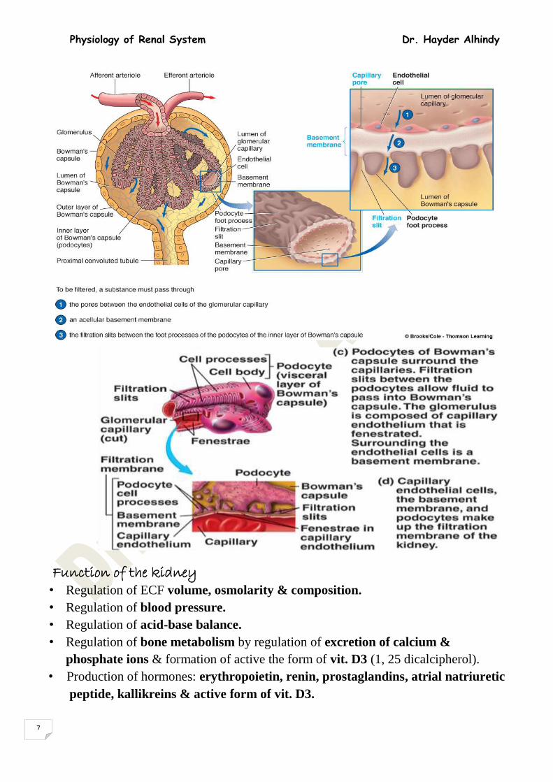
Glomerular capillary membrane
It is composed of three layers. The single
layer of capillary endothelial cells lies on
basement membrane and covered by the
visceral layer of Bowman's capsule which
is called podocytes. The endothelial layer
is fenestrated such that even some plasma
proteins ﴾but not cells﴿ can pass through its
fenestrae. Yet; the proteins cant exit the
basement membrane despite its very high
permeability ﴾ more than 100 times the
permeability of other capillary membranes
in human body﴿. Other filtered
materials and ions, however can pass easily
through the membrane and the slit pores between (Glomerular filtration membrane & podocyte)
the feet of podocytes.
Filterability of the Membrane:
Filterability is a term used to describe
membrane selectivity based on the molecular
size and charge. Pore size would favor plasma
protein (albumin) passage, but negative
charge on protein is repelled by the (-)
charged basement membrane (proteoglycan
filaments & podocytes). Loss of this (-)
charge causes proteinuria.
Physiology of Renal System Dr. Hayder Alhindy
7
Function of the kidney • Regulation of ECF volume, osmolarity & composition.
• Regulation of blood pressure.
• Regulation of acid-base balance.
• Regulation of bone metabolism by regulation of excretion of calcium &
phosphate ions & formation of active the form of vit. D3 (1, 25 dicalcipherol).
• Production of hormones: erythropoietin, renin, prostaglandins, atrial natriuretic
peptide, kallikreins & active form of vit. D3.

Physiology of Renal System Dr. Hayder Alhindy
8
• Excretion of various metabolic waste products, drugs, toxic substances:
- Urea from protein breakdown
- Uric acid from nucleic acid breakdown
- Creatinine from muscle creatine breakdown
- End products of hemoglobin breakdown
Basic Renal Terminology Glomerular filtration rate (GFR): amount of fluid pass into Bowman’s capsule / min.
Renal blood flow (RBF): amount of blood flowing through the kidney / (min).
Filtration: process by which substances enter Bowman’s capsule.
Reabsorption: process by which substances move from inside to outside the tubule
(from lumen to blood).
Secretion: process by which substances move from outside to inside the tubule (blood
to lumen) made by epithelial cells itself or blood substance are transported into renal
tubular lumen.
Excretion: substances that pass from the kidney into the bladder.
(Basic renal functions)
The ability of the kidney to move specific substances selectively into & out of the
tubule in a very controlled & coordinated manner makes normal kidney function so
critical to life.
Glomerular filtration:
Fluid and small solutes dissolved in the plasma such as glucose, amino acids, Na, K,
Cl, HCO3- , other salts, and urea pass through the membrane and become part of the
filtrate.
- The glomerular membrane hold back blood cells, platelets and most plasma proteins.
- The filtrate is about 20% of the plasma.
- Ultrafiltrate: Most substances in the plasma (except protein) are freely filtrated, so
that their concentrations in Bowman’s capsule are almost the same as in the plasma.

Physiology of Renal System Dr. Hayder Alhindy
9
Glomerular Filtrate Plasma Substance Ions (mEq/L)
142 142 Na
5 5 K
103 103 Cl
28 28 HCO Organic Molecule (mg/dl)
5-11 1000-5000 Protein
100 100 Glucose
26 26 Urea
3 3 Uric acid
1.1 1.1 creatinine (Composition of Plasma Vis Glomerular Filtrate)
Renal clearance
When certain amount of substance ﴾x﴿ is cleared away from plasma and excreted in
urine, it is called renal clearance of that substance ﴾ Cx﴿ . Not all of the substance filtered
from glomerular capillaries appears in urine, instead; some of this substance may be
reabsorbed back to the blood via peritubular capillaries, while additional amounts of the
same substance may be secreted from tubular cells to tubular lumen to appear in urine.
Cx = GFR – TR + TS
Where: GFR is the glomerular filtration rate of x
TR is the tubular reabsorption of x
TS is the tubular secretion of x
Significance of renal clearance:
- Estimate renal function;
- Determine glomerular filtration rate (GFR )
- Determine renal blood flow (RBF)
- Presume renal tubular transport effect
- Free-water clearance
Glomerular filtration rate ﴾GFR﴿ Glomerular filtration is passive non-selective process. When renal plasma flows from
afferent to efferent arterioles, ≈ 20% of its contents filtered by glomerular filtration from
glomerular capillaries to Bowman's capsule. This is called the filtration fraction ﴾FF ﴿
GFR = Renal Plasma flow ﴾ RPF ﴿ * FF
GFR = RPF * 20%
RPF = Renal Blood Flow ﴾ RBF ﴿ * (1-hematocrit)
= 1250 ml\min * 0.5
= 625 ml\min
GFR = 625 ml\min * 20%
Physiology of Renal System Dr. Hayder Alhindy
10
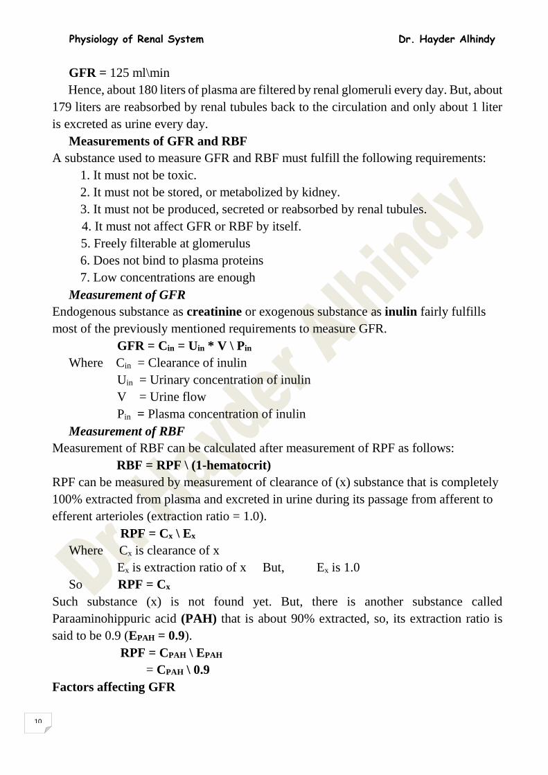
GFR = 125 ml\min
Hence, about 180 liters of plasma are filtered by renal glomeruli every day. But, about
179 liters are reabsorbed by renal tubules back to the circulation and only about 1 liter
is excreted as urine every day.
Measurements of GFR and RBF
A substance used to measure GFR and RBF must fulfill the following requirements:
1. It must not be toxic.
2. It must not be stored, or metabolized by kidney.
3. It must not be produced, secreted or reabsorbed by renal tubules.
4. It must not affect GFR or RBF by itself. 5. Freely filterable at glomerulus
6. Does not bind to plasma proteins
7. Low concentrations are enough
Measurement of GFR
Endogenous substance as creatinine or exogenous substance as inulin fairly fulfills
most of the previously mentioned requirements to measure GFR.
GFR = Cin = Uin * V \ Pin
Where Cin = Clearance of inulin
Uin = Urinary concentration of inulin
V = Urine flow
Pin = Plasma concentration of inulin
Measurement of RBF
Measurement of RBF can be calculated after measurement of RPF as follows:
RBF = RPF \ (1-hematocrit)
RPF can be measured by measurement of clearance of (x) substance that is completely
100% extracted from plasma and excreted in urine during its passage from afferent to
efferent arterioles (extraction ratio = 1.0).
RPF = Cx \ Ex
Where Cx is clearance of x
Ex is extraction ratio of x But, Ex is 1.0
So RPF = Cx
Such substance (x) is not found yet. But, there is another substance called
Paraaminohippuric acid (PAH) that is about 90% extracted, so, its extraction ratio is
said to be 0.9 (EPAH = 0.9).
RPF = CPAH \ EPAH
= CPAH \ 0.9
Factors affecting GFR

Physiology of Renal System Dr. Hayder Alhindy
11
The following equation summarizes some factors influencing GFR
GFR = Kf * (net ultrafiltration pressure)
= Kf * (PG + ΠB – PB - ΠG)
= Kf * (60 + 0 – 18 - 32) respectively
= Kf * (+10 mmHg)
Where: Kf is ultrafiltration coefficient, which is the effective ultrafiltration (surface
area multiplied by glomerular capillary permeability).
PG is glomerular capillaries' hydrostatic pressure
ΠB is osmotic pressure of colloids inside Bowman's capsule
PB is hydrostatic pressure inside Bowman's capsule
ΠG is osmotic pressure of colloids inside glomerular capillaries
The latter four factors are called Starling forces
(Net Filtration Pressure)
Other factors affecting GFR are:
- RBF: when RBF increases GFR also increases
- FF: when FF increases GFR also increases
- Vasoconstriction of afferent arterioles decreases GFR (decreased PG)
- Slight vasoconstriction of efferent arterioles increases GFR (increased PG)
- Severe vasoconstriction of efferent arterioles decreases GFR (increased ΠG)
(Effects of different Afferent
arterioles states on GFR &
Efferent arterioles)
Because GFR=RPF * FF

Physiology of Renal System Dr. Hayder Alhindy
12
Control of GFR
Control of GFR is by one or more of the followings:
Sympathetic nervous activity
Hormones and autacoids
Autoregulation
Plasma levels of amino acids and glucose
1- Sympathetic nervous activity: Strong sympathetic activity decreases GFR e.g.,
in severe hemorrhage and in cerebral ischemia while the role of parasympathetic (vagal)
innervations is yet unknown.
2- Hormones and autacoids: Hormones are small chemical substances that are
secreted from endocrine glands and transported by blood to work at distant organs while
autacoids are chemical substances that are secreted and act locally.
Adrenaline (epinephrine), nor-adrenaline (nor-epinephrine), angiotensin II, aspirin
and endothelin decrease GFR. Nitric oxide, prostaglandin & bradykinin increase GFR.
3- Autoregulation: Autoregulation involves afferent not efferent arterioles.
Autoregulation is important to prevent large changes in GFR that would greatly
affect urinary output. Two regulatory processes are faced in the regulation of GFR:
a. Juxtaglomerular feedback mechanism: Juxtaglomerular apparatus (JGA) consists of
specific distal convoluted tubular epithelial cells called macula densa (which contains
osmoreceptors that are sensitive to any increase or decrease in concentration of NaCl)
in close contact with specific smooth muscle cells in the wall of afferent arterioles called
Juxtaglomerular cells.
Macula Densa cells in distal tubule sense levels of Na with Na-K-2Cl cotransporter .
If Na levels are high, releases adenosine, which acts on afferent & efferent arterioles
(reduces filtration). Adenosine constricts afferent arteriole by A1 adenosine receptor &
adenosine dilates efferent arteriole by A2 adenosine receptor.
Decreased amounts of NaCl near osmoreceptors of macula densa sends impulses to
JG cells to relax resulting in vasodilatation of afferent arterioles, increase blood flow to
glomerular capillaries, and increase GFR.
When GFR is decreased, tubular flow slows down resulting in increased tubular
reabsorption of NaCl. This will decrease NaCl near the osmoreceptors of macula
densa. Macula densa will send impulses to the JG cells to relax resulting in
vasodilatation of afferent arterioles. This will increase blood flow to the glomerular
capillaries which will in turn increase GFR.
The reverse occurs when GFR is increased; tubular flow quickens resulting in
decreased tubular reabsorption of NaCl. This will increase NaCl near the
osmoreceptors of macula densa. Macula densa will send impulses to the JG cells to

Physiology of Renal System Dr. Hayder Alhindy
13
contract resulting in vasoconstriction of afferent arterioles. This will decrease blood
flow to the glomerular capillaries, which will in turn decrease GFR.
Renin is also produced by JG cells in response to any increase in GFR or RBF, which
will result in production of angiotensin I and then angiotensin II to decrease GFR and
RBF.
Impact of Autoregulation:
• Autoregulation:
– GFR = 180 L/day and tubular reabsorption = 178.5 L/day
– Results in 1.5L/day in urine
• Without Autoregulation:
– Small ↑ in BP 100 to 125mm Hg, ↑GFR by 25% (180 to 225L/day)
– If tubular reabsorption constant, urine flow of 46.5 L/day
b. Myogenic mechanism: Increased RBF that causes increase in GFR; is at the same
time the cause of distension of afferent arterioles and stretch of smooth muscles lining
their walls. This stretch results in myogenic contraction of these smooth muscles leading
to vasoconstriction of afferent arterioles and decreased RBF and GFR.
Myogenic Mechanism of the autoregulation
4- Plasma levels of amino acids and glucose: This mechanism depends on the fact
that tubular reabsorption of amino acid and glucose is unlimited and so, when plasma
levels of amino acids and glucose increase; tubular reabsorption will also increase
resulting in decreased amounts of NaCl near the osmoreceptors of macula densa. Macula
densa will send impulses to the JG cells to relax resulting in vasodilatation of afferent
arterioles. This will increase blood flow to the glomerular capillaries which will in turn
increase GFR.
Tubular reabsorption A highly selective process may be passive or active. Some substances are
completely reabsorbed like amino acids and glucose. Some substances are mostly

Physiology of Renal System Dr. Hayder Alhindy
14
reabsorbed like bicarbonates and some other electrolytes. Some other substances are
mostly reabsorbed in the presence of specific factors like hormones but their
reabsorption is reduced when these hormones are reduced or absent like water
reabsorption, which is increased in the presence of antidiuretic hormone, and sodium
ions reabsorption that is increased in the presence of aldosterone and\or angiotensin II
hormones. Many substances are reabsorbed along with other substances like chloride
ions, which follow sodium ions, and sodium chloride salt, which follows water. Some
substances are 50% reabsorbed (and 50% excreted) like urea. Some other substances
are about completely excreted like creatinine and some drugs and poisons.
Why large amounts of solutes are filtered & then reabsorbed by the kidneys?
1- Allows the kidneys to rapidly remove waste products from the body that depend
primarily on GFR
2- Allows all body fluids to be filtered many times each day. Because the entire plasma
volume is only ≈ 3 liters, while the GFR is ≈180 L/day, the entire plasma can be filtered
≈ 60 times each day (precise & rapid) control of the body fluids.
There is a glomerulotubular balance such that when GFR increases; tubular
reabsorption also increases. However, this is not eternal; in reality, the active transport
processes of reabsorption may be saturated when the tubular lumen is overloaded with
filtered substances. The maximum tubular load of certain substance above which an
active transport process of tubular reabsorption is saturated and reabsorption is ceased
is called transport maximum (Tm) of that substance. The maximum plasma
concentration of certain substance above which this substance starts to appear in urine
is called renal threshold of that substance which equals Tm\GFR
Transport maximum of glucose (TmG) is about 325 mg\min and its ideal renal
threshold is 325\125 = 2.6 mg\ ml = 260 mg\100 ml
But the actual renal threshold for glucose is about 180 mg\100 ml and this difference
may be due to that not all of renal tubules have the same Tm and that some of the filtered
glucose molecules before Tm bypass reabsorption.
Factors affecting tubular reabsorption
The same previously mentioned Starling forces affect tubular reabsorption in
addition to other factors. The following equation summarizes some of these factors.
TR = Kf * (net reabsorption pressure)
TR = Kf * (Pif – Pc + Πc - Πif )
TR = Kf * (6 mmHg – 13 mmHg + 32 mmHg – 15 mmHg)
TR = Kf * (+10 mmHg)
Where Kf is a constant that depends on the surface area of effective reabsorption,
distance of reabsorption and tubular capillary permeability

Physiology of Renal System Dr. Hayder Alhindy
15
Pif is interstitial fluid hydrostatic pressure
Pc is peritubular capillaries' hydrostatic pressure
Πc is peritubular capillaries' osmotic pressure of colloids Starling forces
Πif is interstitial fluid osmotic pressure of colloids
Control of tubular reabsorption:
1-Sympathetic activity: Increase tubular reabsorption of sodium ions.
2- Hormonal activity:
A- Aldosterone: Secreted from adrenal cortex and acts on principal cells of
distal tubules to increase reabsorption of sodium ions and excretion of potassium
ions. It increases permeability of luminal membrane to sodium ions and stimulates
sodium- potassium pump in basolateral membrane.
Adrenal insufficiency (Addison's disease) results in excessive sodium loss and
potassium retention while adrenal hyperactivity (Cushing syndrome) results in sodium
retention and potassium depletion.
B- Angiotensin II: Produced by the lungs from angiotensin I this in turn produced
in the liver from angiotensinogen (renin). Angiotensin acts directly (or indirectly after
stimulation of aldosterone) to increase sodium ions reabsorption.
C- Antidiuretic hormone (vasopressin): Produced from posterior pituitary gland
and it acts on distal and collecting tubules and ducts to increase water reabsorption and
urine concentration.
D- Atrial natriuretic peptide (ANP): Produced by cardiac atria in response to any
increase in blood volume and acts especially on collecting ducts to decrease sodium
&water reabsorption and so, increase urine excretion to restore normal blood volume.
A few Situations Encountered in Life:
Increase Fluid Volume /Decrease Osmolarity
- Drink pure H2O
- Excrete dilute urine
Norm Fluid Volume /Increase Osmolarity
- Salty popcorn (no drink)
- Highly concentrated urine of low volume
E- Parathyroid hormones: Produced by parathyroid glands and act especially on
thick ascending limbs of Henle's loops (and distal convoluted tubules) to increase
calcium and magnesium ions reabsorption and decrease phosphate reabsorption.
Sources of Water Output & Input
Water input • Food & drink = 2.2 L/day
• Cellular respiration: Glucose + O2 CO2 + H2O = 0.3 L/day

Physiology of Renal System Dr. Hayder Alhindy
16
Water output • Urine = 1.5 L/day
• Fecal matter = 100 mL/day
• Evaporative{skin & respiration} = 900 mL/day
Regulation of ECF osmolarity
Normal ECF osmolarity is about 280-300 mosm\L and it is mostly dependant on sodium
ions concentration (142 mEq\L). Normal daily sodium ions intake must equals its daily
output = 10-20 mEq.
Plasma osmolarity (Posm) in healthy subjects is calculated from plasma sodium
concentration (PNa+)
Na+= 2.1 * P osmP
However, in patients with renal diseases, plasma concentrations of other substances like
urea and glucose are also calculated.
Na+ intake ECF volume Blood pressure
Angiotensin II Baroreceptors
Na+ excretion Pressure natriuresis Brain stem
Aldosterone Na+ reabsorption Sympathetic activity

Physiology of Renal System Dr. Hayder Alhindy
17
When Posm decreases; kidneys excrete large amounts of diluted urine (down to 50
mosm\L) while when Posm increases; kidneys excrete small amounts of highly
concentrated urine (up to 1200 mosm\L).
The human body must get rid of not less than 600 mosm of metabolic wastes/day. So,
it is very necessary to excrete not less than 0.5 liters of highly concentrated urine daily.
According to this equation, when kidney losses its ability to produce concentrated urine;
the minimum obligatory urine volume will increase resulting in excessive loss of body
fluids (a disease called diabetes insipidus).
According to the same equation, excessive intake of hyperosmotic fluids like
seawater will seriously increase the Posm and, thence, the minimum obligatory urine
volume even with the maximum renal capability of urine concentration resulting in death
from dehydration due to excessive loss of body fluids.
The ability of kidney for concentration of urine requires the presence of
hyperosmotic medulla created by countercurrent mechanism and urea recirculation
in concert with ADH.
Renal regulation of salt and water balance (Relationship of osmolarity and volume)
Countercurrent mechanism
An osmotic gradient is formed in the interstitial space around the loop of Henle that
raises from ‘’the top to the bottom’’ of the loop. The action of the ‘’tritransporter’’ of
the epithelial cells of the ascending limb, the water permeability of the descending limb,
and the shape of the loop give such osmotic gradient. The process by which this occurs
is called counter-current multiplication. The descending and ascending limbs of Henle's
loop and vasa recta run a long distance parallel, counter and in close proximity to each
other carrying solutes toward medulla and water toward systemic circulation resulting

Physiology of Renal System Dr. Hayder Alhindy
18
in hyperosmotic medulla. Ascending limbs of Henle's loop are called countercurrent
multipliers because they continuously bring new NaCl to medulla while ascending vasa
recta are called countercurrent exchangers because they continuously drawback water
(Counter current mechanism)
from medulla to the systemic circulation. The major bulk of tubular reabsorption of
water and solutes (about 65%) occurs in proximal tubules. So, tubular fluid reaches the
thin segments within its original osmolarity (300 mosm\L).
About 15% of water reabsorption occurs in tDHL which is carried back to the
systemic circulation via ascending vasa recta. But tDHL is impermeable to solutes which
stay within thin segment not reabsorbed. So, tubular fluid reaches the ascending limbs
highly hyperosmotic (1200 mosm\L).
Starting from tAHL, all the following segments are impermeable to water in absence
of ADH but very little amounts of solutes are passively reabsorbed in tAHL. So, tubular
fluid reaches the following segment still hyperosmotic (900 mosm\L).
The major active reabsorption of electrolytes occurs in TAHL (about 30%) which is
mainly due to 1Na+-2Cl¯-1K+ active cotransport process which works against as much

Physiology of Renal System Dr. Hayder Alhindy
19
as 200 mosm\L concentration gradient. So, tubular fluid reaches distal tubules
hypoosmotic (100 mosm\L).
The remaining reabsorption processes of electrolytes (5%) occur in distal segments.
The net result is hyperosmotic medulla, which favors further water reabsorption
(about 19%) from collecting ducts by ADH & only about 1% of filtered water is excreted
in urine. While in absence of ADH, about 20% of filtered water is excreted.
Urea recirculation Recirculation of urea is responsible for about 40% of the process of urine
concentration when urea is reabsorbed from medullary colleting tubules to the medullary
interstitium to be secreted again from the tubular cells of thin segments to their lumen
where the cycle is repeated again and again.

Physiology of Renal System Dr. Hayder Alhindy
20
Types of Renal Tubululer Transports :
Passive transport
- Simple: only Na +
- Co-transport: with Cl-, glucose, aminoacids & phosphates
- Anti-transport: Na+ inside, H+ or Ca2+ out
Active transport
- Na+/K+ pump dependent on ATP energy.
Body control of ECF osmolarity
1- Osmoreceptors-ADH feedback
2- Thirst center in brain stem
3- Salt appetite center in brain stem
Osmoreceptor cells lie in anterior hypothalamus, are sensitive to any increase in Na+
concentration, and send signals to supraoptic nuclei, which stimulate the posterior
pituitary gland to increase secretion of ADH that increases water reabsorption.
Vasopressin secretion is also stimulated by decreased blood volume, decreased blood
pressure, nausea, vomiting, morphine and nicotine. Vasopressin secretion is inhibited
by increased blood volume, increased blood pressure and alcohol intake.
Increased osmolarity also stimulates the thirst center in brain stem to increase the
desire for water intake and also to increase secretion of ADH. Thirst center is also
stimulated by decreased ECF volume, decreased blood pressure, angiotensin II and
dryness of mouth, pharynx and esophagus while it is inhibited by decreased ECF
osmolarity, increased ECF volume, increased blood pressure and gastric distension.
Decreased osmolarity stimulates salt appetite center in the brain stem to increase
the desire for salt intake.

Physiology of Renal System Dr. Hayder Alhindy
21
Regulation of blood volume and pressure Blood volume is kept constant despite the tremendous changes in fluids intake from
0.1th to 10 times normal. This constant blood volume is due to:
1- Small increase in blood volume results in large increase in cardiac output.
2- Small increase in cardiac output results in large increase in blood pressure.
3- Small increase in blood pressure results in large increase in urine excretion.
which immediately hinders and reverses the raising blood volume and pressure.
Acute increase in blood pressure is balanced by direct increase in Na+ excretion due
to increase in GFR and decrease in Na+ reabsorption with increase in Na+ leak back to
the tubular lumen. Pressure induced increase in Na+ excretion is called pressure
natriuresis, which is always accompanied by pressure diuresis (pressure-induced
increase in urine excretion).
Chronic increase in blood pressure is balanced by decrease in angiotensin II
production, which results in decrease in Na+ reabsorption directly, or indirectly by
decreasing aldosterone production from adrenal cortex.
Body regulation of acid-base balance General considerations:
- Metabolism of food generates acid.
- Acid in the body is in two forms: fixed and volatile.
- Kidneys remove excess fixed acid;
- Lungs remove excess volatile acid .
- Acidemia is excess H ions in the blood ;
- Alkalemia is excess bicarbonate ions in the blood.

Physiology of Renal System Dr. Hayder Alhindy
22
Any change in hydrogen ions concentration [H+] will affect all cellular and body
functions due to its effects on many reactions. Normal [H+] in ECF is only 0.00000004
mol\L, so; it is better to use pH (which is –log [H+] = 7.4).
Nobody can survive more than hours when pH raises to 8.0 or falls to 6.8 (more or
less than normal by 0.6). Regulation of [H+] is by one or more of the following systems:
Chemical acid-base buffer systems in body fluids, respiratory and renal regulation of
acid-base balance. Buffer system is functioning within seconds up to 2 hours only »
then the respiratory comes within hours » after that the kidney comes within days and
will participate in the process of acid base balance and its affect stay for the long run.
A. Buffer systems are:
1- Bicarbonate buffer system: It is the most important buffer system in ECF. The
following reaction occurs when [H+] is increased:
H+ +HCO3¯ → H2CO3 → H2O + CO2......(CO2 to be expired by the lungs)
While when [OH-] is increased, the following reaction occurs:
OH¯ + H2CO3 → H2O + HCO3¯..........( HCO3¯ to be excreted by kidneys)
The power of dissociation constant (pK) for bicarbonate system is 6.1 and
accordingly, Henderson-Hassel Bach equation states that:
pH = 6.1 + log [HCO3¯]\0.03 PCO2
When HCO3¯ decreases, pH is decreased metabolic acidosis
When PCO2 increases, pH is decreased respiratory acidosis
When HCO3¯ increases, pH is increased metabolic alkalosis
When PCO2 decreases, pH is increased respiratory alkalosis
2- Phosphate buffer system: It is important buffer system in renal tubular fluids &
intracellular. Its pK is 6.8 and the following reaction occurs when [H+] increases:
H+ + HPO42¯ → H2PO4
¯
When [OH¯] is increased the following reaction occurs:
OH¯ + H2PO4¯ → HPO4
2¯ + H2O
3- Protein buffer systems: Are the most available intracellular buffer systems but
also work extracellularly. One of the most important protein buffers is
hemoglobin in red blood cells.
H+ + Hb → HHb
4- Ammonium buffer system: Is the last choice buffer system in renal tubules
when bicarbonate and phosphate buffer systems are saturated. Ammonium is
formed from metabolism of glutamine inside renal tubular cells. The following
reaction occurs when [H+] increases:
H+ + NH3 → NH4+
When [OH-] is increased the following reaction occurs:
OH¯ + NH4+ → NH4OH

Physiology of Renal System Dr. Hayder Alhindy
23
B. Respiratory regulation
Respiratory regulation of acid-base balance is via stimulation or inhibition of the
respiratory center in the brain stem by the central chemosensitive areas which are
bilateral aggregations of neurons beneath the ventral surface of medulla that are sensitive
to changes in H+ and PCO2. The result is hyper- or hypo- ventilation respectively.
Double normal alveolar ventilation reduces PCO2 and raises pH from 7.4 to 7.63
while 1\4th normal alveolar ventilation raises PCO2 and reduces pH to 6.95 because pH
is inversely related to PCO2 according to Henderson-Hasselbach equation.
c. Renal regulation:
Occurs by excretion of acidic or alkaline urine. Normally, daily renal secretion of H+
is about 4400 mmol. Bicarbonates system buffers 4320 mmol in renal tubules and the
other 80 mmol are buffered by phosphates and then ammonium buffer systems. Most of
renal tubular cells utilize secondary active transport to secrete H+ like Na+-H+ antiport,
but the intercalated cells of distal tubules utilize primary active transport called proton
pump.
Physiology of micturition
Urinary excretion is vital function for keeping normal metabolism & homeostasis of
internal environment in the human body. Micturition is the disposing urine from the
bladder through the urethra out of the body & is usually under voluntary control. Urinary
incontinence: inability to control urination & is more in women than men. The reverse
is urinary retention: inability to urinate. While nocturnal enuresis mean incontinence
during the night (effects of emotions).
Urine enters urinary bladder in spurts synchronous with the regular peristaltic
contractions of ureteric smooth muscles (1-5 times per minute) and the oblique insertion
of ureters into the vicinity of bladder walls prevents back flow of urine to the ureters.
Kidney have no vagus nerve fibers innervation. Urinary bladder receives
parasympathetic innervations from S2, S3 and S4 via pelvic nerves. Sympathetic
innervations come from L1, L2 and L3 via hypogastric nerves after relay in inferior
mesenteric ganglion. Somatic sensory and motor innervations come from S2, S3 and S4
via pudendal nerves. Sensory innervations also travel with autonomic innervations via
pelvic and hypogastric nerves. Kidney have no vagus nerve fibers innervation.
The first urge to void is felt at bladder volume of 150 ml and the marked sense of
fullness is at about 400 ml. However, this can be relieved by the property of plasticity
of smooth muscles.

Physiology of Renal System Dr. Hayder Alhindy
24
Voluntary micturition is thought to be initiated after relaxation of muscles of pelvic
floor, which may cause sufficient downward pull on detrusor muscle, which induces
excitation of stretch receptors in the bladder wall to initiate reflex contraction. The
afferent and efferent limbs of voiding reflex travel with pelvic nerves to the sacral
portion of spinal cord and threshold for this reflex is adjusted by the activity of
facilitatory and inhibitory centers in the brain. Facilitatory areas are in pontine region
and posterior hypothalamus while inhibitory area is in midbrain.
Other reflex that share in the physiology of micturition is the ''Renorenal reflex''.
Sensory nerves located in the renal pelvic wall are activated by stretch of the renal pelvic
wall, which may occur during diuresis or ureteral spasm/occlusion . Activation of these
nerves leads to an increase in afferent renal nerve activity, which causes a decrease in
efferent renal nerve activity & an increase in urine flow rate & urinary sodium excretion.
The internal urethral sphincter is made up of bands of smooth muscles on either sides
and plays no role in micturition, but in male, it prevents retrograde ejaculation (reflux
of semen into the urinary bladder during ejaculation). The external sphincter is skeletal
muscle and it contracts voluntarily to delay micturition or interrupt its starting. The
ability to delay micturition until the opportunity to void is available is a learning ability
of brain in adults.
After micturition, female's urethra empties by gravity while male's urethra empties
by several contractions of bulbocavernosus muscle.
Pathological Terms: UTI: Urinary Tract Infection
Nephritis: Inflammation of the kidneys
Hydronephrosis: dilation of the renal pelvis
Anuria: no urinary output
Dysuria: painful urination

Physiology of Renal System Dr. Hayder Alhindy
25
Enuresis: lack of bladder control
Oliguria: scanty urination
Polyuria: excessive urination
Incontinence: involuntary discharge of urine or feces
Diuretic: Increase urine output
Antidiuretic: Decrease urine output
Changes of Renal System with Aging Include
• Decline in the number of functional nephrons
• Reduction of GFR
• Reduced sensitivity to ADH
• Problems with the micturition reflex.