renal physiology comprehensive
TRANSCRIPT

C H A P T E R 2
Renal Physiology
David G. Shirley, Robert J. Unwin
The prime function of the “kidneys” is to maintain a stable milieu intérieur by the selective retention or elimination of water, elec-trolytes, and other solutes. This is achieved by three processes: (1) filtration of circulating blood from the glomerulus to form an ultrafiltrate of plasma in Bowman’s space; (2) selective reab-sorption (from tubular fluid to blood) across the cells lining the renal tubule; and (3) selective secretion (from peritubular capil-lary blood to tubular fluid).
GLOMERULAR STRUCTURE AND ULTRASTRUCTURE
The process of urine formation begins by the production of an ultrafiltrate of plasma. Chapter 1 provides a detailed description of glomerular anatomy and ultrastructure; therefore, only the brief essentials for an understanding of how the ultrafiltrate is formed are given here. The pathway for ultrafiltration of plasma from the glomerulus to Bowman’s space consists of the fenes-trated capillary endothelium, the capillary basement membrane, and the visceral epithelial cell layer (podocytes) of Bowman’s capsule; the podocytes have large cell bodies and make contact with the basement membrane only by cytoplasmic foot pro-cesses. Mesangial cells, which fill the spaces between capillaries, have contractile properties and are capable of altering the capil-lary surface area available for filtration.
Filtration is determined principally by the molecular size and shape of the solute and, to a much lesser extent, by its charge. The size cutoff is not absolute; resistance to filtration begins at an effective molecular radius of slightly less than 2 nm, and substances with an effective radius exceeding ~4 nm are not fil-tered at all. The fenestrations between capillary endothelial cells have a diameter of 50 to 100 nm, and the podocyte foot processes have gaps between them (filtration slits) with a diameter of 25 to 50 nm, but they are bridged by diaphragms (the slit diaphragms), which are themselves penetrated by small pores. The slit dia-phragms constitute the main filtration barrier, although both the endothelium (by preventing the passage of blood cells) and the basement membrane contribute.1 In addition, the podocytes and the endothelial cells are covered by a glycocalyx composed of negatively charged glycoproteins, glycosaminoglycans, and pro-teoglycans, and the basement membrane is rich in heparan sulfate proteoglycans. This accumulation of fixed negative charges further restricts the filtration of large negatively charged ions, mainly proteins (Fig. 2.1). This explains why albumin, despite an effective radius (3.6 nm) that would allow significant filtration based on size alone, is normally virtually excluded. If
these fixed negative charges are lost, as in some forms of early or mild glomerular disease (e.g., minimal change nephropathy), albumin filterability increases and proteinuria results.
GLOMERULAR FILTRATION RATE
At the level of the single glomerulus, the driving force for glo-merular filtration (the net ultrafiltration pressure) is determined by the net hydrostatic and oncotic (colloid osmotic) pressure gradi-ents between glomerular plasma and the filtrate in Bowman’s space. The rate of filtration (single-nephron glomerular filtration rate) is determined by the product of the net ultrafiltration pres-sure and the ultrafiltration coefficient, a composite of the surface area available for filtration and the hydraulic conductivity of the glomerular membranes. Therefore, the single-nephron glomer-ular filtration rate is
K P Pf gc bs gc bs−( ) − −( )[ ]π π
where Kf is the ultrafiltration coefficient, Pgc is glomerular capil-lary hydrostatic pressure (~45 mm Hg), Pbs is Bowman’s space hydrostatic pressure (~10 mm Hg), πgc is glomerular capillary oncotic pressure (~25 mm Hg), and πbs is Bowman’s space oncotic pressure (0 mm Hg). Thus, net ultrafiltration pressure is around 10 mm Hg at the afferent end of the capillary tuft. As filtration of protein-free fluid proceeds along the glomerular capillaries, πgc increases (because plasma proteins are concen-trated into a smaller volume of glomerular plasma) and, at a certain point toward the efferent end, πgc may equal the net hydrostatic pressure gradient; that is, the net ultrafiltration pres-sure may fall to zero: so-called filtration equilibrium (Fig. 2.2). In humans, complete filtration equilibrium is approached but rarely if ever achieved.
The total rate at which fluid is filtered into all the nephrons (glomerular filtration rate [GFR]) is typically ~120 ml/min per 1.73 m2 surface area, but the normal range is wide. GFR can be measured by use of renal clearance techniques. The renal clear-ance of any substance not metabolized by the kidneys is the volume of plasma required to provide that amount of the sub-stance excreted in the urine per unit time; this virtual volume can be expressed mathematically as
C U V Py y y= ×
where Cy is the renal clearance of y, Uy is the urine concentration of y, V is the urine flow rate, and Py is the plasma concentration of y. If a substance is freely filtered by the glomerulus and is not
15

16 S E C T I O N I Essential Renal Anatomy and Physiology
reabsorbed or secreted by the tubule, its renal clearance equals GFR; that is, it measures the volume of plasma filtered through the glomeruli per unit time. The various methods for measure-ment of GFR and their pitfalls are discussed in Chapter 3.
MEASUREMENT OF RENAL PLASMA FLOW
The use of the clearance technique and the availability of sub-stances that undergo both glomerular filtration and virtually complete tubular secretion have made it possible to measure renal plasma flow (RPF; typically ~650 ml/min). p-Aminohippu-rate (PAH) is an organic acid that is filtered by the glomerulus and actively secreted by the proximal tubule. When the plasma concentration of PAH is lower than 10 mg/dl, most of the PAH reaching the peritubular capillaries is cleared by tubular secretion and little PAH appears in renal venous plasma. Under these
Figure 2.1 Size and charge barrier: effects of size and electrical charge on filterability. A, Normal kidney. B, Loss of fixed negative charges. A 100% filterability indicates that the substance is freely filtered, that is, its concentration in Bowman’s space equals that in glomerular capillary plasma. For molecules and small ions (e.g., Na+, Cl−), charge has no effect on filterability; but for ions whose effective molecular radius exceeds ~1.6 nm, anions are filtered less easily than neutral molecules or cations. Thus, insignificant amounts of albumin (anion) are normally filtered. If the fixed negative charges of the glomerular basement mem-branes are lost, as in early minimal change nephropathy, charge no longer influences filterability; consequently, significant albumin filtration occurs.
25
50
75
100
Filt
era
bili
ty (
%)
25
50
75
100
Filt
era
bili
ty (
%)
1 2 3 4Albumin
1 2 3 4Albumin
CationsNeutral moleculesAnions
Anions, cations,neutral molecules
Effective molecular radius (nm)
Effective molecular radius (nm)
Size and Charge Barrier
Normal kidney
Loss of fixed negative charges
A
B
Figure 2.2 Glomerular filtration pressures along a glomerular cap-illary. The hydrostatic pressure gradient (∆P = Pgc − Pbs) is relatively constant along the length of a capillary, whereas the opposing oncotic pressure gradient (∆π = πgc) increases as protein-free fluid is filtered, thereby reducing net ultrafiltration pressure. Two curves are shown, one where filtration equilibrium is reached and one where it is merely approached.
15
25
35
Pre
ssu
re (
mm
Hg
)Distance along capillary
Glomerular Filtration Pressures
Oncotic pressuregradient (∆π)
Hydrostatic pressuregradient (∆P)
Filtrationequilibrium
circumstances, the amount of PAH transferred from the plasma to the tubular lumen through filtration and secretion (i.e., the amount found in the final urine) approximates the amount of PAH delivered to the kidneys in the plasma. Therefore,
RPF PAH PAH× = ×P U V
or
RPF PAH clearancePAH PAH= ×( ) =U V P
where UPAH and PPAH are the concentrations of PAH in the urine and plasma, respectively, and V is the urine flow rate. Renal blood flow (RBF) can be calculated as
RBF RPF hematocrit= −( )[ ] ×100 100
and is typically ~1200 ml/min.The most important limitation of this method is the renal
extraction of PAH, which is always less than 100%. At high plasma concentrations (>10 to 15 mg/dl), fractional tubular secretion of PAH declines and significant amounts appear in the renal veins; under these circumstances PAH clearance seriously underestimates RPF. There are also diseases that can produce either toxins or weak organic acids (e.g., liver and renal failure) that interfere with PAH secretion or cause tubular damage leading to inhibition of PAH transport. Finally, certain drugs, like probenecid, are organic acids and compete with PAH for tubular secretion, thereby reducing PAH clearance.

C H A P T E R 2 Renal Physiology 17
produced angiotensin II (Ang II), nitric oxide, and certain eico-sanoids (see later discussion).
Despite renal autoregulation, a number of extrinsic factors (nervous and humoral) can alter renal hemodynamics. Indepen-dent or unequal changes in the resistance of afferent and efferent arterioles, together with alterations in Kf (thought to result largely from mesangial cell contraction and relaxation), can result in disproportionate or even contrasting changes in RBF and GFR. In addition, within the kidney, changes in vascular resistance in different regions of the renal cortex can alter the distribution of blood flow, for example, diversion of blood from outer to inner cortex in hemorrhagic shock.5 Figure 2.5 indicates how, in principle, changes in afferent and efferent arteriolar resistance will affect net ultrafiltration. Several important vasoac-tive factors that alter renal hemodynamics are listed in Figure 2.6 and discussed at the end of the chapter. In addition, studies suggest that disease of the renal afferent arteriole, such as occurs in hypertension and progressive kidney disease, may also inter-fere with renal autoregulatory mechanisms.
TUBULAR TRANSPORT
Vectorial transport, that is, net movement of substances from tubular fluid to blood (reabsorption) or vice versa (secretion), requires that the cell membrane facing the tubular fluid (luminal or apical) has properties different from those of the membrane facing the blood (peritubular or basolateral). In this polarized epithelium, certain transport proteins are located in one mem-brane, and others are located in the other, thus allowing the net movement of substances across the cell (transcellular route). The tight junction, which is a contact point close to the apical side of adjacent cells, limits water and solute movement between cells (paracellular route).
Solute transport across cell membranes uses either passive or active mechanisms.
Passive Transport
1. Simple diffusion always occurs down an electrochemical gra-dient, which is a composite of the concentration gradient
AUTOREGULATION OF RENAL BLOOD FLOW AND GLOMERULAR FILTRATION RATE
Although acute variations in arterial blood pressure inevitably cause corresponding changes in RBF and GFR, they are short lived, and provided the blood pressure remains within the normal range, compensatory mechanisms come into play after a few seconds to return both RBF and GFR toward normal.2 This is the phenomenon of autoregulation (Fig. 2.3). Autoregulation is effected primarily at the level of the afferent arterioles and is believed to result from a combination of two mechanisms:
1. a myogenic reflex, whereby the afferent arteriolar smooth muscle wall constricts automatically when renal perfusion pressure rises; and
2. tubuloglomerular feedback (TGF), whereby an increased delivery of NaCl to the macula densa region of the nephron (a specialized plaque of cells situated at the distal end of the loop of Henle), resulting from increases in renal perfusion pressure, causes vasoconstriction of the afferent arteriole supplying that nephron’s glomerulus.
Because these mechanisms restore both RBF and Pgc toward normal, the initial change in GFR is also reversed. The TGF system is possible because of the juxtaglomerular apparatus, which consists of the macula densa region of each nephron and the adjacent glomerulus and afferent and efferent arterioles (Fig. 2.4). The primary mediator of TGF is adenosine triphosphate (ATP). Increased NaCl delivery to the macula densa leads to increased NaCl uptake by these cells, which triggers ATP release into the surrounding extracellular space.3 It is thought that ATP has a direct vasoconstrictor effect, acting on P2X1 purinoceptors on afferent arteriolar cells; but there is also good evidence that nucleotidases present in this region degrade ATP to adenosine, which, acting on afferent arteriolar A1 receptors, can also cause vasoconstriction.4 The sensitivity of TGF is modulated by locally
Figure 2.3 Renal autoregulation of renal blood flow and glomeru-lar filtration rate. If mean arterial blood pressure is in the range of ~80 to 180 mm Hg, fluctuations in blood pressure have only marginal effects on renal blood flow and glomerular filtration rate. This is an intrinsic mechanism and can be modulated or overridden by extrinsic factors.
Glomerular filtration rateRenal blood flow
Mean arterial pressure (mm Hg)
Flo
w (
ml/m
in/g
kid
ne
y)
1
2
3
4
5
100 200
Renal Autoregulation
Figure 2.4 Tubuloglomerular feedback. Changes in the delivery of NaCl to the macula densa region of the thick ascending limb of the loop of Henle cause changes in the afferent arteriolar caliber. The response is mediated by adenosine or possibly adenosine triphosphate (ATP), and modulated by other locally produced agents, such as angiotensin II and nitric oxide. Increased macula densa NaCl delivery results in afferent arteriolar constriction, thereby reducing GFR.
Tubuloglomerular Feedback
Glomerulus
Macula
densa
Afferent
arteriole
Efferent
arteriole
Adenosine
ATP
NaCl?

18 S E C T I O N I Essential Renal Anatomy and Physiology
Figure 2.6 Physiologic and pharmacologic influences on glomerular hemodynamics. The overall effect on glomerular filtration rate (GFR) will depend on renal blood flow, net ultrafiltration pressure, and the ultrafiltration coefficient (Kf), which is controlled by mesangial cell contraction and relaxation. The effects shown are those seen when the agents are applied (or inhibited) in isolation; the actual changes that occur are dose-dependent and are modulated by other agents. *In clinical practice, GFR is usually either decreased or unaffected. ACE, angiotensin-converting enzyme; NSAIDs, nonsteroidal anti-inflammatory drugs.
Physiologic and Pharmacologic Factors with Effects on Glomerular Hemodynamics
Afferent ArteriolarResistance
Efferent ArteriolarResistance
Net UltrafiltrationPressure Renal Blood Flow Kf GFR
Renal sympathetic nerves
Epinephrine
Adenosine
Cyclosporine
NSAIDs
Angiotensin II
Endothelin 1
High-protein diet
Nitric oxide (?)
(?)
Atrial natriuretic peptide (high dose)
Prostaglandins E2 /I2
Calcium channel blockers
ACE inhibitors/angiotensin receptor blockers
?*
?
?
?
?
?
?
?
↑
↑
↑
↑
↑
↑
↑
↑
↑
↑
↑↑
↑
↑
↑↑ ↑ ↑
↑
↑
↑
↑
↑
↑
↑
↑
↑ ↑
↑ ↑
↑
↑
↑
↑
↑
↑
↑
↑
↑↑ ↑ ↑
↑
↑
↑
↑
↑
↑
↑
↑
↑
↑
↑
↑
↑
↑
↑ ↑
↑
↑
↑
↑
↑
↑
↑
↑
↑
↑
↑
↑↑
↑↑
↑
↑
↑
Figure 2.5 Glomerular hemodynamics. Changes in afferent or effer-ent arteriolar resistance will alter renal blood flow and (usually) net ultra-filtration pressure. However, the effect on ultrafiltration pressure depends on the relative changes in afferent and efferent arteriolar resistance. The overall effect on glomerular filtration rate will depend not only on renal blood flow and net ultrafiltration pressure, but also on the ultrafiltration coefficient (Kf; see Fig. 2.6).
Glomerular Hemodynamics
Arteriolarresistance
Renal bloodflow
Netultrafiltration
pressure
Control
Increased
afferent
Decreased
afferent
Increased
efferent
Decreased
efferent
Glomerulus
Afferent Efferent
and the electrical gradient. In the case of an undissociated molecule, only the concentration gradient is relevant; whereas for a charged ion, the electrical gradient must also be considered. Simple diffusion does not require a direct energy source, although an active transport process (see later discussion) is usually necessary to establish the initial concentration and electrical gradients.
2. Facilitated diffusion (or carrier-mediated diffusion) depends on an interaction of the molecule or ion with a specific membrane carrier protein that eases, or facilitates, its passage across the cell membrane’s lipid bilayer. In almost all instances of carrier-mediated transport in the kidney, two or more ions or molecules share the carrier, one moiety moving down its electrochemical gradient, the other against (see later discussion).
3. Diffusion through a membrane channel (or pore) formed by specific integral membrane proteins is also a form of facili-tated diffusion because it allows charged and lipophobic molecules to pass through the membrane at a high rate.
Active Transport
When an ion is moved directly against an electrochemical gradi-ent (“uphill”), a source of energy is required, and this is known as active transport. In cells, this energy is derived from metabo-lism: ATP production and its hydrolysis. The most important active cell transport mechanism is the sodium pump, which extrudes Na+ from inside the cell in exchange for K+ from outside the cell.6 In the kidney, it is confined to the basolateral membrane. It derives energy from the enzymatic hydrolysis of ATP, hence its more precise description as Na+,K+-ATPase. It exchanges 3Na+ ions for 2K+ ions, which makes it electrogenic

C H A P T E R 2 Renal Physiology 19
Loop of Henle
The loop of Henle is defined anatomically as comprising the pars recta of the proximal tubule (thick descending limb), the thin descending and ascending limbs (thin ascending limbs are present only in long-looped nephrons; see later), the thick ascending limb (TAL), and the macula densa. In addition to its role in the continuing reabsorption of solutes (Na+, Cl−, K+, Ca2+, Mg2+ [the TAL normally reabsorbs the bulk of filtered Mg2+]), this part of the nephron is responsible for the kidney’s ability to generate a concentrated or dilute urine and is discussed in detail later.
Distal Nephron
The distal tubule is made up of three segments: the distal con-voluted tubule (DCT), where thiazide-sensitive NaCl reabsorp-tion (through an apical cotransporter) occurs (see Fig. 2.7); the connecting tubule (CNT), whose function is essentially interme-diate between that of the DCT and that of the next segment; and the initial collecting duct, which is of the same epithelial type as the cortical collecting duct. Two cell types make up the cortical collecting duct. The predominant type, the principal cell (see Fig. 2.7), is responsible for Na+ reabsorption and K+ secretion (as well as for water reabsorption; see later discussion). Sodium ions enter principal cells from the lumen through apical Na+ channels (ENaC) and are extruded by the basolateral Na+,K+-ATPase. This process is electrogenic and sets up a lumen-negative poten-tial difference. Potassium ions enter principal cells through the same basolateral Na+,K+-ATPase and leave through K+ channels in both membranes; however, the smaller potential difference across the apical membrane (due to Na+ entry) favors K+ secre-tion into the lumen. The other cells in the late distal tubule and cortical collecting duct, the intercalated cells, are responsible for secretion of H+ (by α-intercalated cells) or HCO3
− (by β-intercalated cells) into the final urine (see Fig. 2.7). In the medullary collecting duct, there is a gradual transition in the epithelium. There are fewer and fewer intercalated cells while the “principal cells” are modified; although they reabsorb Na+, they have no apical K+ channels and therefore do not secrete K+.
Figures 2.8 and 2.9 show the sites of Na+ and K+ reabsorption and secretion along the nephron. Figure 2.10 shows the patho-physiologic consequences of known genetic defects in some of the major transporters in the nephron (see Chapter 47 for a detailed account).
GLOMERULOTUBULAR BALANCE
Because the proportion of filtered sodium that is excreted in the urine is so small (normally <1%), it follows that without a com-pensatory change in reabsorption, even small changes in the filtered load would cause major changes in the amount excreted. For example, if GFR were to increase by 10% and the rate of reabsorption remained unchanged, sodium excretion would increase more than 10-fold. However, an intrinsic feature of tubular function is that the extent of sodium reabsorption in a given nephron segment is roughly proportional to the sodium delivery to that segment. This is the phenomenon of glomerulo-tubular balance. Perfect glomerulotubular balance would mean that both sodium reabsorption and sodium excretion change in exactly the same proportion as the change in GFR; but in reality, glomerulotubular balance is usually less than perfect. Most studies of glomerulotubular balance have focused on the proxi-mal tubule. However, succeeding nephron segments exhibit the
because it extrudes a net positive charge from the cell. It is an example of a primary active transport mechanism. Other well-defined primary active transport mechanisms in the kidney are the proton-secreting H+-ATPase, important in H+ secretion in the distal nephron, and Ca2+-ATPase, partly responsible for calcium reabsorption.
Activity of the basolateral Na+,K+-ATPase is key to the opera-tion of all the passive transport processes outlined earlier. It ensures that the intracellular Na+ concentration is kept low (10 to 20 mmol/l) and the K+ concentration high (~150 mmol/l), compared with their extracellular concentrations (~140 and ~4 mmol/l, respectively). Sodium entry into tubular cells down the electrochemical gradient maintained by the sodium pump is either through Na+ channels (in the distal nephron) or linked (coupled) through specific membrane carrier proteins to the influx (cotransport) or efflux (countertransport) of other molecules or ions. In various parts of the nephron, glucose, phosphate, amino acids, K+, and Cl− can all be cotransported with Na+ entry, whereas H+ and Ca2+ can be countertransported against Na+ entry. In each case the non-sodium molecule or ion is trans-ported against its electrochemical gradient by use of energy derived from the “downhill” movement of sodium. Their ulti-mate dependence on the primary active sodium pump makes them secondary active transport mechanisms.
TRANSPORT IN SPECIFIC NEPHRON SEGMENTS
Given a typical GFR, approximately 180 l of largely protein-free plasma is filtered each day, necessitating massive reabsorption by the nephron as a whole. Figure 2.7 shows the major transport mechanisms operating along the nephron (with the exception of the loop of Henle, which is dealt with separately).
Proximal Tubule
The proximal tubule is adapted for bulk reabsorption. The epi-thelial cells have microvilli (brush border) on their apical surface, providing a large absorptive area; the basolateral membrane is thrown into folds that similarly enhance surface area. The cells are rich in mitochondria (concentrated near the basolateral membrane) and lysosomal vacuoles, and the tight junctions between adjacent cells are relatively leaky. The proximal convo-luted tubule (PCT; pars convoluta) makes up the first two thirds of the proximal tubule; the final third is the proximal straight tubule (pars recta).
On the basis of subtle structural and functional differences, the proximal tubule epithelium is subdivided into three types: S1 makes up the initial short segment of the PCT; S2, the remainder of the PCT and the cortical segment of the pars recta; and S3, the medullary segment of the pars recta. The proximal tubule as a whole is responsible for the bulk of Na+, K+, Cl−, and HCO3
− reabsorption, and almost complete reabsorption of glucose, amino acids, and low-molecular-weight proteins (e.g., retinol-binding protein, α- and β-microglobulins) that have penetrated the filtration barrier. Most other filtered solutes are also reab-sorbed to some extent in the proximal tubule (e.g., ~60% of calcium, ~80% of phosphate, ~50% of urea). The proximal tubule is highly permeable to water, so no quantitatively signifi-cant osmotic gradient can be established; thus, most filtered water (~65%) is also reabsorbed at this site. In the final section of the proximal tubule (late S2 and S3), there is some secretion of weak organic acids and bases, including most diuretics and PAH.

20 S E C T I O N I Essential Renal Anatomy and Physiology
Figure 2.7 Major transport mechanisms along the nephron. Major transport proteins for solutes in the apical and basolateral membranes of tubular cells in specific regions of the nephron. Stoichiometry is not indicated; it is not 1:1 in all cases. Red circles represent primary active transport; white circles represent carrier-mediated transport (secondary active); cylinders represent ion channels. In the proximal convoluted tubule (PCT), Na+ enters the cell through a Na+-H+ exchanger and a series of cotransporters; in the distal convoluted tubule (DCT), Na+ enters the cell through the thiazide-sensitive Na+-Cl− cotransporter; and in the principal cells of the cortical collecting duct (CCD), Na+ enters through a channel (ENaC). In all cases, Na+ is extruded from the cells through the basolateral Na+,K+-ATPase. Transporters in the thick ascending limb of Henle are dealt with separately (see Fig. 2.12).
!
Major Transport Mechanisms Along the Nephron
Na+
Lumen Interstitium
Cl−
Na+
Ca2+
Na+
K+
K+
Ca2+Ca2+
Lumen
Principal cells
Interstitium
Na+
K+
Glucose
Amino acids
Na+
Na+
HCO3−
K+
K+
Cl−
K+
Cl−
Lumen Interstitium
Intercalated cells
AlphaPCT
Beta
H+
H+
K+
HCO3−
Cl−
H+
HCO3−
Cl−
Cl−
Cl−
Na+
K+
K+
Na+
Glucose
Na+
Amino acids
Na+
Phosphate
Na+
Citrate
Cl−
Formate/Oxalate
Na+
H+
Cl−
DCT
same property, so if the load to the loop of Henle or to the distal tubule is increased, some of the excess is mopped up. This is part of the reason that diuretics acting on the proximal tubule are relatively ineffective compared with those acting farther along the nephron; with the latter, there is less scope for buffering of their effects downstream. It is also the reason that combining two diuretics acting at different nephron sites causes a more striking diuresis and natriuresis.
The mechanism of glomerulotubular balance is not fully understood. As far as the proximal tubule is concerned, physical factors operating across peritubular capillary walls may be
involved. Glomerular filtration of essentially protein-free fluid means that the plasma leaving the glomeruli in efferent arterioles and supplying the peritubular capillaries has a relatively high oncotic pressure, which favors uptake of fluid reabsorbed from the proximal tubules. If GFR were reduced in the absence of a change in RPF, peritubular capillary oncotic pressure would also be reduced and the tendency to take up fluid reabsorbed from the proximal tubule diminished. It is thought that some of this fluid might leak back through the leaky tight junctions, reducing net reabsorption (Fig. 2.11). However, this mechanism could work only if GFR changed in the absence of a corresponding change in

C H A P T E R 2 Renal Physiology 21
Figure 2.8 Renal sodium handling along the nephron. Figures outside the nephron rep-resent the approximate percentage of the fil-tered load reabsorbed in each region. Figures within the nephron represent the percentages remaining. Most filtered sodium is reabsorbed in the proximal tubule and loop of Henle; normal day-to-day control of sodium excretion is exerted in the distal nephron.
Distal convoluted tubule
Proximalconvoluted
tubule
Proximalstraighttubule
Cortex
Outermedulla
Innermedulla
Collecting ductConnectingtubule
Thindescending
limb
20%15%
50%
7%
Thickascendinglimb
Thinascendinglimb
3%
8%
5%
!1%
Renal Sodium Handling
35%
Figure 2.9 Renal potassium handling along the nephron. Figures are not given for percent-ages reabsorbed or remaining in every region because quantitative information is incomplete, but most filtered potassium is reabsorbed in the proximal convoluted tubule and thick ascending limb of Henle; approximately 10% of the filtered load reaches the early distal tubule. Secretion by connecting tubule cells and principal cells in the late distal tubule–cortical collecting duct is vari-able and is the major determinant of potassium excretion.
1%–100%
10%
Distal convoluted tubule
Proximalconvoluted
tubule
Proximalstraighttubule
Cortex
Outermedulla
Innermedulla
Collecting ductConnectingtubule
Thindescending
limb
45%
Thickascendinglimb
Thinascendinglimb
Renal Potassium Handling

22 S E C T I O N I Essential Renal Anatomy and Physiology
regulatory protein serum- and glucocorticoid-inducible kinase (Sgk1), which increases the number of Na+ channels (ENaC) in the apical membrane (see Fig. 2.7). This stimulates Na+ uptake and further depolarizes the apical membrane, thereby facilitating K+ secretion in the late distal tubule and cortical collecting duct. Aldosterone also stimulates Na+ reabsorption and K+ secretion by upregulating the basolateral Na+,K+-ATPase.
The mineralocorticoid receptors have equal affinity in vitro for aldosterone and adrenal glucocorticoids. The circulating concentrations of adrenal glucocorticoids vastly exceed those of aldosterone, but in vivo, the mineralocorticoid receptors show specificity for aldosterone because of the presence along the distal nephron of the enzyme 11ß-hydroxysteroid dehydrogenase 2, which inactivates glucocorticoids in the vicinity of the receptor.9
COUNTERCURRENT SYSTEM
A major function of the loop of Henle is the generation and main-tenance of the interstitial osmotic gradient that increases from the renal cortex (~290 mOsm/kg) to the tip of the medulla (~1200 mOsm/kg). As indicated in Chapter 1, the loops of Henle of most superficial nephrons turn at the junction between the
RPF; if the two change in parallel (i.e., unchanged filtration frac-tion), there would be no change in oncotic pressure.
A second contributory factor to glomerulotubular balance in the proximal tubule could be the filtered loads of glucose and amino acids; if their loads increase (because of increased GFR), the rates of sodium-coupled glucose and amino acid reabsorption in the proximal tubule will also increase. Finally, it has been proposed that the proximal tubular brush border microvilli serve a mechanosensor function, transmitting changes in torque (caused by altered tubular flow rates) to the cells’ actin cytoskel-eton, which can modulate tubular transporter activity appropri-ately (although the mechanisms are unknown).7
Although the renal sympathetic nerves and certain hormones can influence reabsorption in the proximal tubule and loop of Henle, the combined effects of autoregulation and glomerulotu-bular balance ensure that a relatively constant load of glomerular filtrate is delivered to the distal tubule under normal circum-stances. It is in the final segments of the nephron that normal day-to-day control of sodium excretion is exerted. Evidence points toward important roles for the late DCT and the CNT, in addition to the collecting duct.8 Aldosterone, secreted from the adrenal cortex, stimulates mineralocorticoid receptors within principal cells and CNT cells, which leads to generation of the
Figure 2.10 Genetic defects in transport proteins resulting in renal disease. For more detailed coverage of these clinical conditions, see Chapter 47.
Transporter Consequence of Mutation
Proximal Tubule
Apical Na+-cystine cotransporter Cystinuria
Apical Na+-glucose cotransporter(SGLT2)
Renal glycosuria
Basolateral Na+-HCO3– cotransporter
Proximal renal tubular acidosis
Intracellular H+-Cl– exchanger (CIC5) Dent disease
Thick Ascending Limb
Apical Na+-K+-2Cl– cotransporter
Apical K+ channel
Basolateral Cl– channel
Basolateral Cl– channel accessory protein
Bartter syndrome type 4
Bartter syndrome type 3
Bartter syndrome type 2
Bartter syndrome type 1
Distal Convoluted Tubule
Apical Na+-Cl– cotransporter
Collecting Duct
Apical Na+ channel (principal cells) Overexpression: Liddle’s syndromeUnderexpression: pseudohypoaldosteronism type 1b
Basolateral Cl–/HCO3– exchanger
(intercalated cells)
Nephrogenic diabetes insipidus
Aquaporin-2 channel (principal cells)
Distal renal tubular acidosis
Apical H+-ATPase (intercalated cells)
Gitelman’s syndrome
Distal renal tubular acidosis (with or without deafness)
Defects in Transport Proteins Resultingin Renal Disease
Figure 2.11 Physical factors and proximal tubular reabsorption. Influence of peritubular capillary oncotic pressure on net reabsorption in proximal tubules. Uptake of reabsorbate into peritubular capillaries is determined by the balance of hydrostatic and oncotic pressures across the capillary wall. Compared with those in systemic capillaries, the peri-tubular capillary hydrostatic (Ppc) and oncotic (πpc) pressures are low and high, respectively, so that uptake of proximal tubular reabsorbate into the capillaries is favored. If peritubular capillary oncotic pressure decreases (or hydrostatic pressure increases), less fluid is taken up, interstitial pres-sure increases, and more fluid may leak back into the lumen paracellu-larly; net reabsorption in proximal tubules would therefore be reduced.
Normal
Peritubular Capillaries Modulate Fluid Reabsorption
Lumen Proximal tubule Interstitialfluid
Peritubularcapillary
Na+Na+
K+H2O
Reduced peritubular capillary oncotic pressure
Fluidreabsorbed
Paracellularbackflux
Ppc(low)
πpc(high)
Raisedinterstitialpressure
↓ πpc
Na+
Na+
K+H2O
Less fluidreabsorbed
Increasedparacellular
backflux

C H A P T E R 2 Renal Physiology 23
descending limb of superficial nephrons is relatively impermeable to water.10) Both the thin ascending limb (found only in deep nephrons) and the TAL are essentially impermeable to water; however, Na+ is reabsorbed—passively in the thin ascending limb but actively in the TAL. Active Na+ reabsorption in the TAL is again driven by the basolateral sodium pump, which maintains a low intracellular Na+ concentration, allowing Na+ entry from the lumen through the Na+-2Cl−-K+ cotransporter (NKCC-2) and, to a much lesser extent, the Na+-H+ exchanger (Fig. 2.12). The apical NKCC-2 is unique to this nephron segment and is the site of action of loop diuretics like furosemide and bumetanide. Na+ exits the cell through the sodium pump, and Cl− and K+ exit through basolateral ion channels and a K+-Cl− cotransporter. K+ also re-enters the lumen (recycles) through apical membrane potassium channels. Re-entry of K+ into the tubular lumen is necessary for normal operation of the Na+-2Cl−-K+ cotransporter, presumably because the availability of K+ is a limiting factor for the transporter (the K+ concentration in tubular fluid being much lower than that of Na+ and Cl−). K+ recycling is also partly respon-sible for generating the lumen-positive potential difference found in this segment. This potential difference drives additional Na+ reabsorption through the paracellular pathway; for each Na+ reabsorbed transcellularly, another one is reabsorbed paracellu-larly (see Fig. 2.12).11 Other cations (K+, Ca2+, Mg2+) are also reabsorbed by this route. The reabsorption of NaCl along the TAL in the absence of significant water reabsorption means that the tubular fluid leaving this segment is hypotonic; hence the name diluting segment.
The U-shaped, countercurrent arrangement of the loop of Henle, the differences in permeability of the descending and ascending limbs to Na+ and water, and active Na+ reabsorption in the TAL are the basis of countercurrent multiplication and gen-eration of the medullary osmotic gradient (Fig. 2.13). Fluid
Figure 2.12 Transport mechanisms in the thick ascending limb of Henle. The major cellular entry mechanism is the Na+-K+-2Cl− cotrans-porter. The transepithelial potential difference drives paracellular trans-port of Na+, K+, Ca2+, and Mg2+.
Transport Mechanisms inthe Thick Ascending Limb
Lumen Cells of thick ascending limb Interstitial fluid
K+K+
K+
Na+
Na+
Na+
Na+
K+
Ca2+
Mg2+
H+
K+
K+
Cl−
Cl−
2Cl−
Paracellulardiffusion
Lumen-positivepotential difference
+
Figure 2.13 Countercurrent multiplication by the loop of Henle. The nephron drawn represents a deep (long-looped) nephron. Figures represent approximate osmolali-ties (mOsm/kg). Osmotic equilibration occurs in the thin descending limb, whereas NaCl is reabsorbed in the water-impermeable ascending limb; hypotonic fluid is delivered to the distal tubule. In the absence of vasopressin, this fluid remains hypotonic during its passage through the distal tubule and collecting duct, despite the large osmotic gradi-ent favoring water reabsorption. A large volume of dilute urine is therefore formed. During maximal vasopressin secretion, water is reabsorbed down the osmotic gradient, so that tubular fluid becomes isotonic in the cortical collect-ing duct and hypertonic in the medullary collecting duct. A small volume of concentrated urine is formed.
Countercurrent Multiplication
Cortex
Outermedulla
Innermedulla
Papilla 1200
900
600
290
1200
900
600
290
700
400
100
1200
900
600
290
NaCl NaCl NaCl
NaClNaCl
NaCl
H2O
H2O
H2O
NaCl
H2O
outer and inner medulla, whereas those of deep nephrons (long-looped nephrons) penetrate the inner medulla to varying degrees. The anatomic loops of Henle reabsorb approximately 40% of filtered Na+ (mostly in the pars recta and TAL) and approximately 25% of filtered water (in the pars recta and in the thin descending limbs of deep nephrons). (Recent evidence suggests that the thin
24 S E C T I O N I Essential Renal Anatomy and Physiology
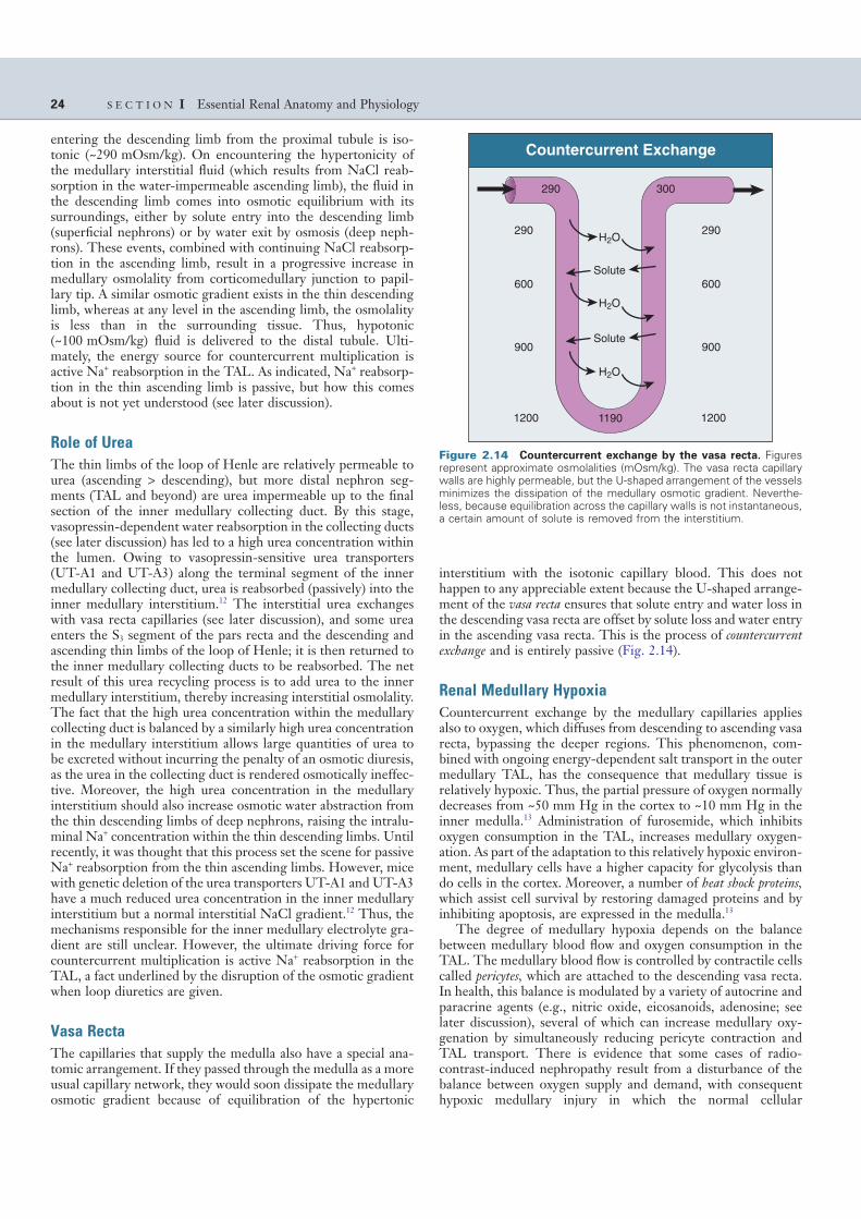
interstitium with the isotonic capillary blood. This does not happen to any appreciable extent because the U-shaped arrange-ment of the vasa recta ensures that solute entry and water loss in the descending vasa recta are offset by solute loss and water entry in the ascending vasa recta. This is the process of countercurrent exchange and is entirely passive (Fig. 2.14).
Renal Medullary Hypoxia
Countercurrent exchange by the medullary capillaries applies also to oxygen, which diffuses from descending to ascending vasa recta, bypassing the deeper regions. This phenomenon, com-bined with ongoing energy-dependent salt transport in the outer medullary TAL, has the consequence that medullary tissue is relatively hypoxic. Thus, the partial pressure of oxygen normally decreases from ~50 mm Hg in the cortex to ~10 mm Hg in the inner medulla.13 Administration of furosemide, which inhibits oxygen consumption in the TAL, increases medullary oxygen-ation. As part of the adaptation to this relatively hypoxic environ-ment, medullary cells have a higher capacity for glycolysis than do cells in the cortex. Moreover, a number of heat shock proteins, which assist cell survival by restoring damaged proteins and by inhibiting apoptosis, are expressed in the medulla.13
The degree of medullary hypoxia depends on the balance between medullary blood flow and oxygen consumption in the TAL. The medullary blood flow is controlled by contractile cells called pericytes, which are attached to the descending vasa recta. In health, this balance is modulated by a variety of autocrine and paracrine agents (e.g., nitric oxide, eicosanoids, adenosine; see later discussion), several of which can increase medullary oxy-genation by simultaneously reducing pericyte contraction and TAL transport. There is evidence that some cases of radio-contrast-induced nephropathy result from a disturbance of the balance between oxygen supply and demand, with consequent hypoxic medullary injury in which the normal cellular
entering the descending limb from the proximal tubule is iso-tonic (~290 mOsm/kg). On encountering the hypertonicity of the medullary interstitial fluid (which results from NaCl reab-sorption in the water-impermeable ascending limb), the fluid in the descending limb comes into osmotic equilibrium with its surroundings, either by solute entry into the descending limb (superficial nephrons) or by water exit by osmosis (deep neph-rons). These events, combined with continuing NaCl reabsorp-tion in the ascending limb, result in a progressive increase in medullary osmolality from corticomedullary junction to papil-lary tip. A similar osmotic gradient exists in the thin descending limb, whereas at any level in the ascending limb, the osmolality is less than in the surrounding tissue. Thus, hypotonic (~100 mOsm/kg) fluid is delivered to the distal tubule. Ulti-mately, the energy source for countercurrent multiplication is active Na+ reabsorption in the TAL. As indicated, Na+ reabsorp-tion in the thin ascending limb is passive, but how this comes about is not yet understood (see later discussion).
Role of Urea
The thin limbs of the loop of Henle are relatively permeable to urea (ascending > descending), but more distal nephron seg-ments (TAL and beyond) are urea impermeable up to the final section of the inner medullary collecting duct. By this stage, vasopressin-dependent water reabsorption in the collecting ducts (see later discussion) has led to a high urea concentration within the lumen. Owing to vasopressin-sensitive urea transporters (UT-A1 and UT-A3) along the terminal segment of the inner medullary collecting duct, urea is reabsorbed (passively) into the inner medullary interstitium.12 The interstitial urea exchanges with vasa recta capillaries (see later discussion), and some urea enters the S3 segment of the pars recta and the descending and ascending thin limbs of the loop of Henle; it is then returned to the inner medullary collecting ducts to be reabsorbed. The net result of this urea recycling process is to add urea to the inner medullary interstitium, thereby increasing interstitial osmolality. The fact that the high urea concentration within the medullary collecting duct is balanced by a similarly high urea concentration in the medullary interstitium allows large quantities of urea to be excreted without incurring the penalty of an osmotic diuresis, as the urea in the collecting duct is rendered osmotically ineffec-tive. Moreover, the high urea concentration in the medullary interstitium should also increase osmotic water abstraction from the thin descending limbs of deep nephrons, raising the intralu-minal Na+ concentration within the thin descending limbs. Until recently, it was thought that this process set the scene for passive Na+ reabsorption from the thin ascending limbs. However, mice with genetic deletion of the urea transporters UT-A1 and UT-A3 have a much reduced urea concentration in the inner medullary interstitium but a normal interstitial NaCl gradient.12 Thus, the mechanisms responsible for the inner medullary electrolyte gra-dient are still unclear. However, the ultimate driving force for countercurrent multiplication is active Na+ reabsorption in the TAL, a fact underlined by the disruption of the osmotic gradient when loop diuretics are given.
Vasa Recta
The capillaries that supply the medulla also have a special ana-tomic arrangement. If they passed through the medulla as a more usual capillary network, they would soon dissipate the medullary osmotic gradient because of equilibration of the hypertonic
Figure 2.14 Countercurrent exchange by the vasa recta. Figures represent approximate osmolalities (mOsm/kg). The vasa recta capillary walls are highly permeable, but the U-shaped arrangement of the vessels minimizes the dissipation of the medullary osmotic gradient. Neverthe-less, because equilibration across the capillary walls is not instantaneous, a certain amount of solute is removed from the interstitium.
290 300
1190
H2O
H2O
H2O
290
600
900
1200
290
600
900
1200
Solute
Solute
Countercurrent Exchange

C H A P T E R 2 Renal Physiology 25
nephrons; it is largely responsible for the permanently high water permeability of these segments. Aquaporin 3 is constitutively expressed in the basolateral membrane of CNT cells and cortical and outer medullary principal cells, and aquaporin 4 is constitu-tively expressed in the basolateral membrane of outer medullary principal cells and inner medullary collecting duct cells; but it is aquaporin 2 that is responsible for the variable water permeabil-ity of the late distal tubule and collecting duct. Acute vasopressin release causes shuttling of aquaporin 2 from intracellular vesicles to the apical membrane, while chronically raised vasopressin levels increase aquaporin 2 expression. The apical insertion of aquaporin 2 allows reabsorption of water, driven by the high interstitial osmolality achieved and maintained by the counter-current system. Vasopressin also contributes to the effectiveness of this system by stimulating Na+ reabsorption in the TAL (although this effect may be functionally significant only in rodents16) and urea reabsorption through the UT-A1 and UT-A3 transporters in the inner medullary collecting duct. In the rare autosomal recessive and even rarer autosomal dominant forms of nephrogenic diabetes insipidus, aquaporin 2 is abnormal or fails to translocate to the apical membrane.15
Aquaporin 2 dysfunction also appears to underlie the well-known urinary concentrating defect associated with hypercalce-mia. Increased intraluminal Ca2+ concentrations, acting through an apically located calcium-sensing receptor, interfere with the insertion of aquaporin 2 channels in the apical membrane of the medullary collecting duct.17 In addition, stimulation of a calcium receptor in the basolateral membrane of the TAL inhibits solute transport in this nephron segment (through inhibition of the apical NKCC-2 and potassium channels), thereby reducing the medullary osmotic gradient.18
INTEGRATED CONTROL OF RENAL FUNCTION
One of the major functions of the kidneys is the regulation of blood volume, through the regulation of effective circulating volume, an unmeasurable, conceptual volume that reflects the degree of fullness of the vasculature. This is achieved largely by control of the sodium content of the body. The mechanisms involved in the regulation of effective circulating volume are discussed in detail in Chapter 7. Some of the more important mediator systems are introduced here.
Renal Interstitial Hydrostatic Pressure and Nitric Oxide
Acute increases in arterial blood pressure lead to natriuresis (pres-sure natriuresis). Because autoregulation is not perfect, part of this response is mediated by increases in RBF and GFR (see Fig. 2.3), but the main cause is reduced tubular reabsorption, which appears to result largely from an increase in renal interstitial hydrostatic pressure (RIHP). An elevated RIHP could reduce net reabsorption in the proximal tubule by increasing paracellular backflux through the tight junctions of the tubular wall (see Fig. 2.11). The increase in RIHP is thought to be dependent on intrarenally produced nitric oxide.19 Moreover, increased nitric oxide production in macula densa cells (which contain the neu-ronal [type I] isoform of nitric oxide synthase [nNOS]) blunts the sensitivity of TGF, thereby allowing increased NaCl delivery to the distal nephron without incurring a TGF-mediated decrease in GFR.20
Another renal action of nitric oxide results from the presence of inducible (type II) nitric oxide synthase in glomerular
adaptations are overwhelmed, with subsequent apoptotic and necrotic cell death.
VASOPRESSIN (ANTIDIURETIC HORMONE) AND WATER REABSORPTION
Vasopressin, or antidiuretic hormone, is a nonapeptide synthe-sized in specialized neurons of the supraoptic and paraventricular nuclei. It is transported from these nuclei to the posterior pitu-itary and released in response to increases in plasma osmolality and decreases in blood pressure. Osmoreceptors are found in the hypothalamus, and there is also input to this region from arterial baroreceptors and atrial stretch receptors. The actions of vaso-pressin are mediated by three receptor subtypes: V1a, V1b, and V2 receptors. V1a receptors are found in vascular smooth muscle and are coupled to the phosphoinositol pathway; they cause an increase in intracellular Ca2+, resulting in contraction. V1a recep-tors have also been identified in the apical membrane of several nephron segments, although their role is not yet clear. V1b recep-tors are found in the anterior pituitary, where vasopressin modu-lates adrenocorticotropic hormone release. V2 receptors are found in the basolateral membrane of principal cells in the late distal tubule and the whole length of the collecting duct; they are coupled by a Gs protein to cyclic adenosine monophosphate generation, which ultimately leads to the insertion of water chan-nels (aquaporins) into the apical membrane of this otherwise water-impermeable segment (Fig. 2.15). In the X-linked form of nephrogenic diabetes insipidus (the most common hereditary cause), the V2 receptor is defective.14
Several aquaporins have been identified in the kidney.15 Aqua-porin 1 is found in apical and basolateral membranes of all proximal tubules and of thin descending limbs of long-looped
Figure 2.15 Mechanism of action of vasopressin (antidiuretic hormone). The hormone binds to V2 receptors on the basolateral mem-brane of collecting duct principal cells and increases intracellular cyclic adenosine monophosphate (cAMP) production, causing, through interme-diate reactions involving protein kinase A, insertion of preformed water channels (aquaporin 2 [AQP2]) into the apical membrane. The water permeability of the basolateral membrane, which contains aquaporins 3 and 4, is permanently high. Therefore, vasopressin secretion allows tran-scellular movement of water from lumen to interstitium. AC, adenylate cyclase.
Action of Vasopressin
InterstitiumLumen Cells
H2O
H2O
Vesiclescontaining
AQP2
AQP2
H2O
H2O
Protein kinase A
ATP
cAMP
AC
H2O
V2 receptor
Vasopressin
AQP4AQP3

26 S E C T I O N I Essential Renal Anatomy and Physiology
two isoforms exist, COX-1 and COX-2, both expressed in the kidney), cytochrome P-450, and lipoxygenase. The major renal eicosanoids produced by the COX system are prostaglandin E2 and prostaglandin I2, both of which are renal vasodilators and act to buffer the effects of renal vasoconstrictor agents such as Ang II and norepinephrine; and thromboxane A2, a vasoconstrictor. Under normal circumstances, prostaglandins E2 and I2 have little effect on renal hemodynamics; but during stressful situations such as hypovolemia, they help protect the kidney from excessive functional changes. Consequently, nonsteroidal anti-inflamma-tory drugs (NSAIDs), which are COX inhibitors, can cause dra-matic falls in GFR. Prostaglandin E2 also has tubular effects, inhibiting Na+ reabsorption in the TAL of the loop of Henle and both Na+ and water reabsorption in the collecting duct.22 Its action in the TAL, together with a dilator effect on vasa recta pericytes, is another paracrine regulatory mechanism that helps protect the renal medulla from hypoxia. This may explain why inhibition of COX-2 can reduce medullary blood flow and cause apoptosis of medullary interstitial cells.
The metabolism of arachidonic acid by renal cytochrome P-450 enzymes yields epoxyeicosatrienoic acids (EETs), 20-hydroxye-icosatetraenoic acid (20-HETE), and dihydroxyeicosatrienoic acids (DHETs). These compounds appear to have a multiplicity of autocrine, paracrine, and second-messenger effects on the renal vasculature and tubules that have not yet been fully unraveled.23 Like prostaglandins, EETs are vasodilator agents, whereas 20-HETE is a potent renal arteriolar constrictor and may be involved in the vasoconstrictor effect of Ang II as well as the TGF mechanism. 20-HETE also constricts vasa recta pericytes and may be involved in the control of medullary blood flow. Some evidence suggests that locally produced 20-HETE and EETs can inhibit sodium reabsorption in the proximal tubule and TAL.24 Indeed, cytochrome P-450 metabolites of arachi-donic acid may contribute to the reduced proximal tubular reab-sorption seen in pressure natriuresis.
The third enzyme system that metabolizes arachidonic acid, the lipoxygenase system, is activated (in leukocytes, mast cells, and macrophages) during inflammation and injury and is not considered here.
COX-2 is present in macula densa cells and has a critical role in the release of renin from juxtaglomerular cells in response to reduced NaCl delivery to the macula densa.22 A low-sodium diet increases COX-2 expression in the macula densa and simultaneously increases renin secretion; the renin response is virtually abolished in COX-2 knockout mice or during pharmacologic inhibition of COX-2. It is likely, there-fore, that the hyporeninemia observed during administration of NSAIDs is largely a consequence of COX-2 inhibition. As well as COX-2, the enzyme prostaglandin E synthase is expressed in macula densa cells, and it is thought that the principal COX-2 product responsible for enhancing renin secretion is prostaglan-din E2, acting on specific receptors that have been identified in juxtaglomerular cells; it is not clear whether prostaglandin I2 is also synthesized in macula densa cells. As already indicated, nNOS (type I) is also present in macula densa cells and produces nitric oxide that blunts TGF.25 Nitric oxide also has a permis-sive role in renin secretion, although the mechanism is not understood. The increase in macula densa COX-2 expression induced by a low-sodium diet is attenuated during admini-stration of selective nNOS inhibitors, which has led to specula-tion that nitric oxide is responsible for the increase in COX-2 activity and the resulting increase in juxtaglomerular renin secretion.26 The established and proposed roles of COX-2 and
mesangial cells: local production of nitric oxide counteracts the mesangial contractile response to agonists such as Ang II and endothelin (see later discussion). Furthermore, nitric oxide may have a role in the regulation of medullary blood flow. Locally synthesized nitric oxide offsets the vasoconstrictor effects of other agents on the pericytes of the descending vasa recta, and it reduces Na+ reabsorption in the TAL; both actions will help protect the renal medulla from hypoxia. Finally, nitric oxide may promote natriuresis and diuresis through direct actions on the renal tubule. Thus, in addition to its effect on the TAL, locally produced nitric oxide inhibits Na+ and water reabsorption in the collecting duct.21
Renal Sympathetic Nerves
Reductions in arterial pressure or central venous pressure result in reduced afferent signaling from arterial baroreceptors or atrial volume receptors, which elicits a reflex increase in renal sympa-thetic nervous discharge. This reduces urinary sodium excretion in at least three ways:
■ Constriction of afferent and efferent arterioles (predomi-nantly afferent), thereby directly reducing RBF and GFR, and indirectly reducing RIHP.
■ Direct stimulation of sodium reabsorption in the proximal tubule and the TAL of the loop of Henle.
■ Stimulation of renin secretion by afferent arteriolar cells (see later discussion).
Renin-Angiotensin-Aldosterone System
The renin-angiotensin-aldosterone system (RAAS) is central to the control of extracellular fluid volume (ECFV) and blood pres-sure. Renin is synthesized and stored in specialized afferent arte-riolar cells that form part of the juxtaglomerular apparatus (see Fig. 2.4) and is released into the circulation in response to
■ Increased renal sympathetic nervous discharge.■ Reduced stretch of the afferent arteriole after a reduction
in renal perfusion pressure.■ Reduced delivery of NaCl to the macula densa region of
the nephron.Renin catalyzes the production of the decapeptide angiotensin
I from circulating angiotensinogen (synthesized in the liver); angiotensin I is in turn converted to the octapeptide Ang II by the ubiquitous angiotensin-converting enzyme. Ang II has a number of actions pertinent to the control of ECFV and blood pressure:
■ It causes general arteriolar vasoconstriction, including renal afferent and (particularly) efferent arterioles, thereby increasing arterial pressure but reducing RBF. The ten-dency of Pgc to increase is offset by Ang II–induced mesan-gial cell contraction and reduced Kf; thus, the overall effect on GFR is unpredictable.
■ It directly stimulates sodium reabsorption in the proximal tubule.
■ It stimulates aldosterone secretion from the zona glomeru-losa of the adrenal cortex. As described earlier, aldosterone stimulates sodium reabsorption in the distal tubule and collecting duct.
Eicosanoids
Eicosanoids are a family of metabolites of arachidonic acid pro-duced enzymatically by three systems: cyclooxygenase (of which

C H A P T E R 2 Renal Physiology 27
excretion. ET-1 levels are highest in the renal medulla—in the TAL and, more prominently, the inner medullary collecting duct. The distribution of renal endothelin receptors (ETA and ETB receptors) reflects the sites of production; the predominant receptor in the inner medulla is ETB.28 Mice with collecting duct–specific deletions of either ET-1 or ETB receptors exhibit salt-sensitive hypertension, whereas collecting duct–specific ETA receptor deletion results in no obvious renal phenotype.21 ET-1 knockout mice also show a greater sensitivity to vasopressin than do wild-type mice. There is mounting evidence that the natri-uretic and diuretic effects of medullary ETB receptor stimulation are mediated by nitric oxide.21 Taken together with evidence that ET-1 can inhibit Na+ reabsorption in the medullary TAL (also likely to be mediated by nitric oxide), these findings highlight the potential importance of ET-1 and nitric oxide interactions in the control of Na+ and water excretion.
Purines
There is increasing evidence that extracellular purines (e.g., ATP, adenosine diphosphate [ADP], adenosine, uric acid) can act as autocrine or paracrine agents within the kidneys. Purinoceptors are subdivided into P1 and P2 receptors. The P1 receptors are responsive to adenosine and are more usually known as adenos-ine receptors (A1, A2a, A2b, and A3); the P2 receptors, responsive to nucleotides (e.g., ATP and ADP), are further subdivided into P2X (ligand-gated ion channels) and P2Y (metabotropic) recep-tors, each category having a number of subtypes. As indicated earlier, A1 and P2X1 receptors are found in afferent arterioles and mediate vasoconstriction. Purinoceptors are also found in the apical and basolateral membranes of renal tubular cells. Stimula-tion of A1 receptors enhances proximal tubular reabsorption and inhibits collecting duct Na+ reabsorption, whereas stimulation of P2 receptors generally has an inhibitory effect on tubular trans-port.29 Thus, luminally applied nucleotides, acting on a variety of P2 receptor subtypes, can inhibit Na+ reabsorption in the proximal tubule, distal tubule, and collecting duct30; and stimula-tion of P2Y2 receptors in the collecting duct inhibits vasopressin-sensitive water reabsorption; an observation reinforced by the report of increased concentrating ability in P2Y2 receptor knock-out mice.31 Despite these clear indications of tubular effects of nucleotides, further studies will be necessary before their roles in normal tubular physiology are clarified.
Finally, there is some evidence that the end product of purine metabolism, uric acid, may cause renal vasoconstriction, possibly by inhibiting endothelial release of nitric oxide and stimulation of renin.32
R E F E R E N C E S
1. Haraldsson B, Nyström J, Deen WM. Properties of the glomerular barrier and mechanisms of proteinuria. Physiol Rev. 2008;88:451-487.
2. Persson PB. Renal blood flow autoregulation in blood pressure control. Curr Opin Nephrol Hypertens. 2002;11:67-72.
3. Bell PD, Komlosi P, Zhang Z. ATP as a mediator of macula densa cell signalling. Purinergic Signal. 2009;5:461-471.
4. Inscho EW. ATP, P2 receptors and the renal microcirculation. Purinergic Signal. 2009;5:447-460.
5. Shirley DG, Walter SJ. A micropuncture study of the renal response to haemorrhage in rats: Assessment of the role of vasopressin. Exp Physiol. 1995;80:619-630.
6. Skou JC. The influence of some cations on an adenosine triphosphatase from peripheral nerves. Biochim Biophys Acta. 1957;23:394-401.
7. Du Z, Yan Q, Duan Y, et al. Axial flow modulates proximal tubule NHE3 and H-ATPase activities by changing microvillus bending moments. Am J Physiol Renal Physiol. 2006;290:F289-F296.
nNOS in the macula densa are shown diagrammatically in Figure 2.16.
Atrial Natriuretic Peptide
If blood volume increases significantly, the resulting atrial stretch stimulates the release of atrial natriuretic peptide from atrial myo-cytes. This hormone increases sodium excretion, partly by sup-pression of renin and aldosterone release and partly by a direct inhibitory effect on sodium reabsorption in the medullary col-lecting duct. Atrial natriuretic peptide may additionally increase GFR because high doses cause afferent arteriolar vasodilation and mesangial cell relaxation (thus increasing Kf; see Fig. 2.6).
Endothelins
Endothelins are potent vasoconstrictor peptides to which the renal vasculature is exquisitely sensitive.27 They function primarily as autocrine or paracrine agents. The kidney is a rich source of endothelins, the predominant isoform being endothelin 1 (ET-1). ET-1 is generated throughout the renal vasculature, including afferent and efferent arterioles (where it causes vasoconstriction, possibly mediated by 20-HETE) and mesangial cells (where it causes contraction, i.e., decreases Kf). Consequently, renal ET-1 can cause profound reductions in RBF and GFR (Fig. 2.6).
In contrast to its effect on GFR, it is now clear that ET-1 can act on the renal tubule to increase urinary Na+ and water
Figure 2.16 Interactions between macula densa and afferent arte-riole: proposed mediators of renin secretion and tubuloglomerular feedback. Both cyclooxygenase 2 (COX-2) and neuronal nitric oxide synthase (nNOS) enzyme systems are present in macula densa cells. Increased NaCl delivery to the macula densa stimulates NaCl entry into the cells through the Na+-K+-2Cl− cotransporter. This causes afferent arteriolar constriction through adenosine or adenosine triphosphate (ATP), and also inhibits COX-2 activity; the latter effect might be mediated partly through inhibition of (nNOS-mediated) nitric oxide (NO) production. Generation of prostaglandin E2 by COX-2 stimulates renin release. Pros-taglandin E2 (PGE2) also modulates vasoconstriction, as does nitric oxide.
!
!
! !?
?
?
Interactions Between Macula Densa
and Afferent Arteriole
K+
K+
Na+
ATP COX-2nNOS
NOATP
Adenosine
Afferent arteriolargranular cell
Macula densa cell
Tubular lumen
VasoconstrictionRenin
2Cl−
PGE2
+
+
+
+
+ ++

28 S E C T I O N I Essential Renal Anatomy and Physiology
21. Pollock JS, Pollock DM. Endothelin and NOS1/nitric oxide signaling and regulation of sodium homeostasis. Curr Opin Nephrol Hypertens. 2008;17:70-75.
22. Hao C-M, Breyer MD. Physiological regulation of prostaglandins in the kidney. Annu Rev Physiol. 2008;70:357-377.
23. Maier KG, Roman RJ. Cytochrome P450 metabolites of arachidonic acid in the control of renal function. Curr Opin Nephrol Hypertens. 2001;10:81-87.
24. Sarkis A, Lopez B, Roman RJ. Role of 20-hydroxyeicosatetraenoic acid and epoxyeicosatrienoic acids in hypertension. Curr Opin Nephrol Hyper-tens. 2004;13:205-214.
25. Vallon V. Tubuloglomerular feedback in the kidney: Insights from gene-targeted mice. Pflugers Arch. 2003;445:470-476.
26. Welch WJ, Wilcox CS. What is brain nitric oxide doing in the kidney? Curr Opin Nephrol Hypertens. 2002;11:109-115.
27. Kohan DE. Endothelins in the normal and diseased kidney. Am J Kidney Dis. 1997;29:2-26.
28. Kohan DE. The renal medullary endothelin system in control of sodium and water excretion and systemic blood pressure. Curr Opin Nephrol Hypertens. 2006;15:34-40.
29. Bailey MA, Shirley DG, King BF, et al. Extracellular nucleotides and renal function. In: Alpern RJ, Hebert SC, eds. The Kidney: Physiology and Pathophysiology. 4th ed. Amsterdam: Elsevier; 2008:425-442.
30. Bailey MA, Shirley DG. Effects of extracellular nucleotides on renal tubular solute transport. Purinergic Signal. 2009;5:473-480.
31. Zhang Y, Sands JM, Kohan DE, et al. Potential role of purinergic signal-ing in urinary concentration in inner medulla: Insights from P2Y2 receptor gene knockout mice. Am J Physiol Renal Physiol. 2008;295:F1715-F1724.
32. Sanchez-Lozada LG, Tapia E, Santamaria J, et al. Mild hyperuricemia induces severe cortical vasoconstriction and perpetuates glomerular hypertension in normal rats and in experimental chronic renal failure. Kidney Int. 2005;67:237-247.
8. Meneton P, Loffing J, Warnock DG. Sodium and potassium handling by the aldosterone-sensitive distal nephron: The pivotal role of the distal and connecting tubule. Am J Physiol Renal Physiol. 2004;287:F593-F601.
9. Bailey MA, Unwin RJ, Shirley DG. In vivo inhibition of renal 11β-hydroxysteroid dehydrogenase in the rat stimulates collecting duct sodium reabsorption. Clin Sci. 2001;101:195-198.
10. Zhai X-Y, Fenton RA, Andreasen A, et al. Aquaporin-1 is not expressed in descending thin limbs of short-loop nephrons. J Am Soc Nephrol. 2007;18:2937-2944.
11. Greger R. Ion transport mechanisms in thick ascending limb of Henle’s loop of mammalian nephron. Physiol Rev. 1985;65:760-795.
12. Fenton RA, Knepper MA. Mouse models and the urinary concentrating mechanism in the new millennium. Physiol Rev. 2007;87:1083-1112.
13. Neuhofer W, Beck F-X. Cell survival in the hostile environment of the renal medulla. Annu Rev Physiol. 2005;67:531-555.
14. Rosenthal W, Seibold A, Antaramian A, et al. Molecular identification of the gene responsible for congenital nephrogenic diabetes insipidus. Nature. 1992;359:233-235.
15. Nielsen S, Frøkiær J, Marples D, et al. Aquaporins in the kidney: From molecules to medicine. Physiol Rev. 2002;82:205-244.
16. Bankir L. Antidiuretic action of vasopressin: Quantitative aspects and interaction between V1a and V2 receptor–mediated effects. Cardiovasc Res. 2001;51:372-390.
17. Valenti G, Procino G, Tamma G, et al. Aquaporin 2 trafficking. Endo-crinology. 2005;146:5063-5070.
18. Ward DT, Riccardi D. Renal physiology of the extracellular calcium-sensing receptor. Pflugers Arch. 2002;445:169-176.
19. Nakamura T, Alberola AM, Salazar FJ, et al. Effects of renal perfusion pressure on renal interstitial hydrostatic pressure and Na+ excretion: Role of endothelium-derived nitric oxide. Nephron. 1998;78:104-111.
20. Thorup C, Persson AEG. Macula densa derived nitric oxide in regulation of glomerular capillary pressure. Kidney Int. 1996;49:430-436.