physiology of big hemispheres. functions of the basal ganglia these ganglia perform essentially all...
TRANSCRIPT

Physiology of big Physiology of big hemisphereshemispheres


FUNCTIONS OF THE BASAL FUNCTIONS OF THE BASAL GANGLIAGANGLIA
These ganglia perform essentially all the motor These ganglia perform essentially all the motor functions, even controlling the voluntary functions, even controlling the voluntary movements in much the same manner that the motor movements in much the same manner that the motor cortex of the human being controls voluntary cortex of the human being controls voluntary movements. Further more, in the cat, and to a lesser movements. Further more, in the cat, and to a lesser extent in the dog, decortication removes only the extent in the dog, decortication removes only the discrete types of motor functions and does not discrete types of motor functions and does not interfere with the animal's ability to walk, eat, fight, interfere with the animal's ability to walk, eat, fight, develop rage, have periodic sleep and wakefulness, develop rage, have periodic sleep and wakefulness, and even participate naturally in sexual activities. and even participate naturally in sexual activities.


Cycle of putamenCycle of putamenProvide an action of separate movement, Provide an action of separate movement,
which need previous studyingwhich need previous studying..
It starts from premotor and addition motor zones It starts from premotor and addition motor zones of cortex go to putamen, than to globus pallidus. of cortex go to putamen, than to globus pallidus. After that can be two ways: After that can be two ways:
1) from globus pallidus impulses go to thalamus; 1) from globus pallidus impulses go to thalamus; 2) from globus pallidus impulses go to substantia 2) from globus pallidus impulses go to substantia nigra and than to thalamus. nigra and than to thalamus.
From thalamus impulses go to primary motor From thalamus impulses go to primary motor zone.zone.

Cycle of caudate nucleusCycle of caudate nucleusRegulate the moving behaviorRegulate the moving behavior (the model (the model
of movement). of movement). It starts from associative zones of cortex go to It starts from associative zones of cortex go to caudate nucleus.caudate nucleus.
After that can be two ways:After that can be two ways:
1) from caudate nucleus impulses go to globus 1) from caudate nucleus impulses go to globus pallidus; pallidus;
2) from caudate nucleus impulses go to putamen and 2) from caudate nucleus impulses go to putamen and than to globus pallidus. than to globus pallidus.
From globus pallidus impulses go to thalamus and From globus pallidus impulses go to thalamus and than to premotor and addition motor zones.than to premotor and addition motor zones.

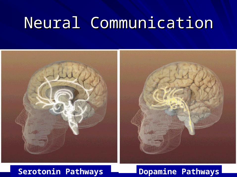
Neural CommunicationNeural Communication
Dopamine PathwaysSerotonin Pathways
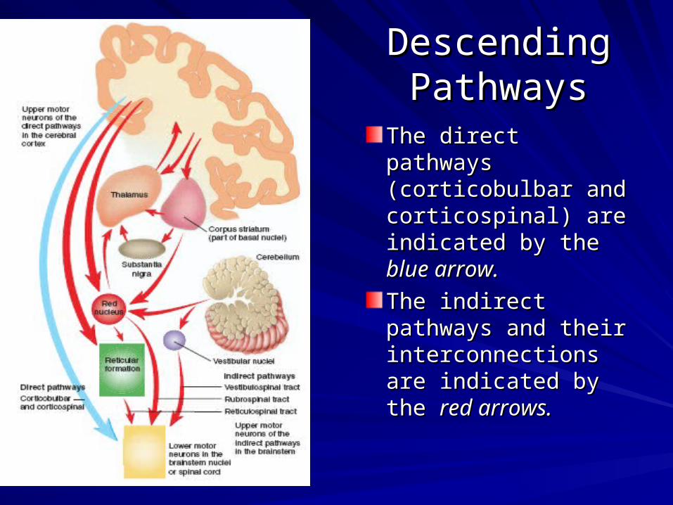
Descending Descending PathwaysPathways
The direct pathways The direct pathways (corticobulbar and (corticobulbar and corticospinal) are corticospinal) are indicated by theindicated by the blue blue arrow. arrow.
The indirect The indirect pathways and their pathways and their interconnections are interconnections are indicated byindicated by the the red red arrows.arrows.


Posterior Posterior Spinocerebellar TractSpinocerebellar Tract
This tract transmits This tract transmits proprioceptive proprioceptive information from information from the thorax, upper the thorax, upper limbs,limbs, and upper and upper lumbar region to lumbar region to the cerebellum. the cerebellum. Lines on the inset Lines on the inset indicate levelsindicate levels of of section.section.

CerebellumCerebellum
Connected to brainstem by cerebellar pedunclesConnected to brainstem by cerebellar peduncles
White matter (arbor vitae) visible in sagittal White matter (arbor vitae) visible in sagittal sectionsection
Sits atop the 4th ventricleSits atop the 4th ventricle

FUNCTION OF CEREBELLUMFUNCTION OF CEREBELLUM
1. Regulation of posture and 1. Regulation of posture and equilibrium, and muscle tone equilibrium, and muscle tone
2. Coordination of posture and slow 2. Coordination of posture and slow determined movementsdetermined movements
3. Coordination of fast determined 3. Coordination of fast determined movementsmovements
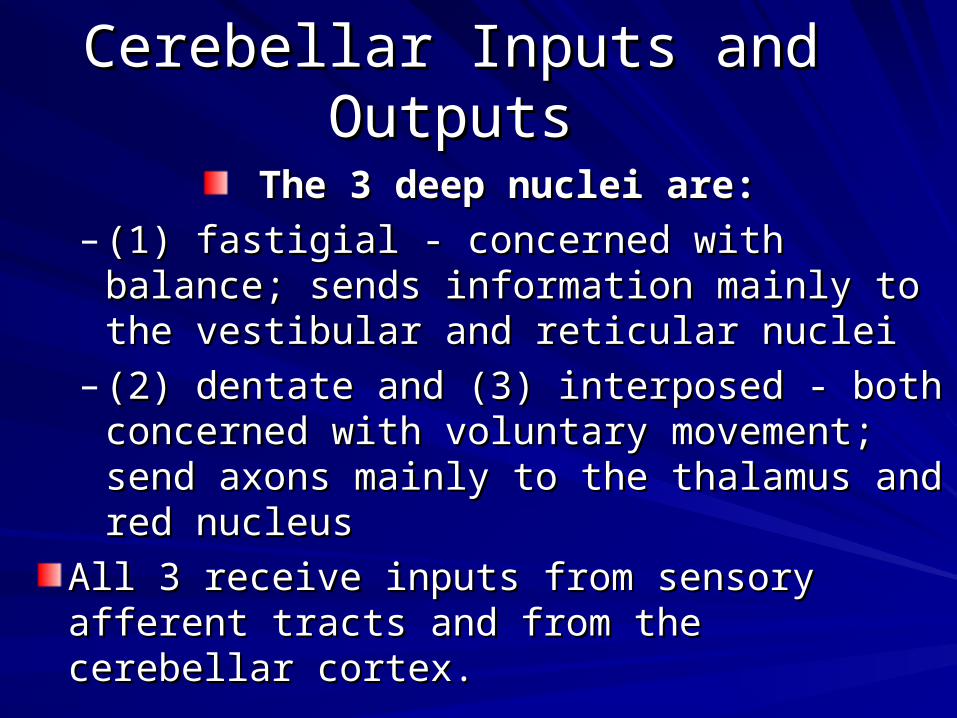
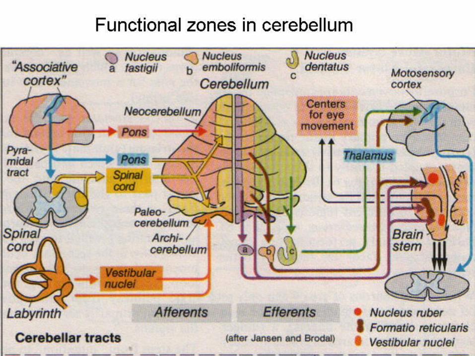
Cerebellar Inputs and OutputsCerebellar Inputs and Outputs
The 3 deep nuclei are:The 3 deep nuclei are:
– (1) fastigial - concerned with balance; sends (1) fastigial - concerned with balance; sends information mainly to the vestibular and information mainly to the vestibular and reticular nucleireticular nuclei
– (2) dentate and (3) interposed - both concerned (2) dentate and (3) interposed - both concerned with voluntary movement; send axons mainly to with voluntary movement; send axons mainly to the thalamus and red nucleusthe thalamus and red nucleus
All 3 receive inputs from sensory afferent tracts All 3 receive inputs from sensory afferent tracts and from the cerebellar cortex.and from the cerebellar cortex.

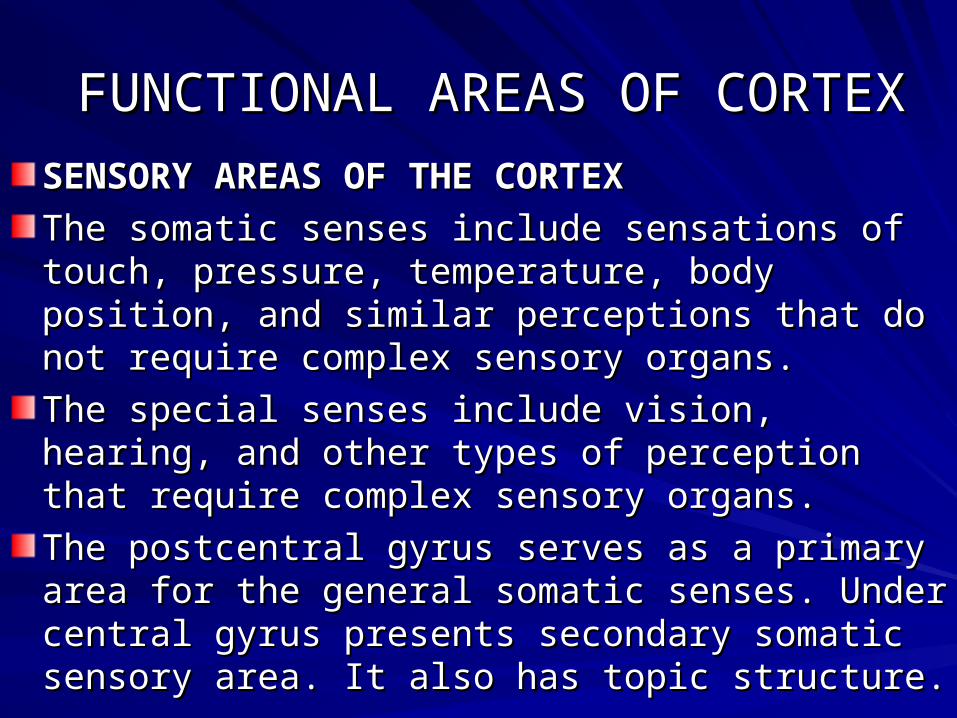
FUNCTIONAL AREAS OF CORTEXFUNCTIONAL AREAS OF CORTEX
SENSORY AREAS OF THE CORTEXSENSORY AREAS OF THE CORTEX
The somatic senses include sensations of touch, The somatic senses include sensations of touch, pressure, temperature, body position, and similar pressure, temperature, body position, and similar perceptions that do not require complex sensory organs.perceptions that do not require complex sensory organs.
The special senses include vision, hearing, and other The special senses include vision, hearing, and other types of perception that require complex sensory types of perception that require complex sensory organs.organs.
The postcentral gyrus serves as a primary area for the The postcentral gyrus serves as a primary area for the general somatic senses. Under central gyrus presents general somatic senses. Under central gyrus presents secondary somatic sensory area. It also has topic secondary somatic sensory area. It also has topic structure.structure.

Functional Regions of the Lateral Functional Regions of the Lateral Side of the Left Cerebral CortexSide of the Left Cerebral Cortex
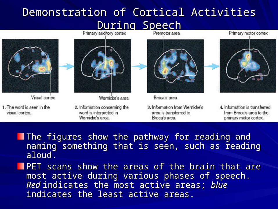
Demonstration of Cortical Activities During SpeechDemonstration of Cortical Activities During Speech
The figures show the pathway for reading and naming The figures show the pathway for reading and naming something that is seen, such as reading aloud. something that is seen, such as reading aloud. PET scans show the areas of the brain that are most PET scans show the areas of the brain that are most activeactive during various phases of speech. during various phases of speech. Red Red indicates indicates the most active areas; the most active areas; blue blue indicates the least active indicates the least active areas.areas.

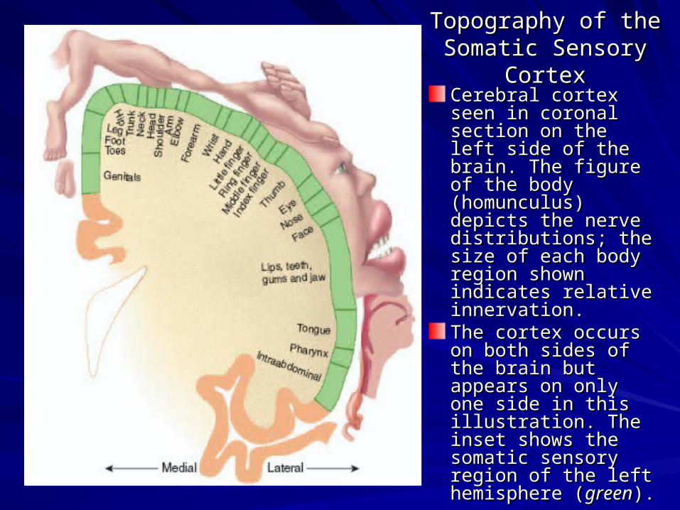
Topography of the Topography of the Somatic Sensory CortexSomatic Sensory Cortex
Cerebral cortex seen Cerebral cortex seen in coronal section on in coronal section on the left side of the the left side of the brain. The figurebrain. The figure of of the body the body (homunculus) depicts (homunculus) depicts the nerve the nerve distributions; the size distributions; the size of eachof each body region body region shown indicates shown indicates relative innervation. relative innervation. The cortex occurs on The cortex occurs on bothboth sides of the brain sides of the brain but appears on only but appears on only one side in this one side in this illustration. The insetillustration. The inset shows the somatic shows the somatic sensory region of the sensory region of the left hemisphere left hemisphere ((greengreen).).

Topography of the Topography of the Primary Motor CortexPrimary Motor Cortex
Cerebral cortex Cerebral cortex seen in coronal seen in coronal section on the section on the left side of the left side of the brain. brain. The figureThe figure of the of the body body (homunculus) (homunculus) depicts the nerve depicts the nerve distributions; the distributions; the size of eachsize of each body region body region shown indicates shown indicates relative relative innervation.innervation.


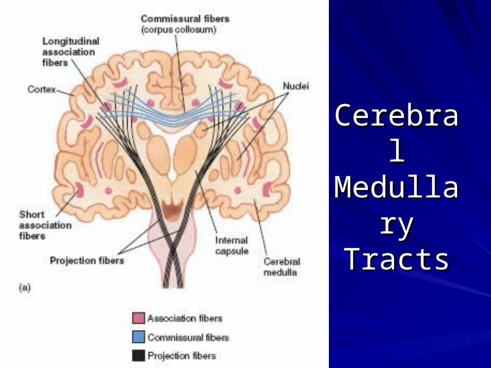
Cerebral Cerebral Medullary Medullary
TractsTracts


The (a) alpha and (b) beta rhythms The (a) alpha and (b) beta rhythms of the of the EEGEEG..

Effects of Aging on theEffects of Aging on the Nervous SystemNervous System
As a person ages, there’s a gradual decline in sensory As a person ages, there’s a gradual decline in sensory functionfunction because the number of sensory neurons declines, because the number of sensory neurons declines, the functionthe function of remaining neurons decreases, and CNS of remaining neurons decreases, and CNS processing decreases. processing decreases. InIn the skin, free nerve endings and hair follicle receptors the skin, free nerve endings and hair follicle receptors remainremain largely unchanged with age. Meissner’s corpuscles largely unchanged with age. Meissner’s corpuscles and pacinianand pacinian corpuscles, however, decrease in number. corpuscles, however, decrease in number. The capsules of thoseThe capsules of those that remain become thicker and that remain become thicker and structurally distorted and, therefore,structurally distorted and, therefore, exhibit reduced exhibit reduced function. As a result of these changes infunction. As a result of these changes in Meissner’s Meissner’s corpuscles and pacinian corpuscles, elderly people arecorpuscles and pacinian corpuscles, elderly people are less conscious of something touching or pressing on the less conscious of something touching or pressing on the skin, haveskin, have a decreased sense of two-point discrimination, a decreased sense of two-point discrimination, and have a moreand have a more difficult time identifying objects by touch. difficult time identifying objects by touch. These functionalThese functional changes leave elderly people more prone changes leave elderly people more prone to skin injuries and with ato skin injuries and with a greater sense of isolation.greater sense of isolation.