physical review e99, 022416 (2019) - … · physical review e99, 022416 (2019) void distributions...
TRANSCRIPT

PHYSICAL REVIEW E 99, 022416 (2019)
Void distributions reveal structural link between jammed packings and protein cores
John D. Treado,1,2 Zhe Mei,2,3 Lynne Regan,4,3,2,* and Corey S. O’Hern1,2,5,6,7,†
1Department of Mechanical Engineering & Materials Science, Yale University, New Haven, Connecticut 06520, USA2Integrated Graduate Program in Physical & Engineering Biology, Yale University, New Haven, Connecticut 06520, USA
3Department of Chemistry, Yale University, New Haven, Connecticut 06520, USA4Department of Molecular Biophysics & Biochemistry, Yale University, New Haven, Connecticut 06520, USA
5Department of Physics, Yale University, New Haven, Connecticut 06520, USA6Department of Applied Physics, Yale University, New Haven, Connecticut 06520, USA
7Program in Computational Biology and Bioinformatics, Yale University, New Haven, Connecticut 06520, USA
(Received 31 October 2018; published 21 February 2019)
Dense packing of hydrophobic residues in the cores of globular proteins determines their stability. Recently,we have shown that protein cores possess packing fraction φ ≈ 0.56, which is the same as dense, random packingof amino-acid-shaped particles. In this article, we compare the structural properties of protein cores and jammedpackings of amino-acid-shaped particles in much greater depth by measuring their local and connected voidregions. We find that the distributions of surface Voronoi cell volumes and local porosities obey similar statisticsin both systems. We also measure the probability that accessible, connected void regions percolate as a functionof the size of a spherical probe particle and show that both systems possess the same critical probe size. Wemeasure the critical exponent τ that characterizes the size distribution of connected void clusters at the onset ofpercolation. We find that the cluster size statistics are similar for void percolation in packings of amino-acid-shaped particles and randomly placed spheres, but different from that for void percolation in jammed spherepackings. We propose that the connected void regions are a defining structural feature of proteins and can be usedto differentiate experimentally observed proteins from decoy structures that are generated using computationalprotein design software. This work emphasizes that jammed packings of amino-acid-shaped particles can serveas structural and mechanical analogs of protein cores, and could therefore be useful in modeling the response ofprotein cores to cavity-expanding and -reducing mutations.
DOI: 10.1103/PhysRevE.99.022416
I. INTRODUCTION
A significant driving force in protein folding is the seques-tration of hydrophobic amino acids from solvent. Moreover,these buried amino acids are densely packed in the proteincore [1]. In fact, the packing of core residues has been linkeddirectly to protein stability [2]. For example, large-to-smallamino acid mutations, which can increase interior proteincavities, or voids, are known to destabilize proteins whenthey are subjected to hydrostatic pressure [3–5] and chemicaldenaturants [6,7]. Determining the connection between densecore packing and voids is therefore crucial to understandingthe physical origins of protein stability and reliably designingnew protein structures that are stable [8]. However, no suchquantitative understanding yet exists, and it is currently diffi-cult to distinguish computational protein designs that are notstable in experiments from experimentally observed structures[9].
In previous studies [10–12], we found, using collective sidechain repacking, that the side chain conformations of residues
*Present address: Centre for Synthetic and Systems Biology, In-stitute for Quantitative Biology, Biochemistry, and Biotechnology,School of Biological Sciences, Edinburgh University, Scotland, UK.
in protein cores (from a collection of high-resolution pro-tein crystal structures) are uniquely specified by hard-sphere,steric interactions. Moreover, we have shown that, when con-sidering hard-sphere optimized atomic radii, the core regionsin proteins possess the same packing fraction φ ≈ 0.56 as thatfound in simulations of dense, random packings of purelyrepulsive, amino-acid-shaped particles. This result suggeststhat the packing fraction of protein cores is determined by thebumpy and nonsymmetric shape of amino acids, and not bythe backbone or local secondary structure.
However, materials that share the same packing fraction donot necessarily possess the same internal structure. In this arti-cle, we characterize the void space in experimentally obtainedand computationally generated protein cores to further test thegeometric similarities between these two systems. We showbelow that dense random packings of amino-acid-shaped par-ticles have the same local packing fraction, void distribution,and percolation of connected void space as protein cores,which indicates structural equivalence.
Our results suggest that the computationally generatedpackings can be used as mechanical analogs of protein coresto predict their collective mechanical response. Further, ourresults emphasize the connection between structurally ar-rested, yet thermally fluctuating, protein cores and the jam-ming transition of highly nonspherical particles [13]. Al-though the similarity between structural glasses and proteins
2470-0045/2019/99(2)/022416(13) 022416-1 ©2019 American Physical Society
TREADO, MEI, REGAN, AND O’HERN PHYSICAL REVIEW E 99, 022416 (2019)
at low temperatures has been known for several decades[14–18], prior computational studies have mainly focused onthe transition from harmonic to anharmonic conformationalfluctuations on length scales spanning the full protein. Incontrast, our studies identify key structural similarities be-tween jammed packings of amino-acid-shaped particles andthe cores of protein crystal structures.
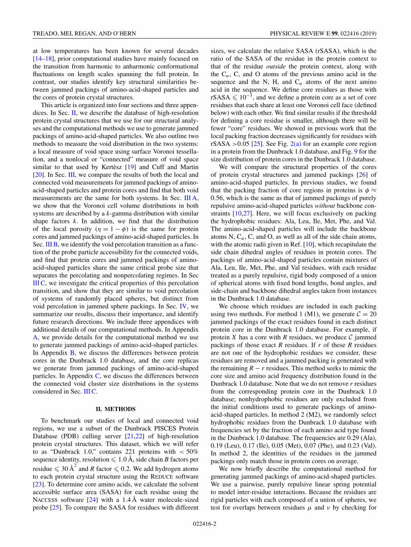
This article is organized into four sections and three appen-dices. In Sec. II, we describe the database of high-resolutionprotein crystal structures that we use for our structural analy-ses and the computational methods we use to generate jammedpackings of amino-acid-shaped particles. We also outline twomethods to measure the void distribution in the two systems:a local measure of void space using surface Voronoi tessella-tion, and a nonlocal or “connected” measure of void spacesimilar to that used by Kertèsz [19] and Cuff and Martin[20]. In Sec. III, we compare the results of both the local andconnected void measurements for jammed packings of amino-acid-shaped particles and protein cores and find that both voidmeasurements are the same for both systems. In Sec. III A,we show that the Voronoi cell volume distributions in bothsystems are described by a k-gamma distribution with similarshape factors k. In addition, we find that the distributionof the local porosity (η = 1 − φ) is the same for proteincores and jammed packings of amino-acid-shaped particles. InSec. III B, we identify the void percolation transition as a func-tion of the probe particle accessibility for the connected voids,and find that protein cores and jammed packings of amino-acid-shaped particles share the same critical probe size thatseparates the percolating and nonpercolating regimes. In SecIII C, we investigate the critical properties of this percolationtransition, and show that they are similar to void percolationof systems of randomly placed spheres, but distinct fromvoid percolation in jammed sphere packings. In Sec. IV, wesummarize our results, discuss their importance, and identifyfuture research directions. We include three appendices withadditional details of our computational methods. In AppendixA, we provide details for the computational method we useto generate jammed packings of amino-acid-shaped particles.In Appendix B, we discuss the differences between proteincores in the Dunbrack 1.0 database, and the core replicaswe generate from jammed packings of amino-acid-shapedparticles. In Appendix C, we discuss the differences betweenthe connected void cluster size distributions in the systemsconsidered in Sec. III C.
II. METHODS
To benchmark our studies of local and connected voidregions, we use a subset of the Dunbrack PISCES ProteinDatabase (PDB) culling server [21,22] of high-resolutionprotein crystal structures. This dataset, which we will referto as “Dunbrack 1.0,” contains 221 proteins with < 50%sequence identity, resolution � 1.0 Å, side chain B factors per
residue � 30 Å2
and R factor � 0.2. We add hydrogen atomsto each protein crystal structure using the REDUCE software[23]. To determine core amino acids, we calculate the solventaccessible surface area (SASA) for each residue using theNACCESS software [24] with a 1.4 Å water molecule-sizedprobe [25]. To compare the SASA for residues with different
sizes, we calculate the relative SASA (rSASA), which is theratio of the SASA of the residue in the protein context tothat of the residue outside the protein context, along withthe Cα , C, and O atoms of the previous amino acid in thesequence and the N, H, and Cα atoms of the next aminoacid in the sequence. We define core residues as those withrSASA � 10−3, and we define a protein core as a set of coreresidues that each share at least one Voronoi cell face (definedbelow) with each other. We find similar results if the thresholdfor defining a core residue is smaller, although there will befewer “core” residues. We showed in previous work that thelocal packing fraction decreases significantly for residues withrSASA >0.05 [25]. See Fig. 2(a) for an example core regionin a protein from the Dunbrack 1.0 database, and Fig. 9 for thesize distribution of protein cores in the Dunbrack 1.0 database.
We will compare the structural properties of the coresof protein crystal structures and jammed packings [26] ofamino-acid-shaped particles. In previous studies, we foundthat the packing fraction of core regions in proteins is φ ≈0.56, which is the same as that of jammed packings of purelyrepulsive amino-acid-shaped particles without backbone con-straints [10,27]. Here, we will focus exclusively on packingthe hydrophobic residues: Ala, Leu, Ile, Met, Phe, and Val.The amino-acid-shaped particles will include the backboneatoms N, Cα , C, and O, as well as all of the side chain atoms,with the atomic radii given in Ref. [10], which recapitulate theside chain dihedral angles of residues in protein cores. Thepackings of amino-acid-shaped particles contain mixtures ofAla, Leu, Ile, Met, Phe, and Val residues, with each residuetreated as a purely repulsive, rigid body composed of a unionof spherical atoms with fixed bond lengths, bond angles, andside-chain and backbone dihedral angles taken from instancesin the Dunbrack 1.0 database.
We choose which residues are included in each packingusing two methods. For method 1 (M1), we generate C = 20jammed packings of the exact residues found in each distinctprotein core in the Dunbrack 1.0 database. For example, ifprotein X has a core with R residues, we produce C jammedpackings of those exact R residues. If r of these R residuesare not one of the hydrophobic residues we consider, theseresidues are removed and a jammed packing is generated withthe remaining R − r residues. This method seeks to mimic thecore size and amino acid frequency distribution found in theDunbrack 1.0 database. Note that we do not remove r residuesfrom the corresponding protein core in the Dunbrack 1.0database; nonhydrophobic residues are only excluded fromthe initial conditions used to generate packings of amino-acid-shaped particles. In method 2 (M2), we randomly selecthydrophobic residues from the Dunbrack 1.0 database withfrequencies set by the fraction of each amino acid type foundin the Dunbrack 1.0 database. The frequencies are 0.29 (Ala),0.19 (Leu), 0.17 (Ile), 0.05 (Met), 0.07 (Phe), and 0.23 (Val).In method 2, the identities of the residues in the jammedpackings only match those in protein cores on average.
We now briefly describe the computational method forgenerating jammed packings of amino-acid-shaped particles.We use a pairwise, purely repulsive linear spring potentialto model inter-residue interactions. Because the residues arerigid particles with each composed of a union of spheres, wetest for overlaps between residues μ and ν by checking for
022416-2

VOID DISTRIBUTIONS REVEAL STRUCTURAL LINK … PHYSICAL REVIEW E 99, 022416 (2019)
overlaps between all atoms i on residue μ and all atoms jon residue ν, respectively. Note that this potential is isotropicand depends only on the distances between atoms on differentresidues. [See Eq. (A1) in Appendix A.]
We place N residues with random initial positions and ori-entations at packing fraction φ0 = 0.40 in a cubic simulationbox with periodic boundary conditions and then increase thepacking fraction in small steps �φ to isotropically compressthe system. After each compression step, we relax the totalpotential energy using FIRE energy minimization [28]. Thismethod is similar to a “fast” thermal quench that finds thenearest local potential energy minimum. We use quaternionsto track the particle orientations for each residue, as describedin Ref. [29]. If the total potential energy per residue is zero af-ter energy minimization, i.e., U/Nε < 10−8, where ε is the en-ergy scale of the atomic interactions, we continue to increasethe packing fraction. If the total potential energy per residueis nonzero, i.e., U/Nε � 10−8 and residues have small over-laps, we decrease the packing fraction. The packing fractionincrement �φ is halved each time the algorithm switches fromcompression to decompression and vice versa. We terminatethe packing-generation protocol when the residue packingssatisfy 10−8 < U/Nε < 2 × 10⁻⁸ and possess a vanishingkinetic energy per residue (i.e., K/Nε < 10−20) [13]. [Anexample jammed packing of amino-acid-shaped particles isshown in Fig. 2(b) and further computational details areincluded in Appendix A.]
To measure the distribution of local voids in packingsof amino-acid-shaped particles and protein cores, we use aVoronoi tessellation, which ascribes to each particle the regionof space that is closer to that particle than all other particlesin the system. For residues, which are highly nonsphericalparticles, we use a generalization of the standard Voronoitessellation known as the surface- or set-Voronoi (SV) tessel-lation [30]. This tessellation partitions the empty space in thesystem using a bounding surface for each residue. An efficientalgorithm to generate this tessellation is outlined in Ref. [30]and implemented using POMELO [31]. To construct the SVtessellation, consider a set of N particles with bounding sur-faces {∂Kμ} for μ = 1, ..., N . The software approximates ∂Kμ
by triangulating points on the particle surfaces, and uses thestandard Voronoi tessellation of the surface points to constructthe SV cell for each residue μ. We find that using 400 surfacepoints per atom, or ≈6400 surface points per residue, gives anaccurate representation of the SV cell, which does not changesignificantly as more surface points are added. An example SVcell from a packing of amino-acid-shaped particles is shown inFig. 1(a). For an SV cell with volume V v
μ surrounding residueμ with volume vμ, the local porosity is given by
ημ = V vμ − vμ
V vμ
= 1 − φμ, (1)
where φμ = vμ/V vμ is the local packing fraction. This quantity
measures the local void space associated with each residue.We also quantify the “connected” void space shared be-
tween residues in packings of amino-acid-shaped particlesand protein cores. To do this, we implement a grid-basedmethod similar to that described by Kertèsz [19] and Cuffand Martin [20], where the “void space” is defined as the
(a)
(b)
Ala
Ala
Val
Ile
Ile
Leu
FIG. 1. Visualization of (a) local and (b) connected voids fromthe same computationally generated packing of N = 64 amino-acid-shaped particles. Only the central Alanine (Ala) with the neighboringAlanine, 2 Isoleucines (Ile), Leucine (Leu), and Valine (Val) areshown for clarity. The neighboring amino acids share at least onecommon surface Voronoi cell face with the central Ala. In (a),the central Ala is enclosed by its surface Voronoi cell. In (b), theconnected void space is visualized using points on a grid. For clarityonly 75% of the points are shown, and the grid spacing (g = 0.7Å)is large compared to values used in the text. In both (a) and (b), theatoms are colored as follows: C (green), O (red), N (blue), and H(white). See Fig. 8 for visualizations of the connected void spacethroughout the entire simulation domain.
region of a system accessible to a spherical probe particlewith radius a. The geometry and distribution of void spacein a system is thus a function of a, the residue positions�rμ, and bounding surfaces ∂Kμ. We define a cubic latticewith G points in each direction within the simulation domain,which gives a lattice spacing g = L/G. For all lattice pointsp, we define the set of void points V to be all points thatcan accommodate a spherical probe particle with radius awithout causing overlaps with any atoms. We label all voidpoints with a 1, and all other points with a 0. After all gridpoints are labeled, we use the Newman-Ziff algorithm [32] tocluster adjacent, similarly-labeled grid points. We consider alladjacent points on the nearest face, edge, and vertex of a cube
022416-3

TREADO, MEI, REGAN, AND O’HERN PHYSICAL REVIEW E 99, 022416 (2019)
(b)(a)
0 1 2 3 40
0.2
0.4
0.6
0.8
1
1.2
0 50
0.5
1
(a) )c()b(
FIG. 2. (a) Core residues in an example globular protein (PDB code: 3F1L). Noncore regions are drawn using the ribbon representation,and the 11 core amino acids are drawn in all-atom representation. (b) Jammed packing of the same 11 core residues in (a). (c) The surfaceVoronoi cell volume V v distribution plotted as a function of x = (V v − V v
min,α )/(〈V v〉α − V vmin,α ) and fit to a k-gamma distribution (black line)
with k = 6.06 ± 0.08 and 5.29 ± 0.27 for packings of amino-acid-shaped particles (circles) and protein cores (squares), respectively. 〈V v〉α isthe average and V v
min,α is the minimum SV cell volume of residue type α. The inset of (c) is the cumulative distribution function F (x) for thedata in the main panel.
of points surrounding each lattice point (i.e., next-to-next-to-nearest neighbor counting with 26 possible adjacenciesfor each point) when merging void clusters and implementperiodic boundary conditions. A sketch of connected voidlattice points in a subset of a packing of amino-acid-shapedparticles is shown in Fig. 1(b).
When measuring void space in protein structures, we im-plement a similar procedure, but we only consider voids incore residues. We construct a box of dimension Lx × Ly × Lz
that circumscribes each protein core, with the box just outsidethe radii of core residues near the box edges. We pick aspherical probe particle of radius a, and label the void spaceas all points that are (a) not contained inside an atom, and(b) contained only within the union of the SV cells of coreresidues. With these constraints, we only consider connectedvoid space specific to the core of the protein. We then use theNewman-Ziff algorithm to merge void clusters, and repeat theprocedure for 100 different random protein orientations.
III. RESULTS
A. Local void analysis
We begin with an analysis of local voids associated witheach amino acid in jammed packings of amino-acid-shapedparticles and protein cores. We measure the distribution ofthe SV cell volumes and show that the distributions in bothsystems can be fit to a k-gamma distribution, which alsodescribes Voronoi cell distributions in jammed packings ofspheres [33,34], ellipsoids [35], attractive emulsion droplets[36], wet granular materials [37], and model cell monolayers[38]. The k-gamma distribution for the SV cell volume V v
μ foreach residue has the form
P(x) = kk
(k)xk−1 exp(−kx), (2)
where x = (V vμ − V v
min,α )/(〈V vμ 〉α − V v
min,α ), which sets thescale factor of the distribution to 1. Here,
⟨V v
μ
⟩α
= 1
Nα
Nα∑μ=1
V vμ (3)
is the average SV cell volume of residue type α. The sum in-volving μ is over all Nα residues of type α in all packings, andV v
min,α is the minimum SV cell volume of residue type α. Weconsider minima and averages for each residue type separatelyto account for the large differences in residue volumes; that is,each residue type α, when considered individually, has a SVcell volume distribution described by Eq. (2).
We measure the shape factor kα for each residue type α ei-ther by fitting the SV cell volume distribution to Eq. (2) usingMaximum Likelihood Estimation (MLE), or by calculating
kα =(⟨
V vμ
⟩α
− V vmin,α
)2
⟨(V v
μ
)2⟩α
− ⟨V v
μ
⟩2α
. (4)
We obtain similar k values using both methods. Althoughthe values of kα depend on the type of amino acid α, whenwe average the values of kα we recover the value of k ob-tained from fitting the combined distribution. We focus on thedistributions of SV cell volumes averaged over all hydropho-bic residues.
In Fig. 2(c), we show the SV cell volume distributions P(x)for packings of core amino-acid-shaped particles modeledafter specific protein cores (method M1) and for all coreresidues in the Dunbrack 1.0 database. We find that thedistributions for these two systems are similar; both obey ak-gamma distribution [Eq. (2)] with similar shape parameters,k = 6.06 ± 0.08 and k = 5.29 ± 0.27, for core residues in theDunbrack 1.0 database and packings of amino-acid-shapedparticles, respectively. As expected, the cumulative distribu-tions F (x) of the SV cell volumes for residues in protein coresand packings of amino-acid-shaped particles are also nearlyindistinguishable.
The strong similarity between the SV cell volume dis-tributions indicates that jammed packings of amino-acid-shaped particles (at φJ ≈ 0.56) and protein cores possess thesame underlying structure. To better understand this result,in Fig. 3 we plot the shape parameter k that describes theform of the Voronoi cell volume distributions for packingsof N = 103 monodisperse spheres (with φJ ≈ 0.64) and ofN = 64 amino-acid-shaped particles versus φ. When φ �φJ , and the systems are sufficiently dilute, the Voronoi cell
022416-4

VOID DISTRIBUTIONS REVEAL STRUCTURAL LINK … PHYSICAL REVIEW E 99, 022416 (2019)
0 0.1 0.2 0.3 0.4 0.5 0.60
5
10
15
FIG. 3. The shape parameter k for fits of the k-gamma distribu-tion [Eq. (2)] to the SV cell volume distributions P(x) for packingsof amino-acid-shaped particles (open circles), monodisperse spheres(filled circles), and both core and surface residues in the Dunbrack1.0 database (open squares) as a function of packing fraction φ.The dashed horizontal line at k = 5.59 is the analytical value of theshape factor for the Voronoi cell volume distribution of a randomPoisson point process [39], and the dashed vertical line at φJ = 0.56is the packing fraction for protein cores and jammed packings ofamino-acid-shaped particles.
volume distributions of the packings of monodisperse spheresand amino-acid-shaped particles resemble that for a randomPoisson point process [39] with k ≈ 5.6. In this regime, freevolume is assigned randomly to each particle since the particlepositions are uncorrelated. However, as φ increases, the kvalues for packings of monodisperse spheres and amino-acid-shaped particles begin to grow, but at different rates, sincethe particle geometry becomes important in determining thelocal free volume. Near φ � φJ , the shape parameter plateausat k ≈ 13 for packings of monodisperse spheres, but theshape parameter decreases strongly to k ≈ 6 for packings ofamino-acid-shaped particles. This decrease in k indicates atransition from having the shape of the Voronoi cell volumedistribution determined by spherical particles (for φ � φJ )to that determined by bumpy, asymmetric amino-acid-shapedparticles (for φ � φJ ). Note, however, that the SV cell volumedistribution of jammed packings of amino-acid-shaped parti-cles is similar (in terms of k value) to that of randomly placedPoisson points. This suggests that the void distribution ofjammed packings of amino-acid-shaped particles and proteincores share structural properties with randomly placed points.We will expand on this similarity in Sec. III C.
In addition, we calculate k for the SV cell volume dis-tributions for residues in the Dunbrack 1.0 database as afunction of packing fraction. In previous studies, we havefound a one-to-one correlation between solvent accessibilityand packing fraction [25]; residues with lower values of φ inFig. 3 are therefore more solvent-exposed, i.e., closer to theprotein surface. For most of the range in φ, k ≈ 2, whereask � 5.6 for packings of monodisperse spheres and amino-acid-shaped particles. In particular, k does not equal the valuefor a random Poisson point process (k = 5.6) in the limitφ � φJ for residues in protein cores. In protein cores, the
0 1 2 3 40
0.2
0.4
0.6
0.8
1
1.2
0 1 20
0.5
1
FIG. 4. Distribution of the scaled local porosity y = (η −ηmin,α )/(〈η〉α − ηmin,α ), where 〈η〉α is the average and ηmin,α is theminimum porosity of residue type α, for packings of amino-acid-shaped particles (circles) and residues in protein cores in the Dun-brack 1.0 database (squares). The solid line is a Weibull distributionwith shape parameter b ≈ 3.2 [Eq. (7)]. The inset is the cumulativedistribution function F (y) of the data in the main panel.
backbone constraint gives rise to correlations in the residuepositions. However, as φ → φJ , k increases, reaching k ≈ 6when φ = φJ . This result shows that there is a fundamentalchange in the SV cell distribution near the onset of jammingin protein cores. For φ � φJ , the backbone determines theshape of the SV cell volume distribution, whereas for φ → φJ ,the shapes of the amino acids determine the SV cell volumedistribution.
We also compare the local porosity distributions for proteincores and packings of amino-acid-shaped particles in Fig. 4.We scale the porosity [as in Eq. (2)] by defining
y = ημ − ηmin,α
〈ημ〉α − ηmin,α
, (5)
where
〈ημ〉α = 1
Nα
Nα∑μ=1
ημ, (6)
and ηmin,α is the minimum porosity over all Nα core residuesof type α. Again, the porosity distributions P(y) [and cumu-lative distributions F (y)] for residues in protein cores andpackings of amino-acid-shaped particles are similar, but hereP(y) has the shape of a Weibull distribution with scale factorλ = 1,
P(y) = byb−1 exp(−yb), (7)
where b is the shape parameter of the Weibull distribution.The small differences in P(x) and P(y) between core
residues in protein crystal structures and packings of amino-acid-shaped particles can be explained by the small differ-ences between the volumes of core residues in crystal struc-tures and in packings. The atoms on neighboring amino acidsinteract differently for free amino acids in packings versusbackbone atoms in protein cores, which form covalent andhydrogen bonds. Thus, we find that the volumes of residuesin protein cores have larger variances and smaller means than
022416-5

TREADO, MEI, REGAN, AND O’HERN PHYSICAL REVIEW E 99, 022416 (2019)
those in packings of amino-acid-shaped particles. Also, theoverlaps between covalently bonded backbone atoms that linkadjacent residues slightly decreases the mean SV cell volume,which gives rise to a larger population of small SV cells anda small deviation between P(x) for residues in protein coresand in packings for small x in Fig. 2(c).
B. Connected void analysis of protein cores
We next quantify the distribution of “connected” voidspace that is shared between residues. Using a grid-basedmethod, we calculate the volume of regions of connected voidspace as a function of the radius a of a spherical probe particle.As we increase a, the connected void space transitions fromhighly connected throughout the system to compact and lo-calized with distinct void regions. We measure the probabilityρ(a) of finding a percolating void region, where we definepercolation as the appearance of a cluster that spans one ofthe system dimensions when the boundary is closed, and acluster that both spans, wraps around the boundary, and self-intersects when the boundaries are periodic. We identify thecritical probe radius ac by setting ρ(ac) = 0.5. Because thedefinition of connected void regions depends on the boundarycondition, the value of ac, especially in systems as small asprotein cores, is affected by the boundary conditions. Thus,to calculate ρ(a), we create packings of amino-acid-shapedparticles with similar boundary conditions as those in pro-tein cores. From a packing of amino-acid-shaped particleswith periodic boundary conditions (N = 64, method M2), weextract a representative protein core of R − r residues thatall share at least one SV cell face. We sample R − r fromthe distribution of core sizes P(R) found in the Dunbrack1.0 database. (See Fig. 9 in Appendix A.) The resultingpackings have boundary conditions similar to protein cores inthe Dunbrack 1.0 database. We then determine the connectedvoid regions as a function of a and identify the critical probesize ac as shown in Fig. 5(a). We find the same critical probesize ac = 0.48 ± 0.01 Å for both protein cores and packingsof amino-acid-shaped particles with similar boundary condi-tions. Note that this value of the critical probe radius is smallerthan that of a water molecule, which is ≈1.4 Å, and thus thevoids we consider here are not accessible by aqueous solvents.However, as we discuss below, this value of the probe radiuscorresponds to a critical point; we will exploit the behavior ofthe voids near this critical point to understand the geometricproperties of the connected voids, and to differentiate betweenthe voids in various systems.
Thus, determining the connected void regions in proteincores is a type of percolation problem. In lattice site perco-lation, sites on a lattice in d spatial dimensions are eitheroccupied randomly with probability p or not occupied withprobability 1 − p. At the percolation threshold pc, adjacentoccupied sites form a percolating cluster that spans the systemand becomes infinite in the large-system limit. Continuumpercolation occurs in systems that are not confined to alattice. Both particle contact and void percolation have beenstudied in randomly placed overlapping spheres [19,40,41]and percolation of particle contacts [42,43] has been studiedin packings of repulsive [44] and adhesive particles [45].
101 103 105100
101
102
103
104
0 0.2 0.4 0.6 0.8 10
0.2
0.4
0.6
0.8
1(a)
(b)
FIG. 5. (a) Percolation probability ρ(a) plotted versus the proberadius a for protein cores from the Dunbrack 1.0 database (crosses)and clusters of core residues extracted from static packings ofN = 64 amino-acid-shaped particles (circles). The horizontal andvertical dashed lines indicate the critical probe radius ac = 0.48 Åthat satisfies ρ(ac ) = 0.5. (b) Cluster size distribution ns with size sat the critical porosity ηc, which scales as ns(ηc ) ∼ s−τ . The Fisherpower-law exponents τ = 1.95 ± 0.06 and 1.85 ± 0.05 for proteincores from the Dunbrack 1.0 database (crosses) and representativeclusters of core residues in packings of amino-acid-shaped particles(circles), respectively. The solid line has slope equal to −1.85.
In this article, we consider percolation of the void spaceaccessible to a spherical probe particle with radius a inpackings of spheres and amino-acid-shaped particles, as wellas systems composed of randomly placed spheres [40,41]. Asthe probe particle radius is increased, the amount of spaceavailable to the probe is restricted and the number of voidlattice sites decreases. We define an effective porosity η as theratio of the number of void lattice sites to the total numberof lattice sites Gd . We determine the percolation thresholdusing a bisection method, where we begin with two initialguesses for the percolation transition, aH and aL with aH >
aL, and iteratively check for percolation of void sites at theprobe radius a = (aH + aL )/2. We set aH = a if we find apercolated cluster of void sites, and aL = a if we do not finda percolated cluster. We terminate the algorithm when thedifference between successive values for ac is within a smalltolerance δa = 10−8 Å. Note that our use of a lattice of pointsto measure the connected void region does not imply thatour model is a lattice model. The lattice is simply a tool to
022416-6

VOID DISTRIBUTIONS REVEAL STRUCTURAL LINK … PHYSICAL REVIEW E 99, 022416 (2019)
0 0.01 0.02 0.03 0.040.03
0.04
0.05
0.06
0.07
0.08
0.09
FIG. 6. Critical porosity 〈ηc(G)〉 using a lattice with G pointsalong each dimension plotted versus G−1 for jammed packings ofN = 64 amino-acid-shaped particles with Na = 1024 atoms (opencircles), N = 103 randomly placed spheres (open squares), and N =103 monodisperse spheres (filled circles). The dashed lines havevertical intercepts that indicate ηc(G → ∞) ≈ 0.0345, 0.0318, and0.0305 for packings of amino-acid-shaped particles, randomly placedspheres, and monodisperse sphere packings, respectively.
calculate the connected void space volume [19]. Furthermore,in the continuum limit (i.e., G → ∞), we recover the criticalporosity ηc ≈ 0.03 measured using Kerstein’s method [40,41]on systems of randomly placed spheres [46]. (See Fig. 6.)Since there is a one-to-one mapping between a and η, we willuse η as the order parameter for continuum void percolation.
C. Connected void analysis of packings
We now focus on the statistical properties of the connectedvoid regions in packings of spheres and amino-acid-shapedparticles prepared in systems with cubic, periodic boundaries.In Fig. 7 and Table I, we summarize the results of this analysis,and we show visualizations of the percolating connected voidsin jammed packings of spheres and amino-acid-shaped parti-cles in Fig. 8. We first measure the correlation length exponentν, where the correlation length ξ is defined as the averagedistance between two points in the largest connected voidcluster. Near ηc, ξ diverges as |η − ηc|−ν . Using finite-sizescaling [47], we can write
ηc(N ) − ηc(∞) ∼ N−1/dν, (8)
where ηc(∞) is the percolation threshold in the large-systemlimit and N ∼ Ld . ηc(N ) is a random variable with standarddeviation �ηc(N ), which will approach ηc(∞) as N → ∞.Thus, we make the ansatz that
�ηc(N ) ∼ N−1/dν, (9)
which can be used to measure ν. [See Fig. 7(a).] We alsomeasure the Fisher exponent τ , defined by
ns(ηc) ∼ s−τ , (10)
where ns is the number of void clusters containing s sites.While we measure this exponent for protein cores and randompackings with representative boundary conditions in Fig. 5(b),in Fig. 7(b) we measure this exponent in systems with cubic,periodic boundary conditions.
We also measure the fractal dimension and percolationstrength of the percolating void clusters. The fractal dimen-sion is defined by
smax(ηc, N ) ∼ ND/d , (11)
where smax(ηc, N ) is the number of sites contained in thelargest void cluster in the system at percolation onset. IfD = d , the largest void cluster is a compact, nonfractal object.However, if D < d , the void cluster is fractal [48]. [SeeFig. 7(c).] The percolation strength is the probability P (η)that a given lattice site is part of the percolating void clusterat a given porosity. Near ηc, the probability scales as P (η) ∼|η − ηc|β . The probability obeys finite size scaling,
P (ηc, N ) ∼ N−β/dν . (12)
Once we determine ν using Eq. (9), we can determine β fromEq. (12). [See Fig. 7(d).] We also expect β, ν, and D to satisfythe hyperscaling relation,
D = d − β
ν. (13)
In Table I, we report our measurements for the criticalexponents ν, τ , D, and β for void percolation (using aspherical probe particle), as well as for d = 3 lattice sitepercolation on a cubic lattice and void percolation in systemsof randomly placed spheres using two methods: the connectedvoid method described previously and the Voronoi vertexmethod introduced by Kerstein [40] and implemented by Rin-toul [41]. Note that protein cores and representative subsetsof jammed packings of amino-acid-shaped particles (denoted“rep.”) are small systems with N < 30, and thus we cannotuse finite-size scaling to measure the critical exponents. Wecan, however, measure the critical exponents for full packingsof amino-acid-shaped particles (denoted “full”), which mimicthe geometric properties of void clusters in protein cores.
We observe that across all models and methods studied, thecorrelation length exponent ν ≈ 0.9–1.0 for void percolation.In particular, ν ≈ 0.90 for packings of amino-acid-shapedparticles is similar to that (0.90) for randomly placed spheres[41], as well as for standard site percolation [47]. In addition,the fractal dimension D ≈ 2.4–2.6 is similar for all modelsand methods for calculating void percolation. We find that thepercolation strength exponent β < 0.5 for randomly placedspheres and packings of amino-acid-shaped particles whenusing the connected void method, but β > 0.5 for packings ofmonodisperse and bidisperse spheres. (The bidisperse systemsinclude N/2 large and N/2 small spheres with diameter ratiod = 1.4.)
However, because of the limited range of system sizesstudied here, it is difficult to determine the critical exponentswith high precision. Thus, given the results for the ν, D, andβ exponents alone, it is difficult to distinguish the statisticalproperties of the void content of packings of jammed spheres,randomly placed spheres, and jammed amino-acid-shapedparticles from each other, or from void percolation on a cubiclattice, for that matter.
We do see a strong distinction in the Fisher exponent τ
[Eq. (10)] between void percolation on a cubic lattice and inpackings of amino-acid-shaped particles. For these two sys-tems, τ = 2.07 ± 0.01 and τ = 1.29 ± 0.06, respectively. For
022416-7

TREADO, MEI, REGAN, AND O’HERN PHYSICAL REVIEW E 99, 022416 (2019)
103 104104
105
106
103 104
4
6
8
10
12
1410-3
(c)
103 104
0.4
0.5
0.6
0.7
0.8 (d)
(a)
100 102 104 106100
102
104
106
(b)
FIG. 7. (a–d) Scaling behavior for jammed packings of amino-acid-shaped particles (open circles), bidisperse spheres (filled circles), andrandomly placed spheres (open squares). In (a), we show that the standard deviation in the critical porosity scales as �ηc(N ) ∼ N−1/dν , whereν is the correlation length exponent. The lines have slopes −0.33 (dotted line) and −0.31 (dot-dashed line). In (b), we show that for randomlyplaced spheres and jammed packings of amino acids, the distribution of connected void clusters of size s at the percolation threshold scalesas ns(ηc ) ∼ s−τ , with a Fisher exponent τ ≈ 1.25. In addition, we plot ns at criticality for standard site percolation on a cubic lattice (redstars), which has an apparent τ ≈ 2.07. We also plot the same distribution for the connected voids in jammed packings of bidisperse andmonodisperse spheres (filled diamonds), which do not display power-law scaling. In (c), we show that the maximum cluster size near thepercolation onset scales as smax(ηc, N ) ∼ ND/d , were D is the fractal dimension. The lines have slopes 0.83 (dotted line) and 0.82 (dot-dashedline). In (d), we show that the probability for a given site to be in the percolating void cluster at ηc scales as P (ηc, N ) ∼ N−β/dν , where β is thepercolation strength scaling exponent. The lines have slopes −0.14 (dotted line) and −0.19 (dot-dashed line).
packings of randomly placed spheres, we find that the Fisherexponent is τ = 1.22 ± 0.05, which is similar to our resultfor packings of amino-acid-shaped particles. (See Table I.)These values for τ were obtained from a cubic lattice withG = 100 sites per box length, packings of amino-acid-shapedpackings with N = 128 particles and, on average, Na = 2048atoms, and systems of N = 2000 randomly placed spheres.These results suggest that the properties of connected voidsare similar in packings of amino-acid-shaped particles andrandomly placed spheres, and in general that connected voidsin these systems are distinct from those for void regions incubic lattices near percolation onset.
We do not report values of τ for jammed packings ofmonodisperse and bidisperse spheres, since we observe non-power-law behavior in the cluster size distributions for thesesystems. As discussed in Appendix C, this behavior is mostlikely due to a residual finite length scale at the percolationthreshold. We also observe non-power-law behavior in thecluster size distribution for void percolation in randomlyplaced spheres using Kerstein’s method, and do not reporta value for τ in Table I. As shown in Appendix C, thisnon-power-law behavior is most likely due to the sparsity of
the Voronoi vertex network, which truncates the cluster sizedistribution.
Our results suggest that the critical exponent τ is ableto distinguish the geometries of connected void regions indifferent systems. In particular, the connected void regionsin packings of amino-acid-shaped particles and systems ofrandomly placed spheres are similar, but distinct from that forjammed sphere packings. In Fig. 8, we show examples of theconnected void surface in packings of (a) amino-acid-shapedparticles, (b) randomly placed spheres, and (c) bidispersespheres. Qualitatively, the connected void surfaces in systemsof randomly placed spheres and amino-acid-shaped particleslook similar, while the connected void surface in jammedpackings of bidisperse spheres looks different, with a char-acteristic void size.
IV. CONCLUSIONS AND FUTURE DIRECTIONS
In this article, we analyzed local and connected voidregions in protein cores and in jammed packings of purelyrepulsive amino-acid-shaped particles and showed that these
022416-8

VOID DISTRIBUTIONS REVEAL STRUCTURAL LINK … PHYSICAL REVIEW E 99, 022416 (2019)
TABLE I. Table of critical exponents ν, τ , D, and β for several models of void percolation. In the last column, we provide the value for thehyperscaling relation, d − β
ν, which matches the fractal dimension D if hyperscaling is satisfied. In the first four rows, we report the critical
exponents for packings of amino-acid-shaped particles with periodic boundary conditions (full) and boundary conditions representative ofprotein cores (rep.). We also report the critical exponents for void percolation in jammed packings of monondisperse (Mono.) and bidisperse(Bidis.) spheres. In the last four rows, we compare these results to those for void percolation in several systems that were studied previously.We report our measurements of the critical exponents for site percolation on a cubic lattice, where only nearest neighbors are counted asadjacent sites. We also report the critical exponents for void percolation and Voronoi vertex percolation in systems composed of randomlyplaced spheres. Previously reported values of the exponents are given in parentheses, and references are given in the footnotes. Throughout thetable, error bars are obtained from the bootstrap method, where we randomly, independently sample 20% of the data over 200 trials, and fit theexponents over each individual trial. Values for the exponents are the average over trials, and the error bars are standard deviations.
System ν τ D β d − β
ν
residue packings (full) 0.90 ± 0.24 1.29 ± 0.06 2.58 ± 0.18 0.37 ± 0.19 2.58 ± 0.33residue packings (rep.) − 1.85 ± 0.05 − − −Protein cores, Dunbrack 1.0 − 1.95 ± 0.06 − − −Mono. Spheres (jammed) 1.05 ± 0.12 − 2.46 ± 0.09 0.60 ± 0.07 2.43 ± 0.15Bidis. Spheres (jammed) 0.93 ± 0.10 − 2.40 ± 0.08 0.56 ± 0.07 2.40 ± 0.14
Cubic Latticea 0.91 ± 0.04 (0.88) 2.07 ± 0.01 (2.18) 2.49 ± 0.03 (2.53) 0.48 ± 0.02 (0.42) 2.47 ± 0.04Randomly Placed Spheres 1.10 ± 0.06 1.22 ± 0.05 2.51 ± 0.03 0.44 ± 0.03 2.60 ± 0.05(connected void method)Randomly Placed Spheres 0.99 ± 0.05 (0.902 ± 0.005b) − 2.44 ± 0.03 0.48 ± 0.02 (0.45 ± 0.2c) 2.52 ± 0.05(Voronoi vertex method)
aRef. [47].bRef. [41].cRef. [40].
two systems share the same void structure. We first inves-tigated the surface-Voronoi (SV) cell volume distributionsand found that in both systems these distributions are well-described by a k-gamma distribution with k ≈ 6. This k valueis much smaller than that (k ≈ 13) obtained for jammedsphere packings, which indicates that packings of amino-acid-shaped particles have a broader distribution of Voronoivolumes. We also studied the SV cell volume distribution asa function of the packing fraction, and found that only nearthe onset of jamming do the SV cell distributions in proteincores and packings of amino-acid-shaped particles match. Inthe dilute case φ � φJ , the local packing environment inprotein cores is determined by the backbone, whereas the localpacking environment of packings of free residues resemblesa Poisson point process. At jamming onset, the local packing
environment is determined by the “bumpy,” asymmetric shapeof amino acids, not the backbone constraints.
Using a grid-based method, we also measured the dis-tribution of nonlocal, connected voids in protein cores andjammed packings of amino-acid-shaped particles. We foundthat when we consider similar boundary conditions in proteincores and jammed packings of amino-acid-shaped particles,the two systems also have the same critical probe size ac (atwhich the accessible, connected void region spans the system)and Fisher exponent τ (which characterizes the scaling ofthe size of the void clusters near percolation onset). We alsocompared the finite-size scaling results for void percolationin packings of amino-acid-shaped particles, in packings ofmonodisperse and bidisperse spheres, and systems of ran-domly placed spheres. We found that the void percolation
(a) (b) (c)
FIG. 8. Visualization of the surface of a connected void region (light domain) at the percolation threshold ηc ≈ 0.03. The dark domainsare the “inside” of the void region, which connects across the periodic boundaries. These three systems are (a) a jammed packing of N = 16amino-acid-shaped particles, with 298 atoms in total, (b) N = 300 randomly placed spheres, and (c) a jammed packing of 300 bidispersespheres.
022416-9

TREADO, MEI, REGAN, AND O’HERN PHYSICAL REVIEW E 99, 022416 (2019)
critical exponents in packings of amino-acid-shaped particlesare similar to those in randomly placed spheres. Specifically,the Fisher exponent τ takes a similar value for these twosystems (≈1.2 and ≈1.3, respectively), while this exponentis significantly different for void percolation in cubic lattices(≈2.1). This result may explain why the distribution of SVcell volumes is similar for jammed packings of amino-acid-shaped particles and randomly placed Poisson points withφ = 0, as seen in the horizontal line Fig. 3. Interestingly, theseresults echo similar observations by Liang and Dill, where theauthors recognize the similarity between the void distributionof randomly-placed spheres and of protein crystal structures,although they did not connect packing in protein cores withrandom close packing of nonspherical particles [2].
In future work, we will use jammed packings of amino-acid-shaped particles to understand the structural and mechan-ical response of protein cores to amino acid mutations. Wecan assess the response in two ways. First, we can preparejammed packings of amino-acid-shaped particles that repre-sent wildtype protein cores, substitute one or more of thewildtype residues with other hydrophobic residues, relax the“mutant” packing using potential energy minimization, andmeasure the changes in void structure. We can also measurethe vibrational density of states (VDOS) in jammed packingsthat represent the wildtype and mutant cores. The VDOS andthe associated eigenmodes can provide detailed informationon how the low-energy collective motions change in responseto mutations. There are several advantages for calculatingthe VDOS in jammed packings of amino-acid-shaped par-ticles. For example, in jammed packings, only hard-sphere-like steric interactions are included. In contrast, moleculardynamics force fields for proteins typically include manyterms in addition to those that enforce protein stereochemistry,which makes it difficult to determine the interactions thatcontrol the collective motions. Studying jammed packings ofamino-acid-shaped particles also decouples the motions ofcore versus surface residues.
Studies of the VDOS in jammed packings of amino-acid-shaped particles will also shed light on the protein“glass” transition, where the root-mean-square deviations inthe atomic positions switch from harmonic to anharmonicbehavior [17] in globular proteins near Tg ≈ 200 K [18]. Wewill investigate the vibrational response of jammed pack-ings of amino-acid-shaped particles to thermal fluctuations.In particular, we will measure the Fourier transform of theposition fluctuations and determine the onset of anharmonicresponse. Additionally, in this work we did not include back-bone connectivity between amino acids in our packings, nordid we treat the side chain dihedral angles as “soft” degrees offreedom with harmonic constraints. In future work, to moreaccurately model the geometrical and topological propertiesof dynamically fluctuating protein cores, we will incorpo-rate harmonic bond length, bond angle, and dihedral angleinteractions (for both backbone and side chain atoms), withstiffnesses taken from bond length, bond angle, and dihedralangle distributions observed in high-resolution protein crystalstructures.
In addition, our analysis of void distributions in proteincores will provide new methods for identifying protein de-coys, which are computationally generated protein structures
that are not observed experimentally. However, it is currentlydifficult to distinguish between real structures and decoys. Forexample, in the most recent Critical Assessment of ProteinStructure Prediction (CASP12), researchers were given a setof target sequences, and were tasked with predicting thestructures of those sequences using a variety of methods [49].Each group was allowed to submit five structures per targetsequence; when assessing which of their submissions werethe most accurate, only 3 groups out of 31 had >50% successat identifying the most accurate structure [50]. The averagesuccess rate was 30%, just slightly better than guessing atrandom. Thus, assessing the viability of computationally-designed structures is an incredibly difficult task.
Since the structure of void regions in the cores of proteincrystal structures is the same as that found in packings ofamino-acid-shaped particles, the properties of void regionscan serve as a benchmark for ranking computationally de-signed protein structures. Recent studies have suggested thatprotein decoys [8] possess local packing fraction inhomo-geneities that are not present in protein crystal structures.In addition, the void-based analyses presented here can beused to evaluate the conformational dynamics of proteinssampled in all-atom molecular dynamics simulations. Anunderstanding of the expected void properties from high-resolution protein crystal structures can improve our abilityto identify unphysical conformational fluctuations that occurduring molecular dynamics trajectories. We propose that de-tailed characterizations of the void space, using the methodsdescribed here, will be a sensitive metric than can be used toassess a variety of protein designs.
ACKNOWLEDGMENTS
The authors acknowledge support from NIH training GrantNo. T32EB019941 (J.D.T.), the Raymond and Beverly Sack-ler Institute for Biological, Physical, and Engineering Sci-ences (Z.M.), and NSF Grant No. PHY-1522467 (C.S.O.).This work also benefited from the facilities and staff of theYale University Faculty of Arts and Sciences High Perfor-mance Computing Center. We thank J. C. Gaines for providingthe code to analyze cores in protein crystal structures and Z.Levine for helpful comments on this research.
APPENDIX A: PACKING-GENERATION PROTOCOL
As described in Sec. II, we generate jammed packingsof amino-acid-shaped particles using successive small stepsof isotropic compression or decompression with each stepfollowed by potential energy minimization. Each residue wasmodeled as a rigid union of spheres with fixed bond lengths,bond angles, and dihedral angles. Forces between atoms iand j on distinct residues μ and ν were calculated using�Fμν
i j = −�∇U (rμνi j ), with the pairwise, purely repulsive linear
spring potential energy,
U(rμν
i j
) = ε
2
(1 − rμν
i j
σμνi j
)2
�
(1 − rμν
i j
σμνi j
). (A1)
In Eq. (A1), ε is the characteristic energy scale of the re-pulsive interactions and σ
μνi j = (σμ
i + σ νj )/2, where σ
μi is the
022416-10

VOID DISTRIBUTIONS REVEAL STRUCTURAL LINK … PHYSICAL REVIEW E 99, 022416 (2019)
diameter of atom i on residue μ. The quantity rμνi j = |�rμ
j −�rνi |
is the separation distance between atoms i and j on distinctresidues μ and ν, and � is the Heaviside step function thatsets the potential energy to zero when atoms i and j arenot in contact. We consider each residue as a rigid body.Forces between pairs of atoms on contacting residues generatetorques. The torque on residue μ arising from a force onatom i on residue μ from atom j on residue ν is �hμ
i × �Fμνi j ,
where �hμi is the position of atom i relative to the center of
mass of residue μ. Note that this pair potential reduces toa hard-sphere-like interaction in the limit of small atomicoverlaps [27]. The total potential energy U is given by
U =∑ν<μ
∑i, j
U(rμν
i j
). (A2)
We use the velocity-Verlet algorithm to integrate the transla-tional equations of motion for each particle’s center of mass,and a quaternion-based variant of the velocity-Verlet methoddescribed in Ref. [29] to integrate the rotational equations ofmotions for each residue.
To simulate isotropic compression, we scale all lengths inthe system (except the box edges) at each iteration m by thescale factor
α =(
φm + �φm
φm
)1/3
, (A3)
where φm is the packing fraction and �φm is the packing frac-tion increment at iteration m. This process uniformly growsor shrinks all atoms, and thus the packing fraction satisfiesφm+1 = φm + �φm. After each compression or decompres-sion step, we use the FIRE algorithm [28] to minimize the po-tential energy in the packing. The packing fraction incrementis halved each time the total poential energy switches fromzero (i.e., U/Nε < 10−8) to nonzero or vice versa. We termi-nate the packing-generation algorithm when the total potentialenergy per residue satisfies 10−8 < U/Nε < 2 × 10⁻⁸ and thekinetic energy per residue is below a small threshold, K/Nε <
10−20. We set the initial values of the packing fraction andpacking fraction increment to be φ0 = 0.4 and �φ0 = 10−3,but our results do not depend sensitively on these values.
APPENDIX B: PROTEIN CORE SIZE DISTRIBUTION
In this Appendix, we show the distributions of the numberof core residues in protein crystal structures from the Dun-brack 1.0 database. (See Fig. 9.) As described in Sec. II, wedefine protein cores as clusters of residues that all share atleast one SV cell face with other residues in the cores, andevery atom in each residue has an rSASA � 10−3. In MethodM1 for generating jammed packings of amino-acid-shapedparticles, we create C replicas of each protein core with thespecific R − r residues found in that core, where R is thenumber of core residues and R − r is the number of Ala,Ile, Leu, Met, Phe, and Val core residues. Before pruningnonhydrophobic residues, the average core size is 〈R〉 ≈ 16residues, and 〈R − r〉 ≈ 12 after pruning.
5 10 15 20 25 300
0.02
0.04
0.06
0.08
0.1
0.12
FIG. 9. Distribution of the number of core residues P(R) in theDunbrack 1.0 database, before (circles) and after (squares) pruningnonhydrophobic residues from the core replicas as described inSec. III A. The mean number of residues before pruning is 〈R〉 ≈ 16,and after pruning is 〈R − r〉 ≈ 12.
APPENDIX C: MEASUREMENT OF THEFISHER EXPONENT τ
In this Appendix, we explain the differences we observein the Fisher exponent τ for different systems. In void perco-lation in cubic lattices, systems of randomly placed spheresand jammed packings of amino-acid-shaped particles, thedistribution of void cluster sizes at percolation onset ns(ηc)has a well defined power-law decay, as shown in Fig. 7(b).We demonstrate this difference further in Fig. 10, where weplot the average Fisher exponent 〈τ 〉 with error bars as afunction of the number of void grid points G measured alongthe box length. Error bars are estimated by the bootstrapmethod (described above). We find that the value of τ forrandomly placed spheres and jammed packings of amino-acid-shaped particles are similar, while markedly differentfrom τ measured for void percolation on cubic lattices.
0 100 200 3001
1.2
1.4
1.6
1.8
2
2.2
FIG. 10. Plot of the average Fisher exponent 〈τ 〉 as a function ofthe number of lattice points along a given direction G for void perco-lation on a cubic lattice (stars), in packings of jammed amino-acid-shaped particles (open circles), and packings of randomly placedspheres (open squares). Error bars are calculated by the bootstrapmethod, where τ is fit to 200 separate trials of independent, randomsubsets of the cluster size data. The average Fisher exponent 〈τ 〉 isthe mean of these fits, and the error bars are the standard deviations.
022416-11

TREADO, MEI, REGAN, AND O’HERN PHYSICAL REVIEW E 99, 022416 (2019)
0.01 0.02 0.03 0.04 0.0510-10
10-8
10-6
10-4
10-2
FIG. 11. Cluster size distribution at percolation ns(ηc ) versuss/〈s〉, normalized such that each curve has unit area, for void percola-tion through randomly placed spheres using Kerstein’s method withN = 300 (dots), 103 (squares), and 104 (open circles). We also showthe normalized ns(ηc ) calculated using the connected void methodfor N = 7 × 10³ randomly placed spheres (filled diamonds).
Non-power-law decay in the void cluster size distribution,as displayed in Fig. 7(b) for jammed sphere packings, may bedue to the existence of competing length scales in the system.The typical form of Eq. (10) at any porosity η is [47]
ns = s−τ exp(−s/sξ ), (C1)
where sξ is the number of sites in a cluster with correlationlength ξ . In systems where ξ is the only length scale, sξ →∞ as η → ηc and Eq. (C1) reduces to Eq. (10). However,if there is another intrinsic length scale in the system thatis still relevant at the void percolation transition, it is notnecessarily true that sξ → ∞. sξ can remain finite and addan exponential tail to ns(ηc). Indeed, this behavior is whatwe find for the connected void size distribution in jammedsphere packings. The “kink” in ns(ηc) in Fig. 7(b) indicatesthat sξ ≈ 20. However, the end of the ns distribution for void
percolation in jammed packings of spheres seems to gaina power-law tail which matches that of void percolation oncubic lattices. Studies of larger systems would be required toconfirm this similarity, but the data in Fig. 7(b) suggests thatvoid percolation in jammed packings of spheres resemblesthat in systems on a cubic lattice.
This second length scale is most likely set by the nearest-neighbor distances between particles. Qualitatively, if thenearest-neighbor distance between particles is a δ function(or a set of δ functions, in the case of polydisperse spheres),there are a limited number of local cavities in the system.In particular, there can be small, particle-scale voids thatpersist even even at the percolation threshold. However, inpackings of amino-acid-shaped particles and in systems ofrandomly placed spheres, there are a wide range of inter-particle distances, and a continuous range of local cavity sizesthat can form. In Fig. 8, we show that the void regions arewell-connected for jammed packings of amino-acid-shapedparticles and randomly placed spheres, while the void re-gions have a characteristic cavity size for jammed spherepackings at percolation onset. Thus, there is a well-definedFisher exponent τ in jammed packings of amino-acid-shapedparticles and randomly placed spheres, but not in jammedmonodisperse and bidisperse sphere packings. Additionally,we do not find power law scaling for ns(ηc) in systemsof randomly placed spheres when the void connectivity isanalyzed using Kerstein’s method [40]. We see in Fig. 11 thatthis is likely due to the sparsity of the Voronoi-based network,as smaller systems display an exponential decay in ns(ηc)rather than a power law decay. However, as the system sizeincreases, the distribution of void cluster sizes approaches thatfor void clusters measured using the lattice-based connected-void method (described in Sec. II in the main text), whichhas many more sites per void cluster. Thus, if we were tostudy larger system sizes (e.g. N ∼ 105), the void-cluster sizedistribution obtained using Kerstein’s method would likelycollapse onto the cluster size distribution we obtain using theconnected-void method.
[1] K. A. Dill, Biochemistry 29, 7133 (1990).[2] J. Liang and K. A. Dill, Biophys. J. 81, 751 (2001).[3] J. Roche, J. A. Caro, D. R. Norberto, P. Barthe, C. Roumestand,
J. L. Schlessman, A. E. Garcia, B. García-Moreno E., and C. A.Royer, Proc. Natl. Acad. Sci. USA 109, 6945 (2012).
[4] N. V. Nucci, B. Fuglestad, E. A. Athanasoula, and A. J. Wand,Proc. Natl. Acad. Sci. USA 111, 13846 (2014).
[5] M. T. Lerch, C. J. López, Z. Yang, M. J. Kreitman, J. Horwitz,and W. L. Hubbell, Proc. Natl. Acad. Sci. USA 112, E2437(2015).
[6] B. Borgo and J. J. Havranek, Proc. Natl. Acad. Sci. USA 109,1494 (2012).
[7] A. E. Eriksson, W. A. Baase, X.-J. Zhang, D. W. Heinz, M.Blaber, E. P. Baldwin, and B. W. Matthews, Science 255, 178(1992).
[8] W. Sheffler and D. Baker, Protein Sci. 18, 229 (2009).[9] S. J. Fleishman et al., J. Mol. Biol. 414, 289 (2011).
[10] J. C. Gaines, W. W. Smith, L. Regan, and C. S. O’Hern, Phys.Rev. E 93, 032415 (2016).
[11] J. Gaines, A. Virrueta, D. Buch, S. Fleishman, C. S.O’Hern, and L. Regan, Protein Eng., Design Select. 30, 387(2017).
[12] D. Caballero, A. Virrueta, C. S. O’Hern, and L. Regan, ProteinEng., Design Select. 29, 367 (2016).
[13] K. VanderWerf, W. Jin, M. D. Shattuck, and C. S. O’Hern, Phys.Rev. E 97, 012909 (2018).
[14] I. E. T. Iben, D. Braunstein, W. Doster, H. Frauenfelder, M.K. Hong, J. B. Johnson, S. Luck, P. Ormos, A. Schulte, P. J.Steinbach, A. H. Xie, and R. D. Young, Phys. Rev. Lett. 62,1916 (1989).
[15] D. L. Stein, Proc. Natl. Acad. Sci. USA 82, 3670 (1985).[16] J. D. Bryngelson and P. G. Wolynes, Proc. Natl. Acad. Sci. USA
84, 7524 (1987).[17] R. J. Loncharich and B. R. Brooks, J. Mol. Biol. 215, 439
(1990).[18] D. Ringe and G. A. Petsko, Biophys. Chem. 105, 667 (2003).[19] J. Kertész, J. Phys. Lett. 42, 393 (1981).[20] A. L. Cuff and A. C. R. Martin, J. Mol. Biol. 344, 1199 (2004).
022416-12

VOID DISTRIBUTIONS REVEAL STRUCTURAL LINK … PHYSICAL REVIEW E 99, 022416 (2019)
[21] G. Wang and R. L. Dunbrack, Jr., Bioinformatics 19, 1589(2003).
[22] G. Wang and R. L. Dunbrack, Jr., Nucleic Acids Res 33, W94(2005).
[23] J. M. Word, S. C. Lovell, J. S. Richardson, and D. C.Richardson, J. Mol. Biol. 285, 1735 (1999).
[24] S. J. Hubbard and J. M. Thornton, “Naccess” (1993), http://wolf.bms.umist.ac.uk/naccess/.
[25] J. C. Gaines, S. Acebes, A. Virrueta, M. Butler, L. Regan, andC. S. O’Hern, Proteins: Struct., Funct., Bioinf. 86, 581 (2018).
[26] C. S. O’Hern, L. E. Silbert, A. J. Liu, and S. R. Nagel, Phys.Rev. E 68, 011306 (2003).
[27] J. C. Gaines, A. H. Clark, L. Regan, and C. S. O’Hern, J. Phys.:Condens. Matter 29, 293001 (2017).
[28] E. Bitzek, P. Koskinen, F. Gähler, M. Moseler, and P. Gumbsch,Phys. Rev. Lett. 97, 170201 (2006).
[29] D. Rozmanov and P. G. Kusalik, Phys. Rev. E 81, 056706(2010).
[30] F. M. Schaller, S. C. Kapfer, M. E. Evans, M. J. F. Hoffmann, T.Aste, M. Saadatfar, K. Mecke, G. W. Delaney, and G. Schröder-Turk, Philos. Mag. 93, 3993 (2013).
[31] S. Weis, P. W. A. Schönhöfer, F. M. Schaller, M. Schröter, andG. E. Schröder-Turk, EPJ Web Conf. 140, 06007 (2017).
[32] M. E. J. Newman and R. M. Ziff, Phys. Rev. E 64, 016706(2001).
[33] T. Aste and T. Di Matteo, Phys. Rev. E 77, 021309 (2008).[34] T. Aste, T. D. Matteo, M. Saadatfar, T. J. Senden, M. Schröter,
and H. L. Swinney, Europhys. Lett. 79, 24003 (2007).[35] F. M. Schaller, R. F. B. Weigel, and S. C. Kapfer, Phys. Rev. X
6, 041032 (2016).
[36] I. Jorjadze, L.-L. Pontani, K. A. Newhall, and J. Brujic, Proc.Natl. Acad. Sci. USA 108, 4286 (2011).
[37] J. Li, Y. Cao, C. Xia, B. Kou, X. Xiao, K. Fezzaa, and Y. Wang,Nat. Commun. 5, 5014 (2014).
[38] A. Boromand, A. Signoriello, F. Ye, C. S. O’Hern, and M. D.Shattuck, Phys. Rev. Lett. 121, 248003 (2018).
[39] E. Pineda, P. Bruna, and D. Crespo, Phys. Rev. E 70, 066119(2004).
[40] A. R. Kerstein, J. Phys. A: Math. Gen. 16, 3071 (1983).[41] M. D. Rintoul, Phys. Rev. E 62, 68 (2000).[42] M. D. Rintoul and S. Torquato, J. Phys. A: Math. Gen. 30, L585
(1997).[43] S. Ostojic, E. Somfai, and B. Nienhuis, Nature 439, 828 EP
(2006).[44] T. Shen, C. S. O’Hern, and M. D. Shattuck, Phys. Rev. E 85,
011308 (2012).[45] G. Lois, J. Blawzdziewicz, and C. S. O’Hern, Phys. Rev. Lett.
100, 028001 (2008).[46] Y. B. Yi, Phys. Rev. E 74, 031112 (2006).[47] D. Stauffer and A. Aharony, Introduction to Percolation Theory
(CRC Press, Boca Raton, FL, 1994).[48] G. Grimmet, Percolation, 2nd ed., Grundlehren der
mathematischen Wissenschaften, Vol. 321 (Springer-Verlag,Berlin/Heidelberg, 1999).
[49] J. Moult, K. Fidelis, A. Kryshtafovych, T. Schwede, andA. Tramontano, Proteins: Struct., Funct., Bioinfo. 86, 7(2018).
[50] L. Hovan, V. Oleinikovas, H. Yalinca, A. Kryshtafovych, G.Saladino, and F. L. Gervasio, Proteins: Struct., Funct., Bioinfo.86, 152 (2018).
022416-13