physical principles of ultrasound - · pdf filephysical principles of ultrasound ... •...
TRANSCRIPT

Physical Principles of Ultrasound
©2000 UIC All Rights Reserved.
Grateful appreciation to Richard A. Lopchinsky, MD, FACS and Nancy H. Van Name, RDMS, RTR, and MarleneKattaron, RDMS
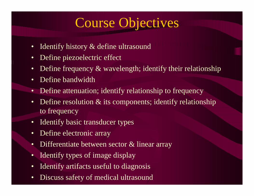
Course Objectives
• Identify history & define ultrasound
• Define piezoelectric effect
• Define frequency & wavelength; identify their relationship
• Define bandwidth
• Define attenuation; identify relationship to frequency
• Define resolution & its components; identify relationship to frequency
• Identify basic transducer types
• Define electronic array
• Differentiate between sector & linear array
• Identify types of image display
• Identify artifacts useful to diagnosis
• Discuss safety of medical ultrasound
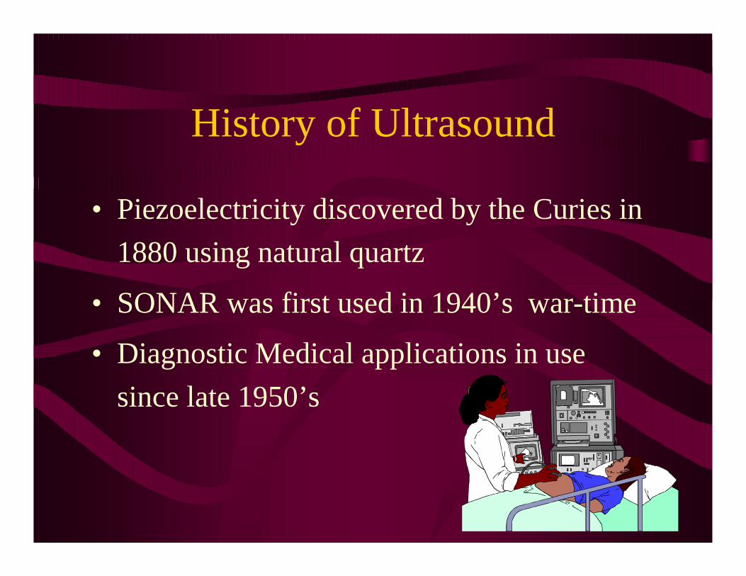
History of Ultrasound
• Piezoelectricity discovered by the Curies in
1880 using natural quartz
• SONAR was first used in 1940’s war-time
• Diagnostic Medical applications in use
since late 1950’s

Ultrasound: Physical Definition
• Sound waves greater than 20,000 Hertz or cycles per second
Infrasound Ultrasound
<20 Hz >20,000 Hz

Ultrasound: Medical Definition
• Diagnostic Medical Ultrasound is the use of high frequency sound to aid in the diagnosis and treatment of patients.
• Frequency ranges used in medical ultrasound imaging are 2 - 15 MHz

Piezoelectric Effect
• Definition: The principle of converting energy by applying pressure to a crystal.
• The reverse of the piezoelectric effect converts the energy back to its original form.

Piezoelectric Effect and Ultrasound Transducers
• A transducer converts one type of energy into another.
• Based upon the pulse-echo principle occurring with ultrasound piezoelectric crystals, ultrasound transducers convert:– Electricity into sound = pulse
– Sound into electricity = echo
Pulse
• Pulse of sound is sent to soft tissues
• Sound interaction with soft tissue = bioeffects
• Pulsing is determined by the transducer or probe crystal(s) and is not operator controlled
Echo
• Echo produced by soft tissues
• Tissue interaction with sound = acoustic propagation properties
• Echoes are received by the transducer crystals
• Echoes are interpreted and processed by the ultrasound machine

Frequency
• Number of complete cycles per unit of time
• Man-made transducer frequency is predetermined by design
• Ultrasound transducers are referred to by the operating, resonant or main frequency
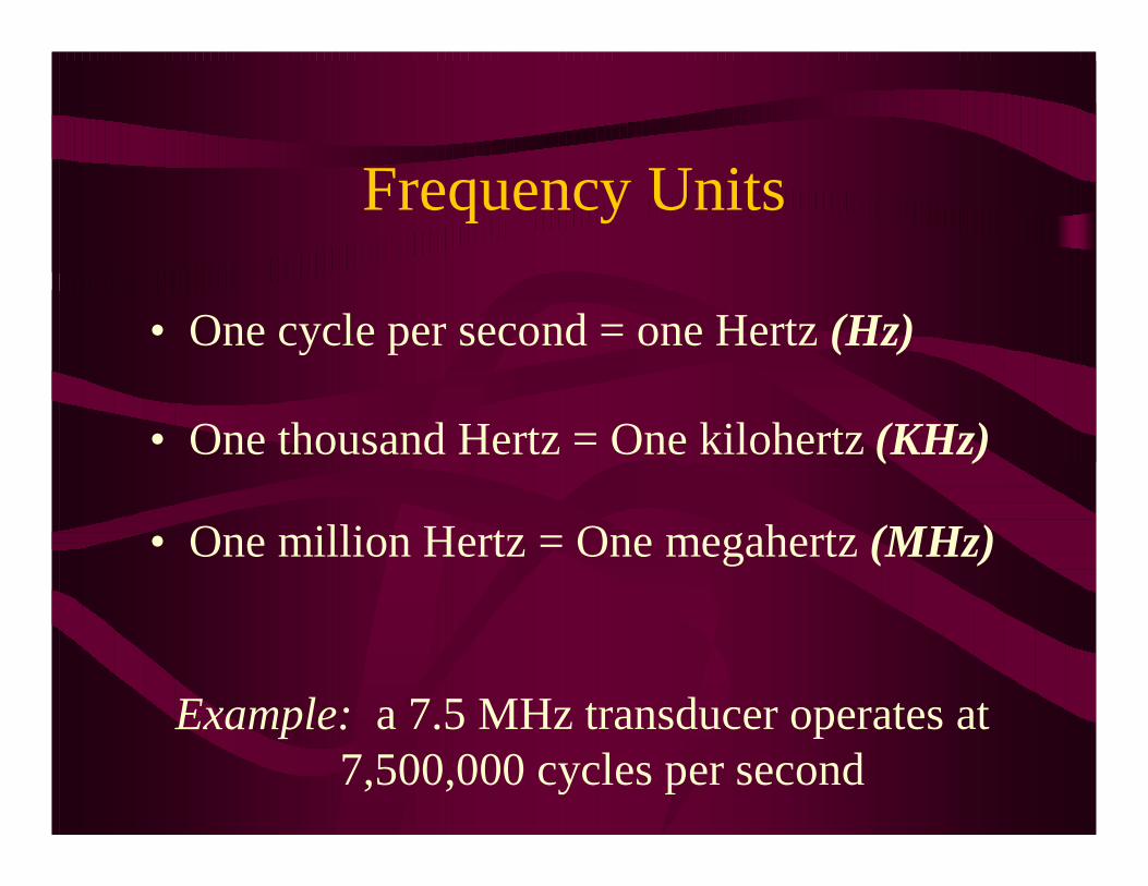
Frequency Units
• One cycle per second = one Hertz (Hz)
• One thousand Hertz = One kilohertz (KHz)
• One million Hertz = One megahertz (MHz)
Example: a 7.5 MHz transducer operates at 7,500,000 cycles per second
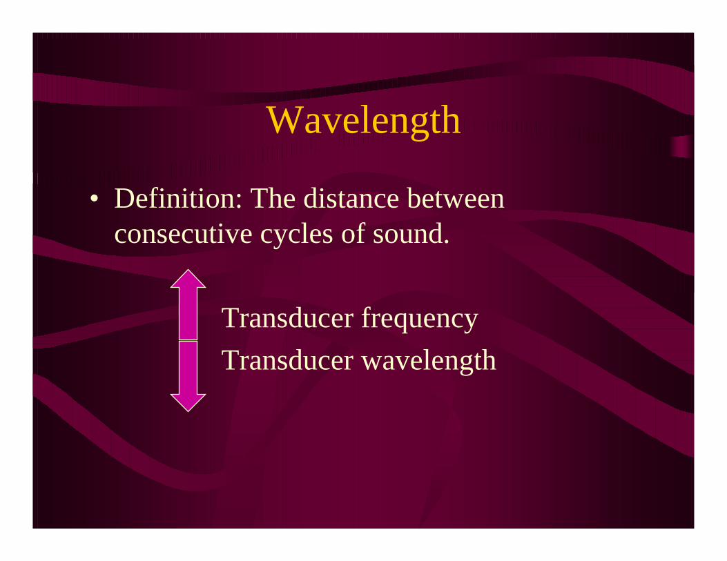
Wavelength
• Definition: The distance between consecutive cycles of sound.
Transducer frequency
Transducer wavelength
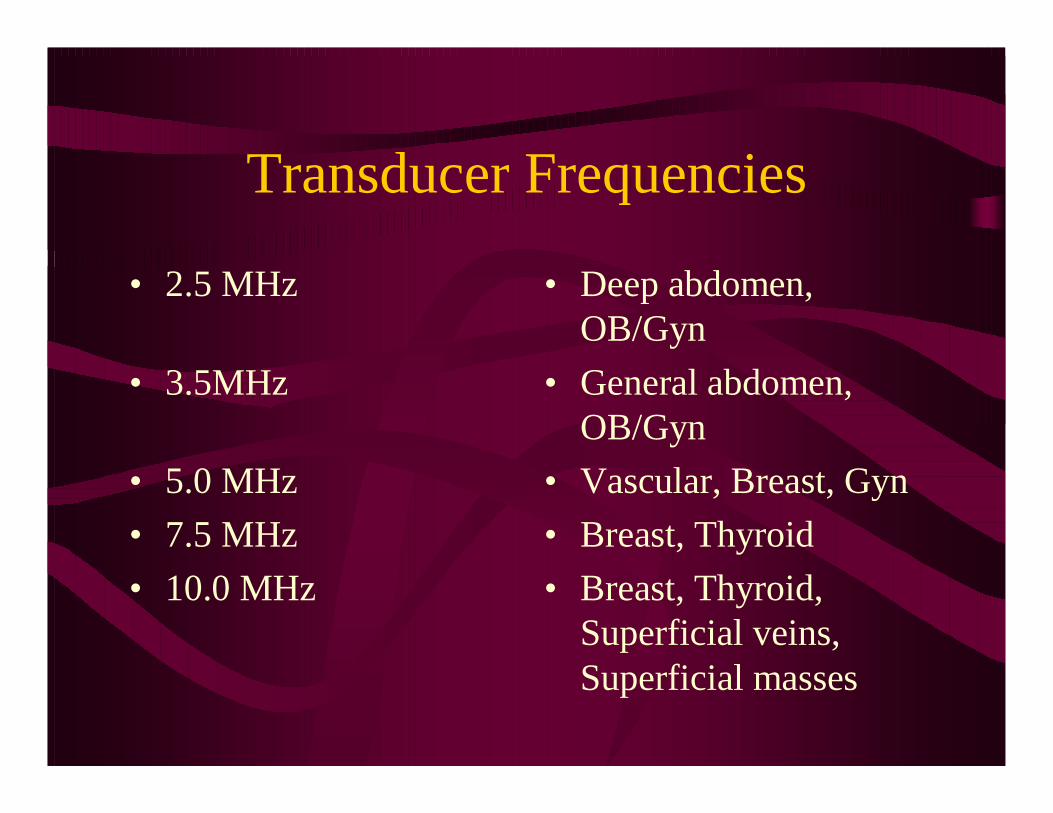
Transducer Frequencies
• 2.5 MHz
• 3.5MHz
• 5.0 MHz
• 7.5 MHz
• 10.0 MHz
• Deep abdomen, OB/Gyn
• General abdomen, OB/Gyn
• Vascular, Breast, Gyn
• Breast, Thyroid
• Breast, Thyroid, Superficial veins, Superficial masses

Bandwidth
• All ultrasound transducers contain a range of frequencies, termed bandwidth
• Broad bandwidth technology produces medical transducers that contain more than one operating frequency, for example:– 2.5 - 3.5 MHz for general abdominal imaging
– 5.0 - 7.5 MHz for superficial imaging

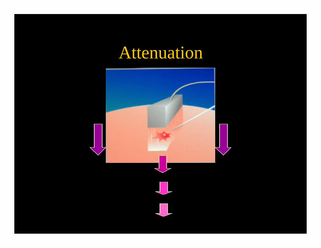
Attenuation
• Definition: The reduction in power and intensity as sound travels through a medium.
Transducer frequency
Depth of penetration
• Higher frequencies attenuate, or are absorbed, faster than lower frequencies
Attenuation

Time Gain Compensation
• Operator controlled adjustment to compensate for the attenuation of sound as it travels into the tissue
• Must be adjusted manually for each tissue type examined and may be manipulated throughout an exam to optimize the image

RESOLUTION
• The ability to differentiate between structures that are closely related, both in terms of space and echo amplitude
• Wavelength (frequency) dependent
– Gray Scale Resolution
– Axial Resolution
– Lateral Resolution
Frequency vs. Resolution
Transducer frequency
Resolution and image detail
• Higher frequency transducers provide better image resolution – better gray scale resolution
– improved ability to distinguish fine detail

Frequency and Resolution
3.5 MHz 7.5 MHz

Gray Scale Resolution
• Adequate gray scale resolution allows for the differentiation of subtle changes in the tissues
• Dynamic Range determines how many shades of gray are demonstrated on an image

Dynamic Range
Decreased DR Increased DR

Axial & Lateral Resolution
• Spatial Resolution describes how physically close two objects can be and displayed separately.
– Axial: along the beam path
– Lateral: perpendicular to beam path
• All current equipment has an overall spatial resolution of 1.0 mm or less.

Frequency Summary
• High frequency– improved
resolution
– depth of penetration loss
– higher frequency transducers for superficial uses
• Low frequency– poorer resolution
– full depth of penetration
– lower frequency transducers for general abdominopelvic uses

Machine Components
Transducer
Beam Former
Receiver
Memory
Display
Transducer Types
• Mechanical– Oscillating
– Rotating
• Electronic– Linear Arrays
– Curved Arrays
– Phased Arrays

Electronic Arrays
• Groups of piezoelectric material working singly or in groups
Transducer
126873 4 5 621
Electronic Transducers
• Sector Array– crystals are placed
parallel or in concentric rings
– transducer face is curved
– produces sector or pie-shaped image
• Linear Array– crystals are placed
parallel
– transducer face is flat
– produces rectangular image

Display Field of View
• Field Of View -- the display of the echo amplitudes
• shape dependent on transducer type and function
Field of View Shapes
• SECTOR FOV
• produced byoscillatingrotatingcurved arraysphased arrays
• typically used in abdominal application
• LINEAR FOV
• produced bylinear arrays
• typically used in superficial application

Sector Linear
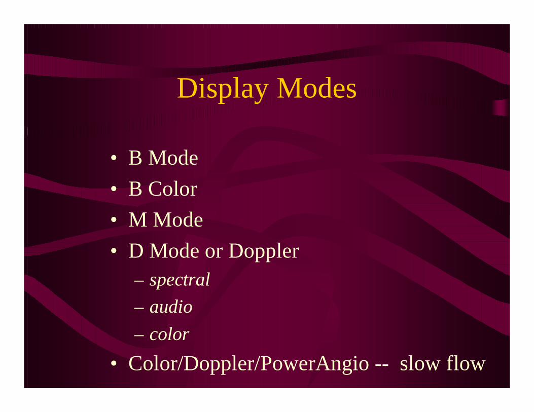
Display Modes
• B Mode
• B Color
• M Mode
• D Mode or Doppler– spectral
– audio
– color
• Color/Doppler/PowerAngio -- slow flow
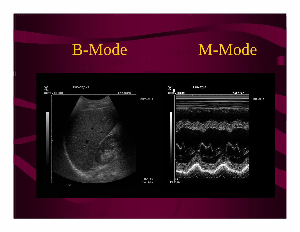
B-Mode M-Mode

Color PowerDoppler Doppler
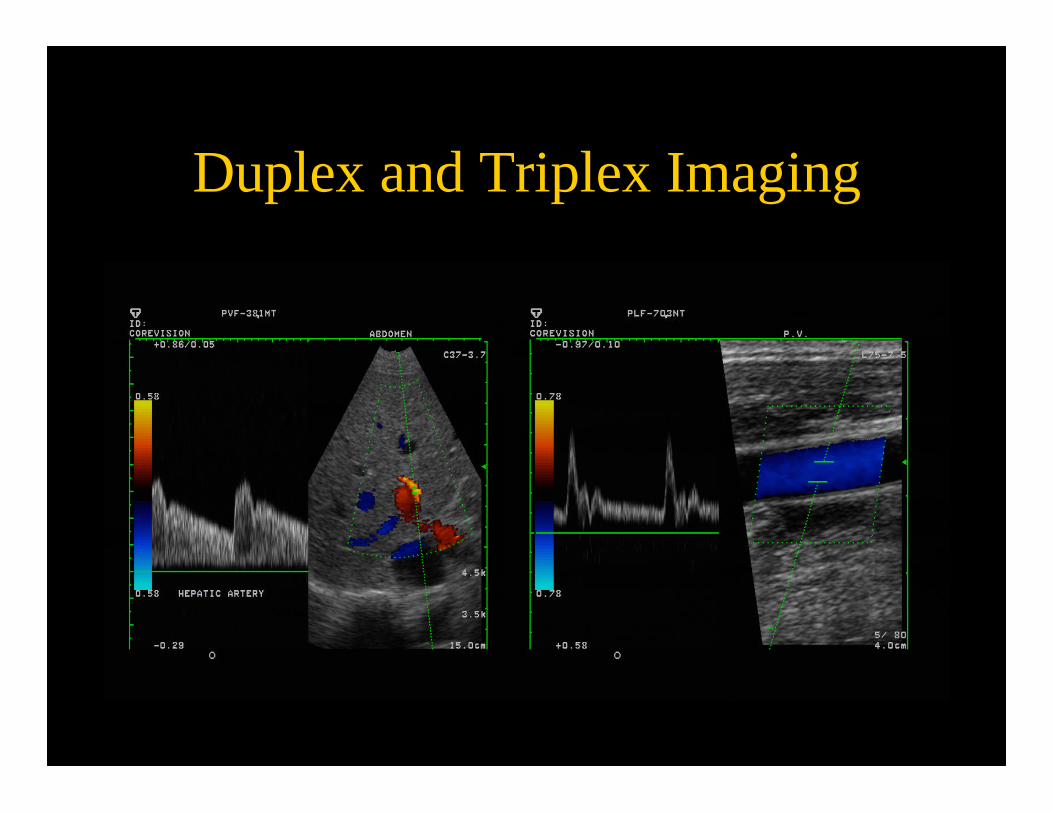
Duplex and Triplex Imaging

Artifacts
• Portions of the display which are not a “true” representation of the tissue imaged
• Medical Diagnostic Ultrasound imaging utilizes certain artifacts to characterize tissue

Artifacts
• The ability to differentiate solid vs. cystic tissue is the hallmark of ultrasound imaging
• Acoustic Shadowingand Acoustic Enhancementare the two artifacts that provide the most useful diagnostic information

Shadowing
• Diminished sound or loss of sound posterior to a strongly reflecting or strongly attenuating structure– Strong reflectors
• large calcifications, bone
– Strong attenuators • solid tissue, significantly dense or malignant masses

Shadowing

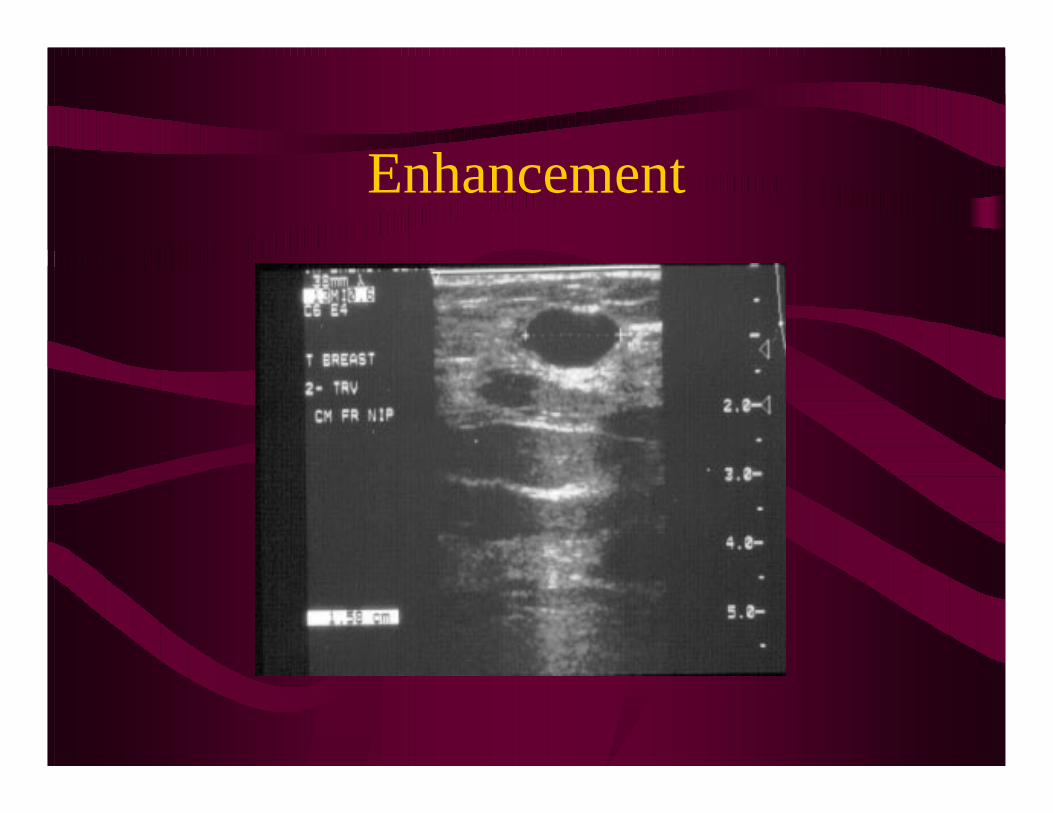
Enhancement
• Increased through transmission of the sound wave posterior to a weakly attenuating structure
• Gain curve expecteda certain loss or attenuation with depth of travel – Occurs posterior to
• simple cysts or weakly attenuating masses
Enhancement
Bioeffects
• Prudent use assures patient safety
• Effects at intensities higher than those used in diagnostic medical ultrasound include:
cavitationsister chromatid exchange

AIUM Statement
• “No confirmed biological effects on patients or operators caused by exposure at intensities typical of diagnostic ultrasound…
• ...current data indicate that the benefits… outweigh the risks.”

Summary
• Ultrasound > 20,000 Hz
• Piezoelectric Effect = pulse-echo principle
• Frequency & wavelength are inversely proportional
• Broad bandwidth enables multihertz probes
• Attenuation & frequency are inversely related
• Resolution determines image clarity
• Electronic Arrays may be sector or linear
• Display mode chosen determines how image is registered
• Shadowing & Enhancement are the artifacts most used in ultrasound diagnosis
• Diagnostic Medical Ultrasound is safe!