the basic principles of ophthalmic ultrasound · pdf filethe basic principles of ophthalmic...
TRANSCRIPT
The Basic Principles of Ophthalmic Ultrasound Devices
ORBIS International
Cheng Xiao Ming
Basics of Ultrasonography
Ultrasound Waves are acoustic waves that have frequencies greater than 20 KHz.
The audible range to humans is 60 Hz to 20 KHz.

Basics of Ultrasonography
Sound Waves
Sound is a mechanical wave (pressure wave) which is created by a vibrating object.

Basics of UltrasonographySound Waves
• The energy of vibration is transported through the mediamedia (for example: air, water, human tissue…).
Basics of Ultrasonography
Tfl.gif
Sound Waves
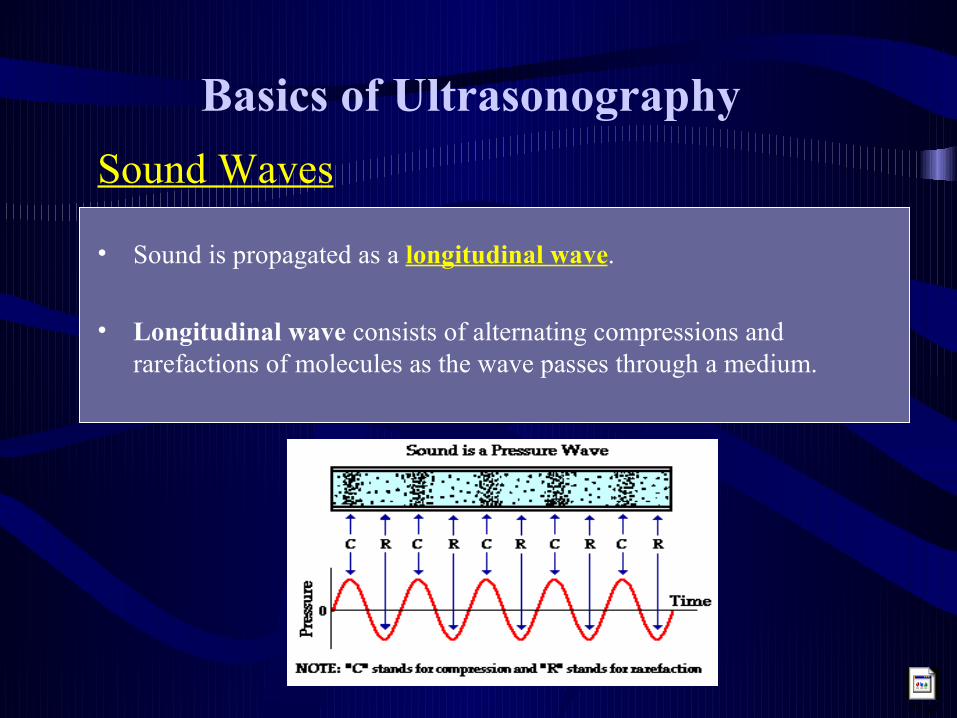
• Sound is propagated as a longitudinal wave.
• Longitudinal wave consists of alternating compressions and rarefactions of molecules as the wave passes through a medium.

Basics of UltrasonographySound Waves
The sound wave is characterized by:velocity (m/s):
frequency (Hz):
wavelength (m):
TTime
P
the speed of propagation of the wave.
number of complete cycles per unit of time.the distance traveled by one cycle.
Velocity = Wavelength x Frequency
Basics of Ultrasonography
Sound Waves
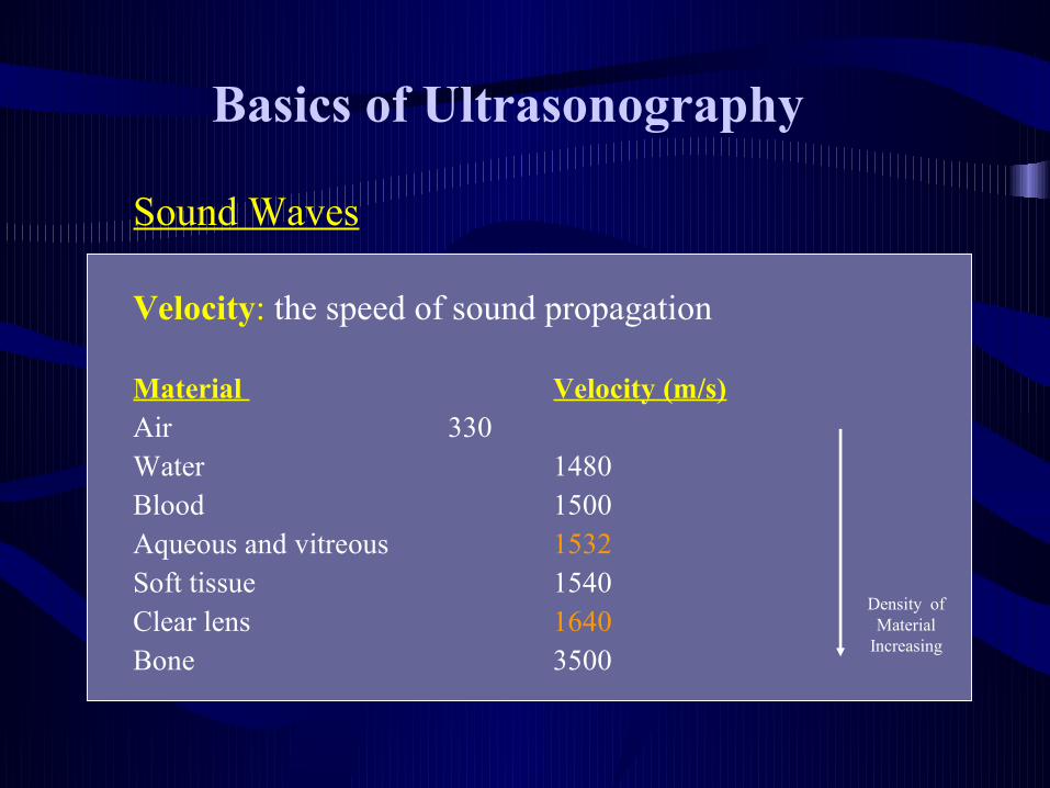
Velocity: the speed of sound propagation
Material Velocity (m/s)Air 330Water 1480Blood 1500Aqueous and vitreous 1532Soft tissue 1540Clear lens 1640Bone 3500
Density of Material
Increasing

Basics of Ultrasonography
Reflection of Sound Wave
Reflected sound waves are produced by acoustic interfaces that have different acoustic impedances.
Acoustic Interface:
Acoustic Impedance of a Medium:
is created at the junction of two media ( for example: air and rock, soft-tissue and bone…).
is a measure of the efficiency with which sound propagates in the material.
Acoustic Impedance = Sound Velocity Acoustic Impedance = Sound Velocity xx Medium Density Medium Density

Basics of Ultrasonography
Acoustic Impedance for Different Materials
Material Acoustic Impedance(106 Rayls)
Air 0.0004
Fat 1.38
Water 1.48
Liver 1.65
Muscle 1.70
Bone 7.80

Basics of Ultrasonography
Propagation of Sound Wave at a Perpendicular Surface
Reflected Sound
Media BMedia A
Incident Sound
Acoustic Interface

Basics of Ultrasonography
Propagation of Sound Wave at a Non-perpendicular Surface
Reflected Sound
Media BMedia A
Refracted Sound
Incident Sound

Basics of Ultrasonography
Propagation of Sound Wave at a Irregular Surface
Reflected Sound
Media BMedia A
Incident Sound
Acoustic Interface
Deviated Sound

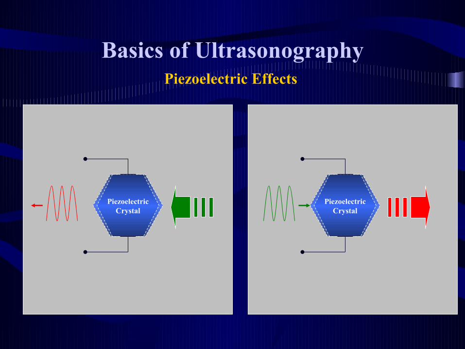
Basics of UltrasonographyPiezoelectric Effects
ForceElectrical Charges
Piezoelectric Crystal
Piezoelectric Crystal
Mechanical Deformation
ForceElectrical Charges
Mechanical Deformation
+ + + +
- - - -
+ + + +
- - - -
Natural quartz, lead zirconate titanate ….
Basics of UltrasonographyPiezoelectric Effects
Piezoelectric Crystal
Piezoelectric Crystal

Basics of Ultrasonography
Focus Zone
Basic Structure of an Ultrasound Transducer
Crystal
Acoustic Coupling Material
Electrical
Pulses

Basics of Ultrasonography
Input Electrical Pulses
Ultrasound
Transducer
Sound Wave
Echoes
Output Electrical Pulses

Basics of Ultrasonography
The greater the difference in the acoustic impedance of the two media that produced the interface, the stronger the reflection of the ultrasound wave.
Medium A
Medium CMedium A
Medium B
Sound
source IC > IB

Ophthalmic Ultrasound Medium A
Medium A
Resolution of Ultrasound Imaging
Frequency of Ultrasound
Depth of Penetration
Resolution of Ultrasound Imaging
Frequency of Ultrasound
Depth of Penetration

Ophthalmic Ultrasound
Ophthalmic ultrasound uses frequencies ranging from 6 to 50 MHz (1MHz=1 million cycles per second)
Type of Ultrasound Device FrequencyA-Scan 6~15 MHz
Ultrasound Biometry 6~15 MHz
B-Scan 8~20 MHz
UBM (Ultrasound Bio-microscopy) 30~50 MHz

Ophthalmic UltrasoundAbsorption and Attenuation of Ultrasound
Energy in the Tissues
Causes of Loss Energy:
•Beamwidth loss ( a = 1/D2 )
•Absorption loss ( Tissue Acoustic Impedance )
•Scattering loss
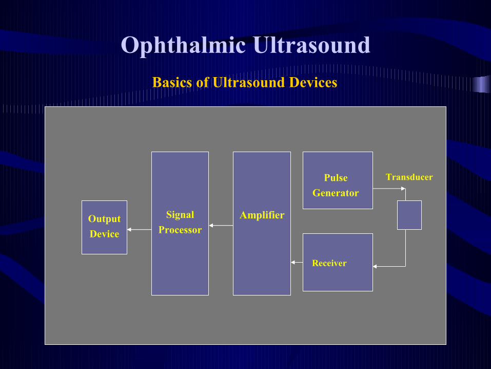
Ophthalmic Ultrasound
Basics of Ultrasound Devices
Pulse
Generator
Receiver
AmplifierSignal
ProcessorOutput
Device
Transducer

Ophthalmic UltrasoundPulse Ultrasound System
ReceiveTransmit Transmit
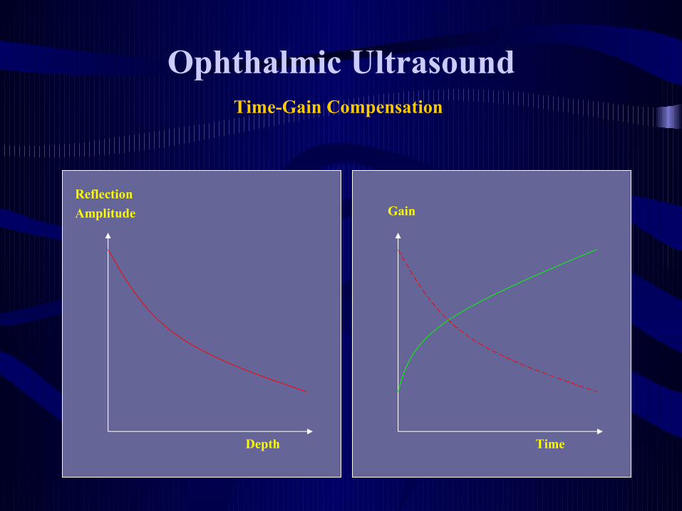
Ophthalmic UltrasoundTime-Gain Compensation
Depth
Reflection
Amplitude
Time
Gain

Ophthalmic Ultrasound
– A scan
– biometry
– B Scan
– UBM
Ultrasound Scan Modes

Anterior and Posterior surface of Cornea
Anterior surface of lensPosterior surface of lens
Retina
ScleraVitreous
Anterior Chamber
A-SCAN

A-SCAN Biometry
A-Scans are used to measure the axial length of the eye for determining the proper power of the intraocular lens to be implanted. Typical axial length measurements are from 21-26 mm.

Ophthalmic UltrasoundA-Scan Example 1

Ophthalmic UltrasoundA-Scan Example 2

The transducer sweeps back and forth.
The ultrasonic beam is equivalent to a knife blade, exposing a cross section of the cut object.
B-SCAN

Ophthalmic UltrasoundB-Scan Example 1
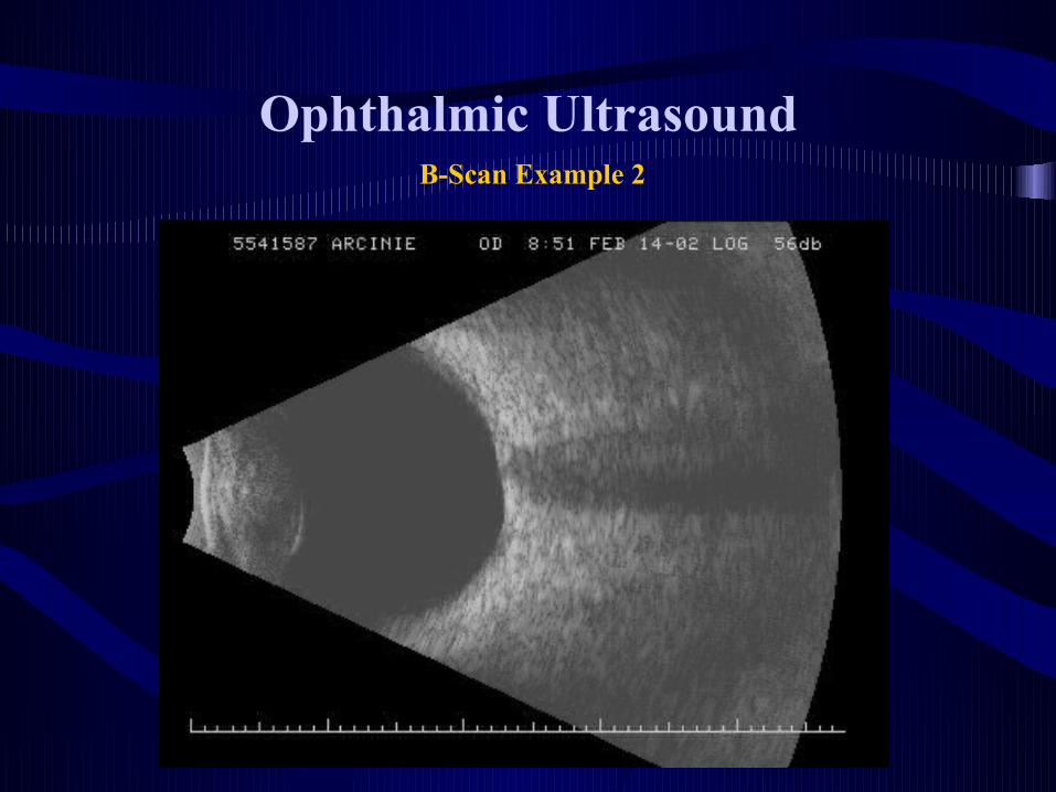
Ophthalmic UltrasoundB-Scan Example 2

Ophthalmic UltrasoundUBM Example 1
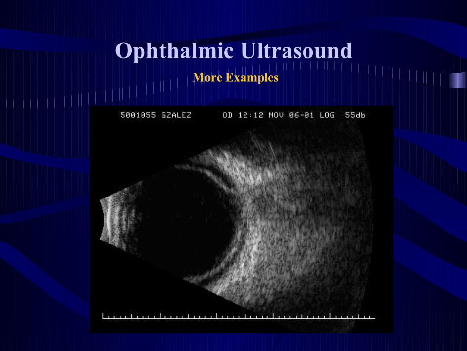
Ophthalmic UltrasoundMore Examples

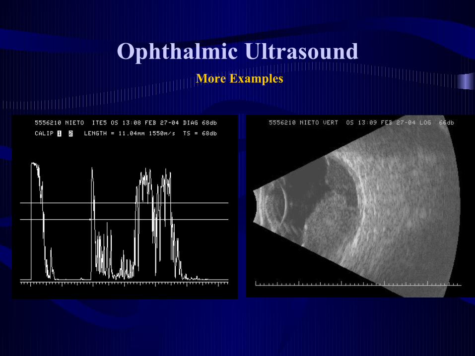
Ophthalmic UltrasoundMore Examples

Ophthalmic UltrasoundMore Examples

Ophthalmic UltrasoundMore Examples
Ophthalmic UltrasoundMore Examples

Thank You !