phyllanthus virgatus l.: an in vitro and...
TRANSCRIPT

Hindawi Publishing CorporationBioMed Research InternationalVolume 2013, Article ID 729393, 12 pageshttp://dx.doi.org/10.1155/2013/729393
Research ArticleAntioxidant and 𝛼-Amylase Inhibitory Property ofPhyllanthus virgatus L.: An In Vitro and MolecularInteraction Study
Arshya Hashim,1 M. Salman Khan,1 Mohd. Sajid Khan,1
Mohd. Hassan Baig,1 and Saheem Ahmad1,2
1 Clinical Biochemistry and Natural Product Research Lab, Department of Biosciences, Integral University, Lucknow 226026, India2Department of Surgery, Cardinal Bernardin Cancer Center, Loyola University Medical Center, Maywood, IL 60153, USA
Correspondence should be addressed to M. Salman Khan; [email protected]
Received 14 April 2013; Accepted 28 May 2013
Academic Editor: Wilson Joao Cunico Filho
Copyright © 2013 Arshya Hashim et al.This is an open access article distributed under the Creative Commons Attribution License,which permits unrestricted use, distribution, and reproduction in any medium, provided the original work is properly cited.
The present study on Phyllanthus virgatus, known traditionally for its remedial potential, for the first time provides descriptions ofthe antioxidant and inhibition of 𝛼-amylase enzyme activity first by in vitro analyses, followed by a confirmatory in silico study tocreate a stronger biochemical rationale. Our results illustrated that P. virgatus methanol extract exhibited strong antioxidant andoxidative DNA damage protective activity than other extracts, which was well correlated with its total phenolic content. In addition,P. virgatusmethanol extract strongly inhibited the 𝛼-amylase activity (IC
5033.20 ± 0.556 𝜇g/mL), in a noncompetitivemanner, than
acarbose (IC5076.88 ± 0.277 𝜇g/mL), which showed competitive inhibition. Moreover, this extract stimulated the glucose uptake
activity in 3T3-L1 cells and also showed a good correlation between antioxidant and 𝛼-amylase activities. The molecular dockingstudies of the major bioactive compounds (9,12-octadecadienoic acid, asarone, 11-octadecenoic acid, and acrylic acid) revealedvia GC-MS analysis from this extract mechanistically suggested that the inhibitory property may be due to the synergistic effect ofthese bioactive compounds.These results provide substantial basis for the future use of P. virgatusmethanol extract and its bioactivecompound in in vivo system for the treatment and management of diabetes as well as in the related condition of oxidative stress.
1. Introduction
Oxidative stress induced by reactive oxygen species (ROS)can cause cell membrane disintegration, protein, lipid, anddeoxyribose nucleic acid (DNA) damage which can furtherinitiate or propagate the development of many chronicand degenerative diseases [1–3]. When there is imbalancebetween ROS generation and antioxidant protection mecha-nism, it leads to cellular dysfunction causing various diseasesinducing diabetes mellitus (DM) [4, 5]. Diabetes is animportant metabolic syndrome affecting about 200 millionpeople worldwide. The critical effect of diabetes is post-prandial hyperglycemia and reduction in antioxidant defensemechanism. So, the management of type 2DM could bedone both by reducing oxidative stress as well as by delayingthe absorption of glucose through the inhibition of any oneof the carbohydrates-hydrolyzing enzymes, 𝛼-glucosidase,
and 𝛼-amylase that are responsible for the breakdown ofoligosaccharides and disaccharides into monosaccharidessuitable for absorption [6–8].
There has been enormous interest in natural antioxidantsdue to their ability to neutralize the effects of ROS thatare not only responsible for alleviating the oxidative stresscondition in diabetes but are also helpful in managing thepostprandial hyperglycemia. The growing interest to combatthe side effect of the drugs available for diabetes leads to thedevelopment of green medicines due to their higher stability,higher antioxidant potential, low cost, and low cytotoxicity.Plants are rich sources of phytochemicals, which possessa variety of biological activities including antioxidant andantidiabetic potential both in vitro and in vivo [7–12].
In the last few decades, plants of genus Phyllanthus(Euphorbiaceae) came in focus due to their wide distribution,diversity in the genus, broad therapeutic potential, and
2 BioMed Research International
variety in the secondarymetabolites [13].This family includesseveral plant species among all the species; P. amarus, P.urinaria, P. maderaspatensis, P. virgatus, and P. fraternus arethe most popular ones due to their antioxidant properties aswell as their extensive use in the treatment of disease relatedto kidney, liver, urinary bladder, intestinal infection, cancer,and diabetes [13–18].
It has been previously reported that P. virgatus is rich inpolyphenols [13] and is known traditionally for its antioxidant[14], antimicrobial, antiseptic, anti-inflammatory agent [19],and anticancer activity [20]. The antidiabetic propertiesof various Phyllanthus species have been investigated inexperimental models [15, 21]. However, only one studyspeculated the antidiabetic property of P. virgatus [21], andstill the detailed investigation pertaining to their mechanismof action is lacking. So, this study was the first integrativeapproach to investigate and correlate the antioxidant, oxida-tive DNA damage protective activity, 𝛼-amylase inhibitoryand glucose uptake property of various extracts of P. virga-tus. Moreover, the mechanistic aspect of these compounds,elucidated via GC-MS analysis, was explored by carryingout docking studies against porcine pancreatic 𝛼-amylasein an array to give molecular mechanism of action of suchinhibitors.
2. Methods
2.1. Chemicals. Chemicals such as 1,1-diphenyl-2-picryl-hydrazyl (DPPH), 2,4,6-tripyridyl-s-triazine (TPTZ), ascor-bic acid, thiobarbituric acid (TBA), Folin-Ciocalteu reagent(FCR), dimethyl sulfoxide (DMSO), dinitro salicylic acid(DNS), Dulbecco’s modified Eagle medium (DMEM), dex-amethasone (DEX), isobutylmethylxanthine (IBMX), foetalbovine serum (FBS), and 3-[4,5-dimethylthiazol-2-yl]-2,5-diphenyl tetrazolium bromide (MTT) were purchased fromHiMedia Laboratories, Mumbai, India. Porcine pancreatic 𝛼-amylase was procured from SRL Pvt. Ltd., Mumbai, India.Methanol (MeOH), acetone, dichloromethane (DCM), n-hexane (n-hex), ethyl acetate (EtOAc), pUC18 plasmid, andMerckotest GOD/POD kit were obtained fromMerck, India.Acarbose was obtained from Bayer Pharmaceuticals, andactrapid insulin was purchased from Torrent Pharmaceuti-cals Ltd., India. All chemicals were of analytical grade.
2.2. Collection and Preparation of Plant Extract. P. virgatuswhole plant was collected from the local area around IntegralUniversity, Lucknow, India, in the months of July-August.The plant was botanically identified and authenticated byDr. Mohd. Tariq, National Botanical Research Institute,Lucknow, India, and a voucher specimen (98195) of the plantwas submitted there. P. virgatus whole plants were shed driedand made in coarse powder, avoiding sun dried due to thesignaturemodification of the biochemicals.Thedried powder(25 g) of the plants was extracted using nonpolar, partiallypolar, and polar solvents successively with the requiredamount of each of n-hex, DCM, EtOAc, MeOH, and watersolvents in soxhlet apparatus until it turned colorless. Thesolvent was removed, filtered, and dried at room temperature,
and residueswere scratched out and stored at−20∘C for futureuse.The percentage yield of different fractions was calculatedby using the formula
% yield =weight of crude extractweight of raw material
× 100. (1)
The percentage yield was found to be n-hex: 2.17%, DCM:0.75%, EtOAc: 0.37%, MeOH: 6.71%, and water: 2.31%.
2.3. Phytochemical Screening and Estimation of Total Phe-nol Content. Qualitative chemical tests were carried outto identify the phytochemicals present in various extractsof P. virgatus using standard procedure [22]. Total phenolcontent (TPC) of the extracts was determined by using Folin-Ciocalteu method [23].
2.4. DPPH Radical Scavenging Activity. The DPPH radicalscavenging capacity of the various extracts of P. virgatus wasdetermined by the method of Brand-Williams et al. [24].Ascorbic acid was used as a reference standard. Percent (%)scavenging of DPPH free radical was measured using thefollowing equation:
% DPPH radical scavenging
=[
(absorbance of control−absorbance of test sample)(absorbance of control)
]
× 100.(2)
Further, IC50
value represented the concentration of theextract that caused 50% inhibition of DPPH radicals and wascalculated by interpolation of linear regression analysis.
2.5. Ferric Reducing Antioxidant Potential. A modifiedmethod of Benzie and Strain [25] was adopted to determinethe ferric reducing antioxidant potential (FRAP) of variousextracts of P. virgatus. Briefly, the FRAP reagent was freshlyprepared by mixing sodium acetate buffer (300mM, pH 3.6),10mM TPTZ solution (in 40mM HCl), and 20mM Fe(III)chloride solution in a volume ratio of 10 : 1 : 1, respectively.Hundred microliters of the sample at various concentrationswas added to 3mL of the FRAP reagent. The absorbance wasmeasured after 30min at room temperature at 593 nm. Thestandard curve was plotted using FeSO
4solution, and results
were expressed as𝜇mole Fe(II)/g dryweight of plantmaterial.
2.6. Hydroxyl Radical Scavenging Assay. Hydroxyl radicalscavenging activity of various extracts of P. virgatus wasevaluated by themethod of Badami et al. [26].The percentageof hydroxyl radical scavenging potential was calculated by

BioMed Research International 3
using the following formula, and IC50
was calculated asdescribed previously:
% Hydroxyl radical scavenging activity
=[
(absorbance of control−absorbance of test sample)(absorbance of control)
]
× 100.(3)
2.7. DNA Protection Assay. DNA protection assay was per-formed using supercoiled pUC18 (2686 bp) plasmid DNAaccording to the method of Lee et al. [27] with slightmodifications. Plasmid DNA (250 ng) was incubated withFenton’s reagent containing H
2O2(30mM), ascorbic acid
(100 𝜇M), and FeCl3(160 𝜇M) in the presence and absence
of the plant extract, and the final volume of the mixturewas raised up to 20𝜇L. The mixture was then incubatedfor 45min at 37∘C followed by the addition of loading dye,and the electrophoresis was carried out in Tris-acetate-EDTAbuffer (40mMTris base, 16mM acetic acid, and 1mMEDTA;pH 8.0). DNA was analyzed followed by ethidium bromidestaining, and mannitol was used as positive control.
2.8. 𝛼-Amylase Inhibition Assay. To determine the in vitro 𝛼-amylase inhibition by various extracts of P. virgatus, the stan-dard procedure [28] was adopted with slight modification.Briefly, porcine pancreatic 𝛼-amylase was dissolved in ice-cold phosphate buffer (20mM), pH 6.7, containing sodiumchloride (6.7mM) to give a concentration of 0.15 unit/mL.Triplicate test tubes including the blank were prepared. Ineach test tube, 250𝜇L of the enzyme preparation was mixedwith 100 𝜇L of each of the extracts except the blank. Themixtures were stirred in a vortex and preincubated in a waterbath at 37∘C for 20 minutes. After incubation, 250𝜇L of thesubstrate preparation (0.5% w/v starch in 20mM phosphatebuffer; pH 6.7) was transferred into each test tube to startthe reaction. The mixture was vortexed and then incubatedat 37∘C for 15 minutes. Two mL of DNS color reagent (DNS40mM, K-Na tartrate 1M, and sodium hydroxide 0.4M)was added, vortexed and boiled in a water bath at 100∘C for10 minutes. Thereafter, the mixture was cooled down, andthe absorbance was read at 540 nm. Acarbose was used asstandard inhibitor.
Inhibition rates were calculated as percentage controlsusing the formula:
% inhibition = 100 −% reaction, (4)
where % reaction = (mean product in sample/mean productin control) × 100.
Further, IC50
value represented the concentration of theextract that caused 50% inhibition of 𝛼-amylase and wascalculated by interpolation of linear regression analysis.
2.9. Determination of Mode of Inhibition. Mode of inhibitionof P. virgatus methanol extract against 𝛼-amylase was deter-mined by the method of Mogale et al. [29]. For the assay, two
sets (A and B) of 6 duplicate test tubes were prepared to deter-mine the enzyme activity in the presence [set A] and absence[set B] of an inhibitor (methanol extract/standard acarbose).In set A, 100 𝜇L of the inhibitor (plant extract or acarbose,1mg/mL) solution was added in each test tube except theblank; this was followed by the addition of 100 𝜇L of theenzyme porcine 𝛼-amylase (0.15 units/mL). In set B, 100 𝜇Lof phosphate buffer (20mM), pH 6.7, containing sodiumchloride (6.7mM) was added in each test tube followed by100 𝜇L of the enzyme solution. Both sets of test tubes werethoroughly mixed in a vortex mixer and preincubated in awater bath at 37∘C for 20 minutes. Serial dilutions of thesubstrate solution were added in both sets of test tubes withconcentration ranging between 2.5𝜇g/mL and 0.156𝜇g/mL.All the tubes were then incubated at 37∘C for 15 minutes,followed by the addition of 2mL of DNS color reagent andthe mixtures were boiled for 10 minutes. Absorbance of thecolored solution was read at 540 nm. Double reciprocal curve(1/V v/s 1/[S]) for both sets was plotted to determine the effectof the plant extract/acarbose on 𝑉max and 𝐾𝑚 of the enzyme,where V and [S] are, respectively, the velocity of the reactionand substrate concentration.
2.10. Cell Culture. 3T3-L1 preadipocytes cell lines are knownto mimic in vivo organs that have an influence on glucosehomeostasis. These cell lines were obtained from NationalCentre for Cell Sciences (NCCS), Pune, India. Cells wereroutinely cultured at 37∘C in a humidified 5% CO
2, 95%
air atmosphere and were grown in DMEM medium sup-plemented with 10% FBS, L-glutamine (8mM), and 2%antimycotic.
2.11. Cell Viability Assay. The standard MTT colorimetricassay [30], which is based on the reduction of MTT by mito-chondrial dehydrogenase to a purple formazan product, wasused to assess the cytotoxic activity of P. virgatus methanolextract. The effect was quantified as follows:
% inhibition
=
absorbance of the control − absorbance of sampleabsorbance of control
× 100.
(5)
2.12. Glucose Uptake Assay. Cells were cultured and plated ata density of 12,000 cells/well in a 24-well plate and incubatedfor 24 hours in the DMEM growth media containing 5mMglucose. On day 1, the growth medium was replaced bysupplemented medium, which consisted of DMEM supple-mentedwith 10%FCS, insulin (10𝜇g/mL), DEX (10−8M), andIBMX (0.1mM). Cells were refed 48 hours later with the samesupplemented medium, and after another 24 hours (day 4),thismediumwas removed and replacedwith growthmediumincluding the treatment protocol [11] (Table 1). After a further48-hour incubation (day 6), the cells were assayed in theirappropriate experiments. Ten microliters of the media wasremoved and placed in the 96-well plates to which 200 𝜇Lof GOD/POD reagent was added and incubated for 15min

4 BioMed Research International
at 37∘C.The change in the color was recorded at 495 nm.Thefollowing equation was used to calculate the glucose content(mg/dL) in each well. Concentration of unknown sample =(concentration of sample/abs. of standard − abs. of reagentblank) × abs. of unknown sample − abs. of reagent blank).Finally, glucose uptake over control was calculated as thedifference between the initial and final glucose content in theincubated medium.
2.13. Gas Chromatography and Mass Spectroscopy (GC-MS)Analysis. In order to know the bioactive metabolites respon-sible for antioxidant and antidiabetic activity, the methanolextract of P. virgatus was subjected to GC-MS analysis. Thesample was injected into an RTX-5 column (60m × 0.25mmi.d., film thickness 0.25 𝜇m) of GC-MS (model GC-MS-QP-2010 plus, Shimadzu Make). Helium was used as carrier gasat a constant column flow of 1.2mL/min at 173 kPa inletpressure. Temperature programming was maintained from100∘C to 200∘C with constant rise of 5∘C/min and then heldisothermal at 200∘C for 6min; further, the temperature wasincreased by 10∘C/min up to 290∘C and again held isothermalat 290∘C for 10min.The injector and ion source temperatureswere 270∘C and 250∘C, respectively. Mass spectra were takenat 70 eV a scan interval of 0.5 s and fragments from 40 to950 Dalton. The final confirmation of constituents was madeby computer matching of the mass spectra of peaks withthe National Institute of Standards and Technology (NIST)libraries mass spectral database.
2.14. Docking Analyses
2.14.1. Preparation of Enzyme and Ligand. The crystal struc-ture of porcine pancreatic 𝛼-amylase (PDB ID: 1DHK)in complex with acarbose was retrieved from ResearchCollaboratory for Structural Bioinformatics (RCSB) proteindatabank. Water molecules as well as other heteroatomswere removed, and the protein was subjected to energyminimization using CharMM force field [31]. The methoddeployed for energy minimization was steepest descent atRMS gradient of 0.1 for 1000 steps. Compounds, whose 3-D structures were available, were extracted from PubChemcompounds database, while those whose structures werenot present were drawn using ChemDraw like hexadecanoicacid, 9,12-octadecadienoic acid, 11-octadecenoic acid, and 6-octadecynoic acid.
2.14.2. Molecular Docking. Molecular docking studies werecarried out using AutoDock program [32] to get the favorablebinding modes for compounds within the active site ofporcine pancreatic 𝛼-amylase. Before conducting moleculardocking, validation was performed, in which the acarbosepresent within the binding site of porcine 𝛼-amylase incrystal structure was extracted. This acarbose was subjectedto redocking within the active site of porcine 𝛼-amylase usingAutoDock. The binding confirmation was visualized usingPyMOL. After complete execution of AutoDock, variousconformations of ligand in complex with the receptor were
Table 1: Treatment protocol for glucose uptake assay.
S. no. Incubation medium
Group 1 1000𝜇L DMEM containing 5mM glucose
Group 2 900𝜇L DMEM + 100 𝜇L insulin (1 IU/mL)
Group 3900𝜇L DMEM + 100𝜇L metformin
(1mg/1mL)
Group 4900𝜇L DMEM + 100𝜇L plant extract
(1mg/mL)
Group 5800 𝜇L DMEM + 100𝜇L insulin (1 IU/mL) +
100𝜇L plant extract (1mg/mL)
Group 6800 𝜇L DMEM + 100𝜇L insulin (1 IU/mL) +
100𝜇L metformin (1mg/1mL)
Group 7700 𝜇L DMEM + 100𝜇L insulin (1 IU/mL) +100 𝜇L metformin (1mg/1mL) + 100 𝜇L
plant extract (1mg/mL)
obtained, which were finally ranked on the basis of bindingenergy.
2.15. Statistical Analysis. The results were analyzed by usingone-way analysis of variance (ANOVA) and two-tailed Stu-dent’s t-test. Statistical significance was expressed as ∗𝑃 <0.05, ∗∗𝑃 < 0.01, and ∗∗∗𝑃 > 0.001.
3. Results
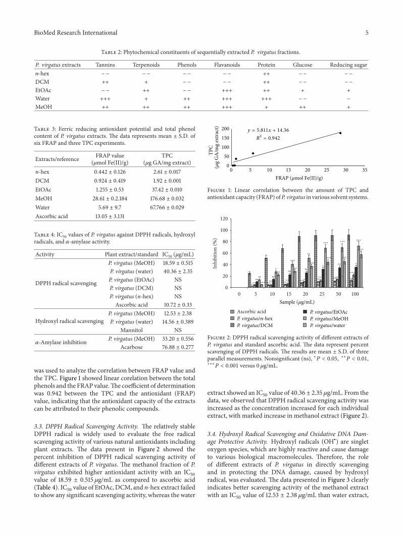
3.1. Phytochemical Estimation and Total Phenol Content. Ourresults illustrated significant presence of tannins, terpenoids,saponins, phenols, carbohydrate, flavanoids, protein, glucose,and reducing sugar in P. virgatus methanol extract (Table 2).Water extract contains all the above phytochemicals exceptglucose and reducing sugar. In addition, EtOAc extractcontains terpenoids, flavanoid, protein, glucose, and reducingsugar, while only tannins, terpenoids, and protein werepresent in DCM extracts. In contrast, n-hex contains onlyprotein content. The TPC (𝜇g/mg GAE) in the variousextracts of P. virgatuswas also determined and found to be inthe following decreasing order: MeOH >water > EtOAc > n-hex>DCM. From the data, it is evident thatmethanol extracthas higher phenolic content (176.68 ± 0.032 𝜇g GA/mg plantextract) than the water and ethyl acetate extracts, whereasDCM and n-hex have the lowest phenolic content (Table 3).
3.2. Total Antioxidant Activity. Antioxidant activities of dif-ferent P. virgatus extracts were assessed by FRAP assay, whichis based on their ability to reduce ferric ions to ferrous form.The results illustrated that methanol extract has significantlyhigher FRAP values (28.61 ± 0.2184𝜇mol Fe(II)/g) as com-pared to other extracts (0.4–6 𝜇mol Fe(II)/g) and standardascorbic acid (Table 3). A simple linear regression analysis
BioMed Research International 5
Table 2: Phytochemical constituents of sequentially extracted P. virgatus fractions.
P. virgatus extracts Tannins Terpenoids Phenols Flavanoids Protein Glucose Reducing sugar𝑛-hex −− −− −− −− ++ −− −−
DCM ++ + −− −− ++ −− −−
EtOAc −− ++ −− +++ ++ + +Water +++ + ++ +++ +++ −− −
MeOH ++ ++ ++ +++ + ++ +
Table 3: Ferric reducing antioxidant potential and total phenolcontent of P. virgatus extracts. The data represents mean ± S.D. ofsix FRAP and three TPC experiments.
Extracts/reference FRAP value(𝜇mol Fe(II)/g)
TPC(𝜇g GA/mg extract)
n-hex 0.442 ± 0.126 2.61 ± 0.017DCM 0.924 ± 0.419 1.92 ± 0.001EtOAc 1.255 ± 0.53 37.42 ± 0.010MeOH 28.61 ± 0.2.184 176.68 ± 0.032Water 5.69 ± 9.7 67.766 ± 0.029Ascorbic acid 13.05 ± 3.131
Table 4: IC50 values of P. virgatus against DPPH radicals, hydroxylradicals, and 𝛼-amylase activity.
Activity Plant extract/standard IC50 (𝜇g/mL)
DPPH radical scavenging
P. virgatus (MeOH) 18.59 ± 0.515P. virgatus (water) 40.36 ± 2.35P. virgatus (EtOAc) NSP. virgatus (DCM) NSP. virgatus (n-hex) NSAscorbic acid 10.72 ± 0.33
Hydroxyl radical scavengingP. virgatus (MeOH) 12.53 ± 2.38P. virgatus (water) 14.56 ± 0.389
Mannitol NS
𝛼-Amylase inhibition P. virgatus (MeOH) 33.20 ± 0.556Acarbose 76.88 ± 0.277
was used to analyze the correlation between FRAP value andthe TPC. Figure 1 showed linear corelation between the totalphenols and the FRAP value.The coefficient of determinationwas 0.942 between the TPC and the antioxidant (FRAP)value, indicating that the antioxidant capacity of the extractscan be attributed to their phenolic compounds.
3.3. DPPH Radical Scavenging Activity. The relatively stableDPPH radical is widely used to evaluate the free radicalscavenging activity of various natural antioxidants includingplant extracts. The data present in Figure 2 showed thepercent inhibition of DPPH radical scavenging activity ofdifferent extracts of P. virgatus. The methanol fraction of P.virgatus exhibited higher antioxidant activity with an IC
50
value of 18.59 ± 0.515 𝜇g/mL as compared to ascorbic acid(Table 4). IC
50value of EtOAc,DCM, and n-hex extract failed
to show any significant scavenging activity, whereas the water
0 5 10 15 20 25 30 35FRAP (𝜇mol Fe(II)/g)
0
50
100
150
200
(𝜇g
GA
/mg
extr
act)
TPC
y = 5.811x + 14.36
R2 = 0.942
Figure 1: Linear correlation between the amount of TPC andantioxidant capacity (FRAP) ofP. virgatus in various solvent systems.
0
20
40
60
80
100
120
0 5 10 15 20 25 50 100
Ascorbic acid
Sample (𝜇g/mL)
P. virgatus/n-hexP. virgatus/DCM
P. virgatus/EtOAcP. virgatus/MeOHP. virgatus/water
nsns ns
nsns
∗
∗∗
∗
∗
∗∗
∗∗
∗∗
∗∗
∗∗∗∗
∗∗
∗∗
∗∗
∗∗
∗∗∗∗ ∗∗∗ ∗∗∗ ∗∗∗
∗∗∗
∗∗∗
∗∗∗
∗∗∗
∗∗∗
∗∗∗ ∗∗∗
∗∗∗
∗∗∗
∗∗∗
Inhi
bitio
n (%
)
Figure 2: DPPH radical scavenging activity of different extracts ofP. virgatus and standard ascorbic acid. The data represent percentscavenging of DPPH radicals. The results are mean ± S.D. of threeparallel measurements. Nonsignificant (ns), ∗𝑃 < 0.05, ∗∗𝑃 < 0.01,∗∗∗𝑃 < 0.001 versus 0 𝜇g/mL.
extract showed an IC50value of 40.36 ± 2.35 𝜇g/mL. From the
data, we observed that DPPH radical scavenging activity wasincreased as the concentration increased for each individualextract, with marked increase in methanol extract (Figure 2).
3.4. Hydroxyl Radical Scavenging and Oxidative DNA Dam-age Protective Activity. Hydroxyl radicals (OH∙) are singletoxygen species, which are highly reactive and cause damageto various biological macromolecules. Therefore, the roleof different extracts of P. virgatus in directly scavengingand in protecting the DNA damage, caused by hydroxylradical, was evaluated. The data presented in Figure 3 clearlyindicates better scavenging activity of the methanol extractwith an IC
50value of 12.53 ± 2.38 𝜇g/mL than water extract,

6 BioMed Research International
01020304050607080
0 5 10 15 20
Mannitol
Sample (𝜇g/mL)
P. virgatus/MeOHP. virgatus/water
∗
∗∗
∗∗
∗∗∗∗∗∗∗∗∗
∗∗∗
∗∗∗
Inhi
bitio
n (%
)
Figure 3: Hydroxyl radical scavenging activity of the P. virgatusMeOH, water extract, and reference compound mannitol. The datarepresents the percentage of inhibition of deoxyribose degradation.The results are expressed as mean ± S.D. (𝑛 = 3). ∗𝑃 < 0.05,∗∗𝑃 < 0.01, ∗∗∗𝑃 < 0.001 versus 0 𝜇g/mL.
Lane 1 2 3 4 5 6 7 8 9
Figure 4: Effect of P. virgatuswater andMeOH extracts on damagedsupercoiled pUC18 plasmid DNA. Lane 1: pUC18 DNA + PBS;lane 2: pUC18 DNA + Fenton’s reagent; lane 3: DNA + Fenton’sreagent + water extract (50 𝜇g/mL); lane 4: DNA + Fenton’s reagent+ water extract (100 𝜇g/mL); lane 5: DNA + Fenton’s reagent +water extract (200𝜇g/mL); lane 6: DNA + Fenton’s reagent +MeOHextract (50 𝜇g/mL); lane 7: DNA + Fenton’s reagent +MeOH extract(100𝜇g/mL); lane 8: DNA + Fenton’s reagent + MeOH extract(200𝜇g/mL); lane 9: Mannitol (200𝜇g/mL).
while the other extracts (data not shown) includingmannitolshowed insignificant scavenging activity of hydroxyl radical.
The oxidative DNA damage protective activity of P. vir-gatus methanol and water extracts showed almost completeand partial protection of OH∙-induced oxidatively damagedplasmid DNA, respectively (Figure 4). Incubation of pUC18plasmid DNA with Fenton’s reagent resulted in the cleavageof supercoiled form to give open circular and linear formsof plasmid DNA, indicating that OH generated from iron-mediated decomposition of H
2O2produced both single-
strand and double-strand DNA breaks. Addition of P. vir-gatus methanol and water extracts (50,100 and 200𝜇g/mL)showed complete and partial protection of supercoiled DNA(Figure 4).
3.5. 𝛼-Amylase Inhibitory Property. In an array to explorethe antidiabetic activity, various extracts of P. virgatus werescreened for the𝛼-amylase inhibitory property. Initial screen-ing of various extracts showed that the methanol extracthas significantly higher percentage of 𝛼-amylase inhibition,that is, 43.2% and 66.09% at 25 and 50 𝜇g/mL, respec-tively (Figure 5). Furthermore, the methanol extract showedconcentration-dependent increase in percent inhibition of𝛼-amylase activity and also exhibited a lower IC
50value
01020304050607080
0 DCM EtOAc MeOH Water AcarboseSample
ns∗∗∗∗
∗∗
∗∗∗
∗∗∗
∗∗∗∗∗∗
∗∗∗∗∗∗
n-hex
25𝜇g/mL50𝜇g/mL
Inhi
bitio
n (%
)
Figure 5: Screening of 𝛼-amylase inhibitory property of variousextracts of P. virgatus. Results are mean ± S.D. of three parallelmeasurements. Nonsignificant (ns), ∗∗𝑃 < 0.01, ∗∗∗𝑃 < 0.001versus 0 𝜇g/mL.
0102030405060708090
0 1 2.5 5 10 25 50 100
Inhi
bitio
n (%
)
Acarbose
ns ns ns
Sample (𝜇g/mL)
P. virgatus/MeOH
∗∗∗∗∗∗
∗∗∗
∗∗∗
Figure 6: Concentration-dependent inhibition of P. virgatusmethanol extract and reference compound acarbose.The results areexpressed as mean ± S.D. of three parallel experiments. Nonsignifi-cant (ns), ∗∗∗𝑃 < 0.001 versus 0 𝜇g/mL.
(33.20 ± 0.556𝜇g/mL) than standard drug acarbose, whichin turn indicates a potent antidiabetic property of this extract(Figure 6; Table 4).
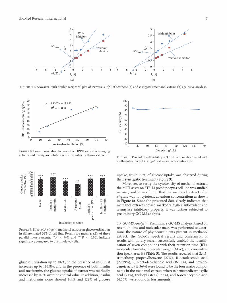
Themode of inhibition of acarbose andmethanol extractsof P. virgatus against porcine 𝛼-amylase was also determinedby means of Lineweaver-Burk double reciprocal plot of 1/vversus 1/[S]. The methanol extract showed a noncovalenttype of noncompetitive inhibition against porcine 𝛼-amylase(Figure 7(b)), whereas acarbose was competitive in nature(Figure 7(a)). In addition, a linear correlation was alsoobserved between DPPH radical scavenging activity and 𝛼-amylase inhibitory property of P. virgatus methanol extract(𝑅2 = 0.885) (Figure 8).
3.6. Glucose Uptake Assay. It was found that the methanolextract of P. virgatus showed strong 𝛼-amylase inhibitoryproperty; further, its role in glucose utilization in differenti-ated 3T3-L1 adipocytes cell line was also studied in vitro.Theability of the plant extract to induce glucose uptake was testedin different combinations (Table 1), and it was found thatthe methanol extract of P. virgatus alone showed significant
BioMed Research International 7
0
1
2
3
4
5
6
7
0 2 4 6 8
Without inhibitor
With inhibitor
−8 −6 −4 −2
1/�1/Vmax
−1/Km 1/[S]
(a)
0
0.5
1
1.5
2
2.5
3
0 2 4 6 8
With inhibitor
Without inhibitor
−8 −6 −4 −2
1/�
1/Vmax
−1/Km 1/[S]
(b)
Figure 7: Lineweaver-Burk double reciprocal plot of 1/v versus 1/[S] of acarbose (a) and P. virgatusmethanol extract (b) against 𝛼-amylase.
0102030405060708090
0 10 20 30 40 50 60 70 80
y = 0.9307x + 11.992
R2 = 0.8858
DPP
H ra
dica
l sca
veng
ing
(%)
𝛼-Amylase inhibition (%)
Figure 8: Linear correlation between the DPPH radical scavengingactivity and 𝛼-amylase inhibition of P. virgatusmethanol extract.
020406080
100120140160180200
over
cont
rol (
%)
Glu
cose
upt
ake
Incubation medium
Insu
lin
Insu
lin+
met
form
in
Met
form
in
P. vi
rgat
usM
eOH
Insu
lin+
plan
t ext
ract
(PE)
Insu
lin+
PE+
met
form
in
∗∗
∗∗∗∗∗∗∗∗∗
∗∗∗∗∗∗
Figure 9: Effect ofP. virgatusmethanol extract on glucose utilizationin differentiated 3T3-L1 cell line. Results are mean ± S.D. of threeparallel measurements. ∗∗𝑃 < 0.01 and ∗∗∗𝑃 < 0.001 indicatesignificance compared to unstimulated cells.
glucose utilization up to 102%; in the presence of insulin itincreases up to 146.8%, and in the presence of both insulinand metformin, the glucose uptake of extract was markedlyincreased by 149% over the control value. In addition, insulinand metformin alone showed 144% and 122% of glucose
102030405060708090
100
0 20 40 60 80 100 120 140 160
Cel
l via
bilit
y (%
)
Sample (𝜇g/mL)
Figure 10: Percent of cell viability of 3T3-L1 adipocytes treated withmethanol extract of P. virgatus at various concentrations.
uptake, while 158% of glucose uptake was observed duringtheir synergistic treatment (Figure 9).
Moreover, to verify the cytotoxicity of methanol extract,the MTT assay on 3T3-L1 preadipocytes cell line was studiedin vitro, and it was found that the methanol extract of P.virgatuswas noncytotoxic at various concentrations as shownin Figure 10. Since the presented data clearly indicates thatmethanol extract showed markedly higher antioxidant and𝛼-amylase inhibitory property, it was further subjected topreliminary GC-MS analysis.
3.7. GC-MS Analysis. Preliminary GC-MS analysis, based onretention time and molecular mass, was performed to deter-mine the nature of phytoconstituents present in methanolextract. The GC-MS spectral results and comparison ofresults with library search successfully enabled the identifi-cation of seven compounds with their retention time (RT),molecular formula, molecular weight (MW), and concentra-tion (peak area %) (Table 5). The results revealed that 2,4,5-trimethoxy propenylbenzene (27%), 11-octadecenoic acid(22.29%), 9,12-octadecadienoic acid (16.91%), and hexade-canoic acid (13.36%) were found to be the four major compo-nents in the methanol extract, whereas benzenedicarboxylicacid (7.1%), tridecyl ester (8.77%), and 6-octadecynoic acid(4.56%) were found in less amounts.

8 BioMed Research International
Table 5: Major constituents of P. virgatusmethanol extract revealed via GC-MS analysis.
Peak R.T Compound Molecularformula Molecular weight Area%
1 16.508 Benzenedicarboxylic acid(synonym: phthalic acid) C12H14O4 222 7.1
2 17.0052,4,5-Trimethoxy propenylbenzene(synonym: asarone)
C12H16O3 208 27
3 18.508 Tridecyl ester(synonym: acrylic acid) C16H30O2 254 8.77
4 22.643 Hexadecanoic acid(synonym: palmitic acid) C17H34O2 270 13.36
5 26.557 9,12-Octadecadienoic acid(synonym: linoleic acid) C19H34O2 294 16.91
6 26.798 11-Octadecenoic acid C19H34O3 294 22.297 27.517 6-Octadecynoic acid C19H34O4 294 4.56
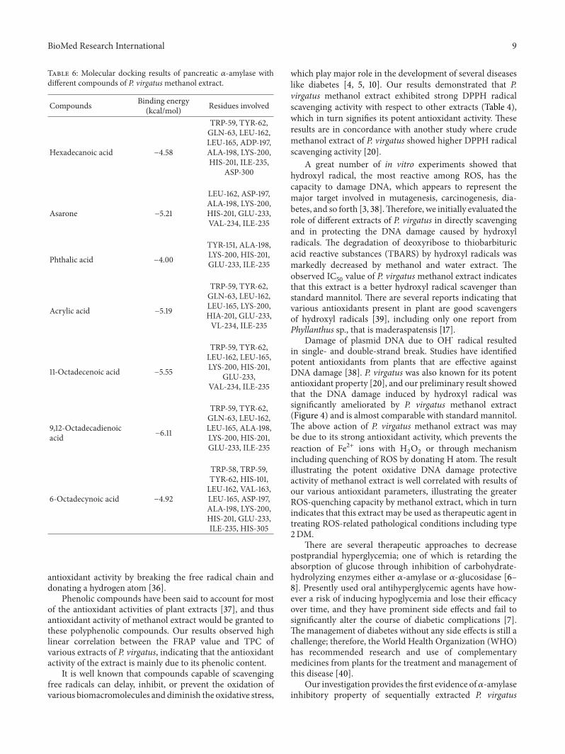
3.8. Docking Analyses. Validation of docking protocol andthe size as well as center of the coordinates of the grid wascarried out in order to ensure that ligands bind to the bindingpocket in the correct conformation. It was performed byredocking cocrystallized acarbose into its respective bindingsite within porcine pancreatic 𝛼-amylase. Redocked inhibitorwas found to interact with the same amino acids of the activesite as was in the crystal structure (Figure 11). The root meansquare deviation (RMSD) of all atoms between these twoconformations was 1.58 A, indicating that the protocol set formolecular docking is accurate. Then, the in silico study wasdone beneath the assumption that a predicted high dockingscore in absolute valuewill be predictive of a strong inhibitionof the enzyme. The results showed that all the moleculesdepicted by GC-MS were found to bind within the activesite of porcine pancreatic 𝛼-amylase with binding free energyranging from −3.27 to −6.11 Kcal/mol (Table 6). The dockingresults illustrated that 9,12-octadecadienoic acid was themostactive compound, followed by 11-octadecenoic acid, asarone,and acrylic acid with binding free energy of −6.11, −5.55,−5.21, and −5.19 Kcal/mol, respectively, against 𝛼-amylase.The following amino acids: Trp57, Tyr62, Leu162, Leu165,Asp197, Lys200, His201, Glu233, and Ile235 were found tobe the key residues playing important role in stabilizing thecomplex (Figure 12 and Table 6).
4. Discussion
Currently used synthetic drugs, which are known to protectagainst type 2DM and oxidative damage, have their adverseside effects. As a result, consumption of natural antioxidants,that are known to be effective scavengers of free radicals,through plants, food, or dietary supplements, interrupts theproduction of ROS and thus helping in the prevention ofvarious diseases including type 2DM [4, 8, 9, 33, 34]. In thecurrent study, a sequential extraction involving the solvent ofdecreasing polarity to extract the bioactive compounds wasused because the nature, polarity, and hence the solubility ofthe bioactive compounds in P. virgatus were unknown. Our
Figure 11: Binding orientation of the crystallized (red) and redocked(blue) acarbose.
Figure 12: Binding pattern of the compounds depicted via GC-MSanalyses within the active site of pancreatic 𝛼-amylase.
data initially indicated that methanol extract of P. virgatusnot only showed significant presence of polyphenols but alsoexhibited the highest amount of TPC than other extracts,which is in well agreement with earlier studies [14, 35].Our results illustrated significantly higher total antioxidantcapacity of the methanol extract of P. virgatus, which is evengreater than the standard ascorbic acid.The reducing capacityof a compound may serve as a significant indicator of itspotential antioxidant activity and generally correlates withthe presence of reductones, which have been shown to exert
BioMed Research International 9
Table 6: Molecular docking results of pancreatic 𝛼-amylase withdifferent compounds of P. virgatusmethanol extract.
Compounds Binding energy(kcal/mol) Residues involved
Hexadecanoic acid −4.58
TRP-59, TYR-62,GLN-63, LEU-162,LEU-165, ADP-197,ALA-198, LYS-200,HIS-201, ILE-235,
ASP-300
Asarone −5.21
LEU-162, ASP-197,ALA-198, LYS-200,HIS-201, GLU-233,VAL-234, ILE-235
Phthalic acid −4.00
TYR-151, ALA-198,LYS-200, HIS-201,GLU-233, ILE-235
Acrylic acid −5.19
TRP-59, TYR-62,GLN-63, LEU-162,LEU-165, LYS-200,HIA-201, GLU-233,VL-234, ILE-235
11-Octadecenoic acid −5.55
TRP-59, TYR-62,LEU-162, LEU-165,LYS-200, HIS-201,
GLU-233,VAL-234, ILE-235
9,12-Octadecadienoicacid −6.11
TRP-59, TYR-62,GLN-63, LEU-162,LEU-165, ALA-198,LYS-200, HIS-201,GLU-233, ILE-235
6-Octadecynoic acid −4.92
TRP-58, TRP-59,TYR-62, HIS-101,LEU-162, VAL-163,LEU-165, ASP-197,ALA-198, LYS-200,HIS-201, GLU-233,ILE-235, HIS-305
antioxidant activity by breaking the free radical chain anddonating a hydrogen atom [36].
Phenolic compounds have been said to account for mostof the antioxidant activities of plant extracts [37], and thusantioxidant activity of methanol extract would be granted tothese polyphenolic compounds. Our results observed highlinear correlation between the FRAP value and TPC ofvarious extracts of P. virgatus, indicating that the antioxidantactivity of the extract is mainly due to its phenolic content.
It is well known that compounds capable of scavengingfree radicals can delay, inhibit, or prevent the oxidation ofvarious biomacromolecules and diminish the oxidative stress,
which play major role in the development of several diseaseslike diabetes [4, 5, 10]. Our results demonstrated that P.virgatus methanol extract exhibited strong DPPH radicalscavenging activity with respect to other extracts (Table 4),which in turn signifies its potent antioxidant activity. Theseresults are in concordance with another study where crudemethanol extract of P. virgatus showed higher DPPH radicalscavenging activity [20].
A great number of in vitro experiments showed thathydroxyl radical, the most reactive among ROS, has thecapacity to damage DNA, which appears to represent themajor target involved in mutagenesis, carcinogenesis, dia-betes, and so forth [3, 38].Therefore, we initially evaluated therole of different extracts of P. virgatus in directly scavengingand in protecting the DNA damage caused by hydroxylradicals. The degradation of deoxyribose to thiobarbituricacid reactive substances (TBARS) by hydroxyl radicals wasmarkedly decreased by methanol and water extract. Theobserved IC
50value of P. virgatusmethanol extract indicates
that this extract is a better hydroxyl radical scavenger thanstandard mannitol. There are several reports indicating thatvarious antioxidants present in plant are good scavengersof hydroxyl radicals [39], including only one report fromPhyllanthus sp., that is maderaspatensis [17].
Damage of plasmid DNA due to OH⋅ radical resultedin single- and double-strand break. Studies have identifiedpotent antioxidants from plants that are effective againstDNA damage [38]. P. virgatus was also known for its potentantioxidant property [20], and our preliminary result showedthat the DNA damage induced by hydroxyl radical wassignificantly ameliorated by P. virgatus methanol extract(Figure 4) and is almost comparable with standard mannitol.The above action of P. virgatus methanol extract was maybe due to its strong antioxidant activity, which prevents thereaction of Fe2+ ions with H
2O2or through mechanism
including quenching of ROS by donating H atom. The resultillustrating the potent oxidative DNA damage protectiveactivity of methanol extract is well correlated with results ofour various antioxidant parameters, illustrating the greaterROS-quenching capacity by methanol extract, which in turnindicates that this extract may be used as therapeutic agent intreating ROS-related pathological conditions including type2DM.
There are several therapeutic approaches to decreasepostprandial hyperglycemia; one of which is retarding theabsorption of glucose through inhibition of carbohydrate-hydrolyzing enzymes either 𝛼-amylase or 𝛼-glucosidase [6–8]. Presently used oral antihyperglycemic agents have how-ever a risk of inducing hypoglycemia and lose their efficacyover time, and they have prominent side effects and fail tosignificantly alter the course of diabetic complications [7].The management of diabetes without any side effects is still achallenge; therefore, the World Health Organization (WHO)has recommended research and use of complementarymedicines from plants for the treatment and management ofthis disease [40].
Our investigation provides the first evidence of𝛼-amylaseinhibitory property of sequentially extracted P. virgatus
10 BioMed Research International
extracts. The initial screening of all extracts of P. virga-tus demonstrated some 𝛼-amylase inhibitory activity withmaximum activity in methanol extract. In addition, thisextract also exhibited a concentration-dependent increasein percent inhibition of 𝛼-amylase activity with an IC
50
value of 33.20 ± 0.556𝜇g/mL, which is quite better than theIC50
value of standard acarbose 76.88 ± 0.277 𝜇g/mL. Thisobservation suggests that porcine pancreatic 𝛼-amylase isinhibited by more polar constituents of P. virgatus, whichis in agreement with another study that reported 𝛼-amylaseinhibitory activities in the more polar extracts of plantmaterials [41, 42]. Thus, the enzyme inhibitory activity ofmethanol extracts could be due to the presence of polyphe-nols, flavonoids, and their glycosides, which are known to besoluble in more polar solvents.
Enzymes inhibitors obeying Michaelis-Menten kineticsare often characterized in terms of their effects on thekinetic constants, 𝐾
𝑚and 𝑉max, using either Lineweaver-
Burk plots or Dixon secondary plots. In the current study,P. virgatusmethanol extracts demonstrated noncovalent typeof noncompetitive (𝑉max decreased whereas 𝐾
𝑚remained
the same) mode of inhibition against porcine pancreatic 𝛼-amylase, whereas acarbose was competitive in nature. Theseobservations might suggest that the 𝛼-amylase inhibitorycomponents ofmethanol extracts do not resemble the normalsubstrates of the enzymes in structure [29]. Further, themechanism of inhibition of acarbose seems to be due tothe unsaturated cyclohexene ring and the glycosidic nitrogenlinkage that mimics the transition state for the enzymaticcleavage of glycosidic linkages [43]. It has been previouslyshown that acarbose is a competitive inhibitor of 𝛼-amylase,which is in strong agreement with our results [29].
Moreover, the strong correlation demonstrated betweentotal antioxidant and polyphenol contents and betweenDPPH radical scavenging and 𝛼-amylase activity ofmethanolextracts suggests that polyphenol compounds involved intotal antioxidant/antiradical activity may also is directly orindirectly intervene in 𝛼-amylase inhibitory activity. Post-prandial blood glucose level is known to be regulated byglucose uptake, a rate limiting step for glucose metabolism.In the present study, we used differentiated 3T3-L1 cell linesbecause it was previously established that glucose uptakewas higher in these cells than in undifferentiated one, whichis probably due to the presence of glucose transporter-4(GLUT4) in their expression [44].
Our result, for the first time, speculated that P. virgatusmethanol extract possesses the ability to improve glucoseuptake in the adipose tissue. The positive controls chosenfor glucose uptake in 3T3-L1 cells due to their antidiabeticactivity were metformin and insulin, as they are knownspecifically for the affirmative effect on the translocation ofGLUT4 to the cell surface thereby promoting glucose uptake.Insulin and metformin significantly promote the glucoseuptake alone, while there is almost a negligible synergisticeffect when given in combination (in presence or absence ofextract). On this basis, a mechanism of action of P. virgatusmay be hypothesized, which could be linked to insulin-mediated glucose transport transduction pathway in which
a series of proteins (phosphatidylinositol-3 kinase, proteinkinase C, and PPAR) are involved [44, 45]. This may perhapslead to the translocation of GLUT4 to the plasma membraneto facilitate the uptake of glucose from the bloodstream intothe cells. Thus, the occurrence of polyphenolic compoundsin methanol extract may be responsible for the activation ofthese signaling proteins [11, 46, 47] and might therefore alsoaccount for their upregulation of these proteins, which in turnis responsible for its glucose uptake activity.
In a search for the source of bioactive compounds respon-sible for the aforementioned actions of P. virgatus methanolextract, preliminary GC-MS analysis was performed. Forthe first time, it was noted that the sequentially extractedP. virgatus methanol extract contains phthalic acid, asarone,acrylic acid, palmitic acid, linoleic acid, 11-octadecenoicacid, and 6-octadecynoic acid (Table 5). Various species ofPhyllanthus reported the presence of these compounds withantioxidant and antidiabetic activity [13, 48, 49].
From the above data, it may be concluded that thesebioactive compounds of P. virgatus methanol extract aloneor in combination possess significant antioxidant activity,which could be responsible in ameliorating all the aboveoxidative damages, including inhibition of amylase activity.However, our in silico investigation is a novel approachto identify the molecular targets involved in inhibition of𝛼-amylase activity by this extract. We previously revealedthe implication of molecular docking studies in elucidatingthe mechanistic aspect of natural products against differentenzymes [50]. Molecular simulation study is considered to bean important vehicle to investigate the mode of interaction ofligand against its target protein that alsomakes us understandtheir binding or inhibitionmechanism. Validation of dockingprotocol was performed by redocking cocrystallized acarboseinto its respective binding site within porcine pancreatic𝛼-amylase. Redocked inhibitor was found to interact withthe same amino acids of the active site as was in thecrystal structure with RMSD of 1.58 A between these twoconformations (Figure 11). Our results demonstrated that9,12-octadecadienoic acid (linoleic acid) was the most potentinhibitor of pancreatic 𝛼-amylase, whereas 11-octadecenoicacid, 2,4,5-trimethoxy propenyl (asarone), and tridecyl esteralso showed good inhibitory activity in terms of their bindingenergy. These results are well supported by wet lab studieswhere asarone, linoleic acid, and acrylic acid have beenreported to exhibit the antidiabetic property [50, 51]. Of thecatalytic triad [52], Asp197 and Glu233 were found to bevery much involved in the positioning of inhibitor withinthe active sites of pancreatic 𝛼-amylase. Our results are ingeneral agreement with the mechanism of action proposedfor acarbose [8], since they showed for 𝛼-amylase that activeligands interact with the side chains of Asp197, Glu233, andAsp300 (Figure 12, Table 6). All the inhibitors were anchoredat the catalytic center, whichmight explainwhy the enzymaticactivity of 𝛼-amylase was successfully blocked.Thus, it is verydifficult to name a single compound responsible for thewholeactivity. Therefore, based on our in vitro and in silico results,we suggest that the 𝛼-amylase inhibitory activity of P. virgatusmethanol extract might be because of the synergistic effect ofthese compounds.
BioMed Research International 11
5. Conclusion
In conclusion, the results for the first time demonstrateda strong antioxidant and 𝛼-amylase inhibitory as well asglucose uptake property of sequentially extracted P. virgatusmethanol fraction, compared to other extracts. The dockingstudies further confirmed the antidiabetic property of thebioactive compounds revealed via GC-MS analysis of thisextract and suggested that 𝛼-amylase inhibitory property ofthis extract was maybe due to the synergistic effect of thesebioactive compounds. Thus, it is a good approach to managetype 2DM as a whole with these compounds/extracts, whichshowed good enzyme inhibitory and antioxidant activities.Further, a thorough and full-fledged in vivo study is neededto explore the role of these extracts and also their bioactivecompounds.
Conflict of Interests
The authors declare that they have no conflict of interests.
Acknowledgments
The authors would like to thank University Grant commis-sion (UGC), New Delhi, for providing predoctoral fellow-ship to Ms. Arshya Hashim in the form of Maulana AzadNational Junior Research Fellowship.The authors would alsolike to thank Professor S. W. Akhtar, Vice Chancellor, forproviding state-of-art research laboratory and Professor A.K. Srivastava, Head, Department of Biosciences, for smoothsuccession of this work.
References
[1] J. Styskal, H. van Remmen, A. Richardson, and A. B. Salmon,“Oxidative stress and diabetes: what can we learn about insulinresistance from antioxidant mutant mouse models?” Free Radi-cal Biology and Medicine, vol. 52, no. 1, pp. 46–58, 2012.
[2] T. Finkel and N. J. Holbrook, “Oxidants, oxidative stress and thebiology of ageing,”Nature, vol. 408, no. 6809, pp. 239–247, 2000.
[3] B. Halliwell, “Free radicals, antioxidants, and human disease:curiosity, cause, or consequence?”TheLancet, vol. 344, no. 8924,pp. 721–724, 1994.
[4] J. S. Johansen, A. K.Harris, D. J. Rychly, andA. Ergul, “Oxidativestress and the use of antioxidants in diabetes: linking basicscience to clinical pratice,” Cardiovascular Diabetology, vol. 4,article 5, 2005.
[5] J. M. C. Gutteridge, “Biological origin of free radicals andmechanisms of antioxidant protection,” Chemico-BiologicalInteractions, vol. 91, no. 2-3, pp. 133–140, 1994.
[6] F. A. van de Laar, P. L. Lucassen, R. P. Akkermans, E. H. vande Lisdonk, G. E. Rutten, and C. van Weel, “Alpha-glucosidaseinhibitors for type 2 diabetes mellitus (Cochrane Review),”TheCochrane Library, 2008.
[7] A. Y. Y. Cheng and I. G. Fantus, “Oral antihyperglycemic ther-apy for type 2 diabetes mellitus,” Canadian Medical AssociationJournal, vol. 172, no. 2, pp. 213–226, 2005.
[8] P. M. de Sales, P. M. de Souza, L. A. Simeoni, P. D. O.Magalhaes, and D. Silveira, “𝛼-amylase inhibitors: a reviewof raw material and isolated compounds from plant source,”
Journal of Pharmacy and Pharmaceutical Sciences, vol. 15, no.1, pp. 141–183, 2012.
[9] S. Kumar, V. Kumar, M. Rana, and D. Kumar, “Enzymesinhibitors from plants: an alternate approach to treat diabetes,”Pharmacognosy Communications, vol. 2, no. 2, pp. 18–33, 2012.
[10] E. Haslam, “Natural polyphenols (vegetable tannins) as drugs:possible modes of action,” Journal of Natural Products, vol. 59,no. 2, pp. 205–215, 1996.
[11] Y.-C. Yang, H.-K. Hsu, J.-H. Hwang, and S.-J. Hong, “Enhance-ment of glucose uptake in 3T3-L1 adipocytes by Toona sinensisleaf extract,” Kaohsiung Journal of Medical Sciences, vol. 19, no.7, pp. 327–333, 2003.
[12] P. Marin, S. S. Rebuffe, U. Smith, and P. Bjorntorp, “Glucoseuptake in human adipose tissue,”Metabolism, vol. 36, pp. 1154–1160, 1987.
[13] J. Calixto, A. R. S. Santos, V. Filbo, and R. A. Yunes, “Reviewof the plants of the genus phyllanthus: their chemistry, pharma-cology, and therapeutic potential,”Medicinal Research Reviews,vol. 18, no. 4, pp. 225–258, 1998.
[14] A. Kumaran and R. Joel Karunakaran, “In vitro antioxidantactivities of methanol extracts of five Phyllanthus species fromIndia,” LWT-Food Science and Technology, vol. 40, no. 2, pp.344–352, 2007.
[15] J. Shabeer, R. S. Srivastava, and S. K. Singh, “Antidiabetic andantioxidant effect of various fractions of Phyllanthus simplex inalloxan diabetic rats,” Journal of Ethnopharmacology, vol. 124,no. 1, pp. 34–38, 2009.
[16] N. V. Rajeshkumar, K. L. Joy, G. Kuttan, R. S. Ramsewak, M. G.Nair, and R. Kuttan, “Antitumour and anticarcinogenic activityof Phyllanthus amarus extract,” Journal of Ethnopharmacology,vol. 81, no. 1, pp. 17–22, 2002.
[17] V. V. Asha, M. S. Sheeba, V. Suresh, and P. J. Wills, “Hepatopro-tection of Phyllanthus maderaspatensis against experimentallyinduced liver injury in rats,” Fitoterapia, vol. 78, no. 2, pp. 134–141, 2007.
[18] A. Prakash, K. S. Satyan, S. P. Wahi, and R. P. Singh, “Com-parative hepatoprotective activity of three phyllanthus species,P. urinaria, P. niruri and P. simplex, on carbon tetrachlorideinduced liver injury in the rat,” Phytotherapy Research, vol. 9,no. 8, pp. 594–596, 1995.
[19] H. S. Chouhan and S. K. Singh, “Phytochemical analysis,antioxidant and anti-inflammatory activities of Phyllanthussimplex,” Journal of Ethnopharmacology, vol. 137, no. 3, pp. 1337–1344, 2011.
[20] K. Poompachee and N. Chudapongse, “Comparison of theantioxidant and cytotoxic activities of Phyllanthus virgatus andPhyllanthus amarus extracts,” Medical Principles and Practice,vol. 21, no. 1, pp. 24–29, 2011.
[21] K. Regi Raphael, M. C. Sabu, and R. Kuttan, “Hypoglycemiceffect of methanol extract of Phyllanthus amarus Schum &Thonn on alloxan induced diabetes mellitus in rats and its rela-tion with antioxidant potential,” Indian Journal of ExperimentalBiology, vol. 40, no. 8, pp. 905–909, 2002.
[22] J. B. Harborne, Phytochemical Methods: A Guide to ModernTechniques of Plant Analysis, vol. 279, Chapman&Hall, London,UK, 1973.
[23] V. L. Singleton, R. Orthofer, and R. M. Lamuela-Raventos,“Analysis of total phenols and other oxidation substrates andantioxidants by means of folin-ciocalteu reagent,” Methods inEnzymology, vol. 299, pp. 152–178, 1998.

12 BioMed Research International
[24] W. Brand-Williams, M. E. Cuvelier, and C. Benset, “Use of freeradical method to evaluate antioxidant activity,” Lebensmittel-Wissenschaft Und -Technologie, vol. 28, pp. 25–30, 1995.
[25] I. F. F. Benzie and J. J. Strain, “The ferric reducing ability ofplasma (FRAP) as a measure of “antioxidant power”: the FRAPassay,” Analytical Biochemistry, vol. 239, no. 1, pp. 70–76, 1996.
[26] S. Badami, M. K. Gupta, and B. Suresh, “Antioxidant activityof the ethanolic extract of Striga orobanchioides,” Journal ofEthnopharmacology, vol. 85, no. 2-3, pp. 227–230, 2003.
[27] J.-C. Lee, H.-R. Kim, J. Kim, and Y.-S. Jang, “Antioxidantproperty of an ethanol extract of the stem of Opuntia ficus-indica var. saboten,” Journal of Agricultural and Food Chemistry,vol. 50, no. 22, pp. 6490–6496, 2002.
[28] P. Bernfeld, “Amylases 𝛼 and 𝛽,” inMethods in Enzymology, S. P.Colowick and N. O. Kaplan, Eds., vol. 1, p. 149, Academic Press,New York, NY, USA, 1955.
[29] M. A. Mogale, H. M. Mkhombo, S. L. Lebelo, L. J. Shai, M. A.Chauke, and A. Freitas, “The effects of Clausena anisata (Wild)Hook leaf extracts on selected diabetic related metabolizingenzymes,” Journal of Medicinal Plants Research, vol. 6, no. 25,pp. 4200–4207, 2012.
[30] T. Mosmann, “Rapid colorimetric assay for cellular growth andsurvival: application to proliferation and cytotoxicity assays,”Journal of Immunological Methods, vol. 65, no. 1-2, pp. 55–63,1983.
[31] B. R. Brooks, R. E. Bruccoleri, B. D. Olafson, D. J. Sate, S.Swaminathan, and M. Karplus, “CHARMM: a program formacromolecular energy minimization and dynamics calcula-tions,” Journal of Computational Chemistry, vol. 4, pp. 187–217,1983.
[32] G. M. Morris, D. S. Goodsell, R. S. Halliday et al., “Automateddocking using a Lamarckian genetic algorithm and an empiricalbinding free energy function,” Journal of Computational Chem-istry, vol. 19, no. 14, pp. 1639–1662, 1998.
[33] M. S. Khan, I. A. Ansari, S. Ahmed, F. Akhtar, A. Hashim, andA. K. Srivastava, “Chemotherapeutic potential of Boeerhaavidiffussa L: a review,” Journal of Applied Pharmaceutical Science,vol. 3, no. 01, pp. 133–139, 2013.
[34] J. Iqbal, M. S. Khan, andA. Khan, “Protection of oxidative stressinduced LDL oxidation and erythrocyte damage from type 2diabetic subjects by in vitro tocotrienol treatment,” Journal ofPharmacy Research, vol. 5, no. 01, pp. 30–37, 2012.
[35] C. L. Narasimhudu and R. R. V. Raju, “Preliminary phytochem-ical studies on leaves of phyllanthus species (euphorbiaceae),used by the local tribals of Andhra Pradesh,” InternationalJournal of Pharmaceutical Studies and Research, vol. 2, no. 4, pp.6–9, 2011.
[36] M. H. Gordon, “The mechanism of antioxidant action in vitro,”in Food Antioxidants, B. J. F. Hudson, Ed., pp. 1–18, ElsevierApplied Science, London, UK, 1990.
[37] J. Dai and R. J. Mumper, “Plant phenolics: extraction, analysisand their antioxidant and anticancer properties,”Molecules, vol.15, no. 10, pp. 7313–7352, 2010.
[38] B. N. Singh, B. R. Singh, R. L. Singh et al., “Oxidative DNAdam-age protective activity, antioxidant and anti-quorum sensingpotentials of Moringa oleifera,” Food and Chemical Toxicology,vol. 47, no. 6, pp. 1109–1116, 2009.
[39] D. Zhang, T. Yasuda, Y. Yu et al., “Ginseng extract scavengeshydroxyl radical and protects unsaturated fatty acids fromdecomposition caused by iron-mediated lipid peroxidation,”Free Radical Biology and Medicine, vol. 20, no. 1, pp. 145–150,1996.
[40] A. K. Tiwari, “Wisdom of Ayurveda in perceiving diabetes:enigma of therapeutic recognition,” Current Science, vol. 88, no.7, pp. 1043–1051, 2005.
[41] M. R. Bhandari, N. Jong-Anurakkun, G. Hong, and J. Kawabata,“𝛼-Glucosidase and 𝛼-amylase inhibitory activities of Nepalesemedicinal herb Pakhanbhed (Bergenia ciliata, Haw.),” FoodChemistry, vol. 106, no. 1, pp. 247–252, 2008.
[42] M. Jung, M. Park, H. C. Lee, Y.-H. Kan, E. S. Kang, and S.K. Kim, “Antidiabetic agents from medicinal plants,” CurrentMedicinal Chemistry, vol. 13, no. 10, pp. 1203–1218, 2006.
[43] E. Lo Piparo, H. Scheib, N. Frei, G. Williamson, M. Grigorov,and C. J. Chou, “Flavonoids for controlling starch digestion:structural requirements for inhibiting human 𝛼-amylase,” Jour-nal of Medicinal Chemistry, vol. 51, no. 12, pp. 3555–3561, 2008.
[44] H. Eldar-Finkelman and E. G. Krebs, “Phosphorylation ofinsulin receptor substrate 1 by glycogen synthase kinase 3impairs insulin action,” Proceedings of the National Academy ofSciences of the United States of America, vol. 94, no. 18, pp. 9660–9664, 1997.
[45] J. R. Zierath and H. Wallberg-Henriksson, “From receptor toeffector: Insulin signal transduction in skeletal muscle fromtype II diabetic patients,” Annals of the New York Academy ofSciences, vol. 967, pp. 120–134, 2002.
[46] P. L. Owen, L. C. Martineau, D. Caves, P. S. Haddad, T.Matainaho, and T. Johns, “Consumption of guava (Psidiumguajava L) and noni (Morinda citrifolia L) may protect betelquid-chewing Papua New Guineans against diabetes,” AsiaPacific Journal of Clinical Nutrition, vol. 17, no. 4, pp. 635–643,2008.
[47] A. P. Jorge, H. Horst, E. D. Sousa, M. G. Pizzolatti, and F. R. M.B. Silva, “Insulinomimetic effects of kaempferitrin on glycaemiaand on 14C-glucose uptake in rat soleus muscle,” Chemico-Biological Interactions, vol. 149, no. 2-3, pp. 89–96, 2004.
[48] S. Hasan, M. Danishuddin, M. Adil, K. Singh, P. K. Verma,and A. U. Khan, “Efficacy of E. officinalis on the cariogenicproperties of streptococcus mutans: a novel and alternativeapproach to suppress quorum-sensingmechanism,” PLoS ONE,vol. 7, no. 7, pp. 1–12, 2012.
[49] S. H. Lee, K.-Y. Kim, S. Y. Ryu et al., “Asarone inhibitsadipogenesis and stimulates lipolysis in 3T3-L1 adipocytes,”Cellular and Molecular Biology, vol. 56, no. 1, pp. OL1215–OL1222, 2010.
[50] M. Salman Khan, S. Akhtar, O. A. Al-Sagair, and J. M. Arif,“Protective effect of dietary tocotrienols against infection andinflammation-induced hyperlipidemia: an in vivo and in silicostudy,”Phytotherapy Research, vol. 25, no. 11, pp. 1586–1595, 2011.
[51] J. W. Ryder, C. P. Portocarrero, X. M. Song et al., “Isomer-specific antidiabetic properties of conjugated linoleic acid:Improved glucose tolerance, skeletal muscle insulin action, andUCP-2 gene expression,” Diabetes, vol. 50, no. 5, pp. 1149–1157,2001.
[52] S. Janecek, B. Svensson, and B. Henrissat, “Domain evolution inthe 𝛼-amylase family,” Journal of Molecular Evolution, vol. 45,no. 3, pp. 322–331, 1997.

Submit your manuscripts athttp://www.hindawi.com
PainResearch and TreatmentHindawi Publishing Corporationhttp://www.hindawi.com Volume 2014
The Scientific World JournalHindawi Publishing Corporation http://www.hindawi.com Volume 2014
Hindawi Publishing Corporationhttp://www.hindawi.com
Volume 2014
ToxinsJournal of
VaccinesJournal of
Hindawi Publishing Corporation http://www.hindawi.com Volume 2014
Hindawi Publishing Corporationhttp://www.hindawi.com Volume 2014
AntibioticsInternational Journal of
ToxicologyJournal of
Hindawi Publishing Corporationhttp://www.hindawi.com Volume 2014
StrokeResearch and TreatmentHindawi Publishing Corporationhttp://www.hindawi.com Volume 2014
Drug DeliveryJournal of
Hindawi Publishing Corporationhttp://www.hindawi.com Volume 2014
Hindawi Publishing Corporationhttp://www.hindawi.com Volume 2014
Advances in Pharmacological Sciences
Tropical MedicineJournal of
Hindawi Publishing Corporationhttp://www.hindawi.com Volume 2014
Medicinal ChemistryInternational Journal of
Hindawi Publishing Corporationhttp://www.hindawi.com Volume 2014
AddictionJournal of
Hindawi Publishing Corporationhttp://www.hindawi.com Volume 2014
Hindawi Publishing Corporationhttp://www.hindawi.com Volume 2014
BioMed Research International
Emergency Medicine InternationalHindawi Publishing Corporationhttp://www.hindawi.com Volume 2014
Hindawi Publishing Corporationhttp://www.hindawi.com Volume 2014
Autoimmune Diseases
Hindawi Publishing Corporationhttp://www.hindawi.com Volume 2014
Anesthesiology Research and Practice
ScientificaHindawi Publishing Corporationhttp://www.hindawi.com Volume 2014
Journal of
Hindawi Publishing Corporationhttp://www.hindawi.com Volume 2014
Pharmaceutics
Hindawi Publishing Corporationhttp://www.hindawi.com Volume 2014
MEDIATORSINFLAMMATION
of