effects of an extract from phyllanthus niruri on hepatitis b and woodchuck hepatitis viruses
TRANSCRIPT

Proc. Nati. Acad. Sci. USAVol. 84, pp. 274-278, January 1987Medical Sciences
Effects of an extract from Phyllanthus niruri on hepatitis B andwoodchuck hepatitis viruses: In vitro and in vivo studies
(antiviral agent/Marmota monax/DNA polymerase/hepatitis B surface antigen/woodchuck hepatitis surface antigen)
P. S. VENKATESWARAN*, I. MILLMAN, AND B. S. BLUMBERGFox Chase Cancer Center, Philadelphia, PA 19111
Contributed by B. S. Blumberg, September 18, 1986
ABSTRACT An aqueous extract of the plant PhyUanthusniruri inhibits endogenous DNA polymerase of hepatitis B virusand binds to the surface antigen of hepatitis B virus in vitro. Theextract also inhibits woodchuck hepatitis virus (WHV) DNApolymerase and binds to the surface antigen of WHV in vitro.The extract, nontoxic to mice, was tested for antiviral activityin woodchucks (Marmota monax). In a trial using six long-termWHV-carrier woodchucks, five treated animals showed afaster decrease in woodchuck hepatitis virus surface antigentiter compared to one untreated control. In animals recentlyinfected with WHV, the extract was effective when adminis-tered i.p. in three out of four animals in reducing and within3-6 weeks eliminating both the surface antigen titer and DNApolymerase activity in serum. The treatment was discontinuedafter 10 weeks, and the treated animals have remained free ofdetectable markers of WHV for more than 45 weeks. Incontrast, three untreated controls remained positive for bothmarkers for WHV. One of the controls died after 8 weeks; theother two controls have remained positive for WHV markersfor more than 45 weeks. In a third trial with long-term carriers,test animals treated subcutaneously with the extract for 12weeks did not respond; but on switching the mode of admin-istration to i.p., two out of the five animals showed a significantdecrease in woodchuck hepatitis virus surface antigen titercompared to controls.
Chronic carriers of the hepatitis B virus (HBV) may remainasymptomatic for long periods, but many are at high risk ofeventually developing post-hepatitic cirrhosis and primaryhepatocellular carcinoma. Carriers are often infected withinthe first few years of life, but symptoms of chronic liverdisease and primary hepatocellular carcinoma may not beperceived until the third, fourth, or later decades; pathogen-esis, even though relentless, is slow (1, 2).
Materials of animal, bacterial, and plant origin (3) havebeen described that appeared to interfere with the binding ofthe HBV surface antigen (HBsAg) to the HBsAg antibody(anti-HBs). Subsequently, about 1200 species of plant weretested, and about one-third were found to inhibit anti-HBs-HBsAg binding. To obtain more specificity and in-crease the probability of obtaining an effective therapeuticagent, in addition to the inhibition of HBsAg-anti-HBsbinding, we examined plant extracts in vitro to determine ifthey inhibited the endogenous DNA polymerase (DNAp) ofHBV, which is necessary for its replication. The first planttested was Phyllanthus niruri, which has been and is usedwidely (4) in southern India and elsewhere for the treatmentof jaundice. The treatment of HBV carriers has not beenrecognized in traditional indigenous medical systems. Theinhibition of anti-HBs-HBsAg binding by P. niruri in vitrohas been reported by Thyagarajan et al. (5).
To assess the effects of P. niruri on the replication ofHBV-like viruses in vivo, we used the woodchuck (Marmotamonax) as an animal model. The carrier state in woodchucksand humans is similar. Liver diseases including primaryhepatocellular carcinoma induced by woodchuck hepatitisvirus (WHV) in woodchucks are very similar to thoseinduced by HBV in humans. WHV is similar to HBV (6, 7)with substantial immunological cross-reactivity (8) and sig-nificant homology of DNA (9). The endogenous DNAp ofboth viruses exhibited optimal activities in the same range ofpH, MgCl2 concentration, and showed similar sensitivity toinhibitors like phosphonoformic acid and arabinofuranosylnucleotides (10).
In this paper we report that P. niruri has profound effectsin vitro on HBsAg, on woodchuck hepatitis virus surfaceantigen (WHsAg), and on the DNAp of both viruses and invivo on the replication ofWHV and on liver histopathology.In some controlled studies, it appeared to eliminate WHVfrom carriers.
MATERIALS AND METHODSPreparation of the Aqueous Extract ofP. niruri. Dried whole
plant (40 g) was pulverized in a Waring blender and mixedwith 200 ml of water. The mixture was shaken periodically(60'C) for 2 hr and filtered through nylon mesh. The filtratewas centrifuged at 8000 rpm for 1 hr in a Beckman JA10 rotorat 20'C. The supernatant was filtered through a 0.45-rim filter(Millipore) for in vivo studies.
Assay for HBsAg or WHsAg Binding Activity. Serial dilu-tions of the aqueous extract of P. niruri were mixed with anequal volume of sera positive for HBsAg or WHsAg, and themixture was incubated for 1 hr at 20'C. The mixture wasassayed directly for HBsAg or cross-reacting WHsAg usingAusria II ELISA kits (Abbott). Binding activity was ex-pressed as the decrease (in percent) in the absorption of thetest sample compared to that of the control composed of 1:1(vol/vol) mixture of surface antigen positive serum and PBS.(PBS = 0.01 M sodium phosphate/0.85% NaCl, pH 7.2.)Assay of WHsAg Titers in Serum. Serum titers of WHsAg
were determined by assaying serial dilutions of serum withthe Ausria II kit using the value obtained for sera ofuninfected woodchucks as controls.
Inhibition of Endogenous Viral DNA Polymerase Activity.Suspensions of the virus were added to a reaction mixturecontaining the nucleotides required for DNA synthesis byDNAp. The formation of DNA was determined by gelelectrophoresis. Serial dilutions of the extracts of the plantwere added to determine their inhibitory ability.
Abbreviations: DNAp, DNA polymerase; HBV, hepatitis B virus;WHV, woodchuck hepatitis virus; WHsAg, woodchuck hepatitissurface antigen; HBsAg, hepatitis B surface antigen; anti-HBs,antibody to HBsAg.*To whom reprint requests should be addressed.
274
The publication costs of this article were defrayed in part by page chargepayment. This article must therefore be hereby marked "advertisement"in accordance with 18 U.S.C. §1734 solely to indicate this fact.
Proc. Natl. Acad. Sci. USA 84 (1987) 275
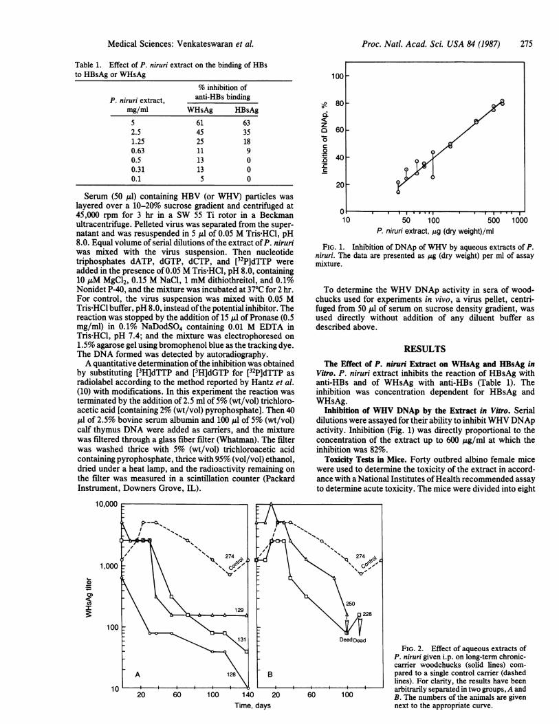
Table 1. Effect of P. niruri extract on the binding of HBsto HBsAg or WHsAg
% inhibition ofP. niruri extract, anti-HBs binding
mg/ml WHsAg HBsAg
5 61 632.5 45 351.25 25 180.63 11 90.5 13 00.31 13 00.1 5 0
Serum (50 ul) containing HBV (or WHV) particles waslayered over a 10-20% sucrose gradient and centrifuged at45,000 rpm for 3 hr in a SW 55 Ti rotor in a Beckmanultracentrifuge. Pelleted virus was separated from the super-natant and was resuspended in 5 dul of 0.05 M Tris HCl, pH8.0. Equal volume of serial dilutions ofthe extract ofP. niruriwas mixed with the virus suspension. Then nucleotidetriphosphates dATP, dGTP, dCTP, and [32P]dTTP wereadded in the presence of 0.05 M Tris HCl, pH 8.0, containing10 AuM MgCl2, 0.15 M NaCl, 1 mM dithiothreitol, and 0.1%Nonidet P-40, and the mixture was incubated at 370C for 2 hr.For control, the virus suspension was mixed with 0.05 MTris HCl buffer, pH 8.0, instead ofthe potential inhibitor. Thereaction was stopped by the addition of 15 A.l of Pronase (0.5mg/ml) in 0.1% NaDodSO4 containing 0.01 M EDTA inTris-HCl, pH 7.4; and the mixture was electrophoresed on1.5% agarose gel using bromophenol blue as the tracking dye.The DNA formed was detected by autoradiography.A quantitative determination ofthe inhibition was obtained
by substituting [3H]dTTP and [3H]dGTP for [32P]dTTP asradiolabel according to the method reported by Hantz et al.(10) with modifications. In this experiment the reaction wasterminated by the addition of 2.5 ml of5% (wt/vol) trichloro-acetic acid [containing 2% (wt/vol) pyrophosphate]. Then 40,ul of 2.5% bovine serum albumin and 100 Al of 5% (wt/vol)calf thymus DNA were added as carriers, and the mixturewas filtered through a glass fiber filter (Whatman). The filterwas washed thrice with 5% (wt/vol) trichloroacetic acidcontaining pyrophosphate, thrice with 95% (vol/vol) ethanol,dried under a heat lamp, and the radioactivity remaining onthe filter was measured in a scintillation counter (PackardInstrument, Downers Grove, IL).
10,000
1,000
4)0)
C0)In
100
1020 60 100 140 20 60 100
Time, days
100
80 _z0-
0.0c
60 _
40 _
20 _
10I I I I I q II I II I * I
50 100 500 1000P. niruri extract, mg (dry weight)/ml
FIG. 1. Inhibition of DNAp of WHV by aqueous extracts of P.niruri. The data are presented as jig (dry weight) per ml of assaymixture.
To determine the WHV DNAp activity in sera of wood-chucks used for experiments in vivo, a virus pellet, centri-fuged from 50 ,4 of serum on sucrose density gradient, wasused directly without addition of any diluent buffer asdescribed above.
RESULTSThe Effect of P. niruri Extract on WHsAg and HBsAg in
Vitro. P. niruri extract inhibits the reaction of HBsAg withanti-HBs and of WHsAg with anti-HBs (Table 1). Theinhibition was concentration dependent for HBsAg andWHsAg.
Inhibition of WHV DNAp by the Extract in Vitro. Serialdilutions were assayed for their ability to inhibitWHV DNApactivity. Inhibition (Fig. 1) was directly proportional to theconcentration of the extract up to 600 ,ug/ml at which theinhibition was 82%.
Toxicity Tests in Mice. Forty outbred albino female micewere used to determine the toxicity of the extract in accord-ance with a National Institutes ofHealth recommended assayto determine acute toxicity. The mice were divided into eight
FIG. 2. Effect of aqueous extracts ofP. niruri given i.p. on long-term chronic-carrier woodchucks (solid lines) com-pared to a single control carrier (dashedlines). For clarity, the results have beenarbitrarily separated in two groups, A andB. The numbers of the animals are givennext to the appropriate curve.
Medical Sciences: Venkateswaran et al.
276 Medical Sciences: Venkateswaran et al. Proc. Natl. Acad. Sci. USA 84 (1987)
TREATMENT
TREATMENT ENDS t0BEGINS
F/
//,370
_)bdI)
TREATMENTENDS
319
-20 0 20 40 60 8o 260 280
(4+)
\CONTROLTREATMENT_BEANS TREATMENT
ENDS
O. ) I DEAD+ + + - +
I>-°-SR I>
W(4.) b b,
44.)
TREATMENTENDS
1 339
-20 20 40
Time, days20 40 60 80 - 260 200 S30
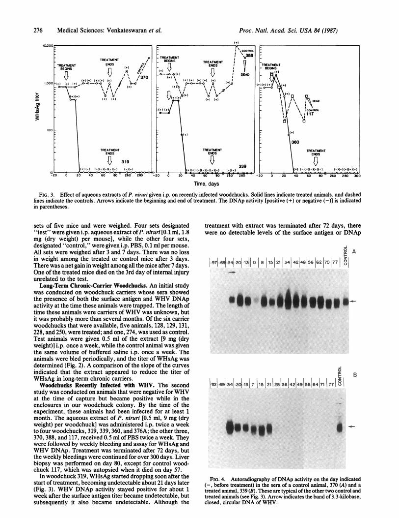
FIG. 3. Effect of aqueous extracts of P. niruri given i.p. on recently infected woodchucks. Solid lines indicate treated animals, and dashedlines indicate the controls. Arrows indicate the beginning and end of treatment. The DNAp activity [positive (+) or negative (-)] is indicatedin parentheses.
sets of five mice and were weighed. Four sets designated"test" were given i.p. aqueous extract ofP. niruri [0.1 ml, 1.8mg (dry weight) per mouse], while the other four sets,designated "control," were given i.p. PBS, 0.1 ml per mouse.All sets were weighed after 3 and 7 days. There was no lossin weight among the treated or control mice after 3 days.There was a net gain in weight among all the mice after 7 days.One of the treated mice died on the 3rd day of internal injuryunrelated to the test.Long-Term Chronic-Carrier Woodchucks. An initial study
was conducted on woodchuck carriers whose sera showedthe presence of both the surface antigen and WHV DNApactivity at the time these animals were trapped. The length oftime these animals were carriers ofWHV was unknown, butit was probably more than several months. Of the six carrierwoodchucks that were available, five animals, 128, 129, 131,228, and 250, were treated; and one, 274, was used as control.Test animals were given 0.5 ml of the extract [9 mg (dryweight)] i.p. once a week, while the control animal was giventhe same volume of buffered saline i.p. once a week. Theanimals were bled periodically, and the titer of WHsAg wasdetermined (Fig. 2). A comparison of the slope of the curvesindicated that the extract appeared to reduce the titer ofWHsAg in long-term chronic carriers.Woodchucks Recently Infected with WHV. The second
study was conducted on animals that were negative for WHVat the time of capture but became positive while in theenclosures in our woodchuck colony. By the time of theexperiment, these animals had been infected for at least 1month. The aqueous extract of P. niruri [0.5 ml, 9 mg (dryweight) per woodchuck] was administered i.p. twice a weekto four woodchucks, 319, 339, 360, and 376A; the other three,370, 388, and 117, received 0.5 ml ofPBS twice a week. Theywere followed by weekly bleeding and assay for WHsAg andWHV DNAp. Treatment was terminated after 72 days, butthe weekly bleedings were continued for over 300 days. Liverbiopsy was performed on day 80, except for control wood-chuck 117, which was autopsied when it died on day 57.
In woodchuck 319, WHsAg started dropping soon after thestart of treatment, becoming undetectable about 21 days later(Fig. 3). WHV DNAp activity stayed positive for about 1week after the surface antigen titer became undetectable, butsubsequently it also became undetectable. Although the
treatment with extract was terminated after 72 days, therewere no detectable levels of the surface antigen or DNAp
I-j°I A
_97-_6+3+2+131 0 8 15 21 34 42 48 56 62 7 77
..g.:jI"Xg u|" .-
B
p21-6-341-21-1317 1i5 12i1 2813614214915616417 1771 ° I
FIG. 4. Autoradiography of DNAp activity on the day indicated(-, before treatment) in the sera of a control animal, 370 (A) and atreated animal, 339 (B). These are typical ofthe other two control andtreated animals (see Fig. 3). Arrow indicates the band of 3.3-kilobase,closed, circular DNA of WHV.
1,000
01)
CO)X
00
TREATMENTBEGINS
MAI' 1~OAD
iI X (CONTRO#u117
360
TREATMENTENDS

Proc. Natl. Acad. Sci. USA 84 (1987) 277
; He r~~~~.-- % are,*w 'Z A.W O W !N, Aa.w %,e.-. .
FIG. 5. Comparison of livers from the untreated woodchuck 370(A and B) and the treated woodchuck 339 (C and D) that were infectedwith WHV and obtained at biopsy 8 days after the termination oftreatment. All preparations were sectioned in paraffin at 5 Am andstained with hematoxylin and eosin. (A) Typical pattern of chronicprogressive viral hepatitis with granulomatous inflammation cen-tered chiefly around portal triads with frequent "bridging" interven-ing spaces between them (b). Note blurring of intralobular cords ascompared with C (treated), owing to inflammatory swelling ofindividual hepatocytes. (x75.) (B) A periportal lesion, with anagglomeration of lymphocytes, plasmacytes, histiocytes, fibroblasts,and necrotic hepatocytes (n); proliferating biliary ductules (d) areconspicuous, which, along with fibrosis, indicate a progression tocirrhosis. (x300.) (C) Minimal inflammatory foci are barely visible inthe portal triads of this treated animal. Hepatocytic swelling and lossof crispness of cords is seen to the right of center. (x75.) Overall,these effects are much less than in the untreated liver. (D) Smallperiportal granuloma; hepatocyte and cords are well preserved, apartfrom rare necrosis (n). (x300.)
activity in 319 up to 300 days after the start of treatment (i.e.,228 days after termination of treatment). The control animal370, on the other hand, did not show a drop in either WHsAgtiter or DNAp activity up to 300 days.
Treated animals 339 and 360 showed a similar drop inWHsAg between 21 and 35 days after the start of treatment,followed 7-14 days later by a drop in WHV DNAp; themarkers stayed undetectable up to 300 days. Control animals117 and 388, however, showed high levels of the WHsAg andDNAp during the same period. Autoradiography of theproduct of DNAp reaction in the serial bleedings of controlwoodchuck 370 and P. niruri extract-treated woodchuck 339is given in Fig. 4. The presence of a band at 3.3 kilobases(arrow) indicated WHV DNAp activity up to 21 and 28 days.These became undetectable thereafter. DNAp of controlanimal 370 did not change.Woodchuck 376A, one of the four treated animals, became
bacteremic early in the experiment. Chloramphenicol wasadministered, but the animal succumbed to the infection. Itdid not respond to the extract.The histopathology of the livers of control woodchuck 370
Table 2. Data on liver biopsies performed 8 days aftertermination of treatment in experiment involvingwoodchucks recently infected with WHV
Pathology
Portal Focalinfiltrate necrosis Diagnosis
Control animal370 +++ ++ Chronic, active
hepatitis388 ++ ++ Chronic, active
hepatitis117 +++ + + Chronic, active
hepatitisP. niruri extract-
treated animal319 + + Mild viral
hepatitis339 + + Active hepatitis360 + 0 Minimal, portal
hepatitis0, None. ±, Marginal. +, Minimal positive. + +, Positive. +++,
Extensive.
and treated animal 339 are shown in Fig. 5. Data on the liverbiopsy performed 8 days after the termination of treatmentare given in Table 2. (Liver biopsies before treatment werenot available.) The three untreated controls, 370, 388, and117, showed extensive portal infiltration and focal necrosis;all three were diagnosed as chronic active hepatitis. Thelivers of the treated animals 319, 339, and 360, on the otherhand, showed marginal or negative portal infiltration andfocal necrosis. The diagnosis of woodchuck 319 was earlymild viral hepatitis, of 339 was early active hepatitis, and of360 was minimal portal hepatitis.
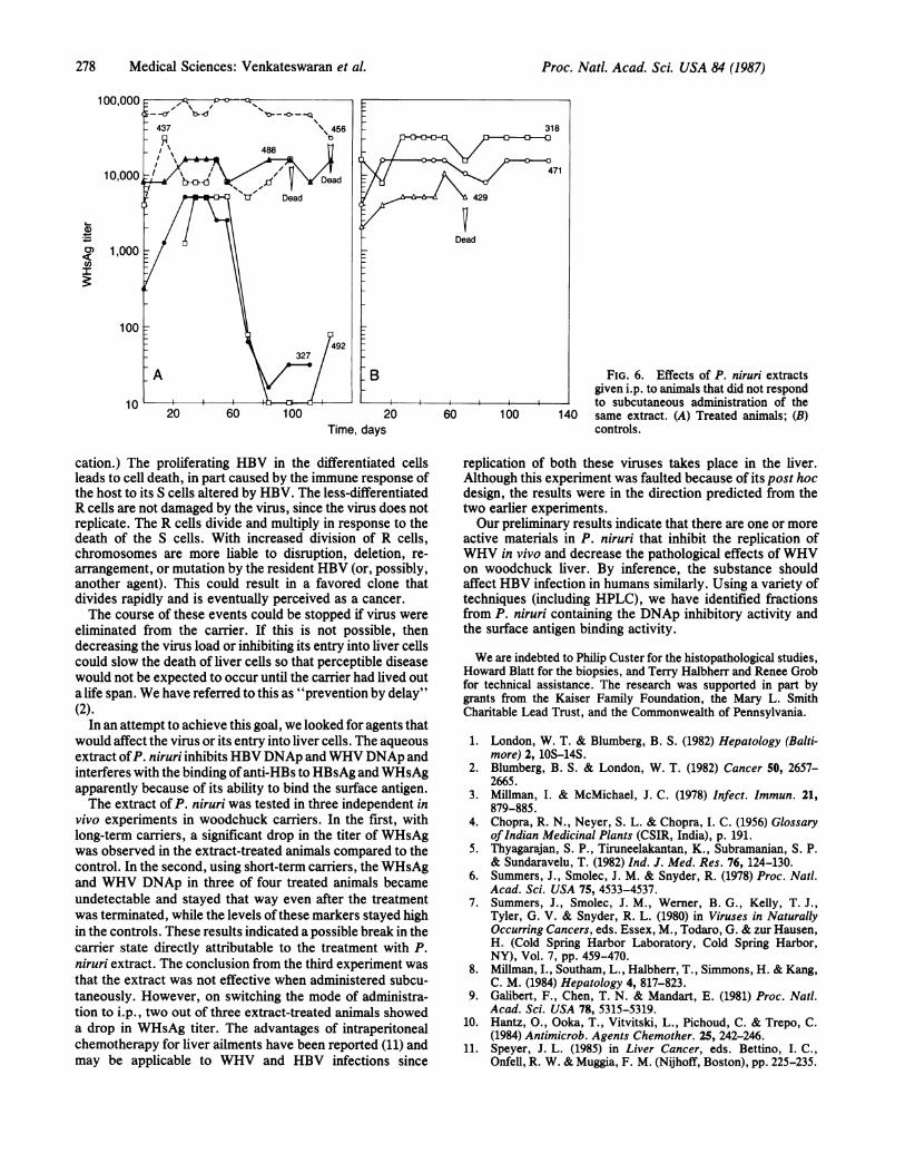
Subcutaneous Administration of the Extract in Long-TermCarrier Woodchucks. Five of eight long-term WHV-carrierwoodchucks (327, 437, 456, 488, and 492) were administered0.5 ml [9 mg (dry weight)] of extract subcutaneously twice aweek. The remaining three (318, 429, and 471) were given 0.5ml of PBS subcutaneously twice a week. The animals werebled weekly, and the titer of WHsAg and WHV DNAp wasmonitored. During 3 months of treatment, there was noappreciable change in either marker in the treated or controlanimals.We concluded that subcutaneous administration was inef-
fective. We hypothesized that either the active principle wasnot absorbed by this route, that antibodies were developedagainst the extract, or some other mechanism rendered itineffective. After 90 days the mode of administration waschanged to the intraperitoneal route that had apparently beensuccessful in the previous studies. Two of the treated animalsshowed a drop in WHsAg about 60 days after switching to i.p.administration (Fig. 6). One control and two treated animalsdied due to bacteremic infections. None of the controlanimals showed any significant change in WHsAg.
DISCUSSIONLondon and Blumberg (1) have proposed a model to explainthe observations on the relation between primary hepatocel-lular carcinoma and HBV. It postulates the existence of fullydifferentiated liver cells which, when infected, allow com-plete replication of HBV. (They are designated S cells; i.e.,susceptible to replication.) Less-differentiated liver cells(common in the fetus and newborn but less so in the adultliver), when infected, do not allow replication, althoughpenetration of the virus and integration of virus DNA mayoccur. (They are designated R cells; i.e., resistant to repli-
Medical Sciences: Venkateswaran et al.
278 Medical Sciences: Venkateswaran et al.
FIG. 6. Effects of P. niruri extractsgiven i.p. to animals that did not respond
- to subcutaneous administration of the140 same extract. (A) Treated animals; (B)
controls.
cation.) The proliferating HBV in the differentiated cellsleads to cell death, in part caused by the immune response ofthe host to its S cells altered by HBV. The less-differentiatedR cells are not damaged by the virus, since the virus does notreplicate. The R cells divide and multiply in response to thedeath of the S cells. With increased division of R cells,chromosomes are more liable to disruption, deletion, re-arrangement, or mutation by the resident HBV (or, possibly,another agent). This could result in a favored clone thatdivides rapidly and is eventually perceived as a cancer.The course of these events could be stopped if virus were
eliminated from the carrier. If this is not possible, thendecreasing the virus load or inhibiting its entry into liver cellscould slow the death of liver cells so that perceptible diseasewould not be expected to occur until the carrier had lived outa life span. We have referred to this as "prevention by delay"(2).
In an attempt to achieve this goal, we looked for agents thatwould affect the virus or its entry into liver cells. The aqueousextract ofP. niruri inhibits HBV DNAp and WHV DNAp andinterferes with the binding ofanti-HBs to HBsAg and WHsAgapparently because of its ability to bind the surface antigen.The extract of P. niruri was tested in three independent in
vivo experiments in woodchuck carriers. In the first, withlong-term carriers, a significant drop in the titer of WHsAgwas observed in the extract-treated animals compared to thecontrol. In the second, using short-term carriers, the WHsAgand WHV DNAp in three of four treated animals becameundetectable and stayed that way even after the treatmentwas terminated, while the levels of these markers stayed highin the controls. These results indicated a possible break in thecarrier state directly attributable to the treatment with P.niruri extract. The conclusion from the third experiment wasthat the extract was not effective when administered subcu-taneously. However, on switching the mode of administra-tion to i.p., two out of three extract-treated animals showeda drop in WHsAg titer. The advantages of intraperitonealchemotherapy for liver ailments have been reported (11) andmay be applicable to WHV and HBV infections since
replication of both these viruses takes place in the liver.Although this experiment was faulted because of its post hocdesign, the results were in the direction predicted from thetwo earlier experiments.Our preliminary results indicate that there are one or more
active materials in P. niruri that inhibit the replication ofWHV in vivo and decrease the pathological effects of WHVon woodchuck liver. By inference, the substance shouldaffect HBV infection in humans similarly. Using a variety oftechniques (including HPLC), we have identified fractionsfrom P. niruri containing the DNAp inhibitory activity andthe surface antigen binding activity.
We are indebted to Philip Custer for the histopathological studies,Howard Blatt for the biopsies, and Terry Halbherr and Renee Grobfor technical assistance. The research was supported in part bygrants from the Kaiser Family Foundation, the Mary L. SmithCharitable Lead Trust, and the Commonwealth of Pennsylvania.
1. London, W. T. & Blumberg, B. S. (1982) Hepatology (Balti-more) 2, 1OS-14S.
2. Blumberg, B. S. & London, W. T. (1982) Cancer 50, 2657-2665.
3. Millman, I. & McMichael, J. C. (1978) Infect. Immun. 21,879-885.
4. Chopra, R. N., Neyer, S. L. & Chopra, I. C. (1956) GlossaryofIndian Medicinal Plants (CSIR, India), p. 191.
5. Thyagarajan, S. P., Tiruneelakantan, K., Subramanian, S. P.& Sundaravelu, T. (1982) Ind. J. Med. Res. 76, 124-130.
6. Summers, J., Smolec, J. M. & Snyder, R. (1978) Proc. Natl.Acad. Sci. USA 75, 4533-4537.
7. Summers, J., Smolec, J. M., Werner, B. G., Kelly, T. J.,Tyler, G. V. & Snyder, R. L. (1980) in Viruses in NaturallyOccurring Cancers, eds. Essex, M., Todaro, G. & zur Hausen,H. (Cold Spring Harbor Laboratory, Cold Spring Harbor,NY), Vol. 7, pp. 459-470.
8. Millman, I., Southam, L., Halbherr, T., Simmons, H. & Kang,C. M. (1984) Hepatology 4, 817-823.
9. Galibert, F., Chen, T. N. & Mandart, E. (1981) Proc. Natl.Acad. Sci. USA 78, 5315-5319.
10. Hantz, O., Ooka, T., Vitvitski, L., Pichoud, C. & Trepo, C.(1984) Antimicrob. Agents Chemother. 25, 242-246.
11. Speyer, J. L. (1985) in Liver Cancer, eds. Bettino, I. C.,Onfell, R. W. & Muggia, F. M. (Nijhoff, Boston), pp. 225-235.
11
0)
C,)-
20Time, days
Proc. Natl. Acad. Sci. USA 84 (1987)