photosynthesis - matthias-schleiden-institut
TRANSCRIPT

Photosynthesis
1. Introduction and evolution2. Light reactions at the thylakoid membrane3. Dark reactions: C3 photosynthesis and
photorespiration4. C4 Photosynthesis – CAM plants5. N and S metabolism6. Plastid gene expression
CO2
O2
C6H12O16

1. Introduction and evolution

Electro-magnetic irradiance and sunlight

CO2 and O2 fixation by Rubisco
’ Development of C4 photosynthesis

Oxygenic photosynthesis was established in Cyanobacteria

Plastids derive from endosymbiosis
Localisation of the photosynthetic complexes
- 100 chloroplasts/cell
- chloroplasts have two membranes with different origin(lipids, proteins)
- thylakoids derive from inner (procaryotic) membrane- grana- and stroma thylakoids
-new compartment: lumen vs. stroma
- different transport processes for proteins into the two plastid compartments
-photosynthesis in thylakoid membranes

Pigments: chlorophyll, carotinoid, phycobilin

Biosynthesis of chlorophylls
- starts with glutamate
- porphyrin
- tetrapyrrole

POR is light-dependent
POR“
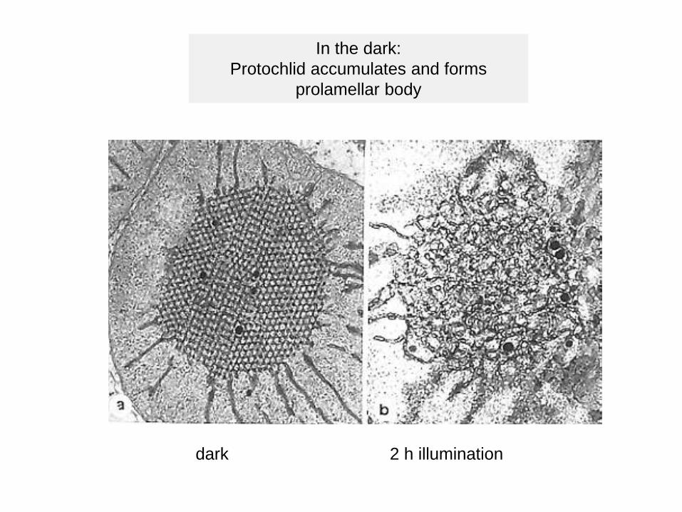
In the dark: Protochlid accumulates and forms
prolamellar body
dark 2 h illumination


Chl absorbs visible light

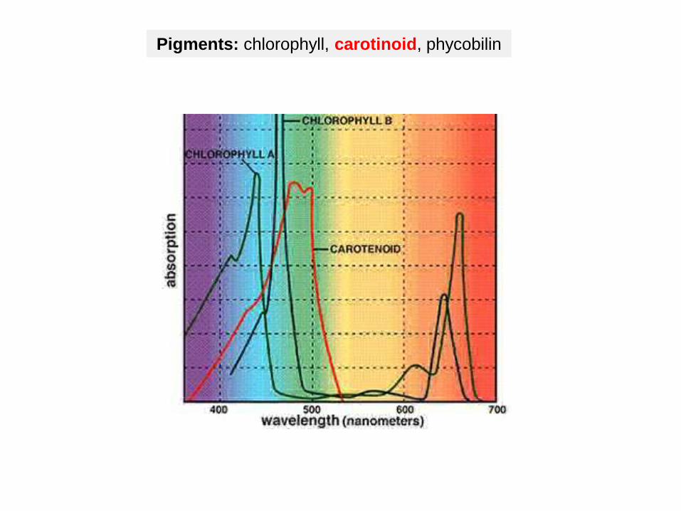
Pigments: chlorophyll, carotinoid, phycobilin

Light absorption


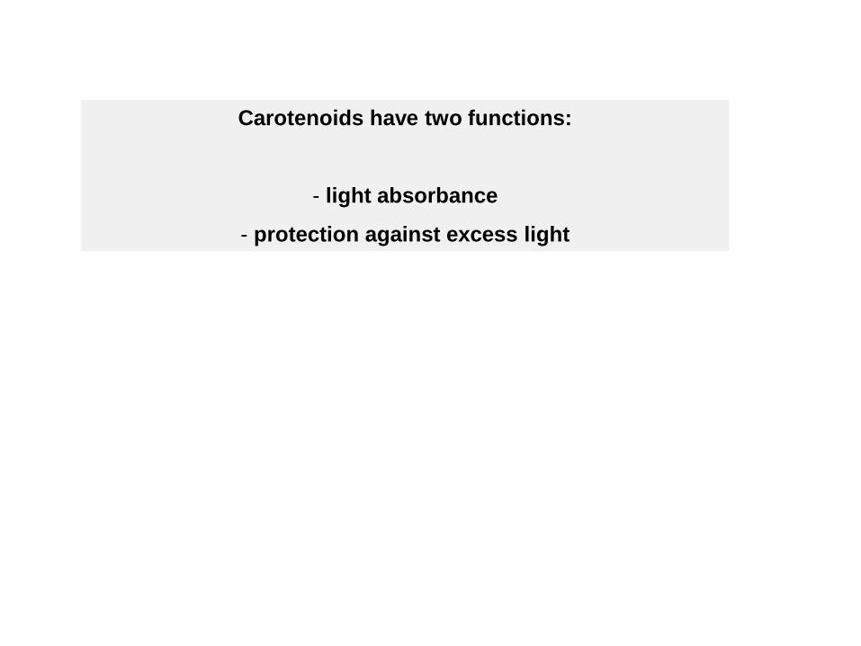
Carotenoids have two functions:
- light absorbance
- protection against excess light

Energy dissipation by xanthophylls
Low light = Violaxanthin is present in and around PSII
High light = Zeaxanthin synthesis in and around PSII

In response to high light, plants have evolved photo protection mechanisms to dissipate the excess absorbed light energy and thus avoid damages to the photosynthetic apparatus. One of
the mechanisms is through transfer of the absorbed energy from chlorophyll a to xanthophyll pigment zeaxanthin since excited zeaxanthin decays to the ground level much more rapidly than excited chlorophyll a. Under excessive light condition, violaxanthin is converted to zeaxanthin in the xanthophyll cycle, and thus accelerates the energy
dissipation from excited chlorophyll a to zeaxanthin.

Phycocyanin: chromophor is bound to the proteinOpen chain tetrapyrol system
Pigments: chlorophyll, carotinoid, phycobilin
phycoerythrinphycocyanin
allophycocyanin

Phycoerythrin, 545 nm
Phycocyanin, 615 nm
Allophycocyanin, 650 nm

Organisation of light harvesting complexes in cyanobacteria and higher plants
cyanobacteria higher plants
Antenna with phycobilisomes Antenna of higher plantsON the thylakoid membrane IN the thylkoid membrane

Cyanobacteria
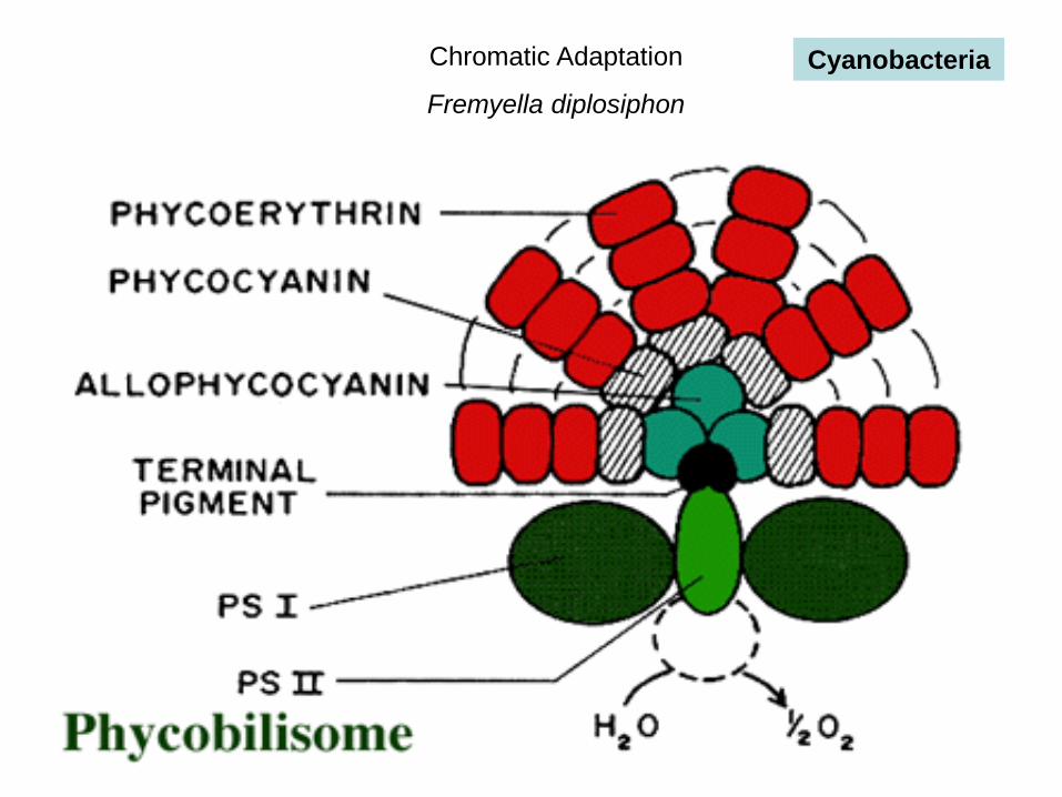
Chromatic Adaptation
Fremyella diplosiphon
Cyanobacteria

Antenna harvest light energy and transfer it to the reaction centers of the photosystems II or I
Pigments in antenne:Energy transfer
Pigment (P680 or P700) in the photosystemsPhotochemical work
Light harvesting complexes Higher plants

Most of the chlorophylls and carotenoids are organized in light-harvesting complexesof the photosystems I and II

Chl a and b (and carotenoids) not covalently bound to light-harvesting proteins of antenna
Stable antenna for the photosystems I and II
Mobile antenna migrates between photosystems
Pigments are also bound to the two photosystems
-inner antenna
- reaction center pigmnents
-Light harvesting proteins are encoded by multigene families
-Specificity for the two photosystems
-Phyologenetic analyses
-Early light-induced proteins (ELIPs)

Mobile antenna
Non-mobile antenna
Both contain Chl, car, and LHCPs
LHCPs evolved from ELIPs

Mutants in photosynthesis
Light is absorbed by antenna
and emited as fluorescence
hcf (high chlorophyll fluorescence) phenotype

Pigments in PSII and PSI reaction centers are excited by different wavelengths
P680, P700
Generation of redox signals

Redox signals regulate plastid and nuclear gene expression

Adaptation to unbalanced excitation of PSII and PSI
(1) – state transition (fast): relocation of mobile antenna
(2) – change in plastid gene expression: genes for limiting PS complex are upregulated
(3) – change in nuclear gene expression

More PSII excitation > PSI activity is limiting
Phosphorylation of mobile antenna
migration to PS I

The four photosynthetic complexes are evolutionary conserved fromcyanobacteria to higher plants
Photosystem II
Cytochrom b6/f-complex
Photosystem I
ATP-synthase
2. Light reactions at the thylakoid membrane


In eukaryotic organism: thylakoid proteins are of dual genetic origin


Photosystem II
Yz = Tyr 161

D1PsbA


D1 protein
- high turnover
- de novo synthesis in the stroma thylakoids during insertion into membrane
- co-factor bindung
- translational arrest when no chlorophyll is available
- transport to grana thylakoids
- association with antenna
- high light: photodestruction
- D2: similar regulation

Two electron transporters: QA and QB

The Mn cluster splits
water and is
responsible for
O2 evolution


1
2
3

Cytochrome b6/f-complex


2 Hämgruppen vom b-Typ
(Cytochrom b)
1 Hämgruppe vom
c-Typ
(Cytochrom f)

Photosystem I


Cyclic electron transport
- electrons from PSI to cytochrome-b6f-complex, back to P700
- no oxygen evolution, no NADPH synthesis
- generation of proton gradient

ATP synthase


Paul D. Boyer and John E. Walker have shown how the enzyme ATP synthase makes ATP. ATP synthase is found in chloroplast and mitochondrial membranes and in the cytoplasmic membrane of bacteria. A difference in hydrogen ion concentration across the membrane drives the enzyme to synthesise ATP.
"The Binding Change Mechanism"Using chemical methods Paul Boyer proposed that ATP synthase is like a cylinder with alternating alpha and beta subunits. An asymmetrical gamma subunit in the middle of the cylinder causes changes in the structure of the beta subunits when it rotates (100 r.p.s.). He termed these structures open (betaO), loose (betaL) and tight (betaT).
Four stages in ATP synthesis


3. Dark reaction: C3 photosynthesis and photorespiration

Ribulose-1,5-bisphosphate-carboxylase fixes CO2

Bifunctionality of Rubisco
- binding O2 instead of CO2
- photorespiration

Photorespiration


4. C4 photosynthesis &CAM plants
CO2 compensation point
CO2 concentration[%, v/v]
photosynthesisrate

- High light intensity ’ high photorespiration, stomata closure
- C4 plants:more efficient CO2 fixation by phosphoenolpyruvate (PEP) carboxylase
- Polyphyletic origin, convergent development
- C4 vs.Crassulaceae acid metabolism (CAM) plants:samle biochemistry, different morphology and CO2 fixation
- Examples for C4 plants: maize, sugar cane, Sorghum, Chenopodiaceae, Euphorbiacecae
- Examples for CAM plants: succulent plants of Crassulacecae, Cactaceae, Compositae, Euphorbiaceae

Mesophyll cell:Normal chloroplasts with grana
Chloroplast dimorphism
Bundle sheath cell:Starch-rich plastids without grana (no PS II)


mesophyll cell bundle sheath cell
1 - PEP carboxylase2 – malate dehydrogenase
3 – malate enzyme4 - Calvin cycle
CO2e-

Biochemistry in different cellular compartments

Summary C4 photosynthesis
- Spacial separation of CO2 fixation and carbohydrate biosynthesis
Mesophyll cells:
- PEP carboxylase high affinity to CO2, no O2 fixation- More efficient CO2 fixation in spite of closed stomata than C3
plants- First C fixation product: C4 compound oxalacetate- Reduction of oxalacetate to malate in plastids of mesophyll
cells requires NADPH from photosynthesis- Malate is transported to bundle sheath cells via
plasmodesmata
Bundle sheath cells:
- Oxidation of malate to pyruvate and CO2 generates NAPDH- ’ chloroplast dimorphism

CAM plants
Temporal separation of CO2 fixation and carbohydrate biosynthesis
Night: stomata open: CO2 fixation
Day: stomata closed: carbohydrate biosynthesis and photosynthetic light reactions
Storage of CO2 fixation product: malate in vacuole (acidification during night)


Evolution of PEP carboxylase
A PEP carboxylase gene (PEPC) is present in many eukaryotic cells, but the gene product does not play an important role in the metabolism.
PEPC gene
1- Stronger promoter: higher expression level2- Tissue- specific cis-elements in promoter: expression in
mesophyll cells
PEPC enzyme
3- Optimization of CO2 binding side: higher affinity to CO2

5. N and S metabolism
NADPH and reduced ferredoxin from light reaction is also used to reduce NO2
- and SO42-.
Nitrate reduction
NO3- NO2
- NH4+
Sulfate reduction
SO42- SO3
2- SH+
NR NiR
SR SiR

5. N and S metabolism
NADPH and reduced ferredoxin from light reaction is also used to reduce NO2
- and SO42-.
Nitrate reduction
NO3- NO2
- NH4+
Sulfate reduction
SO42- SO3
2- SH+
NR NiR
SR SiR
cytosol

5. N and S metabolism
NADPH and reduced ferredoxin from light reaction is also used to reduce NO2
- and SO42-.
Nitrate reduction
NO3- NO2
- NH4+
Sulfate reduction
SO42- SO3
2- SH+
NR NiR
SR SiR
plastid
plastid

NO3-
NO2-
NH4+
Nitrate assimilation

NO3-
NO2-
NH4+
cytoplasmic energy source photosynthesis

NO3-
NO2-
NH4+
co-factors

Ammonium is toxic and rapidly metabolized

Sulfate assimilation
Siroheme, e- from ferredoxin

Life on earth is dependent on sulphur (S) and nitrogen (N). In plants, the second step in the reduction of sulphate and nitrate are mediated by the enzymes sulphite and nitrite reductases, which contain the iron (Fe)-containing siroheme as a cofactor. It is synthesized from the tetrapyrrole primogenitor uroporphyrinogen III in the plastids via three enzymatic reactions, methylation, oxidation and ferrochelatation. Without siroheme biosynthesis, there would be no life on earth.
Baishnab C Tripathy, Irena Sherameti, and Ralf Oelmüller (2010) Siroheme. An essential component for life on earthPlant Signal Behav. 2010 Jan; 5(1): 14–20.
2
Siroheme

6. Plastid geneexpression
Plastids contain DNA – plastome
- Maternal inheritance
(advantage for biotechnological application)(Mirabilis japonica, Correns 1909)
- 100 x 100 plastoms/cell
- prokaryotic origin, procaryotic genes and expression
- gene transfer to the nucleus

Gene expression in plastids is procaryotic

Inheritance in plastids
- Pelargonium:
- biparental
- maternal (most of the angisperms)
- paternal (gymnosperms, Sequoia, Pinus)

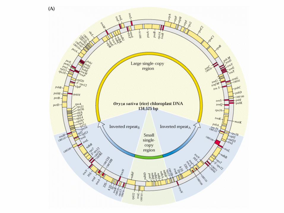
plastome
- DNA is attached to thylakoid membrane (nucleoid)
- 15 nucleoids/plastid, 10 DNA molecules/nucleotid (polyploid)
- circular DNA
- 130 bis 160 kb
- inverse duplication
- small and large single copy region
- loss of inverse duplication e.g. conifers, Papilionaceae
Epiphagus

The plastome of the holoparasite Epifagus virginiana is substantially reduced
Model system for plastid genetics

Plastomes of land plants

Genes of the plastome

Gene expression in plastids requires pro- and eukaryotic elements
Operons und Introns

Most of the promoters are procaryotic – but not all of them
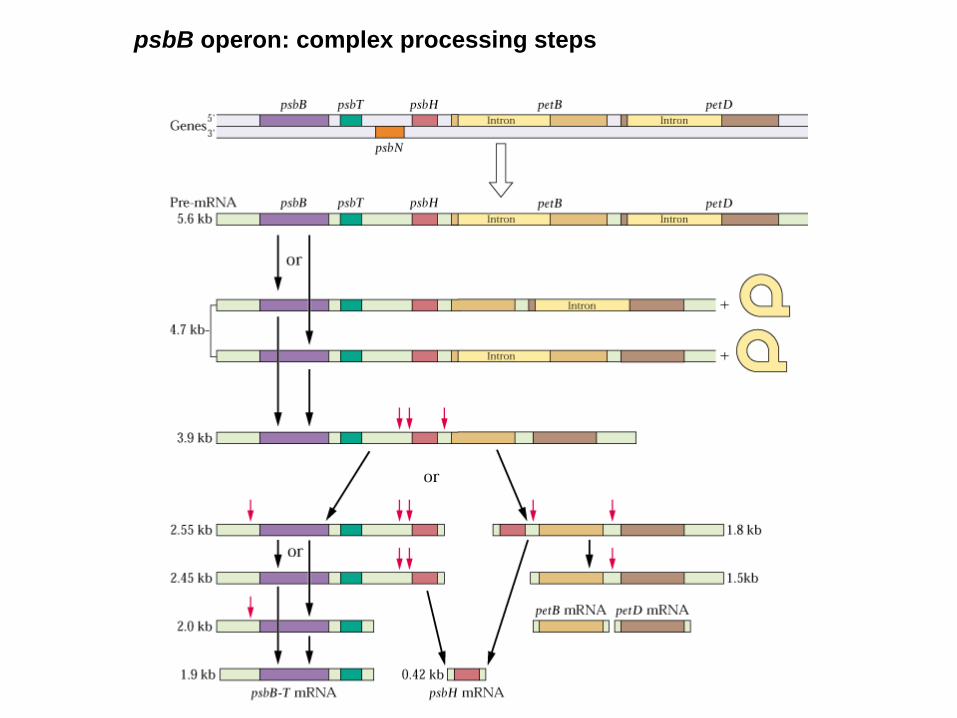
psbB operon: complex processing steps

psbB operon- multiple promotors, multiple transcription start sites
- both strands encode genes
- polycistronic transcripts
- primary transcript is large and unstable
- RNA codes for independent proteins
- transcript ripening, oligocistronic transcripts
- monocistronic transcripts
- specific endonucleases
- Exonucleases: processing of 3´-ends
- hair pin loops stabilizes RNA
- secondary structures prevent degradation

Plastids contain two RNA-polymerases
- Epiphagus: lost the genes for RNA-polymerases, but still contain white plastids
- nuclear-encoded RNA-polymerase - plastid-encoded RNA-polymerase
- phage type - bacterial type - one subunit - ~13 subunits, nuclear- and plastid-encoded
- sigma factors (bacteria-like) - Expression of early genes - Expression of late genes
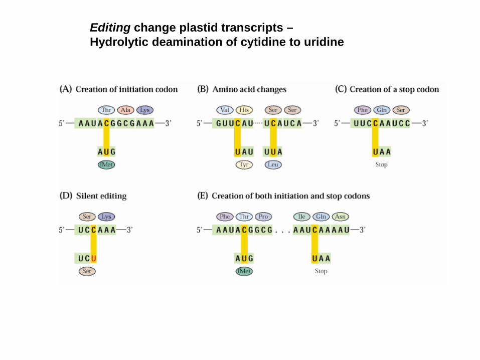
Editing change plastid transcripts –Hydrolytic deamination of cytidine to uridine

Most of the transcripts are stable

Many genes from plastids were transferred to the nucleus
- DNA fragment
- as RNA after reverse transcription (e.g. as edited transcripts)