photodynamic therapy in the canine prostate using...
TRANSCRIPT

Photodynamic Therapy in the Canine Prostate UsingMotexafin Lutetium
R. Alex Hsi,1 Amy Kapatkin, John Strandberg,Timothy Zhu, Theodor Vulcan,Michael Solonenko, Carmen Rodriguez,John Chang, Mark Saunders, Nicole Mason, andStephen HahnDepartment of Radiation Oncology, Virginia Mason Medical Center,Seattle, Washington 98101 [R. A. H.]; Department of RadiationOncology [T. Z., T. V., M. S., C. R., J. C., S. H.], University ofPennsylvania School of Medicine, Department of Clinical Studies[A. K., M. S., N. M], University of Pennsylvania School of VeterinaryMedicine, Philadelphia, Pennsylvania 19104; and Division ofComparative Medicine, Johns Hopkins University School ofMedicine, Baltimore, Maryland 21205 [J. S.]
ABSTRACTOur purpose was to determine the feasibility of com-
prehensive treatment of the canine prostate with photody-namic therapy (PDT) using motexafin lutetium (Lu-Tex)and to evaluate the toxicity and tissue effects associated withthis treatment. Twenty-five adult male beagles with normalprostate glands were given an i.v. injection of the second-generation photosensitizer Lu-Tex (2–6 mg/kg). An addi-tional two dogs were used as controls and did not receive anyphotosensitizing drug. All 27 dogs underwent laparotomy toexpose the prostate. Three hours postinjection, a total doseof 75–150 J/cm of 732 nm laser light was delivered intersti-tially and/or transurethrally to the prostate via cylindricaldiffusing fibers. Dogs were euthanized between 2 days and 3months after PDT. All subjects were monitored for clinicalevidence of toxicity. Specimens were examined macroscop-ically and microscopically to characterize the tissue reactionand assess extent of tissue effect as a result of treatment.Interstitial and/or transurethral PDT were successfully de-livered in all dogs with no perioperative complications. Noclinical evidence of acute urinary obstruction or rectalbleeding was noted. At all dose levels, macroscopic andmicroscopic evaluation revealed a prostatic tissue reactioncharacterized initially (within 48 h) by inflammation andnecrosis followed by fibrosis and glandular epithelial atro-phy. Comprehensive treatment of the entire prostate couldbe achieved using the interstitial alone approach or com-bined transurethral and interstitial approach. The transure-
thral alone approach did not result in complete coverage ofthe prostate. Dogs receiving transurethral or combined in-terstitial and transurethral treatment developed erythemaand urethral epithelial disruption at all dose levels. Thosereceiving combined treatment at the highest dose level (Lu-Tex 6 mg/kg, 150 J/cm light) developed urethral fistulae andperitonitis. Dogs treated with the interstitial alone approachwere found to have the least amount of urethral damage.Comprehensive treatment of the canine prostate with Lu-Tex PDT is feasible using an interstitial alone or combinedinterstitial and transurethral approach. The interstitialalone technique results in the least amount of toxicity. Theprostatic tissue reaction to treatment is characterized byinitial inflammation and necrosis followed by fibrosis andglandular epithelial atrophy.
INTRODUCTIONPDT2 is a treatment modality using light of an appropriate
wavelength to activate a photosensitizer in the presence ofoxygen, resulting in localized tissue necrosis. PDT has beenapproved by the Food and Drug Administration for the treat-ment of selected patients with esophageal cancer and lungcancer (1). Other potential applications and techniques for PDTare being actively explored in the clinic (2–7).
The placement of optical fibers directly into a tumor ororgan (interstitial light delivery) is likely the most suitablemethod for the volumetric treatment of bulk tumor using PDT.Interstitial light delivery to the prostate gland is potentiallyfeasible based on existing techniques used for radioactive im-plants (8). Clinically, PDT has potential as a treatment forprimary localized prostate cancer or for locally recurrent diseasefor which treatment options are limited (9). Several preclinicalstudies have evaluated the feasibility of delivering PDT via aninterstitial approach in a canine model (10–13). Light-diffusingfibers were introduced directly into the prostate gland and/or viathe urethra to deliver the light to the gland. To date, however, nostudy has directly addressed whether PDT has the potential forcomprehensively treating the prostate gland. This issue is im-portant if one wishes to treat prostate cancer (14).
Various photosensitizers have been used for PDT of theprostate gland in preclinical studies. The first-generation pho-tosensitizer, Photofrin, has been shown to cause glandular ne-crosis in the canine prostate (10). There are several limitationsfor the use of Photofrin-mediated PDT in the prostate gland.Photofrin is activated by 630 nm light, which has a limited depthof penetration into tissues and is absorbed by hemoglobin. Thisis potentially a significant problem in the prostate where place-ment of interstitial needles may lead to bleeding within the
Received 10/3/00; revised 12/18/00; accepted 12/28/00.The costs of publication of this article were defrayed in part by thepayment of page charges. This article must therefore be hereby markedadvertisementin accordance with 18 U.S.C. Section 1734 solely toindicate this fact.1 To whom requests for reprints should be addressed, at Departmentof Radiation Oncology, Virginia Mason Medical Center, 1100 NinthAvenue, CB-RO, Seattle, WA 98101. E-mail: [email protected].
2 The abbreviations used are: PDT, photodynamic therapy; Lu-Tex,motexafin lutetium; mTHPC,meso-tetra(m-hydroxyphenyl)chlorin.
651Vol. 7, 651–660, March 2001 Clinical Cancer Research
Research. on June 22, 2018. © 2001 American Association for Cancerclincancerres.aacrjournals.org Downloaded from

tissues. Furthermore, Photofrin is a partially purified mixture ofporphyrin monomers and oligomers and is clinically associatedwith prolonged (6–8 weeks) skin photosensitivity.
Second-generation photosensitizers may have some advan-tages for interstitial prostate PDT compared with Photofrin. Ingeneral, the second-generation photosensitizers are pure com-pounds and are associated with shorter skin photosensitivity.Many of these compounds are activated by longer wavelengthsof light; thus, the treatment effect may have deeper tissuepenetration. Lu-Tex is a second-generation photosensitizer withreported efficacy in murine tumor models (15, 16) and humanclinical trials (17). Lu-Tex is a tripyrrolic pentaaza-expandedporphyrin with an absorption band at 732 nm. This longerwavelength of activating light in the far red range may permitmore optimal interstitial light delivery than other wavelengthsbecause of greater depth of penetration. Another advantage offar red light is that there is less absorption of light by hemoglo-bin compared with shorter wavelengths. Skin photosensitivity isrelatively minor, with Lu-Tex lasting;24–48 h. These featuressupport the investigation of Lu-Tex as a photosensitizer forinterstitial PDT of the prostate gland.
In this study, we evaluated Lu-Tex-mediated PDT of theprostate in a canine model. The primary end points of this studywere: (a) to determine the feasibility of comprehensive treat-ment of the entire prostate gland using an interstitial, transure-thral, or combined interstitial and transurethral light deliveryapproach; (b) to determine the toxicities associated with Lu-TexPDT in the canine prostate; and (c) to characterize the histolog-ical effect of Lu-Tex PDT on prostatic tissue. The results of thisstudy form the basis on which a future human trial will beinitiated.
MATERIALS AND METHODSAnimals, Anesthesia, and Surgical Procedure. Exper-
iments were conducted at the University of Pennsylvania undera protocol approved by the Institutional Animal Care and UseCommittee. A total of 27 adult male beagles weighing between8.8 and 15.0 kg (median, 12.0 kg) were used. All dogs were 1–3years old. Six dogs were from Harlan Sprague Dawley (Indian-apolis, IN), 6 from Marshall Farms (North Rose, NY), 11 fromSummitt Ridge Farm (Susquehanna, PA), 2 from RidglanFarms, Inc. (Mount Horeb, WI), and 2 from White Eagle Lab-oratories, Inc. (Doylestown, PA). Dogs were quarantined for 2weeks. All dogs received complete blood counts, blood chem-istry, and fecal testing to ensure their health. All were vacci-nated withLeptospira canicola-icterohaemorrhagiaebacterinand canine distemper/adenovirus type/parainfluenza/parvovirusvaccine from Fort Dodge Laboratories, Inc. (Fort Dodge, IA).All surgical procedures were performed at a University ofPennsylvania animal surgical facility using standard sterile sur-gical technique.
Biopsies of the prostate were performed under sedation(i.v. Propofol 10 mg/ml to effect) using transabdominal ultra-sound-guided 14-gauge biopsy needles (Precision Cut; BectonDickinson, Rutherford, NJ) 2–4 weeks preceding Lu-Tex ad-ministration. All prostates were sized and assessed as normal.
The dogs were premedicated with morphine sulfate, atro-pine, and acepromazine before surgery and PDT. Anesthesia
was induced with i.v. thiopental and maintained with 2% isoflu-rane inhalation via an endotracheal tube. All dogs were moni-tored by electrocardiography, indirect blood pressure, rectaltemperature probe, and pulse oximetry. A preoperative colonos-copy and cystoscopy were performed to clinically assess thegenitourinary and gastrointestinal tracts preceding PDT. Eachdog was then placed in dorsal recumbency, and the skin wasprepared for surgery using standard aseptic technique. A heatingpad was used to maintain body temperature. A midline incisionwas made through the skin extending from the umbilicus to thepubis symphysis and through the linea alba. The connectivetissue and fat were dissected to expose the prostate gland(Fig. 1).
Photodynamic Therapy. A total of 27 dogs weretreated. Lu-Tex (Pharmacyclics, Inc., Sunnyvale, CA) was ad-ministered (2 or 6 mg/kg i.v.) 3 h prior to light administration.A 3-h drug-light interval was chosen because preclinical studiesin other model systems demonstrated antitumor efficacy withthis timing (15–17). Two light sources were used during thecourse of this study. In the first 15 dogs, a KTP/532 laserpumping a model 630 XP Dye Module (LaserScope, Inc., SanJose, CA) tuned to emit 732 nm (2 W maximum power) was
Fig. 1 Exposure of canine prostate at laparatomy.
652 Photodynamic Therapy in the Canine Prostate
Research. on June 22, 2018. © 2001 American Association for Cancerclincancerres.aacrjournals.org Downloaded from

used. For the remaining 12 dogs, a diode laser, model 730 (2 Wmaximum power; Diomed, Ltd., Cambridge, United Kingdom,kindly provided by Pharmacyclics, Inc.) was used. A 2.5-cmcylindrical diffusing fiber with an outside diameter of 800mm(Rare Earth Medical, Inc., West Yarmouth, MA) was used forlight delivery. All fibers were calibrated before and after theprocedure using an integrating sphere (Diomed, Ltd.). The trans-mission efficiency of each fiber, defined as the ratio betweenlight output (as measured from the integrating sphere) and input,was found to be between 58 and 61%. No changes in efficiencywere noted between pre- and posttreatment measurements.
After the prostate was exposed, the diffusing fibers wereplaced in the gland. Interstitial placement of the diffusing fiberswas performed using a custom-made polyurethan template withevenly spaced holes which was attached to a pole and centeredover the prostate (Fig. 2). Plastic 17-gauge needles (Best Indus-tries, Inc., Springfield, VA) containing metal trocars wereplaced in parallel at the desired locations in the prostate throughthe template. Each needle was presoaked in heparin (HeparinLock Flush Solution; Abbott Laboratories, Chicago, IL) to helpprevent thrombus formation around the needle as had beennoted in a previous report by Chenet al. (18). The needles werethen pushed through the prostatic tissue from a ventral to dorsaldirection until the tip could be felt on the opposite side. Thetrocars were removed and replaced with the light diffusers.There was initial concern that the opaque plastic needles mightreduce light output from the cylindrical diffusing tips. Outputwas therefore measured in an integrating sphere with a barefiber and a fiber sheathed in a needle. Transmission of lightthrough the opaque plastic needles was 95% as compared withthat of the bare fibers. For light fluence/fluence rate specifica-tion, output was specified as output from the bare fiber.
Transurethral treatments were performed by placing thediffusing fiber into a clear plastic 5 French 15-inch feeding tube
(Premature Infant Feeding Tube; CR Bard, Inc., Covington,GA) and inserting the tube in the urethra until the fiber tipreached just below the bladder neck.
The light fluence was prescribed based on the unit length ofradial diffusing fiber (J/cm). The dogs received total fluencesranging from 75 to 150 J/cm at a fluence rate of 75 or 150mW/cm. The temperature of the prostate during light deliverywas monitored (;5 mm from the light source) using a thermalmicroprobe (Physitemp Instruments, Clifton, NJ).
Table 1 summarizes the treatment delivered to all dogs.Two control dogs were treated with light alone and received noLu-Tex. When comprehensive treatment of the entire prostatewas attempted, the number of interstitial sites used ranged fromfour to six depending on the size of the prostate gland.
Postoperatively, dogs were observed by a veterinarian forsigns of pain/distress or urinary symptoms twice daily for 1week and once daily thereafter. Butorphenol tartrate (10 mg/ml)was administered 0.4 mg/kg i.v. or i.m. as needed. Selected dogsunderwent cystoscopy and colonoscopy days to weeks afterPDT to document any visible urethral or rectal toxicity. Dogswere sacrificed from 2 days to 3 months after PDT with i.v.pentobarbital sodium (389 mg/ml) 1 ml/3 kg.
Fig. 2 Polyurethane templateplaced over canine prostate toguide interstitial light sourceplacement.
Table 1 Treatment schema
Lu-Tex dose (mg/kg)Light dose
(J/cm) ITa TU IT 1 TU
6 75 3 16 150 2 1 70 (control) 150 12 100 6 3 20 (control) 100 1
a IT, interstitial light delivery; TU, transurethral light delivery;IT 1 TU, interstitial and transurethral light delivery.
653Clinical Cancer Research
Research. on June 22, 2018. © 2001 American Association for Cancerclincancerres.aacrjournals.org Downloaded from

Pathology Review. At necropsy, the bladder, prostate,and rectum were removed and fixeden bloc in 10% neutralbuffered formalin. The prostate and urinary bladder were dis-sected from the rectum, taking care to observe adhesions orevidence of gross injury to the organs and surrounding connec-tive tissues. The urinary bladder and prostate were dividedserially in 3–4-mm blocks, photographed, and embedded inparaffin for histological examination. Sections 6mm thick werestained with H&E and examined to determine the presence ofnecrosis, hemorrhage, fibrosis, glandular atrophy, inflammation,and other abnormalities.
RESULTSLu-Tex Administration and Toxicity. No evidence of
skin photosensitivity was observed during the follow-up periodof any animal. All dogs were kept in normally illuminated cagesin an approved animal facility, although they were not exposedto direct sunlight. An apparent allergic reaction to the photo-sensitizer injection was noted in 10 of 14 dogs receiving 6mg/kg Lu-Tex. This reaction was manifested by generalizedhives as well as muzzle and paw edema. No respiratory prob-lems were encountered. However, 5 dogs briefly required i.v.fluids to maintain mean blood pressure.60 mm Hg at the timeof these reactions. Only 5 of 11 dogs receiving 2 mg/kg Lu-Texexperienced this reaction. None of these dogs required anyspecific treatment as a result of the reaction.
Clinical Results and Toxicity. In the initial experiments,four dogs were treated (three interstitial alone and one transure-thral alone light source arrangement) with Lu-Tex 6 mg/kg i.v.followed 3 h later by light delivery with a fluence of 75 J/cm andfluence rate of 150 mW/cm (Table 1). The goals of these initialexperiments were to identify the clinical tolerance of this doseof drug and light within a 2–3-week time frame and to evaluatethe zone of necrosis around both interstitial and transurethraltreatment fibers. This study showed that the zone of tissuedamage surrounding the fibers was irregular but;1.2 cm indiameter. Given that the diameter of the prostates were signif-icantly larger than the zone of tissue damage created by a singlefiber, it was concluded from these studies that transurethral lightdelivery alone would not achieve the goal of comprehensivetreatment of the entire prostate gland. Our initial measurementsof the zone of necrosis, however, suggested that interstitial fiberspacing of 1 cm would allow for comprehensive treatment of thegland at this dose level of light.
Based on these initial results, the light fluence was esca-lated to 150 J/cm at a fluence rate of 150 mW/cm and theLu-Tex dose remained at 6 mg/kg. The next experiments weredesigned to evaluate the acute (2 days after PDT) clinical andhistological effects from this increased light fluence. Three dogswere treated with a combination of interstitial and transurethrallight delivery in an attempt to illuminate the entire gland. Thefibers were spaced 1 cm apart craniocaudally and were no morethan 5 mm away from the capsule edge. Immediately after PDT,the gland looked dark and dusky. No acute clinical toxicitieswere observed. In addition, two dogs were treated with a singleinterstitial fiber on either side of the gland to assess the zone oftissue necrosis at the higher light dose. Another dog was alsotreated with a single transurethral fiber to assess the same effect.
In each of these dogs, the diameter of necrosis (1.2–1.5 cm) wassimilar to that in the dogs treated with the lower dose of light.
Additional experiments were designed to evaluate the3-month clinical and histological effects of the combined tran-surethral and interstitial light delivery. Four dogs were treatedwith the combined approach to a total light fluence of 150 J/cm.Two dogs received the light at a fluence rate of 75 mW/cm andtwo received light at a fluence rate of 150 mW/cm. Immediatelyafter treatment, the prostate looked dark and dusky. All fourdogs developed peritonitis (confirmed on necropsy) and eitherdied or were euthanized 3 days to 7 weeks after therapy.Complete, severe, diffuse necrosis of the prostate was notedwith complete destruction of the urethra in each dog.
Based on these results, the doses of Lu-Tex and light weredecreased. A total of 11 dogs were treated with Lu-Tex 2 mg/kg,with a light fluence of 100 J/cm at a fluence rate of 150 mW/cm.Six dogs received interstitial alone treatment, three dogs re-ceived transurethral alone treatment, and two dogs receivedcombined interstitial and transurethral treatment. These dogswere followed clinically from 2 days to 3 months after PDT. Noacute or long-term clinical toxicities were observed.
Postoperatively, 24 dogs required one dose of butorphenolfor pain. Two dogs required two doses, and one dog requiredfour doses. All dogs were able to urinate and defecate sponta-neously after the procedure (on the same day as the surgery).Three dogs had urinary dribbling postoperatively. Each of thosedogs received both transurethral and interstitial treatments.
Endoscopic Findings. A total of 10 dogs underwentendoscopic evaluation at various intervals after PDT rangingfrom 2 days to 10 weeks. Colonoscopies were performed at 2days to 4 weeks after treatment. No gross rectal abnormalitiesrelated to the PDT treatment were noted in any animals. Cys-toscopies were also performed at 4 days to 10 weeks aftertreatment. No gross bladder abnormalities related to PDT treat-ment were noted. Results of cystoscopic evaluation of the ure-thras were as follows: (a) two dogs received a transurethral onlytreatment using either 6 mg/kg Lu-Tex and 150 J/cm light or 2mg/kg Lu-Tex and 100 J/cm light. At 1 week postoperatively,erythema, hyperemia, and mild necrosis was noted within thelumen of the urethra. By 3 weeks only residual hyperemiaremained, and by 5 weeks it had nearly completely resolved.The final cystoscopy performed at 7 weeks was normal with noevidence of stricture; (b) two dogs received interstitial onlytreatment at 2 mg/kg and 100 J/cm light. Cystoscopies at 1 weekrevealed mild erythema, edema and minimal necrosis within theurethral lumen. At 4 weeks only mild erythema remained, and at8 weeks the urethra was normal with no evidence of stricture;(c) three dogs received both interstitial and transurethral treat-ment at 6 mg/kg and 150 J/cm light. This group had severeerythema and necrosis within the urethral lumen, which per-sisted at 4 weeks postoperatively. No further endoscopies wereperformed because each of these dogs died before their nextscheduled endoscopy due to treatment complications; (d) twodogs received both interstitial and transurethral treatment at 2mg/kg and 100 J/cm. This group had erythema, edema, andnecrosis at 1 week, with persistent erythema and edema found at4 weeks. Healing was noted by 7 weeks, although one of thosetwo dogs suffered from a persistent urethral stricture; (e) a lightcontrol animal treated with interstitial and transurethral light
654 Photodynamic Therapy in the Canine Prostate
Research. on June 22, 2018. © 2001 American Association for Cancerclincancerres.aacrjournals.org Downloaded from

(100 J/cm) but no Lu-Tex had a completely normal cystoscopy1 week after treatment.
Dogs were scheduled to be euthanized at;2 days, 2weeks, or 3 months after treatment. All but four animals wereeuthanized as scheduled. The four animals that died prematurelywere scheduled to be followed for 3 months in an effort toevaluate for long-term toxicity. All four were treated with acombination of interstitial and transurethral light delivery at thehighest dose level (6 mg/kg and 150 J/cm) as described above.One dog was found dead in its cage 3 weeks postoperatively,whereas the other three developed peritonitis and had to beeuthanized at 3 days, 6 weeks, and 7 weeks, respectively. Itappeared that all four dogs died of treatment-related toxicity,which was confirmed by necropsy results described under“Macroscopic Findings.”
Macroscopic Findings. A total of 11 dogs were treatedvia interstitial light delivery alone. Six of those dogs receivedonly partial gland treatment (one light source placed in the leftupper and right lower quadrants of each prostate) to evaluatelesion size. Groups of two dogs each received 6 mg/kg and 75J/cm, 6 mg/kg and 150 J/cm, or 2 mg/kg and 100 J/cm. Dogswere euthanized 2 days after treatment. The lesions were well-circumscribed necrotic areas (Fig. 3) ranging in diameter from12 to 15 mm around the axis of the cylindrical diffusing lightsource. There appeared to be no difference in diameter ofnecrosis among the various drug/light doses delivered.
Another five dogs were treated via interstitial light deliveryalone with the intent of comprehensive coverage of the entiregland. The one dog receiving 6 mg/kg, 75 J/cm was treated atfour sites and was euthanized at 2 weeks. Residual lesions werenoted with areas of resolving necrosis throughout the gland. Theremaining four dogs received 2 mg/kg, 100 J/cm. One dog waseuthanized at 2 days, one at 2 weeks, and two at 3 months. At2 days, necrosis was found throughout the entire gland (Fig. 4A).No obvious skip areas were noted in the dogs with smallerprostates. Dogs with larger prostates did have microscopic areasof unaffected tissue in “watershed” areas between light fibersites. At 2 weeks, the lesions appeared to be resolving (Fig. 4B);and by 3 months, again the tissue appeared pale, but grosslynormal (Fig. 4C).
A total of 11 dogs were treated by a combination of aninterstitial and transurethral approach. Two of those dogs servedas controls and were treated with light only (100 or 150 J/cm).Neither dog had visible lesions on gross sectioning. Anotherseven dogs received 6 mg/kg and 150 J/cm. Three of the sevenwere euthanized at 2 days. Each was treated with four to six
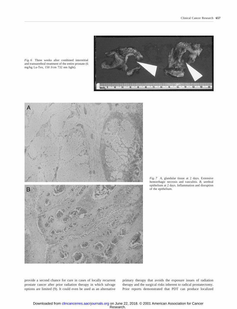
interstitial sites and showed coalescence of necrotic lesions. Noskip areas were noted (Fig. 5). The remaining four dogs treatedwith 6 mg/kg and 150 J/cm were scheduled to be euthanized at3 months, but due to complications (as stated previously) theyeither died or were euthanized at 3 days, 3 weeks, 6 weeks, or7 weeks after treatment. These specimens revealed completedestruction of urethral and central prostatic tissue with largefistulae between the urethra/prostate and peritoneum. Only anouter rim of prostate tissue could be identified grossly (Fig. 6).Finally, two dogs received 2 mg/kg, 100 J/cm and were eutha-nized at 3 months. These specimens showed intact urethral andprostate tissue, no residual necrosis, and pale-appearing tissuethroughout the gland.
Five dogs were treated with transurethral light deliveryalone. The dogs receiving 6 mg/kg, 75 J/cm and 6 mg/kg, 150J/cm were euthanized at 1 and 2 weeks, respectively. Theremaining three dogs received 2 mg/kg, 100 J/cm and wereeuthanized at 2 days, 2 weeks, and 3 months after treatment.Irrespective of drug and light dose, all dogs acutely showedzones of periurethral necrosis similar in size to those seen in thedogs treated via the interstitial alone approach. Healing of theselesions was again seen by 2 weeks with near complete resolutionby 3 months.
Twenty dogs also underwent temperature measurementswithin the prostate tissue. At;5 mm from the light source, thetemperature increased from 0.5°C to 3.1°C from initiation tocompletion of light administration.
Microscopic Findings. No microscopic abnormalitieswere noted in either of the light only control dogs that wereeuthanized at 2 weeks and 3 months. In the treated dogs, at 2days there was extensive hemorrhagic necrosis of the glandulartissue in the treatment areas (Fig. 7A) with marked local hem-orrhagic vasculitis. The size of the induced lesions was similarregardless of dose of light or drug as was noted in the macro-scopic findings. The fibromuscular connective tissue appearedto be affected in the same manner as the glandular tissue,showing diffuse and extensive necrosis. The prostatic capsulewas also affected, showing a local inflammatory reaction. Treat-ment via the transurethral approach resulted acutely in disrup-tion of the urethral epithelium (Fig. 7B).
At 2 weeks, the areas treated interstitially showed resolvingnecrosis with atrophic glandular epithelium (Fig. 8A). The areastreated transurethrally showed complete regeneration ofthe urethral epithelium with periurethral glandular atrophy(Fig. 8B).
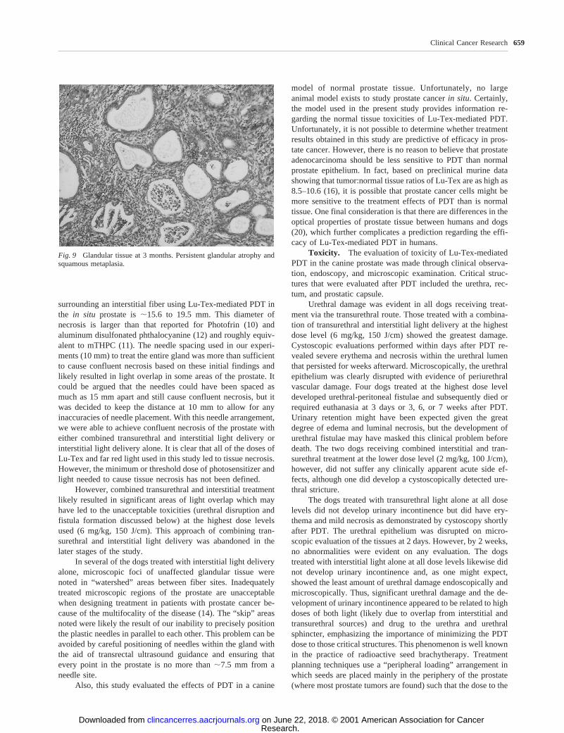
At 3 months, the areas treated interstitially showed resolu-tion of the necrosis with persistent glandular atrophy and squa-mous metaplasia (Fig. 9). The urethral epithelium was com-pletely normal in all dogs with some persistent periurethralglandular atrophy. Fibrosis and loss of stromal connective tissuewere noted throughout the prostate. Areas of chronic inflamma-tion were noted throughout the gland. The prostatic capsuleremained intact but also showed evidence of a low grade chronicinflammatory reaction.
The bladder neck appeared to be affected most markedly inthe dogs receiving transurethral treatments. At 2 days, erythemaand submucosal hemorrhage was noted in most of those dogs.Minimal erythema was noted in only one of the dogs treatedinterstitially alone. At 3 months, those dogs treated both tran-
Fig. 3 Area of acute necrosis caused by Lu-Tex PDT.
655Clinical Cancer Research
Research. on June 22, 2018. © 2001 American Association for Cancerclincancerres.aacrjournals.org Downloaded from

surethrally and interstitially exhibited smooth muscle hypertro-phy of the bladder neck. No abnormalities were seen in the dogstreated via either the interstitial or transurethral route alone.
The rectum appeared to be affected in four dogs, all ofwhich received interstitial treatment. Three dogs had been eu-thanized at 2 days and showed focal areas of hemorrhageextending into the muscle layer which appeared to correspondwith the position of the needles containing the diffusers. Onedog was euthanized at 3 months and showed focal scarring intothe muscle layer of the rectum, but no evidence of fistulaformation.
Finally, local temperature elevations of 0.5°C-3.1°C
caused by heat generated by the diffuser tips produced noobservable tissue damage in the control dogs (who receivedlight but no Lu-Tex) as analyzed pathologically 2 weeks and 3months after treatment.
DISCUSSION
Photodynamic therapy in solid organs has considerablepotential for treating a wide variety of cancers. The prostate isa good target organ for PDT because cancers are often locallyconfined and techniques already exist for the interstitial admin-istration of radiation which could easily be adapted. PDT could
Fig. 4 A, 2 days after interstitial treatment ofthe entire prostate;B, 2 weeks after interstitialtreatment of the entire prostate;C, 3 monthsafter interstitial treatment of the entire pros-tate.
Fig. 5 Two days after combined interstitial and transurethral treatment of the entire prostate (6 mg/kg Lu-Tex, 150 J/cm 732 nm light).
656 Photodynamic Therapy in the Canine Prostate
Research. on June 22, 2018. © 2001 American Association for Cancerclincancerres.aacrjournals.org Downloaded from
provide a second chance for cure in cases of locally recurrentprostate cancer after prior radiation therapy in which salvageoptions are limited (9). It could even be used as an alternative
primary therapy that avoids the exposure issues of radiationtherapy and the surgical risks inherent to radical prostatectomy.Prior reports demonstrated that PDT can produce localized
Fig. 6 Three weeks after combined interstitialand transurethral treatment of the entire prostate (6mg/kg Lu-Tex, 150 J/cm 732 nm light).
Fig. 7 A, glandular tissue at 2 days. Extensivehemorrhagic necrosis and vasculitis.B, urethralepithelium at 2 days. Inflammation and disruptionof the epithelium.
657Clinical Cancer Research
Research. on June 22, 2018. © 2001 American Association for Cancerclincancerres.aacrjournals.org Downloaded from

necrosis in a canine prostate model using photosensitizers suchas Photofrin, mTHPC, tin(II)ethyletiopurpurin dichloride, andaluminum disulfonated phthalocyanine (10–13). We chose toinvestigate the effect of the second-generation photosensitizer,Lu-Tex, due to its longer wavelength of activation, thus provid-ing potentially greater light penetration through tissue as well asits short (24 h) duration of skin photosensitivity.
Prostate cancer has a high propensity to be multifocalwithin the prostate (14); thus, the entire gland must be treated toeradicate the tumor. Therefore, the primary goal of this studywas to establish a photosensitizer/light dose and light deliverysystem that would allow complete treatment of the entire glandwhile maintaining an acceptable level of toxicity.
Treatment Parameters and Technique. No prior stud-ies have been reported in the literature using Lu-Tex for PDT ofthe in situ prostate. Therefore, the initial dose of Lu-Tex (6mg/kg) and 732 nm light fluence (75 J/cm at 150 mW/cm) waschosen based on experience in a murine model as well as humantrials involving of a number of different tumor types (15–17).
We chose a light delivery system that could be easily adapted toinstrumentation currently used in patients to deliver interstitialradioactive seeds (transperineally) to the human prostate. How-ever, an open transabdominal approach was chosen in our dogmodel, rather than the transperineal approach used in humans,simply for better access to the canine prostate. Plastic catheters(17-gauge) with a sharp end and a metal trocar were used in thepresent study because these catheters can be used with existinghuman prostate brachytherapy templates and can be seen ontransrectal ultrasound imaging for accurate placement via thetransperineal approach. Once positioned correctly, the metaltrocars can be replaced with the laser diffusers.
Studies to evaluate lesion size produced by a single fiberrevealed circular areas of well-demarcated necrosis 12–15 mmin diameter. Lesions of similar size were found at both the high(6 mg/kg, 150 J/cm) and low (2 mg/kg, 100 J/cm) dose levels.Due to the fixation process, a tissue shrinkage factor of;23%(19) should be accounted for when assessing the true diameterof necrosis in the prostate. Therefore, the diameter of necrosis
Fig. 8 A, glandular tissue at 2 weeks. Resolvingnecrosis and atrophic glandular epithelium.B, ure-thral epithelium at 2 weeks. Complete regenera-tion of urethral epithelium.
658 Photodynamic Therapy in the Canine Prostate
Research. on June 22, 2018. © 2001 American Association for Cancerclincancerres.aacrjournals.org Downloaded from
surrounding an interstitial fiber using Lu-Tex-mediated PDT inthe in situ prostate is;15.6 to 19.5 mm. This diameter ofnecrosis is larger than that reported for Photofrin (10) andaluminum disulfonated phthalocyanine (12) and roughly equiv-alent to mTHPC (11). The needle spacing used in our experi-ments (10 mm) to treat the entire gland was more than sufficientto cause confluent necrosis based on these initial findings andlikely resulted in light overlap in some areas of the prostate. Itcould be argued that the needles could have been spaced asmuch as 15 mm apart and still cause confluent necrosis, but itwas decided to keep the distance at 10 mm to allow for anyinaccuracies of needle placement. With this needle arrangement,we were able to achieve confluent necrosis of the prostate witheither combined transurethral and interstitial light delivery orinterstitial light delivery alone. It is clear that all of the doses ofLu-Tex and far red light used in this study led to tissue necrosis.However, the minimum or threshold dose of photosensitizer andlight needed to cause tissue necrosis has not been defined.
However, combined transurethral and interstitial treatmentlikely resulted in significant areas of light overlap which mayhave led to the unacceptable toxicities (urethral disruption andfistula formation discussed below) at the highest dose levelsused (6 mg/kg, 150 J/cm). This approach of combining tran-surethral and interstitial light delivery was abandoned in thelater stages of the study.
In several of the dogs treated with interstitial light deliveryalone, microscopic foci of unaffected glandular tissue werenoted in “watershed” areas between fiber sites. Inadequatelytreated microscopic regions of the prostate are unacceptablewhen designing treatment in patients with prostate cancer be-cause of the multifocality of the disease (14). The “skip” areasnoted were likely the result of our inability to precisely positionthe plastic needles in parallel to each other. This problem can beavoided by careful positioning of needles within the gland withthe aid of transrectal ultrasound guidance and ensuring thatevery point in the prostate is no more than;7.5 mm from aneedle site.
Also, this study evaluated the effects of PDT in a canine
model of normal prostate tissue. Unfortunately, no largeanimal model exists to study prostate cancerin situ. Certainly,the model used in the present study provides information re-garding the normal tissue toxicities of Lu-Tex-mediated PDT.Unfortunately, it is not possible to determine whether treatmentresults obtained in this study are predictive of efficacy in pros-tate cancer. However, there is no reason to believe that prostateadenocarcinoma should be less sensitive to PDT than normalprostate epithelium. In fact, based on preclinical murine datashowing that tumor:normal tissue ratios of Lu-Tex are as high as8.5–10.6 (16), it is possible that prostate cancer cells might bemore sensitive to the treatment effects of PDT than is normaltissue. One final consideration is that there are differences in theoptical properties of prostate tissue between humans and dogs(20), which further complicates a prediction regarding the effi-cacy of Lu-Tex-mediated PDT in humans.
Toxicity. The evaluation of toxicity of Lu-Tex-mediatedPDT in the canine prostate was made through clinical observa-tion, endoscopy, and microscopic examination. Critical struc-tures that were evaluated after PDT included the urethra, rec-tum, and prostatic capsule.
Urethral damage was evident in all dogs receiving treat-ment via the transurethral route. Those treated with a combina-tion of transurethral and interstitial light delivery at the highestdose level (6 mg/kg, 150 J/cm) showed the greatest damage.Cystoscopic evaluations performed within days after PDT re-vealed severe erythema and necrosis within the urethral lumenthat persisted for weeks afterward. Microscopically, the urethralepithelium was clearly disrupted with evidence of periurethralvascular damage. Four dogs treated at the highest dose leveldeveloped urethral-peritoneal fistulae and subsequently died orrequired euthanasia at 3 days or 3, 6, or 7 weeks after PDT.Urinary retention might have been expected given the greatdegree of edema and luminal necrosis, but the development ofurethral fistulae may have masked this clinical problem beforedeath. The two dogs receiving combined interstitial and tran-surethral treatment at the lower dose level (2 mg/kg, 100 J/cm),however, did not suffer any clinically apparent acute side ef-fects, although one did develop a cystoscopically detected ure-thral stricture.
The dogs treated with transurethral light alone at all doselevels did not develop urinary incontinence but did have ery-thema and mild necrosis as demonstrated by cystoscopy shortlyafter PDT. The urethral epithelium was disrupted on micro-scopic evaluation of the tissues at 2 days. However, by 2 weeks,no abnormalities were evident on any evaluation. The dogstreated with interstitial light alone at all dose levels likewise didnot develop urinary incontinence and, as one might expect,showed the least amount of urethral damage endoscopically andmicroscopically. Thus, significant urethral damage and the de-velopment of urinary incontinence appeared to be related to highdoses of both light (likely due to overlap from interstitial andtransurethral sources) and drug to the urethra and urethralsphincter, emphasizing the importance of minimizing the PDTdose to those critical structures. This phenomenon is well knownin the practice of radioactive seed brachytherapy. Treatmentplanning techniques use a “peripheral loading” arrangement inwhich seeds are placed mainly in the periphery of the prostate(where most prostate tumors are found) such that the dose to the
Fig. 9 Glandular tissue at 3 months. Persistent glandular atrophy andsquamous metaplasia.
659Clinical Cancer Research
Research. on June 22, 2018. © 2001 American Association for Cancerclincancerres.aacrjournals.org Downloaded from

urethra is no more than 100% of the prescribed dose (21). Sucha system for interstitial prostate PDT might also help to limiturethral toxicity.
Rectal damage was not apparent on clinical and endoscopicevaluations of all dogs. However, in the dogs receiving inter-stitial treatment, focal areas of hemorrhage and eventually scarwere noted microscopically and appeared to correspond to nee-dle tracks in which the needles were pushed too far through theprostate and into rectum. This was a consequence of the trans-abdominal technique used in this study and likely could beavoided with a transperineal approach with transrectal ultra-sound guidance.
The prostatic capsule showed both acute and chronic dif-fuse capsular inflammation with pericapsular vasculitis in allareas treated via an interstitial approach. This finding is contraryto that found by Changet al. (11), who noted no connectivetissue (including capsule) effect using mTHPC PDT in thecanine prostate. This difference may be due to differences inconnective tissue uptake of mTHPC and Lu-Tex, although itcould also be related to the PDT doses that were delivered.
The results of this study demonstrated that Lu-Tex PDT isfeasible in the canine prostate. Its biological effect is character-ized initially by inflammation and necrosis followed by glan-dular atrophy and fibrosis. This tissue effect was similar to thatof PDT mediated by other photosensitizers. In addition, thisstudy showed that both the interstitial alone and the combinedinterstitial and transurethral technique of light delivery couldprovide comprehensive treatment of the entire prostate gland,whereas the interstitial alone technique resulted in the leastamount of urethral damage. Although questions remain regard-ing the PDT effect on prostatic carcinoma, it seems reasonablebased on our data to proceed with Phase I human trials in aselected population such as those patients with locally recurrentdisease after radical radiotherapy.
REFERENCES1. Dougherty, T. J., Gomer, C. J., Henderson, B. W., Jori, G., Kessel,D., Korbelik, M., Moan, J., and Peng, O. Photodynamic therapy. J. Natl.Cancer Inst.,90: 889–905, 1998.2. Friedberg, J. S., Hsi, R. A., Glatstein, E., and Hahn, S. M. Pleuralphotodynamic therapy (PDT) as a component of multimodality therapyfor non-small cell lung cancer: a Phase II trial. Proc. Am. Soc. Clin.Oncol.,18: 503a, 1999.3. Hahn, S., Hsi, R. A., Fraker, D., Rubin, S. C., and Glatstein, E. APhase II trial of intraperitoneal (IP) PDT for the treatment of IP malig-nancies (Abstract RC8).In: Seventh Biennial Congress of the Interna-tional Photodynamic Association, p. 53, 1998.4. Overholt, B. F., and Panjehpour, M. Photodynamic therapy forBarrett’s esophagus. Gastrointest. Endosc. Clin. North Am.,2: 207–220,1997.5. Cortese, D. A., Edell, E. S., and Kinsey, J. H. Photodynamic therapyfor early stage squamous cell carcinoma of the lung. Mayo Clin. Proc.,72: 595–602, 1997.6. Biel, M. A. Photodynamic therapy and the treatment of head andneck neoplasia. Laryngoscope,108: 1259–1268, 1998.
7. Cairnduff, F., Stringer, M. R., Hudson, E. J., Ash, D. V., and Brown,S. B. Superficial photodynamic therapy with topical 5-aminolevulinicacid for superficial primary and secondary skin cancer. Br. J. Cancer,69: 605–608, 1994.
8. D’Amico, A. V. Role of interstitial radiotherapy in the managementof clinically organ-confined prostate cancer: the jury is still out. J. Clin.Oncol.,14: 304–315, 1996.
9. Ornstein, D. K., Oh, J., Herschman, J. D., and Andriole, G. L.Evaluation and management of the man who has failed primary curativetherapy for prostate cancer. Urol. Clin. North Am.,25: 591–601, 1998.
10. Lee, L. K., Whitehurst, C., Chen, Q., Pantelides, M. L., Hetzel,F. W., and Moore, J. V. Interstitial photodynamic therapy in the canineprostate. Br. J. Urol.,80: 898–902, 1997.
11. Chang, S. C., Buonaccorsi, G., MacRobert, A., and Bown, S. G.Interstitial and transurethral photodynamic therapy of the canine pros-tate usingmeso-tetra-(m-hydroxyphenyl) chlorin. Int. J. Cancer,67:555–562, 1996.
12. Chang, S. C., Buonaccorsi, G., MacRobert, A., and Bown, S. G.Interstitial photodynamic therapy in the canine prostate with disulfon-ated aluminum phthalocyanine and 5-aminolevulinic acid-induced pro-toporphyrin IX. Prostate,32: 89–98, 1997.
13. Selman, S. H., Keck, R. W., and Hampton, J. A. Transperinealphotodynamic ablation of the canine prostate. J. Urol.,156: 258–260,1996.
14. Djavan, B., Susani, M., Bursa, B., Basharkhah, A., Simak, R., andMarberger, M. Predictability and significance of multifocal prostatecancer in the radical prostatectomy specimen. Tech. Urol.,5: 139–142,1999.
15. Woodburn, K. W., Fan, Q., Miles, D. R., Kessel, D., Luo, Y., andYoung, S. W. Localization and efficacy analysis of the phototherapeuticlutetium texaphyrin (PCI-0123)in the murine EMT6 sarcoma model.Photochem. Photobiol.,65: 410–415, 1997.
16. Young, S. W., Woodburn, K. W., Wright, M., Moody, T. D., Fan,Q., Sessler, J. L., Dow, W. C., and Miller, R. A. Lutetium texaphyrin(PCI-0123): a near-infrared water-soluble photosensitizer. Photochem.Photobiol.,63: 892–897, 1996.
17. Renschler, M. F., Yuen, A., Panella, T. J., Julius, M., Wieman, C.J., Panjehpour, S., Taber, S., Fingar, V., Horning, S., and Miller, R. A.Photodynamic therapy trials with lutetium texaphyrin PCI0123 (LuTex)(Abstract MPM-F6).In: 25th Annual Meeting of the American Societyfor Photobiology, p. 47S, 1997.
18. Chen, Q., Wilson, B. C., Shetty, S. D., Patterson, M. S., Cerny,J. C., and Hetzel, F. W. Changes inin vivo optical properties and lightdistributions in normal canine prostate during photodynamic therapy.Radiat. Res.,147: 86–91, 1997.
19. Anderson, J. H., Strandberg, J. D., Wong, D. F., Conti, P. S.,Barker, P. B., Blackband, S. J., Hilton, J., Natarajan, T. K., Dannals,R. F., Samphilipo, M. A., Jr.,et al. Multimodality correlative study ofcanine brain tumors: proton magnetic resonance spectroscopy, positronemission tomography, and histology. Invest. Radiol.,29: 597–605,1994.
20. Whitehurst, C., Pantelides, M. L., Moore, J. V., Brooman, P. J., andBlacklock, N. J. In vivo laser light distribution in human prostaticcarcinoma. J. Urol.,151: 1411–1415, 1994.
21. Stone, N. N., Stock, R. G., DeWyngaert, J. K., and Tabert, A.Prostate brachytherapy: improvements in prostate volume measure-ments and dose distribution using interactive ultrasound guided implan-tation and three-dimensional dosimetry. Radiat. Oncol. Invest.,3: 185–195, 1995.
660 Photodynamic Therapy in the Canine Prostate
Research. on June 22, 2018. © 2001 American Association for Cancerclincancerres.aacrjournals.org Downloaded from

2001;7:651-660. Clin Cancer Res R. Alex Hsi, Amy Kapatkin, John Strandberg, et al. LutetiumPhotodynamic Therapy in the Canine Prostate Using Motexafin
Updated version
http://clincancerres.aacrjournals.org/content/7/3/651
Access the most recent version of this article at:
Cited articles
http://clincancerres.aacrjournals.org/content/7/3/651.full#ref-list-1
This article cites 19 articles, 1 of which you can access for free at:
Citing articles
http://clincancerres.aacrjournals.org/content/7/3/651.full#related-urls
This article has been cited by 1 HighWire-hosted articles. Access the articles at:
E-mail alerts related to this article or journal.Sign up to receive free email-alerts
Subscriptions
Reprints and
To order reprints of this article or to subscribe to the journal, contact the AACR Publications
Permissions
Rightslink site. Click on "Request Permissions" which will take you to the Copyright Clearance Center's (CCC)
.http://clincancerres.aacrjournals.org/content/7/3/651To request permission to re-use all or part of this article, use this link
Research. on June 22, 2018. © 2001 American Association for Cancerclincancerres.aacrjournals.org Downloaded from