canine physical therapy and rehabilitationcanine physical therapy and rehabilitation canine physical...
TRANSCRIPT
Canine Physical Therapy and Rehabilitation
Canine physical therapy and rehabilitation involves designing and implementing a protocol that will allow one to alleviate impairment and improve function of an injured dog. Achieving the desired outcome depends on careful monitoring and modification of the protocol put in place in order to tailor it to the individual patient, owner, and injury. It is also critical that the protocol be tailored to the rate and level of progress being made by the patient. Being able to design and modify these protocols appropriately requires training in both veterinary care and physical therapy. The techniques that are involved should be done with the aid and guidance of a veterinary professional. Physical therapy and rehabilitation begins with a protocol or therapeutic intervention. These interventions consist of numerous techniques.
• Therapeutic exercise: range of motion (ROM), aerobic conditioning, gait training, aquatic therapy,
muscle strengthening, balance, coordination, posture, motor control. • Manual therapy techniques: joint mobilization and manipulation, massage, remodeling scar tissue. • Wound management: dressings, topical agents, debridement, oxygen therapy. • Airway clearance techniques: postural drainage, percussion, vibration. • Orthotic and prosthetic intervention. • Electrotherapeutic modalities: electrical stimulation, laser. • Thermal modalities: superficial heat and cold, ultrasound, diathermy.
The goals of physical therapy vary with the patient and the owner. A professional or athletic dog may have
much different goals of therapy than a sedentary house pet. Regardless of use or activity level, in general, the goals of physical therapy are to decrease pain, improve strength, slow muscle atrophy, reduce swelling, speed healing, remodel scar tissue, and improve function (whatever that function may be). Physical therapy is also used to prevent re-injury and maintain the progress that is made during recovery.
The primary person responsible for the decisions being made during the rehabilitation process should be the
veterinarian overseeing your dogs care. Depending on the injury and the repair, recommendations can be given to you, the owner, regarding what must be done. Often times there are specific precautions that need to be made and considered when performing the recommended therapy. Initially, therapy should be carried out together with your veterinarian. Once care is turned over to you, routine follow-ups are necessary to ensure the proper adjustments can be made in the therapy.
Documentation of progress is essential when planning and modifying a protocol. The information should
include functional status, ROM, treatment given, and patient progress. Below is a list of key information that should be documented.
• History of illness, including precautionary information or contraindications for certain treatments. • Objective information such as ROM, function, girth measurements, lameness scores. • Primary problems to be addressed and goals for treatment. • Details of the treatment provided. • Response to treatment.
A thorough understanding of canine behavior is an important part of interpreting response to therapy and
motivating the patient to engage in therapy. Body position, vocalization, tail position, willingness to participate, ear position, and eye movement are all cues that can be used to interpret response to therapy. You can use this information to then influence or encourage your dog to participate in therapy or make decisions about modifications to the protocol.
There are numerous modalities that can be used to achieve the desired goals of therapy. In the home
environment, slow controlled leash walks and swimming are commonly used to begin rehabilitation. Passive range of motion and thermal therapy are also easily performed at home. Modifications that can be made to these simple techniques include walking in high grass, sand, uphill, or walking for extended periods of time at a faster pace. Some treatment modalities that can supplement those therapies performed at home include underwater treadmills, ultrasound therapy, and neuromuscular electro-stimulation.
What modifications to make and when to make them is not an easy question to answer and depends a great
deal on the patient, the injury and the progress that is made. One must consider the type and severity of injury, number of limbs affected, stability of surgical repair, size of the patient, pre-existing conditions, and available

facilities. With the help of a veterinary professional, one can tailor a protocol depending on the circumstances involved. Goals should be used as objectives to therapy. For example, the initial goal should be to bear weight on the affected limb. Next, one may work on assisted active ambulation followed then by unassisted active ambulation.
Setbacks in progress are usually an indication that the protocol has advanced too far or too quickly. In these
situations it is important to determine how severe of a set back has occurred. Complications could be as serious as a catastrophic failure of a surgical repair which requires immediate medical attention or it may be as simple as over use of muscle groups that have not been challenged until beginning new therapy. Once the seriousness of the setback can be gauged, one is then able to use that information to determine how much to back-up and decide how long to rest before resuming activity.
A rehabilitation program need not be elaborate or costly. Consideration should be given to the patient,
owner, and therapist needs. Protocol development greatly depends on the available facilities and equipment, the willingness of you as owners to help with rehabilitation, and the education level of the professionals assisting you. Therapeutic exercises are certainly the most important aspects of rehabilitation. They may be incorporated with other modalities to enhance recovery but it is the exercises that help improve muscle strength, joint mobility, limb use, nerve function, and muscle endurance.
TTuummoorrss ooff tthhee BBooxxeerr BBrreeeedd:: AAnn eemmpphhaassiiss oonn LLyymmpphhoommaa DDaavviidd MM.. LLuurriiee,, BBVVSScc,, DDAACCVVIIMM ((oonnccoollooggyy)),, DDAACCVVRR ((rraaddiiaattiioonn oonnccoollooggyy))
HHiillll’’ss CClliinniiccaall AAssssiissttaanntt PPrrooffeessssoorr ooff OOnnccoollooggyy
UUnniivveerrssiittyy ooff FFlloorriiddaa
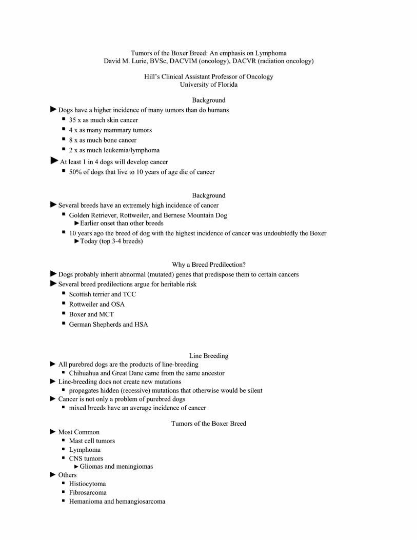
BBaacckkggrroouunndd ►► DDooggss hhaavvee aa hhiigghheerr iinncciiddeennccee ooff mmaannyy ttuummoorrss tthhaann ddoo hhuummaannss
3355 xx aass mmuucchh sskkiinn ccaanncceerr 44 xx aass mmaannyy mmaammmmaarryy ttuummoorrss 88 xx aass mmuucchh bboonnee ccaanncceerr 22 xx aass mmuucchh lleeuukkeemmiiaa//llyymmpphhoommaa
►►AAtt lleeaasstt 11 iinn 44 ddooggss wwiillll ddeevveelloopp ccaanncceerr 5500%% ooff ddooggss tthhaatt lliivvee ttoo 1100 yyeeaarrss ooff aaggee ddiiee ooff ccaanncceerr
BBaacckkggrroouunndd ►► SSeevveerraall bbrreeeeddss hhaavvee aann eexxttrreemmeellyy hhiigghh iinncciiddeennccee ooff ccaanncceerr
GGoollddeenn RReettrriieevveerr,, RRoottttwweeiilleerr,, aanndd BBeerrnneessee MMoouunnttaaiinn DDoogg ►►EEaarrlliieerr oonnsseett tthhaann ootthheerr bbrreeeeddss
1100 yyeeaarrss aaggoo tthhee bbrreeeedd ooff ddoogg wwiitthh tthhee hhiigghheesstt iinncciiddeennccee ooff ccaanncceerr wwaass uunnddoouubbtteeddllyy tthhee BBooxxeerr ►►TTooddaayy ((ttoopp 33--44 bbrreeeeddss))
WWhhyy aa BBrreeeedd PPrreeddiilleeccttiioonn?? ►► DDooggss pprroobbaabbllyy iinnhheerriitt aabbnnoorrmmaall ((mmuuttaatteedd)) ggeenneess tthhaatt pprreeddiissppoossee tthheemm ttoo cceerrttaaiinn ccaanncceerrss ►► SSeevveerraall bbrreeeedd pprreeddiilleeccttiioonnss aarrgguuee ffoorr hheerriittaabbllee rriisskk
SSccoottttiisshh tteerrrriieerr aanndd TTCCCC RRoottttwweeiilleerr aanndd OOSSAA BBooxxeerr aanndd MMCCTT GGeerrmmaann SShheepphheerrddss aanndd HHSSAA
LLiinnee BBrreeeeddiinngg ►► AAllll ppuurreebbrreedd ddooggss aarree tthhee pprroodduuccttss ooff lliinnee--bbrreeeeddiinngg
CChhiihhuuaahhuuaa aanndd GGrreeaatt DDaannee ccaammee ffrroomm tthhee ssaammee aanncceessttoorr ►► LLiinnee--bbrreeeeddiinngg ddooeess nnoott ccrreeaattee nneeww mmuuttaattiioonnss
pprrooppaaggaatteess hhiiddddeenn ((rreecceessssiivvee)) mmuuttaattiioonnss tthhaatt ootthheerrwwiissee wwoouulldd bbee ssiilleenntt ►► CCaanncceerr iiss nnoott oonnllyy aa pprroobblleemm ooff ppuurreebbrreedd ddooggss
mmiixxeedd bbrreeeeddss hhaavvee aann aavveerraaggee iinncciiddeennccee ooff ccaanncceerr
TTuummoorrss ooff tthhee BBooxxeerr BBrreeeedd ►► MMoosstt CCoommmmoonn
MMaasstt cceellll ttuummoorrss LLyymmpphhoommaa CCNNSS ttuummoorrss
►► GGlliioommaass aanndd mmeenniinnggiioommaass ►► OOtthheerrss
HHiissttiiooccyyttoommaa FFiibbrroossaarrccoommaa HHeemmaanniioommaa aanndd hheemmaannggiioossaarrccoommaa
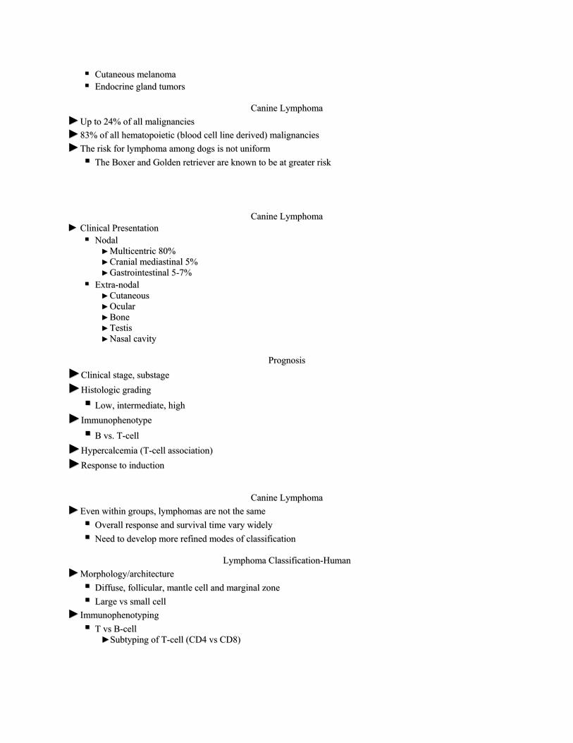
CCuuttaanneeoouuss mmeellaannoommaa EEnnddooccrriinnee ggllaanndd ttuummoorrss
CCaanniinnee LLyymmpphhoommaa
►► UUpp ttoo 2244%% ooff aallll mmaalliiggnnaanncciieess ►► 8833%% ooff aallll hheemmaattooppooiieettiicc ((bblloooodd cceellll lliinnee ddeerriivveedd)) mmaalliiggnnaanncciieess ►► TThhee rriisskk ffoorr llyymmpphhoommaa aammoonngg ddooggss iiss nnoott uunniiffoorrmm
TThhee BBooxxeerr aanndd GGoollddeenn rreettrriieevveerr aarree kknnoowwnn ttoo bbee aatt ggrreeaatteerr rriisskk
CCaanniinnee LLyymmpphhoommaa ►► CClliinniiccaall PPrreesseennttaattiioonn
NNooddaall ►► MMuullttiicceennttrriicc 8800%% ►► CCrraanniiaall mmeeddiiaassttiinnaall 55%% ►► GGaassttrrooiinntteessttiinnaall 55--77%%
EExxttrraa--nnooddaall ►► CCuuttaanneeoouuss ►► OOccuullaarr ►► BBoonnee ►► TTeessttiiss ►► NNaassaall ccaavviittyy
PPrrooggnnoossiiss
►►CClliinniiccaall ssttaaggee,, ssuubbssttaaggee ►►HHiissttoollooggiicc ggrraaddiinngg
LLooww,, iinntteerrmmeeddiiaattee,, hhiigghh ►►IImmmmuunnoopphheennoottyyppee
BB vvss.. TT--cceellll ►►HHyyppeerrccaallcceemmiiaa ((TT--cceellll aassssoocciiaattiioonn)) ►►RReessppoonnssee ttoo iinndduuccttiioonn
CCaanniinnee LLyymmpphhoommaa ►► EEvveenn wwiitthhiinn ggrroouuppss,, llyymmpphhoommaass aarree nnoott tthhee ssaammee
OOvveerraallll rreessppoonnssee aanndd ssuurrvviivvaall ttiimmee vvaarryy wwiiddeellyy NNeeeedd ttoo ddeevveelloopp mmoorree rreeffiinneedd mmooddeess ooff ccllaassssiiffiiccaattiioonn
LLyymmpphhoommaa CCllaassssiiffiiccaattiioonn--HHuummaann
►► MMoorrpphhoollooggyy//aarrcchhiitteeccttuurree DDiiffffuussee,, ffoolllliiccuullaarr,, mmaannttllee cceellll aanndd mmaarrggiinnaall zzoonnee LLaarrggee vvss ssmmaallll cceellll
►► IImmmmuunnoopphheennoottyyppiinngg TT vvss BB--cceellll
►►SSuubbttyyppiinngg ooff TT--cceellll ((CCDD44 vvss CCDD88))
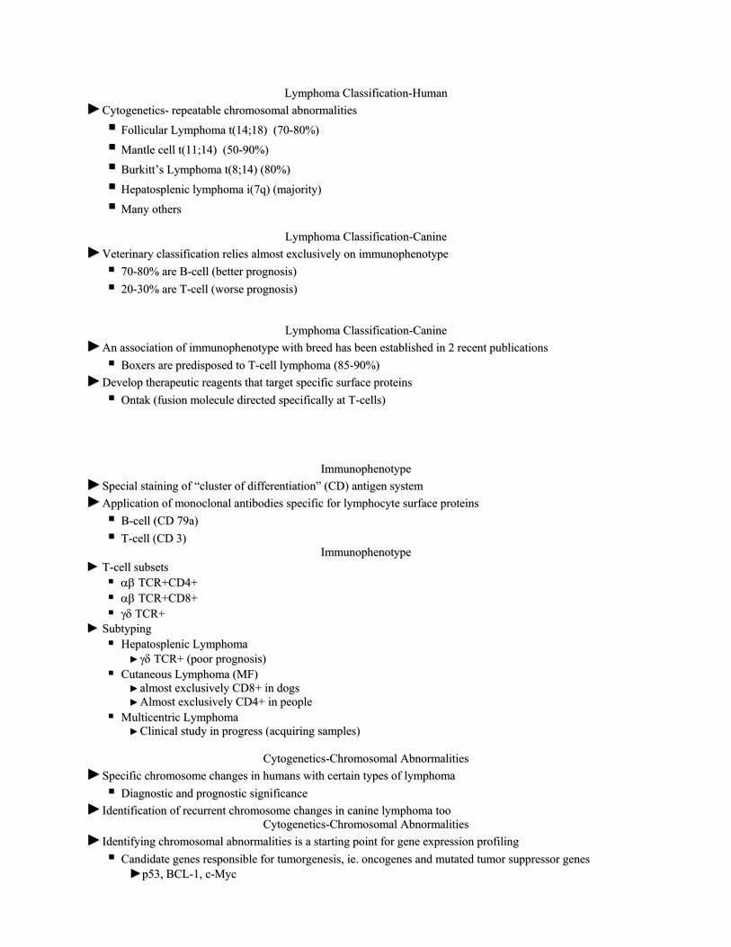
LLyymmpphhoommaa CCllaassssiiffiiccaattiioonn--HHuummaann ►► CCyyttooggeenneettiiccss-- rreeppeeaattaabbllee cchhrroommoossoommaall aabbnnoorrmmaalliittiieess
FFoolllliiccuullaarr LLyymmpphhoommaa tt((1144;;1188)) ((7700--8800%%))
MMaannttllee cceellll tt((1111;;1144)) ((5500--9900%%))
BBuurrkkiitttt’’ss LLyymmpphhoommaa tt((88;;1144)) ((8800%%))
HHeeppaattoosspplleenniicc llyymmpphhoommaa ii((77qq)) ((mmaajjoorriittyy))
MMaannyy ootthheerrss
LLyymmpphhoommaa CCllaassssiiffiiccaattiioonn--CCaanniinnee ►► VVeetteerriinnaarryy ccllaassssiiffiiccaattiioonn rreelliieess aallmmoosstt eexxcclluussiivveellyy oonn iimmmmuunnoopphheennoottyyppee
7700--8800%% aarree BB--cceellll ((bbeetttteerr pprrooggnnoossiiss)) 2200--3300%% aarree TT--cceellll ((wwoorrssee pprrooggnnoossiiss))
LLyymmpphhoommaa CCllaassssiiffiiccaattiioonn--CCaanniinnee ►► AAnn aassssoocciiaattiioonn ooff iimmmmuunnoopphheennoottyyppee wwiitthh bbrreeeedd hhaass bbeeeenn eessttaabblliisshheedd iinn 22 rreecceenntt ppuubblliiccaattiioonnss
BBooxxeerrss aarree pprreeddiissppoosseedd ttoo TT--cceellll llyymmpphhoommaa ((8855--9900%%)) ►► DDeevveelloopp tthheerraappeeuuttiicc rreeaaggeennttss tthhaatt ttaarrggeett ssppeecciiffiicc ssuurrffaaccee pprrootteeiinnss
OOnnttaakk ((ffuussiioonn mmoolleeccuullee ddiirreecctteedd ssppeecciiffiiccaallllyy aatt TT--cceellllss))
IImmmmuunnoopphheennoottyyppee ►► SSppeecciiaall ssttaaiinniinngg ooff ““cclluusstteerr ooff ddiiffffeerreennttiiaattiioonn”” ((CCDD)) aannttiiggeenn ssyysstteemm ►► AApppplliiccaattiioonn ooff mmoonnoocclloonnaall aannttiibbooddiieess ssppeecciiffiicc ffoorr llyymmpphhooccyyttee ssuurrffaaccee pprrootteeiinnss
BB--cceellll ((CCDD 7799aa)) TT--cceellll ((CCDD 33))
IImmmmuunnoopphheennoottyyppee ►► TT--cceellll ssuubbsseettss
ααββ TTCCRR++CCDD44++ ααββ TTCCRR++CCDD88++ γγδδ TTCCRR++
►► SSuubbttyyppiinngg HHeeppaattoosspplleenniicc LLyymmpphhoommaa
►► γγδδ TTCCRR++ ((ppoooorr pprrooggnnoossiiss)) CCuuttaanneeoouuss LLyymmpphhoommaa ((MMFF))
►► aallmmoosstt eexxcclluussiivveellyy CCDD88++ iinn ddooggss ►► AAllmmoosstt eexxcclluussiivveellyy CCDD44++ iinn ppeeooppllee
MMuullttiicceennttrriicc LLyymmpphhoommaa ►► CClliinniiccaall ssttuuddyy iinn pprrooggrreessss ((aaccqquuiirriinngg ssaammpplleess))
CCyyttooggeenneettiiccss--CChhrroommoossoommaall AAbbnnoorrmmaalliittiieess ►► SSppeecciiffiicc cchhrroommoossoommee cchhaannggeess iinn hhuummaannss wwiitthh cceerrttaaiinn ttyyppeess ooff llyymmpphhoommaa
DDiiaaggnnoossttiicc aanndd pprrooggnnoossttiicc ssiiggnniiffiiccaannccee ►► IIddeennttiiffiiccaattiioonn ooff rreeccuurrrreenntt cchhrroommoossoommee cchhaannggeess iinn ccaanniinnee llyymmpphhoommaa ttoooo
CCyyttooggeenneettiiccss--CChhrroommoossoommaall AAbbnnoorrmmaalliittiieess ►► IIddeennttiiffyyiinngg cchhrroommoossoommaall aabbnnoorrmmaalliittiieess iiss aa ssttaarrttiinngg ppooiinntt ffoorr ggeennee eexxpprreessssiioonn pprrooffiilliinngg
CCaannddiiddaattee ggeenneess rreessppoonnssiibbllee ffoorr ttuummoorrggeenneessiiss,, iiee.. oonnccooggeenneess aanndd mmuuttaatteedd ttuummoorr ssuupppprreessssoorr ggeenneess ►►pp5533,, BBCCLL--11,, cc--MMyycc
TThhiiss iiss ppaarrttiiccuullaarrllyy aattttrraaccttiivvee iinn bbrreeeeddss lliikkee tthhee BBooxxeerr ►►HHiigghhllyy hhoommooggeennoouuss ggeenneettiicc mmaatteerriiaall
CCoommppaarraattiivvee AAssppeeccttss
►► WWhhiillee tthhee eexxaacctt ttuummoorr ttyyppeess mmaayy nnoott bbee tthhee ssaammee iinn ddooggss ccoommppaarreedd ttoo ppeeooppllee,, tthhee mmoolleeccuullaarr aabbnnoorrmmaalliittiieess aarree oofftteenn iiddeennttiiccaall
pp5533 mmuuttaattiioonnss ►►OOsstteeoossaarrccoommaa,, vvaacccciinnee aassssoocciiaatteedd ssaarrccoommaass
KKiitt mmuuttaattiioonnss ►►MMaasstt cceellll ttuummoorrss,, ggaassttrrooiinntteessttiinnaall ssttrroommaall ttuummoorrss
AAbbeerrrraanntt MMeett eexxpprreessssiioonn ►►OOsstteeoossaarrccoommaa,, mmaasstt cceellll ttuummoorrss
FFuuttuurree DDiirreeccttiioonn
►► FFeeddeerraallllyy ffuunnddeedd ccaanniinnee ggeennoommee pprroojjeecctt ffiinniisshheedd DDeecc 88 22000055 (($$3300 mmiilllliioonn))
►► BBooxxeerr bbrreeeedd wwaass cchhoosseenn aass aa rreepprreesseennttaattiivvee ooff tthhee aavveerraaggee ppuurreebbrreedd ddoogg LLiittttllee vvaarriiaattiioonn iinn ggeennoommee FFeemmaallee BBooxxeerr nnaammeedd ““TTaasshhaa””
►►22 bbiilllliioonn nnuucclleeoottiiddeess sseeqquueenncceedd
FFuuttuurree DDiirreeccttiioonn ►► IIddeennttiiffyyiinngg cchhrroommoossoommaall aabbnnoorrmmaalliittiieess aanndd uullttiimmaatteellyy ggeennee mmuuttaattiioonnss mmaayy ggiivvee uuss mmoorree iinnffoorrmmaattiioonn aabboouutt tthhee
cclliinniiccaall bbeehhaavviioorr ooff ssppeecciiffiicc ttuummoorrss ►► SSuucchh aabbnnoorrmmaalliittiieess aarree wweellll kknnoowwnn iinn ppeeooppllee aanndd pprroovviiddee vvaalluuaabbllee iinnffoorrmmaattiioonn ffoorr ddeetteerrmmiinniinngg tthheerraappyy ►► AApppplliiccaattiioonn iinn ddooggss mmaayy hhaavvee pprreeddiiccttiivvee vvaalluuee rreeggaarrddiinngg ttuummoorr ssttaaggee aanndd ggrraaddee,, pphheennoottyyppee,, rreessppoonnssee ttoo tthheerraappyy,,
aanndd ssuurrvviivvaall
QQuueessttiioonnss??
FLORIDA DOG BREEDER’S SYMPOSIUM
JULY 2006
THE INTERPRETATION OF CLINICAL SIGNS
Michael Schaer, DVM, Diplomate ACVIM, ACVECC
University of Florida, College of Veterinary Medicine, Gainesville, FL
The key to making a correct diagnosis depends on obtaining an accurate patient history and doing a
complete physical examination. Obtaining a history is as much an art form as it is a scientific inquiry. It must be
done as objectively as possible in order to obtain a truly accurate description of the patient's problems. Failure to do
so can result in a misinterpretation of the facts, which can lead to misdiagnosis, needless procedures and perhaps
patient discomfort, excessive expense for the client, and sometimes a less than optimal client-doctor relationship.
The following discussion will identify several clinical abnormalities that tend to be subject to misinterpretation.
PLATELET DISORDERS
Petechia are foci of pinpoint hemorrhages that can involve any of the soft tissues of the body. They usually
signal the presence of a platelet disorder although they can also occur with vasculitis. Platelet disorders can arise
from inadequate numbers (quantitative) or from impaired function (qualitative). Quantitative problems result from
either peripheral destruction, impaired production or increased consumption. Platelet disorders commonly present
clinically as small skin or mucous membrane pinpoint or blotchy (ecchymotic) hemorrhages or as black-colored
associated with upper gastrointestinal bleeding. Rarely, the bleeding can be more dramatic as with frank bloody
urine. The most important diagnostic tests include the platelet count and the bleeding time.
FACTOR DEFICIENCIES
Coagulation factor deficiencies characterize as frank hemorrhaging into the body tissues; blood
accumulations under the skin (hematomas) are of rather common occurrence. Ecchymoses and overt profuse
bleeding require a rapid evaluation of the coagulation system with tests such as the prothrombin (PT) and partial
thromboplastin times (PTT) and the activating clotting time (ACT). Factor deficiencies do not cause petechia unless
a disorder involving platelets coexists. The history (including a drug history) should help to differentiate between
congenital and acquired disorders.
HEMOLYSIS
In vivo hemolysis is the destruction of red blood cells within the vascular and extravascular spaces. Its
causes are many ranging from toxins, infections, and drugs to autoimmunity. The classic picture of intravascular
hemolysis includes: weakness, nausea, anemia, pink colored serum, yellow mucous membranes, and hemoglobin-
and bilirubinuria. Note that "HEMOLYZERS" HEMOLYZE while "BLEEDERS” BLEED. Coagulogram profiles
are generally normal with hemolytic disease unless a co-existing bleeding disorder is present.
HEMOGLOBINURIA VS. HEMATURIA VS MYOGLOBINURIA VS BILIRUBINURIA
Hemoglobinuria is a product of hemolytic disease characterized as a dark port-wine-like colored urine that
contains few intact RBC's. Hematuria is a typical red color and reflects bleeding anywhere along the urinary tract.
Patients with a bleeding tendency can also bleed into their normal urinary tract. It is possible for some upper urinary
tract lesions, such as a bleeding renal tumor, to have both hemoglobinuria (as a result of the blood becoming
hemolyzed in the urinary bladder during storage) and hematuria. It is also possible for a fresh kidney bleed to form
clots in the urinary bladder and cause urinary outflow obstruction.
Myoglobinuria represents muscle pigment in urine and is associated with muscle destruction. It will cause
reddish brown pigmenturia and a positive test for blood on dipstick, but the sediment is devoid of RBC’s.
VOMITING VS. REGURGITATION VS. RETCHING
Vomiting is the actual forceful oral expulsion of gastrointestinal contents accompanying many different
digestive tract disorders as well as those involving other organ systems as well.
Regurgitation is the more passive bringing up of esophageal contents that most commonly accompanies esophageal
disorders. Retching is a forceful but unproductive attempt to vomit that can occur with constrictive esophageal
pathology. It can also occur following a coughing episode.
LOCALIZING SIGNIFICANCE OF THE VARIOUS TYPES OF VOMITUS
Clear mucoid-esophageal, gastric. Red blood-esophagus, stomach. Dark red ("coffee grounds")-gastric,
pylorus, very proximal duodenum. Bile stained-small bowel; bile in vomitus attests to pyloric patency. Brown,
malodorous (feculent)-distal small bowel, large bowel.
BLOOD IN STOOL
In general, bright red blood can originate anywhere distal to the mid-bowel while dark brown-to-black stool
(melena) comes from the stomach and proximal small bowel. However, a major bleed in the proximal bowel and its
subsequent rapid passage can show as bright red blood in the stools. Oral or nasal bleeding can cause melena from
swallowed blood.
STRANGURIA VS. OBSTIPATION
The most important situation where this differentiation becomes significant is that involving the cat or dog
with urinary obstruction. Strainful urination (stranguria) in male dogs and cats almost always signifies urinary outlet
obstruction and is therefore always considered as a medical emergency. The male cat with urethral obstruction will
make repeated trips to the litter box, sometimes accompanied by vocalization reflecting the animal's discomfort.
Over a matter of hours the cat will become anorectic, begin to vomit, and become mentally depressed. The
obstipated patient shows a better systemic tolerance for its GI dilemma. It should be pointed out that females can
also experience urinary outflow obstruction and trying to decide on patency by way of telephone might compromise
the patient’s well being.
POLYURIA VS. INCONTINENCE
The polyuric patient typically produces and voluntarily passes copious volumes of dilute urine often
accompanied by increased thirst (polydipsia). Urinary incontinence is an unconscious passage of urine that occurs
while the patient is lying down or sleeping; these animals urinate normally. Females generally release urine while
laying down while males can do it anytime.
BLINDNESS VS. DEMENTIA
At first glance, these two conditions might resemble each other to the uncritical observer. The blind patient
will certainly bump into objects, especially in unfamiliar surroundings, and it will fail to respond to a menace
gesture, but its other neurological functions are normal. The demented patient might not menace mainly because the
brain is malfunctioning and consequently
cannot register the threatening gesture while its visual pathways might very well be normal. This patient will have
accompanying neurological abnormalities.
NEUROLOGIC VS. METABOLIC WEAKNESS
The signs accompanying neurological weakness will depend on the nature of the primary disorder. Most
are usually continuous, sometimes progressive. They are commonly segmental or lateralizing in their distribution
when caused by a focal lesion. Other neurological signs such as pain might be present if there is meningeal or dorsal
nerve root pathology.
Metabolic weakness can also be continuous and progressive, but it can also be periodic as well. Patients
with metabolic weakness tend not to segmentalize or lateralize their neurological
abnormalities. The eye movement and pupillary light reflexes are usually retained with metabolic brain dysfunction.
LAMENESS VS. WEAKNESS

Lameness is the favoring of a limb because of discomfort. Depending on the distribution of the lesion, one
or more limbs may be involved. In general the more distal the lesion, the less weight bearing that occurs. Pain and
gross morphologic abnormalities can usually be detected. Weakness can be accompanied by pain, but it more
commonly present in the absence of pain and any favoring of a particular limb. Weakness can be regional or diffuse
in its distribution.
PAIN
Pain is any localized discomfort associated with a bodily disorder. It can originate from any organ system
and be a source of major patient incapacitation. Animals will show its presence with either outward signs of
discomfort (vocalization, biting, various motor movements), or they will show opposite signs characterized by a
withdrawn behavior along with anorexia and mental depression (as commonly seen in cats). Identifying the source
can sometimes be a challenge to the most experienced diagnostician.
SEIZURE VS. SYNCOPE
Seizures are usually complex motor movements associated with abnormal cerebral function; alterations in
the state of consciousness to varying degrees are commonly present. Typically, seizures have three components.
Syncope is a loss of consciousness that lacks the three components of a seizure. Although most syncopal disorders
are due to cardiac dysfunction, it is possible where a cardiac cause of syncope can be accompanied by enough
hypoxia to establish a seizure focus in the same patient. It is most commonly caused by abnormal cardiac excitation
or conduction although it can more rarely occur with certain metabolic disorders as well. It is important (but not
always easy) to be able to differentiate narcolepsy and cataplexy from seizures and syncope.
PRIMUM NON NOCERE
Approach to anemia Andrew Specht, DVM, DACVIM
Anemia ~ decreased number of red blood cells (RBC). This is a clinical problem and a pathologic state, but it is not a diagnosis (e.g. it does not imply a specific disease, but rather is one possible result of a number of different disease processes) The clinical diagnosis may be based on a low PCV, Hct, [RBC], or Hgb.
●PCV = packed cell volume. This is a manual reading of the percentage of blood volume that is taken up by red blood cells (vs. plasma). The normal range for dogs is ≈ 35-55% (there may be in this range slight variation between different laboratories) ●Hct = hematocrit. This is essentially synonymous with PCV, but implies that the relative volume of RBC has been measured by a calibrated machine. The normal range is essentially the same ● [RBC] = RBC count. This is the concentration of red blood cells (number of RBC in a specific volume of blood). ●Hgb = Hemoglobin concentration. This is the amount of hemoglobin in a specific volume of blood. This should be directly proportional to the [RBC] in almost all cases.
The primary purpose of RBC is to carry oxygen from the lungs to the rest of the tissues of the body. The primary problem with anemia is that there is not enough oxygen getting to the tissues. For this reason the primary symptoms are increased heart rate and increased respiratory rate. Other common signs might include pale mucous membrane (e.g. gums…) color, lethargy, weakness, decreased energy or exercise tolerance, and decreased appetite. The severity of many of these signs depends upon how long it took the animal to become anemic. There may also be other more specific clinical signs associated with anemia depending upon the cause. For example, with gastrointestinal ulcers and bleeding the feces may turn dark or black, or in cases of hemolytic anemia there may be evidence of jaundice. Once we have established that a particular patient is anemic there are several important questions we need to ask. The two most important are:
Why is the patient anemic? AND
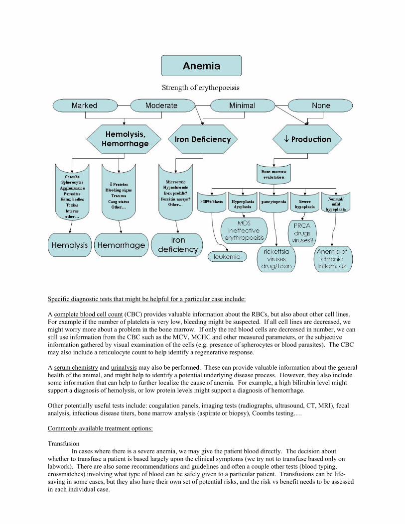
How do we fix the problem? The primary purpose of the talk to today is to explain how we answer these two important questions. Sometimes the answer is obvious (e.g. bleeding from trauma), but often there may not be an obvious historical or external reason for the anemia. In those cases it helps to have a plan. Diagnostic plan for anemia: There are three ways in which anemia is commonly classified further in order to provide more information about the potential underlying cause(s):
1. Classification by marrow responsiveness (regenerative vs. non-regenrative) 2. Classification by morphologic features (size and “color”; MCV and MCHC) 3. Classification by basic pathophysiologic mechanism (loss, destruction, production)
Classification by marrow responsiveness Definitive classification of the regenerative response to anemia is based upon a reticulocyte count. Reticulocytes are immature RBC that still have fragments of RNA which stains differently than the rest of the cell. The absolute reticulocyte count, corrected reticulocyte percentage, or reticulocyte index could each be used to determine if there are as many reticulocytes as there should be. If the marrow is functioning normally, there should be significant production of new red blood cells as a response to anemia, and these should start to appear in the blood within 3-4 days. Before this time, the reticulocyte count may be misleadingly low. There are other changes
that can suggest a regenerative response (e.g. variations in size and color of RBC observed under the microscope or by machines), but none are as specific as reticulocytes.
One common misunderstanding is to assume that nucleated RBC (nRBC) are a sign of a regenerative anemia. NRBC are very immature and if they are present in the blood it is because they were released prematurely from the bone marrow. Although this can happen during periods of strong regeneration, it may be due to the anemia and not the regenerative response. More appropriately, these cells should be viewed as a sign of damage to the blood/bone marrow barrier. This can be due to a number of causes, including low oxygen levels (often associated with significant anemia), primary bone marrow diseases, or other causes. It is possible to have nRBC in circulation, but not have an appropriate regenerative response.
Regenerative responses are more likely to occur with blood loss or destruction (hemorrhage or hemoysis), or to a lesser degree with iron deficiency anemia.
A non-regenerative anemia is more likely to be seen with various bone marrow diseases or with a condition known as “anemia of chronic disease”.
Classification by morphologic features For this classification two values are considered: size and “color”. The size can be subjectively assessed by visualization under the microscope or measured in the laboratory as a value called the MCV. Color can also be assessed subjectively via microscopy, but can also be quantified in the laboratory as a value called the MCHC.
●MCV = mean cell volume. This is the average volume of the individual RBCs. The terms microcytic, macrocytic, and normocytic are used to describe big, small, or normal cells respectively. ●MCHC = mean cell hemoglobin concentration. This is the average amount of hemoglobin per unit of surface area of the red blood cells. The terms hypochromic and normochromic are used to describe cells with decreased or normal hemoglobin concentrations respectively. There is no such thing as a hyperchromic cell –high MCHC is due to artifact.
Any combination of terms can suggest a certain subset of conditions that are more likely to be responsible for the patient’s anemia. Classification by basic pathophysiologic mechanism This is the goal in any case, and it may be very basic (loss, destruction, production…), or very specific (e.g. hemorrhage from a GI leiomyosarcoma, hemolysis secondary to zinc toxicity, pure red cell aplasia…). In general, the more specific the diagnosis… the more specific the treatment options and the better our ability to predict the response to them. Example of diagnostic algorithm of anima in dogs.
Specific diagnostic tests that might be helpful for a particular case include: A complete blood cell count (CBC) provides valuable information about the RBCs, but also about other cell lines. For example if the number of platelets is very low, bleeding might be suspected. If all cell lines are decreased, we might worry more about a problem in the bone marrow. If only the red blood cells are decreased in number, we can still use information from the CBC such as the MCV, MCHC and other measured parameters, or the subjective information gathered by visual examination of the cells (e.g. presence of spherocytes or blood parasites). The CBC may also include a reticulocyte count to help identify a regenerative response. A serum chemistry and urinalysis may also be performed. These can provide valuable information about the general health of the animal, and might help to identify a potential underlying disease process. However, they also include some information that can help to further localize the cause of anemia. For example, a high bilirubin level might support a diagnosis of hemolysis, or low protein levels might support a diagnosis of hemorrhage. Other potentially useful tests include: coagulation panels, imaging tests (radiographs, ultrasound, CT, MRI), fecal analysis, infectious disease titers, bone marrow analysis (aspirate or biopsy), Coombs testing…. Commonly available treatment options: Transfusion In cases where there is a severe anemia, we may give the patient blood directly. The decision about whether to transfuse a patient is based largely upon the clinical symptoms (we try not to transfuse based only on labwork). There are also some recommendations and guidelines and often a couple other tests (blood typing, crossmatches) involving what type of blood can be safely given to a particular patient. Transfusions can be life-saving in some cases, but they also have their own set of potential risks, and the risk vs benefit needs to be assessed in each individual case.

Treatment of underlying cause This seems obvious, but it is important not to overlook this. If an animal is bleeding, we want to know where and why and then address that problem. Similarly, if the animal is destroying RBC due to a toxicity or infection, then removing the toxin or treating the infection may be all the therapy that is necessary. Erythropoeitin (EPO) The recombinant human EPO protein is commercially available. It does seem to exert several effects in dogs. One potential risk is due to the fact that it is a foreign (non-dog) protein. The immune system of some patients may recognize this protein as foreign and start to produce antibodies against it. If this happens, it can eliminate the positive effects of the EPO and in more serious cases these antibodies can start to abnormally recognize and inactivate the dog’s own EPO as well, leaving no stimulus for new red blood cell production. This medication is therefore only used for certain diseases and in cases where there are not other good treatment options. Iron Iron supplementation only helps when there is an actual iron deficiency. This may seem obvious, but it is a key point. Like all other medications, there are some potential side effects of iron supplements. Also, anemia of chronic disease is probably not a true iron deficiency as much as a relative deficiency in which the normal processing and transport of iron is altered. For this reason, we do not recommend giving extra iron to these patients.
One Medicine: Integration of East and West
R.M. Clemmons, DVM, PhD Associate Professor of Neurology & Neurosurgery
Certified in Veterinary Acupuncture SACS, University of Florida
Some people say, “There is only one medicine: medicine which is proven; medicine which is scientific; and medicine which is good”, indicating that all other forms of medicine are bad, unscientific and unproven. I disagree. There are many forms of medicine which have been practiced (or are currently being practiced) all over the world which these people would explain away as superstition and quackery; however, some of these practices are as valid today as they were thousands of years ago. While antibiotics, computers and other advances in equipment have revolutionized medicine, these have only been available in the last 60 years. So, what we think of as modern medicine is barely 200 years old. People lived healthy lives long before that and had existing health care systems which relied on herbal medicines, foods and body manipulations for treatment and prevention of disease. Only in the United States has modern medicine completely replaced older forms of medicine. The World Health Organization recently indicated that 80% of the World’s population relies on herbal medications as part of their primary health care. A new movement today in medicine is the incorporate modern, Western medicine with the best of other forms of healing into a single more expansive, integrative medical system. This is based upon the concept that there is only one medicine, medicine that helps patients recover from injury and disease. Practitioners of integrative medicine combine traditional medicine with alternative forms of healing to treat their patients based upon what the problem the patient has. Traditional medicine, that medicine taught by modern, Western medical schools, is great at diagnosing and treating acute disease. On the other hand, it is not always the best at preventing disease. Certainly, judicious use of vaccinations has helped protect against diseases of early life; but, short of this, modern medicine has not yet embraced methods to keep most diseases from happening, particularly chronic diseases like auto-immune disorders and cancer. Only now are diet, exercise and nutritional supplements being considered as part of health and physicians are beginning to encourage patients to seek help from less "traditional" medical systems. Veterinarians have lagged behind this movement in human medicine toward integrative care. Of course there are a number of veterinarians who practice non-traditional forms of medicine; but most of these veterinarians do not practice conventional medicine as well as complementary medicine. Often, they wear 2 hats, one for conventional medical practice and another for alternative medical practice or they abandon conventional medicine altogether. This leads to a division in veterinary care rather than integration of this care. Hopefully, the movement toward integrative medicine will bridge the gap and bring both sides of traditional and complementary veterinary medicine together. Rather than to argue who has the best way to treat a patient, veterinarians can focus on how best to resolve any current disease and, then, how to keep the patient healthy in the future. This is, to me, the goal of integrative medicine. We know that the application of recent and future advances in modern medicine will not stop. We must continue to examine the inner workings of the body in terms of new developments, concepts and scientific knowledge. On the other hand, Eastern philosophers would argue that to treat the body while ignoring the spirit is not practicing healthy medicine. This is at the heart of the controversy and the movement toward incorporating alternative medicine into patient care. Many people perceive alternative medicine as a kinder and gentler approach. Science can be cold, calculating and heartless. Medicine should not be. Medicine deals with people and pets, who are not cold or heartless. The best science is no good if the patient is ignored. The movement toward specialty practices in veterinary medicine, providing veterinarians with additional training in a specific area of medicine has furthered this division. The patient can get lost in the scientific struggle to characterized and identify the disease. Some patients are called “a great liver case” or “a case of congestive heart failure”. That is why holistic veterinary medicine was created. It said, “No, it is a pet with a bad liver. We must take care of the whole patient, not just the liver.” But, integrative medicine goes farther. In integrative medicine, it is understood that not every veterinarian can be an expert in all aspects of medicine, either traditional or non-traditional. On the other hand, the primary veterinarian does have the responsibility to know enough about the disease process and the various traditional and non-traditional approaches that can be taken in the diagnosis and treatment of the patient so that the best recommendation can be made for each patient. In that way, the patient can be referred to the best veterinary health care team, including traditional veterinary specialists and practitioners trained in non-traditional medicine so that the patient can receive the benefits from each approach. Test procedures and therapies can be coordinated and prioritized based upon the patient’s individual needs.
In that way, the body, mind and spirit can be served for both the short and the long term good of the patient. Acute care is most likely to take the form of traditional medical care, while long term health is probably best achieved with changes in the patients life-style, including dietary modifications, vitamin therapy, exercise, energy work (acupuncture, homeopathy and healing touch), and manual therapy (massage, physical therapy and veterinary chiropractic). Developing a comprehensive health care approach for each patient provides integrative medical care. We still have a distance to go to see integrative medicine gain its proper place in the care of veterinary patients. The sides are still divided; however, the ground swell is beginning and many more veterinarians are embracing the concept. It is, after all, that patient that counts. Here are some areas where integrative medicine can be applied. Exercise The importance of regular aerobic exercise in the prevention of chronic degenerative diseases and maintenance of good health should not be overlooked. Many studies in human beings have demonstrated improved muscle performance, memory and cerebral blood flow in patients who undertake aerobic exercise. Many of the goals of treatment in chronic neurodegenerative diseases are obtainable through regular exercise. Two forms of exercise seem the most useful: walking and swimming. Both have their merits and they may not be exclusive. A number of pet owners have reported that swimming assists dogs beyond the exercise of mere walking. Swimming generally increases muscle tone and allows movement without stress on joints. Walking, on the other hand, helps build strength, since gravity is involved. In older patients, particularly those with arthritis, gradually building the exercise program is important. In addition, allowing a day of rest between heavy workouts can help the patient recover faster from the exercise. A good general reference of exercise physiology and exercise programs is a book by Jeff Galloway: Galloway's Book on Running, Shelter Publications, Inc., Bolinas, CA, 1984. Start out with 5-10 minutes of walking or swimming every other day for 2 weeks. Then, increase the length of exercise time to a goal of 30 minutes twice a week and a long walk of 1 hour once a week. If your dog already exceeds this limits, that is fine. However, remember to provide a day of easier exercise between vigorous workouts. This is particularly important as the patient gets older. It is sustained exercise which is important, walking in the backyard is not adequate. Many patients with chronic spinal disorders have remained functional because of exercise alone. Diet The best dog food is fresh food, prepared to provide optimal nutrients while reducing risks of disease transmission. Not everyone can home prepare the diets for their pet. It does take time and extra planning. Millions of years ago, dogs caught their own food and ate it raw. Today, however, processed raw food is not as safe as the fresh-killed food our dogs’ ancestors ate. I think that all dog food should be cooked (at least on the outside) to reduce the chances for contagion and to increase the food’s palatability. Modern dogs have evolved with us and have adapted to eating what we eat. It is best to feed them with diets that have been checked for their unique requirements and balanced for them. Too much variety may lead to gastrointestinal upset and diarrhea. On the other hand, adding some variety helps prevent deficiency of vital nutrients. Commercial foods (particularly premium, natural pet foods) offer the advantage that they are convenient and they do meet the minimum daily requirements (MDR) for dogs. On the other hand, even the best commercial food does not provide extra nutrients beyond those needed to prevent specific nutritional deficiencies. In addition, the MDR for dogs were established prior to the increase in pollutants and stresses that our pets are exposed to today. These commercial foods can, therefore, benefit with the addition of fresh food and supplements, making them more complete and healthy. To improve the quality of any commercial dog food, add tofu (a good source of soy lecithin, phytoestrogens and bioflavonoids), carrots (a good source of beta carotene), greens (like spinach which provides many trace minerals), and broccoli (a good source of bioflavonoids which act as anti-cancer compounds). These can be mixed by the following formula and added to make up 1/3 of the total diet (reducing the commercial food by 1/3 in amount). $ 4 oz Tofu (soybean curd) $ 2 Whole Carrots $ 1 cup Spinach (cooked) $ 4 Tbs. Green Bell Pepper $ 4 Broccoli Spears (½ cup)
The tofu can be fried in olive oil and the other vegetables cooked to help in their digestibility. Most dogs will enjoy this combination and benefit from the extra nutrition provided. One way to provide this conveniently would be to get prepared stir-fry vegetables and add tofu during their preparation. Herbal therapy Many of our modern day drugs originally came from plants. Even Hippocrates, the father of modern medicine, suggested that health could be maintained with regular exercise, a good night’s rest, a healthy diet and a few good herbs. Certainly, herbal medications help maintain the health of most of the people on the planet and most animals know instinctively about certain plants. Dogs eat grass to sooth their stomachs. The opponents of herbal medicines point to the inconsistencies in certain preparations, variation in plant contents brought on by seasonal variations and lack of standardization from manufacturer to manufacturer. They state that herbal remedies are not FDA approved and can, therefore, be unsafe. All of this is potentially true. On the other hand, none of these problems is sufficient to warrant not using certain herbal remedies to help maintain health. In the cases where the herbal ingredients can be toxic, yet very beneficial, the ingredients should be isolated and reduced to the active ingredient. This is true for drugs like digitalis from the foxglove plant or vincristine from periwinkles. For many of the other herbs, reducing them to one ingredient may actually stop their action, since it is the combination of materials which make them work. Herbal medicines can generally be separated into those which are safe for everyone, those which are safe unless there is a pre-existing medical problem, and those which are safe if used under medical supervision. The culinary herbs, if used in moderation, can be highly beneficial to health and usually cause little concern. These would include herbs like ginger and garlic. Fresh ginger is an important antiemetic drug which soothes the stomach and reduces nausea. Dry ginger can be helpful in controlling mild diarrhea. Fresh, crushed garlic is antibacterial and antifungal and can be used to help control infection. There is, however, a single report of a single cat who developed a Heinz body anemia on high doses of garlic. Herbs like Ginkgo biloba are probably safe unless there are medical reasons not to use it. Ginkgo improves blood flow to tissues and has anti-asthma properties. As an antioxidant, it appears to be as potent as many of our modern medications. In older people, it can improve cerebral blood flow by up to 70%, improving memory and reducing progression of Alzheimer’s disease. Certainly, it has great potential in treating Canine Cognitive Disorder in older dogs. Ginkgo does have the potential, like other antioxidants, to reduce platelet function and lead to prolongation of the bleeding time. It should, therefore, be used with caution in dogs with von Willebrand’s disease. Hawthorn, Crataegus oxyacantha, is a heart tonic that can low blood pressure, reduce chest pain, moderate cardiac arrhythmia and increase blood flow to the heart, itself. It can improve exercise and stress tolerance. Hawthorn provides at least 4 benefits to the heart, all of which are the goals of modern heart patient therapy. It appears to be safe, can be used with other heart medications (although it can be synergistic with digitalis and, therefore, digitalis doses should be reduced if used with hawthorn), and does not loose its effectiveness over time. In studies of human patients in Germany who had Type II congestive heart failure, hawthorn was as effective as any other therapy. However, because it is used to treat (as well as prevent) heart problems, it should be used under the guidance of your veterinary health care team. Awareness and use of herbal medications in people and animals is increasing, particularly in light of the expense of modern medications, when sometimes there are cheaper herbal alternatives. Many conditions do respond to herbal treatments and herbs can help prevent some disease processes from progressing to the point where more aggressive interventions are needed. Part of integrative medicine is to provide data where available or to continue to investigate and make the data available in the future where it is not about which herbs have effects that can help maintain health and which do not appear to have efficacy. With limits on veterinary interventions that can be undertaken, decision about what herbal remedies to use must be made wisely and frugally. Orthomolecular Medicine Orthomolecular medicine (OM) is an emerging tool of the 21st century. OM is the preservation of health and prevention of disease through the provision of the optimum molecular constituents of the body. Literally, it means “right molecule”. Practitioners of OM believe that nutrition must come first in health, that each individual has a biochemical optimum, that drugs can be toxic and should be minimized where possible, and that pollution cannot be escaped. As such, they advocate the use of prescribed qualitites of vital nutrients at levels sufficient to prevent, treat or control certain diseases. MDR of antioxidants, membrane stabilizers and cofactors (many of which are vitamins) are not enough to fulfill the bodies requirements and supplementation of these levels is necessary for health.

Antioxidants include vitamin E, vitamin C, selenium, beta carotene (vitamin A), superoxide dismutase, glutathione peroxidase, acetylcysteine, and L-methionine. Membrane stabilizers include omega-3 fatty acids, gamma-linolenic acid, coenzyme Q-10, L-carnitine, and L-taurine. Cofactors include B vitamins (niacin, folic acid thiamin, and cyanocobalamin) and trace minerals (zinc, iron, copper, and cobalt). All of these can be manipulated to provide the right individual balance for each pet. Vitamin E is an important nutrient which has been shown to have a number of physiologic and pharmacologic effects. It in a potent antioxidant and reduces fat oxidation and increases the production of HDL cholesterol. At higher doses, it also reduces cyclooxygenase and lipooxygenase activities, decreasing production of prostaglandins and leukotreines. As such, it is a potent anti-inflammatory drug. It will reduce platelet function and prolong the bleeding time slightly in healthy individuals. There is no known side-effects to vitamin E at levels less than 4000-6000 IU per day. Preventative levels in dogs is around 10-20 IU/kg, while therapeutic levels can be between 50-100 IU/kg. Vitamin C works with vitamin E and helps regenerate vitamin E, potentiating its antioxidant effect. Vitamin C appears to stabilize and strengthen the collagen fibers of blood vessels, maintaining their flexibility and compliance. Vitamin C supplementation does no harm, since the excess is excreted through the kidney. While dogs produce vitamin C in their bodies (unlike human beings and guinea pigs who must have it in their diet), under stress or disease, they may need vitamin C in excess of their manufacturing capacity. In excessive dose, vitamin C can cause flatulence and diarrhea. This intestinal tolerance level varies among dogs, but is generally around 3000 mg per day in an adult German Shepherd. The dose of vitamin C to start with is around 25 mg/kg twice a day. Omega-3 fatty acids like EPA (eicosapentaenoic acid) and DHA (docosahexaenoic acid) are the constituents of fish oils that act as anti-inflammatory agents and may be worth trying if your dog has an autoimmune disorder or arthritis. Many versions of these substances are on the shelves of health-food stores, from salmon oil to capsules of concentrated EPA. However, eating some cooked salmon or sardines may have benefits over capsular forms of the fish oils. Alternatively, you can give ground flax seeds, flax oil, or hemp oil as a dietary supplement; rather than fish oils. These materials will reduce platelet function for a brief period in dogs, but it seems that dogs compensate for this within about 8 weeks. Omega-3 fatty acids replace the 2-series fatty acids over time. As such, cellular stimulation produces 3-series prostaglandins and thromboxanes. The later does not cause inflammation and reduce blood flow like the 2-series thromboxanes. Try 10-15 mg/kg of fish oil, 1 T ground flax seeds, or feed 2 sardines every day. Since some studies have demonstrated negative or adverse effects using fish oil capsules (due to spoilage), I prefer giving sardines or ground flax seeds as the supplement source. B complex is a balanced form of vitamin B supplementation; which is the only way B vitamins should be given, unless specifically instructed to give one of the B vitamins by your veterinarian. B vitamins are cofactors for a number of important biological processes. They are important in maintaining a positive environment for neural regenerative efforts. In addition, they are water soluble so that any excess is merely eliminated in the urine. I recommend that all dogs receive B complex supplements twice a day. For small dogs, use the regular B complex. For medium size dogs, use high potency B complex (B 50s). For large dogs, use high potency stress formula B complex (B 100s). The advantages of OM therapy is that the ingredients can be optimized for each patient, supporting their optimal healing system function. The components can be used both to treat and to prevent disease, while remaining safe and cost effective. OM practitioners still need to validate efficacy of each component, demonstrate whether drug interactions exist, and provide safety information where lacking. On the other hand, OM has been practiced in one form or another for around 40 years which seems to be the minimum time for acceptance into mainstream medicine. Human-Animal Bond An important aspect of your pets development is play and attention from you, the owner. Not only do human beings benefit from contact with animals, animals benefit from the care and interaction with their owners. Companionship and care given mutually will help the owner and the pet live happier and healthier lives. No matter how busy or hectic things seem to be, be sure to spend time with your pet. It is best to set aside play time. This can be part of the regular exercise period, but also make time to cuddle, hold and touch your pet. It is also good to "practice" manipulations which might be needed in times of injury or illness so that they will be less stressful should they be needed. Don't worry, your pet will welcome the attention. Vaccinations There are two things have been ingrained in the teaching of veterinarians for years: 1) dogs should eat dog
food and 2) dogs and cats should be vaccinated yearly for every disease imaginable. There is actually a lack of scientific evidence to support the current practice of annual vaccination and increasing documentation showing that over-vaccinating has been associated with harmful side effects. While vaccinations is one of the 20th century’s greatest advances in medicine, saving thousands of lives by preventing childhood infectious disease, there is mounting evidence that these vaccinations may play a role in the increasing incidence of autoimmune diseases and even the cancers that we see today. Prime examples are the association of autoimmune hemolytic anemia with vaccination in dogs and vaccine-associated sarcomas in cats -- both of which are often fatal. The vaccine contains adjuvants that boost the body’s response to the altered vaccine materials (proteins derived from the infectious organism). This material is injected into the body, which can lead to local trauma and release of tissue antigens at the site of injection. As a result, the adjuvant can stimulate the body’s immune response at these released body antigens as well as the vaccine material. Except for rabies vaccine, the yearly revaccination recommendation on vaccine labels is only a recommendation without supporting data of long-term immune studies. It is not a legal requirement. Only rabies vaccines have required duration, immunity studies that must be carried out before they can be licensed in the United States. Even with rabies vaccines, a three-year duration of immunity product may also be labeled and sold as a one-year product. Legally, rabies vaccination is required in many areas and the accepted duration of immunity varies greatly. Working with local governments to achieve reasonable vaccination schedules for rabies is the only way to change this. On the other hand, your veterinarian can provide documentation to bypass this legal requirement, if vaccinating your pet could be medically unsafe. Unfortunately, no one knows the real need for vaccination, but yearly boosters for all infectious diseases are overkill. Clearly, in many cases, the vaccinations are not necessary and giving them may cause problems. The risk of not giving vaccinations (once the healthy young dog has been adequately immunized) is becoming less than the risk of giving them. What appears to be the prevailing view is that dogs and cats should receive their puppy and kitten series against the major canine and feline diseases. These vaccinations should be repeated at 1 year of age. After that time, only necessary vaccines should be given. That includes, of course, the legally required rabies vaccinations. Your local veterinarian is your best resource to develop a vaccination program tailored for your pet. The health status and infectious disease risks of your pet should be considered in the selection of a vaccination program. Infectious disease risk may with differing localities. In addition, recent studies clearly indicate that not all vaccines perform equally. Once puppihood is over, further parvovirus vaccination is probably unwarranted. The disease in adults is mild and self-limiting. Intranasal vaccination for bordetella may provide life-long immunity (although more frequent intranasal vaccination may not carry the same risk as injected vaccines). In areas where Lyme's disease or leptospirosis are not prevalent, vaccination for these agents seems unnecessary. On the other hand, vaccination for canine distemper and canine hepatitis virus is probably warranted at some time while the animal ages. There are currently 3 ways to do this: 1) monitor titers and vaccinate when the IgG antibody titer drops below 1:50 (although this may not be any more valid than guessing), 2) revaccinate when the dog gets 10-12 years old (which in many cases will be adequate), or 3) play the odds and vaccinate every 3 years. Recent studies with the major feline vaccines indicated that the worse vaccine had, at least, a three-year duration of immunity in health cats. The best vaccine protected cats for over eight years. The American Association of Feline Practitioners as a result recommends a three-year vaccination schedule for cats. No one wants their pet to contract a preventable disease, yet most healthy animals do not need vaccination as often as is currently practiced. Immunodeficient animals may not respond adequately regardless of the vaccination schedule. Discuss these options with your veterinarian and make an informed choice about vaccination. Hopefully, your veterinarian will have thought and struggled with these issues and be able to support your decision about your pet's health. Remember: Just because you pet does not need yearly vaccinations, they should still have a yearly check-up by your veterinarian! Additional Measures Acupuncture: Acupuncture is one form of ancient medicine which has now become mainstream and is widely accepted as a method to provide analgesia without the side-effects of drugs. Acupuncture has local effects, segmental effects at the spinal cord level and systemic effects mediated through brainstem connections with acupuncture points. Connections with the body surface and internal organs (referred pain pathways) allows stimulation of surface acupuncture points to influence the function of internal organs. In addition, dysfunction of the internal organs can be manifested by sensitivity of points on the body surface. Acupuncture can help treat gastrointestinal and urinary tract dysfunction. It stabilizes the adrenal gland function and may increase endogenous

corticosteroid secretion without the side-effects of exogenous steroid medication. Electrical acupuncture will stimulate reflex activity, improving muscle strength and allowing more rapid return of function. Generally, acupuncture is given over several treatments. If it does not provide benefits within 3-5 treatments, then further therapy may not be warranted. Acupuncture should be performed only by a veterinarian who is trained and certified in its use; your veterinarian should be able to refer you to a qualified veterinary acupuncturist in your area. Chiropractic Care: Veterinary Chiropractic is a rapidly emerging field in treating equine patients and is expanding in its role in treating small animals. It should be performed by a licensed Veterinary Chiropractor. In general, veterinary chiropractic involves the manual adjustments of the vertebrae to correct chiropractic, vertebral subluxations. It is felt that these subluxations result in a series of events beginning with vertebral misalignment and sequentially progressing to neuropathy, kinesiopathy (changes in normal vertebral movement), neurologic or biomechanical dysfunction, and tissue degeneration. Correcting these subluxations may reverse this process and stimulate healing. The application of chiropractic manipulations to dogs with chondrodystrophy early in life may help prevent the development of intervertebral disc (IVD) disease by maintaining vertebral flexibility. It is likely that the dietary changes and supplements discussed above will be synergistic with this effort, also. Since chiropractic is limited to manual spinal column adjustments, you will need a veterinarian who can integrate these methods. Once IVD disease as already occurred, chiropractic manipulations should not be performed during the acute phases, but be limited to the assistance of recovery following surgery or once the patient has sufficiently healed so that manipulations will be less likely to cause further IVD herniation. This may be only after "strict rest" has been enforced for 3 weeks after the patient is normal. Physical & Massage Therapy: Massage therapy improves muscle and joint flexibility, increases blood supply (improving nutrient delivery and waste removal), and help prevent or breakdown scar tissue formation. It also helps relax muscle spasms and aids in patient comfort levels. Massage therapy for animals should be performed by massage therapist trained in animal behavior and anatomy, under the supervision of your veterinarian. Many of the basic principles can be learned by the owner under proper instruction. Physical therapy is often initiated by your veterinarian, who will instruct the owner in how to continue the therapy at home. There are several physical techniques which are beneficial. Passive movement of all joints of legs can be performed. Each joint should be gently brought through its full range of motion. This will stimulate blood circulation and help maintain muscle and joint flexibility. Muscles must fatigue to gain strength. Standing exercises with weight resistance can sometimes help build muscle strength. Using warm water, hydrotherapy helps loosen muscles and increase circulation. Hydrotherapy also can be combined with passive movements during the early stages increasing the benefits of each. By removing gravity, movements may be easier for the patient to initiate with reduced discomfort. Healing Touch: Healing touch is based upon the capacity of human beings to pass "life-force" from themselves into others willing to accept this gift. Although many forms of healing touch are taught in the West, they represent teachings of the same physical process. Many studies have indicated that human contact can help lower blood pressure, reduce stress and improve the state of well-being of the recipient. Human contact has also been shown to increase the immune resistance of others. These principles can be used to help animal patients heal, as well. While it is not easy to demonstrate measurable results in all cases, certainly healing touch does no harm. When done as taught by practitioners of healing touch, it does not cost the "giver" personal energy, since the "giver" acts as a conduit of "universal" life-force which is freely available from a limitless supply of life-force within the cosmos. The "recipient" is free to accept and use this life-force energy. Most Eastern philosophies of healing are based upon the concept that living beings are based upon energy which flows in the body. When the energy level is low or there is a blockage of energy flow, disease develops. Healing touch, by providing life-force energy above or below this blockage, can re-establish the natural flow of energy, allowing healing to take place. While healing touch has a spiritual aspect, it is not a religious practice nor does it require any particular belief by the giver or recipient. What is required is a recognition by the giver that this process can occur and for the giver to practice the technique to establish pathways for energy flow from them to the recipient. Distant healing touch can also be beneficial to patients. In this form of healing touch, the giver establishes a "psychic" connection with the recipient and mentally visualizes offering the life-force to the patient. Many double blind studies have shown that prayers directed at patients in human intensive care units reduce the complication rates of those patients and their ultimate length of stay in the intensive care unit. Distant healing touch and prayer seem to work through similar mechanisms, in their benefits to patients. On the other hand, belief in any specific religion is unnecessary to practice healing touch. Any person can learn and practice healing touch. In fact, most people perform healing touch without knowledge of doing so. Healing touch may be helpful to maintain normal health in dogs. It also will assist in speeding and maximizing recovery if disease occurs. Since this can be done without risk of injury, it will do no harm; yet healing touch may increase the chances of full recovery. It also helps develop the human-animal bond. The outcome of
healing touch is non-judgmental. It is a gift which is shared between the patient and healer. Summary Maintaining health is becoming increasingly difficult. All animals are born with a tremendous capacity to heal. In fact, most (up to 80%) patients who experience a temporary illness will overcome the illness without costly intervention. This healing system is now beginning to be understood and involves an integrated system of immune regulation by the body, offering resistance to disease and injury. Unfortunately, this healing system can be overwhelmed by many factors including poor diet, bad hygiene and chronic exposure to environmental stresses. Pollution in the environment leads to internal pollution as the pollutants are concentrated over time. Internal pollution poisons the healing system. In the worst cases, one of two outcomes can be predicted. The immune system can be increased, leading to chronic immune diseases. Alternatively, the immune system can be shutdown, leading to cancer. It is not always possible to live in a pollution-free environment, it can come into the body through air, food or water. On the other hand, the latter sources of pollution can be minimized through healthy nutrition and safe drinking water. Traditional Western medicine is excellent in diagnosing disease and in treating acute disease. However, the treatment of chronic immune disease and cancer have yet to achieve the same level of success. Part of this is due to the fact that these conditions respond slowly and best when the healing system is taken into account during the treatment process. Eastern medicine, which involves long-term changes in "life-style", has many aspects which make it better in treating chronic conditions, since the goal of Eastern medicine is to support the healing system. Integrative medicine combines the best of both Western and Eastern medicine to offer the patient the best chances of returning to health. If an animal breaks its leg, it needs to be taken to an emergency facility to have it diagnosed and "set". Once this has been performed, then the patient needs to heal, by whatever means supports that best. Integrative medicine supports the patient, providing both the sophistication of modern care without ignoring the wisdom of age-old medicine. We must continually update and expand what upon what has gone before. Things which seemed unimaginable yesterday are the technologies of today; yet, in medicine, the patient must always come before technology and patient care must provide the best it has to offer. It matters not whether medicine is old or new; it matters only that the patient has the chance to live a long and happy life.
Heat Stroke Kirsten Cooke
Clinical Assistant Professor Dept. Small Animal Clinical Sciences
Heat stroke is a severe elevation of body temperature (usually > 104.9) that results when an animal is exposed to high environmental temperatures. It occurs most commonly in animals that are confined in places without adequate shade/cooling (ie. closed cars, outdoor pens without shade on hot days). However, heat stroke can also occur as a result of exercise during hot, humid weather or as a result of an inability to dissipate heat.
The human body dissipates heat by sweating which leads to evaporative heat loss. Dogs do not sweat to a significant degree and so, must rely largely on panting for evaporative heat loss. Other mechanisms of heat loss include dilation of blood vessels as well as increased circulation to the skin which results in radiant heat loss. Anything that impairs airflow in the upper airway (nose, larynx, trachea), or prevents radiant heat loss can increase the risk of heat stroke.
Diagnosis of heat stroke is based on a combination of the patient=s history as well as the clinical signs or symptoms.
Initial signs of heat stroke (or impending heat stroke) are: rapid panting, rapid heart rate (usually >160-180 in a large-breed dog), hyperdynamic (Abounding@) pulses, bright or brick red gums that may be dry to the touch. Rectal temperature is usually > 104.9 (104.9-109 F).
Later signs, as the condition worsens, include: mental depression, seizures, shock (weak pulses, pale-grey mucus membranes, vomiting and diarrhea.
Early treatment of heat stroke is vital in stabilizing the patient. The most important initial treatment is to
lower the core body temperature. The patient should be moved to the shade or cool environment. The coat should be soaked with cool (NOT COLD) water and, if possible a fan should be used to maximize evaporative heat loss. Placing cool (again NOT COLD) compresses or using cool water from a hose over the axillary (armpit) and inguinal areas may help to cool blood flowing through several large vessels in those areas. Use of cold water (ie. an ice bath) can be detrimental for several reasons:
1. If the skin becomes cold, the superficial blood vessels constrict which reduces radiant heat loss. 2. If the rapid cooling may cause the animal to begin shivering which leads to increased heat production. 3. Rapid cooling has been associated with the development of serious complications such as disseminated intravascular coagulation (DIC).
Once the patient=s temperature reaches 103-103.5 F cooling efforts should be discontinued and the rectal temperature monitored closely as it may continue to decrease.
Other means of lowering body temperature include chilled intravenous fluids, cold water enemas or gastric lavage. However, these have not been shown consistently to be of benefit and may do more harm than good and thus, are not currently recommended. Once cooling has been initiated it is essential that the dog be evaluated by a veterinarian as soon as possible. In Ashocky@ patients, a veterinarian may recommend oxygen therapy as well as judicious use of IV fluids to help stabilize the patient.
Heat stroke can also lead to several life-threatening complications including kidney damage/failure, gastrointestinal ulceration (which can then lead to bacterial infection and sepsis), liver damage/failure, cerebral edema and DIC. These complications can occur anywhere from 12-24 hours to 5-7 days. Because of the risk of these complications, heat stroke carries a guarded prognosis and it is important that veterinary care be sought even if the animal appears to be recovering. Early detection and aggressive treatment of these complications offers the best chance at a successful recovery.
Canine Social Organization, Communication, & Aggression
Terry Marie Curtis DVM, MS, DACVB
College of Veterinary Medicine University of Florida
Socialization During the “Sensitive” Period, an animal is plastic and open to new experiences. This period is 3-8 weeks for dog-dog and 5-12 weeks for dog-human. At 10-20 weeks a puppy explores its environment. If dog is deprived of these experiences it is at risk for developing inappropriate or abnormal behaviors. Puppies handled at 5-7 weeks were most responsive to humans. Pups not handled until 14 weeks were fearful of humans and never formed close attachments. Pups raised only with kittens from 2.5 to 13 weeks do not recognize dogs as conspecifics and prefer to be around cats. Early separation from the bitch – at 6 weeks of age results in a negative effect on physical condition and interfered with human bonding. Attachment to location and companions occurs at 6-7 weeks. Elimination substrate/location preference occurs by 8.5 weeks. The best response to novel objects, such as leashes, occurs at 5-9 weeks. Stabilization of the pup-pup social hierarchy occurs at 11-15 weeks. General recommendations for socialization include: handling puppies from birth, exposing pups to variety of situations in a positive or benign way, exposing them to other dogs at 3+ weeks, exposing pups to people at 5+ weeks, and watching the individual puppy for signs of distress. Wolves: Ancestors of the Dog They engage in pack living. They are predatory and hierarchical. Disputes may be settled through ritual signaling or attacks that cause injury. Ritual signaling involves the use of ears, tail, head, lips, stance, eye contact, licking, and mounting. Ears that are up and forward signal alertness or dominance; those that are down and back signal fear/submission or excitement/anxiety. It is important to remember that when entering into an attack, even dominant animals will lay their ears back to protect them from damage. A Tail that is up signals alertness or dominance; if it is midlevel, the dog is relaxed, attentive; a tail that is down is indicative of fear/submission. When a dog’s Head is up, this is a signal of alertness or dominance; when it is down or turned away, this is a signal of fear/submission. Lips - elevation of lips without retraction of the commissure is a dominant aggressive threat; retraction of the commissure is a sign of submission; and retraction of the commissure with exposure of the teeth is a defensive threat. Stance – when a dog is upright/leaning forward, this is a sign of alertness or dominance; when crouched, the dog is fearful/submissive. Lying down is the most submissive position, short of rolling over. Mounting is a signal of dominance, not sexual behavior, with the exception of an intact male mounting an estrous female. Rolling over – Mothers roll their puppies over to clean them. This behavior continues into adulthood as a submissive signal. Eyes – dogs that stare at are signaling dominance; those that look away, blink are signaling submission. Licking - As puppies are being weaned, older wolves regurgitate partially digested food for them. Puppies solicit regurgitation by licking the lips of the older wolf. Licking remains in adults as a form of active submission. Dogs which are not allowed to lick faces may lick hands instead. Metacommunication is a form of communication in which information is provided that modifies the meaning of subsequent communication. The playbow is a form of metacommunication. It means “what I do next is play”. Diagnosis and Treatment of Aggressive Behavior in Dogs
Numerous considerations are involved, such as the human-animal bond, public safety, and euthanasia. When treating aggression in dogs, all of the following should be taken into account: the attitude of the owner, the presence of vulnerable individuals in the household, the size of the dog, the type of aggression, the intensity of the aggression, and special logistical issues for preventing bites (such as doors, fences, gates, collars, muzzles). With the treatment of any aggression, it is important to caution owners of the unpredictability of any attempt to treat. NO TREATMENT IS 100% EFFECTIVE. Any dog may bite, whether they have done so previously or not. It is important to obtain “Permission to Treat” from the owner, in writing.
Aggression Directed at Humans Categories include dominance, fear, possessive, territorial/protective, maternal, and predatory. Remember the “ritual signals”… If the dog signals with its eyes, ears, head, body, tail and the threatening person doesn’t go away, what’s left? With some dogs: growling, snapping, and biting. If, at that point the person retreats, the
behavior has been negatively reinforced and the dog is more likely to perform that behavior in the future. Not all aggression is the same. A diagnosis is imperative! Dominance Aggression
It is a current issue and over diagnosed. In many cases it is presumed to be the cause of aggression when no diagnostic process has been conducted. In “Dominance Aggression Syndrome” there is persistent aggression (growling, snapping, biting), accompanied by multiple ritual dominance signals directed toward the owner. The most common signalment is a male (intact, at least when problem began) that is 1-3 years of age. The most common breeds represented are the German Shepherd, Doberman Pinscher, and Springer Spaniel. But it is important to remember that it can occur in any breed, in either sex, and at any age. If it is seen in a female, it’s usually younger, < 6 months old. A dominant dog will often block the owner’s movements while in the home – by standing or lying in doorways, halls. The dominant dog may lie on the bed and growl when approached. But so will the fearful dog, so you need to look at postures! There is to be no punishment of the dominant dog, as the aggression is often exacerbated by physical punishment. Again, it is important to distinguish from fear aggression, which can also escalate when punishment is used. Dominance motivated aggression is a problem of relationships, so the aggression is most likely directed at family members. It is not likely to be the cause of aggression to you during an exam in your clinic, as you are not a “member of the pack”. A dominant dog will resist submissive postures, and there may be a history of not learning the ‘down’ command in obedience class. Food guarding in the absence of ritual dominance signals or aggression to people in any other situation is simply food guarding (possessive aggression). Many food guarders are quite submissive and non-aggressive in all other situations.
Treatment: Don’t advise an owner of a dog with DAS to “teach the dog who’s boss”, or “have it out” with the dog, as the owner is likely to be bitten. There is never to be any “alpha rolling”. Treatment involves avoiding all situations in which the dog behaves aggressively. It is important to counsel the owner that avoidance ≠ “giving in”. In fact it is actually the owner taking the power back by not allowing the dog to respond in an aggressive manner and denying him the opportunity to practice. There is a significant learning component to the behavior, so the dog needs to “re-learn”. If the dog is an intact male, neuter; however neutering an intact female may not help - there is evidence of a small effect of making the problem worse or more persistent if the dog is showing dominance aggression when a puppy. Behavior Modification involves giving a high frequency of low intensity dominance signals. It is important to identify what ritual signals the owner can safely exhibit to the dog, and have them do this many times a day. In a structured situation, gradually train the dog to exhibit a progressively more submissive posture, e.g. ‘down’. Institute the “nothing in life is free” program: the dog must obey a command, preferably ‘down’, to get anything it wants – food, petting, into the house, out of the house, etc. It is important to reward submissive postures. The Gentle Leader® head collar is designed to provide subtle dominance signals and helps to decrease overall level of anxiety. Any pharmacological treatment is extra-label use. However, increased serotonergic activity should produce a decrease in affective aggressive behavior (i.e. non-predatory behavior), so pharmacological options include the tricyclic antidepressants such as clomipramine and the selective serotonin reuptake inhibitors such as fluoxetine (Prozac®) and paroxetine (Paxil ®). Another option is the progestin, megestrol acetate (Ovaban®). The typical response is a decrease in aggressive behavior with mild sedation. The mild side-effects, which are common, include: polydipsia, polyphagia, polyuria, and mammary hyperplasia. The possible serious side-effects include: elevated blood glucose leading to diabetes and carcinomas. Progestins should generally be restricted to cases in which euthanasia is highly probable if there is not significant, rapid improvement. With long-term use, the dog’s life-span as an acceptable family pet may be extended, but life-threatening medical complications are very likely to eventually occur
Fear Aggression is the most common motivation for aggression directed at people and it is characterized by aggression coupled with signals of fear and submission: avoidance, ears back/down, tail down, retraction of commissure of lip – “grin”, looking away, turning away, and licking lips, yawning. Treatment: Do not punish! Avoid situations that are likely to trigger fearful, defensive behavior. Implement desensitization and counter-conditioning. Use of the Gentle Leader® head collar may help to decrease the overall level of anxiety. Avoid threatening gestures such as standing over, reaching out/over, loud voices, direct eye stare, etc. Interact in a non-threatening manner: pet under chin, chest – on dog’s level, talk softly, etc. Treatment: Desensitization & Counter-Conditioning The animal is exposed to a stimulus that elicits a given response, but at such a low level that the response is not elicited. Over time and successive repetitions, the intensity of the stimulus is gradually increased, ideally without eliciting the response. A response is elicited which is behaviorally and physiologically incompatible with another
response. The dog cannot be relaxed and anxious at same time. Teach the dog to relax and reward dog for being relaxed – the dog “re-learns”. Pharmacological Intervention Used to decrease the dog’s level of anxiety so that it can learn what it needs to. Options include the tricyclic antidepressants and the SSRIs – again, off-label use, so must get owner consent. The DAPP
® diffuser may also help to decrease the dog’s overall level of anxiety. Possessive Aggression The dog defends specific items (food, bones, chewies, toys, etc.), but otherwise does not exhibit aggression or ritual dominance signals. The behavior is often fear-based. Treatment: If there is a limited number or type of items defended, remove them, otherwise, desensitize and counter-condition so that the dog re-learns what interactions mean - at the food bowl, with toys, etc. Territorial Aggression The dog protects an inappropriate location or protects an appropriate location in an inappropriate context. The aggression can be directed at humans, other dogs, other animals, or a combination of targets. The dog can be territorial of the house, the yard, its crate, its sleeping place, of a confined place, the car. It may also protect an individual approach distance – a “mobile territory”. Treatment: Never leave dog outside alone “with a territory to protect”. The Gentle Leader® head collar can be useful in owner-control. The dog can be desensitized and counter-conditioned to people approaching its territory. Protective Aggression An extension of territorial aggression where the dog perceives that the owner is threatened when there is no real threat – such as with a stranger at door, when the dog is approached when in a car with the owner, when another dog approaches owner, when a person raises its voice to owner, or when a person hugs the owner. Treatment: Avoid situations where the dog believes it needs to protect the owner. Implement command control. DS&CC for approaches from strangers, dogs, etc. with the owner present, and the Gentle Leader® head collar. Maternal Aggression This is normal behavior that typically wanes as the puppies mature. Make sure prepartum bitch is familiar with whoever will be caring for her and the puppies postpartum. Predatory Aggression Canis familiaris is a predator. This type of aggression results in a number of fatalities each year, in addition to many injuries. Common targets include: joggers, bicyclers, and running children. Risk factors include: a loose dog and any history of predatory behavior Treatment: Appropriately contain the dog and implement command control. DS&CC to the object of predation may be effective. Use of the Gentle Leader® head collar with a muzzle is recommended. In laboratory studies, serotonin and GABA have been found to have an inhibitory effect on predatory behavior (Eichelman, 1987; Olivier et al., 1987; Miczek et al.,
1989). Therefore, theoretically, serotonergic medications and GABA agonists should reduce probability that this type of behavior will occur. Inter-Dog Aggression Can be status-related, fear-motivated, arousal-related, possessive, protective, territorial, redirected, and/or predatory. Fear Secondary to a lack of socialization or previous experience. Therefore, focus on the early history, upbringing, the time of adoption, and the interaction with other dogs. Look at the aggressor’s body posture and the victim’s body posture. It is important to get a description of both dogs’ behavior before and after the fight – was it defensive? offensive?
Treatment consists of decreasing the dog’s overall level of anxiety using a head collar, DS&CC, ± anxiolytic medication. Arousal Some dogs respond either more quickly or more intensely to a given stimulus than other dogs. The incidents may be situational – such as at feeding time, at owner arrival, going inside/outside, in a hallway, stairwell, on walks, etc. Treatment involves the anticipation of behaviors, redirecting the dogs, and rewarding appropriate behaviors. It is important to avoid certain situations and contexts. Use of a head collar will give the owner better control, and the dogs can be desensitized and counter-conditioned to one another. Possessive Protecting food or other resource may be an ancestral adaptive behavior and food-related aggression is tightly coupled to canine evolutionary history - canids are binge-and-gorge eaters. Some dogs from large litters that are fed from one bowl may learn to be aggressive to each other to successfully compete for food. Free-ranging street dogs may get enough food only by successfully fending off competitors. With possessive aggression, the dog reacts when food or other resource is approached and/or reached for by growling, lip lifting, snarling, lunging, and/or biting. Generally, the higher the quality of the resource, the more pronounced the aggression. Some dogs exhibit possessive-related aggression only to other dogs and there may not be a problem with humans. Treatment - It is much easier to avoid the consequences than to treat the problem. Deny access to “illicit” items such as rawhides, bones, etc. Feed dogs separately or at a distance at which they are not aggressive. Desensitization and counter-conditioning. Protective/Territorial This type of aggression is most obvious when the dog is in the yard and another dog passes, or when the dog is inside and a dog passes outside. Most or all dogs will bark, as this is the normal first step in the sequence of behavior characterizing protection. The problem occurs when the dog refuses to stop barking on command and/or becomes defensive and aggressive, and/or when the dog persists in the behavior despite cues indicating a contrary context. Some dogs become protective of their crates, places they sleep and/or an individual approach distance. Confined spaces may intensify the response – cars, restrictive chains. The hallmark of territorial aggression is that the dog is not aggressive when it is removed from the territory – the dog is fine with other dogs off-property. Treatment involves avoiding situations that dog perceives that it must defend or protect. Never leave dog outside alone, and realize that the dog can become more aggressive in a fenced area. It is important that the owner have command control. DS&CC to approaches by other dogs can be done. A head collar is recommended ± Anxiolytic medication Redirected Often seen when a dog is yelled at, physically punished, or otherwise thwarted from pursuing another aggressive behavior. For example, a person stops dog from chasing cat and dog redirects its aggression to other dog. The dog usually goes after the nearest individual who is not involved, so a person, another dog, cat. It can be difficult to diagnose as the circumstances that precipitate it are not often witnessed, and therefore it is not always recognized by the owner for what it is. When one dog is aggressive to another dog, the owner often thinks that the dog is jealous or being competitive for attention. However, few or no data exist to support this. Treatment involves identifying the primary source of the dog’s initial upset, if possible. Avoidance is key. If possible, address the behavior that is interrupted in the first place, and separate the dog and target animal so that there is no chase to interrupt. Separate the individuals involved in the redirected aggression when unsupervised and make sure that the recipient of the aggression has the most freedom. It is important to reward the aggressor for ignoring the victim. Other things such as a head collar and a bell on the aggressor to warn the victim, may also help. Predatory Predation is a maintenance behavior in carnivores, and therefore not all behaviorists agree that it’s a “true” aggression. It is a non-affective aggression with no autonomic activation that is necessary for survival. Affective aggression is categorized as that involving an intense, patterned autonomic activation associated with sympathetic and adrenal stimulation, such as fear, pain, protective, territorial, and dominance. There are two classes of predatory aggression: 1) Dogs that stalk, stare at, or silently pursue small animals - birds, squirrels, cats, other dogs, and sometimes infants, and 2) Dogs that chase moving objects. Treatment - Dogs that are predatory to other animals should never be off lead, unsupervised, or confined in a fence that other animals might cross. There needs to be adequate control using a head collar and voice command.
Status-Related Inter-Dog Aggression Intact males are aggressive mostly to other males and show more aggression than neutered males. Females are aggressive mostly to other females. Borchelt, 1983 Aggression between familiar dogs involve dogs in the same household. It is also called “Social Aggression”, “Intraspecific Aggression”, and “Sibling Rivalry”. It is a manifestation of canid hierarchical conflicts and underlying anxiety. The dogs are uncertain of their role in the hierarchy. The aggression is most commonly limited to one pair of dogs – even if other dogs are present. It is common between same-sex dogs and early spaying/neutering may help. Voith 1980, Hart 1981 Intra-household aggression is more severe than aggression between non-housemates with female-female aggression being the most severe. The aggressor tends to be younger and tends to have arrived more recently in the household. The fights are less frequent but more injurious than fights between non-household dogs. Sherman et al. 1996 Common triggers include excitement, feeding, walking, owner arrival, control over resources, physical proximity, confining areas (doorways, hallways, etc.), and the owner’s presence (the dogs may compete for attention). The owner tends to support victim (subordinate) and punish the aggressor (dominant). This can increase the aggression if the victim perceives a “coalition” between itself and the owner causing it to act more confidently. In many cases, the owner’s presence and behavior exacerbates the instability between the two dogs and fights may occur when the owner is present. Rarely, the aggression may persist in the owner’s absence and fights may occur then, also. Common triggers for fights include: owner interference when the dogs interact in an attempt to change an established hierarchy, the owner inadvertently or deliberately encouraging a subordinate dog to try to establish dominance over the higher-ranking dog. It is important that the two dogs be allowed to “talk” to one another. Often, there is not a problem between the dogs as to which will be dominant - they understand the hierarchy and signal each other accordingly and appropriately. But, if the “normal talk” is interrupted and/or punished, what is left? There can be an escalation of the communication with growling, snapping and biting. Onset usually occurs when the younger dog reaches social maturity (18 – 24 months old) and the hierarchy is not clearly established. It can also occur in evenly matched dogs, or when the “dominant” dog is aging or ill. In this case, the advanced age and/or illness do not allow the dog to maintain its status and this can result in ↑irritability → ↓tolerance to conspecifics. Treatment involves separating the dogs when not supervised. It is important to establish owner control with head collars or harnesses ± muzzles. If the male dogs are intact, neuter them. It is imperative to stabilize the pack hierarchy by identifying and supporting the dominant dog. This dog is to get attention first, be fed first, be given access to preferred resting places, allowed outside/inside first, and walked in front of the other dog. It is important to allow and reward ritualized signaling – the dogs need to be allowed to “talk to one another”. Client education is key! It may be difficult for the owner to comply, as they may have been punishing the aggressor and comforting the victim. Attempting to superimpose a system of equality may make matters worse. There is a misconception that the dog that has “seniority” should dominate the new dog. In fact, a dog’s social rank is determined by its ability to defend priority access to resources and not by seniority per se. An older or sick dog may not be capable of defending these privileges, or, the other dog may not offer subordinance. In most cases, the dogs are getting “mixed signals”: Dog A is dominant to Dog B. Dog A knows it and Dog B knows it and they signal each other appropriately. However the owner reinforces Dog B as dominant. Dog B knows that is submissive to Dog A, but it is getting reverse signals from “BIG Alpha”. This becomes a source of confusion and anxiety… Bottom Line… The aggression typically occurs in situations that include competition over valuable resources and aims at establishing a dominance-subordinance relationship. The owner should interfere if there are excessive dominance displays or if the aggressive displays do not cease when the subordinate dog defers. They should call the dominant dog away and diffuse aggression in an “upbeat and jolly” manner, so as not to add to the arousal. They are to give preferential attention to the dominant dog and reinforce their own status. The problem may not be resolvable with two evenly matched dogs that are strongly motivated to be alpha. They are likely to fight until one succeeds in injuring the other. In this case, the owner should withdraw privileges from both dogs and interrupt dominance displays by both dogs. They should randomize order of feeding and handling and desensitize and counter-condition to each other’s proximity. It is important to look for ritualized signals and reward them. The prognosis is poorer if the initiator is younger than target, if a person has been bitten, and/or if the aggression is unpredictable.
Collapsing Trachea
Gary Ellison, DVM, MS, Dip. ACVS
University of Florida, Gainesville, FL 32610
Learning Objectives
What is the signalment and what are the clinical signs associated with collapsing trachea. What are the four grades
of tracheal collapse. How is tracheal collapse diagnosed. What are the indications for surgery and what are
the three basic methods devised to surgically manage tracheal collapse.
CCoollllaappssiinngg TTrraacchheeaa
Background: Collapsing trachea is a disease process recognized predominately in miniature or toy breeds
(Pomeranian, miniature and toy poodle, Yorkshire terrier, Chihuahuas, pug). The condition is reported in dogs of all
ages, with the average being 7 years. Although the exact etiology is not known, histologically the tracheal ring
cartilage is hypocellular and deficient in glycoprotein and glycosaminoglycan content. The clinical picture is
variable with early signs generally mild with a productive cough and mild exercise intolerance in a normally active
dog, progressing to more severe exercise intolerance and a characteristic Agoose-honk@ cough. Dyspnea is often
initiated by anxiety or excitement.
Classification of collapsing trachea:
Grade I - tracheal membrane is slightly pendulous, cartilage maintains normal AC@ shape, lumen
reduced approximately 25%
Grade II - tracheal membrane widened and pendulous, cartilage is partially
flattened, lumen reduced approximately 50%
Grade III - tracheal membrane is almost in contact with dorsal trachea, cartilage is nearly flat,
lumen is reduced approximately 75%
Grade IV - tracheal membrane is lying on dorsal cartilage, cartilage is flattened and may invert,
lumen is essentially obliterated
The result of tracheal collapse is an extremely small cross-sectional area of functional tracheal lumen and
high airway resistance. Abnormal rings in the cervical trachea collapse on inspiration, while those in the thoracic
trachea collapse on expiration. When the intrathoracic trachea collapses on expiration a higher expiratory pressure
and an increase pulmonary vascular resistance results (cor pulmonale). This increase in resistance along with
chronic hypoxia causes increased right ventricular work and can lead to enlargement (hypertrophy) of the right side
of the heart.
Diagnosis: Tracheal collapse is suspected with an appropriate signalment, history and eliciting coughing reflexes
with simple digital palpation of the trachea. Radiographs and fluoroscopy of the lateral cervical and thoracic trachea
in an unanesthetized patient during inspiration and expiration can be diagnostic. Evaluation of laryngeal function
under a light plane of anesthesia should also be performed to rule out laryngeal paralysis or laryngeal collapse.
Unfortunately not all cases can be diagnosed easily and it may be necessary to elicit a cough while obtaining
radiographs to demonstrate tracheal collapse. Endoscopy/tracheoscopy is an excellent technique to evaluate the
trachea and bronchi. Cytology and culture of the airway should be obtained to determine if a bacterial component is
involved. Recurrent bacterial tracheitis can occur with severe tracheal collapse.
Medical management of tracheal collapse involves symptomatic therapy using antitussive medication,
corticosteroids, bronchodilators, sedatives, and weight loss. It can be effective in mild cases; however, more
advanced stages of collapse (grades III and IV) usually do not respond well. Aggressive medical therapy should be
attempted before surgery is considered.
Surgery: Should not be undertaken unless the remainder of the upper respiratory system is free of disease.
Correction of any other components of upper respiratory obstruction (stenotic nares, everted laryngeal saccules,
elongated soft palate, laryngeal paralysis or collapse) may relieve the dyspnea sufficiently to eliminate the need for
surgical correction of the collapsing trachea. Surgical therapy is also controversial with respect to efficacy. Deep
thoracic tracheal and major bronchial collapse have a poor prognosis, whereas isolated cervical collapse has a fair to
good prognosis for improvement. If bronchial collapse is present, surgical support of the trachea may not
sufficiently alter the clinical condition. Reconstruction of one segment of the trachea may be followed by collapse of
another segment.
Three surgical procedures are described: dorsal tracheal membrane plication, internal trachea support, and
external trachea support. These methods provide clinical relief for varying periods of time. Dorsal tracheal
membrane plication is used with ocassional success in patients with Grade I or Grade II collapsing trachea. The
condition usually worsens with time however and other methods of surgical support are usually required. If this
technique is used in animals with more severe disease the plication technique can cause severe narrowing of the
lumen as the tips of the cartilage rings are drawn together. Internal stents to support a collapsing trachea have not yet
been successful in the dog. Stents become dislodged, coughed out, obstructive, or movement of the stent can incite a
granulation reaction.
Extraluminal ring prosthesis: was the most widely used technique for years because it does not interfere with the
mucociliary apparatus and dislodgement is not usually a problem. The goal is to provide external support to prevent
collapse of the trachea without interfering with segmental motion or vascular supply. Ring prostheses are made by
either making individual C-shaped rings (Hobson technique) (5-8 mm wide) or a continuous spiral prosthesis
(Fingland technique). We have modified the spiral rings by cutting an endotracheal tube with a spring insert. The
internal diameter is chosen according to the tracheal ring diameter. Both prosthesis types can be made from syringe
cases (polypropylene). The prosthesis is applied taking great care not to interfere with the vascular or nerve supply
of the larynx or trachea. This technique has produced favorable results, however, when complications occur they can
be life-threatening. Complications: Intraluminal hemorrhage (suture penetration), peritracheal
swelling/inflammation, damage to the recurrent laryngeal nerve resulting in laryngeal paralysis and tracheal necrosis
and slough from ischemia if the blood supply to the trachea is severely compromised. The immediate post-op
recovery period can be challenging, especially if laryngeal paralysis has resulted from recurrent laryngeal nerve
damage. If paralysis occurs, surgery (laryngeal tieback or permanent tracheostomy) is usually necessary for survival.
Coughing usually improves after surgery, however, this procedure does not usually make these dogs normal.
Intra tracheal Stents: The combination of surgical risk and the inability to adequately treat intra-thoracic tracheal
collapse led to the evaluation of minimally-invasive surgical techniques used in humans for potential treatment
options. Interventional radiology involves the use of contemporary imaging modalities such as fluoroscopy to gain
access to different structures in order to deliver materials for therapeutic purposes. Specially designed intra-luminal
metallic stents have been placed within the human tracheobronchial system using these techniques to treat
chondromalacia, malignant obstruction, or strictures and stenoses. A number of stents have been previously
evaluated in the canine trachea, including both balloon-expandable stents (Palmaz) and self-expanding (Stainless
steel, Laser-cut nitinol, Knitted nitinol) Ultrflex stents.2-4 The vastly superior flexibility makes the use of self-
expanding metallic stents (SEMS) particularly appealing for tracheal use. Clinical improvement rates in 75%-90%
of animals treated with intra-luminal stents have been reported.3,4 Immediate complications were typically minor
although there was a reported peri-operative mortality rate of approximately 10%, a rather high figure compared to
the author’s experience. Late complications included stent shortening, excessive granulation tissue, progressive
tracheal collapse, and stent fracture. The made for Human stents are extremely expensive often costing $2,500-2,800
per unit not including the cost of deployment. However a newer made for veterinary stent is currently being
marketed by Infinity medical for a much reduced cost of $800-900.
Patient Selection: Neither surgery nor stenting are cures for tracheal collapse, and to the author’s knowledge, neither
has been shown to slow the progression of the disease. When used appropriately in the proper patients, both can
significantly improve the patients’ quality of life when medications alone are no longer adequate. Below are this
author’s criteria for patient selection, method of stent selection, and technique for placing intra-luminal tracheal
stents.
Whether considering surgical rings or intra-luminal stenting it is imperative that aggressive medical management
has been attempted and has failed at providing a “reasonable quality of life” for the patient. In the author’s opinion,
this includes anti-inflammatory doses of corticosteroids, anti-tussives, and the general management considerations
described above. An exception to this rule is the emergent, intubated patient which has failed attempts at extubation.
An owner’s inability to administer medication is not a valid reason to perform one of these invasive procedures as
the majority of patients will still require medication following treatment. In addition, while the “grade” of tracheal
collapse (Grades I, II, III, or IV) has been described in the literature, the author will not treat based upon the grade of
collapse alone. The success of either of these procedures must be evaluated in light of the owners’ expectations. It is
the veterinarian’s responsibility to properly inform the owner that these are largely palliative procedures and the
disease is likely to progress. Hoever many patients can be significantly improved for years after the procedures are
performed.
Post-op Care: Strict cage rest (oxygen if necessary), antitussives, corticosteroids are given for at least two weeks and
longer if coughing persists, and appropriate antibiotics based on a tracheal aspiration sample.
Prognosis: Patients receiving extraluminal rings have a better chance of not surviving the postoperative period and
may be afflicted with life threating larygeal paralysis. However if the surgery goes well many of these patients can
live long lives and have significant relief from their chronic cough. Intraluminal stents have the advantage of
providing immediate relief from airway obstruction but with time the constant motion leads to granuloma formation
and stenosis of the ends of the stent. With overactive dogs fracture of the units has also been reported. Therfore I
tend to reserve stent usage for middle to older aged dogs in which medical management is not longer effective.

References
1. Fingland RB, DeHoff WD, Birchard SJ. Surgical management of cervical and thoracic tracheal collapse in dogs
using extraluminal spiral prostheses. J Am Anim Hosp Assoc 23:163-172, 1987.
2. Nelson AW. Lower Respiratory System. In Slatter DH (ed). Textbook of Small Animal Surgery W.B. Saunders,
Philadelphia, 777-794, 1993.
3. White RAS, Williams JM. Tracheal collapse in the dog- is there really a role for surgery? A survey of 100
cases. J Sm Anim Pract 35:191-196, 1994.

4. White RN. Unilateral arytenoid lateralization and extraluminal polypropylene ring prostheses for
correction of tracheal collapse in the dog. J Sm Anim Pract 36:151-158, 1995.
5. Weiss CA. Intra-Luminal Tracheal Stenting in Bojrab ed Current techniques in Small Animal Surgery 5th edition. Blackwell publishers. In press.
Cancer Treatment Breakthroughs: Stereotactic Radiosurgery for the Treatment of Canine Osteosarcoma
James P. Farese, DVM, DACVS University of Florida, CVM
Osteosarcoma is the most common primary bone tumor affecting dogs. The current recommended
treatments for canine appendicular osteosarcoma are: 1. Amputation (+/- chemotherapy) 2. Limb-sparing surgery and chemotherapy 3. Radiation Therapy (+/- chemotherapy)
- Fractionated radiation therapy - Stereotactic Radiosurgery (SRS)
4. Palliation
Although amputation has become the most common form of therapy, many dogs are not considered suitable candidates for amputation due to concurrent orthopedic or neurological conditions. There is growing interest among owners of dogs affected with osteosarcoma for alternatives to surgery. Stereotactic radiosurgery (SRS): SRS involves the delivery of a single, high dose of radiation in a highly targeted manner while sparing the surrounding tissues. In our initial experience with SRS for the treatment of canine appendicular osteosarcoma eleven dogs were treated: nine distal radius (one pathologic fracture), one distal ulna, and one distal tibia. To perform SRS, a targeting array must be temporarily (overnight) rigidly fixed to the affected bone. This is usually done with two 1.5mm diameter pins and casting material. SRS for osteosarcoma is performed with 30 Gy prescribed to the 75% isodose line. Five dogs received SRS alone and six SRS and chemotherapy (carboplatin 300 mg/m2 30 minutes prior to SRS for radiosensitizing effects). Follow-up chemotherapy (carboplatin alone or carboplatin and doxorubicin) was administered to the six dogs that received carboplatin.
Subjective evaluation of tumoral swelling and lameness showed improvement in all cases treated, usually within three weeks. Our preliminary data indicate that the combination of coverage of the tumor with 30 Gy and chemotherapy consistently provide long-term tumor control and likely complete tumor kill. Overall median survival time in the initial eleven dogs treated was 363 days (range 145 - 763).
We are also starting to use SRS to treat tumors in upper extremity locations. The limiting factors of SRS therapy for appendicular osteosarcoma are the size of the tumor and the condition of the bone at the time of therapy, as adequate coverage of large tumors with the 3,000 cGy isodose line is not always possible and the risk of pathologic fracture remains after treatment. Thus, SRS should ideally be used to treat appendicular osteosarcomas that are relatively small and have caused minimal bone destruction. Cost is similar to that of limb-sparing surgery.

Instructions for Completing Form HR-600 The secondary employer is responsible for ensuring that the form is complete and accurate. Assignments/payments will not be authorized until the form has been approved and signed by the appropriate offices. 1. The employee and primary employer must complete and sign the “EMPLOYEE SIGNATURE” and “PRIMARY EMPLOYER” portions of the form. 2. The completed form must be submitted to the appropriate personnel office for final approval (TEAMS, USPS, OPS) and/or processing (Academic Personnel) as follows: For appointments/payments to TEAMS, USPS, and OPS: Division of Human Resources PO Box 115001, 326 Stadium Gainesville, FL 32611-5001 Phone (352) 392-1213, SC 622-1213 For appointments/payments to Academic Personnel and housestaff: Academic Personnel Office PO Box 113005, 29 Tigert Phone (352) 392-1251, SC 622-1251 Please note that this form does NOT accomplish payment. It simply provides AUTHORIZATION for payment from the University of Florida only. For more information on processing and payment, go to: http://www.hr.ufl.edu/cc/extracomp.htm or contact one of the above personnel offices. HR 600 10/02 Request for Approval of Additional University Employment and State of Florida Employment Contact Person: PO Box: Phone: Employee Name: UF Employee ID Number: REQUEST (check one) Employment of University of Florida employee at greater than 1.00 FTE or simultaneously from OPS and salaries Employment at University of Florida of an employee of other state university or a state agency Employment of University of Florida employee by a state agency or other state university PRIMARY EMPLOYMENT SECONDARY EMPLOYMENT Department/Unit: Class Title: Position Number: LP#: TKL: Rate of Pay (Hourly or Biweekly): Work Schedule: Daily: am pm Weekly: Daily: am pm Weekly: Full Time Equivalency (FTE): Period of Employment: Appropriation Paid From: Salaries OPS Salaries OPS THE SECONDARY MUST COMPLETE THIS SECTION (The secondary employer is responsible for insuring that the form is completed and approved) DUTIES TO BE PERFORMED IN SECONDARY EMPLOYMENT and EXPLANATION/JUSTIFICATION (Attach additional sheets if necessary) _________________________________________ ________________________________________ ________________ SECONDARY EMPLOYER NAME (Please Type) SIGNATURE DATE THE PRIMARY EMPLOYER MUST COMPLETE THIS SECTION (If for any reason this statement is not applicable, a separate statement of explanation from the primary employer must be attached). This employee has my approval to perform the additional duties indicated

above for the secondary employer. These additional duties will not be performed during the employee’s regular working hours with this university and will not involve a conflict of interest with the employee’s regularly assigned duties. __________________________________ __________________________________ _____________ __________________________________ __________________________________ _____________ _________________________________________ _____________ EMPLOYEE SIGNATURE DATE AUTHORIZATION OF APPROPRIATE PERSONNEL OFFICE APPROVED APPROVED AS MODIFIED DISAPPROVED FLSA OVERTIME REQUIRED REMARKS: ___________________________________________________________ ____________ APPROPRIATE PERSONNEL OFFICE SIGNATURE DATE Send original and one copy to PO Box 113005 for Academic Personnel-related requests Send original and one copy to PO Box 115001 for all other requests
