photodynamic therapy: fundamentals and dosimetry · aapm refresher course (july 28, 2005)...
TRANSCRIPT

AAPM Refresher Course (July 28, 2005) Photodynamic Therapy, Zhu et al
1
Photodynamic Therapy: Fundamentals and Dosimetry
Timothy C Zhu1, Jarod C Finlay1, and Brian C Wilson2
1Dept. of Radiation Oncology, University of Pennsylvania, Philadelphia, PA 2Department of Medical Biophysics, University of Toronto, Toronto, CA
Abbreviations: AK, Actinic Keratosis; ALA, aminolevulinic acid; AMD, age-related macular degeneration; BCC, Basal cell carcinoma; BPD-MA, benzoporphyrin derivative monoacid A, CNV, Choroidal neovascularization; CW, continuous wave; FDA, Food and Drug Administration; Hb, hemoglobin; HPD, hematoporphyrin derivative; ISC, Intersystem crossing; LED, light-emitting diode; MLu, motexafin lutetium; mTHPC, meso-tetrahydroyphenol chlorin; PDT, Photodynamic therapy; PIT, photoimunotherapy; PpIX, protoporphyrin IX; SCC, Squamous cell carcinoma;
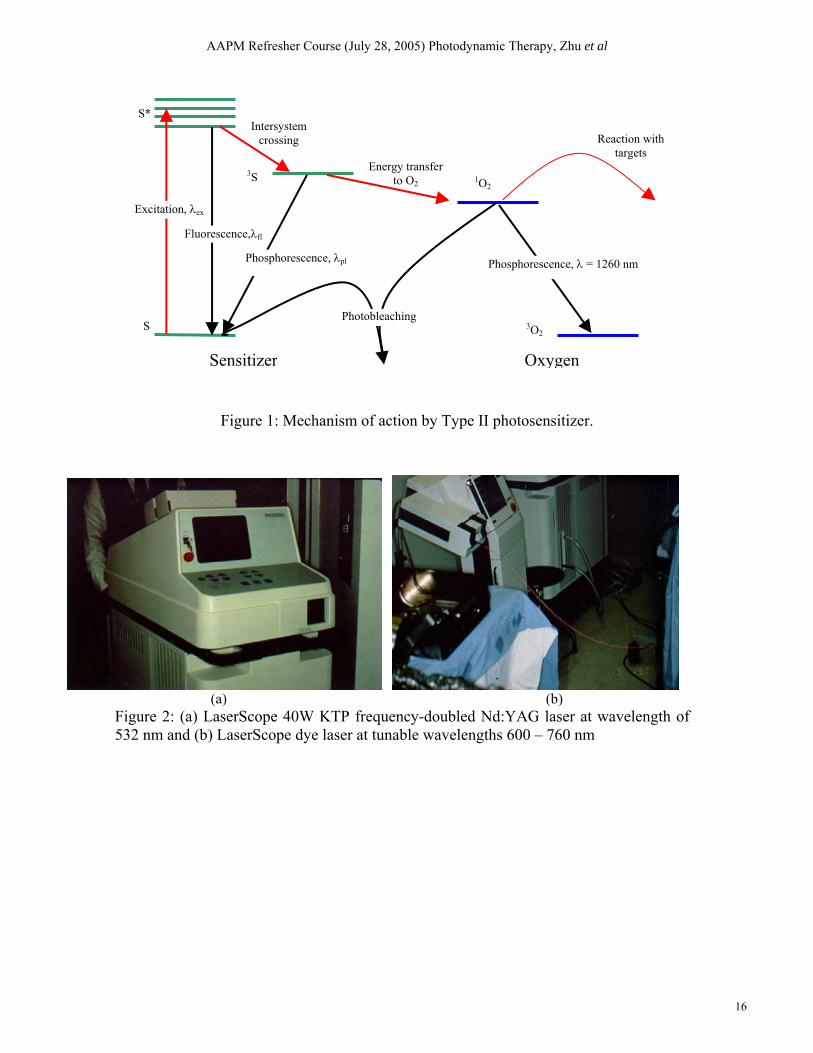
I Introduction Photodynamic therapy (PDT) is an emerging cancer treatment modality based on the interaction of light, a photosensitizing drug, and oxygen.1 The photochemical reactions that result in photodynamic damage can be characterized as either Type I or Type II reactions. In Type I reactions, the photosensitizer in its excited state reacts directly with a substrate present in the tissue, leading to the generation of cytotoxic free radicals.2, 3 The majority of sensitizers available for PDT utilize Type II photodynamic processes, meaning that they accomplish their photodynamic effect through the production of singlet oxygen.2, 4 Singlet oxygen is a highly reactive excited state of the oxygen molecule. Direct optical excitation of oxygen is forbidden by three molecular selection rules, and is practically impossible in living tissue. A photosensitizer can act as an intermediate, allowing the formation of singlet oxygen, see below. The energy level diagram shown in figure 1 summarizes the underlying physical processes involved in type-II PDT. The process begins with the absorption of a photon by photosensitizer in its ground state, exciting it to an excited stated. In general, both the ground state and this excited state are spectroscopic singlets (i.e., states with a spin multiplicity of 1). The sensitizer molecule can return to its ground state by emission of a fluorescence photon, which can be used for fluorescence detection. Alternatively, the molecule may convert to a triplet state (one with a spin multiplicity of 3), a process known as intersystem crossing (ISC). A high intersystem-crossing yield is an essential feature of a good sensitizer. Once in its triplet state, the molecule may undergo a collisional energy transfer with ground state molecular oxygen (type II) or with the substrate (type I). In type II interaction, the photosensitzer returns to its ground state, and oxygen is promoted from its ground state (a triplet state) to its excited (singlet) state. Since the sensitizer is not consumed in this process, the same sensitizer molecule may create many singlet oxygen molecules. Once the singlet oxygen is created, it reacts almost immediately with cellular targets in its immediate vicinity. The majorities of these reactions are irreversible, and lead to consumption of oxygen. This consumption of oxygen is efficient enough to cause measurable decreases in tissue oxygenation if the incident light intensity is high enough. In addition to its reactions with cellular targets, singlet oxygen may react with the sensitizer itself. This leads to its irreversible destruction (photobleaching). Photobleaching can decrease the effectiveness of PDT by reducing the sensitizer concentration, however it can also be useful for dosimetry.5 Because of its high reactivity, singlet oxygen has a very short lifetime in tissue. However, a small fraction of the singlet oxygen produced may return to its ground state via emission of a phosphorescence photon, which can be detected optically.6, 7 PDT has been approved by the US Food and Drug Administration for the treatment of microinvasive lung cancer, obstructing lung cancer, and obstructing esophageal cancer. Studies have shown some efficacy in the treatment of a variety of malignant and premalignant conditions including head and neck cancer,8, 9
AAPM Refresher Course (July 28, 2005) Photodynamic Therapy, Zhu et al
lung cancer,10-12 mesothelioma,13 Barrett’s esophagus,14, 15 prostate,16-18, and brain tumors.15, 19-21 Unlike radiation therapy, PDT is a non-ionizing radiation that can be used repeatedly without cumulative long-term complications since it does not appear to target DNA.
There has been tremendous progress in photodynamic therapy dosimetry. The simplest clinical dose prescription is to quantify the incident fluence (Joules/cm2) for patients treated with a given photosensitizer injection per body weight. However, light dose given in this way does not take into account the light scattering by tissue and usually underestimates light fluence rated. Techniques22, 23 have been developed to characterize the tissue optical properties and the light fluence rate in-vivo. Other optical spectroscopic methods24, 25 have been developed to characterize tissue absorption and scattering spectra, which in term provide information about tissue oxygenation and drug concentration. Fluorescence techniques26 can be used to quantify drug concentration and potentially photobleaching rate of photosensitizers.
The objective of this paper is to present a brief review of the issues related to the application of photodynamic therapy. In particular, we review the current start of art of techniques to quantify light fluence, drug concentration, tissue oxygenation, and PDT efficiency. II. Fundamentals of PDT dosimetry To quantify the complex photodynamic effect, a dosimetric parameter called the "photodynamic dose" is introduced.27 Patterson et al27 have described it as the number of photons absorbed by photosensitizing drug per gram of tissue [ph/g]:
∫ ⋅⋅=t
dth
tcD0
'1)'(ρν
φε , (1)
where ρ is the density of tissue [g/cm3], φ is the light fluence rate [W/cm2], hν is the energy of a photon [J/ph], c is the drug concentration in tissue [µM], ε is the extinction coefficient of the photosensitizer drug [1/cm/µM]. “Photodynamic dose” is the dosimetric parameter most commonly documented. The logic in this choice is that light fluence rate (φ), drug concentration (c), and exposure time (t) are parameters under clinical control. Due to photobleaching effect, the drug concentration is usually a function of light fluence Φ = φt. The exact relationship between drug concentration and the light fluence should be determined by rate equations based on molecular interactions.31-33 For purpose of illustration, one can assume an exponential form between the drug concentration and light fluence, c = c0e-bφt, where the photobleaching rate b is a constant28. One gets from Eq. 1:
)1(10 tbebh
cD φ
νρε −−⋅= . (2)
Here we assume a constant light fluence rate φ. This equation illustrates that the PDT dose has an upper limit for a given photosensitizer beyond that it cannot be increased by simply increasing the light fluence. For photosensitizers with negligible photobleaching rate, i.e., bφt << 1, PDT dose is proportional to the light fluence. Experimental determination of the margins of necrosis induced by a well-defined D can specify the threshold dose (Dth).27, 29
2

AAPM Refresher Course (July 28, 2005) Photodynamic Therapy, Zhu et al
The “photodynamic dose” (D) does not consider the quantum yield (η) of oxidative radicals, the effect of tissue oxygenation on η, or the fraction (f) of radicals that oxidize critical sites. The production of oxidative radicals which are capable of damaging the tissue can be expressed 30 as: , (3) DfO ⋅⋅= η][ 2
1
where f depends on the localization of the photosensitizer at the cell level and thus depends on the photosensitizer and tissue types, the quantum yield η gives the number of singlet oxygen molecules produced per an absorbed photon, which is a constant under ample oxygen supply. However, when insufficient oxygen supply exists, η is also a function of the oxygen concentration, or pO2, in tissue. The relationship between η and oxygen concentration can be derived from differential equations modeling the reaction rates of oxygen and sensitizer in their various states.31-33 Based on our current understanding, the PDT effect is directly proportional to the total concentration of reactions of singlet oxygen [1O2], with biological targets which can be either calculated (Eq. 3) or indirectly measured in tissue via the local [1O2] concentration.6, 7
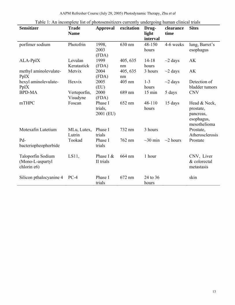
III Photosensitizers Various photosensitizer drugs have been developed. Although Type I photosensitizers have been investigated for antimicrobial applications34, most available oncologic sensitizers achieve their cytotoxic effect primarily via Type II reactions. Table 1 lists several of the more widely used photosensitizers currently available. The first-generation photosensitizer, haematoporphyrin derivative (HPD), is a mixture of porphyrin monomers and oligomers that is partially purified to produce the commercially available product, porfimer sodium, marketed under the tradename Photofrin®. Photofrin was approved for treatment of early stage lung cancer in 1998, and for Barret's esophagus in 2003. The clinical applicability of Photofrin has been limited by two factors. First, its absorption peak occurs at too short a wavelength (630 nm) to allow deep penetration in tissue. Second, administration of photofrin results in cutaneous photosensitivity lasting up to 6 weeks. These limitations have inspired the development of a second generation of photosensitizers with longer-wavelength absorption peaks and more rapid clearance from skin. Among these was benzoporphyrin derivative monoacid A (BPD-MA), or verteporfin. In preclinical trials, it was observed that verteporfin preferentially targeted neovasculature. This selectivity has been exploited for the treatment of choroidal neovascularization (CNV), an abnormal growth of vessels in the retina associated with age-related macular degeneration (AMD), the leading cause of blindness in the developed world. Verteporfn was approved in the US under the tradename Visudyne for CNV treatment in 2000. Another development of note is the prodrug δ-aminolevulinic acid (ALA). Unlike other PDT drugs, ALA itself is not a photosensitizer. When taken up by cells, however, it is converted by a naturally occurring biosynthetic process into the photosensitizer protoporphyrin IX (PpIX). ALA can be applied topically, and was approved by the FDA in 1999 for the treatment of actinic keratosis (AK). ALA has the advantage that it clears from normal tissue within days and can be applied topically, so it causes almost no systemic photosensitivity. In order to improve the uptake of ALA, two variants (methyl- and hexyl-aminolevulinate) have been developed. Methylaminolevulinate (m-ALA) has been approved for treatment of AK under the name Metvix®. The hexyl variant (h-ALA), marketed as Hexvix®, has been approved in the European Union for use in fluorescence cystoscopy. In this case, the preferential accumulation of PpIX in tumors relative to normal bladder endothelium allows tumors to be differentiated by their increased PpIX fluorescence.
3
AAPM Refresher Course (July 28, 2005) Photodynamic Therapy, Zhu et al
4
Other second-generation sensitizers include mTHPC (Foscan®), which has been investigated in clinical trials for a variety of tumors and has been approved for palliative treatment in Europe, and Tookad® and Motexafin Lutetium® (MLu), which are both currently undergoing Phase I trials for prostate cancer treatment. This list is certainly not exhaustive. As understanding of the mechanisms of photosensitizer uptake and preferential sensitization of tumors increases, drugs designed to increase selectivity and light penetration while minimizing sensitization of normal tissue will continue to be developed. Photoimunotherapy (PIT) is a promising approach to improving the selectivity of PDT agents.35-37 In PIT, a sensitizer is conjugated to a monoclonal antibody, allowing it to target a particular molecular marker. By choosing antibodies that target molecules selectively expressed by tumors, an increased tumor selectivity can be achieved. This approach brings with it additional challenges, including the difficulty of conjugating a sensitizer to an antibody without compromising the integrity of either molecule. PIT is still in the early stages of development, however the initial results in animal and cell models are encouraging. IV Light source and delivery devices A. Light sources Various light sources have been developed for photodynamic therapy. Most PDT procedures are carried out at wavelengths between 600 and 850 nm, also called the “therapeutic window”, where the penetration of light in tissue is the greatest and yet the photon energy (>1.5 eV) is high enough to cause photoactivation.38 Because PDT requires intense light with preferably monochromic wavelength, laser is the most common light source Early laser sources were very bulky argon-pumped dye systems. Solid-state lasers have been used lately, which can be made small enough to be transportable (see Fig. 2a). A dye laser provides tunable wavelengths (see Fig. 2b). The current state of art laser is the diode laser, which is very compact and have long lifetime but has slightly wider line width (~5 nm) (Fig. 3). Diode lasers with high power of upto 15W are commercially available. One drawback of the diode laser is fixed wavelength per diode module. New development in high power fiber lasers is advancing rapidly. They will conceivably become the light source of choice in the futuret.39 Besides lasers, there are several other light sources used in clinic: Broadband light sources, including various lamps and light-emitting diodes (LED’s), which generate non-coherent light. Some common terms used to describe light sources as described by AAPM TG5 are listed below:30 Continuous wave (cw): A source, which emits light continuously. Examples applicable to PDT are diode lasers, LED’s, lamps and argon-pumped dye lasers. Pulsed: A source which emits light as a series of pulses, for example, a dye laser pumped by a frequency-doubled Nd:YAG laser. Pulsed sources are characterized by their pulse repetition frequency (in Hz), the pulse width (definition may vary), the pulse energy (typically in mJ), the peak power within a pulse (in W), and the average power (in W). If the pulse energy is low enough, a pulsed source will produce the same biological PDT effect as a cw source with the same average power. Broadband: A source with a wide spectral output compared to typical laser line widths (less or greater? than 1 nm). Tunable: A source whose output wavelength may be adjusted – typically over a range of tens of nanometers.
AAPM Refresher Course (July 28, 2005) Photodynamic Therapy, Zhu et al
5
Bandwidth: A term used to characterize the width of the source’s output spectrum. A variety of definitions are used in practice. For example, the bandwidth of a laser source could be quoted as the wavelength range over which the power is greater than 50% of the power at the peak wavelength. B. Light delivery devices Photodynamic therapy has been greatly facilitated by the development of optical fibers, which allows light to be directed easily to deep-lying tissues, both within body cavities and interstitially. Figure 4 shows some common light delivery fiber optical devices used in photodynamic therapy (see details below). 1. Point sources A fiber designed for intracavitary use, consisting of a small spherical scattering material at the fiber tip. Ideally, such a fiber acts as a point source of illumination. For some special applications, such as PDT of mesothelioma, lung, brain, and bladder cancers, it is preferable to create a uniform illuminating spherical source with finite fluence rate at the sphere boundary. A balloon or a modified endotracheal tube filled with intralipid solution is often used for this purpose (Fig. 4).
2. Linear sources An optical fiber modified to emit light along some portion of its length. The “active” length may be several centimeters. Such fibers are used in intralumenal treatments and may also be made robust enough for interstitial implants.
3. Others Often, a fiber fitted with a microlens in close proximity to the cleaved end is used. This design produces a uniform circular field at a convenient distance from the fiber, and is often used for surface irradiation. A good review of different types of light sources and methods of light fluence profile shaping can be found in AAPM Monograph 19.40
V Light transport in tissue
A. Dosimetry Quantities AAPM TG530 describes the dosimetric quantities used to characterize light in turbid medium based on earlier definitions summarized by Star41: Radiant energy (Q): Total energy emitted, transferred or received as electromagnetic radiation. SI unit is J. Radiant power (P): Power emitted, transferred or received as electromagnetic radiation. SI unit is W. Energy radiance (L): Radiant power transported at a given field point in a given direction per unit solid angle per unit area perpendicular to that direction. The SI unit is W m-2 sr-1. The radiance provides a complete description of the light field and is the fundamental quantity in the radiative transport equation. While important from a theoretical standpoint it is rarely measured directly.
AAPM Refresher Course (July 28, 2005) Photodynamic Therapy, Zhu et al
Energy fluence rate (φ): Ratio of total power incident on an infinitesimal sphere (containing the point of interest) to the cross-sectional area of that sphere. It can also be defined as the integral of the radiance over 4π solid angled. The SI unit is W m-2, although the unit mW cm-2 is still more common in PDT. The fluence rate is the fundamental parameter in PDT dosimetry as it determines the local interaction rate of photons. It can be measured using a specialized detector, which has an isotropic response. Energy fluence (Φ): Total radiant energy incident on an infinitesimal sphere (containing the point of interest) divided by the cross-sectional area of that sphere. SI unit is J m-2 but the unit J cm-2 is common in PDT. Obviously, the fluence is the time integral of the fluence rated. Irradiance (E): Radiant power incident on an infinitesimal surface element (containing the point of interest) divided by the area of that element. The SI unit is W m-2 but the unit mW cm-2 is commonly used in PDT. Note that the irradiance and the fluence rate have the same physical units (power per unit area) but they are not the same quantity. The irradiance is defined for a particular surface whereas the fluence rate can be defined and measured in free space or the interior of an object. Terms such as power density, flux density and intensity, which have been used to describe the irradiance, should be avoided. Radiant exposure (H): Radiant energy incident on an infinitesimal surface element (containing the point of interest) divided by the area of that element. The SI unit is J m-2 but, in PDT, the unit J cm-2 is more common. The radiant exposure is the time integral of the irradiance. The term “energy density” which has been applied to this quantity should be avoided. The radiant exposure is specified for PDT treatments using surface irradiation. Among those quantities the most important quantity is the energy fluence (Φ) since this quantity is directly associated with PDT dose. In a collimated beam, energy fluence = irradiance. However, in an integrating sphere, energy fluence = 4* irradiance since irradiance takes into account the direction of light incidencet.41 B. Diffusion Approximation The most widely used model of light transport in tissue is the radiative transport equation42. Because analytic solutions to this equation exist for only very simple geometries, it is generally solved by a Taylor expansion. A first-order expansion yields the commonly used diffusion approximation. In the near infrared (NIR) region, tissue scattering dominates over tissue absorption, so that the diffusion approximation is valid.38 Under diffusion approximation, the light fluence rate, φ, can be described as
( ) ( ) ( ) ( trvStrvtrDt
tra ,,,,
+−∇⋅∇=∂
∂ φµφ )φ, (4)
where v is the speed of light in the turbid medium; ( )[ ]asvD µµ += '3 is the photon diffusion coefficient; S is an isotropic source term which gives the number of photons emitted at position r and time t per unit volume per unit time. Jacques showed that the light distribution using diffusion approximation is still not accurate in this wavelength region, compared to Monte-Carlo simulation.43 This is especially true near the source, in the so-called near-source field. Monte-Carlo simulation, however, is not suitable for real-time light fluence calculation because of the long computing times required.44 To address this deficiency, several researchers have investigated a 3rd-order approximation of the transport equation (P3 approximation) for a point source to improve both the speed and accuracy of light fluence calculation.45-48
6
AAPM Refresher Course (July 28, 2005) Photodynamic Therapy, Zhu et al
7
VI PDT Dosimetry The concept of explicit and implicit PDT dosimetry was introduced in the 1990s by Wilson et al.5 Explicit dosimetry refers to measurement of physical quantities that are well-defined, e.g. light fluence, photosensitizer drug concentration, and tissue oxygenation. Because it is a well-defined physical quantity, one may design methods to measure each quantity independently. Implicit dosimetry refers to the use of a measurable quantity, such as the extent of sensitizer photobleaching, which is sensitive to some or all of the factors influencing photodynamic efficacy but which does not require independent measurements of each of these quantities.
A. Explicit dosimetry From physics point of view, the explicit PDT dose is defined as the light energy deposited to photosensitizer, i.e. it is proportional to the product of the absorption coefficient of the photosensitizer and light fluence (Eq. 1). The absorption coefficient of the photosensitizer is, in turn, proportional to the photosensitizer concentration. PDT dose calculated in this way is a good marker if one is operating in a drug- or light- limiting regime when there is ample oxygen supply. However, if oxygen delivery is the factor limiting PDT effect, singlet oxygen production rate (Eq. 3) is a better marker for predicting PDT efficiency.
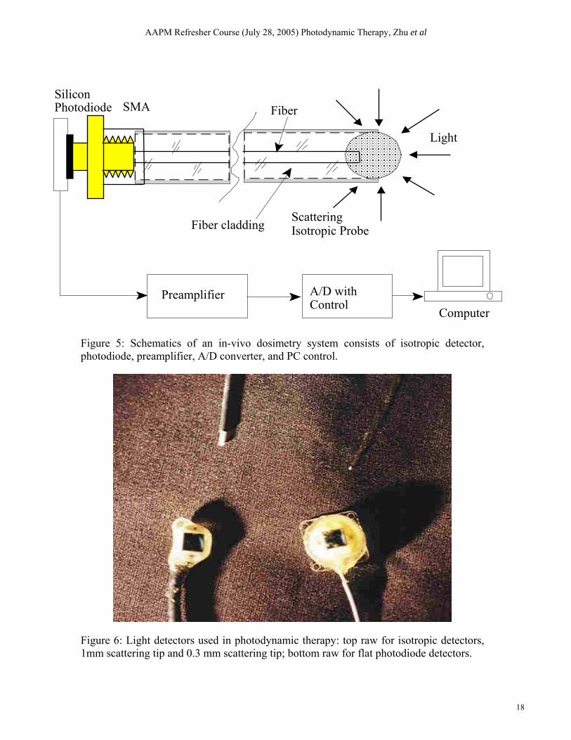
In-vivo light Dosimetry in PDT Light is an important quantity that determines the outcome of PDT treatment. The light fluence (expressed in J/cm2) is proportional to the light energy deposited in tissue. The total fluence in tissues is a function not only of the incident light delivered by the laser but also of scattered light. Often clinical PDT treatments are prescribed in terms of the incident light delivered from the laser rather than the total fluence of light the tissues receive which is a combination of scattered and incident light. Substantial differences in total fluence to tissues can be observed among patients if the clinician accounts only for incident light.41, 49 Dosimetry systems using isotropic light detectors have been developed to measure both incident and scattered light.50, 51 A 16-channel system developed at the University of Pennsylvania is shown in Fig. 5. These systems should begin to allow clinical researchers to measure and therefore prescribe a consistent total fluence to the tissues. Isotropic detectors are often used to measure the light fluence rate directly (see Fig. 6).52 These detectors have the advantage of detecting light from all directions vs. the flat photodiodes (Fig. 6) that can only detect light from normal incidencet.53
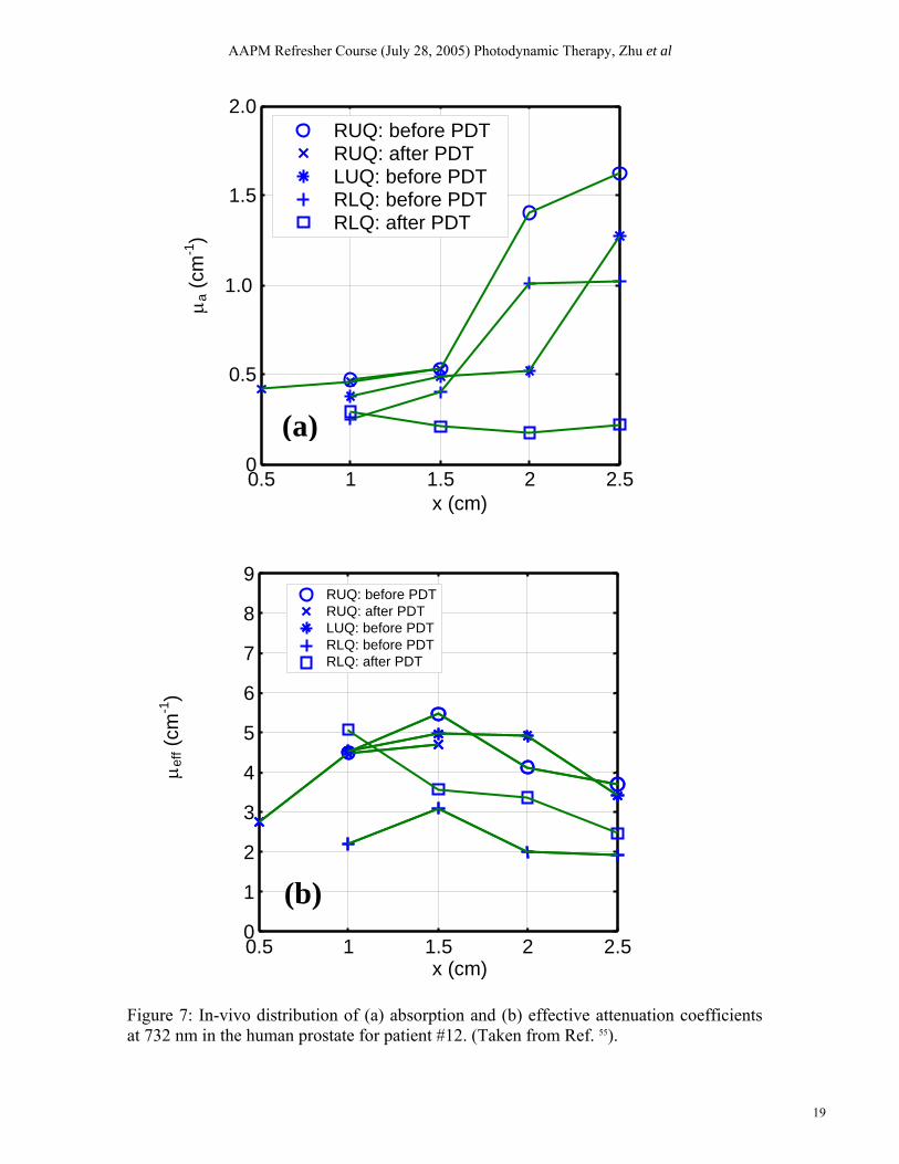
In vivo characterization of tissue optical properties The measurement of light fluence rate in vivo is necessary but not sufficient to quantify light fluence rate distribution. Volumetric determination of the light fluence rate in the entire treatment volume requires accurate characterization of the in vivo tissue optical properties as input (µa, µs’ in Eq.4). Several techniques have been developed to determine the optical properties in vivo.24, 54 Figure 7 shows measured distribution of optical properties in human prostate.55 Clearly, there is a significant difference (up to 3 times) between optical properties measured in different locations, which will affect the light fluence distribution.
More advanced non-invasive technology such as diffuse optical tomography has shown promise for determining the 3D distribution of optical properties (µa and µs’) in brain56 and breast57. However, this technique can be applied to limited anatomic sites because of the limited tissue penetration depth, ~ 10 cm.
Quantification of drug concentration Determination of drug concentration is important for PDT efficacy. Early PDT clinical protocols only specify this quantity in terms of the amount of photosensitizer given to patient per body weight. Recent in-vivo studies have show large variation of photosensitizer concentration in different tissue types, thus
AAPM Refresher Course (July 28, 2005) Photodynamic Therapy, Zhu et al
8
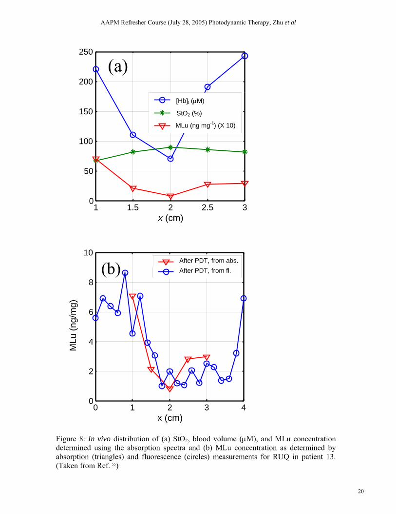
suggesting determination of this quantity in-vivo in the region of treatment directly.23, 55 To include the drug concentration in the evaluation of PDT dose, in situ fluorescent58 or absorption25, 59-62 measurements of photosensitizer can be made interstitially using optical fibers. Figure 8 shows measured distribution of MLu drug concentration in prostate using absorption (Fig. 8a) and fluorescence (Fig. 8b) measurement.55 The results of the two methods agreed well for MLu drug concentration.55
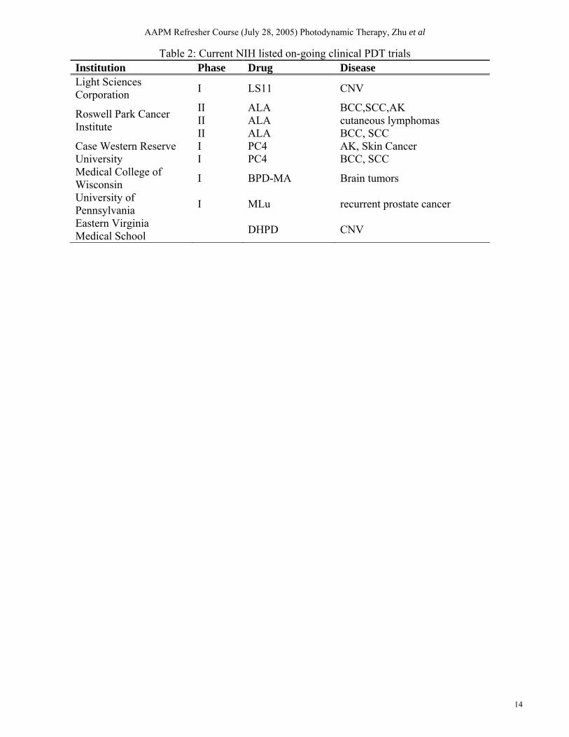
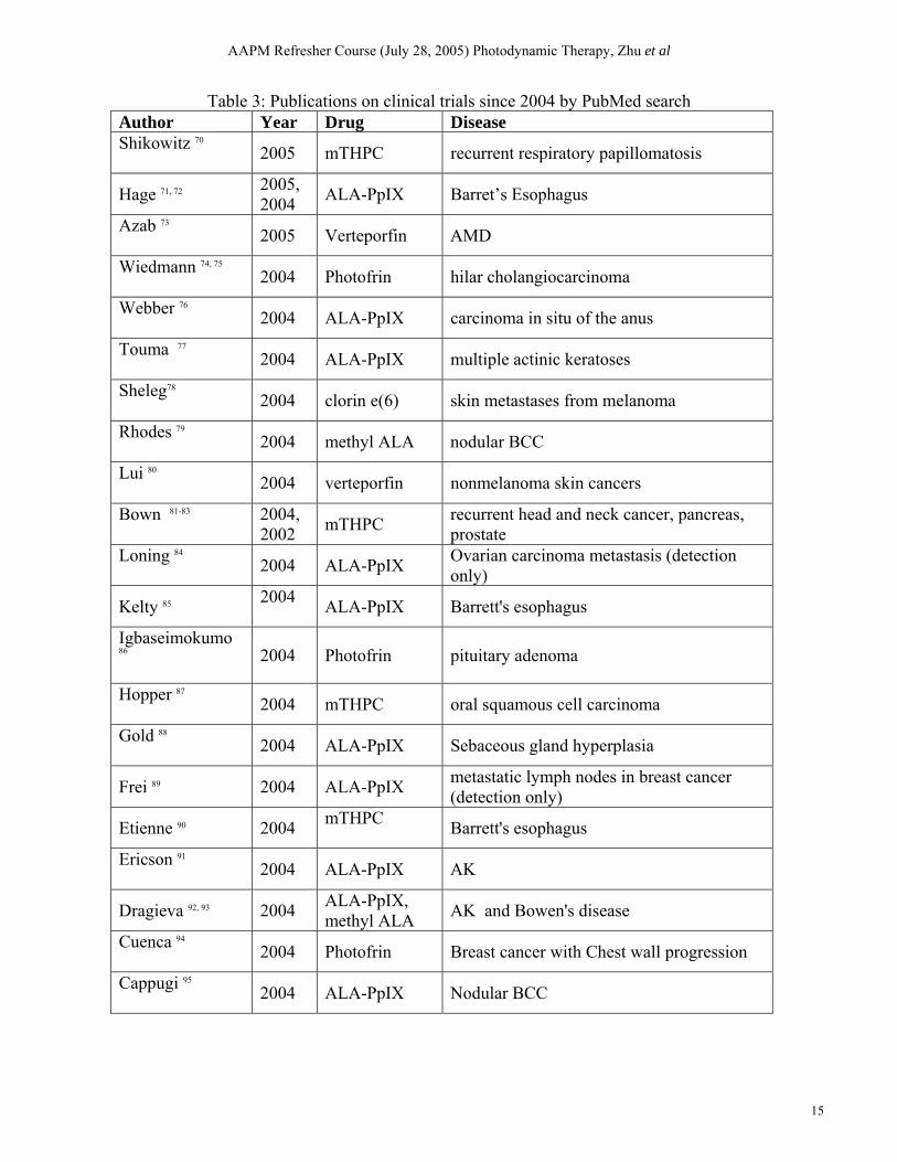
Quantification of tissue oxygenation Tissue oxygenation is known to affect PDT efficacy31, 63 in vitro. In addition, changes in tissue oxygenation due to photochemical oxygen consumption during PDT have been observed directly,64, 65 and indirectly through their effect on the photobleaching ratet.66, 67 Recent studies have shown that one can determine the concentration of hemoglobin (Hb), HbO2, and H2O from absorption measurement.59-62 Figure 8a shows measured distribution of Hb and StO2 = HbO2/Hb measured in human prostate.55 B. Implicit dosimetry Implicit dosimetry5 relies on a surrogate indicator of damage to measure the photodynamic effect, rather than explicitly quantifying all the parameters needed to calculate the dose. One such mechanism is the measurement of fluorescence photobleaching. In particular, if a sensitizer’s photobleaching and the damage to tissue are both caused primarily by reactions with singlet oxygen, it is reasonable to assume that the rate of photobleaching will be indicative of the rate of deposition of singlet oxygen-mediated damage in tissue. In a simplified model, Georgakoudi et al. showed that the fraction of the initial photosenstizer bleached could be related to the absolute concentration of reacted singlet oxygen in tissue32. While oxygen transport, tissue heterogeneity, and light diffusion complicate the issue in vivo, the correlation between photobleaching and PDT-induced damage holds in animal models 66, 68. When designing a dosimetry protocol, it is important to remember that the usefulness of a particular method or model depends on the drug, light fluence rate, and tissue being investigated. There is experimental evidence that some sensitizers may photobleach by singlet oxygen-independent mechanisms, in which case implicit dosimetry based on photobleaching will provide incorrect conclusions concerning singlet oxygen dose.33 Different sensitizers may exhibit very different bleaching behaviors, even in the same animal model.33, 67, 69 Furthermore, even if a sensitizer does bleach purely through singlet-oxygen mediated mechanisms, the relationship between photobleaching and clinical outcome will depend on a host of factors, including cellular and vascular distribution and tumor selectivity, which may be drug- and tissue-specific. Therefore, any implicit dosimetry method must be verified for the specific drug and irradiation scheme being used before it can be implemented clinically. VII PDT clinical protocols Tables 2 and 3 list the current oncological PDT clinical trials listed by the NIH as ongoing in the US, and the papers published since 2004 on clinical trial in PDT, as listed by PubMed. Trials investigating PDT for cosmetic and ocular applications have not been included. While the majority of PDT trials and approvals have been for cancers and other diseases of the skin, more recent studies have begun to expand the range of diseases treated with PDT to include solid tumors of the head and neck, prostate, pancreas and breast. As new drugs and new light delivery devices are developed, we can expect to see trials of PDT for an increasing number of diseases that were previously considered inaccessible to PDT. These sites generally involve the treatment of large, solid tumors, requiring illumination by intra-cavity or interstitial light delivery devices. The wide variation in optical properties within and among tumors and the variation in tumor geometry from patient to patient make accurate quantitative dosimetry even more important in these cases. It is increasingly becoming appreciated that the PDT treatment of the future will

AAPM Refresher Course (July 28, 2005) Photodynamic Therapy, Zhu et al
9
incorporate real-time dosimetry and tissue optics monitoring into the light delivery system, allowing the light distribution to be optimized as treatment progresses. VIII Acknowledgment The authors wish to thank members of the AAPM task group 5, especially Fred Hetzel and Steve Jacques, for their contributions to establish methodology for PDT dosimetry. IX Bibliography 1T. J. Dougherty, D. J. Gomer, B. W. Henderson, and et. al., "Photodynamic Therapy," J. Natl. Cancer Inst. 90, 889-905 (1998). 2M. Ochsner, "Photophysical and photobiological processes in the photodynamic therapy of tumours," J Photochem Photobiol B 39, 1-18 (1997). 3H. I. Pass, "Photodynamic therapy in oncology: mechanisms and clinical use," J Natl Cancer Inst 85, 443-56 (1993). 4C. S. Foote, "Photosenstizied oxidations and the role of singlet oxygen," Accounts Chem. Res. 1, 104-110 (1967). 5B. C. Wilson, M. S. Patterson, and L. Lilge, "Implicit and explicit dosimetry in photodynamic therapy: A new paradigm," Lasers Med. Sci. 12, 182-199 (1997). 6M. J. Niedre, M. S. Patterson, A. Giles, and B. C. Wilson, "Imaging of Photodynamically Generated Singlet Oxygen Luminescence In Vivo," Photochem Photobiol, (2005). 7M. Niedre, M. S. Patterson, and B. C. Wilson, "Direct near-infrared luminescence detection of singlet oxygen generated by photodynamic therapy in cells in vitro and tissues in vivo," Photochem Photobiol 75, 382-91 (2002). 8M. A. Biel, "Photodynamic therapy and the treatment of head and neck cancers," J Clin Laser Med Surg 14, 239-244 (1996). 9W. E. Grant, P. M. Speight, C. Hopper, and et. al., "Photodynamic therapy: an effective, but non-selective treatment for superficial cancers of the oral cavity," Int J Cancer 71, 937-942 (1997). 10J. S. Friedberg, R. Mick, J. P. Steveson, T. Zhu, T. M. Busch, D. Shin, D. Smith, M. Culligan, A. Dimofte, E. Glatstein, and S. M. Hahn, "A Phase II Trial of Pleural Photodynamic Therapy (PDT) and Surgery for Patients with Non-small Cell Lung Cancer (NSCLC) with Pleural Spread," J Clin Oncol 22, 2192-2201 (2004). 11T. L. Moskal, T. J. Dougherty, J. D. Urschel, J. G. Antkowiak, A. M. Regal, D. L. Driscoll, and H. Takita, "Operation and photodynamic therapy for pleural mesothelioma: 6-year follow-up," Ann Thorac Surg 66, 1128-33 (1998). 12H. I. Pass, T. F. Delaney, Z. Tochner, and et. al., "Intrapleural photodynamic therapy: Results of a phase I trial," Ann Surg Oncol 1, 28-37 (1996). 13J. S. Friedberg, R. Mick, J. Stevenson, J. Metz, T. Zhu, J. Buyske, D. H. Sterman, H. I. Pass, E. Glatstein, and S. M. Hahn, "A phase I study of Foscan-mediated photodynamic therapy and surgery in patients with mesothelioma," Ann Thorac Surg 75, 952-9 (2003). 14R. Ackroyd, N. J. Brown, M. F. Davis, T. J. Stephenson, S. L. Marcus, C. J. Stoddard, A. G. Johnson, and M. W. Reed, "Photodynamic therapy for dysplastic Barrett's esophagus: a prospective, double blind, randomised, placebo controlled trial," Gut 47, 612-617 (2000). 15M. Panjehpour, B. F. Overholt, J. M. Haydek, and S. G. Lee, "Results of photodynamic therapy for ablation of dysplasia and early cancer in Barrett's esophagus and effect of oral steroids on stricture formation," Am J Gastroenterol 95, 2177-2184 (2000). 16T. R. Nathan, D. E. Whitelaw, S. C. Chang, W. R. Lees, P. M. Ripley, H. Payne, L. Jones, M. C. Parkinson, M. Emberton, A. R. Gillams, A. R. Mundy, and S. G. Browen, "Photodynamic therapy for prostate cancer recurrence after radiotherapy: A Phase I study," J Urol 168, 1427-1432 (2002). 17D. C. H. Stripp, R. Mick, T. C. Zhu, R. Whittington, D. Smith, A. Dimofte, J. C. Finlay, J. Miles, T. M. Busch, D. Shin, A. Kachur, Z. A. Tochner, S. B. Malkowicz, E. Glatstein, and S. M. Hahn, "Phase I trial of Motexafin lutetium-mediated interstitial photodyanmic therapy in patients with locally recurrent prostate cancer," Proc. SPIE 5315, 88-99 (2004). 18R. A. Weersink, A. Bogaards, M. Gertner, S. R. Davidson, K. Zhang, G. Netchev, J. Trachtenberg, and B. C. Wilson, "Techniques for delivery and monitoring of TOOKAD (WST09)-mediated photodynamic therapy of the prostate: clinical experience and practicalities," J. Photochem. Photobiol B 79, 211-222 (2005). 19M. A. Rosenthal, B. Kayar, J. S. Hill, D. J. Morgan, R. L. Nation, S. S. Stylli, R. L. Basser, S. Uren, H. Geldard, M. D. Green, S. B. Kahl, and A. H. Kaye, "Phase I and pharmacokinetic study of photodynamic therapy for high-grade gliomas using a noval boronated porphyrin," J Clin Oncol 19, 519-524 (2001). 20T. T. Goodell, and P. J. Muller, "Photodynamic therapy: a novel treatment for primary brain malignancy," J Neurosci Nurs. 33, 296-300 (2001). 21A. Bogaards, A. Varma, K. Zhang, D. Zach, S. K. Bisland, E. H. Moriyama, L. Lilge, P. J. Muller, and B. C. Wilson, "Fluorescence image-guided brain tumour resection with adjuvant metronomic photodynamic therapy:pre-clinical model and technology development," Photochem Photobiol Sci. 4, 438-442 (2005).
AAPM Refresher Course (July 28, 2005) Photodynamic Therapy, Zhu et al
10
22A. Dimofte, J. C. Finlay, and T. C. Zhu, "A method for determination of the absorption and scattering properties interstitially in turbid media," Phys. Med. Biol. 50, 2291-2311 (2005). 23T. C. Zhu, A. Dimofte, J. C. Finlay, D. Stripp, T. M. Busch, J. Miles, R. Whittington, S. B. Malkowicz, Z. Tochner, E. Glatstein, and S. M. Hahn, "Optical properties of Human Prostate at 732nm measured in vivo during motexafin lutetium-mediated photodynamic therapy," Photochem Photobiol 81, 96-105 (2005). 24H. W. Wang, T. C. Zhu, M. E. Putt, M. Solonenko, J. Metz, A. Dimofte, J. Miles, D. L. Fraker, E. Glatstein, S. M. Hahn, and A. G. Yodh, "In-vivo broadband reflectance measurements of light penetration depth, blood oxygenation, hemoglobin concentration, and drug concentration in human tissues before and after photodynamic therapy," J Biomed Opt 10, (2005). 25J. C. Finlay, T. C. Zhu, A. Dimofte, D. Stripp, S. B. Malkowicz, R. Whittington, J. Miles, E. Glatstein, and S. M. Hahn, "In vivo determination of the absorption and scattering spectra of the human prostate during photodynamic therapy," Proc. SPIE 5315, 132-142 (2004). 26J. C. Finlay, T. C. Zhu, A. Dimofte, D. Stripp, S. B. Malkowicz, R. Whittington, J. Miles, E. Glatstein, and S. M. Hahn, "In vivo measurement of fluorescence emission in the human prostate during photodynamic therapy," Proc. SPIE 5689, 299-310 (2005). 27M. S. Patterson, B. C. Wilson, and R. Graff, " In vivo tests of the concept of photodynamic threshold dose in normal rat liver photosensitized by aluminum chlorosulphonated phthalocyanine," Photochem Photobiol 51, 343-349 (1990). 28L. O. Svaasand, "Light Delivery and Dosimetry," in Phototherapy of Cancer Volume, edited by, G. Morsty, and H. Kaye, (Harwood Academic Publisher, Chur, 1990) pp. 73-98. 29T. J. Ferrell, B. C. Wilson, M. S. Patterson, and et. al., "Comparison of the in vivo photodynamic threshold dose for photofrin, mono- and tetrasulfonated aluminum phthalocyanine using a rat liver model," Photochem Photobiol 68, 394-399 (1998). 30S. Brahmavar, Q. Chen, T. C. Zhu, F. W. Hetzel, S. Jacque, M. S. Patterson, and B. C. Wilson, AAPM Photodynamic Therapy Dosimetry, Report 88, (Med Phys Publishing, city, 2005). 31I. Georgakoudi, and T. H. Foster, "Singlet Oxygen -versus nonsinglet oxygen-mediated mechanisms of sensitizer photobleaching and their effects on photodynamic dosimetry," Photochem Photobiol 67, 612-625 (1998). 32I. Georgakoudi, M. G. Nichols, and T. H. Foster, "The mechanism of Photofrin photobleaching and its consequences for photodynamic dosimetry," Photochem. Photobiol. 65, 135-144 (1997). 33J. C. Finlay, S. Mitra, and T. H. Foster, "Photobleaching kinetics of Photofrin in vivo and in multicell tumor spheroids indicate multiple simultaneous bleaching mechanisms," Phys. Med. Biol. 49, 4837-4860 (2004). 34M. Wainwright, "Photodynamic antimicrobial chemotherapy (PACT)," J Antimicrob Chemother 42, 13-28 (1998). 35G. A. van Dongen, G. W. Visser, and M. B. Vrouenraets, "Photosensitizer-antibody conjugates for detection and therapy of cancer," Adv Drug Deliv Rev 56, 31-52 (2004). 36M. D. Savellano, and T. Hasan, "Photochemical targeting of epidermal growth factor receptor: a mechanistic study," Clin Cancer Res 11, 1658-68 (2005). 37R. Hudson, M. Carcenac, K. Smith, L. Madden, O. J. Clarke, A. Pelegrin, J. Greenman, and R. W. Boyle, "The development and characterisation of porphyrin isothiocyanate-monoclonal antibody conjugates for photoimmunotherapy," Br J Cancer 92, 1442-9 (2005). 38B. C. Wilson, and M. S. Patterson, "The determination of light fluence distribution in photodynamic therapy," in Photodynamic Therapy of Neoplastic Disease Volume 1, edited by, D. Kessel, (CRC Press, Inc., Boca Raton, 1990) pp. 129-144. 39N. Peyghambarian, and A. Schulzgen, "High-power devices in compact packages," OPN 16, 36-41 (2005). 40P. R. Almond, "Photodynamic therapy: Equipment and Dosimetry," in Advances in radiation oncology physics: Dosimetry, Treatment planning, and Brachytherapy Volume AAPM monograph No. 19, edited by, J. A. Purdy, (American Institute of Physics, Inc., Woodbury, NY, 1990) pp. 852-885. 41W. M. Star, "Light dosimetry in vivo," Phys. Med. Biol. 42, 763-787 (1997). 42A. Ishimaru, Wave propagation and scattering in random media, (IEEE Press, New York, 1997). 43S. Jacque, "Light Distributions from Point, Line and Plane Sources for Photochemical reactions and fluorescence in turbid biological tissues," Photochem Photobiol 67, 23-32 (1998). 44C. M. Gardner, S. Jacque, and A. J. Welch, "Light transport in tissue:Accurate expressions for one-dimensional fluence rate and escape function based upon Monte Carlo simulation," Lasers Surg Med 18, 129-138 (1996). 45W. M. Star, "Comparing the P3-approximation with diffusion theory and with Monte Carlo calculations of light propagation in a slab geometry," SPIE Institute Series IS5, 46-54 (1989). 46E. L. Hull, and T. H. Foster, "Steady-state reflectance spectroscopy in the P3 approximation," JOSA A 18, 584-599 (2001). 47D. J. Dickey, R. B. Moore, D. C. Rayner, and J. Tulip, "Light dosimetry using the P3 approximation," Phys. Med. Biol. 46, 2359-2370 (2001). 48D. A. Boas, H. Lui, M. A. O'Leary, B. Chance, and A. G. Yodh, "Photon Migration within the P3 approximation," Proc. SPIE 2389, 240-247 (1995). 49A. Dimofte, T. C. Zhu, S. M. Hahn, and R. A. Lustig, "In-vivo light dosimetry for motexafin lutetium-mediated PDT of Recurrent Breast Cancer," Lasers Surg Med 31, 305-312 (2002).
AAPM Refresher Course (July 28, 2005) Photodynamic Therapy, Zhu et al
11
50P. Baas, L. Muller, Z. F.A.N., and et. al., "Photodynamic therapy as adjuvant therapy in surgically treated pleural malignancies," Br J Cancer 76, 819-826 (1997). 51M. Solonenko, T. C. Zhu, and T. G. Vulcan, "Commissioning of the isotropic light dosimetry system for photodynamic therapy," Med Phys 26, 1124 (1999). 52H. J. van Staveren, H. P. A. Marijnissen, M. C. Aalders, and W. M. Star, "Construction, Quality Assurance, and Calibrationof Spherical isotropic fibre optic light diffusers," Lasers Med. Sci. 10, 137-147 (1995). 53T. G. Vulcan, T. C. Zhu, C. E. Rodriguez, A. Hsi, D. L. Fraker, P. Baas, L. H. Murrer, W. M. Star, E. Glatstein, A. G. Yodh, and S. M. Hahn, "Comparison between isotropic and nonisotropic dosimetry systems during intraperitoneal photodynamic therapy," Lasers Surg Med 26, 292-301 (2000). 54T. C. Zhu, S. M. Hahn, A. S. Kapatkin, A. Dimofte, C. E. Rodriguez, T. G. Vulcan, E. Glatstein, and A. Hsi, "In-vivo Optical properties of Normal Canine Prostate at 732 nm using motexafin lutetium-mediated photodynamic therapy," Photochem Photobiol 77, 81-88 (2003). 55T. C. Zhu, J. C. Finlay, and S. M. Hahn, "Determination of the Distribution of light, optical properties, drug concentration, and tissue oxygenation in-vivo in human prostate during motexafin lutetium-mediated photodynamic therapy," J Photochem Photobiol B 79, 231-241 (2005). 56M. Schweiger, and S. R. Arridge, "Optical tomographic reconstruction in a complex head model using a priori region boundary information," Phys. Med. Biol. 44, 2703-2721 (1999). 57M. J. Holboke, B. J. Tromberg, X. Li, N. Shah, J. Fishkin, D. Kidney, J. Butler, B. Chance, and A. G. Yodh, "Three-dimensional diffuse optical mammography with ultrasound localization in a human subject," J Biomed Opt 5, 237-247 (2000). 58K. R. Diamond, M. S. Patterson, and T. J. Farrell, "Quantification of fluorophore concentration in tissue-simulating media by fluorescence measurements with a single optical fiber," Appl. Opt. 42, 2436-2442 (2003). 59R. M. P. Doornbos, R. lang, M. C. Aalders, F. W. Cross, and H. J. Sterenborg, "The determination of in vivo human tissue optical properties and absolute chromophore concentrations using spatially resolved steady-state diffuse reflectance spectroscopy," Phys. Med. Biol. 44, 967-981 (1999). 60A. Kienle, L. Lilge, M. S. Patterson, R. Hibst, R. A. Steiner, and B. C. Wilson, "Spatially resolved absolute diffuse reflectance measurements for noninvasive determination of the optical scattering and absorptions of biological tissue," Appl. Opt. 35, 2304-2313 (1996). 61H. Liu, D. A. Boas, Y. Zhang, A. G. Yodh, and B. Chance, "Determination of Optical Properties and blood oxygenation in tissue using continuous near-infraread light," Phys. Med. Biol. 40, 1983-1993 (1995). 62M. Solonenko, R. Cheung, T. M. Busch, A. Kachur, G. M. Griffin, T. G. Vulcan, T. C. Zhu, H. W. Wang, S. M. Hahn, and A. G. Yodh, "In-vivo reflectance measurement of motexafin lutetium uptake, optical properties, and oxygenation of canine large bowels, kidneys, and prostates," Phys. Med. Biol. 47, 857-873 (2002). 63M. G. Nichols, and T. H. Foster, "Oxygen diffusion and reaction kinetics in the photodynamic therapy of multicell tumour spheroids," Phys. Med. Biol. 39, 2161-2181 (1994). 64B. J. Tromberg, A. Orenstein, S. Kimel, S. J. Barker, J. Hyatt, J. S. Nelson, and M. Berns, "In vivo tumor oxygen tension measurements for the evaluation of the efficiency of photodynamic therapy," Photochem Photobiol 52, 375-385 (1990). 65B. W. Henderson, T. M. Busch, L. A. Vaughan, N. P. Frawley, D. Babich, T. A. Sosa, J. D. Zollo, A. S. Dee, M. T. Cooper, D. A. Bellnier, W. R. Greco, and A. R. Oseroff, "Photofrin photodynamic therapy can significantly deplete or preserve oxygenation in human basal cell carcinomas during treatment, depending on fluence rate," Cancer Res. 60, 525-529 (2000). 66D. J. Robinson, H. S. de Bruijn, N. van der Veen, M. R. Stringer, S. B. Brown, and W. M. Star, "Fluorescence photobleaching of ALA-induced protoporphyrin IX during photodynamic therapy of normal hairless mouse skin: The effect of light dose and irradiance and the resulting biological effect," Photochem. Photobiol. 67, 140-149 (1998). 67J. C. Finlay, S. Mitra, and T. H. Foster, "In vivo mTHPC photobleaching in normal rat skin exhibits unique irradiance-dependent features," Photochem Photobiol 75, 282-288 (2002). 68D. J. Robinson, H. S. de Bruijn, N. van der Veen, M. R. Stringer, S. B. Brown, and W. M. Star, "Protoporphyrin IX fluorescence photobleaching during ALA-mediated photodynamic therapy of UVB-induced tumors in hairless mouse skin," Photochem. Photobiol. 69, 61-70 (1999). 69J. C. Finlay, D. L. Conover, E. L. Hull, and T. H. Foster, "Porphyrin bleaching and PDT-induced spectral changes are irradiance dependent in ALA-sensitized normal rat skin in vivo," Photochem. Photobiol. 73, 54-63 (2001). 70M. J. Shikowitz, A. L. Abramson, B. M. Steinberg, J. DeVoti, V. R. Bonagura, V. Mullooly, M. Nouri, A. M. Ronn, A. Inglis, J. McClay, and K. Freeman, "Clinical trial of photodynamic therapy with meso-tetra (hydroxyphenyl) chlorin for respiratory papillomatosis," Arch Otolaryngol Head Neck Surg 131, 99-105 (2005). 71M. Hage, P. D. Siersema, K. J. Vissers, E. W. Steyerberg, J. Haringsma, E. J. Kuipers, and H. van Dekken, "Molecular evaluation of ablative therapy of Barrett's oesophagus," J Pathol 205, 57-64 (2005). 72M. Hage, P. D. Siersema, H. van Dekken, E. W. Steyerberg, J. Haringsma, W. van de Vrie, T. E. Grool, R. L. van Veen, H. J. Sterenborg, and E. J. Kuipers, "5-aminolevulinic acid photodynamic therapy versus argon plasma coagulation for ablation of Barrett's oesophagus: a randomised trial," Gut 53, 785-90 (2004). 73M. Azab, D. S. Boyer, N. M. Bressler, S. B. Bressler, I. Cihelkova, Y. Hao, I. Immonen, J. I. Lim, U. Menchini, J. Naor, M. J. Potter, A. Reaves, P. J. Rosenfeld, J. S. Slakter, P. Soucek, H. A. Strong, A. Wenkstern, X. Y. Su, and Y. C. Yang,
AAPM Refresher Course (July 28, 2005) Photodynamic Therapy, Zhu et al
12
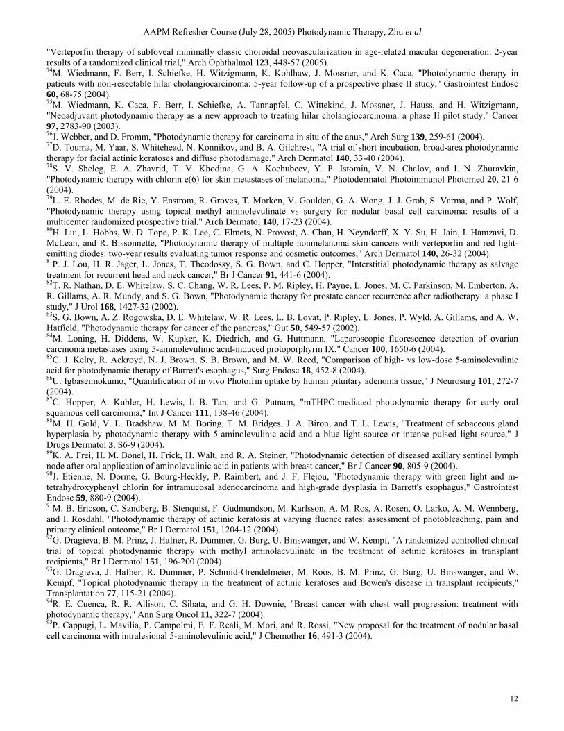
"Verteporfin therapy of subfoveal minimally classic choroidal neovascularization in age-related macular degeneration: 2-year results of a randomized clinical trial," Arch Ophthalmol 123, 448-57 (2005). 74M. Wiedmann, F. Berr, I. Schiefke, H. Witzigmann, K. Kohlhaw, J. Mossner, and K. Caca, "Photodynamic therapy in patients with non-resectable hilar cholangiocarcinoma: 5-year follow-up of a prospective phase II study," Gastrointest Endosc 60, 68-75 (2004). 75M. Wiedmann, K. Caca, F. Berr, I. Schiefke, A. Tannapfel, C. Wittekind, J. Mossner, J. Hauss, and H. Witzigmann, "Neoadjuvant photodynamic therapy as a new approach to treating hilar cholangiocarcinoma: a phase II pilot study," Cancer 97, 2783-90 (2003). 76J. Webber, and D. Fromm, "Photodynamic therapy for carcinoma in situ of the anus," Arch Surg 139, 259-61 (2004). 77D. Touma, M. Yaar, S. Whitehead, N. Konnikov, and B. A. Gilchrest, "A trial of short incubation, broad-area photodynamic therapy for facial actinic keratoses and diffuse photodamage," Arch Dermatol 140, 33-40 (2004). 78S. V. Sheleg, E. A. Zhavrid, T. V. Khodina, G. A. Kochubeev, Y. P. Istomin, V. N. Chalov, and I. N. Zhuravkin, "Photodynamic therapy with chlorin e(6) for skin metastases of melanoma," Photodermatol Photoimmunol Photomed 20, 21-6 (2004). 79L. E. Rhodes, M. de Rie, Y. Enstrom, R. Groves, T. Morken, V. Goulden, G. A. Wong, J. J. Grob, S. Varma, and P. Wolf, "Photodynamic therapy using topical methyl aminolevulinate vs surgery for nodular basal cell carcinoma: results of a multicenter randomized prospective trial," Arch Dermatol 140, 17-23 (2004). 80H. Lui, L. Hobbs, W. D. Tope, P. K. Lee, C. Elmets, N. Provost, A. Chan, H. Neyndorff, X. Y. Su, H. Jain, I. Hamzavi, D. McLean, and R. Bissonnette, "Photodynamic therapy of multiple nonmelanoma skin cancers with verteporfin and red light-emitting diodes: two-year results evaluating tumor response and cosmetic outcomes," Arch Dermatol 140, 26-32 (2004). 81P. J. Lou, H. R. Jager, L. Jones, T. Theodossy, S. G. Bown, and C. Hopper, "Interstitial photodynamic therapy as salvage treatment for recurrent head and neck cancer," Br J Cancer 91, 441-6 (2004). 82T. R. Nathan, D. E. Whitelaw, S. C. Chang, W. R. Lees, P. M. Ripley, H. Payne, L. Jones, M. C. Parkinson, M. Emberton, A. R. Gillams, A. R. Mundy, and S. G. Bown, "Photodynamic therapy for prostate cancer recurrence after radiotherapy: a phase I study," J Urol 168, 1427-32 (2002). 83S. G. Bown, A. Z. Rogowska, D. E. Whitelaw, W. R. Lees, L. B. Lovat, P. Ripley, L. Jones, P. Wyld, A. Gillams, and A. W. Hatfield, "Photodynamic therapy for cancer of the pancreas," Gut 50, 549-57 (2002). 84M. Loning, H. Diddens, W. Kupker, K. Diedrich, and G. Huttmann, "Laparoscopic fluorescence detection of ovarian carcinoma metastases using 5-aminolevulinic acid-induced protoporphyrin IX," Cancer 100, 1650-6 (2004). 85C. J. Kelty, R. Ackroyd, N. J. Brown, S. B. Brown, and M. W. Reed, "Comparison of high- vs low-dose 5-aminolevulinic acid for photodynamic therapy of Barrett's esophagus," Surg Endosc 18, 452-8 (2004). 86U. Igbaseimokumo, "Quantification of in vivo Photofrin uptake by human pituitary adenoma tissue," J Neurosurg 101, 272-7 (2004). 87C. Hopper, A. Kubler, H. Lewis, I. B. Tan, and G. Putnam, "mTHPC-mediated photodynamic therapy for early oral squamous cell carcinoma," Int J Cancer 111, 138-46 (2004). 88M. H. Gold, V. L. Bradshaw, M. M. Boring, T. M. Bridges, J. A. Biron, and T. L. Lewis, "Treatment of sebaceous gland hyperplasia by photodynamic therapy with 5-aminolevulinic acid and a blue light source or intense pulsed light source," J Drugs Dermatol 3, S6-9 (2004). 89K. A. Frei, H. M. Bonel, H. Frick, H. Walt, and R. A. Steiner, "Photodynamic detection of diseased axillary sentinel lymph node after oral application of aminolevulinic acid in patients with breast cancer," Br J Cancer 90, 805-9 (2004). 90J. Etienne, N. Dorme, G. Bourg-Heckly, P. Raimbert, and J. F. Flejou, "Photodynamic therapy with green light and m-tetrahydroxyphenyl chlorin for intramucosal adenocarcinoma and high-grade dysplasia in Barrett's esophagus," Gastrointest Endosc 59, 880-9 (2004). 91M. B. Ericson, C. Sandberg, B. Stenquist, F. Gudmundson, M. Karlsson, A. M. Ros, A. Rosen, O. Larko, A. M. Wennberg, and I. Rosdahl, "Photodynamic therapy of actinic keratosis at varying fluence rates: assessment of photobleaching, pain and primary clinical outcome," Br J Dermatol 151, 1204-12 (2004). 92G. Dragieva, B. M. Prinz, J. Hafner, R. Dummer, G. Burg, U. Binswanger, and W. Kempf, "A randomized controlled clinical trial of topical photodynamic therapy with methyl aminolaevulinate in the treatment of actinic keratoses in transplant recipients," Br J Dermatol 151, 196-200 (2004). 93G. Dragieva, J. Hafner, R. Dummer, P. Schmid-Grendelmeier, M. Roos, B. M. Prinz, G. Burg, U. Binswanger, and W. Kempf, "Topical photodynamic therapy in the treatment of actinic keratoses and Bowen's disease in transplant recipients," Transplantation 77, 115-21 (2004). 94R. E. Cuenca, R. R. Allison, C. Sibata, and G. H. Downie, "Breast cancer with chest wall progression: treatment with photodynamic therapy," Ann Surg Oncol 11, 322-7 (2004). 95P. Cappugi, L. Mavilia, P. Campolmi, E. F. Reali, M. Mori, and R. Rossi, "New proposal for the treatment of nodular basal cell carcinoma with intralesional 5-aminolevulinic acid," J Chemother 16, 491-3 (2004).
AAPM Refresher Course (July 28, 2005) Photodynamic Therapy, Zhu et al
13
Table 1: An incomplete list of photosensitizers currently undergoing human clinical trials Sensitizer Trade
Name Approval excitation Drug-
light interval
clearance time
Sites
porfimer sodium Photofrin 1998, 2003 (FDA)
630 nm 48-150 hours
4-6 weeks lung, Barret’s esophagus
ALA-PpIX Levulan Keratastick
1999 (FDA)
405, 635 nm
14-18 hours
~2 days AK
methyl aminolevulate-PpIX
Metvix 2004 (FDA)
405, 635 nm
3 hours ~2 days AK
hexyl aminolevulate-PpIX
Hexvix 2005 (EU)
405 nm 1-3 hours
~2 days Detection of bladder tumors
BPD-MA Verteporfin, Visudyne
2000 (FDA)
689 nm 15 min 5 days CNV
mTHPC Foscan Phase I trials, 2001 (EU)
652 nm 48-110 hours
15 days Head & Neck, prostate, pancreas, esophagus, mesothelioma
Motexafin Lutetium MLu, Lutex, Lutrin
Phase I trials
732 nm 3 hours Prostate, Atherosclerosis
Pd-bacteriopheophorbide
Tookad Phase I trials
762 nm ~30 min ~2 hours Prostate
Taloporfin Sodium (Mono-L-aspartyl chlorin e6)
LS11, Phase I & II trials
664 nm 1 hour CNV, Liver & colorectal metastasis
Silicon pthalocyanine 4 PC-4 Phase I trials
672 nm 24 to 36 hours
skin
AAPM Refresher Course (July 28, 2005) Photodynamic Therapy, Zhu et al
14
Table 2: Current NIH listed on-going clinical PDT trials Institution Phase Drug Disease Light Sciences Corporation I LS11 CNV
II ALA BCC,SCC,AK II ALA cutaneous lymphomas Roswell Park Cancer
Institute II ALA BCC, SCC I PC4 AK, Skin Cancer Case Western Reserve
University I PC4 BCC, SCC Medical College of Wisconsin I BPD-MA Brain tumors
University of Pennsylvania I MLu recurrent prostate cancer
Eastern Virginia Medical School DHPD CNV
AAPM Refresher Course (July 28, 2005) Photodynamic Therapy, Zhu et al
15
Table 3: Publications on clinical trials since 2004 by PubMed search
Author Year Drug Disease Shikowitz 70 2005 mTHPC recurrent respiratory papillomatosis
Hage 71, 72 2005, 2004 ALA-PpIX Barret’s Esophagus
Azab 73 2005 Verteporfin AMD
Wiedmann 74, 75 2004 Photofrin hilar cholangiocarcinoma
Webber 76 2004 ALA-PpIX carcinoma in situ of the anus
Touma 77 2004 ALA-PpIX multiple actinic keratoses
Sheleg78 2004 clorin e(6) skin metastases from melanoma
Rhodes 79 2004 methyl ALA nodular BCC
Lui 80 2004 verteporfin nonmelanoma skin cancers
Bown 81-83
2004, 2002 mTHPC recurrent head and neck cancer, pancreas,
prostate Loning 84 2004 ALA-PpIX Ovarian carcinoma metastasis (detection
only)
Kelty 85 2004 ALA-PpIX Barrett's esophagus
Igbaseimokumo 86
2004 Photofrin pituitary adenoma
Hopper 87 2004 mTHPC oral squamous cell carcinoma
Gold 88 2004 ALA-PpIX Sebaceous gland hyperplasia
Frei 89 2004 ALA-PpIX metastatic lymph nodes in breast cancer (detection only)
Etienne 90 2004 mTHPC Barrett's esophagus
Ericson 91 2004 ALA-PpIX AK
Dragieva 92, 93 2004 ALA-PpIX, methyl ALA AK and Bowen's disease
Cuenca 94 2004 Photofrin Breast cancer with Chest wall progression
Cappugi 95 2004 ALA-PpIX Nodular BCC
AAPM Refresher Course (July 28, 2005) Photodynamic Therapy, Zhu et al
16
Figure 1: Mechanism of action by Type II photosensitizer.
(a) (b)
Figure 2: (a) LaserScope 40W KTP frequency-doubled Nd:YAG laser at wavelength of 532 nm and (b) LaserScope dye laser at tunable wavelengths 600 – 760 nm
S* Intersystem
crossing Reaction with targets
Energy transfer to O2
Sensitizer Oxygen
Excitation, λex
Fluorescence,λfl
Phosphorescence, λ = 1260 nm Phosphorescence, λpl
3S 1O2
Photobleaching S 3O2

AAPM Refresher Course (July 28, 2005) Photodynamic Therapy, Zhu et al
17
Figure 3: Diomed 2W iode laser at 732 nm
Figure 4: Various light delivery devices that are connected to an optical fiber (from left to
d
right): point source, microlens, cylindrically diffusing fiber, and modified endotracheal point source.
AAPM Refresher Course (July 28, 2005) Photodynamic Therapy, Zhu et al
18
Figure 5: Schematics of an in-vivo dosimetry system consists of isotropic detector, photodiode, preamplifier, A/D converter, and PC control.
Silicon
Figure 6: Light detectors used in photodynamic therapy: top raw for isotropic detectors, 1mm scattering tip and 0.3 mm scattering tip; bottom raw for flat photodiode detectors.
ComputerPreamplifier
Photodiode Fiber
ScatteringIsotropic ProbeFiber cladding
Light
SMA
A/D with Control
AAPM Refresher Course (July 28, 2005) Photodynamic Therapy, Zhu et al
19
Figure 7: In-vivo distribution of (a) absorption and (b) effective attenuation coefficients at 732 nm in the human prostate for patient #12. (Taken from Ref. 55).
x (cm)0.5 1 1.5 2
2.0
2.5 0
0.5
1.0
1.5
RUQ: before PDTRUQ: after PDTLUQ: before PDTRLQ: before PDTRLQ: after PDT
µ a (c
m-1
)
(a)
x (cm)0.5 1 1.5 2
9
2.5 0
1
2
3
4
5
6
7
8 RUQ: before PDTRUQ: after PDTLUQ: before PDTRLQ: before PDTRLQ: after PDT
µ eff (
cm-1
)
(b)
AAPM Refresher Course (July 28, 2005) Photodynamic Therapy, Zhu et al
20
Figure 8: In vivo distribution of (a) StO2, blood volume (µM), and MLu concentration determined using the absorption spectra and (b) MLu concentration as determined by absorption (triangles) and fluorescence (circles) measurements for RUQ in patient 13. (Taken from Ref. 55)
1 1.5 2 2.5
250
30
50
100
150
200
(a)
[Hb]t (µM)
StO2 (%)
MLu (ng mg-1) (X 10)
x (cm)
0 1 2 3
10
40
2
4
6
8
After PDT, from abs.
(b) After PDT, from fl.
MLu
(ng/
mg)
x (cm)