photo-irradiatedbiosynthesisofsilver ...downloads.hindawi.com/journals/bca/2011/650979.pdf ·...
TRANSCRIPT
Hindawi Publishing CorporationBioinorganic Chemistry and ApplicationsVolume 2011, Article ID 650979, 7 pagesdoi:10.1155/2011/650979
Research Article
Photo-Irradiated Biosynthesis of SilverNanoparticles Using Edible Mushroom Pleurotus florida andTheir Antibacterial Activity Studies
Ravishankar Bhat,1 Raghunandan Deshpande,2 Sharanabasava V. Ganachari,1
Do Sung Huh,3 and A. Venkataraman1
1 Materials Chemistry Laboratory, Department of Material Science, Gulbarga University, Gulbarga 585106, Karnataka, India2 H.K.E.’s Matoshree Taradevi Rampure Institute of Pharmaceutical Sciences, Sedam Road, Gulbarga 585105, Karnataka, India3 Department of Chemistry, Inje University, Kimhae, Kyungnam 621749, Republic of Korea
Correspondence should be addressed to A. Venkataraman, raman [email protected]
Received 14 July 2011; Accepted 19 September 2011
Academic Editor: Takao Yagi
Copyright © 2011 Ravishankar Bhat et al. This is an open access article distributed under the Creative Commons AttributionLicense, which permits unrestricted use, distribution, and reproduction in any medium, provided the original work is properlycited.
This is a report on photo-irradiated extracellular synthesis of silver nanoparticles using the aqueous extract of edible oystermushroom (Pleurotus florida) as a reducing agent. The appearance, size, and shape of the silver nanoparticles are understoodby UV-visible spectroscopy, field emission scanning electron microscopy, transmission electron microscopy, and atomic forcemicroscopy. The X-ray diffraction studies, energy dispersive X-ray analysis indicate that particles are crystalline in nature. Fouriertransform infrared spectroscopy analysis revealed that the nanoparticles are covered with biomoieties on their surface. As can beseen from our studies, the biofunctionalized silver nanoparticles thus produced have shown admirable antimicrobial effects, andthe synthetic procedure involved is eco-friendly and simple, and hence high range production of the same can be considered forusing them in many pharmaceutical applications.
1. Introduction
Nanotechnology is a rapidly growing field which has ledto promising revolutionary applications in medical andengineering in terms of their efficacy, safety and economy.Nanobiotechnology is an offspring of nanotechnology thathas emerged at the interface of nanotechnology and biology.Nanoparticles smeared with chemicals or biomoieties aregaining interest in the field of nanodrug delivery systemswhich have specific and localized applications without harm-ing the cells of the surrounding areas of the body organs.Hence, there is a need to develop green chemistry approachesin the synthesis for the nanomaterials [1]. In this aspect,synthetic methods based on naturally occurring biomaterialsoffer better alternatives [2]. The bioroutes for the synthesisof the nanoparticles include employing microorganisms suchas Pseudomonas stutzeri [3], Plectonema boryanum UTEX485[4], Verticillium sp. [5], Fusarium oxysporium [6], Fusarium
semitectum [7], MKY3 (yeast) [8], Thermomonospora sp. [9],and also different plants like Avena sativa [10], Aloe vera [11],Azadiracta indica (neem) [12], Psdium guajava (Guava plant)[13], and so forth. Few mushrooms (spore-bearing fruitingbody of fungus) are also used for this purpose, namely,Volvariella volvacea [14], Pleurotus sajor [15]. In this work,we have used extract of edible mushroom Pleurotus floridawhich is also known as Oyster mushroom (Figure 1) for thesynthesis of biofunctionalized silver nanoparticles (AgNPs)as this mushroom is known for its medicinal property [16]as an antioxidant and antitumor agent [17].
Since antiquity silver in different forms has been exten-sively used as a medicine for curing diseases and promotewound healing [18]. AgNPs have high specific area than theirvolume, which will lead to excellent antimicrobial activity ascompared with bulk Ag metal [19]. Antimicrobial propertiesof AgNPs are well demonstrated against both bacteria [20]and viruses [21]. This is because of close attachment of the

2 Bioinorganic Chemistry and Applications
Figure 1: Image of mushroom Pleurotus florida.
nanoparticles surface with microbial cells or viruses, andhence its antimicrobial property is size dependent. AgNPsare currently used as an active drug in targeted drug delivery[22], gene delivery [23], and artificial implants [24] and as adiagnostic agent for imaging and sensing in different diseasesat their early stages. Owing to their mutation-resistantantimicrobial activity, they are being used in different phar-maceutical formulations as antibacterial clothing [25], burnointments [26], and coating for medical devices [27]. Asstudies of AgNPs improved, several medical applicationshave been developed to prevent the onset of infectionand promote faster wound healing [28]. In this project,we have made an effort to develop eco-friendly methodfor the synthesis of AgNPs and studied using differentsophisticated instruments for the characterization of theparticles.
AgNPs can be successfully synthesized by using a varietyof synthesis methods like heating technique [29], laserirradiation [30], ionizing radiation [31], and radiolysis [32].The chemical and physical methods for nanomaterial manu-facturing are found to be quite expensive and nonfriendly toenvironment.
The upcoming bottom-up self-assembly biosynthesisprocedures are maceration, heating, or exposure to laserfor the production of AgNPs [33], which are simple, eco-friendly, cost effective, and also time saving. In line with this,we have synthesized stable biofunctionalized AgNPs usingedible mushroom P. florida extract and silver salt (AgNO3)with the aid of photo irradiation, that is, exposing thereaction mixture to direct sunlight. These AgNPs have beentested for the antimicrobial property using stock cultures ofStaphylococcus aureus, Salmonella typhi, Providencia alcalifa-ciens, and Proteus mirabilis bacteria’s and MIC; changes inbacterial count are measured and reported.
2. Experimental
2.1. Materials. Fresh mushrooms Pleurotus florida (Oystermushroom) are obtained from Forest College, Sirsi, NorthCanara district. Silver Nitrate (AgNO3) A.R grade is procuredfrom Hi-Media Laboratories.
2.2. Methodology. The 5 gm fresh mushrooms washed re-peatedly with distilled water to remove any organic im-purities present on it. The cleaned mushrooms are thencrushed to small pieces with a sterilized knife. The smallpieces of mushrooms are then taken in to 1 L beakercontaining 500 mL double distilled water and thoroughlystirred for about half an hour. The same is kept for overnightand filtered twice with Waltman filter paper no. 42. Theresultant filtrate is the extract of mushroom used for thereduction of Ag+ to Ag0. The mushroom extract is treatedwith aq. 10−3 M AgNO3 solution and exposed to brightsunlight; the change of color takes place within few minutesfrom colourless to reddish brown colour, whereas no colorchange is observed in the solution kept in dark room.
2.3. Antimicrobial Activity. The nutrient broth is preparedusing Peptone 10 g; NaCl 10 g and yeast extract 5 g, agar20 g in 1 L of distilled water. Initially, the stock cultures ofStaphylococcus aureus, Salmonella typhi, Providencia alcalifa-ciens, and Proteus mirabilis were revived by inoculating thebroth media and grown at 37◦C for 18 h. The media wasautoclaved and cooled to ∼55◦C. The required volume oftest samples containing functionalized AgNPs (produced asabove) at different concentrations (viz., 2.5, 5, 10, 20 μg/mL)were added and mixed well. The media was poured intothe preautoclaved petri dishes. The 104 CFU/mL culture wasinoculated and grown at 37◦C for 24 h. The control plate(without sample) and standard plate (with standard sample)were also studied for comparison purpose.
2.4. Characterization. The formation of AgNPs is verifiedby using UV-vis 5704SS ECIL spectrophotometer operatedat 1 nm resolution with an optical length of 10 mm. UV-vis analysis of the reaction mixture was observed for aperiod of 300 sec. For the study of crystallinity, X-raydiffraction (XRD) studies were conducted using Siemens X-ray diffractrometer (Japan), operated at 30 kV and 20 mAcurrent with CuKα (I = 1.54 A◦). Films of colloidal formAgNPs were tested by drop coating on Si (III) substrates, anddata were recorded. The images of Atomic Force Microscope(AFM) were collected under ambient conditions on a Veeco-Innova scanning probe microscope, etched Si-nano probetips (RTESPA-M) were used for the same. The transmissionelectron microscopy (TEM) images were obtained usingTechnai-20 Philips instrument operated at 190 Kev. Samplesfor this analysis were prepared by coating the aqueous AgNPson carbon-coated copper grids, after 5 min the extra solutionwas removed using blotting paper, and then the films of thegrids are exposed to IR light for drying.
For FTIR studies, the powder sample of AgNPs wasprepared by centrifuging the synthesized AgNP solution at10,000 rpm for 20 min. The solid residue obtained is thenwashed with deionized water to remove any unattachedbiological moieties to the surface of the nanoparticles, whichare not responsible for biofunctionalization and capping.The resultant residue is then dried completely, and thepowder obtained is used for FTIR measurements carried outon a Perkin-Elmer spectrum one, instrument at a spectralresolution of 4 cm−1 in KBr pellets.

Bioinorganic Chemistry and Applications 3
1
0.8
0.6
0.4
0.2
0
300 400 500 600
Sun light Dark room
10h
1h
1h
10h
10m
in
1m
in
0m
in
1m
in
10m
in
Wavelength (nm)
(a.u
.)A
bsor
ban
ce
Figure 2: UV-vis spectra indicating the photo-irradiated AgNPsynthesis recorded as a function of time. The change in color ofthe reaction mixture and its respective surface plasmon resonanceis shown with the lines.
3. Results and Discussion
The change in color of the reaction mixture from colorlessto reddish brown is observed within minutes of photoirradiation. AgNP shows light brown color in water which is aclear indication for the formation of AgNPs. This color arisesdue to excitation of surface plasmon vibrations in metalnanoparticles [34]. This is observed in UV-visible spectra.Absorbance intensity of reddish brown color increasessteadily as a function of reaction time. Figure 2 shows theUV-visible spectra recorded as absorbance versus reactiontime during the synthesis of AgNPs from aq. 10−3 M AgNO3
and extracellular filtrate of the mushroom Pleurotus floridabiomass mixture. It is observed that the band correspondingto surface plasmon resonance occurs at 435 nm which clearlyindicates the formation of AgNPs in the reaction mixture[34]. For comparison purpose, 20 mL of aqueous 10−3 MAgNO3 solution was exposed to sunlight for the blankanalysis and subjected to UV-vis study. It is observed thatthere is no colour change in aq. AgNO3 solution sincenanoparticles are not formed.
In order to verify the results of the UV-vis spectral stud-ies, the colloidal suspensions of AgNPs was examined byXRD to confirm crystallinity. Figure 3 shows that XRDpattern is obtained for biologically synthesized AgNPs. Anumber of Bragg reflections corresponding to the (111),(200), (220), and (311) sets of lattice planes are observedwhich may be indexed based on the FCC structures of silver(JCPDS files no. 03-0921). Thus, the crystalline nature ofAgNPs formed is confirmed. The Energy dispersive X-rayanalysis study (EDAX) shown in the Figure 4 reconfirmedthat the particles formed are crystalline in nature and areindeed metallic AgNPs. The occurrences of carbon and oxy-gen peaks reveal the presence of covering organic moieties onthe metallic nanoparticles. The appearance of “Cu” in figure
80
60
40
20
0
10 20 30 40 50 60 70 80 90
[111]
[200]
[220] [311]
2θ
Lin
(cou
nts
)
Figure 3: XRD pattern of crystalline AgNPs.
0.8 1.6 2.4 3.2 4 4.8 5.6 6.4 7.2
(keV)
C OCu
Ai AgAg
Ag
Figure 4: Energy dispersive X-ray spectrum (EDAX) of metallicAgNP.
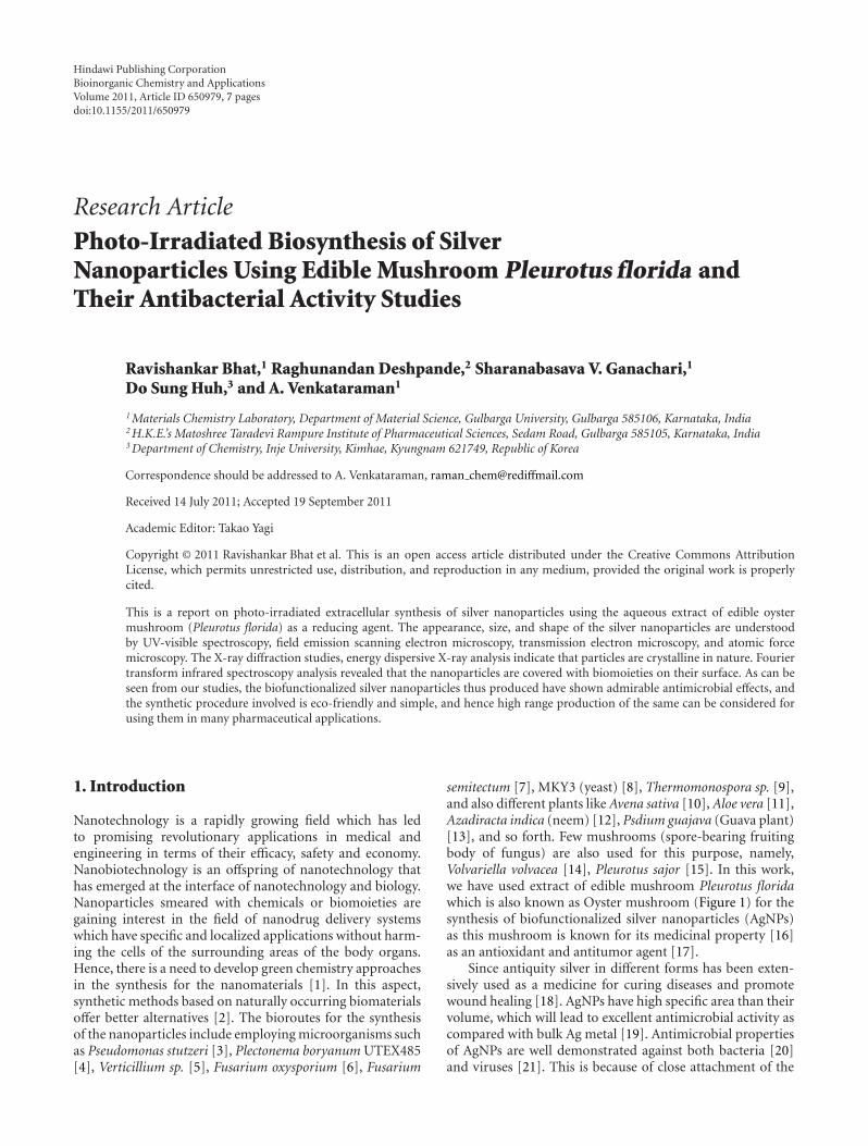
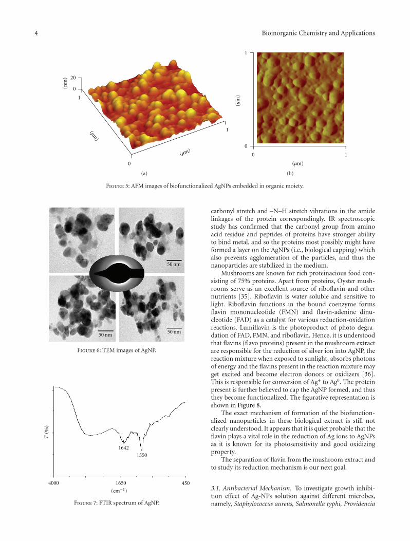
is because of the copper grid base used for the analysis. Atpresent, AFM is a powerful tool to study the morphology ofbiofunctionalized particles. The biofunctionalization of theAgNPs prepared by using mushroom extract is further con-firmed by AFM measurements. The three-dimensional studyof biofunctionalized nanoparticles were made on tappingmode technique developed especially for studying biofunc-tionalization. Figure 5 shows the AgNPs biofunctionalizedhaving organic layer which consists of lot of organic moietiesat the surface. From this figure, we can predict that the shapeof the nanoparticles is nearly spherical with some irregularshaped particles and is randomly distributed. The typicalTEM image showing the size and morphology of AgNPs isgiven in Figure 6; the morphology of AgNP is apparentlyspherical. From the TEM micrograph it is observed that theAgNPs formed are in a size range of 20 ± 5 nm and poly-dispersed. The selected diffraction pattern from one of thesilver nanoparticle, is shown in the inset of Figure 6, whichsuggests that the AgNP is apparently in amorphous conditionsince organic moiety is covered on it. The crystallinity can beachieved by repeated centrifugation and washings.
FTIR spectroscopy from the absorption of IR radia-tion through resonance of noncentrosymmetric (IR active)modes of vibration is useful technique to study the core-shell morphology of AgNPs. Figure 7 shows FTIR spectrumof AgNPs. The two bands at ∼1642 and ∼1550 are observedand are recognized as amide I and amide II and arise due to
4 Bioinorganic Chemistry and Applications
20
0(nm
)
1
0
1(μm)
(μm)
(a)
1
0
10
(μm)
(μm
)
(b)
Figure 5: AFM images of biofunctionalized AgNPs embedded in organic moiety.
50 nm
50 nm50 nm
Figure 6: TEM images of AgNP.
1642
1550
4000 1650 450
(cm−1)
T(%
)
Figure 7: FTIR spectrum of AgNP.
carbonyl stretch and –N–H stretch vibrations in the amidelinkages of the protein correspondingly. IR spectroscopicstudy has confirmed that the carbonyl group from aminoacid residue and peptides of proteins have stronger abilityto bind metal, and so the proteins most possibly might haveformed a layer on the AgNPs (i.e., biological capping) whichalso prevents agglomeration of the particles, and thus thenanoparticles are stabilized in the medium.
Mushrooms are known for rich proteinacious food con-sisting of 75% proteins. Apart from proteins, Oyster mush-rooms serve as an excellent source of riboflavin and othernutrients [35]. Riboflavin is water soluble and sensitive tolight. Riboflavin functions in the bound coenzyme formsflavin mononucleotide (FMN) and flavin-adenine dinu-cleotide (FAD) as a catalyst for various reduction-oxidationreactions. Lumiflavin is the photoproduct of photo degra-dation of FAD, FMN, and riboflavin. Hence, it is understoodthat flavins (flavo proteins) present in the mushroom extractare responsible for the reduction of silver ion into AgNP, thereaction mixture when exposed to sunlight, absorbs photonsof energy and the flavins present in the reaction mixture mayget excited and become electron donors or oxidizers [36].This is responsible for conversion of Ag+ to Ag0. The proteinpresent is further believed to cap the AgNP formed, and thusthey become functionalized. The figurative representation isshown in Figure 8.
The exact mechanism of formation of the biofunction-alized nanoparticles in these biological extract is still notclearly understood. It appears that it is quiet probable that theflavin plays a vital role in the reduction of Ag ions to AgNPsas it is known for its photosensitivity and good oxidizingproperty.
The separation of flavin from the mushroom extract andto study its reduction mechanism is our next goal.
3.1. Antibacterial Mechanism. To investigate growth inhibi-tion effect of Ag-NPs solution against different microbes,namely, Staphylococcus aureus, Salmonella typhi, Providencia

Bioinorganic Chemistry and Applications 5
Flavin
Ag+
Ag+
Ag+
Ag+Ag+ Ag0
Ag0
Ag0
Ag0
hυ
Figure 8: Probable pathway of synthesis mechanism.
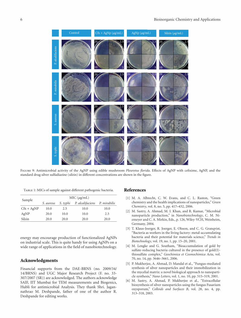
alcalifaciens, and Proteus mirabilis, we measured the MIC andthe changes of bacterial counts at different concentrations ofcfx + AgNP (equal quantities of cefixime antibiotic drug andfunctionalized silver nanoparticles), AgNPs (functionalizedsilver nanoparticles), and with silzin (standard silver sulfadi-azine drug), (2.5 μg to 20.0 μg/mL concentration) shown inFigure 9. The control plates are prepared without addition ofany drug for the comparative study of the inhibitory effects ofthe cfx + AgNP, AgNP, and silzin. These results exposed thatthe MIC of cfx + AgNPs showed higher-inhibition kineticsagainst both P. alcalifaciens, P. mirabilis (Gr +ve). S. aureus,and S. typhi (Gram−ve) when compared with other samplesshown in Table 1. Only 2.5 μg/mL cfx + AgNP is sufficientfor the complete prevention of bacterial growth of S. typhi,whereas in case of other three bacteria 10.0 μg/mL of cfx +AgNP is to be added for the growth inhibition. From theresult, it is noted that AgNP synthesized from mushroomis showing decent bactericidal activity against gram +vebacteria, specially in case of P. mirabilis 2.5 μg/mL is sufficientfor complete effect for reduction of bacterial colony, and incase of gram −ve bacteria the antimicrobial activity of AgNPis moderate. When the MIC result of both the samples (cfx+ AgNP and AgNP) is compared with standard silzin, it isobserved that the MIC of silzin is poor and more quantityof samples is required for complete growth reduction of
bacterial colony. So, it is clear that biofunctionalized AgNPshows excellent antimicrobial property either by mixing withantibiotic drug or by using directly as a drug.
It is presumed that the influence of free electronsproduced by the surface of the functionalized AgNPs willhave lethal effect on the electronegative surface membraneof the microorganism causing its death. The outcome ofantibiogram analysis of compounds against different bacteriashows that biologically prepared AgNP can become analternative agent in antibacterial treatment, because of its lessside effect and potential action.
4. Conclusion
In this study, AgNPs were synthesized by the photo-irradiation technique using the edible mushroom, Pleurotusflorida, as a bioreductant. The biosynthetic method devel-oped in this study for producing silver nanoparticles hasdistinct advantages over chemical methods such as highbiosafety and being ecofriendly and nontoxic to the environ-ment. Furthermore, these functionalized silver nanoparticlesshowed a noticeable antimicrobial activity against differentclinically important pathogenic microorganisms. Hence,such type of synthesis methods for the production ofnanostructured materials at lower cost and with natural
6 Bioinorganic Chemistry and Applications
Control
20 2.5
10 5
20 2.5
10 5
20 2.5
10 5
20 2.5
10 5
20 2.5
10 5
20 2.5
10 5
20 2.5
10 5
20 2.5
10 5
20 2.5
10 5
20 2.5
10 5
20 2.5
10 5
20 2.5
10 5
P.al
calif
acie
ns
P.m
irab
ilis
S.au
reu
sS.
typh
i
Cfx + AgNp (μg/mL) AgNp (μg/mL) Silzin (μg/mL)
Figure 9: Antimicrobial activity of the AgNP using edible mushroom Pleurotus florida. Effects of AgNP with cefixime, AgNP, and thestandard drug silver sulfadiazine (silzin) in different concentrations are shown in the figure.
Table 1: MICs of sample against different pathogenic bacteria.
SampleMIC (μg/mL)
S. aureus S. typhi P. alcalifaciens P. mirabilis
Cfx + AgNP 10.0 2.5 10.0 10.0
AgNP 20.0 10.0 10.0 2.5
Silzin 20.0 20.0 20.0 20.0
energy may encourage production of functionalized AgNPson industrial scale. This is quite handy for using AgNPs on awide range of applications in the field of nanobiotechnology.
Acknowledgments
Financial supports from the DAE-BRNS (no. 2009/34/14/BRNS) and UGC Major Research Project (F. no. 33-307/2007 (SR)) are acknowledged. The authors acknowledgeSAIF, IIT Mumbai for TEM measurements and Biogenics,Hubli for antimicrobial Analysis. They thank Shri. Jagan-nathrao M. Deshpande, father of one of the author R.Deshpande for editing works.
References
[1] M. A. Albrecht, C. W. Evans, and C. L. Raston, “Greenchemistry and the health implications of nanoparticles,” GreenChemistry, vol. 8, no. 5, pp. 417–432, 2006.
[2] M. Sastry, A. Ahmad, M. I. Khan, and R. Kumar, “Microbialnanoparticle production,” in Nanobiotechnology, C. M. Ni-emeyer and C. A. Mirkin, Eds., p. 126, Wiley-VCH, Weinheim,Germany, 2004.
[3] T. Klaus-Joerger, R. Joerger, E. Olsson, and C. G. Granqvist,“Bacteria as workers in the living factory: metal-accumulatingbacteria and their potential for materials science,” Trends inBiotechnology, vol. 19, no. 1, pp. 15–20, 2001.
[4] M. Lengke and G. Southam, “Bioaccumulation of gold bysulfate-reducing bacteria cultured in the presence of gold(I)-thiosulfate complex,” Geochimica et Cosmochimica Acta, vol.70, no. 14, pp. 3646–3661, 2006.
[5] P. Mukherjee, A. Ahmad, D. Mandal et al., “Fungus-mediatedsynthesis of silver nanoparticles and their immobilization inthe mycelial matrix: a novel biological approach to nanoparti-cle synthesis,” Nano Letters, vol. 1, no. 10, pp. 515–519, 2001.
[6] M. Sastry, A. Ahmad, P. Mukherjee et al., “Extracellularbiosynthesis of silver nanoparticles using the fungus Fusariumoxysporum,” Colloids and Surfaces B, vol. 28, no. 4, pp.313–318, 2003.
Bioinorganic Chemistry and Applications 7
[7] S. Basavaraja, S. D. Balaji, L. Arunkumar, A. H. Rajasab,and A. Venkataraman, “Extracellular biosynthesis of silvernanoparticles using the fungus Fusarium semitectum,”Materials Research Bulletin, vol. 43, no. 5, pp. 1164–1170, 2008.
[8] M. Kowshik, S. Ashtaputre, S. Kharrazi et al., “Extracellularsynthesis of silver nanoparticles by a silver-tolerant yeast strainMKY3,” Nanotechnology, vol. 14, no. 1, pp. 95–100, 2003.
[9] A. Ahmad, S. Senapati, M. I. Khan, R. Kumar, and M.Sastry, “Extracellular biosynthesis of monodisperse goldnanoparticles by a novel extremophilic actinomycete,thermomonospora sp,” Langmuir, vol. 19, no. 8, pp. 3550–3553, 2003.
[10] V. Armendariz, I. Herrera, J. R. Peralta-Videa et al., “Sizecontrolled gold nanoparticle formation by Avena sativabiomass: use of plants in nanobiotechnology,” Journal ofNanoparticle Research, vol. 6, no. 4, pp. 377–382, 2004.
[11] S. P. Chandran, M. Chaudhary, R. Pasricha, A. Ahmad,and M. Sastry, “Synthesis of gold nanotriangles and silvernanoparticles using Aloe vera plant extract,” BiotechnologyProgress, vol. 22, no. 2, pp. 577–583, 2006.
[12] M. Sastry, S. S. Shankar, A. Rai, and A. Absar, “Rapid synthesisof Au, Ag, and bimetallic Au core-Ag shell nanoparticles usingNeem (Azadirachta indica) leaf broth,” Journal of Colloid andInterface Science, vol. 275, no. 2, pp. 496–502, 2004.
[13] D. Raghunandan, B. D. Mahesh, S. Basavaraja, S. D. Balaji,S. Y. Manjunath, and A. Venkataraman, “Microwave-assistedrapid extracellular synthesis of stable bio-functionalized silvernanoparticles from guava (Psidium guajava) leaf extract,”Journal of Nanoparticle Research, vol. 13, no. 5, pp. 2021–2028,2011.
[14] D. Philip, “Biosynthesis of Au, Ag and Au-Ag nanoparticlesusing edible mushroom extract,” Spectrochimica Acta—PartA: Molecular and Biomolecular Spectroscopy, vol. 73, no. 2, pp.374–381, 2009.
[15] R. Nithya and R. Ragunathan, “Synthesis of silver nanoparticleusing Pleurotus sajor caju and its antimicrobial study,” DigestJournal of Nanomaterials and Biostructures, vol. 4, no. 4, pp.623–629, 2009.
[16] T. A. Ajith and K. K. Janardhanan, “Indian medicinalmushrooms as a source of antioxidant and antitumor agents,”Journal of Clinical Biochemistry and Nutrition, vol. 40, no. 3,pp. 157–162, 2007.
[17] K. K. Janardhanan and J. Nayana, “Antioxidant and anti-tumour activity of Pleurotus florida,” Current Science, vol. 79,no. 7, pp. 941–943, 2000.
[18] H. Vermeulen, J. M. van Hattem, M. N. Storm-Versloot, D.T. Ubbink, and S. J. Westerbos, Cochrane Database of SystemicReviews, vol. 7, Willey, City, State, USA, 2010.
[19] R. Mahendra, Y. Alka, and G. Aniket, “Silver nanoparticles asa new generation of antimicrobials,” Biotechnology Advances,vol. 27, no. 1, pp. 76–83, 2009.
[20] I. Sondi and B. Salopek-Sondi, “Silver nanoparticles asantimicrobial agent: a case study on E. coli as a model forGram-negative bacteria,” Journal of Colloid and InterfaceScience, vol. 275, no. 1, pp. 177–182, 2004.
[21] H. H. Lara, N. V. Ayala-Nunez, L. Ixtepan-Turrent, andC. Rodriguez-Padilla, “Mode of antiviral action of silvernanoparticles against HIV-1,” Journal of Nanobiotechnology,vol. 8, no. 1, 2010.
[22] J. Jain, S. Arora, J. M. Rajwade, P. Omray, S. Khandelwal, andK. M. Paknikar, “Silver nanoparticles in therapeutics: devel-opment of an antimicrobial gel formulation for topical use,”Molecular Pharmaceutics, vol. 6, no. 5, pp. 1388–1401, 2009.
[23] K. Roy, H. Q. Mao, S. K. Huang, and K. W. Leong, “Oralgene delivery with chitosan-DNA nanoparticles generates
immunologic protection in a murine model of peanut allergy,”Nature Medicine, vol. 5, no. 4, pp. 387–391, 1999.
[24] E. Sachlos, D. Gotora, and J. T. Czernuszka, “Collagenscaffolds reinforced with biomimetic composite nano-sizedcarbonate-substituted hydroxyapatite crystals and shaped byrapid prototyping to contain internal microchannels,” TissueEngineering, vol. 12, no. 9, pp. 2479–2487, 2006.
[25] N. Vigneshwaran, A. A. Kathe, P. V. Varadarajan, R. P.Nachane, and R. H. Balasubramanya, “Functional finishingof cotton fabrics using silver nanoparticles,” Journal ofNanoscience and Nanotechnology, vol. 7, no. 6, pp. 1893–1897,2007.
[26] M. Ip, S. L. Lui, V. K. M. Poon, I. Lung, and A. Burd, “Antimi-crobial activities of silver dressings: an in vitro comparison,”Journal of Medical Microbiology, vol. 55, no. 1, pp. 59–63, 2006.
[27] F. Furno, K. S. Morley, B. Wong et al., “Silver nanoparticlesand polymeric medical devices: a new approach to preventionof infection?” Journal of Antimicrobial Chemotherapy, vol. 54,no. 6, pp. 1019–1024, 2004.
[28] P. Rujitanaroj, N. Pimpha, and P. Supaphol, “Wound-dressingmaterials with antibacterial activity from electrospun gelatinfiber mats containing silver nanoparticles,” Polymer, vol. 49,no. 21, pp. 4723–4732, 2008.
[29] H. Huang and X. Yang, “Synthesis of polysaccharide-stabilizedgold and silver nanoparticles: a green method,” CarbohydrateResearch, vol. 339, no. 15, pp. 2627–2631, 2004.
[30] J. P. Abid, A. W. Wark, P. F. Brevet, and H. H. Girault,“Preparation of silver nanoparticles in solution from a silversalt by laser irradiation,” Chemical Communications, no. 7, pp.792–793, 2002.
[31] S. S. Gasaymeh, S. Radiman, L. Y. Heng, E. Saion, and G.H. Mohamed Saeed, “Synthesis and characterization ofsilver/Polyvinilpirrolidone (AG/PVP) nanoparticles using γirradiation techniques,” American Journal of Applied Sciences,vol. 7, no. 7, pp. 892–901, 2010.
[32] B. Soroushian, I. Lampre, J. Belloni, and M. Mostafavi,“Radiolysis of silver ion solutions in ethylene glycol: solvatedelectron and radical scavenging yields,” Radiation Physics andChemistry, vol. 72, no. 2-3, pp. 111–118, 2005.
[33] S. K. Sahi and M. Patra, “microbially synthsized bioactivenanoparticles AND their formulation active against humanpathogenic fungi,” Reviews on Advanced Materials Science, vol.5, pp. 501–509, 2003.
[34] P. Mulvaney, “Surface plasmon spectroscopy of nanosizedmetal particles,” Langmuir, vol. 12, no. 3, pp. 788–800, 1996.
[35] M. M. Imran, M. M. M. Raja, J. A. Basith, and A. Asarudeen,“Determination of total phenol, flavonoid and antioxidantactivity of edible mushrooms Pleurotus florida and Pleurotuseous,” International Food Research Journal, vol. 18, no. 2, pp.574–577, 2011.
[36] A. N. Woodmansee and J. A. Imlay, “Reduced flavins promoteoxidative DNA damage in non-respiring Escherichia coliby delivering electrons to intracellular free iron,” Journal ofBiological Chemistry, vol. 277, no. 37, pp. 34055–34066, 2002.

Submit your manuscripts athttp://www.hindawi.com
Hindawi Publishing Corporationhttp://www.hindawi.com Volume 2014
Inorganic ChemistryInternational Journal of
Hindawi Publishing Corporation http://www.hindawi.com Volume 2014
International Journal ofPhotoenergy
Hindawi Publishing Corporationhttp://www.hindawi.com Volume 2014
Carbohydrate Chemistry
International Journal of
Hindawi Publishing Corporationhttp://www.hindawi.com Volume 2014
Journal of
Chemistry
Hindawi Publishing Corporationhttp://www.hindawi.com Volume 2014
Advances in
Physical Chemistry
Hindawi Publishing Corporationhttp://www.hindawi.com
Analytical Methods in Chemistry
Journal of
Volume 2014
Bioinorganic Chemistry and ApplicationsHindawi Publishing Corporationhttp://www.hindawi.com Volume 2014
SpectroscopyInternational Journal of
Hindawi Publishing Corporationhttp://www.hindawi.com Volume 2014
The Scientific World JournalHindawi Publishing Corporation http://www.hindawi.com Volume 2014
Medicinal ChemistryInternational Journal of
Hindawi Publishing Corporationhttp://www.hindawi.com Volume 2014
Chromatography Research International
Hindawi Publishing Corporationhttp://www.hindawi.com Volume 2014
Applied ChemistryJournal of
Hindawi Publishing Corporationhttp://www.hindawi.com Volume 2014
Hindawi Publishing Corporationhttp://www.hindawi.com Volume 2014
Theoretical ChemistryJournal of
Hindawi Publishing Corporationhttp://www.hindawi.com Volume 2014
Journal of
Spectroscopy
Analytical ChemistryInternational Journal of
Hindawi Publishing Corporationhttp://www.hindawi.com Volume 2014
Journal of
Hindawi Publishing Corporationhttp://www.hindawi.com Volume 2014
Quantum Chemistry
Hindawi Publishing Corporationhttp://www.hindawi.com Volume 2014
Organic Chemistry International
ElectrochemistryInternational Journal of
Hindawi Publishing Corporation http://www.hindawi.com Volume 2014
Hindawi Publishing Corporationhttp://www.hindawi.com Volume 2014
CatalystsJournal of