phase diagram of self-assembled viral capsid … diagram of self-assembled viral capsid protein...
TRANSCRIPT
Phase Diagram of Self-assembled Viral Capsid Protein Polymorphs†
L. Lavelle,‡ M. Gingery,§ M. Phillips,§ W. M. Gelbart,‡ and C. M. Knobler*,‡
Department of Chemistry and Biochemistry, UniVersity of California, Los Angeles,California 90095-1569, Molecular Biology Institute, UniVersity of California, Los Angeles,California 90095-1570
R. D. Cadena-Nava,⊥ J. R. Vega-Acosta,⊥ L. A. Pinedo-Torres, and J. Ruiz-Garcia*Instituto de Fısica, UniVersidad Autonoma de San Luis Potosı, AlVaro Obregon 64,San Luis Potosı, S.L.P., 78000 Mexico
ReceiVed: September 8, 2008; ReVised Manuscript ReceiVed: NoVember 12, 2008
We present an experimental study of the self-assembly of capsid proteins of the cowpea chlorotic mosaicvirus (CCMV), in the absence of the viral genome, as a function of pH and ionic strength. In accord withprevious measurements, a wide range of polymorphs can be identified by electron microscopy, among themsingle and multiwalled shells and tubes. The images are analyzed with respect to size and shape of aggregates,and evidence is given that equilibrium has been achieved, allowing a phase diagram to be constructed. Somepreviously unreported structures are also described. The range and stability of the polymorphs can be understoodin terms of electrostatic interactions and the way they affect the spontaneous curvature of protein networksand the relative stabilities of pentamers and hexamers.
Introduction
The simplest viruses consist of a single-protein-thick shell,the capsid, that surrounds and protects the viral genome. Intobacco mosaic virus (TMV), the capsid is a hollow cylinder,made up of many copies of a single protein, that assemblesaround its single-stranded RNA genome. In 1955 the demon-stration by Fraenkel-Conrat and Williams1 that infectious TMVcould spontaneously form in solutions of the pure capsid proteinand RNA made it clear that the general physical principles ofself-assembly could apply to a virus. A dozen years later,Bancroft and Hiebert2 showed that infectious cowpea chloroticmosaic virus (CCMV), a spherical virus, would also spontane-ously assemble from appropriately buffered solutions of its pureprotein and RNA.
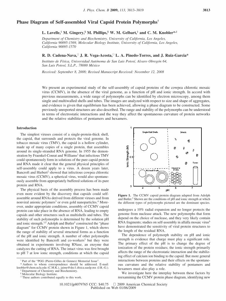
The physical basis of the assembly process has been madeeven more evident by the discovery that capsids could self-assemble around RNAs derived from different viruses and fromnonviral anionic polymers3 or even gold nanoparticles.4 More-over, under appropriate conditions, assembly of CCMV capsidprotein can take place in the absence of RNA, leading to emptycapsids and other structures such as multishells and tubes. Thestability of such polymorphs is determined by the solution pHand ionic strength.3,5 Adolph and Butler5 constructed the “phasediagram” for CCMV protein shown in Figure 1, which showsthe range of stability of several structural forms as a functionof the pH and ionic strength. A number of other polymorphswere identified by Bancroft and co-workers3 but they wereobtained in experiments involving RNase, an enzyme thatcatalyzes the cutting of RNA. The intact virus was first broughtto pH 7 at low ionic strength, conditions at which the capsid
undergoes a 10% radial expansion and no longer protects thegenome from nuclease attack. The new polymorphs that formdepend on the choice of nuclease, and they very likely containRNA fragments; studies on self-assembly in alfalfa mosaic virus6
have demonstrated the sensitivity of viral protein structures tothe length of the residual RNA.
The dependence of polymorph stability on pH and ionicstrength is evidence that charge must play a significant role.The primary effect of the pH is to change the degree ofionization of the protein residues; the ionic strength primarilyaffects the range of the electrostatic interaction and the stabiliz-ing effect of calcium ion binding to the capsid. But more generalinteractions between proteins and their effects on the spontane-ous curvature and the relative stability of pentamers andhexamers must also play a role.
We investigate here the interplay between these factors byreexamining the CCMV protein phase diagram, identifying new
† Part of the “PGG (Pierre-Gilles de Gennes) Memorial Issue”.* Authors to whom correspondence should be addressed. E-mail:
[email protected] (C.M.K.), [email protected] (J.R.-G.).‡ Department of Chemistry and Biochemistry.§ Molecular Biology Institute.⊥ These authors contributed equally to this work.
Figure 1. The CCMV capsid protein diagram adapted from Adolphand Butler.5 Shown are the conditions of pH and ionic strength at whichthe different types of polymorphs pictured are the dominant species.
J. Phys. Chem. B 2009, 113, 3813–3819 3813
10.1021/jp8079765 CCC: $40.75 2009 American Chemical SocietyPublished on Web 01/06/2009
forms of aggregates, and focusing largely on the low-ionic-strength region where the range of structures is greatest. Weinterpret the diagram in terms of a recent theoretical treatmentof viral protein assembly.7
Experimental Results
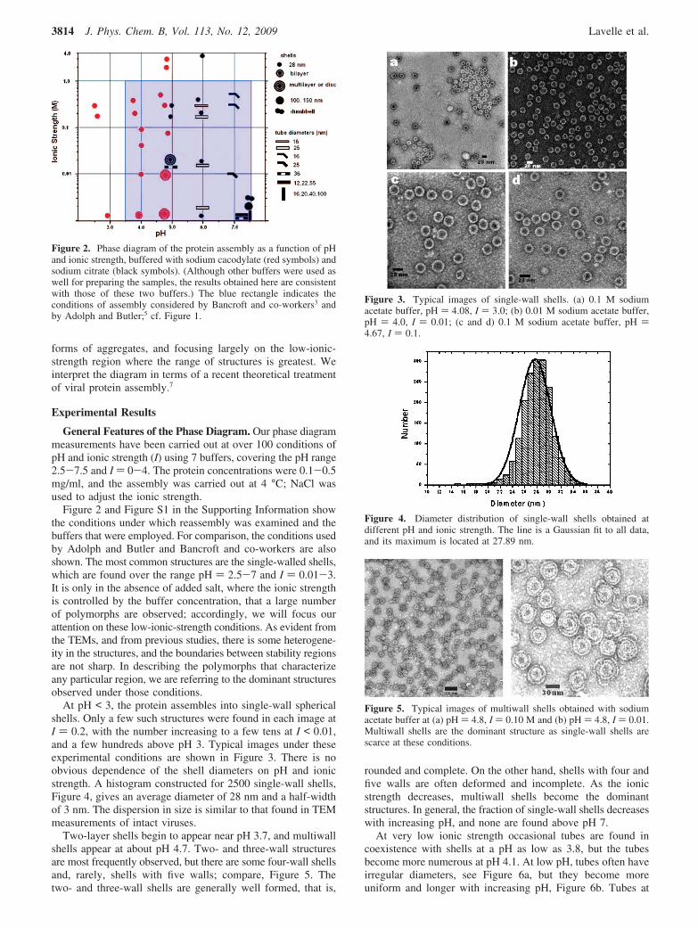
General Features of the Phase Diagram. Our phase diagrammeasurements have been carried out at over 100 conditions ofpH and ionic strength (I) using 7 buffers, covering the pH range2.5-7.5 and I ) 0-4. The protein concentrations were 0.1-0.5mg/ml, and the assembly was carried out at 4 °C; NaCl wasused to adjust the ionic strength.
Figure 2 and Figure S1 in the Supporting Information showthe conditions under which reassembly was examined and thebuffers that were employed. For comparison, the conditions usedby Adolph and Butler and Bancroft and co-workers are alsoshown. The most common structures are the single-walled shells,which are found over the range pH ) 2.5-7 and I ) 0.01-3.It is only in the absence of added salt, where the ionic strengthis controlled by the buffer concentration, that a large numberof polymorphs are observed; accordingly, we will focus ourattention on these low-ionic-strength conditions. As evident fromthe TEMs, and from previous studies, there is some heterogene-ity in the structures, and the boundaries between stability regionsare not sharp. In describing the polymorphs that characterizeany particular region, we are referring to the dominant structuresobserved under those conditions.
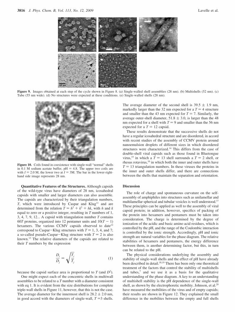
At pH < 3, the protein assembles into single-wall sphericalshells. Only a few such structures were found in each image atI ) 0.2, with the number increasing to a few tens at I < 0.01,and a few hundreds above pH 3. Typical images under theseexperimental conditions are shown in Figure 3. There is noobvious dependence of the shell diameters on pH and ionicstrength. A histogram constructed for 2500 single-wall shells,Figure 4, gives an average diameter of 28 nm and a half-widthof 3 nm. The dispersion in size is similar to that found in TEMmeasurements of intact viruses.
Two-layer shells begin to appear near pH 3.7, and multiwallshells appear at about pH 4.7. Two- and three-wall structuresare most frequently observed, but there are some four-wall shellsand, rarely, shells with five walls; compare, Figure 5. Thetwo- and three-wall shells are generally well formed, that is,
rounded and complete. On the other hand, shells with four andfive walls are often deformed and incomplete. As the ionicstrength decreases, multiwall shells become the dominantstructures. In general, the fraction of single-wall shells decreaseswith increasing pH, and none are found above pH 7.
At very low ionic strength occasional tubes are found incoexistence with shells at a pH as low as 3.8, but the tubesbecome more numerous at pH 4.1. At low pH, tubes often haveirregular diameters, see Figure 6a, but they become moreuniform and longer with increasing pH, Figure 6b. Tubes at
Figure 2. Phase diagram of the protein assembly as a function of pHand ionic strength, buffered with sodium cacodylate (red symbols) andsodium citrate (black symbols). (Although other buffers were used aswell for preparing the samples, the results obtained here are consistentwith those of these two buffers.) The blue rectangle indicates theconditions of assembly considered by Bancroft and co-workers3 andby Adolph and Butler;5 cf. Figure 1.
Figure 3. Typical images of single-wall shells. (a) 0.1 M sodiumacetate buffer, pH ) 4.08, I ) 3.0; (b) 0.01 M sodium acetate buffer,pH ) 4.0, I ) 0.01; (c and d) 0.1 M sodium acetate buffer, pH )4.67, I ) 0.1.
Figure 4. Diameter distribution of single-wall shells obtained atdifferent pH and ionic strength. The line is a Gaussian fit to all data,and its maximum is located at 27.89 nm.
Figure 5. Typical images of multiwall shells obtained with sodiumacetate buffer at (a) pH ) 4.8, I ) 0.10 M and (b) pH ) 4.8, I ) 0.01.Multiwall shells are the dominant structure as single-wall shells arescarce at these conditions.
3814 J. Phys. Chem. B, Vol. 113, No. 12, 2009 Lavelle et al.

higher ionic strength tend to be narrower, 12-16 nm in averagediameter; at low pH they are bent/kinked and their ends tend tobe sharp, see Figure 6c. As the buffer concentration and ionicstrength decrease, the tubes are better-formed and are wider,about 22-25 nm in diameter. Tube ends are rounded and canbe bulbous, Figure 6, panels a and c; as in Figure 6b, theysometimes give the appearance of having grown out of a shellor a cluster of shells. Tube lengths range from 70 to 900 nm.At higher pH values (>7.2) long, open-ended tubes, Figure 6d,with diameters as large as 100 nm are formed.
Table S1 in the Supporting Information summarizes thecharacteristics of the tubes observed with cacodylate buffer inthis study. Very well formed tubes always appear at low ionicstrength in a wide range of pHs. As we increase the ionicstrength, the tubes become narrower and they tend to involvebends or kinks; some develop sharp or “spiky” ends.
Novel Structures. We have observed three novel structuresthat have not been previously reported. Disk-like structures suchas those shown in Figure 7a were observed at pH values rangingfrom 4.75 to 5.0 for ionic strengths up to 0.02. Fouriertransforms of the images show a hexagonal arrangement of theproteins that is expected for a lamellar structure. AlthoughBancroft et al. did not observe disks, they reported the formationof a large number of laminar and plate forms in the same rangeof conditions, thought to be formed on surfaces; the number ofplate structures they observed is large compared with the numberof disks we observed.
At very low ionic strength and pH ) 7.5 we found very largeshells that are not well rounded and have 100-150 nmdiameters; dumbbell-like shells, Figure 7b, were found underthe same conditions.
Thermodynamic Stability. In ascribing a phase diagram tothe polymorphs we are implicitly assuming that the structuresare thermodynamically stable and are not kinetically controlled.Bancroft3 reported that CCMV protein dialyzed at pH 6.0 and0.5 M NaCl did not assemble. However, protein that was firstdialyzed at pH 5.0 and 0.5 M NaCl and then dialysed to pH6.0 at the same ionic strength assembled into single-wall shells.Although it is not clear how long dialyses were carried out inthis work, in some experiments by the Bancroft group8 it wasnoted that dialyses were carried out for 2-4 h. Adolph andButler5 argued that equilibrium could be attained by sufficientlylong (48 h) dialysis. This is in accord with our observation thatthere was no significant difference between the results of dialysisfor 24 or 120 h.
As a test of thermodynamic stability we have carried out thefour-step cycle shown in Figure 8, each time waiting 24 h afterchanging conditions, removing small aliquots, and determiningthe dominant structures in them by TEM. At the initialconditions (0.01 M sodium acetate buffer, pH 4.8, I ) 0.5), theprotein assembled into empty single-walled shells, Figure 9a.After dialysis at the same pH in the absence of added salt,multiwall shells formed, Figure 9b. Dialysis now against a 0.01M sodium cacodylate buffer (pH 5.95) led to reassembly intotubes, Figure 9c. When the system was then dialyzed to pH7.12, I )0.5 M with sodium cacodylate, the protein disassembledand no structures were observed, Figure 9d. Finally, when thesystem was dialysed to the initial conditions, single-walled shellswere again obtained, Figure 9e. Particle size distributions atpoints a and e, as determined by light scattering, Table S2 inthe Supporting Information, are essentially identical. Weconclude then that the structures we have observed are repre-sentative of equilibrium.
Although the observed morphologies are controlled bysolution conditions, defects in the structures, such as bends intubes, are also observed. A striking example, the coil structuresshown in Figure 10 are observed in a very narrow range of pH,4.7-4.8, and over a broad range of ionic strength, ∼0 to 3.0M. Plastic framework models of shells demonstrate that suchstructures can arise from an error in assembly in which a singlepentamer is replaced by a hexamer. A variety of such misas-sembled structuressincluding dumbbellsshave also been ob-served in molecular dynamics simulations of coarse-grainedmodels of capsid proteins.9 These have been compared to so-called “monster” structures that were found in in vitro studiesof the assembly of turnip crinkle virus and capsid protein aroundthe viral RNA.10
Figure 6. Tube structures obtained with sodium cacodylate buffer andno added salt: (a) pH ) 5.65, 0.01 M buffer; (b) pH ) 6.0, 0.01 Mbuffer; (c) pH ) 7.1, 0.01 M buffer; (d) pH ) 7.5, 0.001 M buffer.
Figure 7. (a) Disks formed in 0.01 M sodium citrate buffer pH )4.75, no added salt. (b) Dumbbell structures at pH ) 7.5, 0.001 Mcacodylate buffer, no added salt.
Figure 8. Path followed in test of thermodynamic equilibrium. Theconditions are those given in each box and the arrows indicate dialysisfor 24 h.
Self-assembled Viral Capsid Protein Polymorphs J. Phys. Chem. B, Vol. 113, No. 12, 2009 3815
Quantitative Features of the Structures. Although capsidsof the wild-type virus have diameters of 28 nm, icosahedralcapsids with smaller and larger diameters can also assemble.The capsids are characterized by their triangulation numbers,T, which were introduced by Caspar and Klug11 and aredetermined from the relation T ) h2 + k2 + hk, with h and kequal to zero or a positive integer, resulting in T numbers of 1,3, 4, 7, 9, 12... A capsid with triangulation number T contains60T proteins, organized into 12 pentamer units and 10(T - 1)hexamers. The various CCMV capsids observed to date12
correspond to Caspar-Klug structures with T ) 1, 3, 4, and 7;a so-called pseudo-Caspar-Klug structure with T ) 2 is alsoknown.13 The relative diameters of the capsids are related totheir T numbers by the expression
because the capsid surface area is proportional to T (and D2).One might expect each of the concentric shells in multiwall
assemblies to be related to a T number with a diameter consistentwith eq 1. It is evident from the size distributions for completetriple-wall shells in Figure 11, however, that this is not the case.The average diameter for the innermost shell is 28.2 ( 2.0 nm,in good accord with the diameters of single-wall, T )3 shells.
The average diameter of the second shell is 39.5 ( 1.9 nm,markedly larger than the 32 nm expected for a T ) 4 structureand smaller than the 43 nm expected for T ) 7. Similarly, theaverage outer-shell diameter, 51.8 ( 3.0, is larger than the 48nm expected for a shell with T ) 9 and smaller than the 56 nmexpected for a T ) 12 capsid.
These results demonstrate that the successive shells do nothave a regular icosahedral structure and are disordered, in accordwith recent studies of the assembly of CCMV protein aroundnanoemulsion droplets of different sizes in which disorderedstructures were characterized.14 This differs from the case ofdouble-shell viral capsids such as those found in Bluetonguevirus,15 in which a T ) 13 shell surrounds a T ) 2 shell, orrhesus rotavirus,16 in which both the inner and outer shells haveT ) 13 triangulation numbers. In these viruses the proteins inthe inner and outer shells differ, and there are connectionsbetween the shells that maintain the separation and orientation.
Discussion
The role of charge and spontaneous curvature on the self-assembly of amphiphiles into structures such as unilamellar andmultilamellar spherical and tubular vesicles is well understood.17
These principles can be applied as well to the assembly of viralcapsid protein; in addition, however, specifics of packing ofthe protein into hexamers and pentamers must be taken intoconsideration. The charge is determined by the degree ofionization of the acidic and basic amino acid residues, which iscontrolled by the pH, and the range of the Coulombic interactionis controlled by the ionic strength. Accordingly, pH and ionicstrength are natural variables for the phase diagram. The relativestabilities of hexamers and pentamers, the energy differencebetween them, is another determining factor, but this, in turncan be related to the pH.
The physical considerations underlying the assembly andstability of single-wall shells and the effect of pH have alreadybeen described in detail.18,19 There has been only one theoreticaltreatment of the factors that control the stability of multishellsand tubes,7 and we use it as a basis for the qualitativeunderstanding of the phase diagram. A key to an understandingof multishell stability is the pH dependence of the single-wallshell, as shown by the electrophoretic mobility. Johnson, et al.20
have measured the mobilities of the virus and of empty capsids;their results are shown in Figure 12. They explained the smalldifference in the mobilites between the empty and full shells
Figure 9. Images obtained at each step of the cycle shown in Figure 8. (a) Single-walled shell assemblies (28 nm). (b) Multishells (52 nm). (c)Tube (55 nm wide). (d) No structures were expected at these conditions. (e) Single-walled shells (28 nm).
Figure 10. Coils found in coexistence with single-wall “normal” shellsin 0.1 M sodium acetate buffer, pH ) 4.8. The upper two coils arewith I ) 2.0 M, the lower two at I ) 3M. The bar in the lower-right-hand side image represents 28 nm.
Di
Dj) �Ti
Tj(1)
3816 J. Phys. Chem. B, Vol. 113, No. 12, 2009 Lavelle et al.

as the result of the dependence of the mobility on the surfacecharge density rather than on the net charge.
In CCMV, the surface charge arises from the acidic aminoacid residues, which lie predominantly on the capsid exterior.In contrast, most of the basic residues are near the aminoterminus and project into the capsid interior.21 A simplecalculation of the pH dependence of the charge on a CCMVcapsid on the pH from the pKa values of the isolated residuesin which the contribution of the basic (largely interior) lysineand arginine residues is omitted - gives an isoelectric point of3.6, which is essentially identical to that of CCMV. Moreover,as shown in Figure 12, the shape of the curve is closely similarto the measured electrophoretic mobility. These results areconsistent with the argument that the electrophoretic mobilityis associated with the surface charge density.
The electrophoretic mobility measurements in Figure 12 werecarried out only at 0.2 M NaCl. However, recent studies on thespherical bacteriophage MS2 and accompanying theory22 showthat for a layered structure such as a virus the magnitude of theelectrophoretic mobility depends on the ionic strength butthe isoelectric point shows little change.
The net charge on the capsid is determined by the balancebetween the charges of the acidic residues, which lie predomi-nantly on the exterior of the capsid, and the basic residues, lysineand arginine, have pKa values in excess of 10 and are thereforecharged over the entire pH range that has been examined in theassembly reactions. The pKa values of the acidic residues,aspartic acid and glutamic acid, are much lower, and theircontribution to the charge therefore varies at acidic pH. (Notethat the pKa values are not those of the isolated residues andare dependent on the local environment.23)
We have observed that multishell formation begins near pH3.7, which is the isoelectric point of empty capsids (Figure 12),
that is, single-wall shells. This is related to the fact that, as thepH increases, the capsid exterior becomes increasingly nega-tively charged because of the ionization of acidic residues. Theaddition of a second shell around the capsid is then favoredbecause of the electrostatic interaction between the exteriornegative surface charge and the positive charge on the proteinsat the interior surface. In the related case of oppositely chargedflat membranes24 there is a stable equilibrium separation that isdetermined by the difference in the charge densities and thecounterion concentration. Here, however, there is an additionalfeature, the spontaneous curvature of the protein shell, whichprefers a radius of 14 nm. The successive shells must have largerradii of curvature, and the energy cost for the bending awayfrom the spontaneous curvature will also play a role incontrolling the spacing. With increasing pH the charge differencebetween the interior and exterior surfaces becomes larger,favoring the formation of more shells, but at the same time theshell curvature must be decreased and the energy penalty grows.Multishell stability will decrease with increasing ionic strengthbecause the screening of charge reduces the strength of theCoulombic interaction.
Implicit in these arguments about the effect of the spontaneouscurvature is the assumption that the interactions between capsidproteins that determine the curvature are not significantlyaffected by the pH. It is known21 that the diameter of CCMVcapsids does not change until the pH is close to 7, at whichpoint the capsid diameter expands by 10%. Thus, the interproteininteractions are not affected in the range of pH at whichmultishells are stable. The curvature may well change, however,if the capsid structure is disordered, which appears to be thecase for successive shells. This would have a quantitative effecton the curvature but is unlikely to affect the qualitative featuresof the competing interactions.
The leveling out of the electrophoretic mobility near pH 5,as shown in Figure 12, limits the strength of the electrostaticinteraction between shells; additional layers cannot grow becausethe curvature energy cannot be compensated. This is seen inthe structures (called “rosettes” by Bancroft) shown in Figure13, in which incomplete shells surround a multishell. Ingeneral, the rosette “petals” (see arrows) have a higher curvaturethan the last complete shell and therefore bend out.
The transition from spherical to tubular capsids is related tothe difference in energy between hexamers and pentamers ofproteins. Crudely speaking, this difference arises becausepentamers have five interactions with their neighbors rather thansix. The energy of a capsid then depends on the hexamer-pentamer energy penalty, which increases with increasing pH.25
As shown by Bruinsma, et al.,7 if the penalty is sufficiently large(i.e., at a sufficiently high pH) an assembly of spherical capsidsbecomes unstable with respect to spherocylindrical capsids withthe same mean curvature and the same total area.
Using optical Fourier transforms of electron micrographs,Bancroft et al.8 showed that CCMV tubes have a hexagonalstructure. Tubes with rounded ends are composed of a hexagonal
Figure 11. Diameter distributions of the (a) innermost, (b) middle, and (c) outermost shells of triple-wall shells.
Figure 12. Electrophoretic mobility of CCMV capsids as a functionof pH. The full red line is the mobility for the intact virus, and thedashed line is that for empty capids (single-walled shells) measuredby Johnson, et al.20 in the presence of 0.2 M NaCl. The net charge,represented by the blue line, has been calculated from the pKa valuesof the isolated amino acid residues of the capsid protein with theomission of the 12 lysine and 9 arginine residues. To compare themeasured mobilities and the calculated net charge, the right-handordinate has been multiplied by an arbitrary scale factor.
Self-assembled Viral Capsid Protein Polymorphs J. Phys. Chem. B, Vol. 113, No. 12, 2009 3817

sheet rolled into a cylinder and capped by hemispherescomposed of pentamers and hexamers. The tube diameters canbe understood in terms of a balance between the curvature ofthe caps, which depends on the hexamer-pentamer penalty, andthe spontaneous curvature of a sheet composed of hexamers,which is related to the charge asymmetry between the two sides.We expect discrete diameters because, like complete capsids,hemispherical caps can exist only for specific T numbers andbecause the joining of sheets to form a tube requires that therebe a continuous hexagonal structure.
In their seminal paper on the symmetry of viral capsids,Caspar and Klug11 suggested that one of the most probable“mistakes” of assembly was a tube produced by rolling ahexagonal plane lattice into a tube. If the tubes were capped byicosahedra from pentamers and hexamers they would havediameters comparable to those of fully assembled capsids. Asfirst pointed out by Bancroft,3 the majority of the tubes fall intotwo classes: narrow tubes with diameters about 16 nm and widetubes about 25 nm in diameter. The small diameter correspondsto that of a T ) 1 cap, which from eq 1 is 16 nm (based on D) 28 nm for T ) 3), and the larger diameter to a T ) 2 cap,which has a 24 nm diameter. The smaller diameter is favoredat higher ionic strength, where the electrostatic screening reducesthe spontaneous curvature of the hexagonal sheet by reducingthe effect of the inner surface-outer surface charge asymmetry.The diameters of the structures of the uncapped tubes aredetermined by the curvature of the hexagonal sheet and by therequirement that the hexagonal structure be continuous.
Kieslev and Klug26 also found small and large diameter tubesfor the capsid protein of rabbit papilloma virus. Although thisvirus has T ) 7 capsids, the larger tubes correspond to T ) 3caps and the smaller to T ) 1. Fourier transforms of the electronmicrographs showed that the larger tubes are hexagonal. Thesmaller tubes, which are capped by six pentamers, have anunusual structure in which pentamers are organized as hexava-lent units, allowing the tube surface to be tiled. Our measure-ments do not allow us to distinguish between this structure andone in which a hexagonal sheet of hexamers is capped by sixpentamers.
When the energy penalty for pentamers becomes sufficientlylarge, the spherical caps will be lost, leaving tubes with straightends. (There is, of course, an energy penalty for the unbonded
hexamers at the ends, so strictly speaking it is the differencebetween it and the cost for pentamers that controls theuncapping.) The diameters of the tubes then depend only onthe spontaneous curvature of the hexagonal sheet, which, asalready pointed out, decreases with increasing salt.
Although we have discussed the origin of the most commonof the polymorphs, still others are found in small quantities,underlining the richness of the viral protein assemblies. Thesensitivity to charge suggests that still other structures mightbe found if the assembly was carried out with protein mutants.A variety of polymorphs has been found for a number ofdifferent viral proteins and the arguments that we have madeabout the factors that determine CCMV assemblies should applybroadly. It should be noted as well that beside pH and ionicstrength, the relative stability of hexamers and pentamers maybe affected by the presence of small molecules that can bind tothe capsid proteins. This is the mechanism proposed by Strayet al.27 for the formation of hexagonal sheets and tubes fromhepatitis B viral proteins. In general, polymorphs of viral capsidproteins have little direct impact on our understanding of thebiological function of viruses, but they may provide anopportunity for materials science applications, some of whichhave been explored for single-wall capsids.28
Experimental Details
CCMV was harvested from cowpea california black eye No.5 plants infected both in Los Angeles and San Luis Potosi andpurified by ultrafiltration using the detailed procedures developedand described by Michel, et al.29 (see Sections 2.1, 2.2 and 2.4).
The purified CCMV was disassembled, and the capsid proteinwas isolated using the detailed methods described by Lavelleet al.30 (see Section 2.4). Protein purity and integrity weredetermined by SDS-PAGE and MALDI-TOF mass spectrom-etry. In this study, typical UV-absorbance ratios (Abs280nm/Abs260nm) were between 1.5 (0.5% nucleic acid contamination)and 1.6 (0.25% nucleic acid contamination). Protein concentra-tions were determined from the absorbance at 280 nm aspreviously described,30 and the reassembly was carried out atconcentrations of 0.1-0.5 mg ml-1.
Reassembly of the CCMV protein capsid was as previouslydescribed30 (see Section 2.5). However we note the following.The disassembly buffer (0.9 M NaCl, 0.02 M Tris-HCl, pH7.4, 1 mM DTT, 0.5 mM PMSF) contains DDT (dithiothreitolor Clealand’s reagent) to prevent the formation of disulfidebonds and PMSF (phenylmethylsulfonylfluoride) to inhibitproteases. Prior to the reassembly experiments discussed in thispaper, the DTT and PMSF were removed from the proteinsample by centrifugation at 4 °C using a Centriplus YM-3 filterby dilution of the protein sample and centrifugation (three times)using disassembly buffer without DDT and PMSF.
The assembly reactions are carried out by dialyzing theprotein in 200 to 300 µL of the disassembly protein buffersolution, without PMSF and DTT in a 0.5 mL dialysis cassette(Pierce Slide-A-Lyser, 3.5 kDa membrane) against a large excessof buffer (typically 0.5 L) at a given ionic strength and pH for24 h at 4 °C. NaCl was used to adjust the ionic strength. FigureS1 of the Supporting Information shows the buffers used in thiswork, the pH range at which they were used, and a comparisonwith those used by Bancroft and co-workers3 and by Adolphand Butler.5
Aliquots of the samples were deposited on grids madehydrophilic by glow discharge and were stained with uranylacetate (1% w/v). Images were obtained with a Hitachi H-7000TEM (UCLA) and JEOL TEMs (JEM-1010 and JEM-1230)
Figure 13. Rosette structures in which incomplete shells form petals(see arrows in panels a and b) with different curvature surrounding acompleted shell.
3818 J. Phys. Chem. B, Vol. 113, No. 12, 2009 Lavelle et al.

(UASLP) and enhanced by Fourier transformation with theprogram Digital Micrograph (Gatan Inc., Pleasanton, CA),application of a mask, and inverse Fourier transformation. Theintensity and contrast were adjusted to optimize the imagequality. The diameter of spherical particles was obtained bytaking the geometric mean of at least two orthogonal measure-ments. To construct the particle size distribution histograms ofthe shell capsids, about 2000 particles were measured, whereasfor the tubes several measurements of the diameter were takenalong their length. A binning interval of 1 nm was used toconstruct each of the size distribution histograms.
Acknowledgment. The work described in this paper wassupported by the U.S. National Science Foundation (Grants CHE0400363 and CHE 0714411, to W.M.G. and C.M.K.), CONA-CYT (Grant 60833 to J.R.G.) and a UC MEXUS-CONACYTCollaborative Grant (to J.R.G., W.M.G., and C.M.K.). We thankProfessor Joseph Loo for his assistance in carrying out the massspectrometric measurements of the protein.
Supporting Information Available: This material is avail-able free of charge via the Internet at http://pubs.acs.org.
References and Notes
(1) Fraenkel-Conrat, H.; Williams, C. Proc. Nat. Acad. Sci. USA 1955,41, 690–4.
(2) Bancroft, J. B.; Hiebert, E. Virology 1967, 32, 354–6.(3) Bancroft, J. B. AdV. Virus Res. 1970, 16, 99–134.(4) See, e.g., Chen, C.; Daniel, M. C.; Quinkert, Z. T.; De, M.; Stein,
B.; Bowman, V. D.; Chipman, P. R.; Rotello, V. M.; Kao, C. C.; Dragnea,B. Nano Lett. 2006, 6, 611–5.
(5) Adolph, K. W.; Butler, P. J. G. J. Mol. Biol. 1974, 88, 327–41.(6) Lebeurier, G.; Fraenkel-Conrat, H.; Wurtz, M.; Hirth, L. Virology
1971, 43, 51–61.(7) Bruinsma, R. F.; Gelbart, W. M.; Reguera, D.; Rudnick, J.; Zandi,
R. Phys. ReV. Lett. 2003, 90, 248101.
(8) Bancroft, J. B.; Hills, G. J.; Markham, R. Virology 1967, 31, 354–379.
(9) Nguyen, H. D.; Reddy, V. S.; Brooks III, C. L. Nano Lett. 2007,7, 338–44.
(10) Sorger, P. K.; Stockley, P. G.; Harrison, S. C. J. Mol. Biol. 1986,191, 639–58.
(11) Caspar, D. L. D.; Klug, A. Cold Spring Harbor Symp. Quant. Biol.1962, 27, 1–24.
(12) Hu, Y.; Zandi, R.; Anavitarte, A.; Knobler, C. M.; Gelbart, W. M.Biophys. J. 2008, 94, 1428–36.
(13) Tang, J.; Johnson, J. M.; Dryden, K. A.; Young, M. J.; Zlotnick,A.; Johnson, J. E. J. Struct. Biol. 2006, 154, 59–67.
(14) Chang, C. B.; Knobler, C. M.; Gelbart, W. M.; Mason, T. G. ACSNano 2008, 2, 281–6.
(15) Grimes, J. M.; Burroughs, J. N.; Gouet, P.; Diprose, J. M.; Malby,R.; Zientra, S.; Mertens, P. P. C.; Stuart, D. I. Nature 1998, 395, 470–8.
(16) Yeager, M.; Dryden, K. A.; Olson, N. H.; Greenberg, H. B.; Baker,T. S. J. Cell Biol. 1990, 110, 2133–44.
(17) See, e.g., Gelbart, W. M.; Ben-Shaul, A. J. Phys. Chem. B 1996,100, 13169–89.
(18) Kegel, W. K.; van der Schoot, P. Biophys. J. 2004, 86, 3905–13.(19) Siber, A.; Podgornick, R. Phys. ReV. E 2007, 76, 061906.(20) Johnson, M. W.; Wagner, G. W.; Bancroft, J. B. J. Gen. Virol.
1973, 19, 263–73.(21) Speir, J. A.; Munshi, S.; Wang, G.; Baker, T. S.; Johnson, J. E.
Structure 1995, 3, 63–77.(22) Langlet, J.; Gaboriaud, F.; Gantzer, C.; Duval, J. F. L. Biophys. J.
2008, 94, 3293–312.(23) Tama, F.; Brooks III, C. L. J. Mol. Biol. 2002, 318, 733–67.(24) Ben Yaakov, D.; Burak, Y.; Andelman, D.; Safran, S. A. EPL 2007,
79, 48002.(25) Johnson, J.; Speir, J. A J. Mol. Biol, 1997, 269, 665–75.(26) Kiselev, N. A.; Klug, A. J. Mol. Biol. 1969, 40, 155–71.(27) Stray, S, J.; Bourne, C. R.; Punna, S.; Lewis, W. G.; Finn, M. G.;
Zlotnick, A. Proc. Natl. Acad. Sci. USA 2005, 102, 8138–8143.(28) See, e.g Douglas, T.; Young, M. AdV. Mater. 1999, 11, 679–81.(29) Michel, J. P.; Gingery, M.; Lavelle, L. J. Virol. Methods 2004,
122, 195–8.(30) Lavelle, L.; Michel, J. P.; Gingery, M. J. Virol. Methods 2007,
146, 311–16.
JP8079765
Self-assembled Viral Capsid Protein Polymorphs J. Phys. Chem. B, Vol. 113, No. 12, 2009 3819