pharmacology review(s) - food and drug administration€¦ · department of health and human...
TRANSCRIPT

CENTER FOR DRUG EVALUATION AND RESEARCH
APPLICATION NUMBER: 22-268
PHARMACOLOGY REVIEW(S)

DEPARTMENT OF HEALTH AND HUMAN SERVICES
PUBLIC HEALTH SERVICE FOOD AND DRUG ADMINISTRATION
CENTER FOR DRUG EVALUATION AND RESEARCH
PHARMACOLOGY/TOXICOLOGY ADDENDUM
NDA NUMBER: 22-268
SERIAL NUMBER: 000
REVIEW NUMBER 2: Pharmacology/Toxicology Addendum
DATE RECEIVED BY THE CENTER: November 15, 2007 PRODUCT: Coartem®
INTENDED CLINICAL POPULATION: Patients of 5 kg bodyweight and above with acute, uncomplicated infections due to Plasmodium falciparum or mixed infections including P. falciparum.
APPLICANT: Novartis Pharmaceuticals Corporation One Health Plaza East Hanover, NJ 07936-1080 DOCUMENTS REVIEWED: Nonclinical Pharmacology, Toxicology and Pharmacokinetics data.
REVIEW DIVISION: Division of Special Pathogen and Transplant
Products (HFD-590)
PHARM/TOX REVIEWERS: Owen McMaster, Ph.D.
PHARM/TOX SUPERVISOR: William Taylor, Ph.D.
DIVISION DIRECTOR: Renata Albrecht, M.D.
PROJECT MANAGER: Gregory DiBernardo
Date of review addendum submission to Division File System (DFS): March 6, 2009

NDA 22-268 COARTEM
2
The original Pharmacology/Toxicology review of nonclinical safety studies submitted to NDA 22-268 was entered into the Division File System (DFS) on November 25, 2008. Recommendations for post-marketing nonclinical studies and one unresolved issue – potentially genotoxic impurities associated with the final product – were presented on page 246 of the review. This addendum serves to: (1) Modify our recommendation for a juvenile toxicology study of Coartemether in dogs; (2) Withdraw our recommendation that the applicant conduct an intramuscular
neurotoxicity study of artemether in beagle dogs to assess whether neurologic deterioration occurs following discontinuation of drug;
(3) Recommend bacterial mutagenicity assays (i.e., Ames assays) for particular artemether degradants and lumefantrine process impurities which have structural alerts for genetic toxicity; and
(4) Address the advice provided by our colleagues from the Division of Neurology Products.
(1) Modify our recommendation for a juvenile toxicology study of Coartemether in dogs: In study 0570013, “Artemether: An oral juvenile development study in rats”, oral artemether administration to juvenile rats resulted in severe toxicity, including increased mortality/moribundity, brain hemorrhage and renal histopathology at doses ≥ 30 mg/kg/day. Several problems were encountered during this study, including excessive clotting of blood drawn for hematology and pharmacokinetics determinations, limited histopathology examination of animals in the high dose group and poor tissue fixation. Since a number of animals were found dead, postmortem changes precluded proper evaluations. If properly conducted, the results of this study could help to determine if neonates are as vulnerable to the neurotoxic effects of artemether as adults, and help determine the pharmacokinetics of artemether in juvenile animals. While Coartem has been administered to many thousands of children, without any drug-related clinical signs of neurotoxicity, it is not clear if any neurodegenerative effects are occurring but remaining undetected. The study should be repeated but a few modifications are proposed: • The Pharmacology/Toxicology review team proposed in the original review that the
study be conducted in neonatal dogs because they are larger than rats, and therefore, a study using young dogs might be easier to complete successfully than one using rats. We have reconsidered and decided the study should be done in rats because we want to specifically explore the severe hemorrhaging observed in study 0570013.
• We will require that the applicant conduct a neurotoxicity study of oral artemether in juvenile rats to assess how exposure and toxicity in young animals compares with older animals and humans, and whether neurologic deterioration occurs following the terminal dose. This study should consist of a main study group, a toxicokinetic group, and a recovery group. In this study, comprehensive histopathological examination of the central nervous system should be conducted.

NDA 22-268 COARTEM
3
• This Post Marketing Requirement was shared with the applicant by email on December 22, 2008.
(2) Withdrawal of the request for a neurotoxicity study of intramuscular artemether in
dogs: In Studies 97-0024 and 0510001, artemether induced histopathologic lesions in multiple regions of the brain of beagle dogs following 8 days of intramuscular dosing at 20, 40 and 80 mg/kg/day, but not at 10 mg/kg/day. Neurophysiologic changes were not observed in tests conducted in either study. However, neither study examined clinical neurologic function or brain histopathology beyond the last day of dosing. Therefore, we do not know whether lesions develop further or whether clinical effects develop after dosing ends. The Pharmacology/Toxicology team, therefore, recommended in the original review that the applicant repeat the beagle study to evaluate whether neurologic function and neurodegenerative changes develop one or two weeks following the cessation of intramuscular dosing of artemether. The recommended study would close the information gap stated above and the results would be available in the record. The reviewers’ concerns are that without this information, we have an incomplete understanding of the toxicity of artemether. Should the applicant (or another) come back at a later time with an intramuscular formulation, there is a programmatic risk that doses might be viewed as safe, when the studies are insufficient to reach that conclusion. After considering the comments at the Coartem Advisory Committee meeting, and those from Dr. Abby Jacobs, ODE Associate Director for Pharmacology/Toxicology, and following internal team discussions, we decided to not require the dog study at this time. Our reasons are as follows: • When artemether was orally administered to rats and dogs for three months, brain
lesions were not observed. Measurements of artemether in plasma (AUC’s) of animals after intramuscular administration were considerably higher than after oral administration.
• Gastrointestinal and first pass (liver) metabolism may be responsible for the observed differences in artemether exposure between the two routes of administration; therefore, neurotoxicity following intramuscular administration of artemether may be irrelevant following oral administration. Coartem is clinically formulated for oral administration. The applicant does not currently have an intramuscular formulation for Coartem.
We acknowledge that young children (and juvenile animals) may absorb and/or metabolize artemether differently from adults. If absorption of artemether is increased and/or if metabolism is decreased in young children (juvenile animals), and if the brain barrier is preferentially permeable to parent artemether (compared with metabolites), then

NDA 22-268 COARTEM
4
children may be at greater risk for neurologic toxicity than adults. For this reason, Pharmacology/Toxicology reviewers met with Novartis representatives via teleconference (January 30, 2009) to discuss this (and other) Pharm/Tox issues. (See Meeting Minutes in the Division File System for NDA 22-268.) Novartis stated they consider that the issue was adequately addressed previously, and before they would undertake such a study at this time they would need to discuss it further internally. (3) Degradants and process impurities Novartis has indicated that they would like to set the following shelf life limits for certain artemether-related degradants (Table 1) and lumefantrine-related impurities (Table 2). Table 1. Proposed specifications for artemether-related degradation products. _______________________________________________________________________ Degradation product Proposed
Release limit (%) Proposed Control limit (shelf life)
Nonclinical Qualification levels *
Clinical Qualification levels **
_____________________________________________________________________________________
* Highest levels detected in batches used for nonclinical studies. ** Highest levels detected in batches used for clinical studies Table 2. Proposed specifications for lumefantrine-related compounds. _______________________________________________________________________ Impurity Proposed
Release limit (%) Proposed Control limit (shelf life)
Nonclinical Qualification levels *
Clinical Qualification levels **
____________________________________________________________________________________
_ *Highest levels detected in batches used for nonclinical studies **Highest levels detected in batches used for clinical studies. ND- not determined
(b) (4)
(b) (4)

NDA 22-268 COARTEM
5
The proposed limits for are higher than the levels that are qualified by the
available toxicology studies. In some instances, these compounds were detected in clinical batches and the qualification levels based on the clinical batches are higher since these clinical batches have higher levels of these impurities. According to the ICH “Guidance for Industry: Q3A (R2) Impurities in New Drug Substances (October 2006), “a level of a qualified impurity higher than that present in a new drug substance can also be justified based on an analysis of the actual amount of impurity administered in previous relevant safety studies.” The levels qualified by previous clinical studies are indicated in Table 2 above. The Chemistry reviewers have proposed new shelf-life limits based on (1) the qualified levels and (2) the levels detected in previously manufactured batches. Please see the addendum to the Coartem Chemistry review for details. FDA guidelines are slightly different. Based on information contained in FDA’s draft “Guidance for Industry Genotoxic and Carcinogenic Impurities in Drug Substances and Products: Recommended Approaches and Acceptable Limits” (January 2007), the recommended approach to impurities detected in a drug product at the marketing application stage is as follows: (1) Evaluate identified impurities for genotoxic and carcinogenic risk via SAR
assessment and if an impurity with genotoxic and carcinogenic potential is identified,
(2) Conduct genotoxicity assays to characterize the genotoxic potential and/or to set specification to that associated with a potential daily impurity exposure supported by compound-specific risk assessment or 1.5 µg per day threshold.
Structure-activity relationships (SAR) evaluation by the Chemistry review team has indicated that several artemether- and benflumetol-related compounds possess potentially genotoxic moieties. The FDA guidance indicates that these compounds should be considered for genotoxicity testing. We therefore recommend that the applicant evaluate the genotoxic potential of
using the Ames assay. Dihydroartemisinin was not recommended for genotoxic testing because dihydroartemisinin is a metabolite of artemether and may have antimicrobial activity and therefore, is clinically beneficial; was not recommended for genotoxic testing because it varies from artemether by the orientation of a single side chain, which we do not consider is likely to be biologically significantly different from artemether. • This Post Marketing Requirement was shared with the applicant by email on
December 22, 2008.
(b) (4)
(b) (4)
(b) (4)

NDA 22-268 COARTEM
6
(4) Advice for the Division of Neurology Products (DNP) In his consultative review and evaluation of the nonclinical neurotoxicity of Coartem, Pharmacology/Toxicology reviewer, Dr. David Hawver concluded that the applicant’s nonclinical evaluation of neurotoxicity was not adequate and that the drug should not be approved until such deficiencies are remedied. Dr. Hawver recommended that the applicant “...should conduct a thorough evaluation of the time course of neurotoxicity induced in dogs by AUC exposures to artemether, DHA, and lumefantrine equivalent to (and at multiples of 2- to 10-fold above) those expected in humans...” Please see Appendix A to this review taken from the DNP consultation for the details of the proposed study. Comments were also provided by the Deputy Division Director, Dr. Eric Bastings, MD, who concluded that while the available nonclinical data raised a safety concern, “The study suggested by Dr. Hawver would not substantially decrease or strengthen this concern, unless an oral study could be conducted at doses producing high multiples of the anticipated human plasma exposure (to artemether and DHA).” This reviewer agrees that the study recommended by Dr. Hawver would not provide new information that would change the safety concern for possible neurotoxicity. We already know that nonclinical dosing that results in lesions after 8 days (such as i.m. dosing at 40 mg/kg) does not result in lesions after three days of dosing (the clinically relevant duration) by either i.m. or oral routes. Dr. Bastings then recommended a nonclinical study comparing oral and i.m. administration and proposed that the applicant identify an oral dose that resulted in similar AUC’s (for both artemether and DHA) to an i.m. dose that reliably resulted in brain lesions. Dr. Bastings proposed that if this oral dose did not result in lesions “then it would indicate that it is the shape of the curve (i.e. sustained exposure) that is associated with neurotoxicity, which would decrease the safety concern for possible human neurotoxicity.” Dr. Bastings’ conclusion ignores other differences between oral and i.m. dosing. For example, repeated oral administration of Coartem or artemether results in an induction of artemether metabolism, while repeated intramuscular injections apparently do not induce artemether metabolism. This phenomenon suggests that oral administration of artemether is subjected to an inducible, gut-related metabolic process that does not play a role in the findings observed after i.m. artemether. Metabolites generated after i.m. artemether may also include a neurotoxin that is absent or reduced when artemether is processed after oral administration. This reviewer therefore concludes that the study proposed by Dr. Bastings would not unequivocally indicate that it is solely the shape of the curve (i.e., exposure duration) that is associated with neurotoxicity, and therefore does not believe that his recommended study would decrease the safety concern for possible human neurotoxicity.

NDA 22-268 COARTEM
7
Dr. Kenneth Bergmann noted that an estimated patients have been treated with Coartem without serious adverse CNS events reported in the literature. However, he acknowledged that the degree of under-reporting is likely to be severe in resource-poor malaria-endemic countries. Neurological toxicity assessment could also be confounded by the clinical effects of malaria. Dr. Bergmann also noted that systematic neurological examination of subjects receiving Coartem had not been performed and recommended that a post marketing study be performed to evaluate “clinical safety of Coartem in children who are potentially more vulnerable to CNS side effects due to their small size.” He recommended that, “[d]etailed neurological examination should be done in a controlled fashion, with special emphasis upon gait, balance, coordination, auditory function, behavior and development. Because neurological toxicity could be confounded by both general and CNS effects of malaria, clinical examinations should be performed before drug administration and at defined periods after treatment.” The clinical review team from Division of Special Pathogen and Transplant Products has considered the advice from the Division of Neurology Products and decided not to recommend conducting a clinical study: “…as collecting such data is difficult (need for a neurologist to conduct exams), and the specificity of such findings, such as behavioral and developmental changes, are questionable in children. Abnormal neurologic findings may be manifestations of malaria, and distinguishing this from drug effect may not be possible. Furthermore, it is unclear what recommendations should be made regarding the management of subclinical neurologic signs detected on physical exam.” -NDA 22-268 Cross-Discipline Team Leader Review by Dr Joette Meyer. Summary and Conclusion
(1) We will require that the applicant conduct a neurotoxicity study of oral artemether in juvenile rats to assess how exposure and toxicity in young animals compares with older animals and humans, and whether neurologic deterioration occurs following the terminal dose.
(2) We have decided to not require that the applicant conduct a beagle study to
evaluate whether neurologic function and neurodegenerative changes develop one or two weeks following the cessation of intramuscular dosing of artemether.
(3) We will require that the applicant evaluate the genotoxic potential of
using the Ames assay. (These studies are to be initiated as soon as feasible.)
(4) New shelf-life limits must be established for Coartem-related impurities and
degradants based on (1) the qualified levels and (2) the levels detected in
(b) (4)
(b) (4)

NDA 22-268 COARTEM
8
previously manufactured batches. Please see the addendum to the Coartem Chemistry review for details.
(5) The clinical review team from Division of Special Pathogen and Transplant
Products has considered the advice from the Division of Neurology Products and decided not to recommend conducting a clinical study. The inherent difficulties in conducting and interpreting such a study contributed to this conclusion.
(6) The nonclinical team has also considered the advice from Dr Hawver and Dr
Bastings of the Division of Neurology Products and has decided not to recommend any of the studies they proposed. We believe that the results from their proposed studies would not significantly improve our understanding of the neuropathology of the clinical product or help us to improve its safe use.
Appears This Way on Original

NDA 22-268 COARTEM
9
Appendix A 1. Recommendations from Dr. David Hawver: Question: DSPTP is planning to request two nonclinical studies as post-marketing commitments (PMCs). Are there nonclinical or clinical studies you would recommend, either prior to approval or as PMC’s? Response from Dr. David Hawver: Yes. The sponsor should conduct a thorough evaluation of the time course of neurotoxicity induced in dogs by AUC exposures to artemether, DHA, and lumefantrine equivalent to (and at multiples of 2- to 10-fold above) those expected in humans given the maximum recommended dosage of Coartem (AUC0-24 hr = 1070 ng*hr/mL artemether, 1208 ng*hr/mL DHA, ~50-200 µg*hr/mL lumefantrine). Pilot studies should be conducted to determine how best to match (and exceed) the human plasma exposures to all three compounds at the same time for 3-8 days. Consideration should be given to escalating the oral dose of artemether each day to compensate for induction of metabolic enzymes. DHA and lumefantrine should also be administered directly to match (and exceed) human plasma exposures over the 3-8 day treatment periods (± recovery periods). Consideration should also be given to increasing the frequency of dosing to twice daily to mimic the clinical dosing regimen. Assessments should include histopathological evaluation of all brain regions shown to be affected by i.m. artemether in previous studies in dog. A group treated with artemether i.m. at 40 mg/kg/day for 8 days without lumefantrine should be included as a positive control. If matching human plasma exposures to artemether, DHA, and lumefantrine for 3-8 days in dog is not feasible, then other species (including rodents) should be explored. From a pharmacology/toxicology perspective, the neurotoxicity study described above should be required prior to approval.

---------------------------------------------------------------------------------------------------------------------This is a representation of an electronic record that was signed electronically andthis page is the manifestation of the electronic signature.--------------------------------------------------------------------------------------------------------------------- /s/---------------------Owen McMaster3/6/2009 10:37:47 AMPHARMACOLOGIST
William Taylor3/6/2009 11:02:31 AMPHARMACOLOGISTI concur with the conclusions and recommendations in this review. At the time of this signing, acceptance criteria for product degradants have not been finalized and are the subject of discussions with the applicant, ONDQA, and pharmacology/toxicology.

David B. Hawver, Ph.D. Coartem for Malaria NDA 22-268
1
Review of Pharmacology and Toxicology Data Consultation to DSPTP Regarding Neurotoxicity of Coartem in Dog
Consultation Request From: Joette Meyer, Acting Clinical Team Leader, and
Gregory DiBernardo, Consumer Safety Officer, Division of Special Pathogen and Transplant Products (DSPTP)
Consult Number: 12508 Date of Request: 02 OCT 2008 NDA No.: 22-268 Date of Documents: 30 OCT 2007 (Nonclinical studies) 01 OCT 2008 (Comparison of human and dog exposures to
artemether and metabolites; response to FDA request of 12 SEP 2008)
Name of Drug: Coartem (artemether/lumefantrine) Dosage: 4 tablets (total of 80 mg artemether + 480 mg lumefantrine)
twice daily for 3 days Priority Consideration: Priority Desired Completion Date: 10 NOV 2008 Name of Firm: Novartis Reason for Request: To assess the adequacy of the studies evaluating the
potential for neurotoxicity of oral coartem in humans based on the findings of neurotoxicity in dogs given artemether intramuscularly at ≥ 20 mg/kg/day for 8 or 30 days (but not after 5 days).
Date of Review: 17 DEC 2008 Reviewer: David B. Hawver, Ph.D., DNP, HFD-120 Supervisor: Lois M. Freed, Ph.D., DNP, HFD-120 Questions to be considered, followed by draft responses:
1. Upon review, do you consider the applicant’s nonclinical and clinical evaluation of neurotoxicity to be adequate? No. The potential for neurotoxicity induced by the combination of lumefantrine, artemether, and dihydroartemesinin (DHA) at (and at multiples above) plasma exposures expected in humans given the recommended clinical dosage of coartem has not been adequately evaluated in nonclinical studies.
2. Do you have any concerns or comments about the monitoring for neurologic
events, or lack of monitoring, in the clinical trials? Do you have any suggestions for testing that should be incorporated into future trials?
See comments from the clinical team.

David B. Hawver, Ph.D. Coartem for Malaria NDA 22-268
2
3. Do you agree with DSPTP’s preliminary nonclinical and clinical analyses? Do you have any additional comments to add?
We have the following additional comments:
a. Intramuscular (i.m.) artemether induced degenerative brain lesions when administered to dogs for 8 days at 20, 40, or 80 mg/kg/day, or for 27-30 days at 20 mg/kg/day, but NOT after 8 days at 10 mg/kg/day (N=3M), after 5 days at 40 mg/kg/day (N=3M) or after 3 days at 40 mg/kg/day (± 6 day recovery period; N=3M/group).
b. The evidence suggests that the neurotoxicity observed in dogs and rats
given artemether i.m. correlates better with the level of sustained plasma exposures to artemether and its active metabolite (DHA) over several days rather than with the maximal plasma exposures.
c. The lowest artemether exposure (AUC0-24 hr) associated with brain lesions
was observed in dogs treated for 8 days at 20 mg/kg/day i.m., and ranged from 1340-5920 ng*hr/mL, increasing over the 8 days of treatment. The NOEL for brain lesions in dogs (10 mg/kg/day i.m. for 8 days) resulted in exposures ranging from 537-2560 ng*hr/mL. Estimated human plasma exposure to artemether at the maximum recommended dose of 80 mg BID p.o. is 1070 ng*hr/mL.
d. Repeated intramuscular administration of artemether to dogs has allowed
evaluation of artemether plasma exposures several-fold above those expected in humans given the recommended dosage of coartem, but the DHA plasma exposures in these studies have generally been lower than those expected in humans.
e. Repeated oral administration of artemether to dogs results in rapid
dramatic reduction in plasma exposure to artemether and DHA due to induction of metabolic enzymes; by 7 days of dosing at 300 or 600 mg/kg/day, plasma exposures to artemether and DHA were much lower than those expected in humans given the recommended dosage of coartem.
f. The mechanism of action of the neurotoxicity induced by artemether and
DHA is not clear; therefore, it possible that other metabolites whose concentrations have not been measured may contribute to the toxicity.
g. The possibility that cotreatment with lumefantrine may alter the level or
duration of plasma exposure of artemether/DHA needed to induce brain lesions in dogs has not been evaluated.
h. The possibility that artemether and DHA plasma exposures could be
maintained near or above those expected in humans by increasing the oral

David B. Hawver, Ph.D. Coartem for Malaria NDA 22-268
3
dose given to dogs each day to compensate for the induction of metabolic enzymes has not been explored.
i. Artemether-induced brain lesions were NOT correlated with treatment-
related changes in neurology evaluations (3 i.m. studies in dog; 1 i.m. study in rat) or audiometric evaluations (including Brainstem Auditory Evoked Potentials; 1 i.m. study in dog).
4. Do you have any comments regarding the labeling of this product if it were
approved for the treatment of acute, uncomplicated malaria in adults and children as a 3 day regimen?
The labeling for coartem should include the findings of degenerative brain lesions in rats and dogs. The nonclinical studies submitted to date have not ruled out the possibility that the proposed 3-day clinical regimen of coartem will be neurotoxic.
5. DSPTP is planning to request two nonclinical studies as post-marketing commitments (PMCs). Are there nonclinical or clinical studies you would recommend, either prior to approval or as PMCs?
Yes. The sponsor should conduct a thorough evaluation of the time course of neurotoxicity induced in dogs by AUC exposures to artemether, DHA, and lumefantrine equivalent to (and at multiples of 2- to 10-fold above) those expected in humans given the maximum recommended dosage of coartem (AUC0-24 hr = 1070 ng*hr/mL artemether, 1208 ng*hr/mL DHA, ~50-200 ug*hr/mL lumefantrine). Pilot studies should be conducted to determine how best to match (and exceed) the human plasma exposures to all three compounds at the same time for 3-8 days. Consideration should be given to escalating the oral dose of artemether each day to compensate for induction of metabolic enzymes. DHA and lumefantrine should also be administered directly to match (and exceed) human plasma exposures over the 3-8 day treatment periods (± recovery periods). Consideration should also be given to increasing the frequency of dosing to twice daily to mimic the clinical dosing regimen. Assessments should include histopathological evaluation of all brain regions shown to be affected by i.m. artemether in previous studies in dog. A group treated with artemether i.m. at 40 mg/kg/day for 8 days without lumefantrine should be included as a positive control. If matching human plasma exposures to artemether, DHA, and lumefantrine for 3-8 days in dog is not feasible, then other species (including rodents) should be explored.

David B. Hawver, Ph.D. Coartem for Malaria NDA 22-268
4
From a pharmacology/toxicology perspective, the neurotoxicity study described above should be required prior to approval.
ADME Issues No in vivo human metabolism studies with radiolabeled coartem or artemether have been performed due to feasibility issues (potential irreversible binding of radioactivity to macromolecules and tissues, after metabolic activation of the peroxide group). In human and dog liver microsomal preparations, the primary metabolite of artemether was the peroxide-containing, 0-demethylated derivative, dihydroartemesinin (DHA), which is pharmacologically more active than artemether. The second largest peak in the total ion chromatogram from human liver microsomal S12 fraction preparation did not contain the peroxide moiety believed to be necessary for pharmacological activity. Therefore, most PK studies in humans included measurement of artemether, DHA, and lumefantrine. Administration of coartem with food substantially increased absorption; exposures after a single dose were increased ~2-fold to artemether and DHA, and ~15-fold to lumefantrine. Therefore, coartem was administered with food in most PK studies in humans, though malaria patients often cannot tolerate food at the beginning of treatment. Healthy volunteers given a single dose of coartem (80 mg artemether/480 mg lumefantrine) with food showed mean Cmax = 104 ± 53 ng/mL artemether and 49.7 ± 23.3 ng/mL DHA and mean AUC0-16 hr = 338 ± 175 ng*hr/mL artemether and 169 ± 57.1 ng*hr/mL DHA (see Table 3-2 on page 20 of sponsor’s summary of Biopharm and Clin Pharm studies). In a study in malaria patients, plasma exposures to artemether were shown to decrease with repeated dosing over the course of the three days of treatment twice daily, while plasma exposures to DHA increased (see Figure 3-14 and Table 3-17 below, from page 46 of sponsor’s summary of Biopharm and Clin Pharm studies). These results also show that the plasma levels of artemether and DHA return to near zero within 8 hours, so the total daily plasma exposure can be approximated by doubling the AUC0-8hr values.
Appears This Way on Original

David B. Hawver, Ph.D. Coartem for Malaria NDA 22-268
5
For comparison with the no-effect and neurotoxic plasma exposures in dog, the most conservative approach would be to use the exposures after the first dose for artemether (Cmax = 186 ng/mL; AUC0-8 hr = 535 ng*hr/mL, X 2 = 1070 ng*hr/mL total daily exposure), and the exposures after the final dose for DHA (Cmax = 205 ng/mL; AUC0-8 hr = 604 ng*hr/mL, X 2 = 1208 ng*hr/mL total daily exposure). The sponsor has used these values in constructing Table 2-3 below.
Appears This Way on Original

David B. Hawver, Ph.D. Coartem for Malaria NDA 22-268
6
(page 13 of sponsor’s response of 01 OCT 2008)
Appears This Way on Original
(b) (4)
(b) (4)
(b) (4)
(b) (4)

David B. Hawver, Ph.D. Coartem for Malaria NDA 22-268
7
Neurotoxicity Studies in Rat 1- or 2-week intramuscular exploratory neurotoxicity study in rats (Study 0410072) Two males per group were administered Artemether Injection (96.3% purity, 80 mg/mL solution) via once daily i.m. injection at 25 mg/kg/day in a dosage volume of 0.31 mL/kg for 7 or 14 days. Assessments included mortality, clinical signs, body weight, food consumption, Functional Observational Battery, macroscopic and microscopic observations. One animal per group was fixed by whole body perfusion, while the other was fixed by immersion. Histopathological evaluation was limited to the cochlea (right and left cochlea spirale, vestibule-cochlear nerve, and Corti’s organ) and brain (40 regions). No treatment-related changes were observed in any parameter except for a progressive and slight decrease in body weight from Day 7 to Day 14, slight decreases in body weight gain and food consumption, and the histopathological changes described below:
The histological evaluation of the brain (of all four animals) revealed various degenerative changes (central or total chromatolysis, eosinophilic cytoplasmic granulation) of neurons present in different nuclei of the brain stem, which are mainly part of the auditory pathway (such as the cochlear nucleus, nucleus of the trapezoid body, the nucleus olivaris and the nuclei of the inferior colliculus). In addition, in some localizations reactive changes consisting of astro-and/or microgliosis were present. Similar changes were present in some other nuclei, such as the nucleus cuneatus, the pontine nuclei, the nuclei facialis and the nuclei vestibularis. Not every nucleus was involved in all animals but the lesions were generally more widespread and severe in animals treated over 14 days. The reactive changes were interpreted as a response to the neuronal damage in the affected areas.
(page 7 of Study Report 0410072)
Appears This Way on Original

David B. Hawver, Ph.D. Coartem for Malaria NDA 22-268
8
Toxicity Studies in Dog Oral neurotoxicity study in dogs (Study 0510009) Three male dogs per group were administered artemether via oral gavage at 600 mg/kg, reduced to 300 mg/kg/day after the first dose, or artemether (143 mg/kg/day) + lumefantrine (857 mg/kg/day), for 3 or 8 consecutive days, or for 3 days followed by a 5-day recovery period. Vehicle was 0.1% Tween 80 in aqueous Klucel HF (0.5%). Control data was obtained from a separate study of dogs given i.m. peanut oil at 0.5 mL/kg/day for 7 days. Assessments included mortality, clinical signs, body weights, food consumption, neurology, audiometric tests, toxicokinetic sampling, necropsy, and macroscopic and microscopic examinations (cochlea, brain, spinal cord, ganglion trigeminale, ganglion coeliacum, and ganglion intramuralis). Histopathology methods are described below:
(pages 125-126 of Study Report 0510009)
Clinical signs observed on the first day of dosing at 600 mg/kg artemether included mild to moderately severe vomiting (7/9), tremors of the head (2/9), cramps and recumbency (1/9), staggering gait (1/9), and salivation (2/9). After dose reduction to 300 mg/kg/day on Day 2, vomiting was observed on Day 2 (4/9) and on Day 7 (2/3), and soft feces was observed on Day 1 (3/9) and Day 2 or 3 (3/9). Dogs receiving the combination treatment showed only vomiting (1/9, Day 2) and soft feces (2/9, Days 1-3). Body weight decreased in all treated groups from Days 1-3, correlated with reduced food consumption, then stabilized.
(b) (4)
(b) (4)
(b) (4)
(b) (4)

David B. Hawver, Ph.D. Coartem for Malaria NDA 22-268
9
Audiometric testing revealed no effect on the hearing function at 90, 80, 60, or 40 dB, but the hearing threshold was increased at 20 dB for the artemether and combined treatment groups. No treatment-related changes were reported in neurology parameters, macroscopic findings, or microscopic findings. Individual data: #151, 600/300 mg/kg/day for 3 days:
vomiting (moderate Day 1, mild Day 2) #152, 600/300 mg/kg/day for 3 days:
vomiting (mild Day 2) #153, 600/300 mg/kg/day for 3 days:
vomiting (mild Day 2) #154, 600/300 mg/kg/day for 3 days, followed by 6 days recovery: vomiting (moderate Day 1)
Artemether Cmax (ng/mL) = D1, D3; AUC0-24 (ng*hr/mL) = 28658 D1, 427 D3 DHA Cmax (ng/mL) = D1, D3; AUC0-24 (ng*hr/mL) = 16781 D1, 269 D3
#155, 600/300 mg/kg/day for 3 days, followed by 6 days recovery: vomiting (moderate Day 1); staggered gaiting (mild Day 1); head tremor (mild Day
1); soft feces (mild Day 1); salivation (moderate Day 2) Artemether Cmax (ng/mL) = D1 D3; AUC0-24 (ng*hr/mL) = 30467 D1, 1169 D3 DHA Cmax (ng/mL) = D1, D3; AUC0-24 (ng*hr/mL) = 19584 D1, 3135 D3
#156, 600/300 mg/kg/day for 3 days, followed by 6 days recovery:
soft feces (mild Day 3) Artemether Cmax (ng/mL) = D1, D3; AUC0-24 (ng*hr/mL) = 26393 D1, 851 D3 DHA Cmax (ng/mL) = D1, D3; AUC0-24 (ng*hr/mL) = 16020 D1, 1540 D3
#157, 600/300 mg/kg/day for 8 days:
vomiting (moderate Day 1, mild Day 7); recumbency (mild Day 1); cramp (mild Day 1); body and head tremor (mild Day 1); salivation (mild Day 1)
Artemether Cmax (ng/mL) = D1, D3, D7; AUC0-24 (ng*hr/mL) = 36174 D1, 936 D3, 101 D7 DHA Cmax (ng/mL) = D1, D3, D7; AUC0-24 (ng*hr/mL) = 14931 D1, 2931 D3, 140 D7 #158, 600/300 mg/kg/day for 8 days: vomiting (mild Days 1 and 2) Artemether Cmax (ng/mL) = D1, D3, D7; AUC0-24 (ng*hr/mL) = 6075 D1, 138 D3, 46 D7 DHA Cmax (ng/mL) = D1, D3, D7; AUC0-24 (ng*hr/mL) = 6734 D1, 99 D3, 181 D7 #159, 600/300 mg/kg/day for 8 days vomiting (mild Days 1 and 7) Artemether Cmax (ng/mL) = D1, D3, D7; AUC0-24 (ng*hr/mL) = 7109 D1, 88 D3, 71 D7 DHA Cmax (ng/mL) = D1, D3, D7; AUC0-24 (ng*hr/mL) = 6202 D1, 357 D3, 236 D7
(b) (4) (b) (4)
(b) (4) (b) (4)
(b) (4) (b) (4) (
b
(b) (4)
(b) (4) (b) (4)(b) (4) (b) (4)
(b) (4) (b) (4)
(b) (4)(b) (4) (b) (4) (b)
(4)
(b) (4) (b) (4)
(b) (4)(b) (4) (b)
(4)(b) (4)
(b) (4) (b) (4)
(b) (4)(b) (4) (b)
(4)(b) (4)

David B. Hawver, Ph.D. Coartem for Malaria NDA 22-268
10
Numbers in bold above are those exceeding the maximum mean plasma exposures expected in humans given the clinical dose regimen of 80/480 mg artemether/lumefantrine twice daily for three days:
Max Mean Cmax = 186 ng/mL Artemether, 205 ng/mL DHA Max Mean AUC0-24 hr = 1070 ng*hr/mL Artemether, 1208 ng*hr/mL DHA
The data above demonstrate a wide variation in plasma exposure among individual dogs given the same dose of 600 mg/kg oral artemether on Day 1, from 6075 ng*hr/mL to 36,174 ng*hr/mL artemether and 6202 ng*hr/mL to 19584 ng*hr/mL DHA. All six dogs for which data was available showed Day 1 AUC values for artemether and DHA greater than those expected in humans. The reduction in AUC exposure observed from Day 1 to Day 3 was much greater than expected for the reduction in dose from 600 to 300 mg/kg/day, due to induction of metabolic enzymes. Only 3 of 6 dogs showed Day 3 values near or greater than those expected in humans. By Day 7 of continued daily treatment (Dogs 157-159), AUC values for artemether and DHA were reduced even further, to 5-fold to 23-fold lower than the expected human plasma exposures. The lack of treatment-related brain lesions in the 3 dogs that showed AUC exposures to artemether and DHA near or greater than those expected in humans through three days of treatment, when examined on Day 9, provides some assurance that the human plasma exposures are not likely to be neurotoxic. The data also showed that it is not possible to match or exceed the expected human plasma exposures for longer than 3 days with standard oral dosing in dogs. However, it may be possible to increase the dose each day to compensate for the increased metabolism. This option should be explored.
Appears This Way on Original

David B. Hawver, Ph.D. Coartem for Malaria NDA 22-268
11
Oral and Intramuscular Neurotoxicity Study in Dogs with Pharmacokinetics (Study 970024) Artemether was administered to Beagle dogs (4/sex/group) once daily for 8 days at 0, 20, 40, or 80 mg/kg/day i.m. (in peanut oil) or at 0, 50, 150, or 600 mg/kg/day p.o. (oral capsule). Assessments included mortality, clinical signs, body weight, food consumption, ECG, neurology, microscopic examinations of brain stem areas shown to be most sensitive (cerebellar roof nuclei, pontine nuclei, vestibular nuclei, and paralemniscal/raphe region), and of thymus. Plasma levels of artemether and DHA were determined on Days 1 and 7. The fixation and tissue preparation methods are detailed below:
(page 20 of Study Report 970024)
Results for i.m. groups: Death occurred in 1/4 HDF (80 mg/kg/day) on Day 8; this animal had shown the most severe clinical signs. Clinical signs observed included reduced activity (4/4 HDF, Days 7 & 8; 4/4 HDM, Day 8; 1/4 MDM Day 8); salivation (some HDM & HDF, Days 7-8; 1/4 MDM, 1/4 MDF, Day 8); and vomiting (some HDM & HDF, Days 7-8). Reduced body weight (M & F) and food consumption (F) were observed in HD dogs. Neurologic examination revealed only a decreased activity in HDF. No treatment-related macroscopic findings were observed. Histopathologic examination revealed dose-related neuronal chromatolysis and microgliosis in the cerebellar roof, pontine, raphe, and vestibular nuclei. The effects were described as “marginal” in LD animals (minimal to slight, in 3/4 LDM & 2/4 LDF), but incidence and severity of brain lesions increased with dose, and occasional necrotic neurons were observed in some nuclei in MD and HD dogs. Neuronal chromatolysis was characterized by abnormal red staining of the neuronal perikarya with hematoxylin-eosin stain and pallor with cresyl violet stain. Necrotic neurons were described as having clumped nuclei and fragmented cytoplasm. Microglial reaction was noted in the form of nodular microglial aggregates surrounding the necrotic neurons, sometimes near blood vessels. No degenerative brain lesions were reported in vehicle control dogs.

David B. Hawver, Ph.D. Coartem for Malaria NDA 22-268
12
M#5, 20 mg/kg/day: No Brain Lesions. Artemether: Cmax = ng/mL (D1), ng/mL (D7) DHA: Cmax = ng/mL (D1), ng/mL (D7) Artemether: AUC0-t = 916 ng*hr/mL (D1), 4120 ng*hr/mL (D7) DHA: AUC0-t = 145 ng*hr/mL (D1), 126 ng*hr/mL (D7) M#6, 20 mg/kg/day: cerebellar roof (chromatolysis: slight; microgliosis: minimal)
pontine n. (chromatolysis: slight) vestibular n. (chromatolysis: minimal; microgliosis: minimal) Artemether: Cmax = ng/mL (D1), ng/mL (D7)
DHA: Cmax = ng/mL (D1), ng/mL (D7) Artemether: AUC0-t = 1340 ng*hr/mL (D1), 5400 ng*hr/mL (D7) DHA: AUC0-t = 369 ng*hr/mL (D1), 385 ng*hr/mL (D7)
M#7, 20 mg/kg/day: cerebellar roof (microgliosis: slight)
pontine n. (chromatolysis: minimal; microgliosis: minimal) vestibular n. (microgliosis: minimal)
M#8, 20 mg/kg/day: pontine n. (chromatolysis: slight) raphe n. (chromatolysis: slight)
vestibular n. (chromatolysis: slight) F#21, 20 mg/kg/day: No Brain Lesions. F#22, 20 mg/kg/day: vestibular n. (chromatolysis: minimal)
Artemether: Cmax = ng/mL (D1), ng/mL (D7) DHA: Cmax = ng/mL (D1), ng/mL (D7) Artemether: AUC0-t = 2820 ng*hr/mL (D1), 5920 ng*hr/mL (D7) DHA: AUC0-t = 217 ng*hr/mL (D1), 274 ng*hr/mL (D7) F#23, 20 mg/kg/day: pontine n. (microgliosis: slight) vestibular n. (chromatolysis: minimal) F#24, 20 mg/kg/day: No Brain Lesions.
Artemether: Cmax = ng/mL (D1), ng/mL (D7) DHA: Cmax = ng/mL (D1), ng/mL (D7) Artemether: AUC0-t = 3120 ng*hr/mL (D1), 5940 ng*hr/mL (D7) DHA: AUC0-t = 523 ng*hr/mL (D1), 441 ng*hr/mL (D7) M#9, 40 mg/kg/day: salivation (Day 7) cerebellar roof (chromatolysis: slight)
pontine n. (chromatolysis: moderate; microgliosis: minimal) raphe n. (chromatolysis: moderate) vestibular n. (chromatol: mod; necrosis: min; microgl: slight) Artemether: Cmax = ng/mL (D1), ng/mL (D7)
DHA: Cmax = ng/mL (D1), ng/mL (D7)
(b) (4) (b) (4)
(b) (4) (b) (4)
(b) (4) (b) (4)
(b) (4) (b) (4)
(b) (4) (b) (4)
(b) (4) (b) (4)
(b) (4) (b) (4)
(b) (4) (b) (4)
(b) (4) (b) (4)
(b) (4) (b) (4)

David B. Hawver, Ph.D. Coartem for Malaria NDA 22-268
13
Artemether: AUC0-t = 4470 ng*hr/mL (D1), 6720 ng*hr/mL (D7) DHA: AUC0-t = 212 ng*hr/mL (D1), 224 ng*hr/mL (D7) M#10, 40 mg/kg/day: cerebellar roof (chromatolysis: moderate) pontine n. (chromatolysis: marked)
vestibular n. (chromatol: massive; necrosis: slight; microgl: mod) M#11, 40 mg/kg/day: cerebellar roof (chromatol: moderate; necrosis: min; microgl: min) pontine n. (chromatolysis: marked)
vestibular n. (chromatol: mod; necrosis: min; microgl: min)
M#12, 40 mg/kg/day: reduced activity (Day 8) cerebellar roof (chromatolysis: slight; microgliosis: minimal)
pontine n. (chromatolysis: massive) raphe n. (chromatolysis: moderate) vestibular n. (chromatol: marked; microgl: min) Artemether: Cmax = ng/mL (D1), ng/mL (D7)
DHA: Cmax = ng/mL (D1), ng/mL (D7) Artemether: AUC0-t = 7850 ng*hr/mL (D1), 17900 ng*hr/mL (D7) DHA: AUC0-t = 511 ng*hr/mL (D1), 525 ng*hr/mL (D7) F#25, 40 mg/kg/day: salivation (Day 2)
cerebellar roof (chromatol: marked; necrosis: min; microgl: min) pontine n. (chromatolysis: minimal; microgliosis: minimal) vestibular n. (chromatol: slight) Artemether: Cmax = ng/mL (D1), ng/mL (D7)
DHA: Cmax = ng/mL (D1), ng/mL (D7) Artemether: AUC0-t = 5090 ng*hr/mL (D1), 11700 ng*hr/mL (D7) DHA: AUC0-t = 423 ng*hr/mL (D1), 193 ng*hr/mL (D7)
F#26, 40 mg/kg/day: cerebellar roof (chromatol: marked; necrosis: min; microgl: min)
pontine n. (chromatolysis: moderate; microgliosis: minimal) raphe n. (chromatolysis: moderate; microgliosis: minimal) vestibular n. (chromatol: mod; necrosis: min; microgl: min) Artemether: Cmax = ng/mL (D1), ng/mL (D7)
DHA: Cmax = ng/mL (D1), ng/mL (D7) Artemether: AUC0-t = 5090 ng*hr/mL (D1), 14900 ng*hr/mL (D7) DHA: AUC0-t = 332 ng*hr/mL (D1), 397 ng*hr/mL (D7)
F#27, 40 mg/kg/day: cerebellar roof (chromatol: massive; microgl: moderate)
pontine n. (chromatolysis: massive; microgliosis: minimal) raphe n. (chromatolysis: marked) vestibular n. (chromatol: massive; necrosis: sl; microgl: marked)
F#28, 40 mg/kg/day: pontine n. (chromatolysis: minimal) raphe n. (chromatolysis: slight)
(b) (4) (b) (4)
(b) (4) (b) (4)
(b) (4)
(b) (4)
(b) (4)
(b) (4)
(b) (4)
(b) (4)
(b) (4)
(b) (4)

David B. Hawver, Ph.D. Coartem for Malaria NDA 22-268
14
vestibular n. (chromatol: massive; necrosis: min; microgl: slight) M#13, 80 mg/kg/day: vomiting (Day 7-8); salivation (Day 7); reduced activity (Day 8)
cerebellar roof (chromatolysis: slight) pontine n. (chromatolysis: massive; microgliosis: minimal) raphe n. (chromatolysis: moderate) vestibular n. (chromatol: marked; necrosis: sl; microgl: sl)
M#14, 80 mg/kg/day: vomiting (Day 7); salivation (Day 7); reduced activity (Day 8) cerebellar roof (chromatol: marked; microgl: minimal) pontine n. (chromatolysis: marked) vestibular n. (chromatol: massive; necrosis: sl; microgl: mod)
M#15, 80 mg/kg/day: cerebellar roof (chromatol: marked; microgl: slight) pontine n. (chromatolysis: massive) raphe n. (chromatolysis: massive) vestibular n. (chromatol: marked; necrosis: sl; microgl: sl) Artemether: Cmax = ng/mL (D1), ng/mL (D7)
DHA: Cmax = ng/mL (D1), ng/mL (D7) Artemether: AUC0-t = 9310 ng*hr/mL (D1), 17500 ng*hr/mL (D7) DHA: AUC0-t = 391 ng*hr/mL (D1), 287 ng*hr/mL (D7)
M#16, 80 mg/kg/day: cerebellar roof (chromatol: marked; microgl: slight)
pontine n. (chromatolysis: marked) vestibular n. (chromatol: marked; necrosis: min; microgl: sl) Artemether: Cmax = ng/mL (D1), ng/mL (D7)
DHA: Cmax = ng/mL (D1), ng/mL (D7) Artemether: AUC0-t = 12700 ng*hr/mL (D1), 19900 ng*hr/mL (D7) DHA: AUC0-t = 557 ng*hr/mL (D1), 593 ng*hr/mL (D7)
F#29, 80 mg/kg/day: reduced activity (Day 7-8)
cerebellar roof (chromatol: slight) pontine n. (chromatolysis: marked; microgliosis: slight) raphe n. (chromatolysis: slight) vestibular n. (chromatol: marked)
F#30, 80 mg/kg/day: reduced activity (Day 7-8) cerebellar roof (chromatol: marked; necrosis: min; microgl: min)
pontine n. (chromatolysis: massive) raphe n. (chromatolysis: slight) vestibular n. (chromatol: marked; necrosis: sl; microgl: mod) Artemether: Cmax = ng/mL (D1), ng/mL (D7)
DHA: Cmax = ng/mL (D1), ng/mL (D7) Artemether: AUC0-t = 10700 ng*hr/mL (D1), 23600 ng*hr/mL (D7) DHA: AUC0-t = 861 ng*hr/mL (D1), 898 ng*hr/mL (D7)
(b) (4)
(b) (4)
(b) (4)
(b) (4)
(b) (4)
(b) (4)
(b) (4)
(b) (4)
(b) (4)
(b) (4)
(b) (4)
(b) (4)
David B. Hawver, Ph.D. Coartem for Malaria NDA 22-268
15
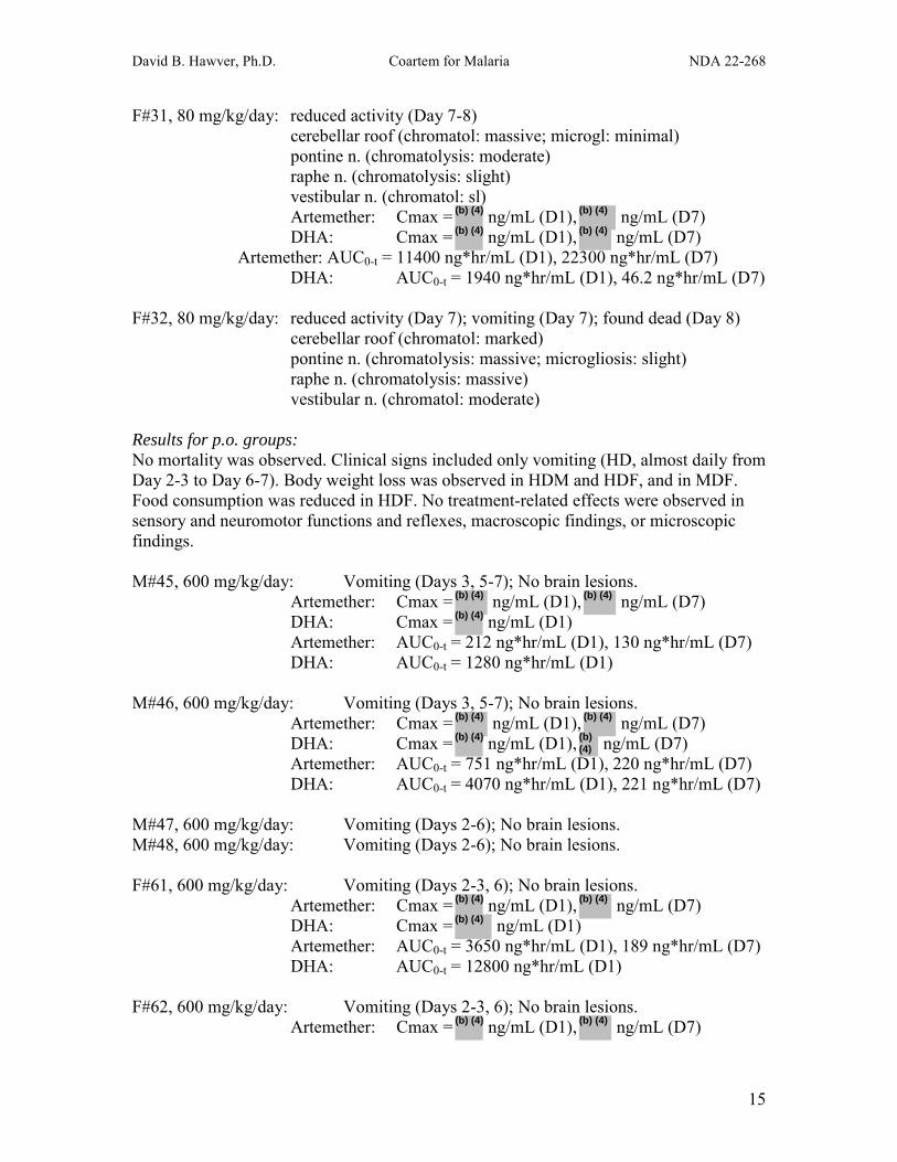
F#31, 80 mg/kg/day: reduced activity (Day 7-8) cerebellar roof (chromatol: massive; microgl: minimal) pontine n. (chromatolysis: moderate) raphe n. (chromatolysis: slight) vestibular n. (chromatol: sl) Artemether: Cmax = ng/mL (D1), ng/mL (D7)
DHA: Cmax = ng/mL (D1), ng/mL (D7) Artemether: AUC0-t = 11400 ng*hr/mL (D1), 22300 ng*hr/mL (D7) DHA: AUC0-t = 1940 ng*hr/mL (D1), 46.2 ng*hr/mL (D7)
F#32, 80 mg/kg/day: reduced activity (Day 7); vomiting (Day 7); found dead (Day 8) cerebellar roof (chromatol: marked)
pontine n. (chromatolysis: massive; microgliosis: slight) raphe n. (chromatolysis: massive) vestibular n. (chromatol: moderate)
Results for p.o. groups: No mortality was observed. Clinical signs included only vomiting (HD, almost daily from Day 2-3 to Day 6-7). Body weight loss was observed in HDM and HDF, and in MDF. Food consumption was reduced in HDF. No treatment-related effects were observed in sensory and neuromotor functions and reflexes, macroscopic findings, or microscopic findings. M#45, 600 mg/kg/day: Vomiting (Days 3, 5-7); No brain lesions.
Artemether: Cmax = ng/mL (D1), ng/mL (D7) DHA: Cmax = ng/mL (D1) Artemether: AUC0-t = 212 ng*hr/mL (D1), 130 ng*hr/mL (D7) DHA: AUC0-t = 1280 ng*hr/mL (D1) M#46, 600 mg/kg/day: Vomiting (Days 3, 5-7); No brain lesions.
Artemether: Cmax = ng/mL (D1), ng/mL (D7) DHA: Cmax = ng/mL (D1), ng/mL (D7) Artemether: AUC0-t = 751 ng*hr/mL (D1), 220 ng*hr/mL (D7) DHA: AUC0-t = 4070 ng*hr/mL (D1), 221 ng*hr/mL (D7) M#47, 600 mg/kg/day: Vomiting (Days 2-6); No brain lesions. M#48, 600 mg/kg/day: Vomiting (Days 2-6); No brain lesions. F#61, 600 mg/kg/day: Vomiting (Days 2-3, 6); No brain lesions.
Artemether: Cmax = ng/mL (D1), ng/mL (D7) DHA: Cmax = ng/mL (D1) Artemether: AUC0-t = 3650 ng*hr/mL (D1), 189 ng*hr/mL (D7) DHA: AUC0-t = 12800 ng*hr/mL (D1) F#62, 600 mg/kg/day: Vomiting (Days 2-3, 6); No brain lesions.
Artemether: Cmax = ng/mL (D1), ng/mL (D7)
(b) (4)
(b) (4)
(b) (4)
(b) (4)
(b) (4)
(b) (4)
(b) (4)
(b) (4)
(b) (4)
(b) (4)
(b) (4)
(b) (4)
(b) (4)
(b) (4)
(b) (4) (b) (4)

David B. Hawver, Ph.D. Coartem for Malaria NDA 22-268
16
DHA: Cmax = ng/mL (D1), ng/mL (D7) Artemether: AUC0-t = 2320 ng*hr/mL (D1), 459 ng*hr/mL (D7) DHA: AUC0-t = 8240 ng*hr/mL (D1), 28.3 ng*hr/mL (D7) F#63, 600 mg/kg/day: Vomiting (Days 2-3, 5-6); No brain lesions. F#64, 600 mg/kg/day: Vomiting (Days 2-3, 5-6); No brain lesions.
(b) (4) (b) (4)
Appears This Way on Original

David B. Hawver, Ph.D. Coartem for Malaria NDA 22-268
17
Pilot Intramuscular Neurotoxicity Study in Dogs with Pharmacokinetics (Study 966141) Artemether (CGP 56696) was administered to Beagle dogs once daily at 20 mg/kg/day i.m. (N=3M/group) for 5 or 30 days or vehicle (peanut oil) for 30 days (N=2M). Assessments included mortality, clinical signs, body weight, food consumption, ophthalmology, ECG, neurology, hematology, clinical biochemistry, urinalysis, organ weights, microscopic and microscopic examinations. PK samples were taken on Days 1, 2, 3, and 4 immediately before dosing, and on Day 4 at 2, 4, 8, 12, and 24 hrs postdose. PK samples were also taken at 2 hrs after the final dose in dogs sacrificed on Day 5. PK samples were also taken immediately before dosing on Days 8, 15, 22, and 29, and at 2, 4, 8, 12, and 24 hrs postdose on Day 29, and at 2 hrs postdose on Day 30 (Day 28 for the dog sacrificed on Day 28). CSF samples were collected from the cisterna magna 2 hrs after the final dose (Day 5 for interim group, Day 30 for main study). The fixation and tissue preparation methods are detailed below:
(page 27-28 of study report)
One dog was sacrificed on Day 28 due to poor health (recumbency, dyspnea, transient episodes of tremor, swaying gait, restless and aggressive behavior), despite discontinuation of treatment on Day 27. Another dog showed clonic-tonic convulsions ending in opisthotonus on Day 30, the day of scheduled sacrifice. Mean food consumption was reduced in weeks 3 and 4, but no changes were observed in ophthalmologic, ECG, or neurologic examinations. Reductions were observed in Week 5 RBCs, hemoglobin, hematocrit, and mean corpuscular volume and hemoglobin, indicating slight hypochromic, microcytic anemia in the treated group. AST and triglycerides were slightly increased in Wk 5, and absolute and relative weights of liver and kidney were slightly increased. Reduced thymus weight correlated with thymic atrophy in 2/3 dogs at 30 days. No drug-related microscopic findings were observed in the group treated for 5 days, except for minimal thymic atrophy in 1/3 dogs. Brain lesions were observed in all 3

David B. Hawver, Ph.D. Coartem for Malaria NDA 22-268
18
animals treated for 27-30 days, characterized by neuronal chromatolysis and microgliosis around damaged cells, occasional degeneration of nerve fibers, and (in the dog sacrificed early), widespread neuronal necrosis in the cerebral cortex and hippocampus. Other findings included hemosiderosis and increased extramedullary hematopoiesis (3/3); deposition of pigment in liver Kupffer cells (2/3, probably hemosiderin); and injection site changes similar to those seen in controls. Artemether concentrations (ng/mL) are listed in the table below. Peak DHA concentrations in plasma did not exceed 21 ng/mL. No AUC values were provided.
(page 14 of PK Study Report F) 1997/004)
Dog #3: Cmax = ng/mL (D4), ng/mL (D29)
Clonic-tonic convulsions, opisthotonus (D30). Neuronal chromatolysis (Grade 2) and Microgliosis (Grade 3) in
cerebellar roof, pontine, and vestibular nuclei. Dog #4: Cmax = ng/mL (D4), ng/mL (D29)
(b) (4)
(b) (4)
(b) (4) (b) (4)
(b) (4) (b) (4)
(b) (4)

David B. Hawver, Ph.D. Coartem for Malaria NDA 22-268
19
Neuronal chromatolysis (Grade 2) and Microgliosis (Grade 3) in cerebellar roof, pontine, cochlear, superior olivary, and vestibular nuclei, and paralemniscal/raphe region.
Dog #5: Cmax = ng/mL (D4)
Whole body tremor, restless (D23); swaying gait, restless, aggressive (D27); recumbency, dyspnea, moribund sacrifice (D28). Cmax = ng/mL (D4) Neuronal chromatolysis (Grade 3) and Microgliosis (Grade 3) in
cerebellar roof, pontine, cochlear, and vestibular nuclei; lateral geniculate body, nucleus ruber substantia nigra, and paralemniscal/raphe region.
Hydrocephalus, Grade 2 Neuronal Necrosis, Grade 4 (hippocampus and cerebral cortex)
Dog #6: Cmax = ng/mL (D4)
No brain lesions. Sacrificed Day 5. Dog #7: Cmax = ng/mL (D4)
No brain lesions. Sacrificed Day 5. Dog #8: Cmax = ng/mL (D4)
No brain lesions. Sacrificed Day 5.
(b) (4)
(b) (4)
(b) (4)
(b) (4)
(b) (4)
Appears This Way on Original

David B. Hawver, Ph.D. Coartem for Malaria NDA 22-268
20
Plasma levels of artemether on Day 4 after 20 mg/kg
0
50
100
150
200
250
300
350
0 5 10 15 20 25
Hours after dosing
Art
emet
her (
ng/m
L)
Dog #3
Dog #4
Dog #5
(Reviewer’s Graph, based on data in table above)
Estimated artemether Day 1 AUC 0-24 hr = ~175 * 24 = ~4200 ng*hr/mL for 30-day group, based on graph above; Day 29 AUC 0-24 hr = ~125 * 24 = ~3000 ng*hr/mL, based on graph below.
Plasma levels of artemether on Day 29 after 20 mg/kg/day
0
50
100
150
200
250
300
0 5 10 15 20 25
Hours after dosing
Art
emet
her (
ng/m
L)
Dog #3
Dog #4
(Reviewer’s Graph, based on data in table above)
(b) (4)
(b) (4)
(b) (4)
(b) (4)
(b) (4)
(b) (4)

David B. Hawver, Ph.D. Coartem for Malaria NDA 22-268
21
Plasma levels of artemether on Day 4 after 20 mg/kg
0
50
100
150
200
250
300
350
0 5 10 15 20 25
Hours after dosing
Art
emet
her (
ng/m
L)
Dog #6
Dog #7
Dog #8
(Reviewer’s Graph, based on data in table above)
Estimated artemether Day 1 AUC 0-24 hr = ~175 * 24 = ~4200 ng*hr/mL for 5-day group, based on graph above
(b) (4)
(b) (4)(b) (4)
Appears This Way on Original

David B. Hawver, Ph.D. Coartem for Malaria NDA 22-268
22
8-Day Exploratory Neurotoxicity Study in Dogs (Study 0410073) Artemether was administered to 2 M dogs at 40 mg/kg/day once daily for 8 days in peanut oil (0.5 mL/kg). Assessments included mortality, clinical signs, body weight, food consumption, neurology, toxicokinetics, necropsy, macroscopic and microscopic observations. Dog #350 was fixed by whole body perfusion, while Dog #349 was fixed by immersion. Histopathology details are given below:
(page 43 of Study Report)
(page 22 of Study Report)
(b) (4)

David B. Hawver, Ph.D. Coartem for Malaria NDA 22-268
23
Degenerative changes (chromatolysis, eosinophilic cytoplasmic granulation, spheroids, apoptosis, and dark neurons) were observed in multiple brain regions in both dogs after eight days of treatment with 40 mg/kg/day i.m. artemether. Perfusion fixation was found to be the preferred fixation method for artemether-induced neurotoxicity, due to fewer artifacts and enhanced ability to use helpful stains. Dog #349: moderate subdued behavior, mild tremor, & mild palpebral closure of eyes
(D8).
(pages 64-65 of Study Report)
Appears This Way on Original

David B. Hawver, Ph.D. Coartem for Malaria NDA 22-268
24
Dog #350: moderate subdued behavior (D8). Progressive body weight loss (↓11%); reduced food consumption (↓30-60%).
(pages 66-67 of Study Report)
Appears This Way on Original

David B. Hawver, Ph.D. Coartem for Malaria NDA 22-268
25
Intramuscular Neurotoxicity Study in Dogs (Study 0510001) Male beagle dogs (3/group) were treated with vehicle (peanut oil, Gr 1) for 8 days; 10 mg/kg/day artemether i.m. for 3 days (Gr 2), 3 days + 6 days recovery (Gr 3), or 8 days (Gr 4); or 40 mg/kg/day artemether i.m. for 3 days (Gr 5), 3 days + 6 days recovery (Gr 6), or 8 days (Gr 7). Assessments included mortality, clinical signs, body weight, food consumption, neurology, audiometric tests (brainstem auditory evoked potentials), toxicokinetics, and macroscopic and microscopic observations. Details of the histopathology evaluation are described below:
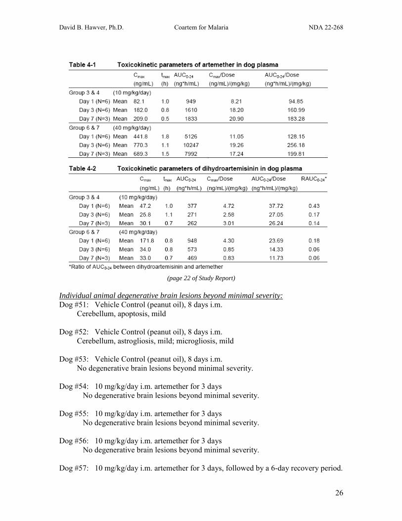
(page 107 of Study Report) Treatment-related changes included vomiting (1 occasion in 1 Gr 7 dog); reduced body weight (Gr 6 & 7), body weight gain (Gr 4-7), and food consumption (Gr 6 & 7); and moderate degeneration (central and/or total chromatolysis, eosinophilic cytoplasmic granulation, and/or neuronal necrosis) in several brain nuclei (Gr 7 only; cochlear, olivary, reticular, vestibular, vestibular, pontine, cuneatus, and cerebellar nuclei, and caudal colliculus). No treatment-related changes were seen in the brainstem auditory evoked potentials, neurology (including acoustic response), or macroscopic evaluation. Neurotoxicity induced by i.m. artemether required sustained treatment at 40 mg/kg/day for 8 days (Artemether AUC0-24 hr = 4252-11426 ng*hr/mL; DHA AUC0-24 hr = 376-885 ng*hr/mL). Degenerative brain lesions above control levels were not observed at 10 mg/kg/day for 8 days (Artemether AUC0-24 hr = 537-2560 ng*hr/mL; DHA AUC0-24 hr = 127-389 ng*hr/mL), or at 40 mg/kg/day for 3 days ± a 6-day recovery period (Artemether AUC0-24 hr = 4575-13492 ng*hr/mL; DHA AUC0-24 hr = 584-1540 ng*hr/mL).
(b) (4)
(b) (4)
(b) (4)
(b) (4)
David B. Hawver, Ph.D. Coartem for Malaria NDA 22-268
26
(page 22 of Study Report)
Individual animal degenerative brain lesions beyond minimal severity: Dog #51: Vehicle Control (peanut oil), 8 days i.m.
Cerebellum, apoptosis, mild
Dog #52: Vehicle Control (peanut oil), 8 days i.m. Cerebellum, astrogliosis, mild; microgliosis, mild
Dog #53: Vehicle Control (peanut oil), 8 days i.m. No degenerative brain lesions beyond minimal severity.
Dog #54: 10 mg/kg/day i.m. artemether for 3 days No degenerative brain lesions beyond minimal severity.
Dog #55: 10 mg/kg/day i.m. artemether for 3 days
No degenerative brain lesions beyond minimal severity.
Dog #56: 10 mg/kg/day i.m. artemether for 3 days No degenerative brain lesions beyond minimal severity.
Dog #57: 10 mg/kg/day i.m. artemether for 3 days, followed by a 6-day recovery period.

David B. Hawver, Ph.D. Coartem for Malaria NDA 22-268
27
Nucleus hypoglossus, neuronal satellitosis, mild Artemether AUC0-24 hr = 892 (D1), 2096 (D3) DHA AUC0-24 hr = 429 (D1), 279 (D3)
Dog #58: 10 mg/kg/day i.m. artemether for 3 days, followed by a 6-day recovery period.
No degenerative brain lesions beyond minimal severity. Artemether AUC0-24 hr = 1359 (D1), 1561 (D3) DHA AUC0-24 hr = 406 (D1), 256 (D3)
Dog #59: 10 mg/kg/day i.m. artemether for 3 days, followed by a 6-day recovery period. Nucleus olivarus, central chromatolyis, mild Artemether AUC0-24 hr = 1313 (D1), 1511 (D3) DHA AUC0-24 hr = 470 (D1), 269 (D3) Dog #60: 10 mg/kg/day i.m. artemether for 8 days.
No degenerative brain lesions beyond minimal severity. Artemether AUC0-24 hr = 585 (D1), 1985 (D3), 2560 ng*hr/mL (D7) DHA AUC0-24 hr = 385 (D1), 382 (D3), 389 ng*hr/mL (D7) Dog #61: 10 mg/kg/day i.m. artemether for 8 days. Nucleus hypoglossus, microgliosis, mild Artemether AUC0-24 hr = 1005 (D1), 987 (D3), 1674 ng*hr/mL (D7) DHA AUC0-24 hr = 336 (D1), 225 (D3), 271 ng*hr/mL (D7) Dog #62: 10 mg/kg/day i.m. artemether for 8 days.
No degenerative brain lesions beyond minimal severity. Artemether AUC0-24 hr = 537 (D1), 1520 (D3), 1264 ng*hr/mL (D7) DHA AUC0-24 hr = 237 (D1), 213 (D3), 127 ng*hr/mL (D7) Dog #63: 40 mg/kg/day i.m. artemether for 3 days.
Nucleus chochlearis, axonal damage, mild; microgliosis, mild. Dog #64: 40 mg/kg/day i.m. artemether for 3 days.
No degenerative brain lesions beyond minimal severity. Dog #65: 40 mg/kg/day i.m. artemether for 3 days. Corpus trapezoideum, microgliosis, mild Nucleus gracilis, microgliosis, mild Cerebellum, microgliosis, mild Artemether AUC0-24 hr = 6723 (D1), 13492 (D3) DHA AUC0-24 hr = 1540 (D1), 770 (D3) Dog #66: 40 mg/kg/day i.m. artemether for 3 days, followed by a 6-day recovery period.
No degenerative brain lesions beyond minimal severity. Artemether AUC0-24 hr = 4575 (D1), 10925 (D3) DHA AUC0-24 hr = 908 (D1), 584 (D3)

David B. Hawver, Ph.D. Coartem for Malaria NDA 22-268
28
Dog #67: 40 mg/kg/day i.m. artemether for 3 days, followed by a 6-day recovery period. Cerebellum, apoptosis, mild Artemether AUC0-24 hr = 4694 (D1), 8404 (D3) DHA AUC0-24 hr = 989 (D1), 608 (D3) Dog #68: 40 mg/kg/day i.m. artemether for 3 days, followed by a 6-day recovery period. Nucleus olivarus, central chromatolyis, mild Artemether AUC0-24 hr = 6723 (D1), 13492 (D3) DHA AUC0-24 hr = 1540 (D1), 770 (D3) Dog #69: 40 mg/kg/day i.m. artemether for 8 days.
Nucleus chochlearis, microgliosis, mild Colliculus caudalis, central chromatolysis, mild; total chromatolysis, moderate Pontine nuclei, central and total chromatolysis, moderate; microgliosis, mild Central grey of pons, microgliosis, mild Nucleus nervi trigemini, total chromatolysis, mild; apoptosis, mild Cerebellar nuclei, central and total chromatolysis, moderate
Nucleus vestibularis, central chromatolysis, mild; total chromatolysis, marked; necrosis of neurons, moderate; microgliosis, moderate
Nucleus hypoglossus, central chromatolysis, mild; total chromatolysis, moderate Nucleus cuneatus, central chromatolysis, moderate; total chromatolysis, marked Nucleus olivaris, central and total chromatolysis, mild; microgliosis, mild Cerebellum, microgliosis, moderate Remaining white, axonal damage, mild; microgliosis, mild; apoptosis, mild Remaining grey, total chromatolysis, mild
Artemether AUC0-24 hr = 5277 (D1), 9690 (D3), 8889 ng*hr/mL (D7) DHA AUC0-24 hr = 649 (D1), 493 (D3), 435 ng*hr/mL (D7)
Dog #70: 40 mg/kg/day i.m. artemether for 8 days.
Nucleus chochlearis, central chromatolysis, mild Pontine nuclei, central and total chromatolysis, mild Nucleus vestibularis, central and total chromatolysis, mild Nucleus cuneatus, central chromatolysis, mild Nucleus vagus, central chromatolysis, mild Cerebellum, apoptosis, mild
Artemether AUC0-24 hr = 4252 (D1), 7546 (D3), 6696 ng*hr/mL (D7) DHA AUC0-24 hr = 885 (D1), 459 (D3), 376 ng*hr/mL (D7) Dog #71: 40 mg/kg/day i.m. artemether for 8 days.
Cortex temporalis, astrogliosis, mild Pontine nuclei, central and total chromatolysis, mild Nucleus vestibularis, central chromatolysis, mild; total chromatolysis, moderate Nucleus hypoglossus, central chromatolysis, mild Nucleus cuneatus, total chromatolysis, moderate Nucleus olivaris, total chromatolysis, mild

David B. Hawver, Ph.D. Coartem for Malaria NDA 22-268
29
Remaining grey, total chromatolysis, mild Artemether AUC0-24 hr = 5235 (D1), 11426 (D3), 8391 ng*hr/mL (D7) DHA AUC0-24 hr = 715 (D1), 524 (D3), 596 ng*hr/mL (D7) Summary of Individual Dog Data Degenerative lesions of minimal severity were observed in multiple brain regions of all groups, including vehicle controls. Degenerative lesions of mild severity were observed in only one brain region in 2/3 controls, 0/3 at 10 mg/kg/day for 3 days, 2/3 at 10 mg/kg/day for 3 days + 6-day recovery period, 1/3 at 10 mg/kg/day for 8 days, 1/3 at 40 mg/kg/day for 3 days (another dog in this group had mild lesions in 3 brain regions), and 2/3 at 40 mg/kg/day for 3 days + 6-day recovery period. In contrast, treatment at 40 mg/kg/day for 8 days resulted in mild to marked degenerative lesions in 6, 7, or 13 brain regions in the three dogs of this group.
Appears This Way on Original

David B. Hawver, Ph.D. Coartem for Malaria NDA 22-268
30
(page 210 of Study Report)

David B. Hawver, Ph.D. Coartem for Malaria NDA 22-268
31
(page 207 of Study Report)
Appears This Way on Original

David B. Hawver, Ph.D. Coartem for Malaria NDA 22-268
32
(page 221 of Study Report)
(b) (4)
(b) (4)
(b) (4)
(b) (4)
(b) (4)
(b) (4)
(b) (4)

David B. Hawver, Ph.D. Coartem for Malaria NDA 22-268
33
CGP 56696: 13-week oral (capsule) toxicity study in dogs (Study 95-6030) Artemether was administered once daily via oral capsule to beagle dogs (3/sex/group + 3/sex Con & HD 4-week recovery) at 0, 30, 100, or 300 mg/kg/day for at least 13 weeks. Treatment-related changes included RBC parameters (HD; minimal decreases in HGB, MCV, MCHC, and increases in reticulocytes); increased circulating nucleated RBCs in peripheral blood smears (HD) and slight to moderate RBC morphology changes (all doses); increased erythropoiesis in bone marrow smears (HD), associated with corresponding increases in hematopoiesis in bone marrow sections and increased extramedullary hematopoiesis in spleen (HD); and thyroid follicular cell hypertrophy (HD). RBC changes were improved, but still abnormal after 4 weeks of recovery. Thyroid, bone marrow, and spleen changes showed recovery. Body weight parameters were decreased in HD dogs (2-4% BW loss in 3/6 HDF). No treatment-related changes were observed in mortality, clinical signs, physical/auditory findings, food consumption, ECG, ophthalmology, or gross pathology. The MD of 100 mg/kg/day was considered the NOAEL, inducing only slight changes in RBC morphology. TK samples were not analyzed.
Appears This Way on Original

David B. Hawver, Ph.D. Coartem for Malaria NDA 22-268
34
CGP 56697: 3-month oral toxicity study in dogs (Study 94-6155) Beagle dogs (3/sex/group) were administered coartem (1 part artemether = 6 parts lumefantrine) at 0, 20, 60, or 200 mg/kg/day (0, 2.9, 8.6, or 29 mg/kg/day artemether) via oral capsule once daily for 13 weeks; a parallel group at 200 mg/kg/day was allowed to recover for 4 weeks prior to sacrifice. Treatment-related changes included diarrhea and pale feces (HD); increased TSH (LD, MD, HD); decreased T4, T3, and rT3 (MD, HD); increased cholesterol (MD, HD); increased weight of thyroid (LDF, MD, HD) and liver (MDM, HD); thyroid follicular cell hypertrophy (LD-HD, reversible); and liver cholangetis (HD; reversible). No changes were observed in mortality, body weight, food consumption, ophthalmology, ECG, neurology, or macroscopic findings. The levels of artemether and DHA were below the level of quantification in all samples from all dogs at all doses on Days 1 and 89, except for one sample from one HDF dog showing 14.0 ng/mL for artemether on Day 1, and another sample from another dog showing 16.7 ng/mL for DHA on Day 1.
Appears This Way on Original

David B. Hawver, Ph.D. Coartem for Malaria NDA 22-268
35
CGP 56697: 1-month oral toxicity study in dogs (Study 94-6154) Beagle dogs (3/sex/group) were administered co-artem (1 part artemether = 6 parts lumefantrine) at 0, 60, 200, or 600 mg/kg/day (0, 8.6, 29, or 86 mg/kg/day artemether) via oral capsule once daily for 28 days; a parallel group at 600 mg/kg/day was allowed to recover for 28 days prior to sacrifice. Treatment-related changes included diarrhea and pale feces (MD, HD); increased AP (slight to moderate, MD, HD, reversible); increased liver weight (MD, HD); thyroid follicular cell hypertrophy (LD-HD, reversible); and focal cellular hypertrophy in pars distalis of pituitary (MD). No changes were observed in mortality, body weight, food consumption, ophthalmology, ECG, neurology, or macroscopic findings. Toxicokinetic data were not provided.
Appears This Way on Original

David B. Hawver, Ph.D. Coartem for Malaria NDA 22-268
36
Summary of Oral Toxicity Studies in Dog In a 3- to 8-day oral gavage neurotoxicity study in dogs (Study 051009), the HD of 600 mg/kg/day artemether was reduced to 300 mg/kg/day after the first dose, due to severe clinical signs in 2/9 dogs (staggered gait and head tremor in one, AUC0-24 hr = 30467 ng*hr/mL artemether, 19584 ng*hr/mL DHA; recumbency, cramp, and body/head tremor in the other, AUC0-24 hr = 36174 ng*hr/mL artemether, 14931 ng*hr/mL DHA). Vomiting on Day 1 was moderate in 4/9, and mild in 4/9. The two dogs with severe signs on Day 1 had the highest artemether AUCs of the six dogs for which TK data was collected; values for the others ranged from 6075-28658 ng*hr/mL artemether. Plasma exposures to artemether and DHA decreased dramatically by Day 3, much more than explained by the two-fold reduction in dose from 600 to 300 mg/kg/day: Day 3 AUC0-24 hr = 88-1169 ng*hr/mL artemether, 99-3135 ng*hr/mL DHA. By Day 7 at 300 mg/kg/day, exposures were reduced even further: Day 7 AUC0-24 hr 46-101 ng*hr/mL artemether, 140-236 ng*hr/mL DHA. Importantly, 3 of the 6 dogs for which Day 3 TK data was available showed plasma exposures near or greater than those expected in humans given coartem for 3 days at the recommended clinical dose (80 mg artemether/480 mg lumefantrine twice daily): Human Max Mean AUC0-24 hr = 1070 ng*hr/mL artemether, 1208 ng*hr/mL DHA; Dogs with highest exposures AUC0-24 hr = 1169, 851, & 936 ng*hr/mL artemether, and 3135, 1540, & 2936 ng*hr/mL DHA, respectively. The lack of brain lesions in these three dogs provides some evidence that the level of plasma exposure expected in humans is not likely to be neurotoxic when limited to 3 days, though it would be prudent to increase the size of the sample before drawing definitive conclusions. In an oral capsule neurotoxicity study in dogs (Study 970024), the HD of 600 mg/kg/day artemether was tolerated for 8 days, with only vomiting and reductions in body weight and food consumption. However, the plasma exposures observed in this study were much lower than those in the previous study: Day 1 AUC0-t = 212-3650 ng*hr/mL artemether, 1280-12800 ng*hr/mL DHA. As in the other oral study, repeated dosing resulted in much lower exposures: Day 7 AUC0-t = 130-459 ng*hr/mL artemether, 28-221 ng*hr/mL DHA In a 13-week oral capsule toxicity study in dogs (Study 95-6030), the HD of 300 mg/kg/day artemether was tolerated with no evidence of neurotoxicity; treatment-related changes included BW loss of 2-4% in 3/6 HDF, RBC changes in blood and bone marrow, and thyroid follicular cell hypertrophy. TK samples were not analyzed. In a 13-week oral capsule toxicity study in dogs (Study 94-6155), the HD of 200 mg/kg/day coartem contained only 29 mg/kg/day artemether, and plasma levels of artemether and DHA were below detectable levels in almost all samples. No treatment-related neurotoxicity was reported. In a 1-month oral capsule toxicity study in dogs (Study 94-6154), the HD of 600 mg/kg/day coartem contained only 86 mg/kg/day artemether. TK data were not provided. No treatment-related neurotoxicity was reported.

David B. Hawver, Ph.D. Coartem for Malaria NDA 22-268
37
Summary of Intramuscular Neurotoxicity Studies in Dog Dogs treated with 20 mg/kg/day artemether i.m. for 8 days (Study 970024) showed minimal to slight lesions (chromatolysis and/or microgliosis in 5/8 dogs) in several brain regions (cerebellar roof, pontine nucleus, vestibular nucleus, and/or raphe nucleus) associated with Day 1 AUC0-t = 1340 & 2829 ng*hr/mL artemether, and 369 & 217 ng*hr/mL DHA, and Day 7 AUC0-t = 5400 & 5920 ng*hr/mL artemether, and 385 & 274 ng*hr/mL DHA (only 2 of the 5 dogs with lesions had TK data). Similar exposures were observed in dogs at the same dose that did not show brain lesions: Day 1 AUC0-t = 916 & 3120 ng*hr/mL artemether, and 145 & 523 ng*hr/mL DHA, and Day 7 AUC0-t = 4120 & 5940 ng*hr/mL artemether, and 126 & 441 ng*hr/mL DHA (only 2 of the 3 dogs without lesions had TK data). Note that repeated i.m. dosing resulted in substantially increased artemether plasma exposures, in contrast to repeated oral dosing, which resulted in dramatically reduced exposure to both artemether and DHA. Also note that DHA plasma exposures after i.m. dosing were much lower than those after oral dosing. Dosing at 40 and 80 mg/kg/day resulted in increased incidence and severity of brain lesions. Dogs treated with 20 mg/kg/day artemether i.m. for 27-30 days (Study 966141) showed mild to moderate brain lesions in several brain regions (3/3 dogs), while those treated at the same dose for only 5 days showed no lesions (3/3 dogs). Artemether AUC values were not provided, but estimates based on graphical analysis are ~4200 ng*hr/mL on Day 1 and ~3000 ng*hr/mL on Day 29. DHA levels were ≤ 21 ng/mL. Dogs (N=2M) treated with 40 mg/kg/day artemether i.m. for 8 days (Study 0410073) showed degenerative lesions in several brain regions associated with artemether AUC0-24
hr = 6780 & 3639 (D1), 12051 & 5777 (D3), 10651 & 5097 (D7) ng*hr/mL, and DHA AUC0-24 hr = 753 & 980 (D1), 481 & 432 (D3), 480 & 378 (D7) ng*hr/mL. Dogs (N=3M/group) treated with 40 mg/kg/day artemether i.m. for 8 days (Study 0510001) showed degenerative brain lesions above control levels, associated with artemether AUC0-24 hr = 4252-11426 ng*hr/mL and DHA AUC0-24 hr = 376-885 ng*hr/mL (ranges reflect lowest to highest individual values on Days 1, 3, or 7 among all 3 dogs). Degenerative brain lesions above control levels were not observed at 10 mg/kg/day for 8 days (Artemether AUC0-24 hr = 537-2560 ng*hr/mL; DHA AUC0-24 hr = 127-389 ng*hr/mL), or at 40 mg/kg/day for 3 days ± a 6-day recovery period (Artemether AUC0-24
hr = 4575-13492 ng*hr/mL; DHA AUC0-24 hr = 584-1540 ng*hr/mL).
Appears This Way on Original

David B. Hawver, Ph.D. Coartem for Malaria NDA 22-268
38
Reviewer’s Summary Table
Species
Artemether Dose
Duration
Artemether AUC0-24 hr ng*hr/mL
DHA
AUC 0-24 hr
Brain Lesion
Study
Number
Human 80 mg BID p.o.
3 Days 1070 1208 ? A028
Dog 10 mg/kg/day i.m.
8 Days 537-2560 127-389 No 0510001
Dog 20 mg/kg/day i.m.
8 Days 1340-5920 217-385 Yes 970024
Dog 20 mg/kg/day i.m.
27-30 Days
3000-4200 ? (low) Yes 966141
Dog 20 mg/kg/day i.m.
5 Days 4200 ? (low) No 966141
Dog 40 mg/kg/day i.m.
8 Days 3639-12051 378-980 Yes 0410073
Dog 40 mg/kg/day i.m.
8 Days 4252-11426 376-885 Yes 0510001
Dog 40 mg/kg/day i.m.
3 Days (± 6 day
recovery)
4575-13492 584-1540 No 0510001
Dog 600/300 mg/kg/day oral gavage
3-8 Days 6075-36174 D1 88-1169 D3 46-101 D7
1690-3220 D1 63-1470 D3 104-191 D7
No 0510009
Dog 600 mg/kg/day
oral capsule
8 Days 212-3650 D1 130-459 D7
1280-12800 D1 28-221 D7
No 970024
Dog 300 mg/kg/day
oral capsule
13 Weeks
No TK
No TK
No
95-6030
Dog 29 mg/kg/day (in coartem) oral capsule
13 Weeks
Below Detectable
Levels
Below Detectable
Levels
No
94-6155
Dog 86 mg/kg/day (in coartem) oral capsule
1 Month
No TK
No TK
No
94-6154

David B. Hawver, Ph.D. Coartem for Malaria NDA 22-268
39
Key Points • The mechanism of action of the neurotoxicity induced by artemether and DHA is not
clear; therefore, it possible that other metabolites whose concentrations have not been measured may contribute to the toxicity.
• Recent publications (Genovese and Newman, 2008, Arch Toxicol 82:379-385; Gordi and Lepist, 2004, Toxicology Letters 147:99-107) have reported that neurotoxicity induced by artemesinins correlates better with the level of sustained plasma exposures to the parent drug and its active metabolite (DHA) over several days rather than with the maximal exposures.
• The possibility that cotreatment with lumefantrine may reduce the level or duration of plasma exposure of artemether/DHA needed to induce brain lesions in dogs has not been evaluated.
• Repeated intramuscular administration of artemether to dogs has allowed evaluation of artemether plasma exposures several-fold above those expected in humans given the recommended dosage of coartem, but the DHA plasma exposures in these studies have generally been lower than those expected in humans.
• Repeated oral administration of artemether to dogs results in rapid dramatic reduction in plasma exposure to artemether and DHA due to induction of metabolic enzymes; by 7 days of dosing at 300 or 600 mg/kg/day, exposures to artemether and DHA were much lower than those expected in humans given the recommended dosage of coartem.
• The possibility that artemether and DHA plasma exposures could be maintained near or above those expected in humans by increasing the oral dose given to dogs each day to compensate for the induction of metabolic enzymes has not been explored.
• Treatment of dogs with artemether i.m. at 20, 40, or 80 mg/kg/day (but NOT 10 mg/kg/day, N=3M) for 8 days resulted in degenerative lesions in several brain regions.
• The lowest artemether exposure (AUC0-24 hr) associated with brain lesions was observed in dogs treated for 8 days at 20 mg/kg/day i.m., and ranged from 1340-5920 ng*hr/mL, increasing over the 8 days of treatment. The NOEL for brain lesions in dogs (10 mg/kg/day i.m. for 8 days) resulted in exposures ranging from 537-2560 ng*hr/mL. Estimated human plasma exposure to artemether at the maximum recommended dose of 80 mg BID p.o. is 1070 ng*hr/mL.
• Treatment of dogs with artemether i.m. at 20 mg/kg/day for 5 days did NOT result in brain lesions (N=3M).
• Treatment of dogs with artemether i.m. at 40 mg/kg/day for 3 days (± 6 day recovery) did NOT result in brain lesions (N=3M/group).
• Artemether-induced brain lesions were NOT correlated with treatment-related changes in neurology evaluations (3 i.m. studies in dog; 1 i.m. study in rat) or audiometric evaluations (including Brainstem Auditory Evoked Potentials; 1 i.m. study in dog).

---------------------------------------------------------------------------------------------------------------------This is a representation of an electronic record that was signed electronically andthis page is the manifestation of the electronic signature.--------------------------------------------------------------------------------------------------------------------- /s/---------------------David Hawver12/19/2008 10:59:30 AMPHARMACOLOGIST
Lois Freed12/19/2008 06:05:01 PMPHARMACOLOGISTI believe the available nonclinical data are sufficient to demonstrate that coartem has neurotoxic potential at clinically relevant exposures. For comments on additional nonclinical studies, please refer to the Memorandum by Dr. Eric Bastings (12/19/08).

Comments on NDA 22-268 Coartem: artemether benflumetol From A. Jacobs 12/15/08 1. I think that there are no pharm/tox issues that would affect approval and I concur that the pregnancy category should be a “C”. 2. It’s not clear to me that there are unresolved toxicology issues a. Acceptance criteria for several components: I could find no discussion of this in the review itself, other than in the conclusions. I don’t know what the levels of the impurities were in the nonclinical and clinical batches nor the basis for the selection of proposed impurity levels. However, if several-fold multiples of the human exposure were used in nonclinical studies, the impurities levels could be covered. More importantly, since the active components are themselves rather toxic, it’s hard to envision that these impurities are more toxic. b. Since brain hemorrhages have been found in several animal studies, I don’t understand what is not resolved. 3. I don’t concur with the recommendations for any future studies: a. I don’t understand the need for any neurotox study in newborn dogs. (I noted that there were peripostnatal studies in rats, including behavioral studies). Coartemether is expected to be neurotoxic, it is noted in the proposed labeling, and generally neonates are more sensitive such effects than adults. I noted that more than 1000 pregnant women have been exposed. b. I don’t understand the need for any neurotox studies after intramuscular administration. The route of administration clinically is oral, with extensive metabolisms, and the potential for neurotox has been demonstrated. c. It’s not clear to me that any further testing of “potentially” genotoxic intermediates, process impurities, or degradants is needed. 4. I have discussed these issues with the pharm/tox team leader, and he will address these issues as he considers appropriate.

---------------------------------------------------------------------------------------------------------------------This is a representation of an electronic record that was signed electronically andthis page is the manifestation of the electronic signature.--------------------------------------------------------------------------------------------------------------------- /s/---------------------Abby Jacobs12/15/2008 11:37:48 AMPHARMACOLOGIST

DEPARTMENT OF HEALTH AND HUMAN SERVICES PUBLIC HEALTH SERVICE
FOOD AND DRUG ADMINISTRATION CENTER FOR DRUG EVALUATION AND RESEARCH
PHARMACOLOGY/TOXICOLOGY REVIEW AND EVALUATION
NDA NUMBER: 22-268
SERIAL NUMBER: 000
DATE RECEIVED BY THE CENTER: November 15, 2007 PRODUCT: Coartem®
INTENDED CLINICAL POPULATION: Patients of 5 kg bodyweight and above with acute, uncomplicated infections due to Plasmodium falciparum or mixed infections including P. falciparum.
APPLICANT: Novartis Pharmaceuticals Corporation One Health Plaza East Hanover, NJ 07936-1080 DOCUMENTS REVIEWED: Nonclinical Pharmacology, Toxicology and Pharmacokinetics data.
REVIEW DIVISION: Division of Special Pathogen and Transplant
Products (HFD-590)
PHARM/TOX REVIEWERS: Owen McMaster, Ph.D.
Rama Dwivedi, Ph.D.
Stephen Hundley, Ph. D.
Terry Miller, Ph.D.
PHARM/TOX SUPERVISOR: William Taylor, Ph.D.
DIVISION DIRECTOR: Renata Albrecht, M.D.
PROJECT MANAGER: Gregory DiBernardo
Date of review submission to Division File System (DFS): November 25, 2008

2
Drug Terminology and Usage in this Review _
Clinical Term Nonclinical Terms lumefantrine benflumetol CGP 56695 artemether artemether CGP 56696 Coartem coartemether CGP 56697
Each clinical Coartem® tablet includes 20 mg artemether and 120 mg lumefantrine; (therefore, in a ratio of 1:6). In the nonclinical studies, “coartemether” was used to designate the combination of the same active substances as in the clinical product, and in the same ratio, when both were administered together. However, as is generally true, the vehicle and/or excipients administered with the nonclinical formulation differ from the clinical formulation and the proportion of each active ingredient sometimes varied slightly from the clinical. Some nonclinical studies were conducted with the individual active components and some used the combination (“coartemether”).
Appears This Way on Original

3
TABLE OF CONTENTS
EXECUTIVE SUMMARY .............................................................................................. 4
2.6 PHARMACOLOGY/TOXICOLOGY REVIEW................................................... 8
2.6.1 INTRODUCTION AND DRUG HISTORY................................................................... 8
2.6.2 PHARMACOLOGY....................................................................................................... 10 2.6.2.1 Brief summary ........................................................................................................................ 9 2.6.2.2 Primary pharmacodynamics ................................................................................................. 10 2.6.2.3 Secondary pharmacodynamics ............................................................................................. 10 2.6.2.4 Safety pharmacology ............................................................................................................ 11 2.6.2.5 Pharmacodynamic drug interactions..................................................................................... 25
2.6.4 PHARMACOKINETICS/TOXICOKINETICS .......................................................... 26 2.6.4.1 Brief summary ...................................................................................................................... 26 2.6.4.2 Methods of Analysis ............................................................................................................. 27 2.6.4.3 Absorption ............................................................................................................................ 27 2.6.4.4 Distribution........................................................................................................................... 58 2.6.4.5 Metabolism ........................................................................................................................... 71 2.6.4.6 Excretion............................................................................................................................... 77 2.6.4.7 Pharmacokinetic drug interactions........................................................................................ 77 2.6.4.8 Other Pharmacokinetic Studies............................................................................................. 77 2.6.4.9 Discussion and Conclusions ................................................................................................. 88
2.6.6 TOXICOLOGY............................................................................................................... 89 2.6.6.1 Overall toxicology summary ................................................................................................ 89 2.6.6.2 Single-dose toxicity .............................................................................................................. 88 2.6.6.3 Repeat-dose toxicity ........................................................................................................... 100 2.6.6.4 Genetic toxicology.............................................................................................................. 146 2.6.6.5 Carcinogenicity................................................................................................................... 186 2.6.6.6 Reproductive and developmental toxicology...................................................................... 186 2.6.6.7 Local tolerance ................................................................................................................... 254 2.6.6.8 Special toxicology studies .................................................................................................. 254 2.6.6.9 Discussion and Conclusions .....................................................................................................
OVERALL CONCLUSIONS AND RECOMMENDATIONS............................................. 244
APPENDIX/ATTACHMENTS ............................................................................................... 248

NDA 22-268 COARTEM _______________________________________________________________________
4
EXECUTIVE SUMMARY
I. Recommendations
A. Recommendation on approvability There are no nonclinical pharmacology or toxicology data that preclude the approval of Coartem. B. Recommendations for nonclinical studies (1) The applicant should conduct a study of oral coartemether in the newborn dog. Dogs should be dosed for both three days and two weeks. Animals should be evaluated at the end of dosing for behavioral and histopathological effects and after a two-week postdose period to determine the reversibility of any findings. Pharmacokinetics (including AUC’s) of both artemether and benflumetol should be followed. A complete draft protocol should be submitted to the Review Division for comments. This study will provide information that was not available from a flawed study in juvenile rats. (2) The sponsor should conduct a neurotoxicity study of intramuscular artemether in beagle dogs that includes post-dosing recovery groups. This study would assess the extent to which neurologic deterioration may occur in the week or weeks following the terminal dose. The post-dosing neurological assessment study may be conducted with either sex. The i.m dosing routines should include a zero-level vehicle control and at least two distinct artemether dose levels that resulted in brain histopathology after 8 days of dosing. The clinical neurologic function tests and brain regions for histopathologic examination should be consistent with the neurotoxicity studies with beagle dogs that were submitted to the NDA. Clinical evaluations of neurologic function should be conducted the day prior to the terminal dose on Day 7, one week after the Day 8 terminal dose, and two weeks after the terminal dose. A complete draft protocol should be submitted to the Review Division for comments. C. Recommendations on labeling Please see the ‘Recommended labeling’ section at the end of this review under the section entitled ‘Overall conclusions and recommendations’.
Appears This Way on Original

NDA 22-268 COARTEM _______________________________________________________________________
5
II. Summary of nonclinical findings
A. Brief overview of nonclinical findings In studies designed to investigate the neurotoxic effects of coartemether, oral dosing with artemether or coartemether at very high multiples of the clinical dose resulted in effects on the auditory system in dogs as well as tremors, staggered gait and recumbency at high exposures. Dosing over thirty days by the intramuscular route resulted in brain lesions, seizures and death. Brain lesions were prominent in the pontine nuclei, cerebellar nuclei, nucleus vestibularis, nucleus hypoglossus, and nucleus cuneatus and included chromatolysis, microgliosis, neuronal necrosis, axonal swelling, neurofilament clumping, eosinophilic cytoplasmic granulation, and spheroids. In vitro binding studies of neurotransmitter receptors have shown that artemether’s interaction with the kainate receptor was among the strongest interactions seen (besides the µ-type opiate receptor). Kainate receptors, are thought to be involved in several pathological processes such as epilepsy and excitotoxic neurodegeneration and could play a role in the brain lesions observed in dogs after intramuscular dosing with artemether. The applicant has shown that artemether-related radioactivity was rapidly taken up into the rat brain after intravenous dosing. It is not clear if these neurotoxic findings with intramuscular artemether are relevant to the oral use of Coartem in patients since the pharmacokinetics of artemether after intramuscular injection is significantly different than after oral administration in the dog. Specifically, repeated oral dosing results in reduced exposure to artemether (presumably due to auto-induction of metabolism) while repeated intramuscular dosing resulted in sustained higher blood levels. No brain lesions were observed in dogs exposed via intramuscular injections to artemether at levels five times clinical exposures (based on AUC comparisons) for up to eight days. In the neurotoxicity studies conducted in dogs and rats, neurological function and/or histology were evaluated during or immediately following the end of dosing. The data do not allow us to determine if neuropathologies develop after dosing ends; therefore, we are requesting a nonclinical post-marketing study to examine the potential for worsening of neurologic effects after dosing ends. In an oral juvenile development study in rats dosed between postpartum days 7 and 21, artemether was associated with increased mortality and brain hemorrhages at 100 mg/kg. Data from this study could not be properly evaluated due to excessive clotting of blood samples, limited histological evaluation of the tissues and poor tissue fixation. Specifically, the data are inadequate to address an important question: Are neonates more susceptible to adverse neurologic effects

NDA 22-268 COARTEM _______________________________________________________________________
6
than older animals? The question has clinical relevance because the indicated clinical population is as low as 5 kg. Additionally, differences in neonatal metabolism or permeability of the blood brain barrier suggest neonates may be more susceptible to neurotoxicity. Therefore, we are requesting a nonclinical safety study to re-examine neonatal neurotoxicity. Toxic effects observed in rats or dogs dosed with coartemether for one or three months were seen in the thyroids, liver, erythrocytes, lymph nodes, kidneys, spleen and brain. A NOAEL was not determined for any of the repeat-dose studies and this was most often due to effects on thyroid function, including increased TSH, decreased T4 or T3, increased thyroid weight and increased thyroid hyperplasia. Other effects seen at the lowest doses tested included hemosiderosis, vacuolation of the pituitary, hyaline droplets in the kidneys and lymph node histiocytosis. Higher doses resulted in enlarged lymph nodes, hypercellularity of the bone marrow, increased spleen weights, increased liver weights, increased kidney weights, increased urea, increased cholesterol, decreased triglycerides, increased α-1-globulin, ß-2-globulin, increased alkaline phosphatase (dogs) decreased alkaline phosphatase (rats) and increases or decreases in glucose levels. It is not clear that these findings indicate potential risks for patients since animals in these studies were dosed for ten to thirty times the recommended clinical duration. Pregnant rats or rabbits dosed with artemether/ benflumetol experienced abortions, preimplantation losses, postimplantation losses and fetal resorptions. Pregnancy rates were reduced by about one half in female rats at about 9 times the clinical dose and male rats dosed for 70 days showed increases in abnormal sperm, increased testes weights, decreased sperm motility. There was no evidence of mutagenicity detected using the Salmonella and Escherichia/mammalian-microsome mutagenicity test, the gene mutation test with Chinese hamster cells V79, the cytogenetic test on Chinese hamster cells in vitro, and the rat micronucleus test, in vivo. Due to the short duration of malaria treatment, carcinogenicity studies were not required. The applicant evaluated the potential of benflumetol to induce QT prolongation. Benflumetol inhibited the IKr tail current in a concentration-dependent manner, but was a weaker inhibitor than comparator antimalarials. The calculated IC50 was approximately 8.1 µM for benflumetol compared to approximately 2.7 µM for chloroquine and mefloquine. B. Pharmacologic activity Artemether and its active metabolite dihydroartemesinin (DHA) both have endoperoxide moieties and are thought to exert their antimalarial activities via the

NDA 22-268 COARTEM _______________________________________________________________________
7
generation of free radicals. The mechanism of the antimalarial effect of lumefantrine is not clear but has been attributed to its interaction with hemin. Artemether and benflumetol are also both known to inhibit nucleic acid and protein synthesis. C. Nonclinical safety issues relevant to clinical use The main safety concerns arising from the nonclinical studies are (1) The potential for neurotoxic effects, (2) Reproductive toxicity, including reduced pregnancy rates, abortions,
preimplantation losses, postimplantation losses, fetal resorptions and abnormal sperms.
(3) Recommendations regarding the testing of potentially genotoxic intermediates, process impurities or degradants will be reflected in an addendum to this review.
Appears This Way on Original

NDA 22-268 COARTEM _______________________________________________________________________
8
2.6 PHARMACOLOGY/TOXICOLOGY REVIEW
2.6.1 INTRODUCTION AND DRUG HISTORY NDA number: 22-268 Review number: 1 Sequence number: 000 Date: November 15, 2007 Type of submission: Original NDA Information to sponsor: Yes Applicant: Novartis Pharmaceuticals Corporation One Health Plaza East Hanover, NJ 07936-1080 Reviewer name: Owen McMaster, Ph.D. Stephen Hundley, Ph. D
Rama Dwivedi, Ph.D. Terry Miller, Ph.D.
Division name: Division of Special Pathogen and Transplant Products (HFD-590)
Review completion date: November 16, 2008 Drug: (artemether/benflumetol) Trade name: Coartem ® Generic name: artemether/benflumetol (benflumetol) Code name (Coartem): CGP 56 697 Chemical name (artemether): (3R,5aS,6R,8aS,9R,10S,12R,12aR)-
decahydro-10-methoxy-3,6,9-trimethyl-3,12-epoxy-12H-pyrano[4,3-j]-1,2-benzodioxepin
CAS registry number (artemether): 71963-77-4

NDA 22-268 COARTEM _______________________________________________________________________
9
Chemical name (benflumetol): (±)-2-dibutylamino-1-[2,7-dichloro-9-(4-
chlorobenzylidene)-9H-fluoren-4-yl]ethanol Empirical formula: C30H32Cll3NO Molecular weight: 528.9 CAS registry number (benflumetol): 82186-77-4 Structure:
Relevant IND’s 75,287 Relevant NDAs None Relevant DMF’s: None Drug class: Antimalarial Intended clinical population: Patients of 5 kg bodyweight and above with acute, uncomplicated infections due to Plasmodium falciparum or mixed infections including P. falciparum. Clinical formulation: Each Coartem tablet contains 20 mg of artemether and 120 mg benflumetol. The inactive ingredients are colloidal silicon dioxide, croscarmellose sodium, hypromellose, magnesium stearate, microcrystalline cellulose, and polysorbate 80.

NDA 22-268 COARTEM _______________________________________________________________________
10
Route of administration: Oral Disclaimer: Tabular and graphical information are constructed by the reviewer unless cited otherwise. Studies reviewed within this submission: Nonclinical studies pertaining to oral dosing of the artemether/ benflumetol combination were reviewed. Please see Appendix A for a complete list. Studies not reviewed within this submission: Studies which pertained to other disciplines such as Microbiology and Clinical Pharmacology were not reviewed.
2.6.2 PHARMACOLOGY 2.6.2.1 Brief summary The majority of the pharmacology studies were reviewed by the microbiologist. The pharmacology studies reviewed below describe artemether/benflumetol interactions with excitotoxic neurotransmitter receptors, the neurotoxic effects and safety pharmacology studies. 2.6.2.2 Primary pharmacodynamics Please see MICROBIOLOGIST’S review.
Appears This Way on Original

NDA 22-268 COARTEM _______________________________________________________________________
11
2.6.2.3 Secondary pharmacodynamics [1.] In Report BS 102, 1996, artemether (CGP 56696) and benflumetol (CGP 56695) were evaluated for their interactions with different neurotransmitter receptors in in vitro receptor binding assays. Results
Although the binding was classified as weak, artemether interaction with the kainate receptor was among the strongest interactions seen (besides its interactions with the µ-type opiate receptor). The kainate receptor is thought to play a central role in pathological processes such as epilepsy and excitotoxic neurodegeneration (Vincent P, Mulle C,

NDA 22-268 COARTEM _______________________________________________________________________
12
Kainate receptors in epilepsy and excitotoxicity. Neuroscience (2008) doi 10.1016/j.neuroscience.2008. 02.066.) Artemether also showed weak interactions at the 5-HT3-serotonergic receptors. Benflumetol showed very weak interactions at α−1-adrenergic, 5-HT3-serotonergic, histamine-1, adenosine-1, and NMDA receptors. Benflumetol showed greatest affinity to interact with the NMDA receptor, which is also known to mediate excitotoxic effects and has been implicated in epilepsy and several neurodegenerative diseases. Conclusion Both artemether and benflumetol showed interactions with excitatory amino acid receptors. These interactions could contribute to the side effect profile observed with coartemether. In particular, the interaction of artemether with kainate receptors contribute to the seizures and neuronal lesions observed after intramuscular artemether. 2.6.2.4 Safety pharmacology Neurological effects: [2.] COA566: 1- or 2-Week Intramuscular Exploratory Neurotoxicity Study in Rats (Study no. 0410072). Summarized Review Artemether was administered to four male rats for one or two weeks (two rats for each duration) at an intramuscular dose of 25 mg/kg/day. Reduced body weight gain was observed during the second week of dose administration. No adverse clinical effects were observed. No compound-related effects were observed in the administration of the standard Functional Observational Battery (FOB). Histopathologic examination concentrated in the brain with detailed examination of the areas associated with auditory function (pars petrosum of the inner ear). Histopathology slides were prepared for the ganglion spirale, vestibule-cochlear nerves, Corti's organ, and 40 different brain locations (such as; mesenphilic nuclei, pontine nuclei, nucleus nervi trigemini, nucleus cochlearis, nucleus vestibularis, nucleus hypoglossus, auditory cortex, visual cortex, motor cortex, hippocampus, etc.). Chromatolysis of neurons was observed in brain stem areas associated with the auditory pathway (cochlear nucleus, reticular nuclei, nucleus olivaris, and nuclei of the inferior colliculus) with greater severity in rats from the 2-week dosing period. However, no effects were observed in the ganglion spirale, vestibulo-cochlear nerves, and Corti's organ. Additional regions of the brain exhibiting compound-related histopathologic effects included: the pontine nuclei, nucleus vestibularis, nucleus of the nervus trigeminus, nucleus facialis, and nucleus of the nervus vagus. The histopathologic effects were possibly compound related but a more extensive study is needed with appropriate zero-level vehicle controls and multiple artemether dose levels.

NDA 22-268 COARTEM _______________________________________________________________________
13
[3.] COA566: 8-Day Exploratory Neurotoxicity Study in Dogs (Study no. 0410073). Summarized Review Two male beagle dogs received intramuscular doses of artemether for eight consecutive days at a dose level of 40 mg/kg/day. Neurological effects were not observed based upon the evaluation of neurological function tests administered prior to the final day of dosing. Clinical effects included lethargy, mild tremor, eye closure, and body weight loss in one of the two animals. Clinical pathology (serum chemistry, hematology, and urinalysis) analyses were not conducted. Extensive brain histopathologic analysis involved tissue samples from the ganglion spirale, vestibule-cochlear nerves, Corti's organ, and 27 additional brain locations (for example; nucleus cochlearis dorsalis, nucleus cochlearis ventralis, formatio reticularis, pontine nuclei, cerebellum, nucleus vestibularis, thalamic nuclei, nucleus nervi trigemini). Compound-related histopathology included chromatolysis, eosinophilic cytoplasmic granulation, and spheroids. These histopathologic effects were observed in eleven different brain locations (nuclei) primarily in the brain stem. Additional observations that were possibly compound-related included apoptotic cells and dark neurons. No compound-related histopathologic effects were noted in the areas associated with hearing function (ganglion spirale, vestibule-cochlear nerves, and Corti's organ). Toxicokinetic values for artemether and dihydroartemisinin were generated on Day 1, 3, and 7. The respective Cmax and AUC values from the two dogs are listed in the following table. Artemether Dihydroartemisinin Cmax (µg/ml) AUC (µg ⋅ hr/ml) Cmax (µg/ml) AUC (µg ⋅ hr/ml) Day 1 0.87 & 0.47 6.8 & 3.7 0.13 & 0.16 0.75 & 0.98 Day 3 1.04 & 0.46 12 & 5.8 0.03 & 0.03 0.48 & 0.43 Day 7 1.1 & 0.42 10 & 5.1 0.03 & 0.02 0.48 & 0.38 The male dog exhibiting the largest artemether AUC values (6.8 to 12 µg ⋅ hr/ml) also exhibited clinical effects during the eight days of repeat dosing. Dihydroartemisinin AUC values were greatest relative to artemether on Day 1 (11 and 26 percent, for the respective animals). The AUC ratios for dihydroartemisinin decreased substantially by Day 3 and 7 (approximately 4 and 7 percent, for the respective animals). [4.] CGP 56696: Pilot Intramuscular Neurotoxicity Study in Dogs with Pharmacokinetics (Test no. 966141) Summarized Review Male beagle dogs 34 to 35 weeks of age weighing 10.3 to 12.8 kg received daily 20 mg/kg i.m injections of artemether (CGP 56696) for 5 or 30 consecutive days. Each dose

NDA 22-268 COARTEM _______________________________________________________________________
14
group consisted of three male dogs. Clinical observations indicated that the dogs tolerated the 20 mg/kg dose for 5 and 30 days without indications of overt toxicity. Notable effects were not observed in the clinical pharmacology analyses (hematology, serum chemistry, and urinalysis). No compound-related effects were observed in the clinical neurological examinations conducted prior to the fifth dose and during week 4 of dosing. Electrocardiograms were generated prior to initiation of the dosing routines, approximately four hours following dose administration on Day 1 and 4, and during the final week of dosing. No compound-related effects were observed on the routinely measured ECG parameters (heart rate, P-wave, PQ interval, QRS interval, QT interval and corrected QT interval). A comprehensive list of organs and tissues were examined for histopathologic effects. Mild thymic atrophy was observed in one of three dogs sacrificed following the fifth dose. No other compound-related effects were observed in animals dosed for five days. Compound-related histopathologic effects following 30 days of dosing included thymic atrophy (2 of 3 dogs), splenic hematopoiesis and hemosiderosis (3 of 3 dogs), brain neuronal chromatolysis and microgliosis (3 of 3 dogs), brain neuronal necrosis (1 of 3 dogs), cervical spinal cord fiber degeneration (3 of 3 dogs) and lumbar spinal cord chromatolysis and microgliosis (1 of 3 dogs). The portions of the brain most affected were the cerebellar roof nuclei and brain stem. The severity of each histopathological effect observed in neurological tissues was classified as minimal to mild. Plasma concentrations were determined for artemether and its metabolite dihydroartemisinin and are reported in the companion report entitled "Artemether and dihydroartemisinin plasma and cerebrospinal fluid concentrations in male dogs in a five days (interim) or 30 days (main) neurotoxicity study following i.m. administration of artemether daily doses of 20 mg/kg body weight; Report no. F)1997/004." Cerebrospinal fluid (CSF) was taken at terminal sacrifices conducted two hours following the final dose (five and 30 days). AUC values were not generated from the serial plasma concentration data. The Cmax for artemether occurred 2 to 4 hours following the i.m administrations and ranged from 0.18 to 0.36 µg/ml. The magnitude of the Cmax values was independent of dose duration (5 or 30 days). The 24-hour post dosing plasma concentrations were approximately 50 percent of the Cmax concentrations indicating a protracted plasma half-life for artemether. Artemether CSF concentrations ranged from 0.013 to 0.025 µg/ml and were determined at the approximate plasma Cmax. The Cmax values for dihydroartemisinin ranged from 0.012 to 0.028 µg/ml. Dihydroartemisinin was not detected in the CSF. [5.] CGP 56696: Oral and Intramuscular Neurotoxicity Study in Dogs with Pharmacokinetics (Test no. 970024). This study was conducted by Novartis in accordance with GLP requirements and was audited by a Quality Assurance group. Artemether (CGP 56696) for injection was produced by (Batch no. 970653-1; 95.6 percent analytical (b) (4)
(b) (4)

NDA 22-268 COARTEM _______________________________________________________________________
15
purity). Artemether for oral administration was produced by Novartis Pharma (Batch no. 845196; 99.7 percent analytical purity). Male and female beagle dogs approximately 42 to 50 weeks of age were used in this study. Their weights at the initiation of the dosing routines ranged from 8.5 to 16 kg. Artemether was administered orally in gelatin capsules or by i.m injection for eight consecutive days at the following dose levels: Orally - 0, 50, 150, and 600 mg/kg/day; intramuscularly - 0, 20, 40, and 80 mg/kg/day. Each dosing regimen consisted of four male and four female beagle dogs. Clinical signs were monitored daily. Clinical pathology was not assessed in this study. Electrocardiograms (ECGs) were generated prior to the initiation of the dosing regimens and on Day 7 of dosing approximately four hours following the dose for that day. The ECGs were generated with limb leads I, II, and III; unipolar leads aVR, aVL, and aVF; and thoracic leads v1, v2, and v3. Neurological function tests were administered prior to the initiation of the dosing regimens and on Day 7 of dosing approximately 3 to 5 hours after dose administration. The examinations consisted of gait anomalies, muscle coordination, acoustic response, activity, extensor strength, flexor reflex, gait, hopping, proprioceptive pacing, triceps reflex, wheel barrowing, landing response, muscle tone, muscle wasting, nystagmus, patellar reflex, pupil reflex, placing reactions, and posture. Pharmacokinetics were generated following the initial dose on Day 1 and following the Day 7 dose. Sequential blood samples were drawn following the i.m dose at the following post-dosing time intervals: 2, 4, 8, 12, and 24 hours. The time intervals following the oral dose were: 5, 15, and 30 minutes, 1, 2, 3, 4, and 24 hours. All animals were sacrificed 2 hours after dosing on Day 8. Blood and cerebrospinal fluid (CSF) were taken at the sacrifice (prior to necropsy) and analyzed for artemether and dihydroartemisinin concentrations. Histopathologic evaluations were limited to the thymus and sections of the brain which included the cerebellar nuclei, pontine nuclei, vestibular nuclei, and the paralemniscal/raphe region. Results Intramuscular Injection Groups The 80 mg/kg/day dose level resulted in clinical observations of excessive salivation, vomiting, and lethargy. One female from this group was found dead on Day 8. These animals lost weight and exhibited decreased activity during the neurological function evaluations. The ECGs revealed increased QT and corrected QT intervals from males and females receiving the 40 and 80 mg/kg day i.m doses. No histopathological effects were noted in the different regions in the brain from the zero-level vehicle control animals. The histopathology from the artemether dosed male

NDA 22-268 COARTEM _______________________________________________________________________
16
and female dogs included neuronal chromatolysis, neuronal necrosis, and microgliosis at each dose level and in all brain regions that were analyzed (cerebellar nuclei, pontine nuclei, vestibular nuclei, and the paralemniscal/raphe region). The frequency and severity of these lesions exhibited a dose response relationship. The severity grades for the neuronal necrosis and microgliosis ranged from minimal to slight. The chromatolysis severity grades were moderate to marked at the 40 and 80 mg/kg/day dose levels. No compound-related thymic effects were observed. Orally Dosed Groups The 600 mg/kg/day dose level resulted in reduced body weight and vomiting. No additional clinical effects were observed. No effects were observed during the neurological function evaluations. The ECGs revealed increased QT and corrected QT intervals from females receiving the 150 and 600 mg/kg/day doses and from males receiving the 600 mg/kg/day dose level. No compound-related histopathological effects were observed in the different brain segments. Slight thymic atrophy was noted in males and females from the 600 mg/kg/day dose levels. Pharmacokinetics The pharmacokinetics are in the companion report entitled "CGP 56696: Artemether and Dihydroartemisinin Plasma Concentrations in Dogs in an 8 Days Neurotoxicity Study Following i.m. and Oral Administration of Artemether Daily Doses ( F) 1998/014)." The artemether plasma concentrations from orally dosed dogs were below the limit of detection or quantitation for all of the samples from the 50 mg/kg/day group and most of the samples from the 150 mg/kg/day group. Therefore, pharmacokinetic values were not generated for these dose levels. The following table lists the average Cmax and AUC values for artemether and dihydroartemisinin following i.m and oral (600 mg/kg) administrations of artemether. Artemether Dihydroartemisinin Cmax
(µg/ml) AUC
(µg ⋅ hr/ml) Cmax
(µg/ml) AUC
(µg ⋅ hr/ml) 20 mg/kg i.m Day1 0.22 2.05 0.04 0.31 20 mg/kg i.m Day 7 0.29 5.35 0.02 0.31 40 mg/kg i.m Day 1 0.46 5.63 0.04 0.37 40 mg/kg i.m Day 7 0.83 12.8 0.02 0.34 80 mg/kg i.m Day 1 0.98 11.0 0.08 0.94 80 mg/kg i.m Day 7 1.18 20.8 0.03 0.46 600 mg/kg Oral Day 1 0.21 1.73 0.79 6.6 600 mg/kg Oral Day 7 0.03 0.25 0.02* 0.12*
(b) (4)

NDA 22-268 COARTEM _______________________________________________________________________
17
*Average for two of four dogs; plasma samples were below the limit of detection for the two remaining dogs. The (CSF) concentrations for artemether were generated from samples taken two hours after the final i.m dose administration (Day 8). The two-hour time post dosing reflected the most common Tmax for artemether following i.m administration. CSF [C] Range
µg/ml Ave. CSF [C]
µg/ml CSF [C]/Cmax*
20 mg/kg i.m 0.017 - 0.032 0.025 0.086 40 mg/kg i.m 0.028 - 0.12 0.060 0.072 80 mg/kg i.m 0.050 - 0.099 0.071 0.060 * Cmax values were taken from the preceding table for average Day 7 Cmax. Dihydroartemisinin was not detected in any of the CSF samples from the i.m-dosed dogs. Artemether was not detected in the CSF samples taken from dogs receiving oral 600 mg/kg doses of artemether. [6.] ARM 566: Intramuscular Neurotoxicity Study in Dogs (Study no. 510001) The study was conducted by the sponsor in accordance with GLP requirements and audited by a Quality Assurance group. ARM 566 (artemether; batch no. 20040303) used in this study had analytical purity of 96.3%. Male beagle dogs ranging in age from 27 to 50 weeks received single i.m doses of artemether for 3 or 8 consecutive days at dose levels of 10 and 40 mg/kg. Each of the following dosing routines consisted of three animals. 1) Zero-level control 8 days of dosing; 24-hour post-dosing sacrifice 2) 10 mg/kg 3 days of dosing; 24-hour post-dosing sacrifice 3) 10 mg/kg 3 days of dosing: 6-day post-dosing recovery sacrifice 4) 10 mg/kg 8 days of dosing; 24-hour post-dosing sacrifice 5) 40 mg/kg 3 days of dosing; 24-hour post-dosing sacrifice 6) 40 mg/kg 3 days of dosing: 6-day post-dosing recovery sacrifice 7) 40 mg/kg 8 days of dosing; 24-hour post-dosing sacrifice Clinical observations were made twice daily and body weights were determined once daily. Neurological functional evaluations were made on days 3 and 7 for groups 1, 3, 4, 6 and 7. Audiometric testing was conducted on Day 2 (except for groups 2 and 5) and Day 6 for groups 1, 3, 4, 6, and 7. The neurological examinations were conducted 1.5 to 3 hours after dose administration and consisted of: gait anomalies, muscle coordination, acoustic response, activity, extensor strength, flexor reflex, gait, hopping, proprioceptive pacing, triceps reflex, wheel barrowing, landing response, muscle tone, muscle wasting, nystagmus, patellar reflex, pupil reflex, placing reactions, and posture. Audiometric testing consisted of measuring brainstem auditory evoked potentials (BAEP). The BAEPs were measured in conscious dogs with an electrophysiologic unit (b) (4)

NDA 22-268 COARTEM
__________________________________________________________________
18
Monoaural clicks were generated at a stimulus rate of 11.1 Hertz at the following decibel normal hearing levels: 20, 40, 60, and 90 dB. The determinations included waves (I to V), latencies of peaks (I, II, III, and V), and interpeak latencies (I - III, III - V, and I - V). The post-dose timing for the audiometric measurements was not specified. Toxicokinetics were generated from blood samples drawn on Day 1, 3, and 7 at the following post-dosing time periods: 5 and 30 minutes, 1, 2, 3, 6, and 24 hours. Artemether and its primary metabolite, dihydroartemisinin, were quantitated in plasma by HPLC-MS/MS analytical techniques. Histopathology included the cochlea (pars petrosum), brain, spinal cord, and the trigeminal, celiac, and intramural ganglions. In addition, approximately 35 sections of the brain that included most of the brain nuclei were evaluated by light microscopy. Results No compound-related clinical effects were observed in dogs receiving the 10 mg/kg/day i.m doses of artemether for 3 or 8 days and dogs receiving the 40 mg/kg/day i.m doses for 3 days. One of three dogs receiving the 40 mg/kg/day i.m dose exhibited a single instance of emesis. Body weight reduction was also observed in dogs receiving the 40 mg/kg/day i.m dose for 8 days. The neurologic functional tests indicated no compound-related effects in all dogs from each dosing routine except for a single dog from the 40 mg/kg/day group dosed for 3 consecutive days with 6 days of recovery. This dog exhibited altered gait (tiptoe walking) and lethargy on day 4 of the six-day post-dosing recovery period. This observation did not appear to be compound related as no effects were observed in the dogs administered the 40 mg/kg/day dose for 8 consecutive days. The audiometric testing based upon BAEP did not indicate compound-related effects at any artemether dosing routine. Compound-related histopathological effects were only observed in dogs dosed for 8 days at the 40 mg/kg/day dose. The effects were prominent in the pontine nuclei, cerebellar nuclei, nucleus vestibularis, nucleus hypoglossus, and nucleus cuneatus. The combined histopathological observations in these regions included: central and total chromatolysis, neuron necrosis, microgliosis, and perivascular cuffs consisting of lymphocytes, neutrophils, and macrophages, astrogliosis, and eosinophilic cytoplasmic granulation in neurons. The severity of these effects ranged from minimal to severe. Peripheral parts of the auditory pathway including the spiral ganglion, nervus vestibulo-cochlearis, and Corti's organ were not affected. The histopathological effects due to artemether at 40 mg/kg/day were widespread in the brain and not restricted to the auditory pathway.
(b) (4)
Appears This Way on Original

NDA 22-268 COARTEM _______________________________________________________________________
19
Toxicokinetic parameters for artemether and dihydroartemisinin are presented in the following table. Artemether 10 mg/kg 40 mg/kg Cmax
(µg/ml) AUC
(µg ⋅ hr/ml) Cmax
(µg/ml) AUC
(µg ⋅ hr/ml) Day 3 0.18 1.6 0.77 10.2 Day 7 0.21 1.8 0.69 8.0 Dihydroartemisinin Day 3 0.026 0.27 0.034 0.57 Day 7 0.030 0.26 0.033 0.47 Systemic exposure to artemether as measured by AUC values increased in relative proportion to the increased dose level. The toxicokinetic values for artemether and dihydroartemisinin did not appreciably change from Day 3 to Day 7. AUC values for dihydroartemisinin were approximately 16 percent and 6 percent of the artemether AUC’s at the 10 mg/kg and 40 mg/kg dose levels, respectively. The NOEL for neurological histopathology was 10 mg/kg and corresponded to plasma artemether AUC values of approximately 2 µg ⋅ hr/ml. [7.] CoA 566 and ARM 566: Oral Neurotoxicity Study in Dogs (Study No. 0510009) The study was conducted by the sponsor in accordance with GLP requirements and audited by a Quality Assurance group. CoA 566 is the designation for a fixed-dose mixture of artemether and benflumetol [1:6 (w/w)]. ARM 566 is the designation for artemether. The artemether (batch no. AL20041035) used in this study had analytical purity of 99.7 percent. The benflumetol (batch no. C0039) had analytical purity of 99.6 percent. The animals were male beagle dogs ranging 6 to 10 months of age and weighing 8.2 to 11.1 kg at the initiation of the dosing routines. The study design included single daily oral doses of artemether at 600 mg/kg/day based upon a preliminary oral study at that dose level. However, the initial (Day 1) 600 mg/kg dose resulted in clinical observations of excessive toxicity (vomiting, head tremors, cramps, staggered gait, recumbency, and excessive salivation). Consequently the dose level was reduced to 300 mg/kg/day for the remainder of the study.
Appears This Way on Original

NDA 22-268 COARTEM _______________________________________________________________________
20
Each dosing routine listed in the following table consisted of three male beagle dogs. 1) Zero-level control 8 days of dosing; 24-hour post-dosing sacrifice 2) Artemether 600/300 mg/kg 3 days of dosing; 24-hour post-dosing sacrifice 3) Artemether 600/300 mg/kg 3 days of dosing: 6-day post-dosing recovery sacrifice 4) Artemether 600/300 mg/kg 8 days of dosing; 24-hour post-dosing sacrifice 5) Artemether 143 mg/kg + Benflumetol 857 mg/kg
3 days of dosing; 24-hour post-dosing sacrifice
6) Artemether 143 mg/kg + Benflumetol 857 mg/kg
3 days of dosing: 6-day post-dosing recovery sacrifice
7) Artemether 143 mg/kg + Benflumetol 857 mg/kg
8 days of dosing; 24-hour post-dosing sacrifice
Clinical observations were made twice daily and body weights were determined once daily. Neurological functional evaluations were made on days 3 and 7 for groups 1, 3, 4, 6 and 7. Audiometric testing was conducted on Day 2 (except for groups 2 and 5) and Day 6 for groups 1, 3, 4, 6, and 7. The neurological examinations were conducted 1.5 to 3 hours after dose administration and consisted of: gait anomalies, muscle coordination, acoustic response, activity, extensor strength, flexor reflex, gait, hopping, proprioceptive pacing, triceps reflex, wheel barrowing, landing response, muscle tone, muscle wasting, nystagmus, patellar reflex, pupil reflex, placing reactions, and posture. Audiometric testing consisted of measuring brainstem auditory evoked potentials (BAEP). The BAEPs were measured in conscious dogs with an electrophysiologic unit
Monoaural clicks were generated at a stimulus rate of 11.1 Hertz at the following decibel normal hearing levels: 20, 40, 60, and 90 dB. The determinations included waves (I to V), latencies of peaks (I, II, III, and V), and interpeak latencies (I - III, III - V, and I - V). The post-dose timing for the audiometric measurements was not specified. Toxicokinetics were generated from blood samples drawn on Day 1, 3, and 7 at the following post-dosing time periods: 5 and 30 minutes, 1, 2, 3, 6, and 24 hours. Artemether, its primary metabolite dihydroartemisinin, and benflumetol were quantitated in plasma by HPLC-MS/MS analytical techniques. Pathology at the terminal sacrifices included the cochlea (pars petrosum), brain, spinal cord, and the trigeminal, celiac, and intramural ganglions. Additionally, approximately 35 sections of the brain that included most of the brain nuclei were evaluated by light microscopy. Results Except for the initial 600 mg/kg oral dose of artemether there were no clinical observations of toxicity resulting from the various dosing routines. The single 600 mg/kg oral artemether dose resulted in the same day observations of vomiting, head tremors, cramps, staggered gait, recumbency, and excessive salivation. The oral artemether dose was reduced to 300 mg/kg starting with the second day of dosing for the three and eight day dosing routines. Body weight reduction resulted from the initial 600 mg/kg dose of
(b) (4)

NDA 22-268 COARTEM _______________________________________________________________________
21
artemether and was stabilized by the end of Day 4, for the three day dosing/six day post-dose recovery and eight day dosing routines. The artemether plus benflumetol dosing routines did not exhibit biologically significant effects on body weight. The BAEP measurements indicated no audiometric effects due to artemether and artemether plus benflumetol dosing at 90, 80, 60, and 40 dB. Hearing function was measured at 90 dB with no treatment-related effects observed in the peripheral and central part to the auditory pathway. The hearing threshold evaluation was evaluated starting at 80 dB with decreasing stimulus in 20 dB steps to 20 dB. Hearing function, as measured by one of the BAEP peaks) was altered (statistically significant) at 20 dB for each dosing routine that was evaluated. The observed changes were not considered biologically significant. No compound-related effects were observed in the clinical neurological function evaluations. Histopathology of the brain, spinal cord, and pars petrosum (with inner ear) did not reveal compound-related effects although various sections of the brain exhibited histopathology (chromatolysis, axonal damage, microgliosis, apoptosis, and perivascular infiltrates) in zero-level vehicle control animals as well as compound treated animals. Toxicokinetic values for Cmax and plasma AUC are presented in the following table for artemether, dihydroartemisinin, and benflumetol. DAY 3 DAY 7 Cmax
(µg/ml) AUC
(µg ⋅ hr/ml) Cmax
(µg/ml) AUC
(µg ⋅ hr/ml) Artemether Artemether 300 mg/kg 0.13 0.60 0.049 0.073 Artemether 143 mg/kg* 0.029 0.052 0.018 0.016 Dihydroartemisinin Artemether 300 mg/kg 0.61 1.39 0.14 0.19 Artemether 143 mg/kg* 0.19 0.36 0.009 0.007 Benflumetol Benflumetol 857 mg/kg* 14.4 264 18.6 337 * Combined dose regimen containing 143 mg/kg artemether plus 857 mg/kg benflumetol. AUC values for artemether and dihydroartemisinin decreased substantially between Day 3 and Day 7 for the 300 mg/kg day artemether and the combined artemether (143 mg/kg / benflumetol 857 mg/kg) dosing regimens. Conversely, the benflumetol AUC values remained relatively constant from Day 3 to 7. In addition, the AUC values for dihydroartemisinin were greater than concurrent AUC values for artemether on Day 3 and 7 of the 300 mg/kg artemether dosing regimen and Day 3 of the 143 mg/kg artemether (with benflumetol) dosing regimen. The AUC observations for orally administered artemether and its major metabolite dihydroartemisinin are difficult to explain.

NDA 22-268 COARTEM _______________________________________________________________________
22
Neurotoxicity Studies in Dogs: Conclusions The lowest i.m dose resulting in histopathologic effects in different regions of the brains from beagle dogs was 20 mg/kg, administered over seven consecutive days. Artemether plasma AUC values from this dose level were initially 2.05 µg ⋅ hr/ml but increased to 5.35 µg ⋅ hr/ml by the seventh dose. These data suggest that artemether AUC levels between 2 to 5 µg ⋅ hr/ml for a relatively short duration of dose administration (seven days) are sufficient to generate brain histopathologic effects such as chromatolysis, neuronal necrosis, and microgliosis. The severity of these lesions increased as dose levels increased to 40 and 80 mg/kg. Plasma and CSF artemether concentrations were determined two hours following the terminal 20, 40, and 80 mg/kg i.m doses. The CSF artemether concentrations at the respective dose levels were 0.025, 0.060, and 0.071 µg/ml. The CSF/plasma ratios at the approximate plasma Tmax ranged from 0.06 to 0.086. No metabolites of artemether were detected in the CSF. These data suggest that a relatively low level of artemether in CSF over a period of seven or more days is sufficient to cause brain histopathology. Orally administered artemether at dose levels as high as 600 mg/kg for seven consecutive days did not result in histopathologic effects in the different regions of the brain and spinal cord. The initial artemether plasma AUC following the 600 mg/kg dose was 1.73 µg ⋅ hr/ml. However, by the seventh dose the plasma AUC dropped to 0.25 µg ⋅ hr/ml. Artemether was not detected in CSF following the terminal 600 mg/kg oral dose. Similar AUC results were observed at an oral dose level of 300 mg/kg where the artemether plasma AUC dropped from 0.60 on Day 3 to 0.073 µg ⋅ hr/ml on Day 7. The reason for the decline in AUC values was not clarified. The feeding schedules indicated that food was presented to the beagle dogs for a period of two to three hours, apparently following oral administration of artemether. In a separate repeat i.m dosing study an apparent NOEL for brain and spinal column histopathology was established at 10 mg/kg for eight consecutive days of dosing. Plasma artemether AUC values averaged 1.6 and 1.8 µg ⋅ hr/ml. CSF was not analyzed in this study. A concurrent 40 mg/kg i.m dose for eight days resulted in extensive histopathology in multiple regions of the brain with effects that included chromatolysis, neuron necrosis, microgliosis, astrogliosis, infiltration of lymphocytes, neutrophils, and macrophages, and cytoplasmic granulation in neurons. Plasma AUC values at the 40 mg/kg i.m dose averaged 10.2 and 8.0 µg ⋅ hr/ml at Day 3 and 7, respectively. The rationale for conducting the in-depth neurotoxicity studies in dogs was to evaluate the effect of artemether on the auditory pathway. The histopathology from the studies conducted by the sponsor indicated histopathologic effects to multiple regions of the brain, most of which are not involved in the auditory pathway. The following table lists the areas of the brain that were examined by histopathology in the dog neurotoxicity studies.

NDA 22-268 COARTEM _______________________________________________________________________
23
Comprehensive Brain Histopathology
Ganglion spirale (left & right)
Corpus geniculatum mediale
Nucleus ruber
Vestibule-Cochlear nerves Cortex temporalis Nucleus ambiguous Corti's Organ Hypothalamic nuclei Nucleus hypoglossus Nucleus cochlearis dorsalis Thalamic nuclei Nucleus cuneatus Nucleus cochlearis ventralis Pontine nuclei Nucleus gracilis Formatio reticularis Pons (central gray) Nucleus vagus Nucleus olivaris superior Nucleus nervi trigemini Nucleus olivaris Corpus trapezoideum Cerebellar nuclei Cerebellum Leminiscus lateralis Nucleus vestibularis
lateralis Remaining white matter
Colliculus caudalis Nucleus vestibularis medialis
Remaining gray matter
Brain Regions with Histopathological Effects
(I.m dosing) Pontine nuclei Nucleus cuneatus Nucleus nervi trigemini Cerebellar nuclei Nucleus cochlearis Nucleus olivaris Nucleus Vestibularis Formatio reticularis Nucleus ambiguous Nucleus hypoglossus Colliculus caudalis Cerebral cortex The effects were prominent in the pontine nuclei, cerebellar nuclei, nucleus vestibularis, nucleus hypoglossus, and nucleus cuneatus. Cardiovascular effects: [8.] Cardiac Action Potential Parameters in Guinea-Pig Isolated Papillary Muscle in vitro. (Sponsor Ref. no., 992004) The sponsor contracted this study to The study was conducted in accordance with GLP requirements and was audited by a Quality Assurance group. Artemether and benflumetol were evaluated for electrophysiological effects on Guinea-pig papillary muscles freshly isolated from right ventricles of hearts from male Dunkin-Hartley guinea pigs. The following parameters were measured: action potential duration at 30, 60, and 90 percent repolarization (APD30,60, or 90); maximum rate of rise of the upstroke (MRD); upstroke amplitude (UA); and diastolic membrane potential (DMP). Benflumetol at in vitro concentrations as high as 300 µg/ml (567 µM) had no effect upon any of the electrophysiological measurements. Artemether at in vitro concentrations as high as 5 µg/ml (17 µM) had no statistically significant effect upon any of the electrophysiological measurements. The positive reference compound, dl-sotalol hydrochloride, caused increases in APD's 30, 60, and 90, confirming the responsiveness of the
(b) (4)

NDA 22-268 COARTEM _______________________________________________________________________
24
test system. Both benflumetol and artemether were negative for electrophysiological effects under the conditions used in the guinea-pig papillary muscle test system. [9.] Effect of Benflumetol, Desbutyl-Benflumetol, Halofantrine, Chloroquine, and Mefloquine on HERG Currents Recorded from Stably Transfected HEK293 Cells. (Sponsor Ref. no. 0120007) The sponsor contracted this study to The study was conducted in accordance with GLP requirements and was audited by a Quality Assurance group. The human embryonic kidney cell line, HEK293, transfected with HERG cDNA was used to evaluate the effect of benflumetol, desbutyl-benflumetol (the major human metabolite of benflumetol), and related anti-malarial compounds on the human cardiac IKr channel (HERG). Inhibition of the IKr tail current by different drug products has been associated with prolongation of the cardiac action potential and the QT interval. The assays were conducted with cell culture benflumetol concentrations of 0.19, 1.9, 5.7, and 18.9 µM while cell culture concentrations for desbutyl-benflumetol were 0.2, 2.1, 6.4, and 21.2 µM. The electrophysiological measurements were made using the whole-cell patch clamp technique with continuously perfused cells in a cell culture chamber. The voltage protocol included initial equilibration at -80 mV then stepping up to +20 mV which was held for 4.8 seconds. The voltage was then stepped down to -50 mV and held for 5 seconds before stepping down to -80 mV for 5.2 seconds for a total pulse length of 15 seconds. The incubations were conducted at ambient room temperature. Benflumetol inhibited the IKr tail current in a concentration-dependent manner. The calculated IC50 was approximately 8.1 µM. Desbutyl-benflumetol exhibited an inhibition profile similar to benflumetol with a calculated IC50 of 5.5 µM. The IC50 value for the comparator anti-malarials, chloroquine and mefloquine, was approximately 2.7 µM. The positive reference compound, E-4031, confirmed the responsiveness of the incubation system with inhibition of the IKr tail current of approximately 76 percent at a cell culture concentration of 0.1 µM. Pulmonary effects: Not evaluated Renal effects: [10.] Evaluation of the Cardiovascular and Renal Effects of Co-Artemether (CGP 56697), Artemether (CGP 56696) and Benflumetol (CGP 56695). (RCC Project no. 626332) Summarized Review Renal effects were assessed in female Wistar rats orally dosed with a 3,000 mg/kg dose of co-artemether, artemether, or benflumetol. The co-artemether dose was 14.6 and 85.4 percent by weight artemether and benflumetol, respectively. Each dose group consisted
(b) (4)

NDA 22-268 COARTEM _______________________________________________________________________
25
of six female rats. Urinalysis was limited to the determination of urine volume, and concentrations of sodium, potassium, and chloride. No compound-related effects were observed on these parameters which were assessed over an eight-hour period following dose administration. Co-artemether, artemether, and benflumetol were evaluated for potential cardiovascular effects in the isolated guinea pig atrium model. The atriums from male albino guinea pigs were isolated and placed into tissue baths for testing. The effects of organ bath concentrations of co-artemether (21 µM), artemether (3 µM), and benflumetol (18 µM) on guinea pig atrium beating frequency and strength of contraction were monitored. No compound-related effects on beating frequency and contraction strength were observed under the conditions used in this study. The effects of co-artemether, artemether, and benflumetol on in vivo cardiovascular function in rats were also assessed. However, rats are not a recommended animal model for assessing cardiovascular effects and the data from this segment of the study did not provide useful safety toxicology insight. Gastrointestinal effects: Not evaluated Abuse liability: Not evaluated 2.6.2.5 Pharmacodynamic drug interactions Please see Clinical Pharmacology Review
Appears This Way on Original

NDA 22-268 COARTEM _______________________________________________________________________
26
2.6.4 PHARMACOKINETICS/TOXICOKINETICS 2.6.4.1 Brief summary Artemether is rapidly absorbed following oral dosing in all species studied and artemether-related radioactivity showed a Cmax between 0.5 and 2 hours in rats, rabbits and dogs. Absorption of artemether-related radioactivity was between 58 and 82 % in rats. Oral absorption of benflumetol was only 4% in rats. Administration with food increased absorption. Benflumetol-related radioactivity increased about 6-fold in rats upon repeated dosing for 10 consecutive days at 20 mg/kg coartemether. Rats dosed with benflumetol between 100 and 1000 mg/kg for 3 months showed increase in benflumetol exposures at 300 mg/kg and above which were less than dose proportional and AUC values which were 36 to 91 % higher in females than in males on Day 91. In the same study, systemic exposure to artemether increased with increasing dose but the exposure was greater than dose proportional. Both Cmax and AUC decreased by 48 to 88 % with repeat dosing, indicating auto induction of metabolism. Five minutes after a single intravenous dose of radiolabeled artemether, radioactivity was highest in the adrenal gland with a notably high uptake of radioactivity into the central nervous system (cerebellum, cerebrum, medulla oblongata and spinal cord, 2-4 times higher 3H concentrations than those in blood.) With repeated dosing, the greatest degree of accumulation was in the blood (5-fold), the brain and the small intestines. Both artemether and benflumetol were detected in the mammary glands. The highest concentrations were recorded in the liver, brown fat and kidney. Plasma protein binding of benflumetol was over 99.8 % in all species tested and over 96 % for artemether. Administration of radiolabeled artemether to dogs resulted in 44 peaks of radioactivity in the feces, urine and bile. The rapid appearance of metabolites and low systemic exposure to artemether is evidence of an extensive first pass metabolism. Repeated oral dosing of Coartemether or artemether resulted in reduced exposures of artemether, presumably due to auto-induction of its metabolism. Paradoxically, repeated intramuscular dosing in dogs did not result in this auto-induction, suggesting that the enzyme that is induced may be in the gut. If the induced enzyme were in the liver, one would expect it to be induced by repeated intramuscular dosing. This suggests that much of the artemether metabolism is occurring before the drug gets into the blood. About 66 % of artemether-related radioactivity was eliminated in the urine, with fecal elimination accounting for about 31 to 44 %. About 96% of the benflumetol dose is eliminated unchanged via the feces and most of this was non-absorbed benflumetol. In rat feces, benflumetol and N-desbutyl-benflumetol (< 1%) accounted for the majority of the radioactivity.

NDA 22-268 COARTEM _______________________________________________________________________
27
2.6.4.2 Methods of Analysis
In studies conducted up to 1998 artemether and DHA were determined by HPLC, with electrochemical detection in reductive mode but more recent studies have used LC-MS/MS. Benflumetol and its metabolite N-desbutyl-benflumetol were detected by HPLC with UV detection or by reversed phase HPLC with MS/MS detection.
2.6.4.3 Absorption
[11.] Study name
Study number Location
Conducting laboratory
Date of study initiation GLP status QA report
Drug Lot #/purity
Vehicle
Species Strain
N
Age/weight Doses
Frequency Route
Disposition studies in rats after administration of Co-artemether (CGP 56697) containing radiolabeled artemether (CGP 56696) and unlabelled benflumetol.
CH) 1997/241 EDR submission dated 30-Oct-2007 Novartis Drug Metabolism and Pharmacokinetics lab, Basel Switzerland. October 1996 Non GLP No Co-artemether Benflumetol lot # 800594. Artemether lots, see below Oral dosing: 0.5 % Klucel HF with 0.1 % Tween 80 IV dosing: N-methyl-2-pyrrolide and polyethylene glycol 400 (3+7;w/w) Rat Male albino Tif:RAIf(SPF) or male pigmented LE/Mol (SPF) rats 3/group 7-8 weeks/214-284g Oral: 20, 100 or 1000 mg/kg. Intravenous: 10 mg/kg. Single Oral or intravenous
The experiments included under (CH) 1997/241 were designed to determine the absorption and the disposition of artemether in rats following administration of single intravenous and oral doses of Co-artemether containing [3H] - or [14C] CGP artemether and unlabelled benflumetol. Radioactivity in blood, plasma, bile and urine was determined by liquid scintillation counting and organ and tissue levels were determined by quantitative whole body autoradiography or dissection, solubilization followed by liquid scintillation counting.
(b) (4)
(b) (4)

NDA 22-268 COARTEM _______________________________________________________________________
28
Table 1: Specific radioactivity and purity of radiolabeled artemether used in study 1997/241
Absorption: After oral administration, the onset of absorption was fast and radioactivity was detected as early as 5 minutes after dosing. Maximum 3H concentrations in plasma occurred at 15 or 30 minutes after dosing followed by a rapid decline which was followed by a slow terminal decline. After the 20 mg/kg oral dose, elimination t ½ from blood (about 130 hours) was markedly slower than that from plasma (about 50 hours) suggesting differential binding to blood cells. T max was 0.5 or 1 hour for the 100 mg/kg dose and 1 hour for the 1000 mg/kg dose. The apparent terminal half-lives in blood were approximately 60 h after administration of 100 and 1000 mg/kg. The AUC of radioactivity did not show a dose-proportional increase. Figure 1. Kinetics of radioactive substances in the blood of male rats dosed with 1000 mg/kg Co-artemether containing radiolabeled artemether.

NDA 22-268 COARTEM _______________________________________________________________________
29
Table 2 Artemether-related 3H concentrations in blood _________________________________________________________
Dose (mg/kg)
Route
Tmax (h)
Cmax (µmol/L)
AUC 0-24h (µmol*h/L)
___________________________________________________________________ 10 20 100 1000
i.v. p.o. p.o. p.o.
- 0.5 0.5 1.0
- 5 13 110
32 32 141 1016
_________________________________________________________ Orally administered artemether was almost completely absorbed. Data from bile cannulated rats (Table 14, below) showed that 86 % of the administered artemether-related radioactivity was absorbed after oral administration in rats. Bioavailability was not measured directly by the applicant, but published literature (Qi-Gui Li et al, The Pharmacokinetics and bioavailability of dihydroartemesinin, artemether, artemether, artesunic acid and artelinic acid in rats. J. Pharm. Pharmacol. 1998, 50:173-182) reports the oral bioavailability of artemether in rats to be about 20 percent. Distribution Five minutes after a single intravenous dose, radioactivity was highest in the adrenal gland and there was a notably high uptake of radioactivity into the central nervous system (cerebellum, cerebrum, medulla oblongata and spinal cord, 2-4 times higher 3H concentrations than in blood.) Thirty minutes after a single oral dose of 20 mg/kg Co-artemether, radioactivity levels in blood and plasma were roughly comparable to those at 5 minutes after an intravenous dose of 10 mg/kg, but radioactivity in the brain was 2-fold lower. This higher brain uptake of artemether-related compounds after parenteral administration of artemether may contribute to the neurotoxic effects which are observed after intramuscular dosing, but not after oral dosing. The highest 3H concentrations were found in the organs and tissues involved in the absorption, metabolism and excretion process such as the esophagus, stomach, small intestine, liver and the kidney. Twenty-four hours after a single intravenous or oral dose, radioactivity levels were lower, but levels in brown fat, adrenal, spleen and tissues involved in the absorption, metabolism and excretion process were highest. See Figure 2 below. Kidney, brown fat, adrenal, liver and spleen retained the highest levels at 168 hours after dosing. With repeated dosing, the greatest radioactivity accumulation was in the blood (5-fold higher than a single dose), the brain and the small intestines. (See Figure 8). The highest concentrations were recorded in the kidney, brown fat, adrenal, liver and spleen.

NDA 22-268 COARTEM _______________________________________________________________________
30
Figure 2: Tissue distribution of artemether-related radioactivity in rats.
Figure 3: Tissue distribution of artemether-related radioactivity in rats: 0.5 and 24 hours post dose.

NDA 22-268 COARTEM _______________________________________________________________________
31

NDA 22-268 COARTEM _______________________________________________________________________
32
Metabolism Unchanged artemether accounted for only a very low proportion of radioactivity in plasma five minutes after administration (see Table 20 below). There were a large number of minor or trace metabolites formed, with the most important 3-5 metabolites in plasma accounting for 5-15% of the extractable plasma radioactivity. In bile-cannulated rats, metabolite P52.5 accounted for 28% of the dose but apparently underwent degradation in the gut. The absence of artemether in plasma 30 min after oral dosing, the high proportion of metabolites in plasma, and the complex metabolite patterns demonstrate rapid and extensive first pass metabolism. The applicant also notes that these findings would also be consistent with some presystemic, degradation, in the gastro-intestinal tract. Appears This Way on Original

NDA 22-268 COARTEM _______________________________________________________________________
33
(b) (4)
(b) (4)

NDA 22-268 COARTEM _______________________________________________________________________
34
Excretion After a single oral administration of 3H Co-artemether, there was almost complete excretion of artemether-related (14C-labeled) compounds, with 28 % being detected in the urine and 58 % in the bile (see Table 14). Most of the radioactivity was excreted within 24 hours, with 82 % of the dose excreted by 72 hours. About 2% of the radioactivity was found in the carcass one week after dosing. Summary Orally administered artemether is almost completely absorbed (86 % in one study) and plasma Cmax is achieved within 30 to 60 minutes. Extensive first pass metabolism results in a very low bioavailability (20 % in the published literature). Highest concentrations were recorded in the kidney, brown fat, adrenal, liver and spleen. There were at least 48 artemether-related peaks were detected in rat blood. After oral dosing, the majority of the administered dose was excreted via the bile and feces (66 %) while 28 % was excreted via the urine. Only 2 % of the radioactivity remained in the carcass one week after dosing.
(
(b) (4) (b) (4) (b) (4)
Appears This Way on Original
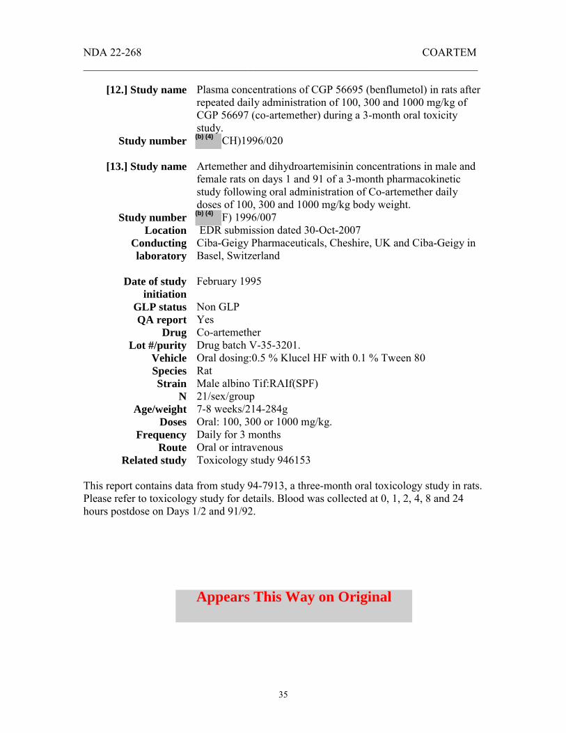
NDA 22-268 COARTEM _______________________________________________________________________
35
[12.] Study name
Study number
[13.] Study name
Study number Location
Conducting laboratory
Date of study
initiation GLP status QA report
Drug Lot #/purity
Vehicle Species Strain
N Age/weight
Doses Frequency
Route Related study
Plasma concentrations of CGP 56695 (benflumetol) in rats after repeated daily administration of 100, 300 and 1000 mg/kg of CGP 56697 (co-artemether) during a 3-month oral toxicity study.
CH)1996/020 Artemether and dihydroartemisinin concentrations in male and female rats on days 1 and 91 of a 3-month pharmacokinetic study following oral administration of Co-artemether daily doses of 100, 300 and 1000 mg/kg body weight.
F) 1996/007 EDR submission dated 30-Oct-2007 Ciba-Geigy Pharmaceuticals, Cheshire, UK and Ciba-Geigy in Basel, Switzerland February 1995 Non GLP Yes Co-artemether Drug batch V-35-3201. Oral dosing:0.5 % Klucel HF with 0.1 % Tween 80 Rat Male albino Tif:RAIf(SPF) 21/sex/group 7-8 weeks/214-284g Oral: 100, 300 or 1000 mg/kg. Daily for 3 months Oral or intravenous Toxicology study 946153
This report contains data from study 94-7913, a three-month oral toxicology study in rats. Please refer to toxicology study for details. Blood was collected at 0, 1, 2, 4, 8 and 24 hours postdose on Days 1/2 and 91/92.
(b) (4)
(b) (4)
Appears This Way on Original

NDA 22-268 COARTEM _______________________________________________________________________
36
Table 1: Benflumetol pharmacokinetics parameters after dosing with Co-artemether
Tmax (h) Cmax (µg/mL)
Trough concentration (µg/mL)
AUC (0-24h) (µg*h/mL)
Dose (mg/kg)
Day 1 Day 91
Day 1 Day 91
Day 1 Day 90
Day 91
Day 1 Day 91
Males Females
100 300 1000 100 300 1000
2 8 24 8 8 24
8 8 4 4 4 2
16 18 14 20 18 14
13 24 19 14 29 36
3 3 14 7 6 14
5 10 10 10 24 32
1 11 12 9 18 20
228 271 227 293 298 210
174 434 337 262 591 645
On Day 1, there was no correlation between Coartemether dose and Cmax or AUC for benflumetol. On Days 90/91, Cmax and AUC values were higher at the two higher doses than at the high dose, but Cmax and AUC values were not dose proportional. Cmax values were generally similar in males and females, except AUC values were 36 to 91 % higher in females than in males on Day 91. There was no clear evidence of accumulation over the 90 day period.
Table 2: Artemether pharmacokinetics parameters after dosing with Co-artemether
Tmax (h) Cmax (µg/mL)
AUC (0-24h) (µg*h/mL)
Dose (mg/kg)
Day 1 Day 91
Day 1 Day 91
Day 1 Day 91
100 300 1000
1 1 1
1 1 1
19 285 954
5 31 327
19 413 2402
5 51 1256
Systemic exposure to artemether increased with increasing dose but the exposure was greater than dose proportional. Both Cmax and AUC decreased by 48 to 88 % with repeat dosing, indicating auto induction of metabolism. No NOAEL could be determined for this drug regimen since microscopic changes in the lymph nodes and kidneys were present at even the lowest dose. Summary Rats dosed with Co-artemether at 100, 300 and 1000 mg/kg for 91 days showed benflumetol Cmax and AUC’s which were not dose-proportional. On Day 1, AUC values were similar across all groups, while on Day 91 AUC values were generally higher at the two higher doses. This lack of increase in benflumetol exposure between administered dose levels may indicate saturation of absorption. In contrast, artemether Cmax and AUC values were greater than dose proportional and were lower on Day 91 than on Day 1. A NOAEL could not be determined due to effects in the lymph nodes and kidneys.

NDA 22-268 COARTEM _______________________________________________________________________
37
[14.] Study name
Study number Location
Conducting laboratory
Date of study initiation
GLP status QA report
Drug Lot #/purity
Vehicle
Species Strain
N Age/weight
Doses Frequency
Route
An oral juvenile development study in rats 0570013 EDR submission dated 30-Oct-2007 Novartis Pharmaceutical, East Hanover New Jersey March 2005 Yes Yes Artemether AL20041043 0.5 % (w/v) hydroxypropylcellulose (Klucel, grade HF), aqueous solution, with 0.1 % (w/v) Tween 80 Rat Wistar Hannover, WI(Glx/BRL/Han)IGS BR 44/sex/group Dams:11 weeks/ 178-241g; Pups 7day-old/16 g 10, 30 or 100 mg/kg Daily Oral
This study was conducted to determine the potential adverse effects of artemether on the postnatal development of the rat. Artemether was administered once daily, by oral gavage at 10, 30 and 100 mg/kg/day to three groups (N=44/sex/group) of juvenile rats [ WI (Glx/BRL/Han) IGS BR] from postpartum day (PPD) 7 through 21 with a recovery period of approximately 5 weeks. Exposure to the test compound was determined in a toxicokinetic analysis and is shown below. Due to significant mortality associated at 100 mg/kg/day, dosing was halted for the high dose group and due to analytical problems no results could be obtained for Day 7. Only plasma concentrations obtained from the Main study animals on Day 21 could be used. Neither artemether nor its metabolite, DHA, was detectable in samples at 10 mg/kg/day. At 30 mg/kg/day, maximal plasma concentrations of artemether and its metabolite were observed 2 hours after dosing in male rats and 1 hour after dosing in female rats. Exposures to artemether were comparable in males and females. Exposure to dihydroartemesinin was higher in males.
(b) (4)
(b) (4)

NDA 22-268 COARTEM _______________________________________________________________________
38
Summary Data collected was limited to just one dose, since artemether and its metabolite were not detected at the low dose and the high dose was eliminated. The available data suggest that exposure to artemether could not be verified in pups receiving 10 mg/kg artemether. At 30 mg/kg blood levels peaked within two hours and AUC values were as high as 263 ng*h/mL. There were no clear sex differences in plasma levels.
[15.] Study name Study number
Location Conducting laboratory
Date of study initiation
GLP status QA report
Drug Lot #/purity
Vehicle
Species Strain
N Age/weight
Doses Frequency
Duration Route
2-week oral (gavage) toxicokinetics study in rats 0570030 EDR submission dated 30-Oct-2007 Novartis Pharmaceutical, East Hanover New Jersey January 2005 Yes Yes Artemether AL20041043 0.5 % (w/v) hydroxypropylcellulose (Klucel, grade HF), aqueous solution, with 0.1 % (w/v) Tween 80 Rat Wistar Hannover, WI(Glx/BRL/Han)IGS BR 10/sex/group 8 weeks old. Males 232-282g; females 166-192g 0, 20 or 200 mg/kg Daily 14 days Oral
The purpose of this study was to establish the toxicokinetic profile of artemether when administered orally to rats for 2 weeks. Groups of Wistar Hannover, WI (Glx/BRL/Han) IGS BR rats were dosed orally, once daily with artemether for two weeks. Blood was obtained from all study animals on study days 1-2, 3-4 and 14-15. Two animals/sex/group were bled at 0.5, 1, 2, 8 and 24 hours post dose. Concentrations of artemether and its metabolite dihydroartemesinin in plasma, AUC, AUC/dose, Cmax, Tmax were measured/calculated using LC-MS/MS. Table 1. Artemether pharmacokinetics parameters in male rats.
AUC 0-24h (ng*h/mL) Cmax (ng/mL) Artemether dose (mg/kg) Day 1 Day 3 Day 14 Day 1 Day 3 Day 14
20 104 203 30 107 132 39 200 3417 700 198 815 635 65
(b) (4)
(b) (4)

NDA 22-268 COARTEM _______________________________________________________________________
39
Table 2. Artemether pharmacokinetics parameters in female rats
AUC 0-24h (ng*h/mL) Cmax (ng/mL) Artemether dose (mg/kg) Day 1 Day 3 Day 14 Day 1 Day 3 Day 14
20 309 240 177 130 174 122 200 16990 2982 1243 3220 1250 402 Exposure to artemether, as measured by Cmax and AUC, decreased with repeated dosing in males and females between Day 1 and Day 14. Artemether exposures increased with dose and were greater than dose proportional on Day 1, but were less than dose proportional on Days 3 and 14. Drug levels in females were higher than those recorded in males. Table 3. Dihydroartemisinin pharmacokinetics parameters in male rats
AUC 0-24h (ng*h/mL) Cmax (ng/mL) Artemether dose (mg/kg) Day 1 Day 3 Day 14 Day 1 Day 3 Day 14
20 234 174 45 194 87 57 200 12103 1080 365 2630 435 141 Table 4. Dihydroartemisinin pharmacokinetics parameters in female rats
AUC 0-24h (ng*h/mL) Cmax (ng/mL) Artemether dose (mg/kg) Day 1 Day 3 Day 14 Day 1 Day 3 Day 14
20 206 115 87 84 97 82 200 7160 4384 2036 1390 1400 592 Dihydroartemesinin exposures (using AUC comparisons) were greater than dose proportional on Day 1 in both sexes, and on Day 3 and Day 14 for females. Tmax was between 0.5 and 2 hours for artemether and DHA. Summary Oral doses of 10 and 200 mg/kg produce measureable artemether levels. This study confirms the finding in other studies that multiple oral doses of artemether induce its own metabolism. In rats dosed for two weeks, exposure, as measured by AUC 0-24h values, were lower for artemether and dihydroartemesinin on Day 14, when compared to Day 1 values. Exposure to artemether as measured by AUC was lower in males.

NDA 22-268 COARTEM _______________________________________________________________________
40
[16.] Study name
Study number Location
Conducting laboratory
Date of study initiation
GLP status QA report
Drug Lot #/purity
Vehicle Species Strain
N Age/weight
Doses Frequency
Duration Route
8-day exploratory neurotoxicity study in dogs 0410073 EDR submission dated 30-Oct-2007
September, 2004 Yes Yes Artemether 20040303/96.3% Not identified (yellow clear oily solution) Dogs Beagle, male 2 16-25 months. 9 kg 40 mg/kg Daily 8 days Intramuscular
In Study 0410073, two male beagle dogs were dosed once daily with intramuscular injections of 40 mg/kg artemether for 8 days. Blood was drawn for pharmacokinetic evaluations at 5 minutes, 30 minutes, 1, 2, 3, 6 (except for Day 3) and 24 hours postdose. Table 1. Mean Toxicokinetics parameters of artemether in dog plasma ________________________________________________________________________ Mean plasma artemether pharmacokinetics ________________________________________________________________________
T max (h)
Cmax (ng/mL)
AUC0-24h) (ng*h/mL
Day 1 1.25 670 5210 Day 3 0.50 749 8914 Day 7 1.5 738 7874
________________________________________________________________________ Table 2. Mean plasma dihydroartemesinin pharmacokinetics ________________________________________________________________________
T max (h)
Cmax (ng/mL)
AUC0-24h) (ng*h/mL)
Day 1 0.75 145 867 Day 3 0.50 33.2 457 Day 7 2.00 28.6 429
________________________________________________________________________
(b) (4)

NDA 22-268 COARTEM _______________________________________________________________________
41
Both animals showed brain lesions which included the following degenerative changes of the neurons, mainly in the brainstem nuclei: chromatolysis, eosinophilic cytoplasmic granulation, speroids, apoptosis and dark neurons. Please see the toxicology review of this study for details. Artemether levels, as measured by Cmax and AUC (0-24h) increased with repeated intramuscular dose in the first few days of dosing (Days 1-3) and then levels begin to plateau or decrease slightly. This increase/plateau is contrasted with the steep decrease in artemether that is observed after repeated oral dosing. Summary Intramuscular injections of 40 mg/kg artemether produced exposures in the range of 5000-8000 ng*h/mL which resulted in brain lesions after 7 days of dosing. Plasma artemether levels appeared to plateau after three days of dosing.
[17.] Study name Study number
Location Conducting laboratory
Date of study initiation
GLP status QA report
Drug Lot #/purity
Vehicle Species Strain
N Age/weight
Doses Frequency
Duration Route
Intramuscular neurotoxicity study in dogs 0510001 EDR submission dated 30-Oct-2007 Novartis Pharma AG, Muttenz, Switzerland January 2005 No Yes Artemether 20040303/96.3% Peanut oil Dogs Beagle, male 3/group 27-50 weeks. 10-16 kg 0, 10, 40 mg/kg Daily 3 or 8 days Intramuscular
In study 0510001 dogs were treated with intramuscular doses of artemether at 10 or 40 mg/kg for 3 or 8 days according to the protocol outlined in Table 10.
Appears This Way on Original

NDA 22-268 COARTEM _______________________________________________________________________
42
Table 1. Study Protocol 0510001 ________________________________________________________________________ Group 1 Group 2 Group 3 Group 4 Group 5 Group 6 Group 7 Control ARM566 ARM566 ARM566 ARM566 ARM566 ARM566 Artemether dose (mg/kg) 0 10 10 10 40 40 40 Dose volume (mL/kg) 0.5 0.125 0.125 0.125 0.5 0.5 0.5 Number of animals 3 3 3 3 3 3 3 Treatment duration (days) 8 3 3 8 3 3 8 Recovery (days) 0 0 6 0 0 6 0 ________________________________________________________________________ On Days 1, 3 and 7, blood was taken at the following time points for pharmacokinetics evaluations: 5 and 30 minutes, 1, 2, 3, 6 and 24 hours postdose. Table 2. Study 0510001: Mean pharmacokinetics parameters for artemether in
beagle dog plasma after 1, 3 or 7 daily intramuscular doses of artemether ________________________________________________________________________ 10 mg/kg/day artemether
Cmax (ng/mL)
T max (h)
AUC(0-24h) (ng*h/mL)
AUC0-24h) //dose (ng*h/mL/mg/kg/day)
Day 1 82 1.0 949 95 Day 3 182 0.8 1610 161 Day 7 209 0.5 1833 183
________________________________________________________________________ 40 mg/kg/day artemether
Cmax (ng/mL)
T max (h)
AUC(0-24h) (ng*h/mL)
AUC0-24h) //dose (ng*h/mL/mg/kg/day)
Day 1 442 1.8 5126 128 Day 3 770 1.1 10247 256 Day 7 689 1.5 7992 200
________________________________________________________________________
Appears This Way on Original

NDA 22-268 COARTEM _______________________________________________________________________
43
Table 3. Study 0510001: Mean pharmacokinetics parameters for dihydroartemisinin
in beagle dog plasma after 1, 3 or 7 daily intramuscular doses of artemether _____________________________________________________________________________________ 10 mg/kg/day artemether
Cmax (ng/mL)
T max (h)
AUC(0-24h) (ng*h/mL)
AUC0-24h) //dose (ng*h/mL/mg/kg/day)
Day 1 47 1.0 377 38 Day 3 26 1.1 271 27 Day 7 30 0.7 262 26
______________________________________________________________________________________ 40 mg/kg/day artemether
Cmax (ng/mL)
T max (h)
AUC(0-24h) (ng*h/mL)
AUC0-24h) //dose (ng*h/mL/mg/kg/day)
Day 1 172 0.8 948 24 Day 3 34 0.8 573 14 Day 7 33 0.7 469 12
________________________________________________________________________ No drug-related lesions were observed following single intramuscular doses of 10 mg/kg or in dogs dosed for 3 days at 10 or 40 mg/kg. Consistent with previous studies, repeated intramuscular doses result in increases in Cmax and AUC between Days 1 and 3, after which values began to decrease. Elimination half life was 7-9 hours on Day 1 and 14-21 hours on Day 7. Conclusions: The NOAEL level for neurodegeneration in dog was 10 mg/kg/day (intramuscular injection) for 8 days or 40 mg/kg/day for 3 days. Repeated intramuscular dosing was associated with an increase in artemether AUC during the first three days, after which AUC levels continued to rise or started to decrease. Elimination half life is also increased after repeated dosing. Animals administered intramuscular doses of 40 artemether for 8 days showed drug-related neuronal damage in several brain areas including the cochlear nucleus, reticular nuclei, the nucleus olivaris and the caudus colliculus. Auto-induction of metabolism, observed with oral dosing, is not observed with intramuscular dosing.
Appears This Way on Original

NDA 22-268 COARTEM _______________________________________________________________________
44
[18.] Study name Study number
Location Conducting laboratory
Date of study initiation
GLP status QA report
Drug Lot #/purity
Vehicle Species Strain
N Age/weight
Doses Frequency
Duration Route
Oral neurotoxicity study in dogs 0510009B EDR submission dated 30-Oct-2007 Novartis Pharma AG, Muttenz, Switzerland January 2005 No Yes Artemether and benflumetol ART batch AL20041035/ 99.7 % Benflumetol batch C0039 99.6 % 0.5 % Klucel HF with 0.1 % Tween 80 Dogs Beagle, male 3/group 6-10 months. 8-11 kg 143, 300 and 600 mg/kg artemether Daily 3 or 8 days Oral
In this study male dogs were treated orally for at least 3 or 8 days with 1000 mg/kg Coartemether (containing 143 mg/kg artemether) or artemether alone (600 mg/kg on day 1 and 300 mg/kg from Day 2 onwards). Animals dosed at 143 mg/kg artemether showed reduced Cmax, Tmax and AUC values with repeated dosing. In the 600/300 group there was also a reduction in Cmax, Tmax and AUC with repeated dosing. No neurodegeneration was observed with these animals. Table 1. Artemether AUC 0-24h and Cmax values measured in dogs dosed orally with Coartemether or artemether for 3 or 8 days. ________________________________________________________________________
AUC 0-24h (ng*h/mL)
Cmax (ng/mL) ______________________________________________________________________________
Artemether dose (mg/kg) Day 1 Day 3 Day 7 Day 1 Day 3 Day 7
143 * 1294 52 16 486 29 18 300 1 602 73 130 49 600 1 22479 3358 ________________________________________________________________________ * Animals dosed with 1000 mg/kg Coartemether containing 143 mg/kg artemether. 1 Animals were dosed with 600 mg/kg artemether on Day 1, but the dose was reduced to 300 mg/kg as of Day 2
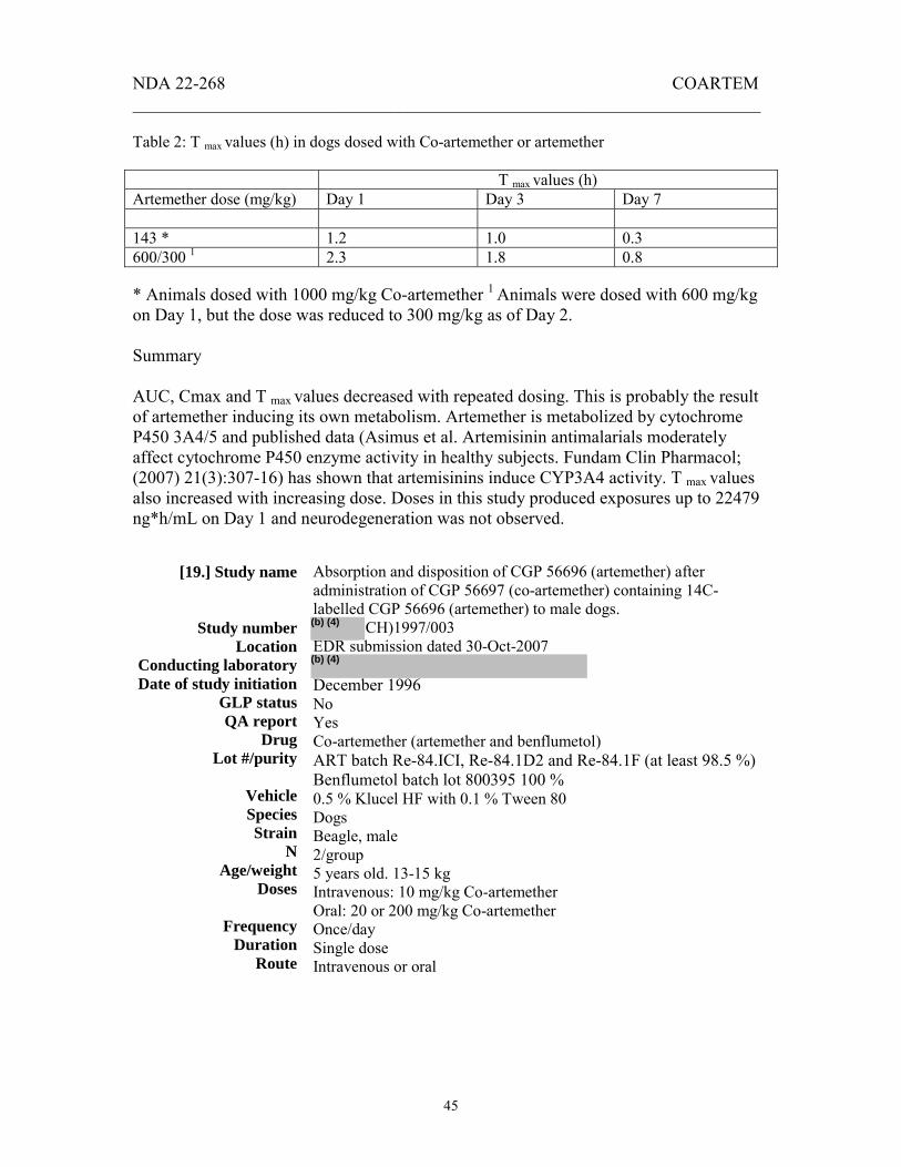
NDA 22-268 COARTEM _______________________________________________________________________
45
Table 2: T max values (h) in dogs dosed with Co-artemether or artemether T max values (h) Artemether dose (mg/kg) Day 1 Day 3 Day 7 143 * 1.2 1.0 0.3 600/300 1 2.3 1.8 0.8 * Animals dosed with 1000 mg/kg Co-artemether 1 Animals were dosed with 600 mg/kg on Day 1, but the dose was reduced to 300 mg/kg as of Day 2. Summary AUC, Cmax and T max values decreased with repeated dosing. This is probably the result of artemether inducing its own metabolism. Artemether is metabolized by cytochrome P450 3A4/5 and published data (Asimus et al. Artemisinin antimalarials moderately affect cytochrome P450 enzyme activity in healthy subjects. Fundam Clin Pharmacol; (2007) 21(3):307-16) has shown that artemisinins induce CYP3A4 activity. T max values also increased with increasing dose. Doses in this study produced exposures up to 22479 ng*h/mL on Day 1 and neurodegeneration was not observed.
[19.] Study name
Study number Location
Conducting laboratory Date of study initiation
GLP status QA report
Drug Lot #/purity
Vehicle Species Strain
N Age/weight
Doses
Frequency Duration
Route
Absorption and disposition of CGP 56696 (artemether) after administration of CGP 56697 (co-artemether) containing 14C-labelled CGP 56696 (artemether) to male dogs.
CH)1997/003 EDR submission dated 30-Oct-2007
December 1996 No Yes Co-artemether (artemether and benflumetol) ART batch Re-84.ICI, Re-84.1D2 and Re-84.1F (at least 98.5 %) Benflumetol batch lot 800395 100 % 0.5 % Klucel HF with 0.1 % Tween 80 Dogs Beagle, male 2/group 5 years old. 13-15 kg Intravenous: 10 mg/kg Co-artemether Oral: 20 or 200 mg/kg Co-artemether Once/day Single dose Intravenous or oral
(b) (4)
(b) (4)(b) (4)

NDA 22-268 COARTEM _______________________________________________________________________
46
In this study (Protocol T96-7029), beagle dogs received a single intravenous dose (10 mg/kg) or oral dose (20 and 200 mg/kg) of co-artemether, consisting of one part [14C]-labeled artemether and six parts unlabelled benflumetol. Artemether-related radioactivity was well absorbed after oral dosing with 63 % of the radioactivity recovered in the urine and 31 % in the feces. Cmax for radiolabel was between 1 and 2 hours postdose. Oral dosing of 20 or 200 mg/kg co-artemether oral dosing resulted in very low plasma levels of dihydroartemesinin (just above the LOQ, 0.034 µmol/L) and did not result in measurable levels of artemether. After intravenous dosing, the Tmax for radioactivity was not achieved at 5 minutes postdose, but Tmax was achieved at 15 or 30 minutes. The applicant interprets this to mean that metabolites were rapidly formed with volumes of distribution smaller than that of artemether. Five minutes after intravenous dosing, artemether accounted for 35 to 45 % of the 14C and dihydroartemesinin accounted for 4 % of the radioactivity in plasma. The AUC’s (0-168 hours) of artemether and dihydroartemesinin accounted for less than 1 % of the AUC of the 14C-radiolabeled substances. Regardless of route of administration, 14C declined slowly between Day 4 and Day 7 with a t½ of 7 days. Most of the administered dose (77 % and 92 % for intravenous and oral dosing respectively) was recovered within 48 hours after dosing. Metabolism About 44 radioactive peaks were detected in the plasma, urine and feces. In the plasma and urine, the vast majority (35 to 40) of these peaks represented 0.1 to 2 % of the administered dose. There were 7-13 peaks that accounted for 2-5 % of dose and two peaks, designated P56.0 and P57.5 represented 8 to 12 % of the dose. No predominant peaks were detected in feces. Incubation of radiolabeled artemether in vitro in the presence of a NADPH regenerating system resulted in the detection of two main metabolizing peaks P80.5 (which had been previously detected after intravenous dosing, representing about 25 % of the radioactivity) and P72.0 (detected in plasma representing about 12 %). Both peaks had been previously detected in plasma after oral and intravenous dosing.
Appears This Way on Original

NDA 22-268 COARTEM _______________________________________________________________________
47
(b) (4) (b) (4)
Appears This Way on Original

NDA 22-268 COARTEM _______________________________________________________________________
48
(b) (4)
(b) (4)
Appears This Way on Original

NDA 22-268 COARTEM _______________________________________________________________________
49
Summary In dogs, artemether-related radioactivity was well absorbed after oral dosing with 63 % of the radioactivity recovered in the urine and 31 % in the feces. Tmax for radiolabel was between 1 and 2 hours postdose. The majority of the radiolabel (92 %) was excreted within 48 hours after oral dosing. Five minutes after intravenous dosing, artemether accounted for 35 to 45 % of the 14C in the plasma. However, the AUC (0-168 h) of artemether and dihydroartemesinin accounted for less than 1 % of the AUC of the 14C radiolabeled substances.
[20.] Study name
Study number Location
Conducting laboratory Date of study initiation
GLP status QA report
Drug Lot #/purity
Vehicle Species Strain
N Age/weight
Doses Frequency
Duration Route
Related study
Artemether and dihydroartemesinin plasma and cerebrospinal fluid concentrations in male dogs in a 5 days (interim) or 30 days (main) neurotoxicity study following i.m. administration of artemether daily doses of 20 mg/kg body weight.
F)1997/004 EDR submission dated 30-Oct-2007 Ciba-Geigy Ltd, Stein Switzerland September 1996 No Yes Artemether 960533-7 Peanut oil Dogs Beagle, male 4/group 27-50 weeks. 10-16 kg 0, 20 mg/kg Daily 5 or 30 days Intramuscular Toxicology study 966141
(b) (4)
(b) (4)

NDA 22-268 COARTEM _______________________________________________________________________
50
In this study, beagle dogs (3 males/group) were administered 20 mg/kg artemether by intramuscular injection for 5 (interim sacrifice group) or 30 days (main study group). Control dogs (1/group) received intramuscular injections of sterile peanut oil. Blood and CSF samples were taken from all animals for artemether pharmacokinetic evaluations. Please see the toxicology report (Study 96-6141) for more details. Results One artemether-treated dog had to be sacrificed on Day 28 after showing tremor, restlessness, swaying gait, aggressiveness, dyspnea, general poor health and evidence of convulsions. A second dog showed convulsions with opisthotonus on Day 30. Microscopic examination of the brains of dosed animals revealed no drug-related neuronal damage after 5 days but neuronal degeneration, chromatolysis and microgliosis after 30 days of dosing. The animal that was sacrificed on Day 28 showed widespread neuronal necrosis in the cerebral cortex and hippocampus. The sponsor concluded that the poor general condition of the animal could have contributed to these latter findings. The plasma pharmacokinetics parameters determined by the applicant are shown below: ________________________________________________________________________ Table 1 Plasma artemether pharmacokinetics ________________________________________________________________________
T max (h)
Cmax (ng/mL)
AUC0-24h) (ng*h/mL)
Day 5 2 254 3952 Day 30 2 223 2777
_______________________________________________________________________ ______________________________________________________________________________________
Table 2 Plasma dihydroartemisinin pharmacokinetics
________________________________________________________________________ T max
(h) Cmax (ng/mL)
AUC0-24h) (ng*h/mL)
Day 5 2 15.0 251 Day 30 3 12.4 45
_______________________________________________________________________ Artemether levels in the CSF were 17 ng/mL on Day 5 and 20 ng/mL on Day 30. No dihydroartemisinin was measured in the CSF on Day 5 or on Day 30.
Appears This Way on Original

NDA 22-268 COARTEM _______________________________________________________________________
51
Figure 1: Trough levels (Predose) of artemether
Artemether levels increased in the plasma in all animals between Days 1 and 5 after which the levels decreased between Days 5 and Day 30. These data suggest that artemether accumulates during the first five days of dosing, after which a change in the metabolism or kinetics reduced the accumulation in the plasma. DHA levels were low compared to artemether (≤ 10 %) and decreased between Days 5 and 30. In the CSF, artemether levels were ≤ 10 % of the corresponding plasma levels and DHA was not detected in the CSF at any time point. Summary Intramuscular injections for 30 days at a dose of 20 mg/kg/day resulted in severe clinical signs, moribundity and convulsions in some animals. Consistent with previous studies, histopathological findings in the brain included neuronal degeneration, chromatolysis and microgliosis. One animal showed widespread neuronal necrosis in the cerebral cortex and hippocampus, but poor general condition of the animal could have contributed to these latter findings. Artemether-related neurotoxic effects appear to become more severe with increased treatment duration. Please see the toxicology review of this study for details. The NOAEL for neurodegeneration was 20 mg/kg (intramuscular injection) for 5 days, a dose which was associated with AUC’s between 2777 and 3952 ng*h/mL.
Appears This Way on Original

NDA 22-268 COARTEM _______________________________________________________________________
52
[21.] Study name
Study number Location
Conducting laboratory Date of study initiation
GLP status QA report
Drug Lot #/purity
Vehicle Species Strain
N Age/weight
Doses
Frequency Duration
Route Related study
Artemether and dihydroartemesinin (DHA) plasma concentrations in dogs in a 8 days neurotoxicity study following i.m. and oral administrations of daily doses of artemether (intermediate results)
F)1998/014 EDR submission dated 30-Oct-2007 Novartis Crop Protection, Stein Switzerland June 1997 No Yes Artemether Intramuscular lot 970653-13/95.95 % Oral batch 845196/99.7 % Gelatin capsules (oral) or peanut oil (intravenous) Dogs Beagle, male 4/sex/group 27-50 weeks. 10-16 kg Intramuscular doses 0, 20, 40 and 80 mg/kg Oral doses 0, 50, 150 and 600 mg/kg Daily 8 days Oral or Intramuscular Toxicology study 970024
In Study 97-0024, groups of beagle dogs, 4/sex/dose group, received artemether for 8 days, either orally as a gelatin capsules, or intramuscular (dissolved in peanut oil) injection. Blood was collected for pharmacokinetics evaluations and animals were sacrificed 2 hours after the last dose on Day 8. One dog that received intramuscular artemether (80 mg/kg) was found dead on Day 8. Other animals showed reduced activity, salivation and vomiting with intramuscular dosing. Food consumption and bodyweight were reduced at 80 mg/kg. QT was prolonged at 40 mg/kg and higher. Signs of neuronal degeneration (chromatolysis, necrosis, and microgliosis) were found at all intramuscular doses but were judged by the sponsor to be marginal in their incidence and grading at the lowest (20 mg/kg) dose. In this study, the only clinical sign observed in the orally dosed animals was vomiting at 600 mg/kg artemether. Oral administration of 600 mg/kg resulted in an AUC of 1730 and 250 ng*h/mL on Days 1 and 7 respectively. Consistent with Study 0510009 above, repeated oral dosing with artemether results in a reduction in the Cmax and AUC between Days 1 and 7 In contrast, repeated intramuscular dosing resulted in increases in Cmax and AUC between Days 1 and 7. The decreased systemic exposure that is observed with repeated oral dosing with artemether is consistent with auto-induction, but it is not clear why this does not occur with repeated intramuscular dosing. One explanation may be that at least some of the artemether metabolism is taking place in the gut. Repeated oral dosing would therefore induce the gut-associated enzyme,
(b) (4)(b) (4)

NDA 22-268 COARTEM _______________________________________________________________________
53
resulting more rapid metabolism of the orally administered drug, while not affecting the metabolism of drug that is administered by the intramuscular route. Table 1 Apparent pharmacokinetics of artemether after oral or intramuscular artemether
in dogs. Route Artemether
dose (mg/kg)
Day Cmax (ng/mL)
T max (h)
AUC0-24h) (ng*h/mL)
T 1/2 (h)
i m. 20 1
7
219 ± 116 294 ± 70
2 5
2050 ±1080 5350 ± 850
6.8 ± 1.8 18.7 ± 5.7
i m. 40 1
7
461 ± 230 825 ± 496
2 5
5630 ± 1510 12800 ± 4800
8.9 ± 2.5 13.6 ±4.4
i m. 80 1
7
985 ± 75 1180 ± 130
2 5
11000 ± 1400 20800 ± 2700
8.1 ± 0.8 20.7 ± 6.2
Oral
50 or 150 No feasible calculation
Oral 600 1 7
208 ± 194 27.8 ± 22.3
3 1.5
1730 ±1560 250 ± 145
It is noteworthy that in the present study, the oral 600 mg/kg dose resulted in lower exposure and significantly fewer neurological signs than the same dose in Study 0510009 above (1730 ng*h/mL and 22479 ng*h/mL, respectively). These findings support the idea that the neurological effects of artemether are exposure-related. Summary The NOAEL for neurodegeneration in dogs was 600 mg/kg for 8 days. Repeated oral doses resulted in Cmax and AUC values which reduced over the 8-days administration period and produced no drug-related neuronal degeneration. In contrast, repeated daily intramuscular dosing with 40 mg/kg artemether in dogs resulted in increases in Cmax and AUC over 8 days and neuronal degeneration in the brain. Please see toxicology review for details.
Appears This Way on Original

NDA 22-268 COARTEM _______________________________________________________________________
54
[22.] Study name
[23.] Study number Location
Conducting laboratory Date of study initiation
GLP status QA report
Drug Lot #/purity
Vehicle Species Strain
N Age/weight
Doses Frequency
Duration Route
Related study
Plasma concentrations of CGP 56695 (benflumetol) in dogs after repeated daily administration of 20, 60 and 200 mg/kg of CGP 56697 during a 3-month oral toxicity study.
CH)1996/024 and 1996/005 EDR submission dated 30-Oct-2007 Ciba-Geigy Limited, Pharma Research, Basel, Switzerland February 13th, 1995. No Yes Co-artemether V-35-3201 Gelatin capsules Dogs Beagle 3/sex/group 7-9 months, 10-13 kg 0, 20, 60 and 200 mg/kg Daily 90 days Oral Toxicology study 946155
Beagle dogs were doses with Co-artemether (capsules) for 90 days. Blood was collected at 0, 1, 2, 4, 8 and 24h on Days 1, 89/90. Table 1 Apparent pharmacokinetics of benflumetol after oral dosing with Co-artemether
in dogs.
Route Co-artemether dose (mg/kg)
Day Cmax (ng/mL)
T max (h)
AUC0-24h) (µg*h/mL)
M F 20 1
89
2.5 6.1
24 1
8 4
30 91
60 1
89
7.2 15.3
24 2
8 8
106 283
200 1
89
8.7 16
24 0
8 8
117 258
(b) (4)

NDA 22-268 COARTEM _______________________________________________________________________
55
Benflumetol levels were extremely variable between animals. Cmax and AUC values increased between 20 and 60 mg/kg at all time points but the increase was slightly less than dose proportional. On Day 1, Cmax and AUC values increased only slightly between the 60 and 200 mg/kg doses. On Day 89 there was no significant difference between the mean Cmax and mean AUC values at the two highest doses. Mean AUC values for females were consistently higher (mean difference 38 %, range +10 to +100 %), than for males. Benflumetol absorption was slow, particularly in males on Day 1 of dosing, where plasma concentration continued to increase at the last time point measured (24 hours) for all but two animals. In females on Day 1, Tmax was between 4 and 8 hours for all but one animal. On Day 89, Tmax was achieved more quickly, between 0 and 8 hours for all but one female. Benflumetol was detected in the plasma of control dogs. The source of this contamination was not determined. Artemether represented 14.3 % of the administered dose and so animals were dosed at 2.9, 8.6 and 29 mg/kg artemether. The limits of quantitation for artemether and dihydroartemesinin were 10.4 and 10.1 ng/mL. Artemether and dihydroartemesinin were only measured above the limit of quantitation in one instance for one male and one female dog at the highest dose. Artemether and dihydroartemesinin were not detected in the plasma of any control animals. The absence of measureable quantities of artemether in the plasma of these dogs may reflect poor absorption or extensive first pass elimination. Other explanations provided by the applicant include low artemether solubility and erythrocyte binding. Summary Dogs treated orally with Coartemether at 20, 60 or 200 mg/kg showed plasma levels of benflumetol which increased between 20 and 80 mg/kg, but then did not show significant additional increase when the dose was increased further to 200 mg/kg. Artemether and DHA concentrations were above the limit of quantitation in only two instances. The lack of exposure to artemether may reflect the extensive first pass metabolism that is known to take place with co-artemether. A review of the toxicology data revealed the NOAEL to be the low dose, which showed AUC’s between 30 and 91 µg*h/mL.
Appears This Way on Original
NDA 22-268 COARTEM _______________________________________________________________________
56
[24.] Report name
Report number Report name
Report number Location
Conducting laboratory Date of study initiation
GLP status QA report
Drug Lot #/purity
Vehicle Species Strain
N Age/weight
Doses Frequency
Route
Benflumetol plasma concentrations in female cynomolgus monkeys from a pilot pharmacokinetic study with a single co-artemether oral dose.
CH) 1996/095 Artemether and dihydroartemesinin plasma concentrations in female cynomolgus monkeys in a pilot pharmacokinetic study with a single co-artemether dose.
F) 1996/029 EDR submission dated 30-Oct-2007 Ciba-Geigy Limited, Stamford Lodge, UK June 1996 No Yes Co-artemether Batch 17/145/1 Gelatin capsules Monkey Macaca fascicularis 3 females 5 year old, 3-4 kg 350 mg/kg Once Oral
This study (protocol 96-7103) examined plasma levels of benflumetol, artemether and dihydroartemisinin in female cynomolgus monkeys in a pilot, single dose pharmacokinetic study of Co-artemether. A single, oral, 350 mg/kg dose of Co-artemether was administered to three female cynomolgus monkeys and blood was collected prior to dosing and at 30 minutes, 1, 2, 4, 8, 12, 24, 36, 48, 72, 96 and 120 hours after the oral dose. Artemether was not detected in plasma from any monkey at any time point and dihydroartemesinin was only detected in one sample each from two monkeys (at 2 and 4 hours). Plasma pharmacokinetics parameters for benflumetol are shown below.
(b) (4)
(b) (4)
Appears This Way on Original

NDA 22-268 COARTEM _______________________________________________________________________
57
Table 1 Benflumetol Pharmacokinetics in monkey _______________________________________________________________________
Pk Parameter Monkey 2000 Monkey 2002 Monkey 2004
_____________________________________________________________________________________
T max (h) Cmax (µg/mL)
AUC(0-24h)x (µg*h/mL) T½ (h)
8 6002 114971 12
12 4214 65463 40
12 5698 125539 28
________________________________________________________________________ Discussion Benflumetol was measureable in monkey plasma two hours after oral administration with peak plasma concentrations reached between 8 and 12 hours. Apparent t½ ranged from 12 to 40 hours. Benflumetol Cmax and AUC values were similar to those calculated in rats and dogs. No systemic exposure to artemether could be demonstrated after the oral administration of 350 mg/kg doses of co-artemether to female cynomolgus monkeys. This lack of systemic exposure after oral administration likely reflects the rapid and extensive first pass metabolism of artemether. This pattern is consistent with findings obtained in dogs, rabbits and rats.
(b) (4)
(b) (4)
Appears This Way on Original

NDA 22-268 COARTEM _______________________________________________________________________
58
2.6.4.4 Distribution
[25.] Study name
Study number Location
Conducting laboratory Date of study initiation
GLP status QA report
Drug Specific activity
Lot #/purity
Vehicle Species Strain
N Age/weight
Doses Duration
Route
Transfer to the embryo-fetal compartment of pregnant rats after peroral administration of 30 mg/kg of CGP 56697 (co-artemether) containing 3H-labeled CGP 56696 (artemether).
CH)1997/099 EDR submission dated 30-Oct-2007
March 1997 No No Co-artemether 435 KBq/mg Artemether batch Re-54.9H/99% Benflumetol lot 95/ 99.9% 0.5% Klucel HF/Tween 80 Rat Tif: RAIf (SPF) 3/time point Day 13 of pregnancy, 176-309g 30 mg/kg Single dose Oral
Study (CH) 1997/099 was conducted to investigate the transfer of artemether-related radioactivity into the embryo-fetal compartment of pregnant Tif: RAIf (SPF) rats after oral administration of 30 mg/kg Co-artemether containing 3H radiolabeled artemether on Day 13 of pregnancy. Distribution of artemether-related radioactivity in the dams was similar to that seen in previous studies, with highest levels being measured in the gastro-intestinal tract, adrenals, kidney and liver. Mean 3H concentrations in the placenta were generally higher than that in the maternal plasma (1.7 fold at 24 hours postdose). Since radioactivity levels in the mammary glands were similar to that in the plasma, artemether-related compounds may be anticipated in the milk. Radioactivity was taken up by the fetus and amniotic fluid, with the Cmax of radioactivity being measured at one hour after dosing. Levels of radioactivity in the fetus and amniotic fluids were as
(b) (4)
(b) (4)
(b) (4)
(b) (4)
Appears This Way on Original
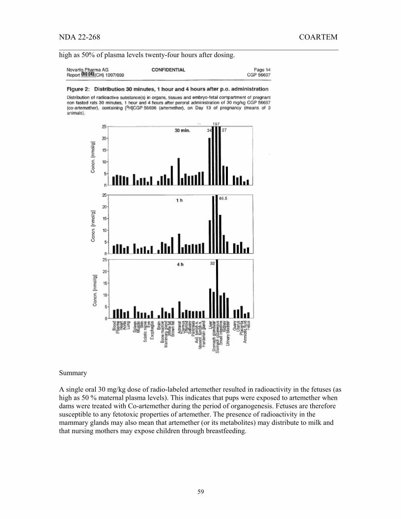
NDA 22-268 COARTEM _______________________________________________________________________
59
high as 50% of plasma levels twenty-four hours after dosing.
Summary A single oral 30 mg/kg dose of radio-labeled artemether resulted in radioactivity in the fetuses (as high as 50 % maternal plasma levels). This indicates that pups were exposed to artemether when dams were treated with Co-artemether during the period of organogenesis. Fetuses are therefore susceptible to any fetotoxic properties of artemether. The presence of radioactivity in the mammary glands may also mean that artemether (or its metabolites) may distribute to milk and that nursing mothers may expose children through breastfeeding.
(b) (4)

NDA 22-268 COARTEM _______________________________________________________________________
60
[26.] Study name
Study number Location
Conducting laboratory Date of study initiation
GLP status QA report
Drug Specific activity
Lot #/purity
Vehicle Species Strain
N Age/weight
Doses Duration
Route
Transfer of radioactive substances to the embryo-fetal compartment of rabbits after peroral administration of CGP 56697 (co-artemether) containing 14C-labeled CGP 56696 (artemether).
CH)1997/004 EDR submission dated 30-Oct-2007
January 1997 No No Co-artemether 43 KBq/mg Artemether batch Re-84.1E2/9H/98.5% Benflumetol lot 800395/100% 0.5% Klucel HF/Tween 80 Rabbit New Zealand Albino 2 Day 17 of pregnancy, 4.3-4.6 kg 175 mg/kg Single dose Oral
This study was designed to investigate the transfer of radiolabeled substances to the embryo-fetal compartment of rabbits after oral administration of Co-artemether containing 14C-labeled artemether. Two fed, pregnant New Zealand White rabbits were treated orally, by gavage with 175 mg/kg radio-labeled Co-artemether on Day 17 of gestation. Blood samples were taken from the rabbits before dosing and 0.25, 0.5, 1, 2, 4, 8 and 24 hours post dose. Urine and feces were also collected. Twenty-four hours after dosing, animals were killed by exsanguination and 14C measured in selected maternal organs (see results below), whole fetus, fetal liver and fetal brain. The mean Cmax of artemether-related radioactivity occurred at 45 minutes after the oral dose. The plasma concentration of unchanged artemether was very low and only measureable in the 15 and 30 minute samples in one of the two rabbits (0.2-0.5 % of the respective 14C concentration, LOQ (0.034 µml/L). Plasma dihydroartemesinin levels were also low and were measureable between 15 minutes and 2 hours postdose. Plasma DHA levels represented 0.1 to 0.7 % of the total 14C-labelled substances. The ratio of the plasma concentrations in blood versus plasma indicates that artemether and dihydroartemesinin have low affinity for blood cells in rabbit. Distribution of artemether-related radioactivity is shown on Figure 1, below. The highest levels of radioactivity were detected in the liver, kidney, placenta, ovary and lungs (41, 17, 5.7, 5.2 and 4.5 nmol/g, respectively. 14C levels in the whole fetuses and amniotic fluid were similar or lower than levels in maternal blood. The pattern of tissue distribution of radioactivity in the fetuses was similar to adult rabbits, with highest levels detected in the fetal liver. HPLC analysis of urine from the two animals revealed 37 metabolite peaks. Three of the peaks each accounted for 6-9 % of the 14C, seven peaks accounting for 2-4 % each, ten peaks accounted for 1-2 % and 17 peaks accounting for less than 1 % each. The chemical structures of the
(b) (4)
(b) (4)
(b) (4)

NDA 22-268 COARTEM _______________________________________________________________________
61
metabolites were not identified, but two peaks P55.0 and P57.0, may be identical to dog metabolites P56.0 and P57.5. Unchanged artemether was only present in trace amounts. Rabbits excreted about 67 % of the dosed radioactivity in the urine and about 2 % in the feces within the first 24 hours. Figure 1. Tissue distribution of artemether-related radioactivity in pregnant rabbits and their fetuses.
Summary

NDA 22-268 COARTEM _______________________________________________________________________
62
Orally administered Co-artemether was very well absorbed and extensively metabolized in pregnant rabbits. Pups were clearly exposed after maternal dosing as 14C levels in the whole fetuses and amniotic fluid were similar or lower than levels in maternal blood. The highest uptake of radioactivity was in the liver in both dams and fetuses. These findings lead to the conclusion that unborn children could be exposed if mothers are dosed with artemether.
[27.] Study name
Study number Location
Conducting laboratory
Date of study initiation GLP status QA report
Drug Specific activity
Lot #/purity Vehicle Species
In vitro binding of artemether to human serum proteins, human erythrocytes and serum proteins from dog, rabbit, rat, and mouse and cynomolgus monkey.
(F) 1998/004 EDR submission dated 30-Oct-2007 Ciba-Geigy Bioanalytics & Pharmacokinetics, Rueil-Malmaison Cedex, France. Not provided Non-GLP No Artemether 1.67 MBq/mg 14 C artemether: Batch Re-84.1A1 (>98 %) Methanol Human, mouse, rat, dog, rabbit, baboon and cynomolgus monkey.
The study was conducted to determine the extent of in vitro binding of artemether to human serum or plasma proteins, isolated albumin, a.1-acid glycoprotein, gamma globulins and lipoproteins, human erythrocytes, serum proteins from dog, rabbit, rat, mouse and cynomolgus monkey. Ultra filtration and erythrocyte partitioning were used. Ultra filtration was used to determine the binding in animal and human serum and in albumin solution.
(b) (4)

NDA 22-268 COARTEM _______________________________________________________________________
63
Table 1: Binding parameters determined in human serum or plasma using ultra filtration or erythrocyte partitioning. Ultra filtration Erythrocyte partitioning method Bound fraction
(%) Bound fraction
(%) Binding capacity
(L/mol) Plasma or serum Albumin α1-acid glycoprotein Gamma globulins High density lipoproteins Low density lipoproteins Very low density lipoproteins
97.9 83-91
95.4 78.3 87.8 (8.0) 72.9 66.8 71.7
6.2 x 10³
3.2 x 10⁵
(1.3 x 10³) 2.1 x 10⁵
1.7 x 10⁶
2.0 x 10⁷
Table 2 Distribution of artemether in human serum proteins after factoring physiological concentrations and binding parameters. Fraction of artemether bound (%) Erythrocytes Albumin α1-acid glycoprotein Gamma globulins High density lipoproteins Low density lipoproteins Very low density lipoproteins
11 17 33 0.4 12 9.3 12
The values of the bound fraction in plasma/serum/albumin determined using ultra filtration or erythrocyte partitioning were similar. Artemether showed strong binding to serum proteins at 14C-artemether concentrations of 1 to 10 µg/mL, levels relevant for clinical exposures. Using ultra filtration data, the bound fraction (%) was between 96 and 98 % for dog, rabbit, rat, monkey and mouse. Table 1 shows that in human blood, the binding capacity of low- and very low density lipoproteins were highest, but considering physiological concentrations of the various components at a hematocrit of 0.40, the largest fraction of the artemether (33 %) would be bound to the to α1-acid glycoprotein (Table 2) . The small fraction bound to erythrocytes (11 %) was consistent with other studies which reported that the ratio of concentrations in the plasma versus blood was 1.6. Only 4.6 % was unbound. Summary Artemether was at least 96 % bound in all the species investigated. In human blood the α1-acid glycoprotein binds the highest fraction of the available artemether.

NDA 22-268 COARTEM _______________________________________________________________________
64
[28.] Study name
Study number Location
Conducting laboratory Date of study initiation
GLP status QA report
Drug Specific activity
Lot #/purity Vehicle Species Strain
N Age/weight
Doses Duration
Route
Transfer of radioactive substance(s) to the embryo-fetal compartment of rats after peroral administration of 30 mg/kg CGP 56697 (co-artemether) containing [14C]CGP 56695 ([14C] benflumetol and unlabeled CGP 56696 (artemether)
CH)1997/139 EDR submission dated 30-Oct-2007
December 1996 No No Co-artemether Benflumetol: Batch Lm-9.3(>98 %) Artemether batch 810294 0.5% Klucel HF/Tween 80 Rat Tif: RAIf (SPF) 3 dams/time point Pregnant dams, 11-14 weeks old, 269-335g 30 mg/kg Single dose Oral
Report (CH) 1997/139: Transfer of radioactive substance(s) to the embryofetal compartment of rats after peroral administration of 30 mg/kg CGP 56697 (Co-artemether) containing [14C]CGP 56695 ([14C] benflumetol) and unlabelled CGP 56696 (artemether). Protocol 967038. April, 1997. Related study report: TIP (US) 95072 The aim of this study was to investigate the transfer of radioactive substances to the embryofetal compartment after oral administration of 30 mg/kg Co-artemether to pregnant rats on Day 13 of gestation. On Day 13 of gestation, 12 rats received 30 mg/kg Co-artemether containing a mixture of 14C-labeled benflumetol as a suspension in 0.5% Klucel HF/Tween 80. Animals were sacrificed 1, 4, 8 and 24 hours postdose and 14C concentrations determined. The 14C concentrations in blood and plasma of the dam were highest at 8 hours. At this time point the highest 14C levels were observed in the cutaneous stomach, the adrenal gland, and the liver. Twenty-four hours after dosing very high 14C concentrations were found in the adrenal gland (221-274 nmol/g), liver (36-50 nmol/g) and the spleen (about 31 nmol/g). In the fetuses, the uptake of radioactivity was very slow, with low levels being detected at 4 hours postdose. While the radioactivity levels continued to increase up to the twenty-four hour time point, the radioactivity levels in the fetuses (0.8-1.2 nmol/g) were still low about 10% of levels detected in maternal blood at the 8 hour time point (t max). Among the reproductive organs, levels were highest in the ovaries and placenta, but were clearly measureable in the mammary glands
(b) (4)
(b) (4)
(b) (4)
(b) (4)

NDA 22-268 COARTEM _______________________________________________________________________
65
and uterus. Only low levels of radiation were detected in the amniotic fluid at 8 and 24 hours post dose.
Summary Oral administration of 30 mg/kg on Day 13 of gestation resulted in measureable 14C concentrations in the ovaries, the uterus, the mammary glands fetuses and the placentas. The uptake of radioactivity into the mammary glands suggests that benflumetol (and/or its metabolite) may be excreted with the milk in lactating animals. Maximum 14C levels in the fetuses were weak and corresponded to about 10% of the maximum amounts detected in maternal blood.
Appears This Way on Original
NDA 22-268 COARTEM _______________________________________________________________________
66
[29.] Report name
Report number Report name
[30.] Report number Location
Conducting laboratory Date of study initiation
GLP status QA report
Drug Specific activity
Lot #/purity Vehicle Species Strain
N Age/weight
Doses Duration
Route
Transfer of radioactive substance(s) to the embryo-fetal compartment of rabbits after peroral administration of CGP 56697 (co-artemether) containing 14C-labeled benflumetol DMET(EU) 29/1996) Plasma concentrations of benflumetol in pregnant rabbits after peroral administration of CGP 56697 (co-artemether) containing 14C-labelled CGP 56695 (benflumetol) and unlabelled CGP 56596 (artemether)
CH)1997/079 EDR submission dated 30-Oct-2007
November 1996 No No Co-artemether Benflumetol: Batch Ko-76.1C Artemether lot # 810294 0.5% Klucel HF/Tween 80 Rabbit New Zealand White 2 Pregnant dams, Day 17 of gestation, 3.5-3.7 kg 175 mg/kg Single dose Oral
Protocol 96-7030 (Reports (CH) 1997/079 and DMET (EU) 29/1996) was designed to investigate the plasma and tissue concentrations of benflumetol after oral administration of Co-artemether containing 14C-labeled benflumetol. Two fed, pregnant New Zealand White rabbits were treated orally, by gavage with 175 mg/kg radio-labeled Co-artemether on Day 17 of gestation. This dose represented the highest dose used in teratogenicity study 94-6157. Blood samples were taken from the rabbits before dosing and 0.5, 2, 4, 8 and 24 hours post dose. Twenty-four hours after dosing, animals were killed by exsanguinations and 14C measured in selected maternal organs (see results below), whole fetus, fetal liver and fetal brain. Benflumetol-related radioactivity was detected in the blood of dams as early as 30 minutes after dosing. The highest concentrations were in the liver, lung, kidney, ovary and plasma. 14C levels in the placenta were about 6-fold higher than the maternal blood, but the levels in the (whole) fetus represented only about 2-3 % of maternal blood levels. Generally, levels were near or below the limit of detection in the amniotic fluid, fetal liver and fetal brain. In the one rabbit where fetal liver concentration was above the limit of detection, it was almost three times the level in the fetus as a whole.
(b) (4)
(b) (4)
(b) (4)
(b) (4)

NDA 22-268 COARTEM _______________________________________________________________________
67
Appears This Way on Original

NDA 22-268 COARTEM _______________________________________________________________________
68
________________________________________________________ [Benflumetol]
14 C concentration (µg/mL) (µmol/L) _____________________________________________________________
Blood Plasma
Time (h)
Rabbit 95354
Rabbit 95442
Rabbit 95354
Rabbit 95442
0
0.5 1 2 4 8
24
0 0
0.01 4.1 12 nd 14
0 0
0.02 0.8 3.2 nd 7.0
0 0.39 0.51 3.25 7.24 5.45 9.44
0 0.47 0.32 0.62 1.75 3.78 4.23
0 0.69 0.87 5.16 11.8
- 12.7
0 0.78 0.62 1.32 3.38 5.74 6.37
AUC(0-24h) ( g*h/mL)
157 78 266 121
____________________________________________________________
Benflumetol Pk Parameter Rabbit 95354 Rabbit 95446 __________________________________________________________
T max (h)
Cmax (µg/mL) AUC(0-24h) (µg*h/mL)
24 14 274
24 7.0 107
________________________________________________ The corresponding benflumetol values (Report (CH) 1997/079) showed that benflumetol-related radioactivity (see table above) was detectable in blood and plasma prior to benflumetol (see Figure 2) and that peak blood benflumetol concentrations were not achieved by 24 hours. Summary Despite measureable benflumetol levels in the maternal blood, pups were exposed to very low levels of benflumetol when pregnant rabbits were treated with 175 mg/kg Co-artemether.
(b) (4)

NDA 22-268 COARTEM _______________________________________________________________________
69
[31.] Study name
Study number Location
Conducting laboratory
Date of study initiation GLP status QA report
Drug Specific activity
Lot #/purity Vehicle Species
In vitro binding of benflumetol to human serum proteins, human erythrocytes and serum proteins from mouse, rat, dog, rabbit, baboon cynomolgus monkey.
F) 1996/044 EDR submission dated 30-Oct-2007 Ciba-Geigy Bioanalytics & Pharmacokinetics, Rueil-Malmaison Cedex, France. Not provided Non-GLP No Benflumetol 911 kBq/mg Benflumetol: Batch Ko-76.1D-1/< 98% DMSO Human, mouse, rat, dog, rabbit, baboon and cynomolgus monkey.
The study was conducted to determine the extent of in vitro, binding of benflumetol to human serum or plasma proteins, isolated albumin, α1-acid glycoprotein, gamma globulins and lipoproteins, human erythrocytes, serum proteins from mouse, rat, dog, rabbit, baboon cynomolgus monkey. Ultra filtration and/or erythrocyte partitioning were used for the determinations. Table 1: Binding parameters determined in human serum or plasma using ultra filtration or erythrocyte partitioning. Ultra filtration Erythrocyte partitioning method Bound fraction
(%) Bound fraction
(%)
Binding capacity (L/µmol)
Distribution
Plasma or serum Albumin α1-acid glycoprotein High density lipoproteins Low density lipoproteins Very low density lipoproteins
99.9 99.9
99.7 62.8 16.8 99.6 96.3 95.9
0.0028
0.0088
27.5 26.0 235
0.47 0.057 77.1 7.3 6.6
Erythrocytes Unbound
8.2
0.28
(b) (4)
Appears This Way on Original

NDA 22-268 COARTEM _______________________________________________________________________
70
Table 2 Distribution of benflumetol in human serum proteins after factoring physiological concentrations and binding parameters. Fraction of benflumetol bound (%) Erythrocytes Albumin α1-acid glycoprotein High density lipoproteins Low density lipoproteins Very low density lipoproteins Unbound
8.2 0.47 0.057 77 7.3 6.6 0.28
The values of the bound fraction in plasma/serum/albumin determined using ultra filtration or erythrocyte partitioning were similar. Benflumetol is largely bound (99.7 % or more) to serum proteins by either method. Using ultra filtration data, the bound fraction (%) was greater that 99 % for all species tested. Table 1 shows that in human blood, the greatest binding capacity resided with the very high density lipoproteins. However, when the concentrations of the proteins were taken into account (Table 2), the highest total binding capacity was in the high density lipoproteins, which bound 77 % of the benflumetol. A small fraction was bound to erythrocytes (8 %). Only 0.28 % was unbound. Figure 1 Distribution of benflumetol in whole blood.
High density lipoproteinsErythocytesLow density lipoproteinsVery low density lipoproteinsHDL 77%
ERYTHROCYTES 8 %
LDL 7 %
VLDL 7%
Conclusion Benflumetol was highly bound in all species tested. Most of the binding capacity was associated with lipoproteins, particularly HDL, which accounted for 77 % of the binding capacity.
Appears This Way on Original

NDA 22-268 COARTEM _______________________________________________________________________
71
2.6.4.5 Metabolism
[32.] Study name
Study number Location
Conducting laboratory Study date GLP status QA report
Administered drug Doses
Dosing frequency Dosing duration
Radionuclide Specific activity/purity
Lot #/purity
Vehicle
Animals Route
Characterization of metabolites of artemether (CGP56696) formed in rats and dogs after single oral doses of Co-artemether containing radiolabeled artemether.
CH)1997/534 EDR submission dated 30-Oct-2007 Ciba-Geigy AG, Basel Switzerland. October 1994 Swiss-GLP Yes Co-artemether Dog: 200 mg/kg; Rat: 20 and 1000 mg/kg Once daily Single dose Rat: 3H, 14C, Dog: 14C Rat: 1390-33560 KBq/mg, >98.5 % Dog: 10 KBq/mg, >98.5 % See Table 1 below. Rats 0.5 % Klucel HF with 0.1 % Tween 80 Dog: Gelatin capsules. 2 albino Tif:RAIf(SPF) rats, 1 beagle dog Oral
Table M1: Specific radioactivity and purity of radiolabeled artemether used in study 1997/241
In study CH) 534, the applicant examined the NMR spectra of various metabolites from rat bile, rat urine, and dog urine in order to determine the structures. In many instances the NMR spectra could not be interpreted and no structure could be proposed for the metabolite. In rat, structures for two metabolites were proposed. The major metabolite in rat bile (26 % of dose) was identified as 9α-hydroxy-artemether-9-O-glucuronide, indicating that 9α-hydroxylation is an important route of metabolism for artemether.
(b) (4)(b) (4)
(b) (4)

NDA 22-268 COARTEM _______________________________________________________________________
72
Although analysis of rat urine metabolites could not provide complete structures from any metabolites, the available data suggest that artemether metabolism includes O-demethylation followed by hemiacetal oxidation, hydroxylations and O-conjugation with glucuronic acid or glucose.
(b) (4)
Appears This Way on Original

NDA 22-268 COARTEM _______________________________________________________________________
73
Analysis of dog urine metabolites identified three metabolites 3ß, 9ß-dihydroxy-deoxyartemesinin-9-O-glucuronide; 3ß-hydroxy-deoxyartemesinin-3-O-glucuronide; 3ß-hydroxy-deoxyartemether-3-glucuronide. In addition to the previously suggested pathways, these metabolites suggest that peroxide deoxygenation and/or rearrangement is an important route of metabolism in dogs. Summary Analysis of the NMR spectra of various metabolites from rat bile, rat urine, and dog urine was unable to identify all the major metabolites of artemether but artemether metabolism was shown to involve peroxide de-oxygenation, O-demethylation, hydroxylation and O-glucuronidation. The proposed scheme is shown below:
(b) (4)

NDA 22-268 COARTEM _______________________________________________________________________
74
[33.] Study name
Study number Location
Conducting laboratory Study date GLP status QA report
Administered drug Doses
Dosing frequency Dosing duration
Radionuclide Specific activity/purity
Animals
Route
In vitro biotransformation of CGP 56696: comparison of three animal species and man.
(CH) 1997/181 EDR submission dated 30-Oct-2007 Ciba-Geigy AG, Basel Switzerland. October 1994 Swiss-GLP Yes Co-artemether Dog: 200 mg/kg; Rat: 20 and 1000 mg/kg Once daily Single dose 14C 499MBq/mmol or 1.67 MBq/mg 12 Tif:MAGf(SPF)Mice, 30g 12 Tif:Ralf(SPF) Rats:200-220 g0-33560 KBq/mg, >98.5 % 1 beagle Dog: 12 kg Liver from 3 male and 3 female kidney donors. See Table 1 below. Rats 0.5 % Klucel HF with 0.1 % Tween 80 Dog: Gelatin capsules. 2 albino Tif:RAIf(SPF) rats, 1 beagle dog Oral
Report In vitro biotransformation of CGP 56696: comparison of three animal species and man. The aim of this study was to investigate the metabolism of 14C-labeled artemether in vitro using the post mitochondrial liver fraction S12 from man and to compare it with that of three animal species (mouse, rat, and dog). Mixtures containing 14C artemether, post mitochondrial liver fraction P12, and NADPH-
(b) (4)

NDA 22-268 COARTEM _______________________________________________________________________
75
regenerating system containing isocitrate-dehydrogenase, NADP, isocitrate and MgCl2 were incubated at 37 °C for 120 minutes. The reaction was stopped and the protein was removed by the addition of
. Radiometry of aliquots of the supernatant and of the total amount of dissolved residue (acidified with 2 N HCI) was performed on a liquid scintillation counter. Mouse
Rat
(b) (4)

NDA 22-268 COARTEM _______________________________________________________________________
76
Dog
Human

NDA 22-268 COARTEM _______________________________________________________________________
77
Comment The pattern of metabolite peaks was qualitatively similar, but quantitatively different across the species. Many of the peaks were super imposable. This metabolite profile represents the effect of liver metabolizing enzymes on artemether. This is one portion of what would take place in vivo in a dosed animal, but other routes of metabolism are also to be considered. In particular, there seems to be pre-systemic metabolism that takes place. These components will also generate other metabolites which will undergo further metabolism in the liver. An accurate representation of the pattern of metabolism in human requires the conduct of a mass balance study in humans. The approximate half lives of l4C-CGP 56696 were 17 min (dog) and 20 min (man), and 38 min (mouse) and 41 min (rat). 2.6.4.6 Excretion Excretion data were incorporated into the studies discussed above. 2.6.4.7 Pharmacokinetic drug interactions See Clinical Pharmacology review. 2.6.4.8 Other Pharmacokinetic Studies
[34.] Study name
Study number Location
Conducting laboratory Study date GLP status QA report
Administered drug Doses
Dosing frequency Dosing duration
Lot #/purity Vehicle Species Strain
N Age
Weight Route
Related study
Plasma concentrations of CGP 56695 (benflumetol) in rats after repeated daily administration of 200, 600 and 1000 mg/kg of CGP 56697 (Co-artemether) during a 1-month oral toxicity study.
CH)1995/079 EDR submission dated 30-Oct-2007 Ciba-Geigy AG, Basel Switzerland. October 1994 Swiss-GLP Yes Co-artemether 0, 200, 600, 1000 mg/kg Once daily 1 month V-25-3187, V-26-3188 0.5% Klucel HF/Tween 80 Rat Tif:RAIf(SPF) 3/sex/dose group 7-11 weeks old Males: 223-282g; Females: 165-241g Oral gavage Toxicology study 946152
(b) (4)

NDA 22-268 COARTEM _______________________________________________________________________
78
Blood sample was collected at 0, 1, 2, 4, 8 and 24 hours on Day 1 and Day 28 to determine the benflumetol and artemether levels in this one month rat toxicology study. Please see Toxicology study 94-6152 for details of study design. Table 1. Pharmacokinetics parameters for benflumetol.
Tmax (h) Cmax (µg/mL)
Trough concentration (µg/mL)
AUC (0-24h) CoartemetherDose (mg/kg)
Day 1 Day 25 Day 1 Day 25 Day 1 Day 25
Day 1 Day 25
Males Females
200 600 1000 200 600 1000
8 24 24 8 8 24
24 8 8 4 0 4
33 21 16 23 20 33
16 21 18 16 25 38
15 21 16 8 18 33
16 9 15 5 14 26
535 423 279 358 402 350
322 386 382 281 449 630
Discussion After administration of 200, 600 and 1000 mg/kg of Co-artemether to rats, the onset of absorption of benflumetol was fast (less than 1 hour) but the absorption continued over an extended period. Table 1 shows that oral doses of 200, 600 and 1000 mg/kg Co-artemether resulted in plasma levels of benflumetol which were not dose proportional. Plasma levels were variable across all dose groups and all time points. Although AUC’s were somewhat higher at the two higher doses in the females on Day 25, there was no clear relationship between dosing and AUC and no differences between the sexes. There was no evidence of accumulation. Similar exposures can be obtained with 200 mg/kg or 1000 mg/kg Co-artemether. Animals in the low dose group showed a 21 % decreases in triglycerides and a slight reduction in bodyweight gain. No NOAEL was determined for this study.
[35.] Report name
[36.] Report number Report name
Report number Location
Conducting laboratory Study date GLP status QA report
Administered drug Doses
Dosing frequency Dosing duration
Lot #/purity
Plasma concentrations of CGP 56695 (benflumetol) in female rabbits after repeated daily administration of 210, 700 and 2100 mg/kg of CGP 56697 (Co-artemether) on day 19 of gestation during a 13-day oral toxicity study.
CH)1996/085 Artemether and dihydroartemisinin plasma concentrations in female rabbits in a follow-up dose-range finding study evaluating effects of Co-artemether on embryo and fetal development.
F) 1996/022 EDR submission dated 30-Oct-2007 Ciba-Geigy AG, Basel Switzerland. October 1995 Swiss-GLP Yes Co-artemether 0, 210, 700, 2100 mg/kg Once daily 13 days V-38-3205
(b) (4)
(b) (4)

NDA 22-268 COARTEM _______________________________________________________________________
79
Vehicle Species Strain
N Age
Weight Route
Related study
0.5% Klucel HF/Tween 80 Rabbits New Zealand 1-4/sex/dose group 27 weeks old 2.8 to 4.2 kg Oral gavage Toxicology study 95059
In study (CH) 1996/085, supplement to study 95-6059, four groups of 8 pregnant New Zealand White rabbits were dosed orally, by suspension with Co-artemether at 0, 210, 700 or 2100 mg/kg between days 7 and 19 of gestation. Benflumetol levels were determined in plasma at 0, 1, 2, 4, 8 and 24 h post dose on the last day of dosing. Results showed that benflumetol was absorbed over an extended period of time with Tmax values between 4 and 8 hours after dosing. Cmax and AUC values did not increase dose proportionately and the Cmax and AUC values for the two highest doses were almost identical. Absorption appears to be limited in this dose range. Pharmacokinetics Parameter
Dose
T max (h) Cmax (µg/mL)
AUC(0-24h)x (µg*h/mL) Trough concentrations (µg/mL)
210 mg/kg
8 23 310 7.4/2.4 (Day 13/14)
700 mg/kg 8 48 841 16/31 (Day 13/14)
2100 mg/kg 4 48 870 31/35 (Day 13/14)
Artemether and dihydroartemesinin levels were also measured on the last day of dosing (Report
(F) 1996/022). No artemether was detected in rabbits at 210 or 700 mg/kg. At 2100 mg/kg, low amounts of artemether (21-61 ng/mL) were detected in four of twenty samples, between 1 and 4 hours. No dihydroartemesinin was detected at 210 mg/kg. At 700 mg/kg, DHA levels were detectable in only 7 of 20 samples, mainly between 1-2 hours postdose; the highest levels were 33 ng/mL. At 2100 mg/kg Co-artemether, DHA levels were detected between 1-4 hours and ranged from below the limit of detection to 220 ng/mL. Artemether Pk Parameter
Coartemether Dose
T max (h) Cmax (ng/mL)
AUC(0-24h)x (µg*h/mL)
210 mg/kg
ND ND ND
700 mg/kg ND ND ND
2100 mg/kg 1 61 ND
ND not determined due to lack of data (many values below the limit of quantitation). Conclusion Benflumetol levels increased in rabbits between 210 and 710 mg/kg, but did not get any higher as the dose was increased to 2100 mg/kg. There appears to be saturation of absorption at these higher doses. The increase was gradual, with Tmax values of 4 to 8 hours and exposures were not
(b) (4)
(b) (4)

NDA 22-268 COARTEM _______________________________________________________________________
80
dose proportional. Rabbits exposed to the two lower doses did not appear to be exposed to any artemether and there was limited exposure at the high dose between 1 and 4 hours.
[37.] Study name
Study numberLocation
Conducting laboratory
Date of study initiationGLP statusQA report
DrugLot #/purity
VehicleSpeciesStrain
NAge/weight
DosesFrequency
Route
Disposition studies in rats and dogs after administration of co-artemether (CGP 56697) containing 14C-labeled benflumetol (CGP 56695) and unlabelled artemether.
CH) 1997/240 EDR submission dated 30-Oct-2007 Novartis Drug Metabolism and Pharmacokinetics lab, Basel Switzerland. November 1995 Non GLP No Co-artemether with radiolabeled benflumetol See Table 1. Oral dosing: 0.5 % Klucel HF with 0.1 % Tween 80 IV dosing: N-methyl-2-pyrrolide and polyethylene glycol 400 (3+7;w/w) Rat, dog Male albino Tif:RAIf(SPF) or male pigmented LE/Mol (SPF) rats 2-3/group Rats 6-8 weeks/186-304 g Dogs: 46 months old 13 and 14 kg Oral: 20, 100 or 1000 mg/kg. Intravenous: 1 mg/kg. Single Oral or intravenous
Report CH) 1997/240: Protocols T95 7016 and T95 7024. Preclinical Safety/Drug Metabolism & Pharmacokinetics, Basle, Switzerland. November 1995 to November 1996. Table 1. Specifications of radioactive benflumetol used in studies
(b) (4)
(b) (4)
(b) (4)
(b) (4)

NDA 22-268 COARTEM _______________________________________________________________________
81
This study was conducted in rats and beagle dogs and was designed to investigate the absorption, kinetics and excretion of radioactive substances after single intravenous and oral administration of Co-artemether, consisting of 14C-labeled benflumetol and unlabelled artemether. 14C distribution after intravenous and oral dosage, and the accumulation of radioactivity upon repeated oral doses in organs and tissues, was also examined in rats. Drug was dissolved in a solution of NMP/PEG 400 [N-methyl-2-pyrrolidone and polyethylene glycol 400 (3+7, w/w)] for intravenous dosing and suspended in 0.5% Klucel HF with 0.1% Tween 80 for oral dosing. After intravenous dosing to non-fasted rats, peak radioactivity was measured at 5 minutes. The decline of radioactivity was multiphasic, with an initial elimination t½ of 1-2 hours and a terminal t½ (after 72 hours) of 50 hours. After oral administration in rats, peak plasma concentrations occurred at 15 minutes and Cmax and AUC values were about two-fold higher than in fasted rats. Terminal half life could not be determined due to low blood levels and physical problems with artery cannulated rats. Figure 1. 14C concentrations in the plasma of rats dosed with co-artemether containing radiolabeled benflumetol.
In dogs, intravenous dosing resulted in maximum blood levels of radioactivity at 5 minutes, but at 15 minutes, there was a decline (to about 33 % of 5 minute values) followed by an increase at 2 hours (to 83 % of 5-minute values), which was then followed by a continuous decline. The significance of this pattern is unclear. When the concentration of 14C was measured in the blood and plasma in dogs or rats, the radioactivity was largely due to benflumetol. Examination of the dogs data in Table 2 below shows that between 70 and 94 % of benflumetol-related radioactivity is due to benflumetol after intravenous or oral dosing. This suggests that little metabolism of benflumetol is taking place after a single dose of benflumetol.
Appears This Way on Original

NDA 22-268 COARTEM _______________________________________________________________________
82
Table 2. Pharmacokinetics parameters for Benflumetol
In fasted, male bile-cannulated rats, the absorption was calculated by combining renal and biliary 14C excretion and was estimated at 4 %. In non-fasted rats, oral absorption was about 15, 7 and 1 % at 20, 100 and 1000 mg/kg respectively. The reason for this dose-related decline in plasma levels remains unclear. The absorption of benflumetol-related 14C was estimated at about 10 % in dogs by comparing 14C after oral and i.v. doses. The bioavailability of benflumetol was estimated at 8-24 % using the specific AUC’s of unchanged benflumetol. Figure 2 illustrates that the effect of food was less pronounced in dogs compared to rats and that plasma 14C concentrations were higher than blood concentrations, indicating no significant uptake of radioactivity into red blood cells.
Appears This Way on Original

NDA 22-268 COARTEM _______________________________________________________________________
83
Five minutes after intravenous dosing, benflumetol-related 14C was detected at highest levels in the liver and the blood. Three hours after oral dosing the highest levels were found in the glandular stomach, mesenteric lymph nodes, kidneys, adrenals and glandular stomach.
Appears This Way on Original

NDA 22-268 COARTEM _______________________________________________________________________
84
Repeated oral dosing in rats for up to 10 consecutive days to rats resulted in increases in radioactivity in all organs and tissues as compared to those after a single dose (see Figure 7). The highest degree of accumulation occurred in the axillary lymph nodes, thymus, white fat, brown fat and testis. The highest radioactivity concentrations were detected in the adrenal gland (about 250 nmol/g) and the brown fat (about 100 nmol/g).

NDA 22-268 COARTEM _______________________________________________________________________
85
Residual benflumetol. One week after a single 1000 mg/kg dose in non-fasted rats, the highest residual 14C levels were measured in the adrenal gland, pituitary gland, brown fat, thyroid and bone marrow. No 14C was detected in the blood. In nonfasted rats treated with 100 mg/kg Co-artemether, about 0.4 % of the administered radiation remained in the body after 7 days. Excretion After oral dosing, the majority of administered benflumetol was excreted in the feces in rats or dogs. In dogs, after a 20 mg/kg oral dose, 86 % of the benflumetol-related radioactivity was excreted in the feces while only 0.7 % was detected in the urine within seven days. In bile cannulated rats dosed orally with 20 mg/kg benflumetol, 1, 3 and 66 % of the administered radioactivity was detected in the urine, bile and feces within the first 48 hours. After intravenous dosing in bile cannulated rats, excretion of benflumetol was largely through the bile (39 %), with 4 and 2 % respectively in the urine and the feces within the first 72 hours after a single dose. This shows that the bile is an important route of elimination of absorbed benflumetol. With only 4% being absorbed, most of the benflumetol in the feces is unabsorbed drug.

NDA 22-268 COARTEM _______________________________________________________________________
86
Summary Absorption of benflumetol was gradual, with Tmax values between 2 and 4 hours after oral dosing in dogs. Food enhances absorption, but the food effect is less pronounced in dogs than rats. Five minutes after intravenous dosing, benflumetol-related radioactivity was highest in the liver, lung or plasma. Cutaneous stomach, maxillary lymph nodes, liver and adrenals had the highest radioactivity after 3 hours. Elimination is multiphasic with elimination half lives around 50 hours in rats. Only 0.4 % of the radioactivity remained in the carcass 72 hours after dosing.
[38.] Study name
Study number Location
Conducting laboratory Study date GLP status QA report
Administered drug Doses
Dosing frequency Dosing duration
Lot #/purity Vehicle Species Strain
N Age
Weight Route
Related study
Plasma concentrations of CGP 56695 (benflumetol) in dogs after repeated daily administration of 60, 200 and 600 mg/kg of CGP 56697 (Co-artemether) during a 1-month oral toxicity study.
CH)1995/080 EDR submission dated 30-Oct-2007 Ciba-Geigy AG, Basel Switzerland. October 1994 Swiss-GLP Yes Co-artemether 0, 60, 200, 600 mg/kg Once daily 1 month V-25-3187, V-26-3188 Gelatin capsules. Dog Beagle 3/sex/dose group 8-12 months old 7-15 kg Oral Toxicology study 946154
Table 1: Pharmacokinetics parameters for benflumetol after 1 or 25 days in the dog.
AUC (0-24h) µg*h/mL
CoartemetherDos
e (mg/kg)
Tmax (h) Day 1
Cmax (µg/mL) Day 1
Day 1 Day 25
Males Females
60 200 600 60 200 600
8 24 24
24 24 16
9.8 14 8.1
4.4 6.2 15
134 234 109 69 65 252
153 463 169 115 106 235
(b) (4)

NDA 22-268 COARTEM _______________________________________________________________________
87
Discussion Oral doses of 60, 200, and 1000 mg/kg co-artemether resulted in plasma levels of benflumetol which were very variable and not dose proportional. Plasma levels were similar across all dose groups and all time points. Eleven of the 12 AUC values were between 65 and 235 µg*h/mL, with one value at 463 µg*h/mL. In many cases benflumetol levels continue to increase even at the 24 hour time point. There was no clear relationship between dose, duration of dosing and AUC. There were no differences between the sexes. No accumulation was observed. In the toxicology portion of the study, no NOAEL could be determined due to changes in the thyroid at all doses. [39.] Study No. CH)1997/177 Conducting laboratory CIBA-GEIGY Limited, Pharmaceuticals Division and location: Basel, Switzerland Date of study initiation: January 29, 1996 GLP compliance: Swiss GLP QA report: Yes Drug: CGP 56697 (Coartemether) Lot No.: 17/093/1 % Active ingredients: CGP 56696/20 mg (14.2% absolute) 99.4% CGP 56695/120 mg (85.2% absolute) 99.4% Doses: 0, 100, 300, 1000 mg/kg/d Species/strain: Albino (Sprague Dawley) rats, Tif: RAIf (SPF) 3 to 5 week old rats weighing 51 to 79 g Route, formulation: Oral gavage in 0.5% suspension of Klucel HF and 0.1% surfactant Tween 80 Study design Weanling rats, 3-5 weeks old, were dosed with CGP 56697 at 0, 100, 300, 1000 mg/kg/d once daily for 13 weeks. Plasma drug levels were measured 8 hours after dosing on Day 1 and on day 91. Plasma benflumetol levels were higher at the two higher doses compared to the lowest dose but the increase was not dose proportional. Benflumetol levels were similar on Day 1 and Day 91.
(b) (4)(b) (4)
Appears This Way on Original

NDA 22-268 COARTEM _______________________________________________________________________
88
Concentrations of benflumetol in plasma 8 hours after dosing in weanling rats ________________________________________________________________________ Plasma concentration
(µg/mL) Dose (mg/kg) Day 1 Day 91 ________________________________________________________________________ 100
Males Females
17 13
11 14
300
Males Females
32 38
19 31
1000
Males Females
32 27
19 31
________________________________________________________________________ 2.6.4.9 Discussion and Conclusions When Coartemether is administered, artemether undergoes rapid extensive first pass metabolism that appears to take place as the drug traverses the gut wall. The lower exposures observed with repeat dosing are consistent with auto induction and the lack of auto induction upon intramuscular dosing suggests that that enzyme is not being exposed when the drug is dosed intramuscularly. This would be consistent with an enzyme in the gut wall. This auto induction and lower drug levels with oral dosing may protect subjects taking artemether from the higher sustained levels of drug and neurodegeneration observed in dogs after intramuscular dosing. The mechanisms behind this phenomenon remain unclear. Both artemether and benflumetol are detected in the mammary glands and this suggests that they would be present in breast milk. Women should decide whether or not to breast feed after considering the relative risk to the child.
Appears This Way on Original

NDA 22-268 COARTEM _______________________________________________________________________
89
2.6.6 TOXICOLOGY 2.6.6.1 Overall toxicology summary The toxicology program for coartemether included safety pharmacology studies, genetic toxicology studies, reproductive toxicology studies, phototoxicity studies as well as single dose, one-month and three-month toxicology studies in rats and dogs. Several studies were conducted to characterize the toxic effects of artemether in juvenile animals as well its neurotoxic effects. The adverse effects of special interest are neurotoxicity and reproductive toxicity. 2.6.6.2 Single Dose Toxicology Studies [40.] Study title: Test No.: 95-6032 “CGP 56 697 Acute oral toxicity study in rats” Key study findings: No toxicity was observed in rats following an oral (gavage) administration of CGP 56 697 (2000 mg/kg/20mL), an antimicrobial compound. Study no.: 95-6032 Conducting laboratory CIBA-GEIGY Limited, Pharmaceutical Division and location: Basel, Switzerland Date of study initiation: April 4, 1995 Study Report Date: November 3, 1995 GLP compliance: Yes QA report: yes (X) no ( ) Drug: CGP 56 697 – Mixture (1:6) of CGP 56 696
(artemether) and CGP 56 695 (benflumetol) Lot No: 810194 (artemether), 800494 (benflumetol) Batch: V38-3205 % Active ingredients: 102.7% (CGP 56 696) and 99.8% (CGP 56 695) Methods: Doses: 2000 mg/kg/20mL
Species/strain: Albino rats, Tif:RAIf (SPF) from CIBA-GEIGY Ltd, Switzerland.
No/sex/group: 5 rats/sex, 105 to 125 gm (33 to 40 day old) or (main study)
Route, formulation By gavage in 0.5% suspension of Klucel HF and volume, and infusion rate: 0.1% surfactant TweenR 80. Satellite groups used for No toxicokinetics or recovery: Study design:

NDA 22-268 COARTEM _______________________________________________________________________
90
One female rat in Group 1 (“Sighting Study”), and 5 rats/sex in Group 2 (“Main Study”), were dosed with CGP 56 697 (2000 mg/kg/20 mL). Group 1 was dosed prior to Group 2, although the duration of observations in Group 1 prior to dosing in Group 2 was not specified in the report. Animals were housed in sterilized cages maintained at 12 hrs light dark cycle period and 220C. Standard pellet diet ( ) and water were provided ad libitum for 5 days prior to treatment. CGP 56 697 was administered once by gavage and animals were sacrificed after 14 days by intraperitoneal injection of followed by exsanguinations. Parameters and endpoints evaluated: All animals were observed for clinical signs, mortality, and food consumption on a regular basis at least once a day. The body weight was recorded twice a week and on the day of sacrifice. Necropsy was performed on day 15 in surviving animals. Results: No treatment related, mortality, reduction in food consumption, clinical signs or body weight changes were observed in this study following an acute administration of CGP 56 697. No gross changes were seen at necropsy. Reviewer’s comment: The present study, “Acute Oral Toxicity Study of CGP 56 697 in Rats” was conducted as per ICH Guideline S5 (R2) and under GLP conditions. No adverse effects were observed following an acute administration of CGP 56 697 in rats. The study appears to have been performed adequately. [41.] Study title: Test No.: 95-6033, “CGP 56 697 Acute oral toxicity study in dogs” Key study findings: No adverse clinical signs were observed in dogs following an oral (capsule) administration of CGP 56 697 (1000 mg/kg), an antimalarial compound. Study no.: 95-6033 Conducting laboratory CIBA-GEIGY Limited, Pharmaceutical Division and location: Basel, Switzerland Date of study initiation: April 21, 1995 Study report date: September 20, 1996. GLP compliance: Yes (Switzerland) QA report: Yes (X) no ( ) Drug: CGP 56 697 Mixture of CGP 56 696 (artemether) and CGP
56 695 (benflumetol) (ratio: 1:6) Batch No. V-38-3205 Lot (Batch) No: 810194 (CGP 56 696) and 800494 (CGP 56 695) % Active ingredients: 102.7 (CGP 56 696) and 99.8 (CGP 56 695)
(b) (4)
(b

NDA 22-268 COARTEM _______________________________________________________________________
91
Methods: Doses: 1000 mg/kg/capsule Species/strain: Female Pedigree Beagle dog from CIBA-GEIGY Ltd,
Switzerland. Number/sex/group or: One female dog (13 kg; 25 months old) time point (main study) Route, formulation: Orally in gelatin capsules volume and infusion rate Satellite groups used for: No toxicokinetics or recovery Study design: One female dog was housed in an individual solid bottom cage (1m x 2m) in an air conditioned room maintained at 20 ± 20 C with 12 hr light dark cycles. Standard pellet diet ( was given at 350 g/animal daily
) while water was provided ad libitum. The animal was given anti-parasitic therapy, and vaccinated against rabies, distemper, leptospirosis, infectious canine hepatitis, kennel cough, PARVO virus and CORONA virus. Animal was acclimatized for 2 weeks prior to treatment, and administered CGP 56 697 (1000 mg/kg/capsule) once orally as gelatin capsules following overnight fasting. The animal was sacrificed at 14 day post dosing. Data were recorded on-line using a DEC/VAX computer software package designed for this study. Parameters and endpoints evaluated: All animals were observed for clinical signs, food consumption, electrocardiography, and mortality at least once a day. The body weight was recorded three times a week and on the day of sacrifice. Necropsy was performed at the end of experiment on day 14. Results: No adverse effects were observed in dogs following an oral dose of CGP 56 697 (1000 mg/kg) in dogs. Body weights were slightly reduced following 14 days post dosing. Electrocardiography did not reveal any treatment related changes. No gross changes were seen at necropsy. Reviewer’s comment: The present study, “Acute Oral Toxicity Study of CGP 56 696 in Dogs” was conducted as per ICH Guideline S5 (R2) and under GLP conditions. The study appears to have been performed adequately. However, in the summary report prepared by the sponsor, a female dog was used for the initial study, but no mention of the sex of the dog was made in the study report by the study director.
(b) (4)(b) (4)

NDA 22-268 COARTEM _______________________________________________________________________
92
[42.] Study title: Test No.: 95-6055, “CGP 56 695 (benflumetol) acute oral toxicity study in rats” Key study findings: No adverse effects were observed in rats following an oral administration of CGP 56 695 (2000 mg/kg/20mL), an active component of antimalarial compound - CGP 56 697. Study no.: 95-6055 Conducting laboratory CIBA-GEIGY Limited, Pharmaceutical Division and location: Basel, Switzerland Date of study initiation: July 31, 1995 Study report date: December 21, 1995. GLP compliance: Yes QA report: Yes (X) no ( ) Drug: CGP 56 695 (benflumetol) Lot No: 800494 % Active ingredients: 99.9% Methods: Doses: 2000 mg/kg/20mL Species/strain: Albino rats, Tif:RAIf (SPF) from CIBA-GEIGY Ltd,
Switzerland. Number/sex/group or 5 rats/sex, 95.6 to 122.4 g (33 to 41 days old) time point (main study): Route, formulation, volume, By gavage in 0.5% suspension of Klucel HF and 0.1% and infusion rate: surfactant TweenR 80. Satellite groups used for No toxicokinetics or recovery: Study design: One female rat in Group 1 (“Sighting Study”), and 5 rats/sex in Group 2 (“Main Study”), were dosed with CGP 56 695 (2000 mg/kg/20mL). Group 1 was dosed prior to Group 2, although the duration of observations in Group 1 prior to dosing Group 2 was not specified in the report. Animals were housed in sterilized cages maintained at 12 hrs light dark cycle period and 22±30C. Standard pellet diet ( ) and water were provided ad libitum except the day prior to treatment. Animals were observed for clinical signs. Animals were sacrificed at day 15 by intraperitoneal injection of followed by exsanguination. Data were recorded on-line using a computer software package wherever possible otherwise entered manually. Parameters and endpoints evaluated: All animals were observed for clinical signs, mortality, and food consumption on a regular basis at least three times on the day of
(b) (4)
(b

NDA 22-268 COARTEM _______________________________________________________________________
93
dosing and at least once each day thereafter. The body weight was recorded twice a week and on the day of sacrifice. Necropsy was performed on day 15 in surviving animals. Results: No toxicity was observed in rats following an acute administration of CGP 56 695 (2000 mg/kg/20mL). No gross changes were seen at necropsy. Reviewer’s comment: The present study, “Acute Oral Toxicity Study of CGP 56 695 in Rats” was conducted as per ICH Guideline S5 (R2) and under GLP conditions. The study appears to have been performed adequately. [43.] Study title: Test No.: 95-6056, “CGP 56 695 (benflumetol) acute oral toxicity study in dogs” Key study findings: No adverse effects were observed in dogs following an oral administration of CGP 56 695 (1000 mg/kg/capsule), an active component of the anti-malarial compound - CGP 56 697. Study no.: 95-6056 Date of study initiation: July 31, 1995 Study report date: February 16, 1996. GLP compliance: Yes (Switzerland) QA report: Yes (X) no ( ) Drug: CGP 56 695 (benflumetol) Lot (Batch) No: 800594 % Active ingredients: 100.1% Methods: Doses: 1000 mg/kg/capsule Species/strain: Male pedigree beagle dog from CIBA-GEIGY Ltd,
Switzerland Number/sex/group or time One male dog (15 kg; 28.5 months old) point (main study): Route, formulation, volume, Orally in gelatin capsules and infusion rate: Satellite groups used for No toxicokinetics or recovery: Study design: The animal was housed in an individual solid bottom cage (1m x 2m) in an air conditioned room maintained at 20 ± 20 C with 12 hr light dark cycles and the relative humidity 45 to 80%. Standard pellet diet was given at 350 g/animal daily while water was provided ad libitum.
(b) (4)
(b) (4)

NDA 22-268 COARTEM _______________________________________________________________________
94
The animal was given anti-parasitic therapy, and vaccinated against rabies, distemper, leptospirosis, infectious canine hepatitis, kennel cough, PARVO virus and CORONA virus. Animal was acclimatized for 2 weeks prior to treatment, and administered CGP 56 695 (1000 mg/kg) once orally as gelatin capsules following overnight fasting. The animal was sacrificed at day 14 post dosing. Data were recorded on-line using a DEC/VAX computer software package designed for this study. Parameters and endpoints evaluated: All animals were observed for clinical signs, food consumption, and mortality at least once a day. Electrocardiography was performed a day prior to treatment, and 2 hrs post dosing. The body weight was recorded three times a week and on the day of sacrifice. Necropsy was performed at the end of experiment on day 14. Results: No adverse effects were observed in this study following an oral administration of CGP 56 695 (1000 mg/kg/capsule) in dogs. Electrocardiography did not reveal any treatment related changes. No gross changes were seen at necropsy. Reviewer’s comment: The present study, “Acute Oral Toxicity Study of CGP 56 695 in Dogs” was conducted as per ICH Guideline S5 (R2) and under GLP conditions. The study appears to have been performed adequately. [44.] Study title: Test No.: 95-6057, “CGP 56 696 (artemether) acute oral toxicity study in rats” Key study findings: No mortality and adverse clinical effects were observed in rats following an oral administration of CGP 56 696 (800 mg/kg/10 mL) an active component of antimalarial compound-CGP56 697. Study no.: 95-6057 Conducting laboratory CIBA-GEIGY Limited, Pharmaceutical Division and location: Basel, Switzerland Date of study initiation: July 31, 1995 Study report date: November 23, 1995 GLP compliance: Yes QA report: Yes (X) no ( ) Drug: CGP 56 696 (artemether) Lot No: 810194 (artemether) % Active ingredients: 98.2% Methods: Doses: 800 mg/kg/10mL

NDA 22-268 COARTEM _______________________________________________________________________
95
Species/strain: Albino rats, Tif:RAIf (SPF) from CIBA-GEIGY Ltd, Switzerland
Number/sex/group or 5 rats/sex, 100.1 to 119.5 g (33 to 40 days old) time point (main study): Route, formulation, volume, By gavage in 0.5% suspension of Klucel HF and 0.1% and infusion rate: surfactant TweenR 80. Satellite groups used for No toxicokinetics or recovery: Study design: One female rat in Group 1 (“Sighting Study”), and 5 rats/sex in Group 2 (“Main Study”), were dosed with single administration of CGP 56 696 (800 mg/kg/10mL). Group 1 was dosed prior to Group 2, although the duration of observations in Group 1 prior to dosing in Group 2 was not specified in the report. Animals were housed in sterilized cages maintained at 12 hrs light dark cycle period and 22±30C. Standard pellet diet and water were provided ad libitum for 5 days prior to treatment. At the end of the study, all animals were sacrificed at day 15 by intraperitoneal injection of followed by exsanguination. Data were recorded on-line using a DEC/VAX computer software package designed for this study. Parameters and endpoints evaluated: All animals were observed for clinical signs, mortality, and food consumption on a regular basis at least once a day. The body weight was recorded twice a week and on the day of sacrifice. Necropsy was performed on day 15 in surviving animals. Results: Slight to moderate clinical signs such as decreased activity, ataxia, dyspnea, piloerection and cool body were observed between 3 to 6 hrs post dosing in this study following an acute dose (800 mg/kg) administration of CGP 56 696. A precise decrease in the body temperature was not documented in this report. No gross changes were seen at necropsy. Reviewer’s comment: The present study, “Acute Oral Toxicity Study of CGP 56 696 in Rats” was conducted as per ICH Guideline S5 (R2) and under GLP conditions. The study appears to have been performed adequately. [45.] Study title: Test No.: 95-6058, “CGP 56 696 (artemether) acute oral toxicity study in dogs” Key study findings: No adverse effects were observed in dogs following an oral administration of CGP 56 696 (200 mg/kg; 600 mg/kg or 1000 mg/kg/capsule), an active component of antimalarial compound - CGP 56 697. Study no.: 95-6058 Conducting laboratory CIBA-GEIGY Limited, Pharmaceutical Division
(b) (4)
(b) (4)

NDA 22-268 COARTEM _______________________________________________________________________
96
and location: Basel, Switzerland Date of study initiation: July 31, 1995 Study report date: February 28, 1996 GLP compliance: Yes (Switzerland) QA report: Yes (X) no ( ) Drug: CGP 56 696 (artemether) Lot (Batch) No: 810194 (artemether) % Active ingredients: 98.2% Methods: Doses: 200 mg/kg; 600 mg/kg or 1000 mg/kg Species/strain: Male Pedigree Beagle dogs from CIBA-GEIGY
Ltd, Switzerland. Number/sex/group or time point One male dog/group (10.45 to 14.10 kg; 16 to 29.5 (main study): months old) Route, formulation, volume, Orally in gelatin capsules and infusion rate: Satellite groups used for No toxicokinetics or recovery: Study design: Animals were housed in individual solid bottom cages (1m x 2m) in an air conditioned room maintained at 20 ± 20 C with 12 hr light and dark cycles. Standard pellet diet
was given at 350 g/animal daily while water was provided ad libitum.
All animals were given anti-parasitic therapy, and vaccinated against rabies, distemper, leptospirosis, infectious canine hepatitis, kennel cough, PARVO virus and CORONA virus. Animals were acclimatized for 2 weeks prior to treatment, and administered CGP 56 696 (200 mg/kg, 600 mg/kg or 1000 mg/kg) once orally as gelatin capsules. All animals were sacrificed at day 14 post dosing. Data were recorded on-line using a DEC/VAX computer software package designed for this study. Parameters and endpoints evaluated: All animals were observed for clinical signs, food consumption, electrocardiography, and mortality at least once a day. The body weight was recorded thrice a week and on the day of sacrifice. Necropsy was performed at the end of experiment on day 14. Results: No adverse effects were observed in dogs following an oral administration of CGP 56 696 (1000 mg/kg/capsule). Electrocardiography did not reveal any treatment related changes. No gross changes were seen at necropsy. Reviewer’s comment:
(b) (4) (b) (4)

NDA 22-268 COARTEM _______________________________________________________________________
97
The present study, “Acute Oral Toxicity Study of CGP 56 696 in Dogs” was conducted as per ICH Guideline S5 (R2) and under GLP conditions. The study appears to have been performed adequately. 2.6.6.3 Repeat-Dose Toxicity Studies [46.] Study title: Test No.: 94-6152 “CGP 56 697 One Month Oral Toxicity Study in Rats” Key study findings: CGP 56 697 (200, 600 or 1000 mg/kg/d), an anti-malarial agent, did not produce any mortality in rats following one month oral (gavage) treatment. Increased hematopoiesis and hemosiderosis were observed in the spleen of males and females at high dose (1000 mg/kg/day) level and were more pronounced in females. Pituitary vacuolation was seen in males only. The clinical signs observed during the dosing period were reversed by the end of recovery period. NOAEL could not be established from this study. Study no.: 94-6152 Conducting laboratory CIBA-GEIGY Limited, Pharmaceutical and location: Division, Basel, Switzerland Date of study initiation: October 5, 1994 Study Report Date: June 29, 1995 GLP compliance: Yes QA report: Yes Drug: CGP 56 697 (Mixture of CGP 56 696 and CGP 56 695 in 1:6 ratio) Batch No.: V– 25-3187, V-26-3188 % Active ingredients: 99.4% (CGP 56 696) and 100.3% (CGP 56 695) Methods Doses: 200, 600, 1000 mg/kg/d/10 mL Species/strain: Albino rats, Tif:RAIf (SPF) from CIBA-GEIGY Ltd, Basel, Switzerland. 7 to 11 week old rats weighing 223.0 to 282.6 g (males) and 165.6 to 241.6 g (females) Route, formulation, By oral gavage in 0.5% suspension of Klucel HF volume, and and 0.1% surfactant Tween 80 (10mL/kg). infusion rate: Study design: Animals were housed in sterilized cages maintained on 12 hrs light and dark cycles at 30 to 70 % relative humidity and 22±30C. Standard pellet diet and water were provided ad libitum. Oral administration of CGP 56 697 (0, 200, 600, and 1000 mg/kg/10mL) was given to male and female rats for 28 consecutive days between 7 AM to 1 PM. Animals were subdivided into the following groups: control; main study; recovery; and satellite group as follows:
(b) (4)

NDA 22-268 COARTEM _______________________________________________________________________
98
Animal Groups and Treatment Plan
• Main study groups were treated for 28 days • The recovery groups were allowed a one month recovery period following dosing
phase • Satellite groups were dosed for 28 days, and were divided in subgroups to allow the
collection of blood All animals including the satellite group were euthanized by CO2 exposure followed by exsanguinations at the end of dosing except the animals from the recovery group were sacrificed at Week 8 after a one month post dosing recovery period. The body weight, food consumption, water consumption and clinical laboratory data were analyzed using standard statistical methods Parameters and endpoints evaluated: All animals were observed for clinical signs and mortality, on a regular basis at least once a day during the dosing and three times per week during the pre-dosing and recovery periods. Body weights were determined three times per week prior to dosing and once daily thereafter. Food consumption was monitored once per week prior to dosing and twice per week during the course of study. Hearing and ophthalmologic tests were conducted prior to dosing, at Week 4 and thereafter at Week 8 (recovery period). Blood samples were collected through orbital plexus puncture using EDTA as anticoagulant at Week 1, 2, 4 (treatment period), and Week 8 at the end of recovery period for clinical chemistry and hematological analyses. The parameters for the clinical chemistry, hematology, urinalysis, and histopathology are listed below. Clinical Chemistry ALAT Calcium Creatinine Potassium* Triglycerides AP Chloride* Electrophoresis/protein* Phosphate Urea ASAT Cholesterol Glucose Sodium* Bilirubin Cholinesterase Magnesium Total protein *Blood samples were not collected during Weeks 1 & 2 for these tests.

NDA 22-268 COARTEM _______________________________________________________________________
99
Hematology Erythrocytes Hemoglobin MCHC Prothrombin
time* Fibrinogen* Inclusion bodies MCV Reticulocytes Hematocrit MCH Partial Thromboplastin
time* Thrombin time*
Thrombocytes White blood cell count *Blood samples were not collected during Weeks 1 & 2 for these tests. Urinalysis Bilirubin Ketone bodies Specific gravity Blood Nitrite Urobilinogen Leukocytes pH Volume Glucose Protein Color Urine samples were collected over a three hour period at the end of Week 4 in the control and high dose groups and at Week 8 in recovery animals. Urine was not collected from satellite group animals. Histopathology Adrenal glands
Epididymis Kidneys Pituitary glands Spleen Thymus
Aorta Esophagus Knee joint Prostate Stomach Thyroid/Parathyroid glands
Sternum Extra orbital Lacrimal glands
Liver Rectum duodenum Tongue
Femur Eyes with optic nerves
Lung Salivary glands Jejunum Trachea
Brain: Pons & Medulla oblongata
Harderian glands
Lymph nodes: axillary
Sciatic nerves Spinal cord Urinary bladder
Cecum Heart Lymph nodes: Mesenteric
Seminal vesicles Testes Uterus
Colon Ileum Ovaries Skin/mammary area
Thigh muscle
Vagina
Animals were sacrificed post-dosing at the end of Week 4 or Week 8 (recovery period) to conduct the necropsy analyses. The adrenal glands, brain, heart, kidney, liver, lung, ovaries, pituitary gland, prostate, spleen, testes, and thymus were weighed while the following organs were sampled and processed for histopathological examination. Toxicokinetics: Blood samples (0.7 mL from control and 0.5 mL from treated) were collected into heparin coated tubes from male (n=3) and female (n=3) control and satellite group animals at 0, 1, 2, 4, 8, and 24 hrs on Days 1 and 28 of dosing. The plasma was separated and processed for toxicokinetics analyses by
. The results are presented in a separate report.
(b) (4)
NDA 22-268 COARTEM _______________________________________________________________________
100
Results: Mortality
No mortality was observed. Clinical signs:
No clinical signs were observed except the pale faces at high dose (1000 mg/kg/d) treatment in both sexes.
Body weights: A reduction in body weight gains was seen in high dose males and females when compared with controls. In high dose (1000 mg/kg/d) males, a 5% reduction in body weight gains was first seen at day 8 and continued till end of the treatment by Week 4 (7%). No reduction in body weight gains was seen in low (200 mg/kg/d) and mid (600 mg/kg/d) dose groups. In females a 9% reduction in body weight gains was first seen at day 3 in low dose (200 mg/kg/d) and mid dose (600 mg/kg/d) groups and continued to 15% at Week 4 in high dose (1000 mg/kg/d) group when compared with controls. Except in mid dose group of females the loss in body weight gains was completely recovered in males and females by Week 8, during one month post dosing recovery period.
Food consumption:
A 21 % reduction in food consumption was observed at Week 1 in high dose (1000 mg/kg/d) treated males, and continued (-17%) by end of the Week 4. No significant loss of food consumption was seen in low (200 mg/kg/d) and mid (600 mg/kg/d) dose males. The same time a 32% loss in food consumption was seen in high dose females at Week 1and continued (-30%) by end of the Week 4. A 25% loss in food consumption was seen at Week 1 in mid dose females and continued till the end of treatment period at Week 4. Loss in food consumption was returned to normal at Week 8, during one month post dosing recovery period.
Water consumption:
No treatment related changes were seen in water consumption of low, mid, and high dosed males and females.
Hearing and ophthalmoscope:
Treatment related hearing and ophthalmologic effects were not observed in rats treated with low, mid, and high dose test compound.
Clinical Biochemistry:
A decrease in alkaline phosphatase was first noticed at Week 1 in mid and high dose treated male animals (-15 and -28% respectively) when compared with controls. By the end of Week 4, the decrease in alkaline phosphatase reached to -19% and -35% in mid and high dose treated males respectively. At Week 1 the decrease in alkaline phosphatase in mid and

NDA 22-268 COARTEM _______________________________________________________________________
101
high dose treated females was -19% and -50% and reached to -26 and -40% respectively at Week 4.
The same time decrease in triglycerides observed in low (-21%), mid (-35%), and in high dosed (-44%) groups of females at Week 1 reached to -37%, -46% and -65% by end of Week 4. Similar effects with lesser magnitude were seen in males. Effects observed in alkaline phosphatase and triglycerides during the dosing phase were more pronounced in females than males. In both sexes, these effects were reversed to normal at Week 8, during one month post dosing recovery period. A slight decrease in alanine aminotransferase (GPT) at Week 1 in mid (-10%) and high (-15%) dosed male animals reached to significant level (-15 and -27% respectively) by end of Week 4 treatment. The same time a -27% decrease seen in GPT levels in high dose treated females at Week 1, remained unchanged at Week 4.
A 10% decrease in glucose level at Week 1 reached to 18% in high dose treated males by end of Week 4. The same time a 12% decrease in glucose at Week 1 reached to 25% in high dose treated females by end of Week 4. Slight to moderate changes were noticed in electrolyte concentration (calcium, phosphate and potassium) and clinical parameters in both sexes following treatment at Week 4. The altered clinical chemistry parameters were found to be reversed at Week 8 following one month post dosing recovery period.
Hematology:
A slight decrease in erythrocyte counts (-7%), hematocrit (-9%) and hemoglobin (-10%) levels, MCV (-3%) and MCH (-3%) as characterized by sideroblastic microcytic hypochromic responsive anemia were seen in males at high dose levels. These changes were accompanied by a marked increase in pronormoblasts (+228%), reticulocytes (+41%), thromboblasts (+34%) and morphological changes in erythrocytes. Similar type of hematological responses was observed in females at high dose treatment. Bone marrow analysis demonstrated reticulocytosis in males and females at high dose level, however, effects were more pronounced in females when compared with respective male groups. The hematology findings were found to be reversed by Week 8 during one month post-dosing recovery period.
Urinalysis:
Urinalysis did not reveal any treatment related changes, however, the presence of hyaline droplets in kidneys in male rats was observed in all treated group of animals at all dose levels. These hyaline droplets were also noticed in some individual control and pretest animals.

NDA 22-268 COARTEM _______________________________________________________________________
102
Organ weights:
Adverse effects were not seen in most of the organ weights except an increase in spleen (+32 %) and decrease in thymus (-18%) in males and females, compared with controls, which were found reversible by the end of the post-dosing recovery period.
Macroscopic & microscopic findings:
Enlarged mesenteric lymph nodes were observed in 20% (2/10) females at high dose (1000 mg/kg/d) group. Microscopic examination of bone marrow revealed hyper-cellular bone marrow in 40% (4/10) males in high dose and hyperplasia and hemosiderosis in all females at all dose levels. A minimal to slight grade cellular vacuolation in the pars distalis of the pituitary gland was seen in male rats only, at all dose levels. Endocrine organs such as thyroid, adrenals, gonads and mammary glands were not affected. Hyaline droplets in kidney tubules were evident in all male rats at all dose levels.
Summary and Comments:
The present study, “One Month Oral Toxicity Study of CGP 56 697 in Rats” was conducted as per ICH Guideline S5 (R2) and under GLP conditions. Results of the study demonstrate that toxicological effects of oral (gavage) treatment of CGP 56 697 in rats were reversed during the post dosing recovery period. The study appears to have been conducted adequately.
One month oral treatment of CGP 56 967 at doses of 200, 600 and 1000 mg/kg/day did not produce any mortality or severe toxic effects in rats. Reduction in body weight gain may be correlated with a loss in food and water consumption during the period of treatment. Histological findings of the bone marrow and spleen reveal effects by CGP 56 967 were primarily on erythrocytes resulting into erythroid hyperplasia. The cellular vacuolation in the pituitary gland in male rats may be the due to an endocrine effect of CGP 56 967 treatment. The presence of hyaline droplets on the kidney in male rats was probably due to accumulation of alpha-2-microglobulins as might be exacerbated by daily gavage treatment, since these changes were also seen in vehicle treated individual control animals.
Alterations in parameters of clinical biochemistry such as alkaline phosphatase, GPT, and liver functions were found to be partly or completely reversed by Week 8 during a four week post dosing recovery period.

NDA 22-268 COARTEM _______________________________________________________________________
103
The NOAEL for this study could not be established because of hematologic effects observed at all dose levels. However, a dose equal to or lower than 6oo mg/kg/day may be considered appropriate for subsequent repeat dose study in rats.
[47.] Study Title: Test No.: 94-6153 “CGP 56 697 three month oral toxicity study in rats” Key study findings: CGP 56 697, an anti-malarial agent, did not produce any mortality or treatment related clinical signs in Sprague Dawley rats following three month oral (gavage) treatment. Increased erythropoiesis in femoral bone marrow, microcytic anemia and associated effects on spleen were observed at high dose (1000 mg/kg/d) level and were more pronounced in females. At low dose (100 mg/kg/d/10mL) treatment, the adverse effects of lesser magnitude were seen in both sexes. Most of the test article related effects were partly or completely reversed during post dosing recovery period. NOAEL could not be established from this study. Study no: 94-6153 Conducting laboratory CIBA Pharmaceuticals, Stamford Lodge, Wilmslow, and location: Cheshire, SK9 4LY, UK. Date of study initiation: February 21, 1995 Study Report Date: February 2, 2996 GLP compliance: Yes QA report: Yes Drug: CGP 56 697 (Mixture of CGP 56 696 and CGP 56 695 in 1:6 ratio) Batch No: V– 35-3201 % Active ingredients: 100.1% (CGP 56 696) and 100.4% (CGP 56 695) Dosing Dose: 100, 300 or 1000 mg/kg/d/10mL Species/strain: Albino Sprague Dawley rats, Tif:RAIf (SPF) from CIBA-
GEIGY Ltd, Basel, Switzerland Sex/group: 10/sex/group. Recovery group: 5/sex/group. Age: 7 to 11 weeks old. Weight: 180 to 268 g (males) and 153 to 189 g (females). Route, formulation, Oral gavage in 0.5% suspension of Klucel HF (cellulose-2- volume, and infusion hydroxypropyl ether) and 0.1% surfactant Tween 80 rate: (10 mL/kg). Study design: Animals were housed in sterilized cages, maintained on 12 hrs light and dark cycles at 30 to 70 % relative humidity, 22±30C and acclimatized for 12 days to laboratory conditions. Standard pellet diet and filtered tap water were provided ad (b) (4) (b) (4)

NDA 22-268 COARTEM _______________________________________________________________________
104
libitum. CGP 56 697 was administered orally (gavage) to male and female rats for 91 consecutive days at 0, 100, 300, and 1000mg/kg dose levels between 7 AM to 1 PM. Animals were subdivided into the following groups: control, main study and one month post dose recovery period. Animal Groups Assignment and Treatment
• Main study groups were treated for 91 days, and subjected to necropsy thereafter • The recovery groups were dosed for 91 days and allowed one month (28 days)
recovery period post-dosing Control animals received only vehicle treatment. The body weight, food & water consumption and clinical laboratory data were entered manually into DEC/VAX 4700a computer using a software package and analyzed using standard statistical methods. All animals were sacrificed post-dosing at the end of Week 13 (main group) or 17 (recovery period) to conduct the necropsy analyses. Parameters and endpoints evaluated: All animals were observed for clinical signs and mortality, on a regular basis at least once a day during the dosing and 3 times per week during the pre dosing and recovery periods. Body weights were determined three times per week prior to dosing and once daily thereafter. Food consumption was monitored once per week prior to dosing and twice per week during the course of study. Hearing and ophthalmologic tests were conducted prior to dosing, at Week 5, 9, 12 and 17 (end of recovery period). Blood samples for clinical chemistry and hematological analyses were collected through orbital plexus puncture using EDTA as anticoagulant at Week 5, 9, 13 (end of treatment period), and Week 17 at the end of recovery period. Urine samples were collected at Week 5, 9, 13 (end of treatment period), and Week 17 at the end of recovery period. Urine was not collected from satellite group animals. The adrenal glands, brain, heart, kidney, liver, lung, ovaries, pituitary gland, prostate, spleen, testes, and thymus were weighed. All tissues (Table 4) were sampled and processed for histopathological examination. The parameters for the clinical chemistry, hematology, urinalysis, and histopathology are listed below. Clinical Chemistry (Table 1)
(b) (4)

NDA 22-268 COARTEM _______________________________________________________________________
105
ALAT Calcium Creatinine Potassium Urea ASAT Chloride Electrophoresis/protein Phosphate AP Cholesterol Glucose Sodium Bilirubin Triglycerides Magnesium Total protein Hematology (Table 2) Erythrocytes Hemoglobin MCH concentration Prothrombin
time Fibrinogen Inclusion bodies* Mean Cell Volume (MCV) Reticulocytes Hematocrit Mean cell
hemoglobin (MCH) Activated partial Thromboplastin time
Thrombocytes
Thrombocytes White blood cell count
White blood cell count (total & differential)
*Counted where abnormalities were present. Urinalysis (Table 3) Bilirubin Erythrocytes Oxalates Specific gravity Blood Glucose pH Urates Casts Ketones Phosphates Urobilinogen Epithelial cell Leucocytes Protein Volume Histopathology (Table 4) Adrenal glands
Epididymis Kidneys Pituitary glands
Spleen Thymus
Aorta Esophagus Knee joint Prostate Stomach Thyroid/Parathyroid glands
Bone/Bone marrow: Sternum
Extra orbital Lacrimal glands
Liver Rectum duodenum Tongue
Bone/Bone marrow: Femur
Eyes with optic nerves
Lung Salivary glands
Jejunum Trachea
Brain Harderian glands
Lymph nodes: axillary
Sciatic nerves Spinal cord
Urinary bladder
Cecum Heart Lymph nodes: Mesenteric
Seminal vesicles
Testes Uterus
Colon Ileum Ovaries Skin/mammary area
Thigh muscle
Vagina
Pancreas Spinal cord Toxicokinetics:

NDA 22-268 COARTEM _______________________________________________________________________
106
Blood samples (0.7 mL from control and 0.5 mL from treated) were collected into heparin coated tubes from additional satellite animals assigned to all dose groups. The plasma was separated and processed for toxicokinetics analyses by
The results are presented in a separate report ( CH) 1996/020). Results Mortality:
No mortality was observed in rats treated with mid (300 mg/kg/d) and high dose (1000 mg/kg/d) test article. One control and one low dose (100 mg/kg/d) male animal died at Week 13 and 9, respectively. These deaths were considered a result of anesthetic overdose, not the treatment.
Clinical Signs: No clinical signs were observed in rats treated with test compound. Body weights:
A slight to moderate reduction in body weight gain was observed in males and females receiving high dose (1000 mg/kg/d) test compound. In males, the mean body weight gain was reduced by 9% at Week 4 and 10 % by the end of Week 13. Females exhibited 8% decrease at Week 6 and 12% at Week 13 in body weight gain when compared with respective controls. No apparent difference was found in body weight gain of animals receiving the mid (300 mg/kg/d) and low (100 mg/kg/d) dosage. The reductions in body weight gains were completely reversed in all male and female groups by end of the recovery period at Week 5.
Food consumption:
No difference was found in food consumption of males and females at low (100 mg/kg/d) and mid (300 mg/kg/d) dose treatment during the study. In high dose (1000mg/kg/d) group, males exhibited a 13% decrease in food consumption during Week 4/5 and recovered to normal thereafter during the study. Starting at Week 1, the food consumption in females was decreased to 20% that continued till the end of study period (Week 13). During the recovery period, food consumption was increased in males (11%) and females (4 to 5%) in high dose treated males when compared with controls, while no differences were noticed at low and mid dose treatment.
Hearing and Ophthalmoscopy:
No abnormalities were noted during the hearing and ophthalmic evaluation of males and females treated with low, mid, and high dose test compound.
Clinical Biochemistry:
(b) (4)
(b) (4)(b) (4)

NDA 22-268 COARTEM _______________________________________________________________________
107
A +37% increase was seen in aspartate aminotransferase (AST) at Week 9 and (+67 %) at Week 13 in high dose (1000 mg/kg/d) treated males while in females a +80% increase was observed at Week 9 when compared with control. Alanine aminotransferase (ALT) showed a significant increase (+24%) at Week 9 in high dose treated males while females did not exhibit any significant alterations at any dose levels during the period of treatment. A slight increase was seen in AST and ALT levels in high dose treated males and females during the period of recovery at Week 17. A moderate (-19 to 22%) decrease was observed in alkaline phosphatase (AP) levels in high dose treated male and female animals at Week 5 and 9. Slight changes were noticed in AST, ALT and AP levels in males and females treated with mid (300 mg/kg/d) and low (100 mg/kg/d) dose levels. AST, ALT and AP levels reached to almost normal levels during the time of recovery at Week 17 when compared with respective controls.
A moderate increase (+15 to 25%) was found in glucose levels at Week 5 and 9 in high dose treated males and females. A decrease in triglyceride concentration was more predominant in males (-40 to 60%) than females (-33%) at Week 5, 9, and 13 treated with high dose test article. Cholesterol and urea levels were found to be increased (+20%) in females rats only treated with high dose at Week 5, 9, and 13. The levels of glucose, triglycerides, cholesterol and urea returned to normal levels by the end of recovery period at Week 17.
Hematology:
In high dose (1000 mg/kg/d) animals, a slight to moderate decrease was observed in hemoglobin (14%), hematocrit (10%), mean cell volume (MCV, 19%), mean cell hemoglobin (MCH, 23%), and mean cell hemoglobin concentration (MCHC, 5%) in males at Weeks 5, and continued throughout the study by Week 13. A slight increase (11-18%) in the total RBC counts and marked increase in reticulocyte counts (>200%) observed in males at Week 5, and continued throughout the study period. A similar trend with smaller changes was seen in hematological parameters evaluated in females at Weeks 5, and persisted by the end of study at Week 13. The total RBC and reticulocyte counts reversed to normal in both sexes during the recovery period at week 17, however, hemoglobin, hematocrit, MCV, MCH, and MCHC remained unaltered. The morphological changes in red cells such as hypochromasia, schistocytes, poikilocytosis, anisocytosis, and polychromasia were more pronounced in females than males. The white blood cell and platelet counts were slightly more elevated in males than females when compared with control animals. Similar findings with a lesser degree, were reported in the mid (300 mg/kg/d) and low (100 mg/kg/d) dose groups throughout the study. Most

NDA 22-268 COARTEM _______________________________________________________________________
108
of the findings returned to normal by end of the recovery period at Week 17 except the morphological changes seen in red cells.
Urinalysis:
An increase in urine volume (53 to 63%) and lower specific gravity of urine was observed in high dose (1000 mg/kg/d) treated females during the time of dosing that returned to normal by end of the post dose recovery period at Week 17. Males did not reveal any treatment related changes.
Organ weights:
The terminal body weights of males (-11%) and females (-13%) were reduced in high dose (1000 mg/kg/d) groups. The absolute weight of kidney (+11%), liver (+19%), and spleen (+52%) were increased while thymus (-25%) was decreased in high dose treated males. The relative to body weight of kidney, liver, and spleen were also increased markedly in high dose males. Similar findings were noted in females of the high dose group. A mild change was noted in organ weights of males and females in mid dose (300 mg/kg/d) groups when compared with controls. Most of the organ weights returned to normal by the end of recovery period at Week 17 except kidney (females) and spleen (males/females). Organ weights did not change in animals treated with low dose (100 mg/kg/d) test article.
Macroscopic findings:
Enlarged mesenteric lymph nodes were observed in high dose (1000 mg/kg/d) treated animals (5/10 males = m, 10/10 females = f) at Week 13 and in some post-dose recovery animals (2/10m) at Week 17. Discoloration of lacrimal glands was noticed in all animals treated with high dose (10/10m & 10/10f) and in some recovery animals (5m/4f). In mid dose (300 mg/kg/d) groups, the enlarged mesenteric lymph nodes were seen in 1/10f at Week 13, discoloration of lacrimal glands in 2/10m and 7/10f at Week 13, and 3/10f at Week 17 (the recovery period). Severe macroscopic changes were not seen in low dose (100 mg/kg/d) treated animals.
Microscopic findings:
Glaucomatous enteritis was seen in small intestine in high dose (1000 mg/kg/d) treated females. Glaucomatous inflammation in the mesenteric lymph nodes was seen in both sexes and continued throughout the recovery period. Altered bile duct epithelium in the liver, and hemosiderin pigments in Kupffer cells were seen in both sexes, however, the effects were more pronounced in females. Hemosiderin in spleen and bone marrow and femoral erythropoiesis were more predominant in males at high dose (1000 mg/kg/d) and in females at mid dose (300mg/kg/d) treatment with a sign of reversal during the recovery period.

NDA 22-268 COARTEM _______________________________________________________________________
109
Increased tubular lipofuscin in kidney, diffused thyroid hyperplasia, and skeletal muscle myopathy with evidence of recovery were seen in males and females at high dose treatment. Thyroid hyperplasia seen in both sexes in mid dose (300 mg/kg/d) group, and pituitary vacuolation seen only in males, were found to be reversed during the recovery, while the kidney lipofuscin found in females did not reverse. Effects with lesser magnitude were seen in both sexes at low dose (100 mg/kg/d) treatment. The histiocytosis in the mesenteric lymph nodes and lipofuscin pigments in kidney of females and thyroid hyperplasia in males were also reported in low dose animals.
Summary and Comments:
The present study, “Three Month Oral Toxicity Study of CGP 56 697 in Rats” was conducted as per ICH Guideline S5 (R2), under GLP conditions. The study appears to have been conducted adequately. Three month oral treatment of test article at high dose (1000 mg/kg/d) did not produce any mortality or severe clinical signs in rats. Reduction in body weight gain may be correlated with a loss in food consumption in high dose animals during the period of treatment. Erythropoiesis, hypochromic microcytic anemia, deposition of hemosiderin in spleen and Kupffer cells of liver, increase in absolute and relative weight of liver and spleen were observed at mid and high dose treatment. No evidence was found to correlate the increased kidney weight or lipofuscin in tubules to renal function. The skeletal muscle cell myopathy observed at high dose treatment may be the result of particulate formation and limited solubility of test compound as suggested by the sponsor. The adverse effects seen as a result of test compound were found to be partly or completely reversed during post dosing recovery period at Week 17. The NOAEL for this study could not be established because of the presence of some microscopic changes such as the mesenteric lymph nodes and lipofuscin pigments in kidney of females and diffused hyperplasia in the thyroids of males even at lowest dose (100 mg/kg/d) tested.
[48.] Study title: Test No.: 94-6154, “CGP 56 697 one month toxicity study in dogs” Key study findings: No adverse clinical signs were observed in dogs following the one month oral administration of CGP 56 697 (60, 200, and 600 mg/capsule/kg, an antimalarial compound. Study no.: 94-6154

NDA 22-268 COARTEM _______________________________________________________________________
110
Conducting laboratory CIBA-GEIGY Limited, Pharmaceutical Division and location: Basel, Switzerland Date of study initiation: September 30, 1994 Study report date: May 23, 1995 GLP compliance: Yes (OECD Guideline No.:409, Switzerland) QA report: Yes Drug: CGP 56 697 (artemether + benflumetol) Batch No. V-253187, V-26-3188 Lot No. 810194 (artemether), 800194 (benflumetol) % Active ingredients: 99.4% (CGP 56 696) and 100.3% (CGP 56 695) Control article: Empty gelatin capsules Methods: Doses: 60, 200, and 600 mg/capsule/kg Species/strain: Pedigree Beagle dogs from CIBA-GEIGY Ltd,
Switzerland. Number/sex/group 3/sex/group or time point (9 to 12 months old, and 9.3 to 13.1 kg) Route, formulation: Orally in gelatin capsules volume and infusion rate Satellite groups used for Yes toxicokinetics or recovery: Study design: Animals were housed in solid bottom cages (1m x 1m) in an air conditioned room maintained at 20 ± 20 C, the relative humidity 45 to 80% and 12 hr light/dark cycles from 6 a.m. to 6 p.m. Standard pellet diet was given daily at 350 g/animal
while water was provided ad libitum. The animals were given anti-parasitic therapy, and vaccinated against rabies, distemper, leptospirosis, infectious canine hepatitis, kennel cough, PARVO and CORONA virus. An indoor run for one hr was given everyday to these animals except on holidays where only 30 min exercise was provided. All animals were acclimatized for 2 weeks prior to treatment. CGP 56 697 (60, 200 or 600 mg/kg/d) was administered orally as gelatin capsules once daily between 7 am and 1 pm in the respective group of experimental animals for 28 consecutive days (Table 1). The control animals received only empty gelatin capsules. The animals of the main group were sacrificed at 29 day after 24 hrs of the post dosing while recovery animals were allowed to recover for 28 days prior to their sacrifice. Data were manually recorded using on-line DEC/VAX computer software package designed for this study.
(b) (4)
(b) (4)
Appears This Way on Original

NDA 22-268 COARTEM _______________________________________________________________________
111
Table. 1. Animal Groups Assignment and Treatment
* Animals in main study groups were dosed for 28 consecutive days. ** The recovery group animals were allowed one month (28 days) post dose recovery period without treatment. Parameters and endpoints evaluated: All animals were observed for clinical signs, food consumption, electrocardiography, and mortality at least once a day. Body weight was recorded three times a week throughout the study. Ophthalmoscopic examination was performed at day 1, end of the Week 3 and 8 (recovery). Electrocardiography was performed at pretest (day 2/3), and weekly during the treatment period (4 hrs postdosing), and at the end of Week 8 (recovery). The clinical parameters for neurology, clinical chemistry (pretest, Week 1, 4, and 8), hematology (pretest, Week 1, 4, and 8) and urinalysis (pretest, Week 4, and 8), were examined and included the followings: Neurology
Acoustic startle response
Head position in righting from lateral recumbent position
Landing or support response
Extensor strength Head position in lateral recumbent position
Placing reactions, tactile and visual pupil reaction to light
Flexor reflex Hopping response Standing on a straight line tonic neck reflexes
Clinical Biochemistry
ALAT Chloride Electrophoresis (proteins) Total Protein AP Cholesterol Glucose Triglycerides
ASAT Cholinesterase Magnesium Urea Bilirubin Creatinine Potassium Calcium Phosphate Sodium
Hematology
Erythrocytes Inclusion bodies Partial thromboplastin time Thrombocytes Fibrinogen MCH Prothrombin time White blood cell count Hematocrit MCHC Thrombin time Hemoglobin MCV Reticulocytes
Urinalysis
Bilirubin Glucose Specific gravity Nitrite Blood Ketone bodies pH Urobilinogen Leucocytes Sediments (microscopic) Proteins Color
Toxicokinetics:

NDA 22-268 COARTEM _______________________________________________________________________
112
Blood samples (2mL) were collected into heparinized tubes from control group of animals (3/sex) at 2 hrs postdosing and from all other groups (3/sex) on day 1 and 25 of treatment at predose, 1, 2, 4, 8 and 24 hrs after dosing. Blood samples from recovery animals were again collected at 24, 48, 72, and 96 hrs after last dosing. Samples were analyzed by
for Benflumetol (CGP 56 695) and Artemether (CGP 56 696), and results are reported separately. Necropsy was performed one day after last administration or at the end of postdosing recovery period. Adrenals glands, brain, heart, kidneys, liver, lung, ovaries, pituitary gland, prostate, spleen, testes, and thyroid gland were weighed and recorded. Following tissue samples were collected, and processed for histopathological examinations. Histopathology
Adrenal glands Liver Lung Testes Trachea Aorta Jejunum Pancreas Thigh muscle Uterus Bone/bone marrow: sternum
Eyes with optic nerve
Lymph nodes: axillary Lacrimal glands Thyroid glands
Bone/bone marrow:articular surface
Knee join capsules Lymph nodes: mesentery
Salivary glands Parathyroid glands
Brain Gallbladder Ovaries Sciatic nerve Urinary bladder Cecum Heart Prostate Skin Vagina Colon Ileum Rectum Spinal cord Thigh muscle Duodenum Kidneys Spleen Thymus Mammary area Epididymides Esophagus Stomach Tongue
Results: No mortality or test article related adverse effects were observed on body weight, food consumption, ophthalmology, electrocardiography and neurology in animals treated with CGP 56 697 at all dose levels (60, 200 or 600 mg/kg/d) for 28 days when compared with control animals. Infrequent diarrhea and feces with apparent compound was noticed in mid and high dose treated animals from time to time. Except an increase in alkaline phosphatase (+46%), cholesterol (+40%), α-1 globulin (+28), and decrease in β-2 globulin (-21%), sum-β-globulin (-8%) at Week 4 in high dose (600 mg/kg/d) treated males, no other findings to clinical significance were observed. Similar patterns, as seen in males were observed in females due to administration of test article. The study director did not consider these changes to any clinical relevance since a complete reversal was seen in these parameters during the course of postdosing recovery period. No hematological findings were observed in animals treated with CGP 56 697 at all dose levels (60, 200 or 600 mg/kg/d) when compared with control animals. The urinalysis demonstrated a slight alkaline pH with positive test for protein urea in urine of females (3/3) treated with high dose (600 mg/kg/d) test compound that returned to normal during the recovery period.
(b) (4)

NDA 22-268 COARTEM _______________________________________________________________________
113
An increase (+17%) in liver weight was first seen in mid dose (200 mg/kg/d), and continued (+24%) in high dose (600 mg/kg/d) treated males. The same time an increase in thyroid was seen in low dose (+20%), mid dose (+21%) and high dose (+18%) males when compared with control animals. Increase in liver and thyroid weights returned to normal during the recovery period. No macroscopic findings were observed in all dosed animals. The microscopic changes seen at the time of necropsy are as follows:
A dose dependent slight to moderate follicular cellular hypertrophy was found in all animals at all dose levels while focal cellular hypertrophy was seen in pituitary at mid dose (200 mg/kg/d) level in an individual male and female animal. The hypercellularity was also evident in the bone marrow of the femur (2/3) and sternum (1/3) of males and females treated with 200 mg/kg/d (mid dose). Chronic inflammation with acinar necrosis was present in periductal areas of high dose females (2/3). All microscopic findings were reversed to normal during the recovery period.

NDA 22-268 COARTEM _______________________________________________________________________
114
Conclusion and Reviewer’s comment: The present study, “One Month Oral Toxicity Study of CGP 56 696 in Dogs” was conducted as per ICH Guideline S5 (R2) and under GLP conditions. The study appears to have been performed adequately. A NOAEL could not be established due to slight to moderate adverse effects of the administration of test article in dogs at mid and high dose levels, however, the mid dose (200 mg/kg/d) may be considered for future studies. [49.] Study title: Test No.: 94-6155, “CGP 56 697 three month toxicity study in dogs” Key study findings: No clinical findings were observed in dogs following three month oral administration of CGP 56 697 (20, 60 or 200 mg/kg/d), an antimalarial compound. Study no.: 94-6155 Conducting laboratory CIBA-GEIGY Limited, Pharmaceutical Division and location: Basel, Switzerland Date of study initiation: January 27, 1995 Study report date: May 15, 1996 GLP compliance: Yes (OECD Guideline No.:409, Switzerland) QA report: Yes Drug: CGP 56 697 (artemether + benflumetol) Batch No. V-35-3201 % Active ingredients: 100.4% (CGP 56 696) and 100.1% (CGP 56 695) Control article: Empty gelatin capsules Methods: Doses: 20, 60, and 200 mg/capsule/kg Species/strain: Pedigree Beagle dogs from CIBA-GEIGY Ltd,
Switzerland. Number/sex/group 3/sex/group or time point (7 to 9.5 months old, and 9.6 to 12.6 kg) Route, formulation: Orally in gelatin capsules volume and infusion rate Satellite groups used for Yes toxicokinetics or recovery:
Study design: Animals were housed in solid bottom cages (1m x 2m) in an air conditioned room maintained at 20 ± 20 C, the relative humidity 45 to 80% and 12 hr light/dark cycles from 6 a.m. to 6 p.m. Standard pellet diet was given daily at 350 g/animal
while water was provided ad libitum. The animals were given anti-parasitic therapy, and vaccinated against rabies, distemper, leptospirosis, infectious canine hepatitis, kennel cough, PARVO and CORONA virus. An indoor run for one hr was given to these animals everyday except on holidays where only 30 min exercise was provided. All animals were acclimatized for 2 weeks prior to treatment.
(b) (4)
(b) (4)
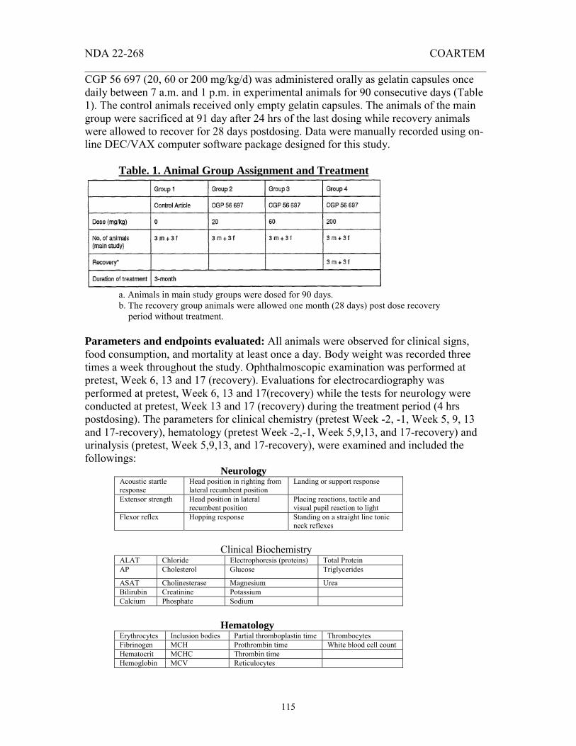
NDA 22-268 COARTEM _______________________________________________________________________
115
CGP 56 697 (20, 60 or 200 mg/kg/d) was administered orally as gelatin capsules once daily between 7 a.m. and 1 p.m. in experimental animals for 90 consecutive days (Table 1). The control animals received only empty gelatin capsules. The animals of the main group were sacrificed at 91 day after 24 hrs of the last dosing while recovery animals were allowed to recover for 28 days postdosing. Data were manually recorded using on-line DEC/VAX computer software package designed for this study. Table. 1. Animal Group Assignment and Treatment
a. Animals in main study groups were dosed for 90 days. b. The recovery group animals were allowed one month (28 days) post dose recovery period without treatment. Parameters and endpoints evaluated: All animals were observed for clinical signs, food consumption, and mortality at least once a day. Body weight was recorded three times a week throughout the study. Ophthalmoscopic examination was performed at pretest, Week 6, 13 and 17 (recovery). Evaluations for electrocardiography was performed at pretest, Week 6, 13 and 17(recovery) while the tests for neurology were conducted at pretest, Week 13 and 17 (recovery) during the treatment period (4 hrs postdosing). The parameters for clinical chemistry (pretest Week -2, -1, Week 5, 9, 13 and 17-recovery), hematology (pretest Week -2,-1, Week 5,9,13, and 17-recovery) and urinalysis (pretest, Week 5,9,13, and 17-recovery), were examined and included the followings: Neurology
Acoustic startle response
Head position in righting from lateral recumbent position
Landing or support response
Extensor strength Head position in lateral recumbent position
Placing reactions, tactile and visual pupil reaction to light
Flexor reflex Hopping response Standing on a straight line tonic neck reflexes
Clinical Biochemistry
ALAT Chloride Electrophoresis (proteins) Total Protein AP Cholesterol Glucose Triglycerides
ASAT Cholinesterase Magnesium Urea Bilirubin Creatinine Potassium Calcium Phosphate Sodium
Hematology
Erythrocytes Inclusion bodies Partial thromboplastin time Thrombocytes Fibrinogen MCH Prothrombin time White blood cell count Hematocrit MCHC Thrombin time Hemoglobin MCV Reticulocytes

NDA 22-268 COARTEM _______________________________________________________________________
116
Urinalysis
Bilirubin Glucose Specific gravity Nitrite Blood Ketone bodies pH Urobilinogen Leucocytes Sediments (microscopic) Proteins Color
Toxicokinetics: Blood samples (2mL) were collected into heparinized tubes from control group of animals (3/sex) at 2 hrs postdosing and from all other groups (3/sex) on day 1 and end of treatment at, 1, 2, 4, 8 and 24 hrs after dosing. Plasma samples were separated and analyzed by
for Benflumetol (CGP 56 695) and Artemether (CGP 56 696), and results are reported separately. Necropsy was performed one day after last administration of the test article or at the end of postdosing recovery period. Adrenals glands, brain, heart, kidneys, liver, lung, ovaries, pituitary gland, prostate, spleen, testes, and thyroid gland were weighed and recorded. Following tissue samples were collected, and processed for histopathological examinations. Histopathology
Adrenal glands Liver Lung Testes Trachea Aorta Jejunum Pancreas Thigh muscle Uterus Bone/bone marrow: sternum
Eyes with optic nerve
Lymph nodes: axillary Lacrimal glands Thyroid glands
Bone/bone marrow:articular surface
Knee join capsules Lymph nodes: mesentery
Salivary glands Parathyroid glands
Brain Gallbladder Ovaries Sciatic nerve Urinary bladder Cecum Heart Prostate Skin Vagina Colon Ileum Rectum Spinal cord Thigh muscle Duodenum Kidneys Spleen Thymus Mammary area Epididymides Esophagus Stomach Tongue
Statistical analysis: Due to limited number of animals in each group, statistical analyses were not performed for the clinical, clinical pathological investigations, and postmortem examinations. Results: No mortality or adverse effects were observed on body weight, food consumption, ophthalmology, electrocardiography and neurology parameters tested in animals treated with CGP 56 697 at all dose levels (20, 60 or 200 mg/kg/d) for three months when compared with control animals. Infrequent diarrhea and feces with apparent compound was noticed in mid and high dose treated animals from time to time. A non-statistical increase in alkaline phosphatase (+18%), urea (+16), cholesterol (+15%), α-1 globulin (+18) was first seen in mid dose (60 mg/kg) treated males at Week 5 of treatment and remained increased by end of the dosing (Week 13). More pronounced changes were seen in these parameters in high dose (200 mg/kg/d) group of animals at Week 5 and 13 when compared with respective controls. Females were found to be more sensitive to these changes than males at all dose levels.
(b) (4)

NDA 22-268 COARTEM _______________________________________________________________________
117
Thyroid stimulating hormone (TSH) was increased at Week 5 and reached to maximum at Week 13 in all dosed animals (161% in 20mg/kg/d; 69% in 60mg/kg/d; 107% in 200 mg/kg/d) No other findings to clinical relevance were observed in males and females due to administration of test articles. A slight to moderate (-10 to -14%) decrease was noticed in levels of thyroxin (T4), triiodothyronine (T3) and reverse triiodothyronine at Week 5 and remained decreased during the period of treatment in both males and females at all dose groups. All these alterations observed during the treatment period returned to normal levels during the recovery period by the end of Week 17 in all males and females. No hematological and urinalysis findings were observed in animals treated with CGP 56 697 at all dose levels (60, 200 or 600 mg/kg/d) when compared with control animals. A +19% increase in absolute and relative liver weight was first seen in males treated with mid dose (60 mg/kg/d) level while +17% increase was observed in high dosed (200 mg/kg/d) females. A dose dependent increase reaching to maximum (+34%) was observed in absolute and relative weight of thyroid in high dose males and being more pronounced in females (+82%). Increase in liver and thyroid weights returned to normal during the recovery period by the end of Week 17. No macroscopic findings were observed in all dosed animals. The microscopic changes seen at the time of necropsy are as follows:
Appears This Way on Original
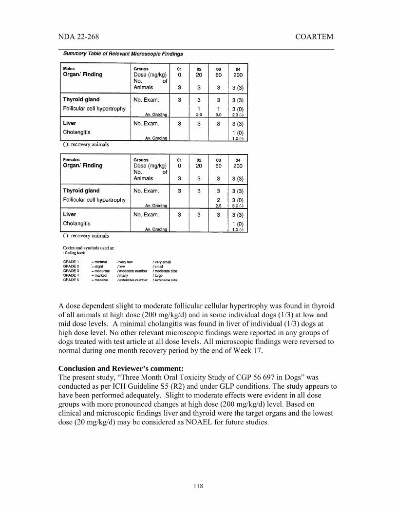
NDA 22-268 COARTEM _______________________________________________________________________
118
A dose dependent slight to moderate follicular cellular hypertrophy was found in thyroid of all animals at high dose (200 mg/kg/d) and in some individual dogs (1/3) at low and mid dose levels. A minimal cholangitis was found in liver of individual (1/3) dogs at high dose level. No other relevant microscopic findings were reported in any groups of dogs treated with test article at all dose levels. All microscopic findings were reversed to normal during one month recovery period by the end of Week 17. Conclusion and Reviewer’s comment: The present study, “Three Month Oral Toxicity Study of CGP 56 697 in Dogs” was conducted as per ICH Guideline S5 (R2) and under GLP conditions. The study appears to have been performed adequately. Slight to moderate effects were evident in all dose groups with more pronounced changes at high dose (200 mg/kg/d) level. Based on clinical and microscopic findings liver and thyroid were the target organs and the lowest dose (20 mg/kg/d) may be considered as NOAEL for future studies.

NDA 22-268 COARTEM _______________________________________________________________________
119
[50.] Study title: Test No.: 95-6031 “CGP 56 696, 13-Week oral toxicity study in rats” Key study findings: Dose dependent effects were seen in rats following 13 Week oral (gavage) administration of CGP 56 696. Increased hematopoiesis and hemosiderosis leading to progressive, hypochromic, and microcytic hemolytic anemia were observed in males and females at high dose (200 mg/kg/d) level. Effects were more pronounced in females compared to males. The clinical effects observed as a result of dosing, were almost reversed during one month recovery period. NOAEL could not be established from this study. Study No.: 95-6031 Conducting laboratory CIBA-GEIGY Limited, Pharmaceutical Division and location: Basel, Switzerland Date of study initiation: November 29, 1995 Study Report Date: September 20, 1996 GLP compliance: Yes QA report: Yes Drug: CGP 56 696 (artemether) Lot No.: 810194 % Active ingredients: 99% (CGP 56 696) Methods Doses: 20, 60, 200 mg/kg/d Species/strain: Albino (Sprague Dawley) rats, Tif: RAIf (SPF) 7 to 11 week old rats weighing 271.0 to 368.0 g (males) and 190.0 to 235 g (females) Route, formulation: By oral gavage in 0.5% suspension of Klucel HF and 0.1% surfactant Tween 80 (10 mL/kg). Study design: Animals were housed in sterilized cages maintained on 12 hrs light and dark cycles (0600 to 1800), 22±30C, and 30 to 70 % relative humidity and acclimatized for 15 days prior to dosing. Standard pellet diet and filtered water were provided ad libitum. Oral administration of CGP 56 696 (0, 20, 60, and 200 mg/kg/d) was given to male and female rats for 91 consecutive days (13 Week) between 7 AM to 1 PM. Animals were subdivided into the following groups: control; main study; recovery; and satellite study. All animals were euthanized by CO2 exposure followed by exsanguinations at the end of dosing (Week 13) while the animals from the recovery group were sacrificed after a one month post dosing recovery period at Week 17. Experimental data were recorded on line (whenever possible) using DEC/VAX 4700a computer and the software package
(b) (4)
(b) (4)
(b) (4) (b) (4)

NDA 22-268 COARTEM
___________________________________________________
120
specifically designed for this study. Statistical analyses were performed using standard battery of statistical tests such as Fisher’s Exact Test, Wilcoxon’s test or Student’s t -test. Animal Groups and Treatment Plan
Main study groups were treated for 13 Weeks *The recovery group animals were allowed to one month post dosing recovery period without any treatment **Dosing was terminated in group-4 females at day 47 due to severity of clinical signs and the extent of anemia. Five of the main group females from group 4 were allowed a four week recovery period without treatment, and terminated at week 11 Control animals received an aqueous solution of 0.5% (w/v) Klucel HF® and Tween 80®
Parameters and endpoints evaluated: All animals were observed for clinical signs, body weight, food consumption and mortality, on a regular basis during the pre-dosing and recovery periods. Hearing and ophthalmologic tests were conducted prior to dosing, at Week 5, 13 and thereafter at Week 17 (recovery period). For clinical chemistry and hematological analyses, blood samples were collected through orbital plexus puncture using EDTA as anticoagulant at Week 5, 9, 11, 13 (treatment period), and Week 17, the end of postdosing recovery period. The parameters for the clinical chemistry, hematology, urinalysis, and histopathology are listed below. Animals were sacrificed postdosing at the end of treatment (Week 13) or recovery period (Week 17) to conduct the necropsy analysis. The adrenal glands, brain, heart, kidney, liver, lung, ovaries, pituitary gland, prostate, spleen, testes, and thymus were weighed while the following organs were sampled, fixed in neutral phosphate buffered formalin
and processed for pathological examination.
(b) (4)
(b) (4)(b) (4)

NDA 22-268 COARTEM _______________________________________________________________________
121
Clinical Chemistry ALAT Calcium Creatinine Potassium Triglycerides AP Chloride Electrophoresis/protein Phosphate Urea ASAT Cholesterol Glucose Sodium Albumin Bilirubin Cholinesterase Magnesium Total
protein Albumin:globulin ratio
Iron TSH T3,T4,rT3 Hematology Erythrocytes Hemoglobin MCHC Fibrinogen Inclusion bodies* White blood cell count Reticulocytes* Hematocrit MCH Partial thromboplastin time Thrombin time* Thrombocytes MCV Prothrombin time *Counted when abnormalities were present. Sodium citrate was used as anticoagulant to measure the clotting factors while EDTA was used to measure other blood parameters Bone Marrow
Cellularity Erythropoiesis predominant cell type
Leucopoiesis predominant cell type
Megakaryocyte numbers Erythropoiesis Myeloid:erythroid ratio Megakaryocyte activity Leucopoiesis Abnormal cell types Urinalysis Bilirubin Ketone bodies Specific gravity Blood Nitrite Urobilinogen Leukocytes pH Volume Glucose Protein Color Urine samples were collected over a three hour period at the end of Week 4 in the control and high dose groups and at Week 8 in recovery animals. Urine was not collected from satellite group animals. Microscopic Examination of Urine Sediments Casts erythrocytes Oxalates Epithelial cells Leucocytes phosphates Leukocytes Urates Toxicokinetics:
Blood samples (2 mL) from animals treated with high dose (200 mg/kg/d) were collected into heparin coated tubes The plasma was separated and processed for toxicokinetics analyses (Ciba-Geigy AG Klybeckstrasse, K-136.1.15, CH-4002, Basel Switzerland). Results are presented in a separate report (# 95-9005).
Statistical evaluations:
Statistical analyses were performed using the SAS programs in accordance to the SOPs of Documentaion and Communication.
(b) (4)

NDA 22-268 COARTEM _______________________________________________________________________
122
Pathology Adrenal glands
Epididymis Kidneys Pituitary glands Spleen Thymus
Aorta Esophagus Knee joint Prostate Stomach Thyroid/Parathyroid glands
Sternum Extra orbital Lachrymal glands
Liver Rectum duodenum Tongue
Femur Eyes with optic nerves
Lung Salivary glands Jejunum Trachea
Brain: Pons & Medulla oblongata
Harderian glands
Lymph nodes: auxiliary
Sciatic nerves Spinal cord Urinary bladder
Cecum Heart Lymph nodes: Mesenteric
Seminal vesicles Testes Uterus
Colon Ileum Ovaries Skin/mammary area
Thigh muscle
Vagina
Results Clinical signs and Mortality:
Five deaths (2 males and 3 females) were recorded in animals treated with high dose (200 mg/kg/d) test compound. Body weight loss, hunched posture, slow respiration, emaciation, piloerection, distended abdomen, varying degree of anemia, pale extremities & eyes were seen in most of these animals. Due to severe anemic condition and apparent clinical signs, the dosing was terminated at Day 47 in females, and five females were put on four week recovery period without any treatment. More pronounced effects were seen in high dose treated females than males.
Liver necrosis and eye damage was reported in high dose animals following histopathological examination. A slight to moderate magnitude of pallor of the extremities was observed in all males from Day 35 and continued throughout the treatment period until Week 13. In females similar magnitude of pallor was recorded at Day 35, and continued until Day 47 when the dosing was terminated in high dose animals. Relevant clinical signs were not observed at low (20 mg/kg/d) and mid (60 mg/kg/d) dose treated animals.
Body weights: A decrease in body weight gains was first seen in males at Week 1 in mid (-6%) and high dose (-9%) treated groups and continued to -10% and -25% in mid and high dose groups respectively at Week 13. In females, the decrease (-6%) in body weight gains was first noticed at Week 3 in high dose group while no decrease was evident in mid dose animals. A dose dependent reduction in body weight gains was seen in males treated with mid (-10/%) and high (-25%) dose test article at Week 13 when

NDA 22-268 COARTEM _______________________________________________________________________
123
compared with controls. Both males and females recovered the loss in body weight gain during one month post dosing recovery period by Week 17.
Food consumption:
Reduction (-25/19%) in food consumption was observed in males and females treated with high dose test article at Week 1. Loss in food consumption was reversed to normal at Week 13, with complete recovery by Week 17.
Hearing and Ophthalmology:
Treatment related hearing and ophthalmologic effects were not seen in rats treated with low, mid, and high dose test compound. However, at Week 13 an ocular pallor was evident in high dose (200 mg/kg/d) males.
Clinical Biochemistry:
An increased ASAT activity (+18/170%) potassium (+17/24%) and bilirubin (+26/154%) concentration with decrease in alkaline phosphatase (-35/50%), cholesterol (-43/24%), triglycerides (+115/149%) and glucose (-14/35%) were observed at Week 5 in high dose (200 mg/kg/d) treated males and females. ALAT activity was increased (+74%) while plasma iron concentration decreased (-70%) in females when compared with untreated controls at Week 5. As evident from the data, females were more sensitive to these alterations than males. Mild to low changes were observed in mid (60 mg/kg/d) and low (20mg/kg/d) dose treated animals. More pronounced effects were seen in treated animals of all groups at Week 9 and 13. Due to severe effects, the study was terminated at day 47 in females treated with high dose of test article. Most of the clinical parameters returned to normal by Week 17 during one month recovery period in males and females except ASAT (+77%), ALAT (+100%) and potassium (+8%) that remained elevated in males.
Hematology:
A significant decrease in erythrocyte counts (-26/52%), hematocrit value (-39/51%) hemoglobin (-61/96%) levels, and MCH (-28/28%) as characterized by microcytic hypochromic hemolytic anemia were seen in males and females at Week 5 in high dose (200 mg/kg/d) groups. These changes were accompanied by a marked increase in reticulocytes (+7.5/9 fold) and WBC (+2.5/1.2 fold) counts in males and females. Similar types of hematological changes with higher magnitude were observed in males and females in high dose groups at Week 9 and 13.

NDA 22-268 COARTEM _______________________________________________________________________
124
Mild to low grade changes were seen in mid (60 mg/kg/d) and low (20 mg/kg) dose treated males and females, however, changes were more pronounced in females when compared with males. Bone marrow analysis demonstrated an increased erythropoiesis, decreased myeloid:erythroid ratio and hyperplasia of normoblast. Changes in bone marrow parameters causing erythropoiesis returned to normal by Week 17 during one month postdosing recovery period.
Urinalysis:
Urinalysis did not reveal any treatment related changes in either group of animals.
Organ weights:
An absolute and relative weight decreases were observed in lung (-22/21%), liver (-13/10%), thymus (-20/18%) and prostate gland (-33/31%) while a significant increase was observed in spleen (+169/177%) of males treated with high dose (200 mg/kg/d) test article when compared with controls. No adverse effects were seen in mid (60 mg/kg/d) and low (20 mg/kg) dose groups. Adverse effects were not seen in most of the organ weights in females except a great decrease in the absolute spleen weight (+88 %) of high dose groups. Most of the organ weights returned to normal during the recovery.
Macroscopic & microscopic findings:
An enlarged spleen was found in low (1/10), mid (4/10), and high (8/10) dose treated males and remained increased (2/5) in high dose group during the recovery period. Hemosiderin pigments were found in ovaries, adrenals, mesenteric lymph nodes, lacrimal glands, liver and spleen of males and females in all dose groups and remained present in high dose groups even after recovery period. Most of the hemosiderin pigments were located in Kupffer cells of liver except in some cases where it was in hepatocytes. Centrilobular hepatic necrosis was seen in two males and three females in high dose groups. Brown lipofuscin pigments were present in the proximal convoluted tubule and epithelium of kidney in all dose groups even in some individual controls. Dose dependent hemopoiesis was seen in spleen of both sexes with reversal to normal after recovery. Examination of bone marrow (sternum and femur) revealed an increased erythropoiesis, presence of hemosiderin pigments and hypercellularity in both sexes at high dose treatment. Lower magnitude of changes were seen in mid and low dose group animals. Most of the animals recovered to normal except the persistence of hemosiderin pigment in some cases. Macrophage aggregation was found within the lumen of larger blood vessels of lung in high dose animals.

NDA 22-268 COARTEM _______________________________________________________________________
125
Most of the microscopic changes were reversed to normal during the recovery.
Summary and Comments:
The present study, “13 Week Oral Toxicity Study of CGP 56 696 in Rats” was conducted according to “Good Laboratory Practice (Switzerland), Procedures and Principles,” March 1986, section 4, 2.2(e) and the UK Department of Health (1989). The study appears to have been conducted adequately. Increased erythropoiesis in bone marrow, deposition of hemosiderin pigments in vital organs, and hemopoiesis in spleen and liver might be due to toxicological effects of test article in rats at mid and high dose levels. Increased reticulocyte and thrombocyte counts in affected animals seemingly, contributed toward the development of progressive, hypochromic, microcytic, hemolytic anemia. Effects of test article were more pronounced in females compared to males. Most of the adverse effects were reversed to normal during one month post dosing recovery period. The NOAEL could not be determined from this study because of hematologic and hemopoietic effects observed at all dose levels.
[51.] Study title: CGP 56 696: 13-Week oral (capsule) toxicity study in dogs Key study findings: Dose dependent effects were seen in beagle dogs following 13 Week consecutive oral (capsule) administration of CGP 56 696, at all dose levels. Effects were more predominant in animals treated with high dose (300 mg/kg/d) test article. Hematological parameters in particular the red blood cells were greatly affected leading to erythropoiesis and increased hematopoietic activity in bone marrow and spleen. The clinical signs observed as a result of dosing, were almost reversed during one month recovery period. NOAEL could not be established from this study. Study no.: MIN 954154: Test No. 95-6030 Conducting laboratory CIBA-GEIGY Limited, Pharmaceutical Division and location: Summit, New Jersey, USA. Date of study initiation: October 10, 1995 Study report date: September 6, 1996 GLP compliance: Yes (OECD Guideline No.:409, Switzerland) QA report: Yes Drug: CGP 56 696 (artemether) Lot No. 810194 % Active ingredients: 99.2% Control article: Empty gelatin capsules

NDA 22-268 COARTEM _______________________________________________________________________
126
Methods: Doses: 30, 100, and 300 mg/capsule/kg Species/strain: Pedigree Beagle dogs
Number/sex/group 3/sex/group (9.0 to 13.6 kg - males; 8.7 to 11.1 kg or time point females at the time of dosing) Route, formulation: Orally in gelatin capsules volume and infusion rate Satellite groups used for Yes toxicokinetics or recovery: Study design: Animals were housed in solid bottom cages (1m x 1m) in an air conditioned room maintained at 69 ± 50 F, the relative humidity 50 ± 20% and 12 hr light/dark cycles. Standard canine pellet diet ( ) was given daily at 350 g/animal while water was provided ad libitum. The animals were given antiparasitic therapy, and vaccinated against rabies, distemper, leptospirosis, infectious canine hepatitis, kennel cough, PARVO and CORONA virus. An indoor run for one hr was given everyday to these animals except on holidays where only 30 min exercise was provided. All animals were acclimatized for 11 weeks prior to dosing. CGP 56 696 (30, 100 or 300 mg/kg/d) was administered orally, once daily between 7 am to 1 pm in the respective group of experimental animals for 13 Weeks (Table 1) as hand filled gelatin capsules . The control animals received only empty gelatin capsules. All animals were weighed weekly, and the dose was administered accordingly. Two animals did not receive the correct dose amount due to vomiting and capsules were recovered from feed bowl. These isolated events had no adverse effects on the study. Animals were allowed to recover for one month prior to their sacrifice at Week 17. Data were manually recorded using on-line computer software package designed for this study. Table. 1. Treatment Schedule
(b) (4)
(b) (4)
(b) (4)
(b) (4)

NDA 22-268 COARTEM _______________________________________________________________________
127
1. Animals in main study groups were dosed for 13 Weeks. Three animals (per sex) from each group were subjected to necropsy at the end of study. 2. The recovery group animals were allowed one month post dose recovery period without treatment. Three animals (per sex) from control and high dose group were subjected to necropsy at the end of study. Parameters and endpoints evaluated: All animals were observed for clinical signs, food consumption, and mortality at least once a day. Body weight was recorded weekly. Ophthalmoscopic examination was performed 29 days prior to initiation of dosing; pretest day 1, and at Week 6 and 13. Electrocardiography (EKG) was recorded prior to initiation of dosing (day 10, 11 and 15), during dosing (4 to 6 hrs after dosing) at Week 5, 9, 13 (main group animals) and at Week 17 (recovery animals). The clinical parameters for clinical chemistry (pretest at day 7 and Week 5, 9, 13 - main group animals and Week 17 - recovery animals), hematology (pretest day 7, Week 5, 10, 13 – main groups and Week 17- recovery animals) and urinalysis (pretest day 8/9, Week 5, 10, 13-main groups, and Week 17-recovery animals), were examined and included the followings. Bone marrow, thyroid, spleen, and gross lesions from recovery animals were subjected to histopathological (H&E stain) examinations. Clinical Biochemistry
ALAT Chloride Glucose Total Protein AP Cholesterol Sodium Triglycerides
ASAT Calcium Magnesium Phosphorus (inorganic) Bilirubin Creatinine Potassium albumin Globulin BUN
Hematology
Erythrocytes Prothrombin time Partial thromboplastin time Thrombocytes
Heinz body MCH Red cell morphology White blood cell count
Hematocrit MCHC Thrombin time Platelet count Hemoglobin MCV Reticulocytes Differential count
Urinalysis
Bilirubin Glucose Specific gravity Nitrite
Blood Ketone bodies pH Urobilinogen
Volume Sediments (microscopic) Proteins Color/clarity
Pathological Examination
Adrenal glands
Epididymis Kidneys Pituitary glands
Spleen Thymus
Aorta Esophagus Knee joint Prostate Stomach Thyroid/Parathyroid glands
Sternum Extraorbital Lachrymal
Liver Rectum duodenum Tongue

NDA 22-268 COARTEM _______________________________________________________________________
128
glands Femur Eyes with
optic nerves
Lung Salivary glands Jejunum Trachea
Brain: Pons & Medulla oblongata
Harderian glands
Lymph nodes: auxiliary
Sciatic nerves Spinal cord
Urinary bladder
Cecum Heart Lymph nodes: Mesenteric
Seminal vesicles
Testes Uterus
Colon Ileum Ovaries Skin/mammary area
Thigh muscle
Vagina
Necropsy:
Animals were fasted overnight (12 hrs), anesthetized with pentobarbital, exsanguinated, and subjected to full gross necropsy one day after last administration of test article at Week 13 or at the end of postdosing recovery period (Week 17). Adrenals glands, brain, heart, kidneys, liver, lung, ovaries, prostate, spleen, testes, pituitary, thyroid and parathyroid glands were weighed and recorded.
Toxicokinetics:
Blood samples (~5mL) were collected into heparinized tubes from the jugular vein of the high dose treated recovery animals at 1, 2, 4, 6, 8, and 24 hrs after dosing on days 2 and 91. The plasma samples were separated and analyzed by
for analysis. Statistical evaluations:
Statistical analyses were performed using the SAS programs in accordance to the SOPs of Documentaion and Communication.
Results Clinical signs and mortality:
No clinical signs or mortality were seen in male and female dogs due to oral (capsule) administration of the test article for 13 consecutive weeks at all dose levels (30, 100 and 300 mg/kg/d).
Body weight:
A lower body weight gain (+5.6 to 10%) was seen in males treated with high dose (300 mg/kg/d) compound in comparison with controls (+11 to 22%) during the exposure period from Week 5 to 13 and returned to normal during the recovery. The body weight gains in females was very low (0.3 to 1.8%) compared to controls (5 to 9%), and returned to normal during one month postdosing recovery period.
(b) (4)

NDA 22-268 COARTEM _______________________________________________________________________
129
Except a very little change in low dose (30 mg/kg/d) group, surprisingly no changes in body weight gains were found at mid dose (100 mg/kg/d) level in males and females. Vomiting was occasionally seen in mid dose treated animals.
Food consumption:
No changes were seen in food consumption of beagle dogs at all dose levels as a result of oral (capsule) administration of test article.
Hematology:
A decrease in hemoglobin (-29.22%), hematocrit (-11%), MCV (-6%), MCHB (-6%) levels and increased reticulocyte counts (+29%) was observed in males and an increase (+84%) in reticulocyte and platelet counts (+45%) was seen in females at week 13 in high dose treated animals. Altered hematological parameters did not return to complete normalcy during one month postdosing recovery period at Week 17.
Clinical pathology:
Clinical pathological findings revealed a mild to moderate effect of test compound on red blood cells at all dose levels, predominantly at high dose (300 mg/kg/d) in both sexes. In addition to a change in the morphology of RBC’s, presence of metarubricytes (nucleated red blood cells) was detected in peripheral blood smears of high dose (300 mg/kg/d) treated males at Weeks 5, 9, and 13 and in females at Week 13. The bone marrow smear analysis demonstrated an increased erythropoiesis in all males (3/3) and two females (2/3) while microscopic examination revealed the presence of hemopoiesis in spleen of two males (2/3) at Week 13 in high dose group. Thyroidal follicular epithelial hypertrophy as evident from colloid filled follicles at the lining of the epithelium was also monitored in high dose treated males at Week 13.
Effects of CGP 56696 on RBC Morphology
The physical/auditory, electrocardiographic, ophthalmoscopic examinations, clinical chemistry and urinalysis did not reveal any abnormalities in either group of beagle dogs related to test compound at any dose levels. Summary and Comments:

NDA 22-268 COARTEM _______________________________________________________________________
130
The present study, “13 Week Oral (capsule) Toxicity Study of CGP 56 696 in Dogs - MIN 954154” was conducted according to Good Laboratory Practice (USA). The study appears to have been conducted adequately. Increased erythropoiesis in bone marrow, hemopoiesis in spleen and hypertrophic changes in thyroid of beagle dogs treated at high dose level (300 mg/kg/d) might be due to toxicological effects of test article. Effects of test article were more pronounced in females compared to males. Most of the adverse effects were reversed during one month postdosing recovery period. The NOAEL could not be determined from this study because of hematologic effects. However, the Sponsor suggests the use of mid dose (100 mg/kg/d) for future studies.
[52.] Study title: Test No.: 95-6026 “CGP 56 695, 13-Week oral toxicity study in rats” Key study findings: Dose dependent effects of CGP 56 695 were seen in rats during the 13 Week oral (gavage) administration. Increased cellular vacuolation in pituitary glands and diffused follicular hypertrophy were seen in both sexes at high dose (1000 mg/kg/d) groups, and were more pronounced in females. The clinical effects observed as a result of dosing, did not completely reverse during one month recovery period. NOAEL could not be established from this study. Study No.: 95-6026 Conducting laboratory CIBA-GEIGY Limited, Pharmaceutical Division and location: Basel, Switzerland Date of study initiation: September 28, 1995 Study Report Date: October 18, 1996 GLP compliance: Yes QA report: Yes Drug: CGP 56 695 Batch No.: 800395 % Active ingredients: 99% CGP 56 695 (benflumetol) Methods Doses: 100, 300, 1000 mg/kg/d Species/strain: Albino (Sprague Dawley) rats, Tif: RAIf (SPF) 6 to 8 week old rats weighing 167.0 to 304.0 g Route, formulation: By oral gavage in 0.5% suspension of Klucel HF and 0.1% surfactant Tween 80 (10 mL/kg). Study design: Animals were housed in sterilized cages maintained on 12 hrs light and dark cycles (0600 to 1800), 22±30C, and 30 to 70 % relative humidity and acclimatized for 2 weeks prior to

NDA 22-268 COARTEM ______________________
131
dosing. Standard pellet diet and filtered water (0.2 µm) were provided ad libitum. Oral administration of CGP 56 695 (0, 100, 300, and 1000 mg/kg/d) was given to male and female rats for 91 consecutive days (13 Week) between 7 AM to 1 PM. Animals were subdivided into the following groups: control; main study; recovery; and satellite study: Animal Groups and Treatment Plan
* The recovery group animals were allowed to one month post dosing recovery period without any treatment ** Main study and Satellite groups were treated for 13 Weeks *** Control animals received an aqueous solution of 0.5% (w/v) Klucel HF® and Tween 80®
All animals were euthanized by CO2 exposure followed by exsanguinations at the end of dosing (Week 13) while the animals from the recovery group were sacrificed at Week 17 after a one month post dosing recovery period. Experimental data were recorded on line (whenever possible) using DEC/VAX 4700a computer and the software package
specifically designed for this type of study. Statistical analyses were performed using standard battery of statistical tests such as Fisher’s Exact Test, Wilcoxon’s test or Student’s t -test. Parameters and endpoints evaluated: All animals were observed for clinical signs, body weight, food consumption and mortality, on a regular basis during the pre-dosing and recovery periods. Hearing and ophthalmologic tests were conducted prior to dosing, at Week 5, 13 and thereafter at Week 17 (recovery period). Blood samples were collected through orbital plexus puncture using EDTA as anticoagulant at Week 1, 5, 9, 13 (treatment period), and Week 17 at the end of recovery for clinical chemistry and hematological analyses. The parameters selected for the clinical chemistry, hematology, urinalysis, and histopathology are listed below. Clinical Chemistry ALAT Calcium Creatinine Potassium Triglycerides AP Chloride Electrophoresis/protein Phosphate Urea ASAT Cholesterol Glucose Sodium TSH Bilirubin Cholinesterase Magnesium Total protein T3,T4,rT3
(b) (4)
(b) (4) (b) (4)

NDA 22-268 COARTEM _______________________________________________________________________
132
Hematology Erythrocytes Hemoglobin MCHC Fibrinogen Inclusion bodies* White blood cell count Reticulocytes* Hematocrit MCH Partial thromboplastin time Thrombin time* Thrombocytes MCV Prothrombin time Counted when abnormalities were present. Sodium citrate was used as anticoagulant to measure the clotting factors while EDTA was used to measure other blood parameters Bone Marrow
Cellularity Erythropoiesis predominant cell type
Leucopoiesis predominant cell type
Megakaryocyte numbers Erythropoiesis Myeloid:erythroid ratio Megakaryocyte activity Leucopoiesis Abnormal cell types Urinalysis Bilirubin Ketone bodies Specific gravity Blood Nitrite Urobilinogen Leukocytes pH Volume Glucose Protein Color Urine samples were collected over a three hour period at the end of Week 4, 8, 13 in control and high dose groups and at Week 17 in recovery animals. Urine samples were not collected from satellite group animals. Microscopic Examination of Urine Sediments Casts erythrocytes Oxalates Epithelial cells Leucocytes phosphates Leukocytes Urates Pathology* Adrenal glands
Epididymis Kidneys Pituitary glands Spleen Thymus
Aorta Esophagus Knee joint Prostate Stomach Thyroid/Parathyroid glands
Sternum Extra orbital Lachrymal glands
Liver Rectum duodenum Tongue
Femur Eyes with optic nerves
Lung Salivary glands Jejunum Trachea
Brain: Pons & Medulla oblongata
Harderian glands
Lymph nodes: auxiliary
Sciatic nerves Spinal cord Urinary bladder
Cecum Heart Lymph nodes: Mesenteric
Seminal vesicles Testes Uterus
Colon Ileum Ovaries Skin/mammary area
Thigh muscle
Vagina
*Animals were sacrificed post-dosing at the end of treatment (Week 13) or recovery period (Week 17) to conduct the necropsy analyses. The adrenal glands, brain, heart, kidney, liver, lung, ovaries, pituitary gland,

NDA 22-268 COARTEM _______________________________________________________________________
133
prostate, spleen, testes, and thymus were weighed and recorded. Following organs were sampled, fixed in 4% neutral phosphate buffered formalin and processed for pathological examinations. Epididymides, lacrimal glands, eyes with optic nerves, Harderian glands, prostate, seminal vesicles and testes were fixed in Davidson solution. Bone marrow demineralization was done with Triplex in Tris buffer or 10% formic acid. Samples (4 micron) were processed for pathological examination and Haematoxylin & Eosin staining. Toxicokinetics:
Animals were divided into 2 subgroups: a & b. Blood samples (0.7 mL) were collected into heparin coated tubes at following time points. Plasma was separated and processed for toxicokinetics analyses
Results are presented in a separate report.
Statistical evaluations:
Statistical analyses were performed using the SAS programs in accordance to the SOPs of Documentaion and Communication.
Results Clinical signs and Mortality:
No mortality and clinical signs were observed in rats due to oral administration of test article for 13 Weeks.
Body weights: No effects were observed on absolute body weight and body weight gains in rats due to oral administration of test article for 13 Weeks.
Food consumption: No effects were observed on food consumption in rats due to oral administration of test article for 13 Weeks.
Hearing and Ophthalmology: No effects on hearing and ophthalmological parameters were observed in rats due to oral administration of test article for 13 Weeks.
(b) (4)
(b) (4)
(b) (4)

NDA 22-268 COARTEM _______________________________________________________________________
134
Clinical Biochemistry: No abnormality was noted in most of the clinical parameters examined due to oral administration of test article in rats for 13 week. A slight increase in ASAT and choline esterase (CHE) was observed in males at all dose levels (30, 100 & 1000 mg/kg/d) being more pronounced at Week 14 and 9 in high dose (+7.3/11.5%) groups. In females the increase (+21%) was noticed only in CHE at Week 9 in high dose (1000 mg/kg/d) groups. Levels of thyroid stimulating hormone (TSH) increased while thyroxin (T4) and reverse triiodothyronine (rT3) decreased in males and females at all dose levels. The most pronounced increase in TSH (~3.5/1.5 fold) and decrease in rT3 (-56/28%) was seen at Week 9 while the T4 (-30/44%) at Week 13 and 9 in high dose treated males and females respectively. Most of these altered parameters returned to their normal levels during one month postdosing recovery period.
Hematology: No effects were observed on hematological parameters in rats due to oral administration of test article for 13 Weeks except a slight decrease in MCHC (-2%) and reticulocyte counts (-15%) in males treated with high dose compound at Week 9. In females, the decrease (-21%) in reticulocyte counts was seen at Week 9 in animals treated with high dose test article.
Urinalysis:
Urinalysis did not reveal any treatment related changes in either group of animals.
Organ weights:
No effects were observed on absolute and relative organ weights in males except a little decrease in spleen (-5%) and thymus (-13%) at Week 9 in high dose groups. In females an increase was seen in heart (+14%) and spleen (+10%) in high dose groups while the increase in liver and ovary weights was seen at all dose levels being more pronounced in high dose groups (+16%) and (+34%) respectively. Most of the organ weights returned to normal during recovery.
Macroscopic & microscopic findings:
No macroscopic changes were seen at the time of necropsy. Slight to moderate granulomatous inflammation with macrophages and multinucleated giant cells having clefts were seen in small intestine (jejunum) and mesenteric lymph nodes in both sexes. Effects were more predominantly seen in high dose (1000 mg/kg/d) females. Cellular vacuolation in the adenohypophysis of pituitary glands were seen in male rats at all dose levels being more pronounced in high dose groups. A minimal to slight diffused follicular cellular hypertrophy was seen in

NDA 22-268 COARTEM _______________________________________________________________________
135
thyroid gland of both sexes at all dose levels. Most of the microscopic changes observed during the treatment, have shown a reversible trend, however, did not completely reverse to normal during the recovery period.
Summary and Comments:
The present study, “13 Week Oral Toxicity Study of CGP 56 695 in Rats” was conducted according to “Good Laboratory Practice (Switzerland), Procedures and Principles,” March 1986, section 4, 2.2(e) and the UK Department of Health (1989). The study appears to have been conducted adequately.
Notable effects of CGP 56 695 were seen on pituitary and thyroid glands, ovary and liver weights. Most of the treatment related effects have shown a reversible trend, however, did not completely reverse to normal during the recovery. The NOAEL could not be determined from this study because of effects of the test article were seen at all dose levels.
[53.] Study title: CGP 56 695: 13-Week oral (capsule) toxicity study in dogs (MIN 954153) Key study findings: No abnormalities were seen in beagle dogs following 13 Week consecutive oral (capsule) administration of CGP 56 695, at all dose levels 60, 200, and 600 mg/capsule/kg/d. Dose related effects were limited to pale discolored fecal changes with the presence of apparent test compound. The clinical signs observed as a result of dosing, were reversed during one month recovery period. Study no.: 95-6025 (MIN 95-4153) Conducting laboratory CIBA-GEIGY Limited, Pharmaceutical Division and location: Summit, New Jersey, USA. Date of study initiation: October 19, 1995 Study report date: July 10, 1997 GLP compliance: Yes QA report: Yes Drug: CGP 56 695 Lot No. 800395 % Active ingredients: 100.0% Control article: Empty gelatin capsules Methods: Doses: 60, 200, and 600 mg/capsule/kg/d Species/strain: Pedigree Beagle dogs from
Number/sex/group 3 /sex/group (10.2 to 12.3 kg - males; 8.5 to 11.8 kg or time point females at the time of dosing) Route, formulation: Orally in gelatin capsules
(b) (4)

NDA 22-268 COARTEM _______________________________________________________________________
136
volume and infusion rate Satellite groups used for Yes toxicokinetics or recovery:
Study design: Animals were housed in solid bottom cages (1m x 1m) in an air conditioned room maintained at 69 ± 50 F, the relative humidity 50 ± 20% and 12 hr light/dark cycles. Standard canine pellet diet was given daily at 350 g/animal while water was provided ad libitum. The animals were given anti-parasitic therapy, and vaccinated against rabies, distemper, leptospirosis, infectious canine hepatitis, kennel cough, PARVO and CORONA virus. An indoor run for one hr was given everyday to these animals except on holidays where only 30 min exercise was provided. All animals were acclimatized for 7 weeks prior to dosing. CGP 56 695 (60, 200 or 600 mg/kg/d) was administered orally once daily between 7 am and 1 pm in the respective group of experimental animals for 13 Weeks (Table 1) as hand filled gelatin capsules The control animals received only empty gelatin capsules. All animals were weighed weekly, and the dose was administered accordingly. Animals were allowed to recover for one month prior to their sacrifice at Week 17. Data were manually recorded using on-line computer software package designed for this study. Table. 1. Treatment Schedule
1. Animals in main study groups were dosed for 13 Weeks. Three animals (per sex) from each group were subjected to necropsy at the end of study. 2. The recovery group animals were allowed one month post dose recovery period without treatment. Three animals (per sex) from control and high dose group were subjected to necropsy at the end of study.
(b) (4)
(b) (4)
(b) (4)

NDA 22-268 COARTEM _______________________________________________________________________
137
Parameters and endpoints evaluated: All animals were observed for clinical signs, food consumption, and mortality at least once a day. Body weight was recorded weekly. Ophthalmoscopic examination was performed 20 days prior to initiation of dosing; pretest day 1, and at Week 6 and 13. Electrocardiography (EKG) was recorded 14 days prior to initiation of dosing, during the period of dosing (4 to 6 hrs after dosing) at Week 5, 9, 13 (main group animals) and at Week 17 (recovery animals). The clinical parameters for clinical chemistry (pretest at day 7 and Week 5, 9, 13 - main group animals and Week 17 - recovery animals), hematology (pretest day 7, Week 5, 10, 13 – main groups and Week 17- recovery animals) and urinalysis (pretest day 8/9, Week 5, 10, 13-main groups, and Week 17-recovery animals), were examined and included the followings. Bone marrow, thyroid, spleen, and gross lesions from recovery animals were processed and subjected to histopathological (H&E stain) examinations. Clinical Biochemistry
ALT Chloride Glucose Total Protein AP Cholesterol Sodium Triglycerides
AST Calcium Magnesium Phosphorus (inorganic) Bilirubin Creatinine Potassium albumin Globulin BUN
Hematology
RBC Prothrombin time Partial thromboplastin time Thrombocytes
Heinz body MCH Red cell morphology WBC
Hematocrit MCHC Thrombin time Platelet count Hemoglobin MCV Reticulocytes Differential count
Urinalysis
Bilirubin Glucose Specific gravity Nitrite
Blood Ketones pH Urobilinogen
Volume Sediments (microscopic) Proteins Color/clarity
Pathological Examination
Adrenal glands
Epididymis Kidneys Pituitary glands
Spleen Thymus
Aorta Esophagus Knee joint Prostate Stomach Thyroid/Parathyroid glands
Sternum Extraorbital Lachrymal glands
Liver Rectum duodenum Tongue
Femur Eyes with optic nerves
Lung Salivary glands Jejunum Trachea
Brain: Pons & Medulla oblongata
Harderian glands
Lymph nodes: auxiliary
Sciatic nerves Spinal cord
Urinary bladder

NDA 22-268 COARTEM _______________________________________________________________________
138
Cecum Heart Lymph nodes: Mesenteric
Seminal vesicles
Testes Uterus
Colon Ileum Ovaries Skin/mammary area
Thigh muscle
Vagina
Necropsy:
Animals were fasted overnight (12 hrs), anesthetized with pentobarbital, exsanguinated, and subjected to full gross necropsy one day after last administration at Week 13 or at the end of postdosing recovery period (Week 17). Adrenals glands, brain, heart, kidneys, liver, lung, ovaries, prostate, spleen, testes, pituitary, thyroid and parathyroid glands were weighed and recorded. Necropsy was performed by the Subdivision of Pathology.
Toxicokinetics:
Blood samples (~5mL) were collected into heparinized tubes from the jugular vein from animals (3/sex) at 1, 2, 4, 6, 8, 12 and 24 hrs after the first dose and postdosing on days 71 and 72. The plasma samples were separated and analyzed by for analysis. Concentrations of CGP 56 695 were measured in plasma samples using compound specific assays.
Statistical evaluations:
Statistical analyses were performed using the SAS programs in accordance to the SOPs of Documentaion and Communication.
Results: Clinical signs and mortality
No clinical signs or mortality were seen in male and female dogs due to oral (capsule) administration of the test article for 13 consecutive weeks. Incidents of vomiting, and pale discolored feces with apparent compound were noticed occasionally in both sexes at mid and high dose levels (200 and 600 mg/kg/d).
Electrocardiographic examinations:
No abnormalities were seen in electrocardiographic parameters of beagle dogs at all dose levels as a result of oral (capsule) administration of test article. Statistically significant decrease in mean PR interval length and increases in mean heart rate and QRS complex in females at 600 mg/kg/d were the isolated events and considered as incidental by the Sponsor.
Ophthalmological examinations:
No ophthalmological abnormalities were seen in beagle dogs at all dose levels due to oral (capsule) administration of test article.
Body weight
(b) (4)

NDA 22-268 COARTEM _______________________________________________________________________
139
No abnormalities were seen in body weight of beagle dogs at all dose levels as a result of oral (capsule) administration of test article.
Food consumption
No abnormalities were seen in food consumption of beagle dogs at all dose levels as a result of oral (capsule) administration of test article.
Hematology
A decrease in hemoglobin (-29.22%), hematocrit (-11%), MCV (-6%), MCHB (-6%) levels and increased reticulocyte counts (+29%) was observed in males and an increase (+84%) in reticulocyte and platelet counts (+45%) was seen in females at week 13 in high dose treated animals. Altered hematological parameters did not return to complete normalcy during one month postdosing recovery period at Week 17.
Clinical pathology
Clinical abnormalities were not observed in beagle dogs at all dose levels as a result of oral (capsule) administration of test article for 13 Weeks. Increased liver weights in females at mid dose (200 mg/kg/d) and pituitary weights in all females were not considered dose related. All other microscopic lesions considered a spontaneous, incidental, and not dose related.
Summary and Comments:
The present study, “13 Week Oral (capsule) Toxicity Study of CGP 56 695 in Dogs - MIN 954153” was conducted according to Good Laboratory Practice (USA). The study appears to have been conducted adequately and no abnormalities were found to be related with test article administration. Dose dependent effects were primarily limited to feces with presence of test compound (seen yellowish) in both male and female dogs, and suggested an incomplete absorption from GI tract. Most of the adverse effects were reversed to normal during one month postdosing recovery period. The NOAEL could not be determined from this study because of hematologic effects at all dose levels. However, the Sponsor suggests the use of mid dose (100 mg/kg/d) for future studies.
Appears This Way on Original
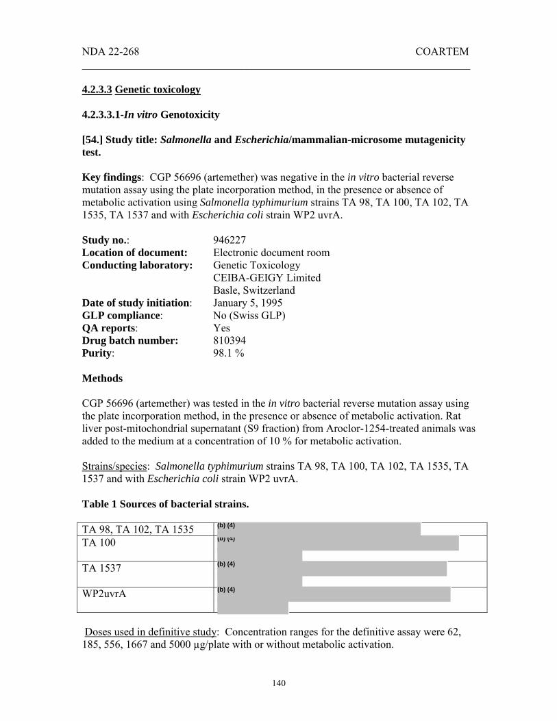
NDA 22-268 COARTEM _______________________________________________________________________
140
4.2.3.3 Genetic toxicology 4.2.3.3.1-In vitro Genotoxicity [54.] Study title: Salmonella and Escherichia/mammalian-microsome mutagenicity test. Key findings: CGP 56696 (artemether) was negative in the in vitro bacterial reverse mutation assay using the plate incorporation method, in the presence or absence of metabolic activation using Salmonella typhimurium strains TA 98, TA 100, TA 102, TA 1535, TA 1537 and with Escherichia coli strain WP2 uvrA. Study no.: 946227 Location of document: Electronic document room Conducting laboratory: Genetic Toxicology CEIBA-GEIGY Limited Basle, Switzerland Date of study initiation: January 5, 1995 GLP compliance: No (Swiss GLP) QA reports: Yes Drug batch number: 810394 Purity: 98.1 % Methods CGP 56696 (artemether) was tested in the in vitro bacterial reverse mutation assay using the plate incorporation method, in the presence or absence of metabolic activation. Rat liver post-mitochondrial supernatant (S9 fraction) from Aroclor-1254-treated animals was added to the medium at a concentration of 10 % for metabolic activation. Strains/species: Salmonella typhimurium strains TA 98, TA 100, TA 102, TA 1535, TA 1537 and with Escherichia coli strain WP2 uvrA. Table 1 Sources of bacterial strains. TA 98, TA 102, TA 1535 TA 100
TA 1537
WP2uvrA
Doses used in definitive study: Concentration ranges for the definitive assay were 62, 185, 556, 1667 and 5000 µg/plate with or without metabolic activation.
(b) (4)
(b) (4)
(b) (4)
(b) (4)

NDA 22-268 COARTEM _______________________________________________________________________
141
Basis of dose selection: In a preliminary, dose-ranging assay, TA 100 and WP2 uvrA were exposed to drug at 21, 62, 185, 556, 1667 and 5000 µg/plate with or without metabolic activation. At 1667 and 5000 µg/plate, the background lawn for TA 100 was reduced (with or without metabolic activation) and the number of revertant colonies was weakly reduced in strain TA 100 (with and without metabolic activation) and in WP2 uvrA (with metabolic activation). At 5000 µg/plate, a precipitate was observed with both TA 100 and WP2 uvrA. Negative controls: Dimethylformamide (DMF) Table 2 Positive controls
Incubation and sampling times: Inoculates from frozen master copies of tester strains were grown in liquid nutrient broth medium (NB-medium) overnight and then plated on NB-agar. After incubation, single colonies were taken from the plates, grown for 12 hours in liquid NB-medium and then used for the experiment. Plates were incubated in triplicates with test compound for 48 hours at 37 ºC in darkness after which the colonies were counted and background lawn was evaluated using an Artek colony counter (or manually in the presence of agar damage or precipitates). Sponsor’s criteria for positive results The test compound was considered positive if the mean number of revertants per plate was increased by 1.5 fold for strains TA 100 or TA 102 and doubled for TA 98, TA 1535, TA 1537 and with Escherichia coli strain WP2 uvrA. Generally, a concentration-related effect should be demonstrable.

NDA 22-268 COARTEM _______________________________________________________________________
142
Results Study validity The bacterial strains used were consistent with ICH Guidance S2 (R1), March 2008, OECD Guideline and CFSAN Redbook. The use of an automatic colony counter to count the triplicates was acceptable. CFSAN guidelines define a positive result in several ways including a concentration-related increase over the range tested and/or a reproducible increase at one or more concentrations. The positive controls used were consistent with CFSAN Redbook guidelines. The highest concentration tested (5000 µg/plate) was based on the limit dose and the precipitates observed at 1667 and 5000 µg/plate. Study outcome: CGP 56696 produced precipitates in plates at 5000 µg/plate. At the upper concentrations the drug occasionally reduced background lawn. CGP 56696 did not increase the number of revertants compared to negative controls in the presence or absence of metabolic activation in the original or confirmatory experiments. Comment: The sponsor listed the expiration date of the test compound as August 31, 1995 but did not verify the accuracy of preparation of the test article or the stability of the test article under the experimental conditions. However, the presence of a precipitate ensured that the highest doses were tested. Conclusion The sponsor concluded that CGP 56696 was negative in the in vitro bacterial reverse mutation assay using the plate incorporation method, in the presence or absence of metabolic activation using Salmonella typhimurium strains TA 98, TA 100, TA 102, TA 1535, TA 1537 and with Escherichia coli strain WP2 uvrA. This reviewer agrees with that conclusion. [55.] Study title: Salmonella and Escherichia/mammalian-microsome mutagenicity test. Key findings: CGP 56697 was negative in the in vitro bacterial reverse mutation assay using the plate incorporation method, in the presence or absence of metabolic activation using Salmonella typhimurium strains TA 98, TA 100, TA 102, TA 1535, TA 1537 and with Escherichia coli strain WP2 uvrA. Study no.: 946169 Location of document: Electronic document room Conducting laboratory: Genetic Toxicology CEIBA-GEIGY Limited Basle, Switzerland Date of study completion: April 6, 1995

NDA 22-268 COARTEM _______________________________________________________________________
143
GLP compliance: No (Swiss GLP) QA reports: Yes Drug batch number: V-32-3197 Purity: CGP 56696: 98.1 % CGP 56695: 99.6 % Methods CGP 56697 (composed of 14.1 % artemether [CGP 56696] and 86.1 % benflumetol [CGP 56695]), was tested in the in vitro bacterial reverse mutation assay using the plate incorporation method, in the presence or absence of metabolic activation. Rat liver post mitochondrial supernatant (S9 fraction) from Aroclor-1254-treated animals was added to the medium at a concentration of 10 % for metabolic activation. Strains/species: Salmonella typhimurium strains TA 98, TA 100, TA 102, TA 1535, TA 1537 and with Escherichia coli strain WP2 uvrA. Table 1 Sources of the bacterial strains TA 98, TA 102, TA 1535 TA 100
TA 1537
WP2uvrA
Doses used in definitive study: Concentration ranges for the definitive assay were 62, 185, 556, 1667 and 5000 µg/plate with or without metabolic activation. Basis of dose selection: In a preliminary, dose-ranging assay, TA 100 and WP2 uvrA were exposed to drug at 21, 62, 185, 556, 1667 and 5000 µg/plate with or without metabolic activation. Background lawn was normal for both strains. Precipitates were observed and the numbers of revertant colonies were variably reduced at concentrations of 556 µg/plate and above. At 5000 µg/plate counting was manual and cytotoxicity in TA 100 was 100 % with metabolic activation and 96 % without metabolic activation. In WP2 uvrA cytotoxicity was 48 % and 8 % with or without metabolic activation respectively. Negative controls: Dimethylformamide (DMF) Table 2 Positive controls
(b) (4)
(b) (4)
(b) (4)
(b) (4)

NDA 22-268 COARTEM _______________________________________________________________________
144
Incubation and sampling times: Inoculates from frozen master copies of tester strains were grown in liquid nutrient broth medium (NB-medium) overnight and then plated on NB-agar. After incubation, single colonies were taken from the plates, grown for 12 hours in liquid NB-medium and then used for the experiment. Plates were incubated in triplicates with test compound for 48 hours at 37 ºC in darkness after which the colonies were counted and background lawn was evaluated using an Artek colony counter (or manually in the presence of agar damage or precipitates). Sponsor’s criteria for positive results The test compound was considered positive if the mean number of revertants per plate was increased by 1.5 fold for strains TA 100 or TA 102 and doubled for TA 98, TA 1535, TA 1537 and with Escherichia coli strain WP2 uvrA. Generally, a concentration-related effect should be demonstrable. Results Study validity The bacterial strains used were appropriate based on ICH Guidance S2 (R1), March 2008, OECD Guideline and CFSAN Redbook. The use of an automatic colony counter to count the triplicates was acceptable. CFSAN guidelines define a positive result in several ways including a concentration-related increase over the range tested and/or a reproducible increase at one or more concentrations. The positive controls used were consistent with CFSAN Redbook guidelines. The highest concentration tested was 5000 µg/plate. This upper limit was adequate as indicated by the precipitates observed at 556, 1667 and 5000 µg/plate.

NDA 22-268 COARTEM _______________________________________________________________________
145
Study outcome: In the definitive experiment, normal background was observed with CGP 56697 in all strains at all concentrations. The number of revertant colonies was reduced with TA 1537 and TA 102 strains of S typihimurium with or without metabolic activation at the highest dose. CGP 56697 formed precipitates at concentrations 556 µg/plate and above. In the confirmatory experiment, normal background growth was observed with all strains at all concentrations. The number of revertant colonies was reduced with TA 1537 and TA 102 at the highest concentration with or without metabolic activation. No increase in the incidence of histidine- or tryptophan-prototropic mutants was observed compared to controls. Comment: The sponsor did not verify the accuracy of preparation of the test article or the stability of the test article under the experimental conditions. However, the presence of a precipitate ensured that the highest doses were tested. Conclusion The sponsor concluded that CGP 56697 was negative in the in vitro bacterial reverse mutation assay using the plate incorporation method, in the presence or absence of metabolic activation using Salmonella typhimurium strains TA 98, TA 100, TA 102, TA 1535, TA 1537 and with Escherichia coli strain WP2 uvrA. This reviewer agrees with that conclusion. [56.] Study title: Salmonella and Escherichia/mammalian-microsome mutagenicity test. Key findings: CGP 56695 (benflumetol) was negative in the in vitro bacterial reverse mutation assay using the plate incorporation method, in the presence or absence of metabolic activation using Salmonella typhimurium strains TA 98, TA 100, TA 102, TA 1535, TA 1537 and with Escherichia coli strain WP2 uvrA. Study no.: 946229 Location of document: Electronic document room Conducting laboratory: Genetic Toxicology CEIBA-GEIGY Limited Basle, Switzerland Date of study initiation: January 5, 1995 GLP compliance: No (Swiss GLP) QA reports: Yes Drug batch number: 800194 Purity: 99.6 % Methods CGP 56695 was tested in the in vitro bacterial reverse mutation assay using the plate incorporation method, in the presence or absence of metabolic activation. Rat liver post mitochondrial supernatant (S9 fraction) from Aroclor-1254-treated animals was added to the medium at a concentration of 10 % for metabolic activation.

NDA 22-268 COARTEM _______________________________________________________________________
146
Strains/species: Salmonella typhimurium strains TA 98, TA 100, TA 102, TA 1535, TA 1537 and with Escherichia coli strain WP2 uvrA. Table 1 Sources of the bacterial strains TA 98, TA 102, TA 1535 TA 100
TA 1537
WP2uvrA
Doses used in definitive study: Concentration ranges for the definitive assay were 62, 185, 556, 1667 and 5000 µg/plate with or without metabolic activation. Basis of dose selection: In the preliminary, dose-ranging assay, (one plate per test substance concentration) TA 100 and WP2 uvrA were exposed to drug at 21, 62, 185, 556, 1667 and 5000 µg/plate with or without metabolic activation. Background lawn was normal for both strains. Precipitates were observed at concentrations at or higher than 556 µg/plate. At no dose was the number of revertant colonies reduced. In WP2 uvrA cytotoxicity was as much as 46 % with metabolic activation and 39 % without metabolic activation. Negative controls: Dimethylformamide (DMF) Table 2 Positive controls
Incubation and sampling times:
(b) (4)
(b) (4)
(b) (4)
(b) (4)

NDA 22-268 COARTEM _______________________________________________________________________
147
Inoculates from frozen master copies of tester strains were grown in liquid nutrient broth medium (NB-medium) overnight and then plated on NB-agar. After incubation, single colonies were taken from the plates, grown for 12 hours in liquid NB-medium and then used for the experiment. Plates were incubated in triplicates with test compound for 48 hours at 37 ºC in darkness after which the colonies were counted and background lawn was evaluated using an Artek colony counter or manually in the presence of agar damage or precipitate. Sponsor’s criteria for positive results The test compound will be considered positive if the mean number of revertants per plate is increased by 1.5 fold for strains TA 100 or TA 102 and doubled for TA 98, TA 1535, TA 1537 and with Escherichia coli strain WP2 uvrA. Generally, a concentration-related effect should be demonstrable. Results Study validity The bacterial strains used were appropriate based on ICH Guidance S2(R1), March 2008, OECD Guideline and CFSAN Redbook. The use of an automatic colony counter (and manually in the presence of precipitate) to count the triplicates was acceptable. CFSAN guidelines define a positive result in several ways including a concentration-related increase over the range tested and/or a reproducible increase at one or more concentrations. The positive controls used produced results consistent with CFSAN Redbook guidelines. The highest concentration tested was 5000 µg/plate. This upper limit was adequate as indicated by the precipitates observed at 556, 1667 and 5000 µg/plate. The sponsor did not verify the accuracy of preparation of the test article or the stability of the test article under the experimental conditions but the presence of a precipitate ensured that the highest doses were tested. Study outcome: In the definitive and confirmatory experiments, normal background was observed with CGP 56695 in all strains at all concentrations. CGP 56695 formed precipitates at concentrations 556 µg/plate and above. No increase in the incidence of histidine- or tryptophan-prototropic mutants was observed compared to controls. An apparent increase in revertants in the original experiment with the TA 1535 strain without metabolic activation (mean counts 10.7, 9.7, 12.3, 9.3 and 11 compared to 7.3 for control) was not confirmed in the confirmatory experiment (9.0, 8.3, 10.3, 9.3, 9.7 compared to 10.3 for control). The discrepancy resulted from a single outlier in the control triplicate from the original experiment. The individual values for the negative controls in the original experiment were 10, 4 and 8 (mean 7.3), while the values in the confirmatory experiment were 9, 13 and 9 (mean 10.3). If we disregard the outlier value 4, there is no difference between the treated values and controls.

NDA 22-268 COARTEM _______________________________________________________________________
148
Conclusion The sponsor concluded that CGP 56695 was negative in the in vitro bacterial reverse mutation assay using the plate incorporation method, in the presence or absence of metabolic activation using Salmonella typhimurium strains TA 98, TA 100, TA 102, TA 1535, TA 1537 and with Escherichia coli strain WP2 uvrA. This reviewer agrees with that conclusion. [57.] Study title: Gene mutation test with Chinese hamster cells V79 Key findings: CGP56697 (artemether [CGP56696] and benflumetol [CGP56695]) was not considered to be genotoxic in the gene mutation test with Chinese hamster cells (V79). Study no.: 946171 Location of Volume: Electronic Document Room Study type: In vitro mammalian cell mutation test Conducting laboratory: Genetic Toxicology
CEIBA-GEIGY Limited, Basle, Switzerland.
Date of study initiation: January 18, 1995 GLP compliance: No. Swiss GLP QA reports: Yes Drug, batch #: V-32-3197 Purity: CGP 56696-14.1 % (98.9 % of declaration)
CGP 56695-86.1 % (100.4 % of declaration) Methods This test was conducted to evaluate the potential of CGP 56697 [CGP 56696 (artemether, 14.1 %) and CGP 56695 (benflumetol, 86.1 %)] to induce gene mutations and was performed according to OECD guidelines (1984), EPA guidelines (1987) and EEC guidelines (1987/88). The study was conducted using V79 Chinese hamster cells, (clone 65/3) and mutagenic effects were indicated by 6-TG resistant colonies appearing in cultures. They were quantified and compared to control cultures. After the preliminary cytotoxicity test, a definitive study was conducted, followed by a confirmatory study. Doses used in definitive study: In the definitive study, concentrations were 3.7, 11.1, 33.1 and 100 µg/ml, with or without metabolic activation. In the confirmatory experiment, concentrations were 12.5, 25, 50 and 100 µg/ml, with or without metabolic activation. Basis of dose selection: In the cytotoxicity assay (12 doses tested between 0.04 and 100 µg/ml), 100 µg/ml resulted in a 74 % cytotoxicity compared to controls without
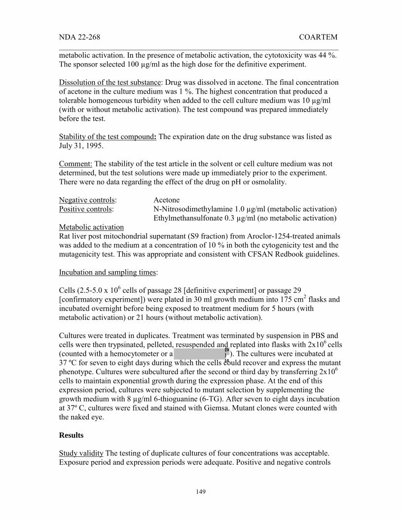
NDA 22-268 COARTEM _______________________________________________________________________
149
metabolic activation. In the presence of metabolic activation, the cytotoxicity was 44 %. The sponsor selected 100 µg/ml as the high dose for the definitive experiment. Dissolution of the test substance: Drug was dissolved in acetone. The final concentration of acetone in the culture medium was 1 %. The highest concentration that produced a tolerable homogeneous turbidity when added to the cell culture medium was 10 µg/ml (with or without metabolic activation). The test compound was prepared immediately before the test. Stability of the test compound: The expiration date on the drug substance was listed as July 31, 1995. Comment: The stability of the test article in the solvent or cell culture medium was not determined, but the test solutions were made up immediately prior to the experiment. There were no data regarding the effect of the drug on pH or osmolality. Negative controls: Acetone Positive controls: N-Nitrosodimethylamine 1.0 µg/ml (metabolic activation)
Ethylmethansulfonate 0.3 µg/ml (no metabolic activation) Metabolic activation Rat liver post mitochondrial supernatant (S9 fraction) from Aroclor-1254-treated animals was added to the medium at a concentration of 10 % in both the cytogenicity test and the mutagenicity test. This was appropriate and consistent with CFSAN Redbook guidelines. Incubation and sampling times: Cells (2.5-5.0 x 10⁶ cells of passage 28 [definitive experiment] or passage 29 [confirmatory experiment]) were plated in 30 ml growth medium into 175 cm2 flasks and incubated overnight before being exposed to treatment medium for 5 hours (with metabolic activation) or 21 hours (without metabolic activation). Cultures were treated in duplicates. Treatment was terminated by suspension in PBS and cells were then trypsinated, pelleted, resuspended and replated into flasks with 2x10⁶ cells (counted with a hemocytometer or a ). The cultures were incubated at 37 ºC for seven to eight days during which the cells could recover and express the mutant phenotype. Cultures were subcultured after the second or third day by transferring 2x10⁶ cells to maintain exponential growth during the expression phase. At the end of this expression period, cultures were subjected to mutant selection by supplementing the growth medium with 8 µg/ml 6-thioguanine (6-TG). After seven to eight days incubation at 37º C, cultures were fixed and stained with Giemsa. Mutant clones were counted with the naked eye. Results Study validity The testing of duplicate cultures of four concentrations was acceptable. Exposure period and expression periods were adequate. Positive and negative controls
(b) (4

NDA 22-268 COARTEM _______________________________________________________________________
150
produced the expected results. The sponsor’s criteria for a positive response included a valid assay in which one or more concentrations is reproducibly, significantly greater than the negative control and the number of normalized mutant clones in the treated and untreated cultures differs by more than 20 and there is a significant dose relationship as indicated by the linear trend analysis. If only some of these criteria are fulfilled, the study director would interpret the results based on his/her own experience. According to OECD/OCDE guidelines a concentration-related or a reproducible increase in mutant frequency is to be considered a positive result considering the biological relevance of the results. Study outcome: Table 1 Cytotoxicity/growth inhibition at 100 µg/ml in definitive experiment Original experiment After treatment After expression + metabolic activation 16 % No cytotoxicity - metabolic activation 19 % No cytotoxicity Table 2 Cytotoxicity-growth inhibition at 100 µg/ml in confirmatory experiment Confirmatory experiment After treatment After expression + metabolic activation 7 % 19 % - metabolic activation 25 % 13 % Mean cytotoxicity at the highest dose in the definitive and confirmatory assays were less than 25 % in all cases. At concentrations up to 100 µg/ml, CGP 56697 did not result in a significant increase in thioguanine-resistant mutants counted compared to controls. According to the sponsor, higher concentrations could not be applied due to solubility limitations in the culture medium. Comments While cytotoxicity did not exceed 25 % in the original or confirmatory experiments, the highest dose tested was the highest dose that produced a tolerable homogeneous turbidity when added to the cell culture medium. The negative and positive controls reacted as expected even though the sponsor’s criteria for a positive result did not follow OECD guidelines. This reviewer is therefore in agreement with the sponsor’s conclusion that CGP56697 was not considered to be genotoxic under these experimental conditions. Study conclusion:

NDA 22-268 COARTEM _______________________________________________________________________
151
CGP56697 was not considered to be genotoxic in the gene mutation test with Chinese hamster cells (V79). This is in agreement with the sponsor’s conclusion. [58.] Study title: Gene mutation test with Chinese hamster cells V79 Key findings: CGP56695 (benflumetol) was not considered to be genotoxic in the gene mutation test with Chinese hamster cells (V79). Study no.: 956038 Location of Volume: Electronic Document Room Study type: In vitro mammalian cell mutation test Conducting laboratory: Genetic Toxicology
CEIBA-GEIGY Limited, Basle, Switzerland.
Date of study initiation: March 10, 1995 GLP compliance: No. Swiss GLP QA reports: Yes Drug, batch 800594 Purity: 100 % Methods This test was conducted to evaluate the potential of CGP 56695 (benflumetol) to induce gene mutations. This study was performed according to SOP no. 30 59 02, CEIBA-GEIGY Limited, Basle, Switzerland and was consistent with OECD guidelines (1984), EPA guidelines (1987) and EEC guidelines (1987/88). The study was conducted using V79 Chinese hamster cells, (clone 65/3) and mutagenic effects were indicated by 6-TG resistant colonies appearing in cultures. They are quantified and compared to control cultures. After the preliminary cytotoxicity test, a definitive study was conducted, followed by a confirmatory study. Doses used in definitive study: In the definitive study, concentrations were 1.9, 5.6, 17 and 50 µg/ml, with or without metabolic activation and in the confirmatory study; concentrations were 6, 13, 25 and 50 µg/ml with or without metabolic activation. Basis of dose selection: In the cytotoxicity assay (12 doses tested between 0.02 and 50 µg/ml), no substantial toxicity was detected in the presence or absence of metabolic activation at any dose. Precipitate (homogenous turbidity) was reported by the sponsor at final concentrations as low as 13 µg/ml. The high dose selected for the definitive experiment was 50 µg/ml. Dissolution of the test substance: Drug was dissolved in acetone. The final concentration of acetone in the culture medium was 1 %. Addition of stock solution produced turbidity in the culture medium at final concentrations down to about 13 µg/ml in the experiments

NDA 22-268 COARTEM _______________________________________________________________________
152
with or without metabolic activation. The test compound was prepared immediately before the test. Stability of the test compound: The expiration date on the drug substance was listed as May 31, 1996. Comment: Although the stability of the test article in the solvent or cell culture medium was not determined, the test solutions were made up immediately prior to the experiment. There were no data regarding the effect of the drug on pH or osmolality. Negative controls: Acetone Positive controls: N-Nitrosodimethylamine 1.0 µg/ml (metabolic activation) Ethylmethansulfonate 0.3 µg/ml (no metabolic activation) Metabolic activation Rat liver post mitochondrial supernatant (S9 fraction) from Aroclor-1254-treated animals was added to the medium at a concentration of 10 % in both the cytogenicity test and the mutagenicity test. This was appropriate and consistent with CFSAN Redbook guidelines. Incubation and sampling times: Cells (2.5-5.0 x 10⁶ cells of passage 28 [definitive experiment] or passage 25 [confirmatory experiment]) were plated in 30 ml growth medium into 175 cm2 flasks and incubated overnight before being exposed to treatment medium for 5 hours with metabolic activation or 21 hours without metabolic activation. Cultures were treated in duplicates. Treatment was terminated by suspension in PBS and cells were then trypsinated, pelleted, resuspended and replated into flasks with 2x10⁶ cells (counted with a hemocytometer or a ). The cultures were incubated at 37 ºC for seven to eight days during which the cells could recover and express the mutant phenotype. Cultures were subcultured after the second or third day by transferring 2x10⁶ cells to maintain exponential growth during the expression phase. At the end of this expression period, cultures were subjected to mutant selection by supplementing the growth medium with 8 µg/ml 6-thioguanine (6-TG). After seven to eight days incubation at 37º C, cultures were fixed and stained with Giemsa. Mutant clones were counted with the naked eye. Results Study validity The testing of duplicate cultures of four concentrations was acceptable. Exposure period and expression periods were adequate. Positive and negative controls produced the expected results. The sponsor’s criteria for a positive response included a valid assay in which one or more concentrations is reproducibly, significantly greater than the negative control and the number of normalized mutant clones in the treated and untreated cultures differs by more than 20 and there is a significant dose relationship as indicated by the linear trend analysis. If only some of these criteria are fulfilled, the study
(b) (4

NDA 22-268 COARTEM _______________________________________________________________________
153
director would interpret the results based on his/her own experience. According to OECD/OCDE guidelines a concentration-related or a reproducible increase in mutant frequency is to be considered a positive result considering the biological relevance of the results. Study outcome: No substantial toxicity was seen at any concentrations. Addition of stock solution produced turbidity in the culture medium at final concentrations down to about 13 µg/ml in the experiments with or without metabolic activation. At concentrations up to 50 µg/ml, CGP 56695 did not result in a significant increase in thioguanine-resistant mutants counted compared to controls. Higher concentrations were not applied due to solubility limitations. Comments The high doses tested produced homogeneous turbidity when added to the cell culture medium. The negative and positive controls reacted as expected even though the sponsor’s criteria for a positive result did not follow OECD guidelines. This reviewer is therefore in agreement with the sponsor’s conclusion that CGP56695 was not considered to be genotoxic under these experimental conditions. Conclusion: CGP56695 was not considered to be genotoxic in the gene mutation test with Chinese hamster cells (V79). This is in agreement with the sponsor’s conclusion. [59.] Study title: Gene mutation test with Chinese hamster cells V79 Key findings: CGP56696 (artemether) was not considered to be genotoxic in the gene mutation test with Chinese hamster cells (V79). Study no.: 956041 Location of Volume: Electronic Document Room Study type: In vitro mammalian cell mutation test Conducting laboratory: Genetic Toxicology
CEIBA-GEIGY Limited, Basle, Switzerland.
Date of study initiation: March 10, 1995 GLP compliance: No. Swiss GLP QA reports: Yes Drug, batch # 810394 Purity: 98.1 %

NDA 22-268 COARTEM _______________________________________________________________________
154
Methods This test was conducted to evaluate the potential of CGP 56696 (artemether) to induce gene mutations. This study was performed according to SOP no. 30 59 02, CEIBA-GEIGY Limited, Basle, Switzerland and was consistent with OECD guidelines (1984), EPA guidelines (1987) and EEC guidelines (1987/88). The study was conducted using V79 Chinese hamster cells, (clone 65/3) and mutagenic effects were indicated by 6-TG resistant colonies appearing in cultures. They are quantified and compared to control cultures. After the preliminary cytotoxicity test, a definitive study was conducted, followed by a confirmatory study. Doses used in definitive study: In the definitive study, concentrations were 11, 33, 100 and 300 µg/ml with metabolic activation and 5, 17, 50 and 150 µg/ml without metabolic activation. In the confirmatory experiment, doses were 31, 63, 125 and 250 µg/ml with metabolic activation and 19, 37, 75 and 150 µg/ml without metabolic activation. Basis of dose selection: In the cytotoxicity assay (12 doses tested between 0.19 and 400 µg/ml), 53 % growth inhibition was observed at 400 µg/ml, in the presence of metabolic activation. The growth inhibition was 63 % at 100 µg/m in the absence of metabolic inhibition. The sponsor therefore selected doses between 11 and 300 µg/ml for the definitive experiment with metabolic activation and doses between 5 and 150 µg/m for the experiment without metabolic activation. Due to severe cytotoxicity, the dose range was reduced to between 13 and 250 µg/m with metabolic activation and 19 to 150 µg/m without metabolic activation for the confirmatory experiments. Dissolution of the test substance: Drug was dissolved in 10 % dimethyl sulfoxide (DMSO). The final concentration of acetone in the culture medium was 1 %. The addition of drug to the culture medium resulted in precipitates at 250 and 300 µg/ml. The test substance solution was prepared immediately before the start of the experiment. Stability of the test compound: The expiration date on the drug substance was listed as August 31, 1995. Comment The stability of the test article in the solvent or cell culture medium was not determined, but the test solution was prepared immediately before the experiment. The sponsor also did not report the effect of the drug on pH or osmolality of the culture medium. Negative controls: DMSO, 10 % Positive controls: With metabolic activation: 1.0 µg/ml N-Nitrosodimethylamine (DMN) Without metabolic activation: 0.3 µg/ml ethylmethansulfonate l (EMS)

NDA 22-268 COARTEM _______________________________________________________________________
155
Metabolic activation Rat liver post mitochondrial supernatant (S9 fraction) from Aroclor-1254-treated animals was added to the medium at a concentration of 10 %. This was appropriate and consistent with CFSAN Redbook guidelines. Incubation and sampling times: Cells (2.5-5.0 x 10⁶ cells of passage 25 [definitive experiment] or passage 26 [confirmatory experiment]) were plated in 30 ml growth medium into 175 cm2 flasks and incubated overnight before being exposed to treatment medium for 5 hours with metabolic activation or 21 hours without metabolic activation. Cultures were treated in duplicates. Treatment was terminated by suspension in PBS and cells were then trypsinated, pelleted, resuspended and replated into flasks with 2x10⁶ cells (counted with a hemocytometer or a ). The cultures were incubated at 37 ºC for seven to eight days during which the cells could recover and express the mutant phenotype. Cultures were subcultured after the second or third day by transferring 2x10⁶ cells to maintain exponential growth during the expression phase. At the end of this expression period, cultures were subjected to mutant selection by supplementing the growth medium with 8 µg/ml 6-thioguanine (6-TG). After seven to eight days incubation at 37º C, cultures were fixed and stained with Giemsa. Mutant clones were counted with the naked eye. Results Study validity The testing of duplicate cultures of four concentrations was acceptable. Exposure period and expression periods were adequate. Positive and negative controls produced the expected results. The sponsor’s criteria for a positive response included a valid assay in which one or more concentrations is reproducibly, significantly greater than the negative control and the number of normalized mutant clones in the treated and untreated cultures differs by more than 20 and there is a significant dose relationship as indicated by the linear trend analysis. If only some of these criteria are fulfilled, the study director would interpret the results based on his/her own experience. According to OECD/OCDE guidelines a concentration-related or a reproducible increase in mutant frequency is to be considered a positive result considering the biological relevance of the results. Study outcome: In the definitive experiment with metabolic activation, the 300 µg/ml dose was toxic, but no severe toxicity was observed at lower doses. Without metabolic activation, toxicity was 69% at the highest concentration. No toxicity was seen after the expression period. In the confirmatory experiment in the presence of metabolic activity, the toxicity at the highest dose (150 µg/ml) was 51 %. After the expression period, the toxicity was 39 %. In the absence of metabolic activation, growth inhibition at the highest level (150 µg/ml), was 85 and 45 % after treatment and expression respectively.
(b) (4

NDA 22-268 COARTEM _______________________________________________________________________
156
At the concentrations tested, CGP 56696 did not result in a significant increase in thioguanine-resistant mutants counted compared to controls. In the definitive and confirmatory experiments, CGP 56696 was associated with slight increases in mean mutant frequency, but these were not dose related and were within the historical range. Table 1 Results from the original experiment with metabolic activation Treatment Mean mutant
frequency(x10E-6) Mutant Factor Cytotoxicity (%)
Negative control 6.7 Positive control 116 17 300 No data No data No data 100 5.8 0.9 25 33 6.2 0.9 18 11 1.9 0.3 16 Historical range for the mean mutant frequency with metabolic activation was 5.76 + 3.95. Table 2 Results from the original experiment without metabolic activation Treatment Mean mutant
frequency(x10E-6) Mutant Factor Cytotoxicity (%)
Negative control 5.9 Positive control 1709 288 150 6.5 1.1 69 50 4.8 0.8 3 17 8.2* 1.4 Not toxic 6 8.0* 1.4 Not toxic * significantly different from control. P<0.05. Historical range for the mean mutant frequency without metabolic activation was 5.36 + 4.62. In the original experiments, there was a slight increase in mutant frequency at the two lower doses, without metabolic activation, but these changes were small and not dose dependent and were within the range of historical control values.

NDA 22-268 COARTEM _______________________________________________________________________
157
Table 3 Results from the confirmatory experiment with metabolic activation Treatment Mean mutant
frequency(x10E-6) Mutant Factor Cytotoxicity
(%) Negative control 5.3 Positive control 132 25 250 6.4 1.2 50 125 4.4 0.8 8 62.5 3.0 0.6 5 31 5.5 1.0 1 Historical range for the mean mutant frequency with metabolic activation was 5.76 + 3.95x10E-6 Table 4 Results from the confirmatory experiment, without metabolic activation Treatment Mean mutant
frequency(x10E-6) Mutant Factor Cytotoxicity
(%) Negative control 4.4 Positive control 1595 360 150 6.4* 1.4 85 75 5.0 1.1 44 38 5.8 1.3 19 19 6.7* 1.5 1 * Significantly different from control. P<0.05. Historical range for the mean mutant frequency without metabolic activation was 5.36 + 4.62x10E-6. In the confirmatory experiment, there were also significant increases in mutant frequencies at 31 µg/ml and 250 µg/ml, but these were also within the range of historical controls. Comments The study was not conducted under GLP, but the sponsor did follow OECD guidelines (1984), EPA guidelines (1987) and EEC guidelines (1987/88). The high dose produced a tolerable homogeneous turbidity when added to the cell culture medium. The negative and positive controls reacted as expected even though the sponsor’s criteria for a positive result did not follow OECD guidelines. This reviewer is therefore in agreement with the sponsor’s conclusion that CGP56696 was not considered to be genotoxic under these experimental conditions.

NDA 22-268 COARTEM _______________________________________________________________________
158
Conclusion: In the absence of a dose related and/or reproducible increase in thioguanine-resistant mutants, CGP56696 was not considered to be genotoxic in the gene mutation test with Chinese hamster cells (V79). The occasional increases observed were not thought to be biologically significant since they were in the range of the historical controls. This is in agreement with the sponsor’s conclusion. [60.] Study title: Cytogenetic Test on Chinese hamster cells in vitro. Key findings: CGP56697 showed no evidence of clastogenic effects under these experimental conditions using Chinese hamster ovary cells in vitro. Study no.: 946170 Conducting laboratory: Genetic Toxicology
CEIBA-GEIGY Limited, Basle, Switzerland.
Date of study initiation: January 19, 1995 GLP compliance: No. Swiss GLP QA reports: Yes Drug, batch #: V-32-3197 Methods This test was conducted to evaluate the potential of CGP 56697 [CGP 56696 (artemether, 14.1 %) and CGP 56695 (benflumetol, 86.1 %)] to induce structural chromosome aberrations in Chinese hamster ovary cells in vitro. The study was conducted in the presence and absence of S9. Table 1 Study protocol Original study Confirmatory Study Experiment # 1 2 1 2 1 2 Metabolic activation - + - + - + Treatment (h) 18 3 18 3 42 3 Recovery (h) - 15 - 15 - 39 # of concentrations 8 8 8 8 8 8 Lowest conc. (µg/ml)
0.8 0.8 0.05 0.8 0.05 0.8
Highest conc. (µg/ml)
100 100 6.3 100 6.3 100
After the preliminary test, a confirmatory study was conducted, followed by a study with a prolonged sampling time, 24 hours after the first one.

NDA 22-268 COARTEM _______________________________________________________________________
159
Doses used in definitive study: In the definitive study, concentrations were 0.8, 1.6, 3.1, 6.3, 12.5, 25, 50, and 100 µg/ml, with or without metabolic activation. In the confirmatory experiment, concentrations were 0.05, 0.1, 0.2, 0.4, 0.8, 1.6, 3.1, 6.3 µg/ml, without metabolic activation and 0.8, 1.6, 3.1, 6.3, 12.5, 25, 50, and 100 µg/ml, with metabolic activation. Basis of dose selection: A toxicity/dose rangefinding study was conducted. Table 2 Results of Toxicity Study Mitotic activity (% control) - metabolic activation + metabolic activation Control 100 100 100 µg/ml 13 12 50 µg/ml 6 44 25 µg/ml 4 54 13 µg/ml 3 101 6 µg/ml 7 74 3 µg/ml 20 76 1.6 µg/ml 54 - 0.8 µg/ml 78 - In the toxicity assay a concentration of 3 µg/ml caused 80 % suppression of mitotic activity in the absence of metabolic activation (18 hour treatment). In the presence of metabolic activity 50 µg/ml caused a 56 % suppression of mitotic activity (3 hours treatment, 15 hours recovery, see Table 2, above). Dissolution of the test substance: Drug was dissolved in acetone. The final concentration of acetone in the culture medium was 1 %. Addition of stock solution produced turbidity in the culture medium at final concentrations down to about 25 µg/ml in the experiments with or without metabolic activation. Stability of the test compound: The expiration date on the drug substance was listed as July 31, 1995. Comments: The stability of the test article in the solvent or cell culture medium was not determined and there was no mention of the effect of the drug on pH or osmolality. Negative controls: Acetone Positive controls: Mitomycin C 0.2 µg/ml (without metabolic activation) or cyclophosphamide 20 µg/ml (with metabolic activation)

NDA 22-268 COARTEM _______________________________________________________________________
160
Metabolic activation Post mitochondrial supernatant (S9 fraction) from Aroclor 1254 induced rat liver was added to the medium at a concentration of 10 %. Incubation and sampling times: Chinese hamster ovary cells were seeded onto quadruplicate culture dishes at a density of at least 1x10⁴ cells/ml (for 18 hour experiments) or 4x10³ cells/ml (for 42 hour experiments) and preincubated for about 24 hours. Test substance or vehicle or positive control was then added to the culture medium (along with S9 where indicated). Two hours prior to sampling, cultures were treated with colcemide (0.4 µg/ml) to arrest cells in metaphase. Treatment was terminated by a hypotonic treatment with 0.075 M KCl followed by fixation. Slides were then dried and stained with orcein. Slides were then examined for aberrations including specific aberrations (chromatid and chromosome deletions), chromatid exchanges, chromosome exchanges), multiple aberrations and unspecific aberrations (chromatid and chromosome gaps). Record was also kept of metaphases with >21 centromeres, polyploidy metaphases and endoduplication figures. Results Study validity The testing of quadruplicate cultures of three concentrations is acceptable according to CFSAN guidelines. Exposure period and expression periods were adequate. Positive and negative controls produced the expected results. Positive controls were consistent with CFSAN guidelines. The sponsor’s criteria for a positive response included an assay in which the percentage of metaphases containing specific aberrations in a treatment group is higher than 6 (based on historical negative control range) and the response is concentration-related. According to OECD/OCDE guidelines a concentration-related or a reproducible increase in mutant frequency is to be considered a positive result considering the biological relevance of the results. Study outcome: In the original mutagenicity study (18 h treatment without metabolic activation) the doses selected for evaluation were 0.8, 1.6 and 3.1 µg/ml, with the highest concentration inhibiting mitosis by 80 % compared to controls. In the original study in the presence of metabolic activity, the concentrations tested were 13, 25 and 50 µg/ml, with the highest concentration resulting in 56 % inhibition of mitotic activity. In the confirmatory test, 1.6 µg/ml resulted in a 73 % reduction in mitotic activity without metabolic activation and 50 µg/ml resulted in a 45 % reduction in mitotic activity with metabolic activation. In the confirmatory test, with 42 hour treatment without metabolic activation, 3.1 µg/ml resulted in 72 % reduction in mitotic activity without metabolic activation and 50 µg/ml resulted in a 22 % reduction in mitotic activity. In the original experiment, without metabolic activation, at 1.6 µg/ml, the drug increased the percentage of cells with chromosome aberrations compared to controls (3.0 vs. 1.5 for controls, but the effect was neither significant nor dose-related (2.0 at 3.1 µg/ml).

NDA 22-268 COARTEM _______________________________________________________________________
161
In the confirmatory study, without metabolic activation, the % cells with aberrations was significantly increased at 1.6 µg/ml (4.5 vs. 1.0 in controls) but the result was within the range of historical solvent controls (as high as 5 for acetone).
Corresponding cytotoxicity values were 14 %, 22 % and 73 % at 0.391, 0.78, and 1.56 µg/ml, respectively. In the presence of metabolic activation, CGP56697did not result in any biologically significant increase in the % of cells with specific aberrations compared to controls. All values were within the range of historical control data.

NDA 22-268 COARTEM _______________________________________________________________________
162
Conclusion CGP56697 was not considered by the sponsor to be clastogenic as determined by the Chinese hamster ovary cells in vitro. This reviewer is in agreement with the sponsor’s conclusion.
Appears This Way on Original

NDA 22-268 COARTEM _______________________________________________________________________
163
[61.] Study title: Cytogenetic Test on Chinese hamster cells in vitro. Key findings: CGP56696 showed evidence of clastogenic effects under these experimental conditions using Chinese hamster ovary cells in vitro. Study no.: 946228 Conducting laboratory: Genetic Toxicology
CEIBA-GEIGY Limited, Basle, Switzerland.
Date of study initiation: January 6, 1995 GLP compliance: No. Swiss GLP QA reports: Yes Drug, batch #: 810394 Purity: 98.1 % Methods This test was conducted to evaluate the potential of CGP 56696 (artemether) to induce structural chromosome aberrations in Chinese hamster ovary cells in vitro. The study was conducted in the presence and absence of S9. Metaphase cells were examined from cultures treated with drug for the presence of structural chromosome aberrations. They are quantified and compared to control cultures. Table 1 Study protocol Original study Confirmatory Study Experiment # 1 2 1 2 1 2 Metabolic activation - + - + - + Treatment (h) 18 3 18 3 42 3 Recovery (h) - 15 - 15 - 39 # of concentrations 8 8 8 8 8 8 Lowest conc. (µg/ml)
3.28 3.28 0.10 0.10 0.10 0.10
Highest conc. (µg/ml)
420 420 210 105 210 105
After the preliminary test, a confirmatory study was conducted, followed by a study with a prolonged sampling time, 24 hours after the first one. Doses used in definitive study: In the definitive study, concentrations were 3.28, 6.56, 13.13, 26.25, 52.5, 105, 210 and 420 µg/ml, with or without metabolic activation. In the confirmatory experiment, concentrations were 0.1, 0.41, 1.64, 6.56, 26.25, 52.5, 105, 210 µg/ml, without metabolic activation and 0.1, 0.41, 1.64, 6.56, 26.25, 52.5 and 105 µg/ml, with metabolic activation. Basis of dose selection: A toxicity/dose rangefinding study was conducted.

NDA 22-268 COARTEM _______________________________________________________________________
164
Table 2 Results of Toxicity Studies Dose
Study Activation
Suppression of mitotic activity (%)
105 µg/ml (18h treatment) Original #1 - 48 53 µg/ml (3h treatment/15h recovery) Original #2 + 59 53 µg/ml (18 h treatment) Confirmatory 1 - 58 53 µg/ml (3h treatment/15 h recovery) Confirmatory 2 + 67 6.6 µg/ml (42 h treatment) Confirmatory 3 - 64 105 µg/ml (3h treatment/39 h recovery)
Confirmatory 4 + 68
In the toxicity assay the high doses produced measurable toxicity. (See Table 2, above). Dissolution of the test substance: Drug was dissolved in DMSO. The final concentration of DMSO in the culture medium was 1 %. Addition of stock solution produced turbidity in the culture medium at final concentrations down to about 420 µg/ml in the experiments with or without metabolic activation. Stability of the test compound: The expiration date on the drug substance was listed as August 31, 1995. Comment: The stability of the test article in the solvent or cell culture medium was not determined and there was no mention of the effect of the drug on pH or osmolality. Negative controls: Acetone Positive controls: Mitomycin C 0.2 µg/ml (without metabolic activation) or cyclophosphamide 20 µg/ml (with metabolic activation) Metabolic activation Post mitochondrial supernatant (S9 fraction) from Aroclor 1254 induced rat liver was added to the medium at a concentration of 10 %. Incubation and sampling times: Chinese hamster ovary cells were seeded onto quadruplicate culture dishes at a density of at least 1x10⁴ cells/ml (for 18 hour experiments) or 4x10³ cells/ml (for 42 hour experiments) and preincubated for about 24 hours. Test substance or vehicle or positive control was then added to the culture medium (along with S9 where indicated). Two hours prior to sampling, cultures were treated with colcemide (0.4 µg/ml) to arrest cells in metaphase. Treatment was terminated by a hypotonic treatment with 0.075 M KCl followed by fixation. Slides were then dried and stained with orcein. Slides were then examined for aberrations including specific aberrations (chromatid and chromosome deletions), chromatid exchanges, chromosome exchanges), multiple aberrations and unspecific aberrations (chromatid and chromosome

NDA 22-268 COARTEM _______________________________________________________________________
165
gaps). Record was also kept of metaphase with >21 centromeres, polyploidy metaphases and endoduplication figures. Results Study validity The testing of quadruplicate cultures of three concentrations is acceptable according to CFSAN guidelines. Exposure period and expression periods were adequate. Positive and negative controls produced the expected results. Positive controls were consistent with CFSAN guidelines. The sponsor’s criteria for a positive response included an assay in which the percentage of metaphases containing specific aberrations in a treatment group is higher than 6 (based on historical negative control range) and the response is concentration-related. According to OECD/OCDE guidelines a concentration-related or a reproducible increase in mutant frequency is to be considered a positive result considering the biological relevance of the results. Study outcome: The stability of the test article itself was reported but the stability of the test article in the solvent or cell culture medium was not determined and there was no mention of the effect of the drug on pH or osmolality of the medium. CGP 56696 did not increase the number of metaphases containing specific chromosome aberrations in the absence of metabolic activation. With metabolic activation, there was an increase at 13.1 and 52.5 µg/ml, but these changes were not statistically significant and fell within the range of historical controls. The corresponding cytotoxicity values were 34, 40 and 59 % at 6.56, 13.13 and 52.5 µg/ml respectively.
Although the increases seen in the original mutagenicity study with metabolic activation were within the range of historical control, a repeat study showed a significant increase in the number of metaphases containing specific chromosome aberrations that went beyond

NDA 22-268 COARTEM _______________________________________________________________________
166
the range of historical controls (historically up to 6 % cells with specific aberrations have been observed in controls). The corresponding cytotoxicity values were 5, 43 and 67 % at 0.41, 26.25 and 52.5 µg/ml respectively.
When the experiment was repeated with longer recovery periods (39 hours instead of 15 hours), there was no increase in chromosome aberrations.
Discussion The sponsor argues that the increase in the percentage of cells containing specific aberrations beyond the limit of the historical controls does not indicate a positive response. They argue that the increase is only slight, is not statistically significant, is not reproducible and is not dose dependent. When the experiment is repeated with a longer recovery period, there is no increase in chromosome aberrations. This reviewer agrees with these conclusions. Study conclusion: CGP56696 was not considered by the sponsor to be clastogenic as determined by the Chinese hamster ovary cells in vitro. This reviewer agrees with the sponsor’s conclusion.

NDA 22-268 COARTEM _______________________________________________________________________
167
[62.] Study title: Cytogenetic Test on Chinese hamster cells in vitro. Key findings: CGP56695 showed no evidence of clastogenic effects under these experimental conditions using Chinese hamster ovary cells in vitro. Study no.: 946230 Conducting laboratory: Genetic Toxicology
CEIBA-GEIGY Limited, Basle, Switzerland.
Date of study initiation: January 6, 1995 GLP compliance: No (Swiss GLP) QA reports: Yes Drug batch #: 800194 Purity: 99.6 % Methods This test was conducted to evaluate the potential of CGP 56695 (benflumetol ) to induce structural chromosome aberrations in Chinese hamster ovary cells in vitro, in the presence and absence of S9. Table 1 Study protocol Original study Confirmatory Study Experiment # 1 2 1 2 1 2 Metabolic activation - + - + - + Treatment (h) 18 3 18 3 42 3 Recovery (h) - 15 - 15 - 39 # of concentrations 8 8 5 5 8 8 Lowest conc. (µg/ml)
0.391 0.391 3.12 3.12 0.391 0.391
Highest conc. (µg/ml)
50 50 50 50 50 50
After the preliminary test, a confirmatory study was conducted, followed by a study with a prolonged sampling time, 24 hours after the first one. Doses used in definitive study: In the definitive study, concentrations were 0.8, 1.6, 3.1, 6.3, 12.5, 25, 50, and 100 µg/ml, with or without metabolic activation. In the confirmatory experiment, concentrations were 0.05, 0.1, 0.2, 0.4, 0.8, 1.6, 3.1, 6.3 µg/ml, without metabolic activation and 0.8, 1.6, 3.1, 6.3, 12.5, 25, 50, and 100 µg/ml, with metabolic activation. Basis of dose selection: A toxicity/dose selection study was conducted

NDA 22-268 COARTEM _______________________________________________________________________
168
Table 2 Results of Toxicity/Dose selection Study: 18 h treatment. Frequency (% control) - metabolic activation + metabolic activation 18 h treatment 3 h treatment; 15 h recovery Control 100 100 50 µg/ml 97 86 25 µg/ml 102 93 13 µg/ml 106 98 0.4 µg/ml 127 109 Dissolution of the test substance: Drug was dissolved in acetone. The final concentration of acetone in the culture medium was 1 %. Addition of stock solution produced turbidity in the culture medium at final concentrations down to about 12.5 µg/ml in the experiments with or without metabolic activation. Stability of the test compound: The expiration date on the drug substance was listed as October 31, 1995, but there was no discussion of the stability of the test article in solvent or cell culture medium. Negative controls: Acetone Positive controls: Mitomycin C 0.2 µg/ml (without metabolic activation)
Cyclophosphamide 20 µg/ml (with metabolic activation) Metabolic activation Post mitochondrial supernatant (S9 fraction) from Aroclor 1254 induced rat liver was added to the medium at a concentration of 10 %. Incubation and sampling times: Chinese hamster ovary cells were seeded onto quadruplicate culture dishes at a density of at least 1x10⁴ cells/ml (for 18 hour experiments) or 4x10³ cells/ml (for 42 hour experiments) and preincubated for about 24 hours. Test substance or vehicle or positive control was then added to the culture medium (along with S9 where indicated). Two hours prior to sampling, cultures were treated with colcemide (0.4 µg/ml) to arrest cells in metaphase. Treatment was terminated by a hypotonic treatment with 0.075 M KCl followed by fixation. Slides were then dried and stained with orcein. Slides were then examined for aberrations including specific aberrations (chromatid and chromosome deletions), chromatid exchanges, chromosome exchanges), multiple aberrations and unspecific aberrations (chromatid and chromosome gaps). Record was also kept of metaphase with >21 centromeres, polyploidy metaphases and endoduplication figures. Results Study validity The testing of quadruplicate cultures of three concentrations is acceptable according to CFSAN guidelines. Exposure period and expression periods were adequate.

NDA 22-268 COARTEM _______________________________________________________________________
169
Positive and negative controls produced the expected results. Positive controls were consistent with CFSAN guidelines. The sponsor’s criteria for a positive response included an assay in which the percentage of metaphases containing specific aberrations in a treatment group is higher than 6 (based on historical negative control range) and the response is concentration-related. According to OECD/OCDE guidelines a concentration-related or a reproducible increase in mutant frequency is to be considered a positive result considering the biological relevance of the results. Study outcome: In the original mutagenicity study (18 h treatment with or without metabolic activation) the doses selected for evaluation were 12.5, 25 and 50 µg/ml. Table 3 Percentage of metaphases with specific chromosomal aberrations (18h treatment) without metabolic activation. [CGP 56695] µg/ml
Original Study Confirmatory Study (%
Control 1 % 3.0 12.5 1 % 3.5 25 2 % 3.0 50 2 % 3.0 Table 4 Percentage of metaphases with specific chromosomal aberrations (3h treatment, 15 h recovery) with metabolic activation. [CGP 56695] µg/ml
Original Study Confirmatory Study
Control 4.5% 2.5 % 12.5 0.5 % 2.0 25 1 % 5.0 50 0.5 % 3.0 Table 5 The percentages of metaphases with specific chromosomal aberrations (3h treatment, 39 h recovery) with metabolic activation are shown below. [CGP 56695] µg/ml
Percentage of metaphases with specific chromosomal aberrations
3 hours treatment/39 hours recovery
42 hours treatment
Control 1.5% 1.0 12.5 1.5 % 3.0 25 2.0 % 2.5 50 4.0 % 3.5

NDA 22-268 COARTEM _______________________________________________________________________
170
Comment: In the original study in the presence of metabolic activity, the concentrations tested were 13, 25 and 50 µg/ml, with the highest concentration resulting in less than 10 % inhibition. The testing of higher doses was hampered by solubility problems. Addition of stock solution produced turbidity in the culture medium at final concentrations down to about 12.5 µg/ml. The sponsor’s criteria for a positive response were an assay in which the percentage of metaphases containing specific aberrations in a treatment group was higher than 6 (based on historical negative control range) and a response that is concentration-related. There were no CGP 56695 concentrations where this criterion for positive was met. This criterion was based on historical control data for a variety of incubation protocols, but the historical control values for the 3 hour treatment/39 hour recovery using acetone as a solvent ranged from 0 to 4. The value 4.0 that was recorded at 50 µg/ml on Table 5 is only marginally outside that range and is therefore likely to be biologically insignificant. For a 42-hour incubation, the range of historical controls is 1 to 4 and so the values achieved in this experiment are within the range of historical control. In the absence of a concentration-related or a reproducible increase in mutant frequency, this drug is considered negative. Conclusion: CGP56695 was not considered by the sponsor to be clastogenic as determined by the Chinese hamster ovary cells in vitro. This reviewer is in agreement with the sponsor’s conclusion.
Appears This Way on Original

NDA 22-268 COARTEM _______________________________________________________________________
171
4.2.3.3.2 – In vivo Genotoxicity [63.] Study title: Micronucleus Test, Rat in vivo study. Key findings: CGP56697 was negative in the rat micronucleus assay. Study number: 946172 Document location: Electronic Document Room Conducting laboratory: Genetic Toxicology
CEIBA-GEIGY Limited, Basle, Switzerland.
Date of study initiation: January 16, 1995 GLP compliance: Swiss GLP QA reports: Yes Drug lot: V 32 3197 Purity: CGP 56696-98.9 % CGP 56695-100.4 % Methods CGP56697 (comprised of 14 % artemether [CGP56696] and 86 % benflumetol [CGP56695]) was evaluated for clastogenicity and its aneuploidy-inducing properties using the rat micronucleus assay. Species/strain: Rat (Tif:RAIf(SPF) Basis of dose selection: A range finding/tolerability study was conducted. Negative controls: Klucel (hydroxypropylcellulose, 0.5 % containing 0.1 %
Tween 80) Positive controls: Cyclophosphamide, 40 mg/kg. Tolerability study: In a tolerability study, rats (1 rat/sex) were treated with drug up to the limit dose (2g/kg). No animals died and no clinical signs of toxicity were observed. Exposure was evidenced by the myelotoxicity which produced a marked reduction in the ratios of polychromatic to normochromatic erythrocytes In the micronucleus study, test substance was administered once by gavage to groups of 5 male and 5 female Tif:RAIf(SPF) rats at doses of 2000, 1000 and 500 mg/kg. Additional groups were treated with vehicle or with cyclophosphamide. High dose and negative control animals were sacrificed 16, 24 and 48 hours after dosing. Animals from the low, intermediate and positive control groups were sacrificed 24 hours after treatment. Animals were killed by CO2 and bone marrow harvested from the shafts of both femurs. Smears were prepared and stained with May-Grünwald/Giemsa solution and mounted. 1000 polychromatic erythrocytes were scored for micronuclei from each animal. The

NDA 22-268 COARTEM _______________________________________________________________________
172
ratio of polychromatic to normochromatic erythrocytes was determined by evaluating 1000 erythrocytes. (see Tables 1 and 2, below). In the original study, with a 24 hour sacrifice, severe myelotoxicity (indicated by the reduced PCE/NCE ratios) was observed beginning at 500 mg/kg (see Table 1). The study was therefore repeated, with treatment 125, 250 and 500 mg/kg for 16 hours (Table 2) and at 250 and 125 mg/kg for 24 hours (data not shown). Table 1 Original micronucleus Study: PCE/NCE ratio* 24h post dose Control 500 mg/kg 1000 mg/kg 2000 mg/kg Male 0.67 0.59 0.14 0.11 Female 0.66 0.06 0.02 0.01 * (number of polychromatic (immature) to normochromatic erythrocytes) Table 2 Confirmatory micronucleus study: PCE/NCE ratio* 16h post dose Control 125 mg/kg 250 mg/kg 500 mg/kg Male 0.77 0.70 0.71 0.24 Female 0.74 0.74 0.72 0.14 * (number of polychromatic (immature) to normochromatic erythrocytes) Results Criteria for positive result: A substance was considered to be positive if the mean number of micronucleated PCE’s exceeds the value of 0.2 % and there is a statistically significant difference of the number of micronucleated PCE’s compared to the negative control. Study validity The study was deemed valid for the following reasons: (1) previous pharmacokinetics assessments demonstrated systemic exposure, (2) Dosing was adequate based on the myelotoxicity seen (as reflected in the PCE/NCE ratio change), the number of animals tested was appropriate, tissue sampling and analysis was acceptable, negative and positive control exhibited appropriate responses and doses were examined where the proportion of immature erythrocytes among total erythrocytes was greater than 20%. Study outcome: No clinical signs of toxicity were observed in the animals treated at up to 2000 mg/kg CGP56697. Exposure was confirmed by myelotoxicity which reduced the PCE/NCE ratio (see Table 1, above). The PCE/NCE ratio was only weakly reduced in males dosed at 500 mg/kg, (24 hours treatment) and in both sexes 48 hours after treatment. There were no increases in the mean number of micronucleated polychromatic erythrocytes in animals treated with CGP56697 compared to control at any doses tested (2000, 1000, 500, 250, and 125 mg/kg) or at any sampling times (16, 24 and 48 hours). Positive and negative controls produced the expected responses.

NDA 22-268 COARTEM _______________________________________________________________________
173
Conclusion CGP56697 was negative in the rat micronucleus assay. [64.] Study title: Micronucleus Test, Rat in vivo study. Key findings: CGP56695 was negative in the rat micronucleus assay. Study number: 956039 Document location: Electronic Document Room Conducting laboratory : Genetic Toxicology
CEIBA-GEIGY Limited, Basle, Switzerland.
Date of study initiation: March 2, 1995 GLP compliance: Swiss GLP QA reports: Yes Drug lot: 800594 Purity: 100 % Methods CGP56695 was evaluated for clastogenicity and its aneuploidy-inducing properties using the rat micronucleus assay. Test substance was administered once by gavage to groups of 5 male and 5 female Tif:RAIf(SPF) rats at doses of 2000, 1000 and 500 mg/kg. Additional groups were treated with vehicle or with cyclophosphamide. High dose and negative control animals were sacrificed 16, 24 and 48 hours after dosing. Animals from the low, intermediate and positive control groups were sacrificed 24 hours after treatment. Animals were killed by CO2 and bone marrow harvested from the shafts of both femurs. Smears were prepared and stained with May-Grünwald/Giemsa solution and mounted. 1000 polychromatic erythrocytes were scored for micronuclei from each animal. The ratio of polychromatic to normochromatic erythrocytes was determined by evaluating 1000 erythrocytes. Species/strain: Rat (Tif: RAIf (SPF) Basis of dose selection: A rangefinding study was conducted. Tolerability study: In a tolerability study, rats (1 rat/sex) were treated with drug up to the limit dose (2g/kg). No animals died and no clinical signs of toxicity were observed. Negative controls: Klucel (hydroxypropylcellulose, 0.5 % containing 0.1 %
Tween 80) Positive controls: Cyclophosphamide, 40 mg/kg

NDA 22-268 COARTEM _______________________________________________________________________
174
Results Criteria for positive result: A substance was considered to be positive if the mean number of micronucleated PCE’s exceeds the value of 0.2 % and there is a statistically significant difference of the number of micronucleated PCE’s in comparison to the negative control. Study validity: The study was deemed valid for the following reasons: (1) previous pharmacokinetics assessments demonstrated systemic exposure, the top dose was appropriate (limit dose, 2000 mg/kg**), the number of animals tested was appropriate, tissue sampling and analysis was acceptable, negative and positive control exhibited appropriate responses and doses were examined where the proportion of immature erythrocytes among total erythrocytes was greater than 20%. Study outcome: No clinical signs of toxicity were observed in the animals treated at up to 2000 mg/kg CGP56695. No myelotoxicity (reduced the PCE/NCE ratio) was observed. There were no statistically significant increases in the mean number of micronucleated polychromatic erythrocytes above 0.2 % in animals treated with CGP56695 compared to controls at any doses tested (2000, 1000, 500 mg/kg) or at any sampling times (16, 24 and 48 hours). All values for % of micronucleated PCE’s were within the range of historical controls and were not statistically significantly different from controls. Positive and negative controls produced the expected responses. Comment: There was no evidence of myelotoxicity (reduced the PCE/NCE ratio) in this study, but a limit dose of 2000 mg/kg was tested. In the absence of statistically significant increases in the number of micronucleated polychromatic erythrocytes, this reviewer concludes that CGP 56695 was negative in the rat micronucleus assay. Conclusion CGP56695 was negative in the rat micronucleus assay. **S2 (R1) Genotoxicity Testing and data interpretation for pharmaceuticals intended for human use (March, 2008).
Appears This Way on Original

NDA 22-268 COARTEM _______________________________________________________________________
175
[65.] Study title: Micronucleus Test, Rat in vivo study. Key findings: CGP56696 was negative in the rat micronucleus assay. Study number: 956040 Document location: Electronic Document Room Conducting laboratory: Genetic Toxicology
CEIBA-GEIGY Limited, Basle, Switzerland.
Date of study initiation: March 2, 1995 GLP compliance: No. Swiss GLP QA reports: Yes Drug lot: 810394 Purity: 98.1 % Methods Artemether [CGP56696] was evaluated for its clastogenicity and its aneuploidy-inducing properties using the rat micronucleus assay. Test substance was administered once by gavage to groups of 5 male and 5 female Tif:RAIf(SPF) rats at doses of 1000 500 and 250 mg/kg. Additional groups were treated with vehicle or with cyclophosphamide. High dose and negative control animals were sacrificed 16, 24 and 48 hours after dosing. Animals from the low, intermediate and positive control groups were sacrificed 24 hours after treatment. Animals were killed by CO2 and bone marrow harvested from the shafts of both femurs. Smears were prepared and stained with May-Grünwald/Giemsa solution and mounted. 1000 polychromatic erythrocytes were scored for micronuclei from each animal. The ratio of polychromatic to normochromatic erythrocytes was determined by evaluating 1000 erythrocytes. Due to the severe myelotoxicity as indicated by the reduced PCE/NCE ratios with the doses up to 1000 mg/kg, the study was repeated at 50, 100 and 200 mg/kg (males) or 50, 25 and 12.5 mg/kg (females). See Tables 2 and 3. High dose and negative control animals were sacrificed 16, 24 and 48 hours after dosing. Animals from the low, intermediate and positive control groups were sacrificed 24 hours after treatment. Species/strain: Rat (Tif: RAIf (SPF) Basis of dose selection: A range finding study was conducted. Tolerability study: In a range finding and confirmatory studies, rats (1 rat/sex)
were treated with 1000 mg/kg). Negative controls: Klucel (hydroxypropylcellulose, 0.5 % with 0.1 % Tween 80) Positive controls: Cyclophosphamide, 40 mg/kg Results

NDA 22-268 COARTEM _______________________________________________________________________
176
Criteria for positive result: A substance was considered to be positive if the mean number of micronucleated PCE’s exceeds the value of 0.2 % and there is a statistically significant difference of the number of micronucleated PCE’s in comparison to the negative control. Tolerability study: Clinical signs of toxicity at 1000 mg/kg included reduced locomotor activity, piloerection and ataxia. The sponsor chose 1000 mg/kg as the proposed highest dose. Exposure was evidenced by the myelotoxicity which produced a marked reduction in the ratios of polychromatic to normochromatic erythrocytes. (See Table 1, below) Table 1 Bone marrow toxicity: PCE/NCE ratio-number of polychromatic (immature) to normochromatic erythrocytes 24h post dose-definitive Study Negative
Control 250 mg/kg 500 mg/kg 1000 mg/kg Positive
control Male 0.97 0.24 0.04 0.2 0.78 Female 0.97 0.14 0.14 0.1 0.64 Study validity: The study was deemed valid for the following reasons: (1) previous pharmacokinetics assessments demonstrated systemic exposure, (2) dosing was adequate based on the myelotoxicity seen (as reflected in the PCE/NCE ratio change), the number of animals tested was appropriate, tissue sampling and analysis was acceptable, negative and positive controls exhibited appropriate responses and doses were examined where the proportion of immature erythrocytes among total erythrocytes was greater than 20%. Study outcome: The animals treated at up to 1000 mg/kg CGP56696 showed reduced locomotor activity, piloerection and ataxia. Adequate exposure was confirmed by myelotoxicity which reduced the PCE/NCE ratio (see Table 1). The PCE/NCE ratio was reduced at all doses in the definitive study (250, 500,100 mg/kg, 24 hours after treatment) and in both sexes. As a result of this severe toxicity, the experiment was repeated with doses reduced to 200, 100 and 50 mg/kg for males and 12.5, 25 and 50 mg/kg for females with animals being sacrificed at 24h after dosing. An additional high dose group (200 mg/kg males and 50 mg/kg females) sacrificed at 24 hours postdose for all the above doses and 48 hours postdose for a 50 mg/kg dose group. In male rats dosed with CGP56696, PCE/NCE ratio was reduced at all doses except the 50 mg/kg dose. Table 2. Bone marrow toxicity: PCE/NCE ratio-number of polychromatic (immature) to normochromatic erythrocytes in male rats. 24h post dose Confirmatory study. Negative
Control 50mg/kg 100mg/kg 200 mg/kg Positive
control Males 0.92 0.88 0.53 0.39 0.67
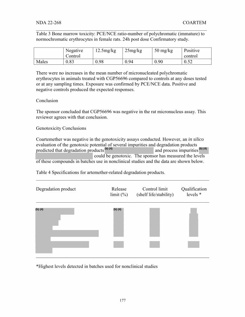
NDA 22-268 COARTEM _______________________________________________________________________
177
Table 3 Bone marrow toxicity: PCE/NCE ratio-number of polychromatic (immature) to normochromatic erythrocytes in female rats. 24h post dose Confirmatory study. Negative
Control 12.5mg/kg 25mg/kg 50 mg/kg Positive
control Males 0.83 0.98 0.94 0.90 0.52 There were no increases in the mean number of micronucleated polychromatic erythrocytes in animals treated with CGP56696 compared to controls at any doses tested or at any sampling times. Exposure was confirmed by PCE/NCE data. Positive and negative controls produced the expected responses. Conclusion The sponsor concluded that CGP56696 was negative in the rat micronucleus assay. This reviewer agrees with that conclusion. Genotoxicity Conclusions Coartemether was negative in the genotoxicity assays conducted. However, an in silico evaluation of the genotoxic potential of several impurities and degradation products predicted that degradation products and process impurities
could be genotoxic. The sponsor has measured the levels of these compounds in batches use in nonclinical studies and the data are shown below. Table 4 Specifications for artemether-related degradation products. ________________________________________________________________________ Degradation product Release
limit (%) Control limit
(shelf life/stability) Qualification
levels * ________________________________________________________________________
________________________________________________________________________ *Highest levels detected in batches used for nonclinical studies
(b) (4) (b) (4)
(b) (4)
(b) (4)

NDA 22-268 COARTEM _______________________________________________________________________
178
Table 5. Specifications for benflumetol-related compounds. ________________________________________________________________________ Impurity Release
limit (%) Control limit
(shelf life/stability) Qualification
levels * ________________________________________________________________________
*Highest levels detected in batches used for nonclinical studies The nonclinical batches contained very low levels of these compounds and so these impurities were essentially not subjected to nonclinical testing. Nonclinical studies need to be conducted using coartemether spiked with these compounds to determine if they do in fact exhibit genotoxic potential. 4.2.3.4 Carcinogenicity Carcinogenicity studies are not required for this three-day treatment regimen. 4.2.3.5. Reproductive and Developmental Toxicity Studies 4.2.3.5.1 Fertility and Early Development [66.] Study title: CGP 56697: An oral study for effects on fertility and early embryonic development in rats (MIN 964020) Key study findings: Dose dependent effects of CGP 56697, an anti-malarial compound were observed on fertility and embryonic development in rats. Reduced sperm motility, decreased pregnancy rates, reduced number of implants, and increased percent implantation losses were observed in high dose (1000 mg/kg/d) treated animals. The parental toxicity such as alopecia and salivation was also evident in both sexes at mid and high dose levels. A NOAEL could not be determined from this study. Study No.: T/P (US) 96064 (95-6089) Conducting laboratory CIBA-GEIGY Limited, Pharmaceutical Division and location: Summit, New Jersey 07901 Date of study initiation: February 6, 1996 Study Report Date: March 31, 1997 GLP compliance: Yes QA report: Yes Drug: CGP 56697 (coartemether)
(b) (4)

NDA 22-268 COARTEM _______________________________________________________________________
179
Lot No.: 17/145/1 and 17/066/1 % Active ingredients: CGP 56696/100.4%/20 mg (14.36% absolute) and CGP 56695/100.6%/120 mg (86.2% absolute) Methods Doses: 0, 30, 100, 300, 1000 mg/kg/d Species/strain: Albino (Sprague Dawley) rats, Tif: RAIf (SPF) 11 to 12 week old rats weighing 358 to 463 g (males) and 204 to 276 g (females) Route, formulation: By oral gavage in 0.5% suspension of Klucel HF and 0.1% surfactant Tween 80 (10mL/kg). Study design: Animals were housed in sterilized cages maintained on 14 hrs light and 10 hrs dark cycles, 73±50F, and 50 ±20 % relative humidity and acclimatized for 2 weeks prior to dosing. Females were co-housed with males in a 1:1 (female: male) ratio for mating, and were confirmed positive when vaginal plug was seen (designated as gestation day 0). Standard pellet diet and filtered water were provided ad libitum. Males were treated for 70 days (once daily) prior to mating, during the two week mating period, and 5 to 9 days thereafter. Females were treated for 14 days (once daily) prior to mating, during the two week mating period, and until day 6 of gestation. Oral administration of CGP 56697 (30, 100, 300, 1000 mg/kg/10mL/d) was given to animals as shown on Table 1. Males were sacrificed at or after 89 days of treatment while the sperm positive females were sacrificed on day 13 of gestation. Reproductive organs were weighed and recorded (Table 2). The experimental data were stored online (whenever possible) using database maintained on the and analyzed by computers for statistical analysis. Standard Operating System(s) were used in analyzing all the data used in this study for statistical analyses.
(b) (4)
(b) (4)
(b) (4) (b) (4)
(b) (4)
Appears This Way on Original

NDA 22-268 COARTEM _______________________________________________________________________
180
Table 1: Animal Groups and Treatment Plan
Table 2: Reproductive Parameters Examined in Female and Male Rats
Females Uterus Placenta Fetuses (UPF)
No. of corpora lutea
No. of implantation sites
No. of resorptions
No. of viable embryos
Males Testes Epididymides Sperm counts Motility Morphology Results: Clinical signs and mortality
Except one female (# 811) treated with high dose (1000 mg/kg/d) at the gestation day 7, no other mortality was observed due to oral administration of test article in rats. The clinical signs including alopecia and salivation were observed in both sexes at mid and high dose (300 and 1000 mg/kg/d) treatment. Other clinical signs

NDA 22-268 COARTEM _______________________________________________________________________
181
observed such as: scratch, broken teeth, blood on cage pan showed no dose response relationship, and were considered isolated events.
Food consumption
A 6 to 18% loss in food consumption was seen on days 0 to 7 in males treated with mid and high doses (300 and 1000 mg/kg/d) of test article, respectively. The loss in food consumption thereafter, remained low to minimal throughout the premating period on days 0-70 at the high dose and on days 0-21 at the mid dose treatment when compared to control animals. No loss in food consumption was seen at lower doses (30 and 100 mg/kg/d) in males.
A 17% loss in food consumption that was seen in pre-mating females on days 0 to 3, increased to 22% on days 3 to 6 in the high dose (1000 mg/kg/d) group, while in animals of the mid dose (300 mg/kg/d) group a 10% loss in food consumption was observed on days 3 to 6. The loss in food consumption returned to normal by day 13 of gestation in females treated with high dose (1000 mg/kg/d) test article. No loss in food consumption was seen at lower doses (30 and 100 mg/kg/d) in females.
Body weights
The mean body weight gain was very low (2.2%) in males of high (1000 mg/kg/d) dose groups when compared with controls (14%) on days 0 to 14. Significant decreases in mean body weight gains were observed on days 17-21, 24-28, 31-35, 38-42, 45-49, 52-56, 59-63, and 66-70 in high dose (1000 mg/kg/d) and on days 3-7, 10-14, and 24-28 in mid dose (300 mg/kg/d) treated males.
There was no gain in the mean body weight of females in the high dose groups while the mean body weight of control animals increased almost 8.5% on pre-mating days 0 to 14. The body weight gains of females were not affected during the gestation period in either group of animals treated with test article when compared with controls.
Effects of CGP 56697 on male reproductive parameters
An increase in the mean testes weight (+10%) and in the percent testes/body weight ratio (+31%) was seen in males treated with high dose (1000 mg/kg/d) test article. A decrease in epididymal weights (-13%), loss in epididymal sperm counts (-42%), and decrease in sperm motility (-55%) was evident in males treated with high dose test article. Counts of abnormal sperms were significantly increased in males treated with 30 (87%), 100 (100%), 300 (100%) and 1000 mg/kg/day (100%) test article when compared with controls (25.5%). Increases of lower magnitude were observed in males of the low and mid dose groups when compared with respective controls.
Effects of CGP 56697 on female reproductive parameters

NDA 22-268 COARTEM _______________________________________________________________________
182
Treatment related effects on reproductive parameters were mostly seen in females of the high dose (1000mg/kg/d) group. A significant decrease in the pregnancy rate (-37.5%), implantation sites (-13%), pre-implantation loss (-10.2%) were seen in females treated with high dose test article when compared with respective controls. Effects of lower magnitude were observed in females treated with 300, 100 and 30 mg/kg/d test article.
Macroscopic & microscopic findings
There were no macroscopic or microscopic findings related with the treatment of the test article on the male and female reproductive parameters.
Summary and Comments
The present study, “CGP 56697: An Oral Study for Effects on Fertility and Early Embryonic Development in Rats (MIN 964020)” was conducted according to the “Good Laboratory Practice regulations of the Food and Drug Administration (21 CFR 58) and the Standard Operating Procedures (SOPs) of the testing Facility.
A decrease in sperm motility, epididymal sperm counts, and increase in abnormal sperm counts were seen in animals treated with high dose (1000 mg/kg/d) test article. Reduced pregnancy rates, pre and post implantation losses resulted in decreased number of live embryos. In addition to effects on male and female reproductive parameters, parental toxicity as evident by clinical signs of alopecia and salivation was also observed in both sexes treated with high dose of CGP 56697. Due to a slight to moderate effects of test article at all dose levels the NOAEL could not be determined from this study. The study appears to have been conducted adequately and the conclusion is consistent with the results.
4.2.3.5.2 Embryo-Fetal Developmental Toxicity Studies [67.] Study title: CGP 56697: A follow-up oral dose range finding study for effects on embryo and fetal development in rats (MIN 954199) Key study findings: Dose dependent effects of CGP 56697, an anti-malarial compound, were observed on embryo and fetal development in rats. Increased number of total resorptions, postimplantation losses along with decreased number of live fetuses were the important findings in rats of the mid (60 mg/kg/d) and high (80 mg/kg/d) dose groups. The lower dose (40 mg/kg/d) level was established as NOAEL for this study. Study No.: T/P (US) 95051 (95-6065) Conducting laboratory CIBA-GEIGY Limited, Pharmaceutical Division and location: Summit, New Jersey 07901 Date of study initiation: April 17, 1995

NDA 22-268 COARTEM _______________________________________________________________________
183
Study Report Date: August 24, 1995 GLP compliance: No QA report: No Drug: CGP 56697 (coartemether) Lot No.: V-30-3192 % Active ingredients: CGP 56696/100.7%/20 mg (14.4% absolute) and CGP 56695/100.4%/120 mg (86.0% absolute) Methods Doses: 0, 40, 60, 80 mg/kg/d Species/strain: Albino (Sprague Dawley) rats, Tif: RAIf (SPF) 9 weeks old rats weighing 451 to 687 g (males) and 222 to 272 g (females) Route, formulation: By oral gavage in 0.5% suspension of Klucel HF and 0.1% surfactant Tween 80 (10mL/kg). Study design: Animals were housed in sterilized cages maintained on 14 hrs light and 10 hrs dark cycles, 73±50F, and 50 ±20 % relative humidity and acclimatized for 15 days prior to dosing. Females were co-housed in a 2:1 (female: male) ratio for mating, and were confirmed positive when vaginal plug was seen (designated as gestation day 0). Standard pellet diet and filtered water were provided ad libitum. Oral administration of CGP 56697 (40, 60, and 80 mg/kg/10mL/d) was given by gavage to animals in group 2, 3, and 4 once daily on gestation days 6 through 17 as follows:
(b) (4)
(b) (4)
Appears This Way on Original

NDA 22-268 COARTEM _______________________________________________________________________
184
Table 1: Animal Groups and Treatment Plan
All animals were sacrificed on day 20 of gestation. Reproductive organs were weighed and recorded (Table 2).
Table 2: Reproductive Parameters Examined in Female Rats Females Uterus Placenta
Fetuses (UPF) No. of corpora lutea
No. of implantation sites
No. of resorptions
No. of dead & viable fetuses
Experimental data were stored online (whenever possible) using database maintained on the and analyzed by computers for statistical analysis. Standard Operating System(s) were used in analyzing all the data used in this study for statistical analyses. Results: Clinical signs and mortality
There were no mortalities or necropsy findings observed due to oral administration of test article in rats. The vaginal blood was seen in one animal each at 60 and 80 mg/kg/d treatment and correlated with the necropsy findings of 100% resorptions in these two animals. The alopecia and chromodacryorrhea (red tears) were occasionally seen in animals of the mid and high dose (60 and 80
(b) (4)
(b) (4) (b) (4)

NDA 22-268 COARTEM _______________________________________________________________________
185
mg/kg/d) groups. Other clinical signs such as: scratch, rales (rattling sound in lungs), blood on cage pan did not exhibit a dose response relationship.
Food consumption There were no changes found in group mean maternal food consumption data or individual food consumption at any dose level.
Body weights
Statistically significant decreases were seen in the mean body weight gain of females in mid (-48%) and high (-42%) dose groups on days 18-20 of treatment.
Effects of CGP 56697 on female reproductive parameters
Treatment related effects on reproductive parameters were seen in females of the mid (60mg/kg/d) and high (80 mg/kg/d) dose groups. The resorptions were significantly increased in rats treated with mid (+5.6 fold) and high (+3.3 fold) dose test article compared with controls. The live fetuses were accordingly decreased in mid (-2.1 fold) and high (-1.5 fold) dose treated animals when compared with respective controls. The same time a 59% and 40% postimplantation loss was seen in rats treated with mid and high dose CGP 56697, respectively. There were no effects of low dose (40 mg/kg/d) treatment on the reproductive parameters of rats in this study.
Macroscopic & microscopic findings
There were no macroscopic or microscopic findings related with the treatment of test article on the female reproductive parameters.
Summary and comments
The present study, “CGP 56697: A Follow up Dose range finding Study for Effects on Embryo and Fetal Development in Rats (MIN 954119)” was not conducted according to the “Good Laboratory Practice regulations of the Food and Drug Administration (21 CFR 58) and the Standard Operating Procedures (SOPs) of the testing facility. Increased number of early and total resorptions, decreased number of live fetuses and increased number of postimplantation losses were seen in rats treated with mid (60 mg/kg/d) and high dose (80 mg/kg/d) test article. The lower dose (40 mg/kg/d) level was established as a NOAEL in this study. The study appears to have been conducted adequately and the conclusion is consistent with the results.

NDA 22-268 COARTEM _______________________________________________________________________
186
[68.] Study title: CGP 56697: An oral embryo and fetal development study in rats (MIN 954003) Key study findings: CGP 56697, an anti-malarial compound was found to have adverse effects on embryo and fetal development in rats at dose 100 mg/kg/d and higher. The total (100%) resorptions (26/26) resulting in 100% postimplantation loss were the primary findings associated with the test article at mid and high dose (300 mg/kg) level. Adverse effects of lower magnitude were noticed at low dose (30 mg/kg/d) level in rats. Study No.: T/P (US) 95072 (94-6158) Conducting laboratory CIBA-GEIGY Limited, Pharmaceutical Division and location: Summit, New Jersey 07901 Date of study initiation: December 28, 1994 Study Report Date: November 21, 1995 GLP compliance: Yes QA report: Yes Drug: CGP 56697 (Coartemether) Lot No.: V-30-3192 % Active ingredients: CGP 56696/100.4%/20 mg (14.36% absolute) and CGP 56695/100.6%/120 mg (86.2% absolute) Methods Doses: 0, 30, 100, 300 mg/kg/d Species/strain: Albino (Sprague Dawley) rats, Tif: RAIf (SPF) 12 weeks old rats weighing 586 to 892 g (males) and 214 to 300 g (females) Route, formulation: By oral gavage in 0.5% suspension of Klucel HF and 0.1% surfactant Tween 80 (10mL/kg) Study design: Animals were housed in sterilized cages maintained on 14 hrs light and 10 hrs dark cycles, 73±50F, and 50 ±20 % relative humidity and acclimatized for 15 days prior to dosing. Females were co-housed in a 2:1 (female: male) ratio for mating, and were confirmed positive when vaginal plug was seen (designated as gestation day 0). Standard pellet diet and filtered water were provided ad libitum. Oral administration of CGP 56697 (30, 100, 300 mg/kg/10mL/d) was given to animals of the group 2, 3, and 4 on gestation days from 6 through 15 as follows (Table 1):
(b) (4)
(b) (4)

NDA 22-268 COARTEM _______________________________________________________________________
187
Table 1: Animal Groups and Treatment Plan
All animals were sacrificed on day 20 of gestation via CO2 asphyxiation followed by exsanguination. All organs were found normal after gross evaluation, and dams were discarded. All fetuses were sacrificed by immersing in fixative media. Reproductive organs were weighed and recorded (Table 2).
Table 2: Reproductive Parameters Examined in Female Rats Females Uterus Placenta
Fetuses (UPF) No. of corpora lutea
No. of implantation sites
No. of resorptions
No. of dead/live fetuses
Experimental data were stored online (whenever possible) using database maintained on the and analyzed by computers for statistical analysis. Standard Operating System(s) were used in analyzing all the data used in this study. Results: Clinical signs and mortality
There was no mortality observed in this study. Some incidental adverse clinical signs occurring unrelated to CGP 56697 treatments such as alopecia, salivation, and rales were seen from time to time, and were considered as isolated events.
Food consumption
(b) (4)
(b) (4)(b) (4)

NDA 22-268 COARTEM _______________________________________________________________________
188
The group mean food consumption was significantly reduced in females treated with high dose (300 mg/kg/d) test article on gestation days 6-8 (-7.7%), 8-12 (-25.5%), and 12-16 (-15%). The loss in food consumption was seen at a lesser extent on the gestation days 16-20 in females treated with high dose and mid dose (100 mg/kg/d) test article, and returned to normal by day 20 of gestation. No loss in food consumption was seen in rats treated with low dose (30 mg/kg/d) of test article.
Body weights
Statistically significant decrease (- 10 to 18%) in the mean body weight was seen in females treated with mid dose (100 mg/kg/d) test article on the gestation days 16-20 while more pronounced decreases (-11 to 21%) were observed in rats treated with high dose (300 mg/kg/d) test article during this time period. Significant decreases in mean body weight gains were observed on days 12-16, 16-20 in mid dose group and on days 6-8, 8-12, 12-16 and 16-20 in high dose treated females.
Effects of CGP 56697 on female reproductive parameters
Treatment related effects on reproductive parameters were mostly seen in females of the mid dose (100 mg/kg/d) and high dose (300mg/kg/d) group. Almost 100% resorption was seen in females treated with mid (24/25) and high (26/26) dose test article when compared with controls. One female at mid dose treatment had an 87% postimplantation loss, and produced only two live fetuses. Effects (visceral and skeletal variations) of lower magnitude were observed in females treated with 30 mg/kg/d test article.
Macroscopic & microscopic findings
The lack of an adequate number of fetuses at mid and high dose levels precluded the full examination to assess the effects of the test article on fetal development. There were no macroscopic or microscopic findings related with the treatment of test article on the male and female reproductive parameters.
Summary and comments
The present study, “CGP 56697: An Oral Study for Effects on Fertility and Early Embryonic Development in Rats (MIN 964020)” was conducted according to the “Good Laboratory Practice regulations of the Food and Drug Administration (21 CFR 58) and the Standard Operating Procedures (SOPs) of the testing Facility.
There were no maternal mortalities and abnormalities noted in this study. The total (100%) resorptions (26/26) resulting into 100% postimplantation loss were the primary findings associated with the test article at the high dose level. Inadequate number of fetuses precluded the complete assessment and effects of test article on fertility and embryonic development. The study appears to have been conducted adequately and the conclusion is consistent with the results.

NDA 22-268 COARTEM _______________________________________________________________________
189
[69.] Study title: CGP 56697: An oral dose-range finding study for effects on embryo and fetal developments in rabbits (MIN 954007) Key study findings: There were no effects of CGP 56697, an anti-malarial compound on fertility and embryonic development in rabbits at daily dose of 0, 7, 35 or 70 mg/kg/d. The maternal mortality, clinical signs, and adverse effects on embryo and fetal development in rabbits were not observed at any of these dose levels. The maximum dose level (70 mg/kg/d) may be considered the NOAEL for the present study. Study No.: T/P (US) 95065 Conducting laboratory CIBA-GEIGY Limited, Pharmaceutical Division and location: Summit, New Jersey 07901 Date of study initiation: December 19, 1994 Study Report Date: November 15, 1995 GLP compliance: No QA report: No Drug: CGP 56697 (Coartemether) Lot No.: V-30-3192 % Active ingredients: CGP 56696/100.6%/20 mg (14.4% absolute) and CGP 56695/100.4%/120 mg (86.0% absolute) Methods Doses: 0, 7, 35, 70 mg/kg/d Species/strain: New Zealand White rabbits (Hra:[NZW]SPF) 6 months age, body weights from 2.58 to 3.82 Kg Route, formulation: By oral gavage in 0.5% suspension of Klucel HF and 0.1% surfactant Tween 80 (5 mL/kg). Study design: Animals were housed in individual sterilized cages maintained on 14 hrs light and 10 hrs dark cycles, 65±50F, and 50 ±20 % relative humidity and acclimatized for 4 weeks prior to dosing. Twenty four females were artificially inseminated from the semen obtained from one male (# 203) and the day of insemination was designated as gestation day 0. Standard pellet diet and filtered water were provided ad libitum. Oral (gavage) administration of CGP 56697 (0, 7, 35, 70 mg/kg/5mL/d) was given to animals on day 7 of gestation through 19 as follows:
(b) (4)
(b) (4)
Appears This Way on Original

NDA 22-268 COARTEM _______________________________________________________________________
190
Table 1: Animal Groups and Treatment Plan
All animals were euthanized by intravenous injection of pentobarbital and sacrificed by exsanguinations on day 29 of gestation. The major viscera and organs were examined grossly and no abnormalities were noticed. Reproductive organs (Table. 2) were weighed and recorded.
Table 2: Reproductive Parameters Examined in Female Rabbits Females Uterus Placenta
Fetuses (UPF) No. of corpora lutea
No. of implantation sites
No. of resorptions
No. of dead/viable embryos
Experimental data were stored online (whenever possible) using database maintained on the and analyzed by computers for statistical analysis. Standard Operating System(s) were used in analyzing all the data used in this study for statistical analyses. Results: Clinical signs and mortality
There were no clinical signs, mortalities or necropsy findings observed due to administration of test article in pregnant rabbits during this study.
Food consumption The maternal food consumption was not affected due to administration of test article in this study.
(b) (4)
(b) (4)(b) (4)

NDA 22-268 COARTEM _______________________________________________________________________
191
Body weights
The mean body weight and body weight gain was not affected due to administration of test article in this study.
Effects of CGP 56697 on female reproductive parameters
Treatment related effects on reproductive parameters were not observed due to administration of test article in this study.
Summary and comments
The present study, “CGP 56697: An oral dose range finding study for effects on embryo and fetal development in rabbits (MIN 954007)” was not conducted according to the “Good Laboratory Practice regulations of the Food and Drug Administration (21 CFR 58) and the Standard Operating Procedures (SOPs) of the testing facility.
There were no effects on maternal mortality, clinical signs, food consumption, body weight and reproductive parameters due to administration of the test article in this study. The maximum dose level (70 mg/kg/d) may be considered the NOAEL. The study appears to have been conducted adequately and the conclusion is consistent with the results.
[70.] Study title: CGP 56697: A follow up dose-range finding study for effects on embryo and fetal development in rabbits (MIN 954092) Key study findings: Adverse effects of CGP 56697, an anti-malarial compound, were seen on the embryo and fetal developments in rabbits at all dose levels (210, 700, 2100 mg/kg/d). A 25% abortion (2/8) rate seen in does of the low dose group increased to 75% (6/8) in the mid dose groups following the administration of test article. Due to 100% resorptions in the mid and high dose groups, fetuses were not available for further examinations. NOAEL could not be determined from this study. Study No.: T/P (US) 95049 (MIN 954092) Conducting laboratory CIBA-GEIGY Limited, Pharmaceutical Division and location: Summit, New Jersey 07901 Date of study initiation: March 2, 1995 Study Report Date: August 18, 1995 GLP compliance: No QA report: No Drug: CGP 56697 (Coartemether) Lot No.: V-38-3205 % Active ingredients: CGP 56696/102.7%/20 mg (14.7% absolute) and CGP 56695/99.8%/120 mg (85.5% absolute)

NDA 22-268 COARTEM _______________________________________________________________________
192
Methods Doses: 0, 210, 700, 2100 mg/kg/d Species/strain: New Zealand White rabbits (Hra: [NZW] SPF) 6/7 months age, body weights from 2.75 to 4.20 Kg Route, formulation: By oral gavage in 0.5% suspension of Klucel (HF) and 0.1% surfactant Tween 80 (10 mL/kg). Study design: Animals were housed in an individual sterilized cages maintained on 14 hrs light and 10 hrs dark cycles, 65 ± 50 F, and 50 ± 20 % relative humidity and acclimatized for 4 weeks prior to dosing. Twenty-four females were artificially inseminated from the semen obtained from one male (#203) and the day of insemination was designated as gestation day 0. Standard rabbit diet and filtered water were provided ad libitum. Oral (gavage) administration of CGP 56697 (0, 210, 700, 2100 mg/kg/d) was given to animals in group 2, 3, and 4 once daily on gestation days 7 through 19 as follows: Table 1: Animal Groups and Treatment Plan
(b) (4)
(b) (4)

NDA 22-268 COARTEM _______________________________________________________________________
193
Toxicokinetics The plasma samples separated as above were subjected to pharmacokinetic analyses by
Concentrations of CGP 56695, CGP 56696 and their metabolites were determined in the plasma by compound specific assays and reported elsewhere ( CH and F, respectively). Statistical evaluations Experimental data were stored online (whenever possible) using database maintained on the and analyzed by computers for statistical analysis. Statistical analyses were performed using SAS programs developed by according to SOPs, and maintained on the
of IBM main frame computer. Necropsy and Pathology All animals were euthanized by intravenous injection of pentobarbital and sacrificed by exsanguinations on day 29 of gestation. Live fetuses were sacrificed via oral administration of pentobarbital solution followed by cervical dislocation. The major viscera and organs were examined grossly and no abnormalities were noticed. Reproductive organs (Table 2) were weighed and recorded.
Table 2: Reproductive Parameters Examined in Female Rabbits Females Uterus Placenta
Fetuses (UPF) corpora lutea
implantation sites
Total resorptions
No. of dead & viable embryos
Results: Clinical signs and mortality
Treatment related abortion was observed at 210 mg/kg (2/8), and 700 mg/kg (6/8), however, no abortion was seen in controls or at 2100 mg/kg dose level. Clinical signs such as blood in cage pan, decreased and yellow brown stool were most often seen in animals of mid and high dose groups.
Food consumption
The mean food consumption was statistically reduced in animals of high dose group on GDs 7-12 and 15-20 compared with controls. Food consumption in the high dose group was increased compared with controls during gestation days 25-29, following cessation of dosing and following complete resorption of implants.
Body weights The corrected mean body weight (the terminal body weight at day 29 minus the weights of the uterus, ovaries, oviducts, placentas and fetuses) of the does was significantly increased in the low (28%), mid (38%), and high (39%) dose groups while the gestational body weight did not change due to administration of the test article at all dose levels in this study when compared with controls.
Effects of CGP 56697 on female reproductive parameters
(b) (4)
(b) (4)
(b) (4)
(b) (4)
(b) (4)
(b) (4)
(b) (4) (b) (4)

NDA 22-268 COARTEM _______________________________________________________________________
194
Abortion rate was significantly increased in animals of the low and mid dose groups (in 2/8 and 6/8 pregnant does, respectively). Does which aborted were sacrificed on the day of abortion. No abortion was noted in control animals or those treated with high dose (2100 mg/kg) test article. Early resorptions were observed among all surviving CGP 56697 treated does on GD 29 (day of C-section): 45%, 100%, and 100% early resorptions in 3, 2, and 8 surviving does in treatment groups 2, 3, and 4, respectively. Due to significant early resorption rates in the does treated with test article, an adequate number of viable fetuses were not available for detailed evaluations and assessment.
Summary and comments
The present study, “CGP 56697: A follow up dose range finding study for effects on embryo and fetal development in Rabbits (MIN 954092)” was not conducted according to the “Good Laboratory Practice Regulations.” Due to a significant increase in the abortion rates, resorptions, and the postimplantation losses at all dose levels, the NOAEL could not be determined. The study appears to have been conducted adequately and the conclusion is consistent with the results.
[71.] Study title: CGP 56697: An oral study for effects on embryo and fetal developments in rabbits (MIN 954158) Key study findings: Adverse effects of CGP 56697, such as abortions, postimplantation losses, and decreased number of live fetuses were seen on embryo and fetal developments in rabbits at high dose level (175 mg/kg/d), while no effects were evident at low (35 mg/kg) or mid dose (105 mg/kg) levels. The mid dose level (105 mg/kg) may be considered as NOAEL for this study. Study No.: T/P (US) 96015 (94-6157) Conducting laboratory CIBA-GEIGY Limited, Pharmaceutical Division and location: Summit, New Jersey 07901 Date of study initiation: June 13, 1995 Study Report Date: May 9, 1996 GLP compliance: Yes QA report: Yes Drug: CGP 56697 (Coartemether) Lot No.: 17/065/1 % Active ingredients: CGP 56696/98.1%/20 mg (14.0% absolute) and CGP 56695/100.3%/120 mg (86.0% absolute) Methods Doses: 0, 35, 105, 175 mg/kg/d Species/strain: New Zealand White rabbits (Hra:[NZW]SPF) 6/7 months age, body weights from 2.90 to 4.71 Kg Route, formulation: By oral gavage in 0.5% suspension of Klucel (HF)

NDA 22-268 COARTEM _______________________________________________________________________
195
and 0.1% surfactant Tween 80 (5 mL/kg). Study design: Animals were housed in individual sterilized cages maintained on 14 hrs light and 10 hrs dark cycles, 65±50F, and 50 ±20 % relative humidity and acclimatized for 4 weeks prior to dosing. Animals (n = 80) were artificially inseminated from the semen obtained from four males (# 113, 115, 182, and 204) and the day of insemination was designated as gestation day 0. Standard rabbit diet and filtered water were provided ad libitum. Oral (gavage) administration of CGP 56697 (35, 105, 175 mg/kg/d) was given to female animals in group 2, 3, and 4 once daily on gestation days 7 through 19 while the control animals received equivalent volumes of vehicle (0.5% Klucel, HF) as follows: Table 1: Animal Groups and Treatment Plan
Statistical evaluations Experimental data were stored online (whenever possible) using database maintained on the and analyzed by computers for statistical analysis. Statistical analyses were performed using SAS programs developed by according to SOPs, and maintained on the
of IBM main frame computer. Necropsy and Pathology
(b) (4)
(b) (4)
(b) (4)(b) (4)
(b) (4)
(b) (4)
(b) (4)

NDA 22-268 COARTEM _______________________________________________________________________
196
All animals were euthanized by intravenous injection of pentobarbital and sacrificed by exsanguinations on day 29 of gestation. Live fetuses were sacrificed via oral administration of pentobarbital solution followed by cervical dislocation. Reproductive organs were weighed and recorded (Table 2).
Table 2: Reproductive Parameters Examined in Female Rabbits Females Uterus Placenta
Fetuses (UPF) Corpora lutea
Implantation sites
Total resorptions
No. of dead & viable embryos
Results: Clinical signs and mortality
There was no mortality related to test article in this study. In addition to alopecia, the clinical signs such as blood in cage pan, decreased and yellow brown stool were occasionally seen in animals of all dose groups. The major viscera and organs were examined grossly and no abnormalities were noticed.
Food consumption
The mean food consumption was not affected due to administration of the test article in this study.
Body weights The mean maternal body weight and body weight gain was not affected due to administration of test article at all dose levels in this study.
Effects of CGP 56697 on female reproductive parameters
No effects of the test article were observed on reproductive parameters examined in low (35 mg/kg/d) and mid (105 mg/kg/d) dose groups. Mottled skin was observed in one of the fetuses in the low dose group, and not considered as related to test article. There were no skeletal and visceral variations observed in fetuses at any dose levels. A small number of abortions (2/20), early and total resorptions (2/20), postimplantation losses, and decrease in mean number of live fetuses were seen in animals treated with high dose (175 mg/kg/d) test article.
Fetal abnormalities:
There were no visceral and skeletal malformations observed due to administration of the test article in this study at all dose levels.
Summary and comments
The present study, “CGP 56697: An oral study for effects on embryo and fetal development in Rabbits (MIN 954158)” was conducted according to the “Good Laboratory Practice regulations of the Food and Drug Administration (21 CFR 58) and the Standard Operating Procedures (SOPs) of the testing Facility. The adverse effects of test article such as abortions, preimplantation losses, and decreased number of live fetuses were seen in animals treated at high dose (175 mg/kg/d) levels, while other groups were not affected.

NDA 22-268 COARTEM _______________________________________________________________________
197
The mid dose (105 /kg/d) level may be considered as the NOAEL. The study appears to have been conducted adequately and the conclusion is consistent with the results.
[72] Study title: CGP 56696: An oral study for effects on embryo and fetal development study in rats (MIN 954224) Key study findings: CGP 56696, an active component of anti-malarial compound CGP 56697, was found to have adverse effects on embryo and fetal development in rats. Increased number of early and total resorptions, postimplantation losses (74%) with reduced number of live fetuses, and increased incidence of skeletal variations (wavy ribs) were the primary findings associated with the test article at high dose level (10 mg/kg). The mid dose (3 mg/kg) level was considered as NOAEL from this study. Study No.: T/P (US) 96036 (95-6159) Conducting laboratory CIBA-GEIGY Corporation, Pharmaceutical Division and location: Summit, New Jersey 07901 Date of study initiation: September 22, 1995 Study Report Date: August 7, 1996 GLP compliance: Yes QA report: Yes Drug: CGP 56696 (artemether) Lot No.: 810194 Active ingredients: CGP 56696 (98.2%) Methods Doses: 0, 1, 3, 10 mg/kg/d Species/strain: Albino (Sprague Dawley) rats, Tif: RAIf (SPF) 12 weeks old rats weighing 586 to 892 g (males) and 214 to 300 g (females) Route, formulation: By oral gavage in 0.5% suspension of Klucel HF and 0.1% surfactant Tween 80 (10mL/kg) Study design: Animals were housed in sterilized cages maintained on 14 hrs light and 10 hrs dark cycles, 73±50F, and 50 ±20 % relative humidity and acclimatized for 15 days prior to dosing. Females were co-housed in a 2:1 (female: male) ratio for mating, and were confirmed positive when vaginal plug was seen (designated as gestation day 0). Standard pellet diet and filtered water
were provided ad libitum. The water analysis by the Subdivision of Toxicology revealed a high level of lead (0.024 mg/L, the acceptable level of lead was 0.015 mg/L) contamination in the drinking water
(b) (4) (b) (4)

NDA 22-268 COARTEM _______________________________________________________________________
198
being used for administration of the test article. This concentration of lead in the drinking water was not considered to have any adverse effects on the outcome of this study. Oral administration of CGP 56696 (1, 3, 10 mg/kg) was given to animals (n = 26/group) of the group 2, 3, and 4 on gestation days from 6 through 17 as follows (Table 1): Table 1: Animal Groups and Treatment Plan
All animals were sacrificed on day 20 of gestation via CO2 asphyxiation followed by exsanguination. All organs were found normal after gross evaluation, and dams were discarded. ~50% of the fetuses were sacrificed by immersing in Bouin’s fixative media for visceral examination and the rest were in EVS media for skeletal evaluations. Following reproductive organs were weighed and recorded (table 2).
Table 2: Reproductive Parameters Examined in Female Rats Females Uterus Placenta
Fetuses (UPF) No. of corpora lutea
No. of implantation sites
No. of resorptions
No. of dead/live fetuses
Experimental data were stored online (whenever possible) using database maintained on the and analyzed by computers for statistical analysis. Standard Operating System(s) were used in analyzing all the data used in this study.
(b) (4)
(b) (4)(b) (4)

NDA 22-268 COARTEM _______________________________________________________________________
199
Results: Clinical signs and Mortality
The clinical signs such as blood in the vaginal area and alopecia were seen in controls (2/26) and treated animals at any dose level (2-4/26), and considered as incidental. An early delivery was seen in one dam (#99) at day 17 of gestation in high dose (10 mg/kg) group, and considered an isolated event not related with treatment. There was no mortality observed in this study.
Food consumption There was no loss in the food consumption of rats treated with test article during the study.
Body weights
Statistically significant decrease (-8%) in the mean body weight was first seen in females treated with the high dose (10 mg/kg) test article on the gestation days 15 to 18 while more pronounced decrease (-12%) was observed in rats on the gestation days 18 to 20. At the same time a significant decrease (-44 to 46%) in the body weight gains was also observed on gestation days 12 to 20. The corrected body weight (the terminal body weight minus the weights of uterus, ovaries, oviducts, placentas and fetuses), and body weight gain were not affected due to test article administration.
Effects of CGP 56696 on female reproductive parameters
Treatment related effects on reproductive parameters were mostly seen in females of the high dose (10 mg/kg/d) group. A significant increase (12/16) in the total resorptions (early and late) in the high dose group lead to ~74% post implantation loss, compared with an ~8% post implantation loss among controls. No effects were seen on fetal sex ratio due to test article administration.
Macroscopic & microscopic findings
There were no abnormalities noticed in fetuses obtained from rats treated with low (1 mg/kg) and mid (3 mg/kg) dose test article. A non-significant decrease (~12.5%) was observed in fetal weights of both sexes treated with high dose (10 mg/kg) test article when compared with controls.
A single finding of gastroschisis was noted in the control group, and considered incidental. No visceral variations and gross malformations were observed in any dose groups due to test article. However, the number of fetuses with wavy ribs were significantly increased (5/51) in the high dose (10 mg/kg) group when compared with mid dose (1/170) group. This event was considered a dose response and indicative of fetotoxicity. Due to increased post-implantation loss (74%) in the high dose (10 mg/kg) group, an inadequate number of fetuses (only from nine litters compared with 16 litters recommended for this type of study) were available and precluded adequate assessment of the effects of test article on embryo and fetal development in rats.

NDA 22-268 COARTEM _______________________________________________________________________
200
Summary and comments
The present study, “CGP 56696: An oral study for effects on embryo and fetal development in rats (MIN 954224)” was conducted according to the “Good Laboratory Practice regulations of the Food and Drug Administration (21 CFR 58) and the Standard Operating Procedures (SOPs) of the testing Facility.
There were no maternal mortalities or abnormalities noted in this study. Significant resorptions (12/16) resulting in 74 % postimplantation loss, and fetuses with wavy ribs (skeletal variations, 50%) indicative of fetotoxicity, were associated with the test article at the high dose level (10 mg/kg).
The mid dose (3 mg/kg) level was considered as NOAEL from this study. The study appears to have been conducted adequately and the conclusion is consistent with the results.
[73.] Study title: CGP 56696: An oral dose-range finding study for effects on embryo and fetal developments in rabbits (MIN 954070) Key study findings: There were no adverse effects of CGP 56696, an active component of anti-malarial compound CGP 56697, on fertility and embryonic development in rabbits at the low (3 mg/kg) and mid (10 mg/kg) dose levels. An increase in the early and total resorptions, abortions, and post implantation losses were evident in animals treated with high dose (30 mg/kg) test article. The mid dose level (10 mg/kg) may be considered the NOAEL for the present study. Study No.: T/P (US) 95074 (956027) Conducting laboratory CIBA-GEIGY Limited, Pharmaceutical Division and location: Summit, New Jersey 07901 Date of study initiation: June 26, 1995 Study Report Date: November 27, 1995 GLP compliance: No QA report: No Drug: CGP 56696 (artemether) Lot No.: 810194 % Active ingredients: CGP 56696 (98.2%) Methods Doses: 0, 3, 10, 30 mg/kg/d Species/strain: New Zealand White rabbits (Hra:[NZW]SPF) 25 weeks old, body weights from 3.22 to 4.57 Kg Route, formulation: By oral gavage in 0.5% suspension of Klucel HF and 0.1% surfactant Tween 80 (10 mL/kg).

NDA 22-268 COARTEM _______________________________________________________________________
201
Study design: Animals were housed in individual sterilized cages maintained on 14 hrs light and 10 hrs dark cycles, 65±50F, and 50 ±20 % relative humidity and acclimatized for 4 weeks prior to dosing. Animals were artificially inseminated from the semen obtained from two males (# 204 & 206) and the day of insemination was designated as gestation day 0. Standard pellet diet and filtered water were provided ad libitum. Oral (gavage) administration of CGP 56696 (0, 3, 10, 30 mg/kg) was given to animals on day 7 of gestation through 19 as follows: Table 1: Animal Groups and Treatment Plan
All animals were euthanized by oral administration of pentobarbital solution and sacrificed by exsanguination on day 29 of gestation. The major viscera including
(b) (4)
(b) (4)
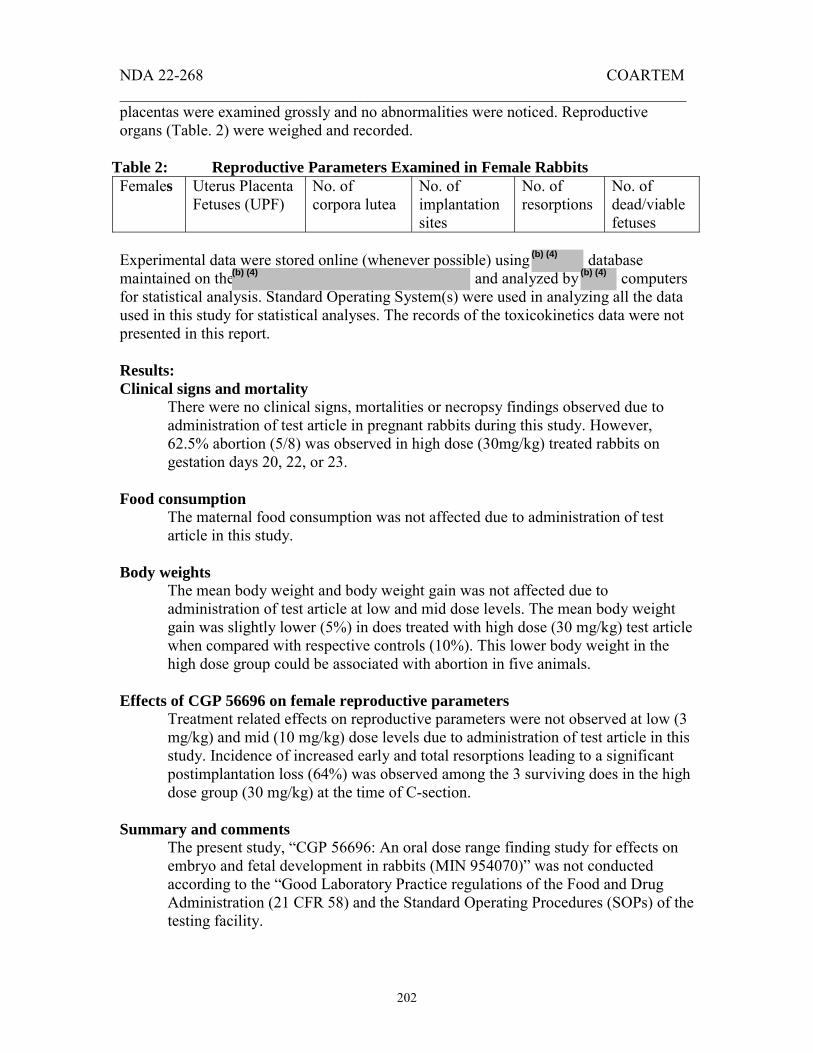
NDA 22-268 COARTEM _______________________________________________________________________
202
placentas were examined grossly and no abnormalities were noticed. Reproductive organs (Table. 2) were weighed and recorded.
Table 2: Reproductive Parameters Examined in Female Rabbits Females Uterus Placenta
Fetuses (UPF) No. of corpora lutea
No. of implantation sites
No. of resorptions
No. of dead/viable fetuses
Experimental data were stored online (whenever possible) using database maintained on the and analyzed by computers for statistical analysis. Standard Operating System(s) were used in analyzing all the data used in this study for statistical analyses. The records of the toxicokinetics data were not presented in this report. Results: Clinical signs and mortality
There were no clinical signs, mortalities or necropsy findings observed due to administration of test article in pregnant rabbits during this study. However, 62.5% abortion (5/8) was observed in high dose (30mg/kg) treated rabbits on gestation days 20, 22, or 23.
Food consumption
The maternal food consumption was not affected due to administration of test article in this study.
Body weights
The mean body weight and body weight gain was not affected due to administration of test article at low and mid dose levels. The mean body weight gain was slightly lower (5%) in does treated with high dose (30 mg/kg) test article when compared with respective controls (10%). This lower body weight in the high dose group could be associated with abortion in five animals.
Effects of CGP 56696 on female reproductive parameters
Treatment related effects on reproductive parameters were not observed at low (3 mg/kg) and mid (10 mg/kg) dose levels due to administration of test article in this study. Incidence of increased early and total resorptions leading to a significant postimplantation loss (64%) was observed among the 3 surviving does in the high dose group (30 mg/kg) at the time of C-section.
Summary and comments
The present study, “CGP 56696: An oral dose range finding study for effects on embryo and fetal development in rabbits (MIN 954070)” was not conducted according to the “Good Laboratory Practice regulations of the Food and Drug Administration (21 CFR 58) and the Standard Operating Procedures (SOPs) of the testing facility.
(b) (4)
(b) (4)(b) (4)

NDA 22-268 COARTEM _______________________________________________________________________
203
No adverse effects were observed on reproductive parameters due to administration of the test article at low (3 mg/kg) and mid (10 mg/kg) levels. Significant increases in abortions and resorptions were seen in females of the high dose (30 mg/kg) group. The mid dose (10 mg/kg/d) level may be considered the NOAEL for this study. The study appears to have been conducted adequately and the conclusion is consistent with the results.
[74.] Study title: CGP 56696: An oral study for effects on embryo and fetal developments in rabbits (MIN 954072) Key study findings: There were no adverse effects of CGP 56696, an active component of anti-malarial compound CGP 56697, on embryonic and fetal development in rabbits at all doses tested (0, 5, 15, 25 mg/kg/d). Administration of test article did not produce any signs of embryotoxicity or fetotoxicity in this study. Study No.: T/P (US) 96033 (956028) Conducting laboratory CIBA-GEIGY Corporation, Pharmaceutical Division and location: Summit, New Jersey 07901 Date of study initiation: September 15, 1995 Study Report Date: August 1, 1996 GLP compliance: Yes QA report: Yes Drug: CGP 56696 (artemether) Lot No.: 810194 % Active ingredients: CGP 56696 (98.2%) Methods Doses: 0, 5, 15, 25 mg/kg/d Species/strain: New Zealand White rabbits (Hra: [NZW] SPF) 24 weeks old, body weights from 2.85 to 4.18 Kg Route, formulation: By oral gavage in 0.5% suspension of Klucel HF and 0.1% surfactant Tween 80 (10 mL/kg). Study design: Animals were housed in individual sterilized cages maintained on 14 hrs light and 10 hrs dark cycles, 65±50F, and 50 ±20 % relative humidity and acclimatized for 4 weeks prior to dosing. Animals were artificially inseminated from the semen obtained from two males (# 204 & 206) and the day of insemination was designated as gestation day 0. Standard pellet diet and filtered water were provided ad libitum. Oral (gavage) administration of CGP 56696 (0, 5, 15, 25 mg/kg) was given to animals on day 7 of gestation through 19, and treated as per table 1.
(b) (4)
(b) (4)

NDA 22-268 COARTEM _______________________________________________________________________
204
All animals were euthanized by intravenous administration of pentobarbital solution and sacrificed by exsanguination on day 29 of gestation. The major viscera including placentas were examined grossly and no abnormalities were noticed. Reproductive organs (Table 2) were weighed and recorded. All fetuses were sacrificed by oral administration of pentobarbital solution. Table 1: Animal Groups and Treatment Plan
Experimental data were stored online (whenever possible) using database maintained on the and analyzed by computers for statistical analysis. Standard Operating System(s) were used in analyzing all the data used in this study for statistical analyses.
Table 2: Reproductive Parameters Examined in Female Rabbits Females Uterus Placenta
Fetuses (UPF) No. of corpora lutea
No. of implantation sites
No. of resorptions
No. of dead/viable fetuses
Results: Clinical signs and mortality
There were no clinical signs, mortalities or necropsy findings observed due to administration of test article in pregnant rabbits during this study. In addition to alopecia (in all groups including the control), blood was noticed in the cage pan of one of the does in the mid (15 mg/kg) and high dose (25 mg/kg) groups. These findings were considered not associated with test article.
(b) (4)
(b) (4) (b) (4)

NDA 22-268 COARTEM _______________________________________________________________________
205
Food consumption The maternal food consumption was not affected due to administration of test article in this study.
Body weights
The mean body weights and body weight gains were not affected due to administration of test article at any dose tested.
Effects of CGP 56696 on female reproductive parameters and fetuses
Treatment related effects were not observed on reproductive parameters of does and fetal sex ratio (% males) at any dose level due to administration of test article in this study. The postimplantation losses observed in mid (11%), and high dose (11%) groups were not significantly abnormal when compared with controls (9%), and considered not related with test article treatment. No effects were seen on fetal weights due to administration of the test article. There were no visceral or skeletal variations observed in the fetuses due to test article administration. An absent gall bladder in one of the fetuses at mid (15 mg/kg) dose and findings of agenesis and misalignment of centrum/vertebrae in fetuses from two litters at the high dose (25 mg/kg) were considered incidental to treatment, and not associated with test article.
Summary and comments
The present study, “CGP 56696: An oral study for effects on embryo and fetal development in rabbits (MIN 954072)” was conducted according to the “Good Laboratory Practice regulations of the Food and Drug Administration (21 CFR 58) and the Standard Operating Procedures (SOPs) of the testing facility.
No adverse effects were observed on reproductive parameters of does and fetuses due to administration of the test article at any dose level. The study appears to have been conducted adequately and the conclusion is consistent with the results.
[75.] Study title: CGP 56695: Embryonic and fetal development study in rats by oral administration (MIN 954224) Key study findings: CGP 56695, an active component of anti-malarial compound CGP 56697, did not produce any adverse effect on the reproductive parameters of the dams, or on embryonic and fetal developments in rats at any dose level (0, 100, 300, 1000 mg/kg/d) tested in this study. Occurrence of kinked ureters and hydronephrosis in treated animals was considered incidental to treatment, since these findings were comparable with the untreated controls.

NDA 22-268 COARTEM _______________________________________________________________________
206
Study No.: 95-6024 Conducting laboratory CIBA-GEIGY Limited, Pharmaceutical Division and location: Basel, Switzerland Date of study initiation: September 07, 1995 Study Report Date: March 19, 1996 GLP compliance: Yes QA report: Yes Drug: CGP 56695 (benflumetol) Lot No.: 800295 Active ingredients: CGP 56695 (100.9%) Methods Doses: 0, 100, 300, 1000 mg/kg/d Species/strain: Albino (Sprague Dawley) rats, Tif: RAIf (SPF) 13/34 weeks old rats weighing 213 to 321 g (males) and 214 to 300 g (females) Route, formulation: By oral gavage in 0.5% suspension of Klucel HF and 0.1% surfactant Tween 80 (10mL/kg) Study design: Animals were housed in sterilized cages maintained on 12 hrs light and 10 hrs dark cycles, 21±20C, and acclimatized to laboratory conditions prior to dosing. Females were co-housed in a 1:1 (female: male) ratio for mating, and were confirmed positive when vaginal plug was seen (designated as gestation day 0). Commercial irradiated diet
and filtered water were provided ad libitum.
The water analysis was routinely carried out for microbial and chemical contaminants by the Ciba, Vision, Macclesfield, UK; ; and by the supplier, Oral administration of CGP 56695 (0, 100, 300, 1000 mg/kg) was given to animals (n = 24/group) of groups 2, 3, and 4 on gestation days from 6 through 17 as follows (Table 1):
(b) (4)
(b) (4)
(b) (4)
(b) (4)
Appears This Way on Original

NDA 22-268 COARTEM _______________________________________________________________________
207
Table 1: Animal Groups and Treatment Plan All animals were sacrificed on day 20 of gestation via CO2 asphyxiation followed by exsanguination. All organs were found normal after gross evaluation, and dams were discarded. Sodium pentabarbitone
was used intravenously to sacrifice the fetuses. Following reproductive organs were weighed and recorded.
Table 2: Reproductive Parameters Examined in Female Rats Females Uterus Placenta
Fetuses (UPF) No. of corpora lutea
No. of implantation sites
No. of resorptions
No. of dead/live fetuses
Experimental data were stored online (whenever possible) using database maintained on the and analyzed by computers and DART system for statistical analysis. Standard Operating System(s) were used in analyzing all the data used in this study. Results: Clinical signs and Mortality
There were no mortalities or clinical signs observed in rats due to administration of test article at any dose level (0, 100, 300, 1000 mg/kg/d).
(b) (4)
(b) (4)
(b) (4)(b) (4)

NDA 22-268 COARTEM _______________________________________________________________________
208
Food consumption There was no loss in the food consumption of rats treated with test article during the study.
Body weights The body weights and body weight gains were not affected due to test article administration in this study. An inconsistent ups and downs in the body weights observed in the mid (300 mg/kg) and high (1000 mg/kg) dose groups were considered incidental to treatment, and not associated with the test article.
Effects of CGP 56695 on female reproductive parameters
There were no effects on reproductive parameters (Table 2) in females treated with the test article. Statistically significant preimplantation loss (30%) observed in the high dose (1000 mg/kg) animals may not be related with the test article administration that started on the day 6 of gestation. No effects were seen on fetal sex ratio due to test article administration.
Macroscopic & microscopic findings
No abnormalities were observed in rats and their fetuses treated with test article at any dose level (100, 300 or 1000 mg/kg). Visceral and skeletal examinations did not reveal any dose related variation in the treated group of animals when compared with the controls. Increased incidences of kinked ureters seen in the low (26%), mid (28%) and high (22%) dose groups were comparable with controls (17%), and considered not related with test article treatment. A single incidence of hydronephrosis (dam 4002 and fetus 12) was noticed in the high dose (1000 mg/kg) group, and considered incidental to treatment. Occurrences of dilated lateral brain ventricles were observed in the mid (11%) and high dose (6%) treated dams, and in fetuses of the mid (2%) and high (1%) dose groups, and considered incidental. Incidence of widened anterior fontanels (22%) seen in fetuses obtained from dams treated with mid dose test article, were also considered incidental by the Study Director.
Summary and comments
The present study, “CGP 56695: Embryonic and fetal development study in rats by oral administration (956024)” was conducted according to the “Good Laboratory Practice regulations of the Food and Drug Administration (21 CFR 58) and the Standard Operating Procedures (SOPs) of the testing Facility.
CGP 56695 treatment at 100, 300 or 1000 mg/kg dose levels did not produce any adverse effects on the reproductive parameters of the dams and embryonic and fetal developments in rats. The study appears to have been conducted adequately and the conclusion is consistent with the results.

NDA 22-268 COARTEM _______________________________________________________________________
209
[76.] Study title: CGP 56695: An oral limit test for effects on embryo and fetal developments in rabbits (MIN 954069) Key study findings: There were no adverse effects of CGP 56695, an active component of anti-malarial compound CGP 56697, on embryonic and fetal developments at the 1000 mg/kg/d dose level. Administration of test article did not produce any signs of embryotoxicity or fetotoxicity in this study. Study No.: T/P (US) 95071 (956022) Conducting laboratory CIBA-GEIGY Corporation, Pharmaceutical Division and location: Summit, New Jersey 07901 Date of study initiation: June 26, 1995 Study Report Date: November 16, 1995 GLP compliance: No QA report: No Drug: CGP 56695 (benflumetol)) Lot No.: 800594 & 800195 % Active ingredients: 100.0% and 99.9% Methods Doses: 0, 1000 mg/kg/d Species/strain: New Zealand White rabbits (Hra: [NZW] SPF) 25 weeks old, body weights from 3.43 to 4.57 Kg Route, formulation: By oral gavage in 0.5% suspension of Klucel HF and 0.1% surfactant Tween 80 (10 mL/kg). Study design: Animals were housed in individual stainless steel wire bottom cages maintained on 14 hrs light and 10 hrs dark cycles, 65±50F, and 50 ±20 % relative humidity and acclimatized for 4 weeks prior to dosing. Animals were artificially inseminated from the semen obtained from two males (# 204 & 206) and the day of insemination was designated as gestation day 0. Standard pellet diet and filtered water were provided ad libitum. Oral (gavage) administration of CGP 56695 (0, 1000 mg/kg) was given to animals (n = 8) on day 7 of gestation through 19, and treated as per table 1. All animals were euthanized by intravenous administration of pentobarbital solution and sacrificed by exsanguination on day 29 of gestation. All fetuses were sacrificed by oral administration of pentobarbital solution. The major viscera including placentas were examined grossly and no abnormalities were noticed. Reproductive organs were weighed and recorded (Table 2).
(b) (4)
(b) (4)

NDA 22-268 COARTEM _______________________________________________________________________
210
Table 1: Animal Groups and Treatment Plan
Experimental data were stored online (whenever possible) using database maintained on the and analyzed by computers for statistical analysis. Standard Operating System(s) were used in analyzing all the data used in this study for statistical analyses.
Table 2: Reproductive Parameters Examined in Female Rabbits Females Uterus Placenta
Fetuses (UPF) No. of corpora lutea
No. of implantation sites
No. of resorptions
No. of dead/viable fetuses
Results: Clinical signs and mortality
(b) (4)
(b) (4)(b) (4)
(b) (4)
(b) (4)
(b) (4)
(b) (4) (b) (4)
(b) (4)

NDA 22-268 COARTEM _______________________________________________________________________
211
There were no clinical signs, mortalities or necropsy findings observed due to administration of test article (1000 mg/kg) in pregnant rabbits in this study. Decreased stool was observed in one of the does (#3), and considered not associated with the test article.
Food consumption
The maternal food consumption was not affected due to administration of test article in this study.
Body weights
The mean body weights of does were not affected due to administration of test article in this study. An increase in the body weight gains in treated does (1000 mg/kg) on gestation days 10-13 and 24-29 was considered not significant due to treatment because of a spontaneous body weight loss observed in controls during this time period.
Effects of CGP 56696 on female reproductive parameters and fetuses
Treatment related effects were not observed in does and their reproductive potential, fetuses and fetal sex ratio (% males) due to administration of test article in this study. There were no visceral or skeletal variations observed in the fetuses due to test article administration.
Summary and comments
The present study, “CGP 56695: An oral limit test for effects on embryo and fetal development in rabbits (MIN 954069)” was not conducted according to the “Good Laboratory Practice regulations of the Food and Drug Administration (21 CFR 58) and the Standard Operating Procedures (SOPs) of the testing facility.
No adverse effects were observed on does, their reproductive potential and fetuses due to administration of the test article at 1000 mg/kg. The study appears to have been conducted adequately and the conclusion is consistent with the results.
[77.] Study title: CGP 56695: An oral study for effects on embryo and fetal developments in rabbits (MIN 954071) Key study findings: There were no adverse effects of CGP 56695, an active component of anti-malarial compound CGP 56697, on does or embryonic and fetal development at any dose level (0, 100, 500 or 1000 mg/kg). Administration of test article did not produce any signs of embryotoxicity or fetotoxicity in this study. Study No.: T/P (US) 96038 (956023) Conducting laboratory CIBA-GEIGY Corporation, Pharmaceutical Division and location: Summit, New Jersey 07901

NDA 22-268 COARTEM _______________________________________________________________________
212
Date of study initiation: September 13, 1995 Study Report Date: August 8, 1996 GLP compliance: Yes QA report: Yes Drug: CGP 56695 (benflumetol)) Lot No.: 800395 % Active ingredients: 100.0% Methods Doses: 0, 100, 500, 1000 mg/kg/d Species/strain: New Zealand White rabbits (Hra: [NZW] SPF) 25 weeks old, body weights from 2.90 to 4.10 Kg Route, formulation: By oral gavage in 0.5% suspension of Klucel HF and 0.1% surfactant Tween 80 (10 mL/kg). Study design: Animals were housed in individual stainless steel wire bottom cages maintained on 14 hrs light and 10 hrs dark cycles, 65±50F, and 50 ±20 % relative humidity and acclimatized for 4 weeks prior to dosing. Animals were artificially inseminated from the semen obtained from four males (# 164, 182, 204 & 206) and the day of insemination was designated as gestation day 0. Standard pellet die and filtered water were provided ad libitum. Oral (gavage) administration of CGP 56695 (0, 1000 mg/kg) was given to animals (n = 20/group) on day 7 of gestation through 19, and treated as per table 1. All animals were euthanized by intravenous administration of pentobarbital solution and sacrificed by exsanguination on day 29 of gestation. All fetuses were sacrificed by oral administration of pentobarbital solution. The major viscera including placentas were examined grossly and no abnormalities were noticed. Reproductive organs were weighed and recorded (Table 2).
(b) (4)
(b) (4)
Appears This Way on Original

NDA 22-268 COARTEM _______________________________________________________________________
213
Table 1: Animal Groups and Treatment Plan
The visceral evaluation of fetuses was performed on the day of C-section and fetuses were cleared in potassium hydroxide solution and stained with alizarin red S for skeletal evaluation. Experimental data were stored online (whenever possible) using database maintained on the and analyzed by computers for statistical analysis. Standard Operating System(s) were used in analyzing all the data used in this study for statistical analyses.
Table 2: Reproductive Parameters Examined in Female Rabbits Females Uterus Placenta
Fetuses (UPF) No. of corpora lutea
No. of implantation sites
No. of resorptions
No. of dead/viable fetuses
Results: Clinical signs and mortality
There were no clinical signs, mortalities or necropsy findings observed in does due to administration of test article at any dose level (100, 500 or 1000 mg/kg) tested in this study. On the day 23 of gestation, one doe (#63) aborted in the high dose (1000 mg/kg) group; however, this was considered incidental.
(b) (4)
(b) (4)(b) (4)

NDA 22-268 COARTEM _______________________________________________________________________
214
Similarly, other clinical signs such as alopecia, blood in cage pan, and decreased, discolored soft stools were also considered incidental since no dose response relationship could be established. Necropsy observations included the findings of mottled lungs in one control animal, and the presence of cyst(s) adjacent to ovaries in two controls and one each in low and high group animals.
Food consumption
The maternal food consumption was not affected due to administration of test article in this study. Because of a random variation of food consumption in control animals, a significant decrease in food consumption (-18%) was noticed in animals of the low dose group (100 mg/kg) on gestation days 20-25 and in high dose group (-10%) on gestation days 11-14.
Body weights
The mean body weights of does were not affected due to administration of test article at any dose level (100, 500 or 1000 mg/kg). An increase in the body weight gains in treated does at 100 (± 06 kg) and 500 mg/kg (± 08 kg) compared with controls (± 02 kg) on gestation days 10-13 was considered incidental.
Effects of CGP 56696 on female reproductive parameters and fetuses
Treatment related effects were not observed on the reproductive parameters of does, fetuses, fetal weights and fetal sex ratio (% males) due to administration of test article at any dose level (100, 500 or 1000 mg/kg). ~18% postimplantation loss observed in controls and the treated group of does, was considered incidental to treatment and not related to test article. Findings of gastroschisis was noted in one of the fetuses each in the control (litter No. 31) and high dose (litter No. 39) groups while umbilical hernia was exhibited in one fetus of the mid dose group (litter No. 22). There were no visceral and skeletal variations observed in the fetuses due to test article administration. However, gall bladder variations observed in controls and all the treated groups were considered not related to test article. Findings of incomplete ossified patellae were observed in the control (44/144), low (33/132), mid (58/167) and high dose (75/155) treated animals, and considered incidental to treatment.
Summary and comments
The present study, “CGP 56695: An oral study for effects on embryo and fetal development in rabbits (MIN 954071)” was conducted according to the “Good Laboratory Practice regulations of the Food and Drug Administration (21 CFR 58) and the Standard Operating Procedures (SOPs) of the testing facility.
No adverse effects were observed on does, their reproductive parameters, and fetuses due to administration of the test article at any dose (100, 500 or 1000

NDA 22-268 COARTEM _______________________________________________________________________
215
mg/kg) level. The study appears to have been conducted adequately and the conclusion is consistent with the results.
4.2.3.5.3 Prenatal and Postnatal Development, Including Maternal function [78.] Title: An oral study for effects on Pre-and Postnatal Development in Rats KEY STUDY FINDINGS: CGP 56697 administered by oral gavage to pregnant rats at daily doses of 5, 25, or 50 mg/kg, from GD 6 to LD 20, resulted in no significant increase in mortality, adverse clinical observations, food consumption, body weights, or gross pathologic findings for any of the three generations (F0, F1, F2) at any dose and sex examined. A slight increase in both gestation duration (1 day) and mean number of still births from F0 maternal animals in the mid (25 mg/kg/day) and high (50 mg/kg/day) dose groups was observed. The sponsor concluded the increase in stillborn rats resulted from a complete loss of a litter from one dam and thus was not considered treatment related. Although not statistically significant, increase in the number and percentage of F0 post-implantation losses and number of viable F1 newborns was reported at the high dose (50 mg/kg). Evaluation of the F1 developmental and reproductive parameters showed no significant treatment related effects different from controls for any dose group or sex examined. Study no.: 94-6159 No. of pages: 282 Conducting laboratory and location: CIBA-GEIGY corp., Pharmaceutical Division, Summit, NJ 07901. Date of study initiation: 5/31/1995 GLP compliance: Yes QA reports: yes (X) no ( ) Drug / lot # / % purity / % of mixture:
CGP 56697 (Lot No. V.38-3205) Mixture comprised of: (1) Artemether / lot # 810194 / purity: 102.7% / 14.7% absolute (2) Benflumetol / lot # 800494 / purity: 99.8% / 85.5% absolute
Vehicle Control: 0.5% (w/v) hydroxypropylcellulose (Klucel, grade HF), aqueous solution plus 0.1% (w/v) Tween 80. Supplier of Test Article: CIBA-GEIGY AG, Basel, Switzerland. METHODS
Doses: 3 doses (5, 25, and 50 mg/kg/day) Species/strain: Rat/Sprague-Dawley (SD) COBS CD [SD] BR). Number/sex/group: (1) F0: Total 104 rats / 26 pregnant females per group / 4 groups.
(2) F1: Total 190 SD rat pups / 23-25 males; 23-25 females per group / 4 groups coinciding with maternal treatment groups [(F1) behavioral/postpartum fertility assessment groups]
(b) (4)

NDA 22-268 COARTEM _______________________________________________________________________
216
Route, formulation, dose volume: Orally by gavage to pregnant rats, suspension of CGP 56697 or vehicle (0.5% Klucel, HF, solution with 0.1% (w/v) Tween 80). Suspensions of 0, 0.5, 2.5, and 5.0 mg/mL CGP 56697 in vehicle was administered in a dosing volume of 10 mL/kg, adjusting for the weight determined for each animal. The sponsor reported that the animals in the vehicle control group received the same dosing volume (mL/kg) as the treatment groups.
(Table 2.1 Dosing Schedule from submitted report page 11)
Animal Age/Weight: F0: 249.61 g (mean at start of study).
F1: Observations began post-partum day 0 (PPD 0).
Sampling times: N/A.
Study design: One hundred and forty-eight virgin female SD rats (11 weeks old) were randomly distributed into 4 dose groups and mated with stock males of same strain in 2:1 ratio. Mating was continued until all groups were filled (26-sperm positive females/group). Remaining animals were euthanized. Test articles or vehicle control were administered orally by gavage once daily on gestation day (GD) 6 through lactation day (LD) 20. Several in-life parameters were measured in both generations of animals, and some selectively for the F1 generation only (see below). Prior to PPD 26, the pups were selected for assignment to one of the behavioral and fertility assessment groups. The four groups were (A) F1 mating, (B) motor activity, (C) passive avoidance, and (D) grip strength. Assignment to each group was based on the number of surviving pups in each litter.
(1) Four or more pups of one sex, the order of assignment: A, B, C, D. (2) Three pups of one sex, the order of assignment: A, D, B, and C. (3) Two pups of one sex, the order of assignment: A, B, D, and C (4) One pup of one sex, the order of assignment: A, B, D. No C was performed.
Once assigned, behavioral assessment was performed with several tests: (1) the passive avoidance acquisition and retention test (shock level: 0.3 ma, and the acquisition and retention phases were 7 days apart); (2) open field motor activity test;

NDA 22-268 COARTEM _______________________________________________________________________
217
and (3) fore/hindpaw grip strength test. Fertility assessments were made in a different group of F1 pups selected for mating at sexual maturity; one male mated with one female within each dose group (random selection but not mated with siblings). Positive mating was confirmed and several reproductive and developmental endpoints were examined as listed below. Upon completion of each phase of the study, animals scheduled for termination at the end of that phase were euthanized via CO2 asphyxiation followed by exsanguination and cervical dislocation. Parameters and endpoints evaluated: • Mortality/Moribundity: Maternal animals: Once daily. F1 generation: Once
daily. • Clinical signs: Maternal animals: Once daily GD 0 to LD 21. F1 generation:
Observations for clinical signs conducted on post-partum day (PPD) 0, 4, 7, 14, 21, 28, and 35, and weekly following selection of F1 animals for the behavioral/fertility phase.
• Food Consumption: Maternal animals: Feeder weights collected on GD 0, 6, 9, 12, 15, 18, and 20, and on LD 0, 4, 7, 10, 14. F1 animals: Pups were observed for the presence of milk in their stomach on PPD 0-7 daily.
• Body weights: Maternal animals: Body weights collected on GD 0, 6, 9, 12, 15, 18, and 20, and on LD 0, 4, 7, 10, 14, 17, and 21. Study animals on day 0 and 4 post partum, daily between post partum days 7 through 21, and on day 24 and 28 post partum. F1 animals: Pup body weights were determined on PPD 0, 4, 7, 14, 21, 28, and 35.
• Special In-life measurements (F1 only): (1) Pup sex and number of live pups were determined on PPD 0, 4, 7, 24, 21,
28, and 35. (2) Pup litter culling: Litter culling (N=8, 4 male, 4 female, when possible)
was conducted PPD 4 to diminish risk confounding effects of unequal litter size.
(3) Pup (F1) postpartum development parameters: Pups were observed daily for each of the parameters below. • Righting reflex: days 0-2 • Pinna detachment: days 3-5. • Ear canal opening: days 13-16. • Eye opening: days 14-17. • Vaginal opening: days 32-40. • Balanopreputial separation: 36-48. • Preyer’s and pupillary reflex on day 21 and 35.
(4) Pup (F1) behavioral assessment: Prior to PPD 26, one pup/sex/litter (when possible) was randomly assigned to 1 of 3 behavioral tests. Each test had an independent schedule that varied by up to 2 days. • Passive avoidance and acquisition and retention (days 26 and 22
respectively) • Open field motor activity (day 40) • Fore/hindpaw grip strength (day 33)

NDA 22-268 COARTEM _______________________________________________________________________
218
(5) Pup (F1) postpartum fertility assessment: Prior to PPD 26, one pup/sex/litter was randomly assigned to the fertility assessment (F1 mating) of the study. At ten weeks of age, one male was mated with one female within each dose group; siblings were not mated. • Daily vaginal washings until sperm positive washing and/or plug
were observed. • Count of number of stillbirths and live F2 newborn pups born in each
litter. • Histopathology: No tissues were evaluated for histopathology. • No other parameters were examined for this study.
Necropsy/Pathology: F0 dams were euthanized on LD 21. F1 generation animals (except for F1 females selected for fertility assessment) were euthanized at the end of the behavioral testing. All F1 animals selected for the fertility phase in addition to all F2 pups were euthanized at the completion of the fertility assessment. A gross evaluation of major viscera was performed by the staff and no organ weights or tissue was analyzed for histopathologic changes. The number of implantation sites (left and right uterine horn counts) for F1 parental females were also recorded.
RESULTS F0 generation in-life (maternal rats) • Mortality: No mortalities were observed in the F0 maternal rats. • Clinical signs: No clinical signs were observed in the F0 maternal rats. • Food Consumption: No treatment related effects on maternal food consumption in F0
rats were observed during the gestation or lactation periods. • Body weights: No treatment related effects on maternal body weight in F0 rats was
observed at any dose level during this study. All treatment animals gained body weight similar to controls for the duration of the study.
• Labor and delivery: A very slight, but statistically significant increase (p ≤ 0.01) in
gestation duration was observed in F0 maternal rats in the high dose group (50 mg/kg). Although the treatment average at this dose (n=25, 23.56±0.10 days) compared to controls (n=25, 23.17±0.10 days) appears minimal, this increased value represents14/25 (56%) of the high dose treated rats with a gestation duration of 24 days, compared to 2/25 (8%) of control rats with a gestation duration of 24 days. The balance of the animals in both groups showed a gestation duration of 23 days. No increase in gestation duration was evident in the lower dose groups.
• Reproductive parameters and pup sex ratios: (see table 7.9 below)
In table 7.9, the reported mean number and % of stillbirths is significantly higher in the mid (0.28, 2.12%) and high (0.19, 5.07%) dose treatment group compared to controls.

NDA 22-268 COARTEM _______________________________________________________________________
219
This increase however is reported to be a result of a single dam that had 11/11 (100%) stillbirths therefore this finding is not believed by the sponsor to be treatment related. This was the only statistically significant finding for any of the parameters measured. Although not statistically significant, a decreased trend in the mean and % post-implantation loss, with a corresponding slight decrease in number of viable newborns was also observed in the high dose treatment group. Two of 26 maternal animals in this high dose F0 group lost all implants, and other F0 lost a significant percentage of implants: 2/26 rats: 60%; 1/26 rats: 40%; 4/26 rats: 22%. Control F0 rats appeared to lose a smaller percentage of their implants (1/26 rats: 20%; 4/26 rats: 15%; 1/26 rats: 6%).
(Table 7.9 on page 39 of the submitted report)
• Necropsy F0 maternal rats: There was no treatment related, gross pathology findings noted in any of the organs collected from the F0 maternal rat population in any treatment group.
• No other measurements including opthalmoscopy, EKG, hematology, clinical chemistry, or histopathology were evaluated in this study.
F1 generation in-life (F1 offspring) • F1 mortality: There was no treatment related mortality observed in pups PPD 0-4, 4-
21, or 4-35. A slight, but statistically significant reduction in male survival on PND 0-4 was not considered biologically meaningful.
• F1 clinical signs: There were no treatment related clinical observations for either sex
of the F1 animals at any dose group examined. The sponsor reported one dam (#45) in the low dose treatment group had a litter of pups that showed dehydration, lethargy, and pale color, believed to be a result from poor maternal care.
• F1 body weights: No treatment effects on body weight were observed in either sex of
the F1 animals at any dose group examined, for the duration of this study.

NDA 22-268 COARTEM _______________________________________________________________________
220
• F1 developmental parameters: No treatment effects on developmental parameters were observed in either sex at any dose level. Evaluation of righting reflex, pinna detachment, ear canal opening, eye opening, preputial separation, vaginal opening, preyer’s reflex, pupillary reflex, and papillary reflex in pups performed at the appropriate developmental period showed similar responses in treated and control animals at every dose level and sex examined.
• F1 fertility and reproductive parameters: No treatment effects on F1 generation
fertility and reproductive parameters were observed at any dose level. In table 7.14 (below), the number of still births is reported to be significantly higher (p ≤ 0.05) in the high dose treated maternal F1 rats than observed in controls; however the sponsor believes that this slight increase is well within the normal range for this species, and not treatment related.
(Table 7.14 on pg 51 of the submitted report) Although most of the reproductive parameters are statistically similar to controls, tests for trends performed by the sponsor for each of these parameters is reported to show mostly a one-sided, increasing direction in response. (See table below).
(Table pg 222, in submitted report)
• F1 behavioral parameters: There were no treatment effects on F1 behavioral parameters at any dose level of either sex. No differences were noted between treated and control animals in open field motor activity testing, passive avoidance behavioral testing, or grip strength testing. A statistically significant finding was observed in the high dose treatment group in the male passive avoidance behavioral testing. However, the sponsor reports that during retention (memory) rats stayed longer in the light side

NDA 22-268 COARTEM _______________________________________________________________________
221
of the chamber per trial and remembered the task better than controls. So this was not considered a toxicologic finding by the sponsor.
• No other measurements such as food consumption, ophthalmoscopy, EKG, hematology, clinical chemistry, or histopathology were evaluated in this study.
• F1 necropsy: There was no treatment related, gross pathology findings noted in any of
the organs collected from the F1 maternal rat population in any treatment group. F2 findings: A statistically significant 3.6% decrease in the percentage of surviving F2 male offspring (PPD 0-4) from treated animals was observed in the high dose group when compared to the time matched surviving F2 offspring from control animals. The sponsor did not include this finding in their summary of findings. Summary of individual study findings: CGP 56697 (artemether 15%:benflumetol 85%), an antimalarial, was evaluated for potential adverse effects on pregnancy/lactating SD rats (F0 generation) and on the development of their offspring (F1 generation), correlating to a duration of exposure from time of implantation until weaning. The compound was administered orally by gavage once daily to the F0 pregnant rats, as a suspension in 0.5% Klucel, HF, solution with 0.1% Tween 80, to three groups of pregnant rats (N=26) at daily doses of 5, 25, or 50 mg/kg from GD 6 to LD 20. The fourth group of animals treated only with the vehicle in equal volumes served as a control. According to the sponsor, there were no significant toxicologic findings reported in any generation (F0, F1, and F2) of rats in either sex at any dose. There was no significant treatment related increase in mortality, clinical observations, food consumption, body weights, or gross pathologic findings for any of the three generations of animals examined that was significantly different from controls. A slight increase in gestation duration (1 day) relative to control animals was reported in a number of high dose treated F0 animals than other treatment groups. Increase in the mean number of still births from F0 maternal animals in the mid (25 mg/kg/day) and high (50 mg/kg/day) dose groups was also reported, but agreeably this most likely resulted from a complete loss of a stillborn litter from 1/26 dams. Although not statistically significant, an interesting dose-dependent trend appeared with increased number (3.5 fold greater) and percentage (2 fold greater) of F0 post-implantation losses compared to controls, and a decreased number of viable F1 newborns at the high dose (50 mg/kg). Several maternal animals in this high dose F0 group lost all (2/26 rats) or a significant percentage of implants; (2/26 rats: 60%; 1/26 rats: 40%; 4/26 rats: 22%). Control F0 rats appeared to lose a fewer percentage of their implants (1/26 rats: 20%; 4/26 rats: 15%; 1/26 rats: 6%). Evaluation of the F1 developmental and reproductive parameters showed no significant treatment related effects different from controls for any dose group.

NDA 22-268 COARTEM _______________________________________________________________________
222
Reviewer comments: The results from this study suggests that CGP 56697 has slight embryotoxic and maternal toxic effects at the high dose (50 mg/kg), not observed in the lower doses and vehicle control groups. Increase in mean number of still births, F0 post implantation losses, and decreased number of viable F1 newborns observed at this highest dose support this finding. Additionally, prolonged duration of gestation in F0 rats observed in the high dose treatment group further hints at a possible effect on labor and delivery. Similarly, a slight but significant increase in the number of stillborn progeny delivered from the F1 maternal animals was observed in both the mid and high dose treatment group, with only the high dose group being statically significant. Although the sponsor dismisses this finding as not treatment related, these data may suggest slight, but long lasting effect on the reproductive capacity of second generation animals to maintain viable pregnancies. However, no treatment related behavioral effects were detected in the F1 progeny and gross evaluation of major viscera from the F0 and F1 generations of rats appeared normal, relative to control. In the F2 generation, the cause(s) or importance of the slight decrease in surviving male F2 offspring from treated F1 rats in the high dose group (PPD 0-4) remains unclear. The results of this study support dosing at 25 mg/kg/day to minimize any reproductive risks associated with CGP 56697 treatments in future rat studies. 4.2.3.5.4. Studies in which the offspring (juvenile animals) are dosed and/or further evaluated [79.] Study title: CGP 56697: 13 Week oral toxicity study in weanling rats Key study findings Dose dependent adverse effects of CGP 56697, an anti-malarial compound, were seen in both sexes of weanling rats on hematopoietic system, liver, kidney, mesenteric lymph node, small intestine, pituitary and thyroid glands at all dose (100, 300,1000 mg/kg) levels. A reversal was seen in some of the adverse findings during the post dosing recovery period, however, recovery was not complete. A NOAEL could not be determined from this study. Study No. 95-6088 Conducting laboratory CIBA-GEIGY Limited, Pharmaceuticals Division and location: Basel, Switzerland Date of study initiation: January 29, 1996 Study Report Date: February 14, 1997 GLP compliance: Yes (OECD guideline No. 408) QA report: Yes Drug: CGP 56697 (Coartemether) Lot No.: 17/093/1 % Active ingredients: CGP 56696/20 mg (14.2% absolute) 99.4% CGP 56695/120 mg (85.2% absolute) 99.4% Methods

NDA 22-268 COARTEM _______________________________________________________________________
223
Doses: 0, 100, 300, 1000 mg/kg/d Species/strain: Albino (Sprague Dawley) rats, Tif: RAIf (SPF) 3 to 5 week old rats weighing 51 to 79 g Route, formulation: By oral gavage in 0.5% suspension of Klucel HF and 0.1% surfactant Tween 80 Satellite: Yes Study design Animals were housed in groups of 5 (except the satellite animals that were in groups of 3) in sterilized cages having softwood particle bedding in air conditioned rooms. They were maintained on 12 hrs light/dark (6 a.m. to 6 p.m.) cycles, 21.4 to 22.10C, and 49.6 to 70 % relative humidity and acclimatized for 2 weeks prior to dosing. Standard pellet diet and water
water supply plant for compliance with Swiss drinking water specifications) were provided ad libitum. Animals were dosed for 13 weeks (once a day) with oral administration of CGP 56697 (0, 100, 300, 1000 mg/kg/d) as follows: Animal Groups and Treatment Plan
All animals were euthanized by CO2 exposure followed by exsanguination at the end of dosing (Week 13) while the animals from the recovery group were sacrificed after a one month post dosing recovery period at Week 17. Experimental data were recorded online (whenever possible) using DEC/VAX 4700a computer and the software package
specifically designed for this study. Statistical analyses were performed using standard battery of statistical tests such as Fisher’s Exact Test, Wilcoxon’s test or Student’s t -test.
(b) (4)
(b) (4) (b) (4)
(b) (4)

NDA 22-268 COARTEM _______________________________________________________________________
224
Parameters and endpoints evaluated All animals were observed for clinical signs, body weight, food consumption and mortality, on a regular basis during the predosing and recovery periods. Hearing was tested using a sound generator and Preyer’s reflex at predosing and Week 13. Ophthalmologic tests were conducted predosing, at Weeks 6 and 13 by inducing the mydriasis and using the Ophthalmoscope
For clinical chemistry and hematological analyses, blood samples were collected through orbital plexus puncture using EDTA as anticoagulant at Weeks 5, 9,14 (treatment period), and Week 18, the end of postdosing recovery period. The parameters for the clinical chemistry, hematology, urinalysis, and histopathology are listed below. Clinical Chemistry (Weeks 5, 9, 14 & 18) ALAT Calcium Creatinine Potassium Triglycerides AP Chloride Electrophoresis/proteins Phosphate Urea ASAT Cholesterol Glucose Sodium Albumin Bilirubin Cholinesterase Magnesium Total
protein Albumin:globulin ratio
Iron rTSH T3,T4,rT3 Hematology (Weeks 5, 9, 14 & 18) Erythrocytes Hemoglobin MCHC Fibrinogen Inclusion bodies* White blood cell count Reticulocytes* Hematocrit MCH Partial thromboplastin time Thrombin time* Thrombocytes MCV Prothrombin time *Counted when abnormalities were present. EDTA was used as anticoagulant to measure the clotting factors while EDTA was used to measure other blood parameters
Urinalysis (Weeks 4, 8, 12/13 & 18) Bilirubin Ketone bodies Specific gravity Blood Nitrite Urobilinogen Leukocytes pH Volume Glucose Protein Color Urine samples were collected over a three hr period, analyzed at Weeks 4, 8, & 12/13 in all dosed groups and at Weeks 4, 8, & 18 in recovery animals. Urine was not collected from satellite group animals. Macroscopic Examination of Sampled Organs Bone marrow (sternum) Exorbital lacrimal glands Kidneys Liver Mesenteric lymph nodes Small intestine Pituitary gland Thymus Thyroid Animals were sacrificed by exposure to CO2 in rising concentration followed by exsanguination at the end of treatment (Week 13) or recovery period (Week 18) to conduct the necropsy analysis.
(b) (4)(b) (4)

NDA 22-268 COARTEM _______________________________________________________________________
225
The adrenal glands, brain, heart, kidney, liver, lung, ovaries, pituitary gland, prostate, spleen, testes, and thymus were weighed while the following organs were sampled, fixed in 4 % neutral phosphate buffered formalin
except epididymides, exorbital lacrimal glands, eyes with optic nerves, Harderian glands, prostate, seminal vesicles and testes that were fixed in Davidson’s solution and processed for pathological examination (4 micron sections). One section from each tissue sample was stained with Hematoxylin and Eosin (H & E). Pathology Adrenal glands
Epididymis Kidneys Pituitary glands Spleen Thymus
Aorta Esophagus Knee joint Prostate Stomach Thyroid/Parathyroid glands
Sternum Exorbital Lachrymal glands
Liver Rectum duodenum Tongue
Femur Eyes with optic nerves
Lung Salivary glands Jejunum Trachea
Brain: Pons & Medulla oblongata
Harderian glands
Lymph nodes: auxiliary
Sciatic nerves Spinal cord Urinary bladder
Cecum Heart Lymph nodes: Mesenteric
Seminal vesicles Testes Uterus
Colon Ileum Ovaries Skin/mammary area
Thigh muscle
Vagina
Toxicokinetics
Blood samples (2 mL) were collected into heparin coated tubes from satellite animals (3m & 3 f) in each group at 8 hrs
post dosing on days 1 and 91. The plasma samples were separated and forwarded to
and to CIBA-GEIGY, Rueil-Malmaison,
France) for toxicokinetics analyses at CH for CGP 56695 (benflumetol) and at F for CGP 56696 (artemether).
Statistical evaluations
Statistical analyses were performed using the SAS programs, including the Dunnett’s test for the comparison between treated groups and control groups. The Steel test in accordance to the SOPs of Documentation and Communication, was applied when the data submitted for analyses did not follow a normal distribution.
Results Clinical signs and Mortality
One female animal (# 670) treated with high dose (1000 mg/kg) test article was found dead on day 14, however, the death was considered incidental due to
(b) (4)
(b) (4)(b) (4)
(b) (4)
(b) (4)
(b) (4)(b) (4)
(b) (4)
(b) (4)

NDA 22-268 COARTEM _______________________________________________________________________
226
intubation. Pale feces were observed in all animals treated with high dose test article from Week 6 through 14.
Body weights
There were no adverse effects of test article administration on the body weight of males at all dose levels (100, 300 or 1000 mg/kg) during the study. At the same time a slight decrease (-5%) in the body weight of females that was first noticed in mid (300 mg/kg) and high dose (1000 mg/kg) groups at Week 5 of treatment became significant (-10%) in the high dose group by end of the treatment at Week 13. The decrease in body weight was reversed to normal by the end of the postdosing recovery period at Week 17/18.
Food consumption
There were no adverse effects of test article administration on the food consumption of animals at all dose levels (100, 300 or 1000 mg/kg) during the study.
Hearing and Ophthalmology:
Treatment related hearing and ophthalmologic effects were not seen in weanling rats treated with low, mid, and high dose test article.
Clinical Biochemistry:
Increased ASAT activity (+21%/39%), choline esterase activity (+21%/36%), potassium (+8%/10%), cholesterol (+17%/13%), & bilirubin (+27%/34%) levels and decreased alkaline phosphatase (-22%/26%) & triglycerides (-27%/50%) were observed in high dose (1000 mg/kg) treated males at Week 5 and 13, respectively. A ~27/26% decrease in thyroxin (T4) and ~180/275% increase in thyroid stimulating hormone (rTSH) levels were also observed in males at Week 5 and 14 respectively.
An increase was observed in ASAT activity (+28%/35%), urea (+53%/26%) and bilirubin levels (46%/70%) while the levels of triglycerides (-46%/40%) were decreased at Week 5 and 14 respectively, in high dose treated females. A significant decrease (-18%/21%) in T4 level and increase (+72%/297%) in the (rTSH) was observed at Week 5 and 9, respectively, in females treated with high dose (1000 mg/kg) test article. As evident from the data, females were more sensitive to these effects than males.
Mild to low magnitude of effects were observed in low (100 mg/kg) and mid (300mg/kg) dose treated animals. Most of the altered clinical parameters showed a tendency to return to normal by Week 18 during postdosing recovery period except ASAT (+95%) in males and rTSH (+77%) in females that remained elevated.
NDA 22-268 COARTEM _______________________________________________________________________
227
Hematology Mild to moderate decreases were observed in hematocrit values (-10/14%) hemoglobin (-16/8%), and MCH (-23/20%) levels in high dose treated (1000 mg/kg) males and females at Week 5. Reticulocyte counts were increased (+68/140%) in high dose treated males and females. Changes in RBCs (+11%), and thrombocyte counts (+21%) were seen in male animals of high dose group at Week 5.
A slight reduction in myeloid/erythroid ratio and thrombocyte counts was noticed in females of the high dose (1000 mg/kg) group at Week 5. Mild to moderate changes were noticed in hematological parameters in both males and females in mid and high dose groups at Weeks 9 and 13.
The bone marrow analyses demonstrated morphological changes in red cells to anisocytosis, macrocytosis, polychromasis, basophilic stippling, Howell Jolly-bodies, and target cells. Changes in hematological parameters showed a tendency to return to their normal levels by Week 18 in both males and females at all dose levels following the postdosing recovery period.
Urinalysis
Urinalysis did not reveal any treatment related changes in either group of animals except an abnormal urobilinogen level (+6.3 fold increase) in high dose treated animals (males/females) at Week 12/13. This increase was considered incidental to treatment.
Organ weights
A significant increase observed in absolute weights of kidney (+20%), liver (+12%), and spleen (+47%) was accompanied by a relative decrease in the thymus (-30%) and prostate (-21%) in males treated with high dose (1000 mg/kg/d) test article when compared with respective controls. Comparable changes were observed in relative weights of high dose treated females. The histological findings could not be correlated with change in organ weights of heart, ovaries, endocrine organs, and were considered incidental to treatment. No adverse effects were seen in low (100 mg/kg/d) and mid (300 mg/kg) dose groups. Most of the organ weights returned to normal during the postdosing recovery period.
Macroscopic & microscopic findings
An enlarged spleen (2m/10, 7f/10), enlarged and discolored mesenteric lymph nodes (4m/10, 9f/10), discolored exorbital lacrimal glands (2m/10, 7f/10) and small thymuses (4m/10) were found in high dose (1000 mg/kg) treated males (m) and females (f) at necropsy. As evident from the macroscopic examination these findings were at greater magnitude in females as males treated with high dose test article. Enlarged spleen (1m/5, 1f/5), discolored mesenteric lymph nodes (2m/5, 2f/5) and discolored exorbital lacrimal glands (4m/5, 5f/5) were present in both sexes even after postdosing recovery period.

NDA 22-268 COARTEM _______________________________________________________________________
228
Hemosiderin pigments were found in bone marrow (5m/10, 9f/10) and spleen (10m/10, 9f/10) of high dose (1000 mg/kg) treated males (m) and females (f). Hemosiderosis was also evident in bone marrow (10f/10) and spleen (9f/10) of females treated with mid dose (300 mg/kg) test article. Increased erythropoiesis as a result of increased turnover of erythrocytes was observed in high dose treated animals. Altered bile duct epithelium in the liver was observed in both sexes at mid and high dose levels while hemosiderosis was evident only at the high dose level.
Tubular pigment deposition was seen in the kidney of females at all dose levels and in males at the high dose level. Granulomatous inflammation was seen in mesenteric lymph nodes of males and female at all dose levels, while in intestines only at mid and high dose levels. Thymic atrophy in males was observed at mid (1/10) and high (7/10) dose levels while in females only at high dose (1/10) level. Dose dependent cellular vacuolation in the pituitary gland was seen in males at all dose levels.
A reversal tendency was seen in most of these findings, however, they did not reverse completely during the post dosing recovery period.
Summary and Comments
The present study, “CGP 56697: 13 Week Oral Toxicity Study in Weanling Rats” was conducted according to “Good Laboratory Practice (Switzerland), Procedures and Principles,” March 1986, section 4, 2.2(e) and the UK Department of Health (1989).
Adverse effects of oral administration of CGP 56697 were observed on vital organs, and included the blood and hematopoietic system, liver, kidney, mesenteric lymph nodes, small intestine, pituitary and thyroid glands of weanling rats. Increased erythropoiesis, deposition of hemosiderin pigments in spleen and liver might be due to toxicological effects of the test article in weanling rats at all dose levels.
A tendency of reversibility was seen in some of the findings during the post dosing recovery period. The NOAEL could not be determined from this study because of the adverse effects observed at all dose levels. The study appears to have been conducted adequately and conclusion is consistent with the results.
[80.] Study 0570031: An oral (gavage) juvenile development dose-range-finding study in rats for Artemether. Key study findings: Artemether administered orally once daily to Wistar rat pups from PPD 7 to PPD 21 at 60, 200, and 600 mg/kg/day resulted in significant dose dependent

NDA 22-268 COARTEM _______________________________________________________________________
229
mortality, greatest at the highest dose (females affected more), with clinical signs of tremors and near equal decrease in body weight observed in both sexes at a dose level of 200 mg/kg/d. Upon cessation of treatment, body weight returned to control levels by PPD 28. Tremors observed in a majority of animals of both sexes in the mid-dose group by PPD 18 spontaneously resolved 1-6 days later, most after cessation of treatment on PPD 21. No treatment related effects were observed in the eye opening exam and acoustic startle response, and gross evaluation of major viscera showed no treatment related macroscopic changes in rats administered artemether. Study no.: 0570031 (Safety Profiling and Assessment of Artemether) No. of pages: 145 Conducting laboratory and location: Novartis Pharmaceuticals Corporation, East Hanover, New Jersey 07936. Date of study initiation: 1/19/2005 GLP compliance: No.
The sponsor states “this nonclinical study was not intended to support a research or marketing permit and therefore compliance with all provisions of the GLPs was not required.”
QA reports: yes ( ) no (X ) Drug, lot #, and % purity: Artemether (a.k.a. Artemether DS 01), Lot #: AL20041043, % purity: 101.8. Control article/vehicle: 0.5% (w/v) hydroxypropylcellulose (grade HF), NF, (Klucel) aqueous solution plus 0.1% (w/v) Tween 80. Supplier of Test Article: Beijing Novartis Pharma Ltd. Methods
Doses: 3 doses (60, 200, 600 mg/kg/day) Species/strain: Rat/Wistar Hannover, WI(Glx/BRL/Han)IGS BR Number/sex/group: (1) F0: 20 timed-pregnant females (untreated)
(2) F1: see table below.
(b) (4)
Appears This Way on Original

NDA 22-268 COARTEM _______________________________________________________________________
230
Route, formulation, dose volume: Orally by gavage, once daily, artemether
suspension in vehicle 0.5% (w/v) hydroxypropylcellulose aqueous solution plus 0.1% (w/v) Tween 80, Groups 1, 2, 3: 2 mL/kg; Group 4: 6 mL/kg.
Age/Weight: F0: 11 weeks (at arrival) / 195-222 g (at arrival) F1: 7 days post partum (at start of dosing) / 6 g (at start of dosing)
Sampling times: N/A Study design: On post partum day 0, litters were cross fostered and assigned to dose groups. Test articles or vehicle control administration were initiated on day 7 post partum and continued until 21 day post partum (14 days), with all the pups in one litter receiving the same dose. On day 28 post partum (7 day recover), study animals were euthanized and gross evaluation of major viscera was conducted. Parameters and endpoints evaluated:
• Mortality/Moribundity: Maternal animals: Once daily. Study animals: Twice daily on weekdays and once daily on weekend.
• Clinical signs: Maternal animals: once daily on body weight days. Study animals: Once daily before dosing, and then twice daily during the dosing, pre-dose, and within 3 hours of post-dose.
• Body weights: Maternal animals: Post partum days 0, 7, 14, and 21. Study animals on day 0 and 4 post partum, daily between post partum days 7 through 21, and on day 24 and 28 post partum.
• Special In-life measurements (F1 only): (1) Eye opening: All pups days 14-17 post partum, (2) Acoustic startle: All pups on day 28 post partum.
• Gross Pathology: A gross evaluation of major viscera was conducted on all study animals. For animals found dead or moribund requiring euthanization, an attempt was made to determine the cause of death or moribundity. A gross evaluation was not performed on maternal animals.
• Histopathology: No tissues were examined for histopathology. • No other parameters were examined for this study.
Results F0 in-life (maternal rats)

NDA 22-268 COARTEM _______________________________________________________________________
231
Mortality: None reported. All maternal animals were euthanized on schedule on day 21 post partum. Clinical signs: None reported. No information was included in the submitted tables. Body weights: None reported. No information was included in the submitted tables. F0 necropsy: No data were reported in this submission. F1 in-life (study animals)
Mortality: Dose dependent moribundity and mortalities were reported in this study. See table 1 below.
Table 1. Mortality rates in study rats receiving Artemether orally.
Males Dosage 0 mg/kg/d 60 mg/kg/d 200 mg/kg/d 600 mg/kg/dFound Dead 0/0 0/0 2/2 2/2Scheduled Sacrifice 12/12 12/12 9/9 10/10*Sacrifice Moribund 0/0 0/0 1/1 0/0
Females Dosage 0 mg/kg/d 60 mg/kg/d 200 mg/kg/d 600 mg/kg/dFound Dead 0/0 3/3 3/3 4/4Scheduled Sacrifice 12/12 9/9 9/9 8/8*Sacrifice Moribund 0/0 0/0 1/1 0/0
*All high dose male and female rats were euthanized on PPD 10 due to significant mortalities early in the study.
Clinical signs: (M: Males, F: Females) (1) Locomotor Activity:
• Tremors: M: 11/12 rats only with 200 mg/kg/d, F: 7/12 only with 200 mg/kg/d. ------Tremors were observed in both sexes in this mid-dose group starting on or near PPD 18 and these effects resolved spontaneously in all affected animals between PPD 19 and PPD 24, days before scheduled euthanasia.
• Decreased Activity: M: 1/12 only with 200 mg/kg/d, F:1/12 only with 200 mg/kg/d (2) Skin/Fur
• Pale: M: 1/12 rats with 200 mg/kg/d and 1/12 600 mg/kg/d group prior to PPD 10. • Cool to touch: M: 2/12 rats with 200 mg/kg/d, and 1/12 at 600 mg/kg/d; F: 2/12 at
200 mg/kg/d and 4/12 at 600 mg/kg/d. Body weights: Treatment related decrease in body weights in males were observed at a dose
of 200 mg/kg/d from PPD (post-partum day) 20 to 24, with a slightly lesser, but not statistically significant decrease observed at Day 28. In females the treatment related decrease in mean body weight was statistically significant from PPD 16 to PPD 24. Both sexes showed near complete recovery of body weight relative to controls after cessation of dosing. (Note statistical comparisons were made on a mean body weight for each group and not necessarily between each individual animal in each treatment group).
Appears This Way on Original

NDA 22-268 COARTEM ____
232
No other measurements such as food consumption, ophthalmoscopy, EKG, hematology, clinical chemistry, or histopathology were evaluated in this study.
F1 necropsy: Only gross examinations of euthanized animals were performed at the termination of this study. No histologic examination of tissues was performed. All gross findings were sporadic and considered unrelated to dosing. No treatment related gross findings were observed in either sex. F1 physical development: No specific measurements other than gross observation were submitted for this study. F1 behavioral evaluation: Two behavioral tests, eye opening and acoustic startle, appeared unaffected by treatment when performed on PPD28 animals. F1 reproduction: No reproductive studies were performed. Summary of individual study findings: Artemether administered orally once daily to Wistar rat pups from PPD 7 to PPD 21 at 60, 200, and 600 mg/kg/day resulted in a dose dependent mortality rate, as supported by the death or morbidity of several animals of both sexes at both 200 (3 males, 3 females), 600 mg/kg/d (2 males, 4 females), and in female pups at 60 mg/kg/day (3 females). By the fourth day of the study, several of the surviving rats (male: 1/10; female 4/8) were observed to be pale, or cool to the touch and the decision was made to euthanize the entire treatment group on PPD 10. Tremors were observed in a majority of rats of both sexes (11 of 12 males and 7 of 12 females) from PPD 18-PPD 24, only in the 200 mg/kg/d dose group. A few animals appeared pale or cool to the touch in the two highest doses at later timepoints in the study. The proposed cause of this finding was not discussed. Treatment related decrease in body weight and locomotor activity (tremors) in the 200 mg/kg/day dose group appeared to be the greatest clinical finding in this study. Both

NDA 22-268 COARTEM _______________________________________________________________________
233
male and female rats were affected, beginning PPD 11-13 and continued until at least PPD 21. However, complete recovery of body weight at PPD 24 and PPD28 was evident upon cessation of treatment. Two other tests, the eye opening and acoustic startle response, performed on the control and treated rat pups showed no treatment related behavioral changes. Gross evaluation of major tissues following euthanasia on PPD 28, showed no macroscopic differences between control and treated rats. In conclusion, the obvious mortalities and signs of toxicity, both in decreased body weight and clinical observations made at a dose of 200 mg/kg/day would suggest that future juvenile rat studies use doses less than 200 mg/kg/day. Reviewer comments: The results from this study show juvenile rats are sensitive to the dose-dependent toxicity of artemether. Treatment at 200 mg/kg/day appeared to show the greatest effects in juvenile animals, however, even at 60 mg/kg/day, two female rats was found dead, potentially as a result of treatment with the drug. The significant treatment related effects on both weight gain and increased locomotor activity observed in the 200 mg/kg/day dose group occurred late during treatment (PPD 18-24). Similar clinical effects likely would have been observed in the highest dose group had these animals survived and had been allowed to complete the study. For some reason yet to be determined, female rats appear slightly more sensitive to the toxic effects of high doses of artemether at all timepoints examined. [81.] Study 0570013: Artemether: An oral juvenile development study in rats. KEY STUDY FINDINGS: Artemether causes adverse toxicity in juvenile animals regardless of sex at doses ≥ 30 mg/kg/day. Significant increases in mortality/moribundity were observed at 100 mg/kg/day, combined with increased clinical tremor and decreased activity, significant loss of body weight, increased liver weight in both sexes and severe gross and histopathologic findings in brain, kidney, lung, liver, spleen, and thymus of animals in this treatment group. Treatment related brain and kidney injury appears to be the primary causes of mortality in the high dose group. Caspase-3 positive nuclei were found in various brain regions in the mid and high dose treated animals. Rats treated with the mid dose, 30 mg/kg/day, showed increased liver hemopoiesis and inflammation. The No Adverse Effect Level (NOAEL) for the test article Artemether in the juvenile rat is 10 mg/kg/day. This study was plagued with problems including: excessive clotting of hematological samples for CBC and TK, limited histopathologic analysis of the high dose treatment group, high mortality preventing proper blood collection/analysis, and poor tissue fixation resulting in severe post-mortem changes. Study no.: 0570013 No. of pages: 1039 Conducting laboratory & location: Novartis Pharmaceuticals Corp. East Hanover, NJ. Date of study initiation: 4/5/2005

NDA 22-268 COARTEM _______________________________________________________________________
234
GLP compliance: Yes QA reports: yes (X) no ( ) Exception: All cochlear processing and cochlear evaluation
will not be performed under GLP regulations. Drug / lot # / % purity: Artemether / (Lot No. AL20041043) / 101.8% Vehicle Control: 0.5% (w/v) hydroxypropylcellulose (grade HF), NF, (Klucel) aqueous solution plus 0.1% (w/v) Tween 80. Supplier of Test Article: Beijing Novartis Pharma METHODS
Doses: 3 doses (10, 30, and 100 mg/kg/day) Species/strain: Rat/Wistar Hannover, WI(Glx/BRL/Han)IGS BR Number/sex/group: (1) F0: Total 110 pregnant rats (11 weeks) (untreated)
(2) F1: Total 176 Wistar rat pups / sex. Litters were cross-fostered on PPD 0, and assigned to Single-dose, Recovery, or Main study groups prior to dosing on PPD7. All animals from one litter assigned to same dose group.
Route, formulation, dose volume: Orally by gavage to juvenile rats (F1), suspension of Artemether or vehicle (0.5% hydroxypropylcellulose, HF, solution with 0.1% (w/v) Tween 80). Solutions of 0, 5, 15, or 50 mg/mL Artemether were administered in a dosing volume of 2 mL/kg, adjusting for the weight determined for each animal.
Dose Volume ConcentrationGroup male female (mg/kg/day) (mL/kg) (mg/mL)
Main study animals1 16 16 0 2 02 16 16 10 2 53 16 16 30 2 154 16 16 100 2 50
Recovery animals1 12 12 0 2 02 12 12 10 2 53 12 12 30 2 154 12 12 100 2 50
Single Dose TK1 16 16 0 2 02 16 16 10 2 53 16 16 30 2 154 16 16 100 2 50
Number/group
Duration of dosing: Main study and Recovery animals - PPD 7 - PPD 21; Single dose animals - PPD 7 only. Recovery period: 5 weeks Terminal Sacrifice: Main study animals – PPD 21/22; Recovery animals – PPD 56 (except group 4 for which dosing was stopped early on PPD 15 due to morbidity/mortality); Single-dose animals – PPD 7/8. Animal Age/Weight: F0: 178-241 g on gestation day (GD) 0
(b) (4)

NDA 22-268 COARTEM _______________________________________________________________________
235
F1: Treatment initiated post partum (PPD) day 7 / 11-21 g
Sampling times: N/A
Study design: One hundred and ten timed pregnant female Wistar rats (F0) (11 weeks old) were allowed to deliver. The day of birth was designated PPD 0. On day 0, the pups were randomized and assigned to each of the three test groups prior to initiation of oral dosing with test article or vehicle on PPD 7. Single dose animals were used for TK studies after receiving one dose of test article or vehicle on PPD 7. Main study animals also began treatment on PPD 7 which was continued daily through PPD 21 with all pups in one litter receiving the same dose. Blood was collected during necropsy (PPD 21/22) for TK analysis, and organs from 10 rats/sex/group were examined for macro- and histopathology. Recovery animals were administered article or vehicle from PPD 7 to PPD 21, but then were allowed 5 weeks to recover untreated before euthanasia on PPD 56. Upon termination of the recovery animals, 6 rats/sex/group were evaluated for organ macro- and histopathology (except for the high dose). Upon completion of each group of the study, animals scheduled for termination were euthanized via CO2 asphyxiation followed by exsanguination and cervical dislocation. Observations and Times: • Mortality/Moribundity:
F0 Maternal animals: Once daily until weaning of the litters. F1 generation: Twice daily on weekdays and once daily on weekends and holidays.
• Clinical signs: F0 Maternal animals: Recorded on body weight days. F1 generation: Main and Recovery animals – Once daily during pre- and post-dose phases. Twice daily during dosing phase, just prior to dosing and 3 hours after post-dose. Single-dose animals were observed just once prior to dosing on PPD 7.
• Body weights: F0 Maternal animals: Body weights collected on GD 0 and PPD 0, 7, 14, and 21. F1 animals: Body weights of Main study and Recovery animals were collected on PPD 0, 4, daily on days 7-21, and PPD 24 (Recovery animals), then twice weekly PPD 28, 31, 35, 38, etc. until terminal sacrifice.
• Food Consumption: N/A • Special In-life measurements (F1 only):
(1) Pre-weaning evaluation: • Eye opening - all remaining animals, PPD 14-15, once daily.
(2) Post-weaning evaluations: • Acoustic startle - all remaining animals, PPD 28 • Pupillary reflex - all remaining animals, PPD 35 • Vaginal opening - all remaining females, PPD 32-34, once daily. • Preputial separation - all remaining males, PPD 42-46, once daily. • (No post-weaning evaluations for group 4 due to moribundity/mortality).
• Toxicokinetics:

NDA 22-268 COARTEM _______________________________________________________________________
236
(1) Single dose animals: Blood was collected 1, 2, 4, and 24 hours after single dose (N=4/sex/group/timepoint) from rats anesthetized with isoflurane/O2 and exsanguinated on PPD 7-8 by cardiac puncture and/or from a blood draw from the descending aorta with heparinized syringes. Note: due to analytic problems with the plasma samples, no results could be obtained in the study.
(2) Main study animals: Blood was obtained in heparinized tubes from rats (N=4/sex/timepoint) on PPD 21, at 1, 2, 4, and 24 hours after the final dose, via orbital bleeds (0.5 mL) under isoflurane/O2 anesthesia.
• Ophthalmoscopy: N/A • EKG: N/A • Hematology: Performed at termination (Recovery study: PPD 56) – Note: All
blood from main study animals collected upon termination on PPD 21 had extensive clotting and were unable to be analyzed.
• Clinical chemistry: Performed at termination (Main study: PPD 21, Recovery study: PPD 56).
• Urinalysis: N/A • Pathology:
(1) Study Groups Evaluated: • Maternal and Single-dose animals were not examined for pathology. • Main Study: Six (N=6) surviving rats/sex/group were perfused with
McDowell-Trump fixative for cochlear evaluation. All others (N=10) were exsanguinated after CO2 asphyxiation for pathology. Note: Due to poor fixation/processing of tissues and post mortem changes of the brain, cochlear evaluations were not possible.
• Recovery Study: Six (N=6) surviving rats/sex/group were perfused for cochlear evaluation. All others (N=6) were exsanguinated after CO2 asphyxiation for pathology.
Notes: -No recovery animals for Group 4 (100 mg/kg/d) surviving rats were perfused on PPD 21.
-Gross evaluation of organs performed on dead/moribund animals when possible.
-Animals were fasted for 12 hours prior to necropsy. • Selected animals for histopathologic review*:

NDA 22-268 COARTEM _______________________________________________________________________
237
(2) Organ Weights: Termination (Main: PPD 21, Recovery: PPD 56). (3) Histopathology: Termination (Main: PPD 21, Recovery: PPD 56) (4) Adequate battery: yes (x), no ( )-explain (5) Peer review: yes (x), no ( )
RESULTS F0 generation in-life (maternal rats) • No information regarding mortality, clinical signs, body weights, hematology, clinical
chemistry, or histopathology were provided for the maternal (F0) animals. F1 generation in-life (F1 offspring) • Mortality: Significant mortality was observed with the high dose (100 mg/kg/day) in
both sexes from PPD 11-16. Dosing was halted at 100 mg/kg/day on PPD 15 due to the unexpected deaths of 22/28 (79%) males and 21/28 (75%) females. 2/28 animals in each sex were found to be missing and presumed cannibalized. At 30 mg/kg/day, one isolated incidence of mortality was observed, reportedly caused by a treatment-related brain hemorrhage.
• Clinical signs: There were clinical observations of tremors in 11/28 (39%) of males and 12/28 (43%) of females treated with 100 mg/kg/day. A single incidence of decreased activity was reported in males (1/28) and 7/28 (25%) of females in the high dose group as well. No significant clinical findings were reported for rats treated with the mid (30 mg/kg/day) or low dose (10 mg/kg/day) of the test article.
• Food consumption: Food consumption data was not recorded. • Body weights: Mean body weight gains were significantly less (p<0.01) in the 100
mg/kg/day dose group between PPD 8 – PPD 17 in both sexes, compared with controls. The surviving animals in this dose group failed to gain any weight on PPD 10-14 in both sexes and on PPD 15-17 showed an increase in body weight but at a slower rate than other treatment and control groups. In addition, prior to any reported moribundity / mortality observed in the high dose, rats showed a sustained daily weight loss of 1-3 g/day in both sexes. Upon cessation of dosing in the high dose group on PPD 15 due to mortality concerns, the body weight of remaining animals (male: n=7, female: n=10) recovered and the mean was no longer statistically different from control animals by PPD 17. No treatment related effects were observed on body weight in the mid and low dose treatment groups at any time.

NDA 22-268 COARTEM _______________________________________________________________________
238
• Developmental parameters: No treatment effects were observed on any of the
developmental parameters in either sex at any dose level. Evaluation of eye opening, acoustic startle, papillary reflex, and vaginal opening showed no differences in development between treated and control animals.
• Ophthalmoscopy: N/A. • EKG: N/A • Hematology: Blood samples collected from the main study for hematology were
clotted and analysis was impossible. No hematology results from Group 4 were reported due to cessation of treatment following increased mortality and moribundity observed early in this dose group. No treatment related effects on hematology parameters were observed in blood samples collected from animals in the Recovery group.
• Clinical chemistry: Several minor treatment effects on clinical chemistry parameters were observed in serum collected from animals in the main study. Serum from rats dosed with 100 mg/kg/day were not included in this analysis. A slight, but statistically significant (p<0.05) decrease (≈ 16%) in aspartate aminotransferase (AST ) was observed in males treated with 10 mg/kg/day artemether compared to controls, which was not different from controls in the higher 30 mg/kg/day dose group. No change in AST was detected in female rats at any dose examined. Dose-responsive decrease in ALP compared to control animals was observed in both sexes (P<0.05), in both the 10 mg/kg/day (male: 21%; female: 16%) and 30 mg/kg/day (male: 32%; female: 28 %) dose groups. At 30 mg/kg/day, there was a significant increase (p<0.05) in serum total bilirubin (TBIL) detected only in males. Evaluation of clinical chemistry from recovery animals (compared with concurrent age-matched controls) yielded values similar to control animals. All other values were within expected limits.
• Urinalysis: N/A • Pathology:
(1) Organ Weights: Treatment related increases in liver organ weights were observed in both sexes treated with 30 mg/kg/day in the main study. When taking into account body weight, mean relative liver weight in this dose group increased significantly (p<0.01) compared to controls by approximately 11.5% and 13.0%, in both male and female rats respectively. However, after the 5 week recovery period, all liver weights for both sexes returned to weights similar to controls. A slight, but significant change (p<0.05) in mean heart weight was also observed in female rats in the 30 mg/kg/day dose group, increasing near 10% above control values when adjusting independently for either mean body weight or mean brain weight. Similar findings were not observed in male rats in this dose group in the main study, however in the male recovery group, a significant increase in heart weight approximately 11% above mean control values was observed after adjusting for mean body weight. Although greater than controls, when normalized to mean brain weights, this value loses its statistical significance. No other treatment related changes in organ weights were observed. Organ weights from rats dosed with 100 mg/kg/day were not used in this analysis.
(2) Gross Pathology: Gross pathologic evaluation of tissues from select animals from the highest dose group of either sex showed several macroscopic changes

NDA 22-268 COARTEM _______________________________________________________________________
239
including brain hemorrhage (M: 9/20, F:5/17; Grade +2 to +4) or renal tubulonephrosis (M: 14/20, F: 9/17; Grade +2 to +4), in combination or alone (see table below). According to the pathology report, 16/20 male and 12/17 female rats evaluated in this high dose group were found dead in their cages, reportedly as a result of one or more of these pathological findings. One female juvenile rat in this dose group also apparently died from a stomach ulcer (grade +3) that was not observed in any other animal of either sex. No treatment related gross pathology findings were observed in any of the other dose groups, with the exception of a single animal (#206) in the 30 mg/kg/day dose group, that developed renal tubulonephrosis (grade +3) and brain hemorrhage and died on PPD 8 of the study.
Group 4: 100 mg/kg/day Sex: MaleAnimal Days on No brain hemmorrhage tubulonephrosis stomach ulcerNumber K0/+1 K1 Study findings grade grade grade
218 x 16 x220 x 10 +4222 x 16 x224 x 6 +4230 x 6 +4 +2232 x 7 +4234 x 6 +4236 x 7 +3238 x 7 +4240 x 5 +3 +3250 x 8 +2 +4252 x 6 +3 +3258 x 7 +2264 x 7 +2 +3266 x 6 +2268 x 16 x270 x 7 +3274 x 5 +4 +3276 x 16 x278 x 7 +4 +4
Gross pathologic findings
Appears This Way on Original

NDA 22-268 COARTEM _______________________________________________________________________
240
Group 4: 100 mg/kg/day Sex: FemaleAnimal Days on No brain hemmorrhage tubulonephrosis stomach ulcerNumber K0/+1 K1 Study findings grade grade grade
217 x 16 x219 x 9 +3221 x 6 +3223 x 16 x225 x 16 x229 x 10 +3237 x 7 +2 +4239 x 7 +1253 x 10 +3255 x 9 +4261 x 6 +3 +3265 x 7 +2271 x 6 +2273 x 16 x275 x 6 +4 +3277 x 6 +4279 x 16 x
Gross pathologic findings
K0 = main study animals, K1 = interim sacrifice, K0/+1 = found dead Grade 1 = minimal; Grade 2 = slight; Grade 3 = moderate; Grade 4 = marked.
(3) Histopathology: Administration of artemether to juvenile rats led to significant
histopathologic alterations in several different organs including kidney, brain, liver, lung, spleen, and thymus, primarily in the high dose group animals of both sexes, and in the brain and kidney of one male animal (#206) in the mid dose group that died.
Brain hemorrhages arising peripherally from the meninges were found in 60-70%
of animals in both sexes in the high dose group, and brain congestion was observed in 20-30% of animals in both sexes, varying in severity from grades +2 to +4. Epithelial degeneration and necrosis in the kidney, termed tubulonephrosis, was observed in male (15/16) and female (11/12) rats in the high dose group with a significant number of animals categorized as grade +3 and +4. Granular/hyaline casts, tubule basophilia, and mineralization were also evident in the kidney in this treatment group in both sexes. Additionally, significant liver changes, including hemopoiesis (male: 12/16; female: 5/12) and cytoplasmic vacuolation (male: 12/16; female: 5/12; grades +2 to +4) was observed in both sexes in the high dose group. Microscopic changes in the lungs of high dose animals showed pulmonary congestion (≈55%) and infiltration by inflammatory cells (80-90%) of all animals in the high dose group with grades of +1 and +2. Histopathologic evaluation of spleen and thymus showed significant depletion of lymphoid cells in approximately 30-40% of high dose animals of both sexes, with grades +2 to +4.

NDA 22-268 COARTEM _______________________________________________________________________
241
In the mid-dose group (30 mg/kg), increased apoptosis in the thymus was observed in 30-40% of the rats. Liver hemopoiesis was observed in both the mid- and low dose groups ( +1 and +2 in severity). After the recovery period, increased hemopoiesis and inflammatory cell infiltration in the liver of both sexes of the low and mid-dose treated animals were the only significant histopathology findings different from controls.
The discovery of caspase-3 positive nuclei consistent with apoptosis in various
brain regions in the mid and high dose treated animals is also reported.
Histopathology Inventory
SpeciesAdrenals X Nasal passage XAorta X Ovary XBone Marrow (in bone) X Pancreas XBrain* X Parathyroid XCecum X Pituitary XCervix X Prostate XColon X Rectum XDuodenum X Salivary glands XEpididymis X Sciatic nerve XEsophagus X Seminal vesicles XEye X Skeletal muscle XFemur/tibia X Skin XHaderian gland X Spinal cord XHeart* X Spleen XIleum X Sternum XJejunum X Stomach XKidneys* X Testes XLachrymal gland X Thymus XLiver* X Thyroid XLungs X Tongue XLymph nodes, bronchial X Trachea XLymph nodes, mandibular X Urinary bladder XLymph nodes, mesenteric X Uterus XMammary gland X Vagina X*Weight collected
Study: 0570013 (Oral juvenile development study in rats)
X: histopathology performed
*organ weight obtained Summary of individual study findings: The investigator concluded that based upon the increased mortality reportedly due to brain hemorrhage and renal histopathology observed at doses ≥ 30 mg/kg/day, the No

NDA 22-268 COARTEM _______________________________________________________________________
242
Adverse Effect Level (NOAEL) for the test article artemether in the juvenile rat is 10 mg/kg/day. Reviewer comments: The results from this study suggest that artemether has defined, adverse toxicologic effect on the postnatal development of juvenile animals regardless of sex at doses ≥ 30 mg/kg/day. Significant increases in mortality/moribundity were observed at 100 mg/kg/day, combined with increased clinical tremor and decreased activity, significant loss of body weight, and severe histopathologic findings in brain, kidney, lung, liver, spleen, and thymus of animals in this treatment group. Treatment related brain and kidney injury appears to be the primary causes of mortality in rats in the high dose group. Rats treated with the mid dose, 30 mg/kg/day, also showed an incidence of mortality in a single male rat, along with increased liver hemopoiesis and inflammation, both in the main study and recovery animals of the low and mid-dose group. In regards to my thorough review of this study, there are several issues I wish to address and comment on: 1) Most if not all of the animals in the high dose group that were affected by the test
article were found dead and were not euthanized when moribund. Whether the opportunity to euthanize a moribund animal was missed or was a conscious decision by the investigator remains unknown. Because the time of death is not known, it is impossible to determine whether the histopathologic changes observed in the high dose treated animals are strictly treatment related and not influenced or caused by severe moribundity and/or death itself.
2) A clear association was made between brain and renal injury and the treatment related mortality observed in the high dose group. Interestingly, other animals that survived in the high dose group were not examined for histopathologic changes. In the absence of any macroscopic effects, a complete histopathology review of the tissues was determined by the sponsor not to be required. However, this data may be helpful in determining whether the neurologic effect we observed is treatment related and not a result of a dosing error or mishandling of animals.
3) The investigator reports that the histologic and immunohistochemical investigation was “impaired” by mild post mortem changes in the brain making scheduled cochlear evaluations not possible. The detection of caspase-3 activation in apoptotic nuclei within various brain regions related to treatment may also be suspect because of unexpected post mortem changes in the brain.
4) The investigator reported that histologic evaluation of tissues in all submitted rats of the main study group and of all high dose recovery animals was limited due to poor tissue preservation. The inability to properly preserve tissues severely limits not only the staining of the tissue, but also the interpretation of the results.
5) Hematology samples from all of the main study animals were unable to be analyzed due to clotting.
6) Clinical chemistry could not be performed on the high dose animals due to the high level of mortality. If rats were euthanized when moribund, perhaps samples from this dose group could be collected and analyzed for this study.
7) The investigator reported that due to analytical problems with the juvenile plasma collected from the single-dose TK rats, no results could be obtained for Day 7. Plasma

NDA 22-268 COARTEM _______________________________________________________________________
243
from day 21 could be used. It is possible that the excessive clot formation observed in the hematology samples on PPD 21 that made hematology analysis impossible, may also adversely affect the preparation of the plasma samples for TK analysis. The validity of the TK results should be evaluated.
4.2.3.7.7 Special Toxicology Studies [82.] ARM 566: Assessment of Contact Sensitizing Potential with the Murine Local Lymph Node Assay (LLNA Tier I). (Study no. 0517008) Summarized Review The study was conducted with female BALB/c mice with six mice in each dermal application group. ARM 566 (artemether) was applied to the ears of mice in solutions of 0.3, 3.0, and 30 percent (w/v). Approximately 25 µl of each concentration was applied to each ear for three consecutive days. Dinitrochlorobenzene (DNCB) was used as the positive reference compound. The zero-level vehicle control was dimethylacetamide, acetone, and ethanol (4:3:3, v/v). The mice were sacrificed and necropsied approximately 24 hours after the final auricular applications. The ears were excised and ear weights, lymph node weights and lymph node cell counts were determined. No significant increases in any of the measurements were observed at any of the artemether concentrations. DNCB resulted in statistically significant increased ear weight, lymph node weight, and lymph node cell count indices. Artemether was considered negative as a skin sensitizer under the conditions used in this assay. [83.] ARM 566: Primary Skin Irritation Study in Rabbits (4-Hour Semi-Occulsive Application). (Study no. 0520004) Summarized Review Male and female New Zealand White rabbits were used in this study. Artemether (ARM 566) was applied to the shaved skin of three rabbits and covered with occlusive gauze for approximately four hours. Skin reactions were evaluated 1, 24, 49, and 72 hours after removal of the artemether. Slight to well-defined erythema and very slight edema were observed one hour after removal of artemether. Very slight erythema was observed in two of three rabbits by 24 hours with no observation of edema. Erythema was not observed by 48 hours post application/removal. Artemether did not cause significant or irreversible dermal effects and was classified as non irritating under the conditions of this test. [84.]CGP 56697: Skin Phototoxicity in Hairless Mice. (Test no. 95-6010) [85.]CGP 56695: Skin Phototoxicity in Hairless Mice. (Test no. 95-6011) Summarized Reviews

NDA 22-268 COARTEM _______________________________________________________________________
244
Both phototoxicity studies were conducted with male and female albino hairless mice (Ibm:MORO-hr strain). Single oral doses of CGP 56697 were administered to male and female mice at dose levels of 100, 300, and 1000 mg/kg (each dose made to contain a 1:6 w/w ratio of artemether to benflumetol). CGP 56695 (benflumetol) was administered as single oral doses of 85, 250, and 850 mg/kg. Both studies contained zero-level vehicle control groups. Each dose routine consisted of only two male and two female mice. Mice were irradiated under simulated solar light approximately one hour after dose administration. The irradiation resulted in a total UVA exposure of 10 J/cm2. None of the dosing routines with artemether/benflumetol and benflumetol gave evidence (edema or erythema) of dermal phototoxicity over the three-day post-irradiation observation period. Benflumetol and the combination of artemether/benflumetol (1:6; w/w) were negative for phototoxicity under the conditions of these two exploratory studies. [86.] ARM 566: Primary Skin Irritation Study in Rabbits (4-Hour Semi-Occulsive Application). (Study no. 0520004) Summarized Review Male and female New Zealand White rabbits were used in this study. Artemether (ARM 566) was applied to the shaved skin of three rabbits and covered with occlusive gauze for approximately four hours. Skin reactions were evaluated 1, 24, 49, and 72 hours after removal of the dermally applied artemether. Slight to well-defined erythema and very slight edema were observed one hour after removal of artemether. Very slight erythema was observed in two of three rabbits by 24 hours with no observation of edema. Erythema was not observed by 48 hours post application/removal. Artemether did not cause significant or irreversible dermal effects and was classified as non irritating under the conditions of this test. [87.] Study title: In silico prediction of potential toxicological properties Key study findings: Using three different quantitative structural activity relationship (QSAR) databases to identify sub structural alerts to predict toxicity, the degradation product of artemether was predicted to be both a potential mutagen and a carcinogen in all three programs. The findings were in disagreement in the potential for genotoxicity with artemether despite the positive comparative findings for carcinogenicity in the MCase program. The second degradant tested, was predicted to be neither a mutagen nor carcinogen in any of the three databases tested. Study no.: 0419343 Document type: Computational toxicology letter report Report Date: 8/10/2004 No. of pages: 5 Author: Glowienke, S. Reviewer: Mueller, L
(b) (4)
(b) (4)

NDA 22-268 COARTEM _______________________________________________________________________
245
Current Project: Evaluation of the degradation products of artemether GLP compliance: No. QA reports: yes Introduction In silico analysis to predict potential toxicological properties of artemether and two degradant metabolites, were performed using three different quantitative structural activity relationship (QSAR) databases. The structures of each of these molecules were input into the following computational programs to determine structure-toxicity relationships to better predict toxicity: DEREK (Deductive Estimation of Risk on Existing Knowledge), MCase (Multiple Computer Automated Structure Evaluation), and In Silico ToxCheck. Each of these programs relies on the use of sub structural “problem” alerts from thousands of known reactive chemicals to “reasonably” predict the potential toxicity of a new molecule. Methods Computer Modeling Programs Utilized for In Silico Evaluation (QSAR) 1. DEREK (Deductive Estimation of Risk on Existing Knowledge) ver. 7.0
• Rule-based structure-toxicity relationship database that generates predictions of various toxic endpoints based on knowledge of reactive substructure of a molecule.
• Endpoints include: Genotoxicity/Mutagenicity (bacterial/mammalian cell systems), carcinogenicity (rodents), others (skin sensitization, irritation, photoallergenicity, neurotoxicity, etc.).
• May not account for influence of adjacent molecule regions on the reactivity of the “alerting substructure”
2. MCase (Multiple Computer Automated Structure Evaluation) MC4PC ver. 1.6 • Predicts toxicity based on biological activity of structural fragments (biophore).
The presence of biophores previously found in active compounds is reasonably indicative of potential activity of new molecules.
• Databases include: Genotoxicity/Mutagenicity (Ames-test only), hERG channel affinity, allergic contact dermatitis
• Generates information on deactivating fragments (biophores) found in inactive compounds.
• Structural fragments in the data base are 3-9 atoms in size; if 2 or more unknown fragments are detected, the prediction uncertainty increases.
• Presently two Modules: a) Mutagenicity module (FDA approved): prediction based on knowledge from
Ames and rodent carcinogenicity tests. b) hERG channel affinity module: 4 classes arranged by hERG channel
affinity/inhibition. 3. In Silico ToxCheck (Novartis) ver. 1.06
• Proprietary rule-based structure-toxicity relationship database that generates predictions of toxicity endpoints based on publicly available and in house data.
(b) (4)
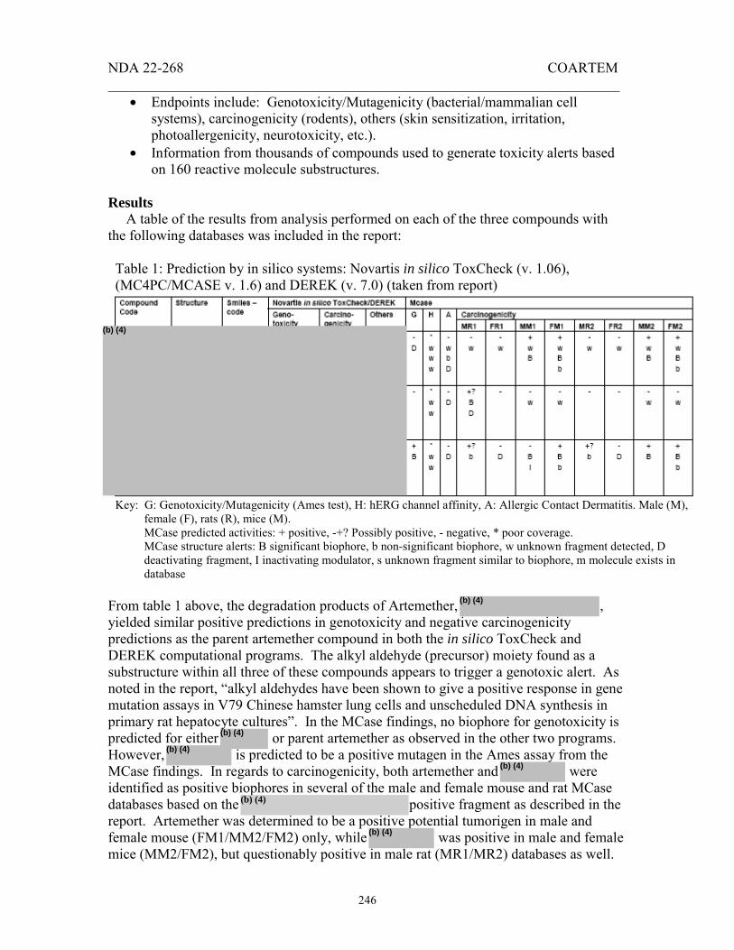
NDA 22-268 COARTEM _______________________________________________________________________
246
• Endpoints include: Genotoxicity/Mutagenicity (bacterial/mammalian cell systems), carcinogenicity (rodents), others (skin sensitization, irritation, photoallergenicity, neurotoxicity, etc.).
• Information from thousands of compounds used to generate toxicity alerts based on 160 reactive molecule substructures.
Results A table of the results from analysis performed on each of the three compounds with the following databases was included in the report:
Table 1: Prediction by in silico systems: Novartis in silico ToxCheck (v. 1.06), (MC4PC/MCASE v. 1.6) and DEREK (v. 7.0) (taken from report)
Key: G: Genotoxicity/Mutagenicity (Ames test), H: hERG channel affinity, A: Allergic Contact Dermatitis. Male (M),
female (F), rats (R), mice (M). MCase predicted activities: + positive, -+? Possibly positive, - negative, * poor coverage. MCase structure alerts: B significant biophore, b non-significant biophore, w unknown fragment detected, D deactivating fragment, I inactivating modulator, s unknown fragment similar to biophore, m molecule exists in database
From table 1 above, the degradation products of Artemether, , yielded similar positive predictions in genotoxicity and negative carcinogenicity predictions as the parent artemether compound in both the in silico ToxCheck and DEREK computational programs. The alkyl aldehyde (precursor) moiety found as a substructure within all three of these compounds appears to trigger a genotoxic alert. As noted in the report, “alkyl aldehydes have been shown to give a positive response in gene mutation assays in V79 Chinese hamster lung cells and unscheduled DNA synthesis in primary rat hepatocyte cultures”. In the MCase findings, no biophore for genotoxicity is predicted for either or parent artemether as observed in the other two programs. However, is predicted to be a positive mutagen in the Ames assay from the MCase findings. In regards to carcinogenicity, both artemether and were identified as positive biophores in several of the male and female mouse and rat MCase databases based on the positive fragment as described in the report. Artemether was determined to be a positive potential tumorigen in male and female mouse (FM1/MM2/FM2) only, while was positive in male and female mice (MM2/FM2), but questionably positive in male rat (MR1/MR2) databases as well.
(b) (4)
(b) (4)
(b) (4)
(b) (4)
(b) (4)
(b) (4)
(b) (4)

NDA 22-268 COARTEM ______________ ___________________________________________________
247
It is unlikely that would be a positive tumorigen as only a single questionably positive response was determined in a single male rat database (MR1). None of the three compounds showed positive results for allergenic contact dermatitis or hERG channel affinity. Conclusions: The results from this in silico study show a clear genotoxic and potential carcinogenic signal for one of the two degradation products, , and to a lesser extent, the parent compound artemether. All three programs similarly conclude from the findings that these two particular compounds, and artemether, may warrant further investigation to determine their capacity to show carcinogenic properties in nonclinical studies. However, unlike artemether was predicted to be negative for mutagenicity in the MCase findings, and in fact was tested in the laboratory and found negative in the Ames and V79 Chinese Hamster Test(s) in Studies 946171, 946169, 946227, 956041. Although untested in the laboratory, is predicted by MCase to test positive for mutagenicity in the Ames assay with high probability near 80%. Therefore the recommendation for further testing of in the Ames assay in isolation is in agreement with investigator findings discussed in this report. The other degradation product, , showed similar genotoxic positive results in the ToxCheck and DEREK programs as artemether and , however, proved mostly negative in the MCase genotoxic and carcinogenicity databases, warranting no further investigation.
(b) (4)
(b) (4)
(b) (4)
(b) (4)
(b) (4)
(b) (4)
(b) (4)
(b) (4)
Appears This Way on Original

NDA 22-268 COARTEM _______________________________________________________________________
248
OVERALL CONCLUSIONS AND RECOMMENDATIONS Conclusions: There are no Pharmacology or Toxicology data that would preclude the approval of Coartem® for the treatment of uncomplicated malaria in patients weighing over 5 kg. Unresolved toxicology issues:
(1) The sponsor is proposing acceptance criteria of not more than % for
respectively. These levels are not qualified by the impurity levels in the batches used for the clinical or nonclinical toxicology studies.
(2) Brain hemorrhages were found in one study of very young rats dosed with
coartemether. The implications of these findings for very young patients are of concern because adult rats and dogs showed brain lesions even though another study of young rats did not show these lesions.
Recommendations:
(1) The applicant should conduct a study of oral Coartemether in the newborn dog to evaluate any neurotoxic or behavioral effects and to assess the reversibility of any findings. Pharmacokinetics (including AUC’s) of both artemether and benflumetol should be followed. (2)The sponsor should conduct a neurotoxicity study of intramuscular artemether in beagle dogs that includes post-dosing recovery groups. This study would assess the extent to which neurological deterioration may occur in the week or weeks following the terminal dose. (3) Recommendations regarding the testing of potentially genotoxic intermediates, process impurities or degradants will be reflected in an addendum to this review.
(b) (4)
(b) (4)
Appears This Way on Original

NDA 22-268 COARTEM _______________________________________________________________________
249
RECOMMENDED LABELING:
8.1 PREGNANCY Pregnancy Category C Data from a pregnancy registry of approximately 500 pregnant women who were exposed to Coartem, and published data of over 1,000 pregnant patients who were exposed to artemisinin derivatives did not show an increase in teratogenic effects over background rate. Coartemether was embryocidal in rats at half the clinical human dose, and three times the clinical human dose in rabbits. Coartem should be used during pregnancy only if the potential benefit justifies the potential risk to the fetus. Data from a pregnancy registry of approximately 500 pregnant women exposed to Coartem, including a third of patients who were exposed in the first trimester, did not show an increase in adverse pregnancy outcomes. This data can not rule out an increased risk for early pregnancy loss. Published data of over 1,000 pregnant patients have not shown an increase in adverse pregnancy outcomes. Pregnant rats dosed during the period of organogenesis at or higher than about half the clinical dose (based on body surface area comparisons), showed increases in the number of dead fetuses, early resorptions and post implantation losses. No adverse effects were observed in animals dosed at about one third the clinical dose. Similarly, dosing in pregnant rabbits at about three times the clinical dose (based on body surface area comparisons) resulted in abortions, preimplantation losses, post implantation losses and decreases in the number of live fetuses. No adverse reproductive effects were detected in rabbits at two times the clinical dose. Embryo-fetal loss is a significant reproductive toxicity. However, because metabolic profiles in animals and humans are dissimilar, artemether exposures in animals may not be predictive of human exposures. [see Nonclinical Toxicology (13.1)].
8.3 NURSING MOTHERS It is not known whether Coartem is excreted in human milk. Because many drugs are excreted in human milk, caution should be exercised when Coartem is administered to a nursing woman. Animal data suggest both artemether and benflumetol are excreted into breast milk. The benefits of breastfeeding to mother and infant should be weighed against potential risk from infant exposure to Coartem through breast milk. 13 NONCLINICAL TOXICOLOGY
13.1 CARCINOGENESIS, MUTAGENESIS, IMPAIRMENT OF FERTILITY Carcinogenesis Carcinogenicity studies were not conducted. Mutagenesis No evidence of mutagenicity was detected. The artemether: benflumetol combination was evaluated using the Salmonella and Escherichia/mammalian-microsome mutagenicity test, the gene mutation test with Chinese hamster cells V79, the cytogenetic test on Chinese hamster cells in vitro, and the rat micronucleus test, in vivo.

NDA 22-268 COARTEM _______________________________________________________________________
250
Impairment of Fertility Pregnancy rates were reduced by about one half in female rats dosed for 2-4 weeks with the artemether: benflumetol combination at 1000 mg/kg (about 9 times the clinical dose based on body surface area comparisons). Male rats dosed for 70 days showed increases in abnormal sperm (87 % abnormal) and increased testes weights at 30 mg/kg doses (about one third of the clinical dose). Higher doses (about 9 times the clinical dose) resulted in decreased sperm motility and 100 % abnormal sperm cells.
13.2 ANIMAL TOXICOLOGY AND/OR PHARMACOLOGY Neurotoxicity Studies in dogs and rats have shown that intramuscular injections of artemether resulted in brain lesions. Changes included degenerative changes of the axons and neurons (chromatolysis, eosinophilic cytoplasmic granulation, speroids, apoptosis and dark neurons) and were located mainly in brainstem nuclei. Lesions were observed in dogs dosed at 20 mg/kg for 8 days or longer, but were not observed after shorter courses of drug or after oral dosing. Brain lesions were also observed in an exploratory rats study. The reason for the difference in susceptibility between oral and intramuscular dosing is unclear but may relate to the pharmacokinetics of the neurotoxic component (artemether). With oral dosing, artemether exposure is reduced upon repeated exposure, perhaps due to auto induction of metabolism. This auto induction is not seen after intramuscular dosing and so these animals achieve consistently high exposures of artemether. Dogs dosed orally with 143 mg/kg artemether showed a statistically measureable effect on the hearing threshold at 20 dB. This dose is equivalent to a 1000 mg/kg dose of artemether/ benflumetol, or about 29 times the clinical dose based on body surface area comparisons. Cardiovascular pharmacology In dogs, evidence of QTc prolongation was detected at doses ≥600mg/kg/day, equivalent to about 17 times the clinical dose based on body surface area comparisons. In an in vitro assay of HERG channels stably expressed in HEK293 cells, benflumetol and the main metabolite desbutyl-benflumetol showed potential to inhibit cardiac repolarization. Signatures (optional): Reviewer Signature ___________________________________ Supervisor Signature_____________________________ Concurrence Yes ___ No ___

NDA 22-268 COARTEM _______________________________________________________________________
251
APPENDIX A Studies reviewed. Report No. 102: Interactions of CGP 56 695 and CGP 56 696, Anti-Malarials, at Neurotransmitter Receptor in vitro. Study 0410072 : Artemether: 1- or 2- week intramuscular exploratory neurotoxicity study in rats. Study No. 0410073 : COA566: 8-day exploratory neurotoxicity study in dogs. Test No. 96-6141 : CGP 56696: Pilot intramuscular neurotoxicity study in dogs with pharmacokinetics. Test No. 97-0024 : CGP 56696: Oral and intramuscular neurotoxicity study in dogs with pharmacokinetics. Study No. 0510001 : ARM566: Intramuscular neurotoxicity study in dogs. Study 0510009 : COA566 and ARM566: Oral neurotoxicity study in dogs. Sponsor number 992004 : Cardiac action potential parameters in guinea-pig isolated papillary muscle in vitro. Sponsor number 0120007 : Effect of lumefantrine, desbutyl-lumefantrine, halofantrine, chloroquine and mefloquine on HERG currents recorded from stably transfected HEK293 cells. Evaluation of the cardiovascular and renal effects of coartemether (CGP 56697), artemether (CGP 56696) and benflumetol (CGP 56695). (RCC Project no. 626332) Study (CH) 1997/241 : Disposition studies in rats after administration of co-artemether (CGP 56697) containing radiolabeled artemether (CGP 56696) and unlabelled benflumetol. Study F) 1996/007 : Artemether and dihydroartemisinin concentrations in male and female rats on days 1 and 91 of a 3-month pharmacokinetic study following oral administration of coartemether daily doses of 100, 300 and 1000 mg/kg body weight. Study CH) 1996/020 : Plasma concentrations of CGP 56695 (benflumetol) in rats after repeated daily administrations of 100, 300 and 1000 mg/kg of CGP 56697 (co-artemether) during a 3-month oral toxicity Study R0570013 : An oral (gavage) juvenile development study in
(b) (4)
(b) (4)
(b) (4)
(b) (4)
(b) (4)
(b) (4)

NDA 22-268 COARTEM _______________________________________________________________________
252
rats. Study R0570030 : Artemether: 2-week oral (gavage) toxicokinetic study in rats. Study F) 1996/022 : Artemether and dihydroartemisinin plasma concentrations in female rabbits in a follow-up dose-range finding study evaluating effects of co-artemether on embryo and fetal development. Study R0410073: 8-day exploratory neurotoxicity study in dogs. Toxicokinetic report: Determination and toxicokinetics of artemether and dihydroartemisinin in dog plasma. Study R0510001: Intramuscular neurotoxicity study in dogs. Toxicokinetic report: Determination and toxicokinetics of artemether and dihydroartemisinin in dog plasma. Study R0510009B: Oral neurotoxicity study in dogs. Toxicokinetic report: Determination and toxicokinetics of artemether and dihydroartemisinin in dog plasma Study CH) 1997-003 : Absorption and disposition of CGP 56696 (artemether) after administration of CGP 56697 (co-artemether) containing 14C-labelled CGP 56696 (artemether) to male dogs. Study F) 1997/004 : Artemether and dihydroartemisinin plasma and cerebrospinal fluid concentrations in male dogs on a 5 days (interim) or 30 (main) neurotoxicity study following i.m. administration of artemether daily doses of 20 mg/kg body weight.
Study F) 1998/014 : Artemether and dihydroartemisinin plasma concentrations in dogs in a 8 day neurotoxicity study following i.m. and oral administrations of artemether daily doses (intermediate results).
Study F) 1996/005 : Artemether and dihydroartemisinin plasma concentrations in male and female dogs on days 1 and 89 of a 3-month toxicity study following oral administration of co-artemether daily doses of 20, 60 and 200 mg/kg body weight. Study CH) 1996/024 : Plasma concentrations of CGP 56695 (benflumetol) in dogs after repeated daily administrations of 6, 20 and 60 mg/kg of CGP 56697 (co-artemether) during a 3-month oral toxicity. Study F) 1996/029 : Artemether and dihydroartemisinin plasma concentrations in female cynomolgus monkeys in a pilot pharmacokinetic study with a single co-artemether oral dose.
(b) (4)
(b) (4)
(b) (4)
(b) (4)
(b) (4)
(b) (4)
(b) (4)
(b) (4)
(b) (4)
(b) (4)
(b) (4)
(b) (4)
(b) (4)
(b) (4)
(b) (4)
(b) (4)

NDA 22-268 COARTEM _______________________________________________________________________
253
Study CH) 1996/095 : Benflumetol plasma concentrations in female cynomolgus monkeys from a pilot pharmacokinetic study with a single coartemether oral dose. Study CH) 1997/099 : Transfer to the embryo-fetal compartment of pregnant rats after peroral administration of 30 mg/kg CGP 56697 (co-artemether) containing 3H-labelled CGP 56696 (artemether)
Study CH) 1997/004 : Transfer of radioactive substance(s) to the embryo-fetal compartment of rabbits after peroral administration of CGP 56697 (co-artemether) containing 14Clabelled CGP 56696 (artemether).
Study F) 1998/004 : In vitro binding of artemether to human serum proteins, human erythrocytes, and serum proteins from dog, rabbit, rat, mouse and cynomolgus monkey.
Study CH) 1997/139 : Transfer of radioactive substance(s) to the embryo-fetal compartment of rats after peroral administration of 30 mg/kg CGP 56697 (co-artemether) containing [14C]- CGP 56695 (14C-benflumetol) and unlabelled CGP 56696 (artemether).
Study DMET(EU) 29/1996 : Transfer of radioactive substance(s) to the Embryofetal compartment of rabbits after peroral administration of CGP 56697 (co-artemether) containing [14C]-CGP 56695 (14C-benflumetol). Study CH)1997/079: Plasma concentrations of benflumetol in pregnant rabbits after peroral administration of CGP 56697 (co-artemether) containing 14C-labelled CGP 56695 (benflumetol) and unlabelled CGP 56596 (artemether) Study F) 1996/044 : In vitro binding of benflumetol to human serum proteins, human erythrocytes, and serum proteins from mouse, rat, dog, rabbit, baboon and cynomolgus monkey.
Study CH) 1997/534 : Characterization of metabolites of artemether (CGP 56696) formed in rats and dogs after single oral doses of coartemether containing 14C-labelled artemeter. Study CH) 1997/181 : In vitro biotransformation of CGP 56696; comparison of three animal species and man. Study CH) 1995/079 : Plasma concentrations of CGP 56695 (benflumetol) in rats after repeated daily administrations of 200, 600 and 1000 mg/kg of CGP 56697 (co-artemether) during a 1-month oral toxicity Study
(b) (4)
(b) (4)
(b) (4)
(b) (4)
(b) (4)
(b) (4)
(b) (4)
(b) (4)
(b) (4)
(b) (4)
(b) (4)
(b) (4)
(b) (4)

NDA 22-268 COARTEM _____ ______________________________________________________________
254
Study CH) 1995/080 : Plasma concentrations of CGP 56695 (benflumetol) in dogs after repeated daily administrations of 6, 20 and 60 mg/kg of CGP 56697 (co-artemether) during a 1-month oral toxicity
Study R0510009A : Oral neurotoxicity study in dogs. Toxicokinetic report: Determination and toxicokinetics of lumefantrine in plasma.
Study CH) 1996/085 : Plasma concentrations of CGP 56695 (benflumetol) in female rabbits after repeated daily administration of 210, 700 and 2100 mg/kg CGP 56697 (coartemether) on day 19 of gestation during a 13-day oral toxicity Study
Study (CH) 1997/240 : Disposition studies in rats and dogs after administration of co-artemether (CGP 56697) containing 14C-labelled benflumetol (CGP 56695) and unlabelled artemether.
Study CH) 1997/209 : Characterization of radioactive substances in disposition studies with 14C-labelled benflumetol (CGP 56695) after administration of co-artemether (CGP 56697) in rats and dogs.
Test No. 95-6032 : CGP 56697: Acute oral toxicity study in rats. Test No. 95-6033 : CGP 56697: Acute oral toxicity study in dogs.
Test No. 95-6057 : CGP 56695: Acute oral toxicity study in rats.
Test No. 95-6058 : CGP 56695: Acute oral toxicity study in dogs.
Test No. 95-6055 : CGP 56696: Acute oral toxicity study in rats.
Test No. 95-6056 : CGP 56696: Acute oral toxicity study in dogs.
Test No. 94-6152 : CGP 56697: 1-month oral toxicity study in rats.
Test No. 94-6153 : CGP 56697: 3-month oral toxicity study in rats.
Test No. 94-6154 : CGP 56697: 1-month oral toxicity study in dogs.
Test No. 94-6155 : CGP 56697: 3-month oral toxicity study in dogs.
Test No. 95-6031 : CGP 56696: 13- week oral toxicity study in rats.
Test No. 95-6030 : CGP 56696: 13- week oral (capsule) toxicity study in dogs.
Test No. 95-6026 : CGP 56695: 13- week oral toxicity study in rats.
Test No. 95-6025 : CGP 56695: 13- week oral (capsule) toxicity study in dogs.
(b) (4)
(b) (4)
(b) (4)
(b) (4)
(b) (4)
(b) (4)
(b) (4)

NDA 22-268 COARTEM _______________________________________________________________________
255
Test No. 94-6227 : CGP 56696: Salmonella and Escherichia / mammalian-microsome mutagenicity test. Test No. 94-6169 : CGP 56697: Salmonella and Escherichia / mammalian-microsome mutagenicity test. Test No. 94-6229 : CGP 56695: Salmonella and Escherichia / mammalian-microsome mutagenicity test.
Test No. 95-6041 : CGP 56696: Gene mutation test with Chinese hamster cells V79. Test No. 94-6171 : CGP 56697: Gene mutation test with Chinese hamster cells V79. Test No. 95-6038 : CGP 56695: Gene mutation test with Chinese hamster cells V79. Test No. 94-6228 : CGP 56696: Cytogenetic test on Chinese hamster cells in vitro. Test No. 94-6230 : CGP 56695: Cytogenetic test on Chinese hamster cells in vitro.
Test No. 94-6170 : CGP 56697: Cytogenetic test on Chinese hamster cells in vitro. Test No. 95-6040 : CGP 56696: Micronucleus test, rat in vivo Study
Test No. 95-6039 : CGP 56695: Micronucleus test, rat in vivo Study Test No. 94-6172 : CGP 56697: Micronucleus test, rat in vivo Study
Test No. 95-6089 : CGP 56697: An oral study for effects on fertility and early embryonic development in rats.
Test No. 95-6065 : CGP 56697: A follow-up oral dose-range finding study for effects on embryo and fetal development in rats.
Study T/P (US) 95072 : CGP 56697: An oral embryo and fetal development study in rats.
Study T/P (US) 95065 : CGP 56697: An oral dose-range finding study for effects on embryo and fetal development in rabbits.
Study T/P (US) 95049 : CGP 56697: A follow up dose range finding study for effects on embryo and fetal development in rabbits.
Test No. 94-6157 : CGP 56697: An oral study for effects on embryo and fetal development in rabbits.
Test No. 95-6159 : CGP 56696: An oral study for effects on embryo and fetal development in rats.

NDA 22-268 COARTEM _______________________________________________________________________
256
Test No. 95-6027 : CGP 56696: An oral dose-range finding study for effects on embryo and fetal development in rabbits.
Test No. 95-6028 : CGP 56696: An oral study for effects on embryo and fetal development in rabbits.
Test No. 95-6024 : CGP 56695 : Embryonic and fetal development study in rats by oral administration.
Test No. 95-6022 : CGP 56695: An oral limit test for effects on embryo and fetal development in rabbits.
Test No. 95-6023 : CGP 56695: An oral study for effects on embryo and fetal development in rabbits.
Test No. 94-6159 : CGP 56697: An oral study for effects on pre- and postnatal development in rats.
Test No. 95-6088 : CGP 56697 : 13- week oral toxicity study in weanling rats.
Study No. 0570031 : Artemether: An oral (gavage) juvenile development dose range-finding study in rats.
Study No. 0570013 : Artemether: An oral juvenile development study in rats.
Study 0419343: In silico prediction of potential toxicological properties
Study No. 0517008 : ARM566: Assessment of contact sensitizing potential with the murine local lymph node assay (LLNA tier I). Study No. 0520004 : ARM566: Primary skin irritation study in rabbits (4-hour semi-occlusive application).
Test No. 95-6010 : CGP 56697: Skin phototoxicity in hairless mice.
Test No 95-6011 : CGP 56695: Skin phototoxicity in hairless mice.
Study CH) 1997/177 : Plasma concentrations of CGP 56695 (benflumetol) in weanling rats after repeated daily administration of 100, 300 and 1000 mg/kg CGP 56697 (coartemether) during a 13-week oral toxicity Studies not reviewed All primary pharmacodynamics studies. Please see Microbiologist’s review.
(b) (4)

NDA 22-268 COARTEM _____ ____________________________________________________________
257
Study CH) 1997/178 : Plasma concentrations of CGP 56695 (benflumetol) in rats after repeated daily administrations of 100, 300 and 1000 mg/kg of CGP 56695 during a 13-week oral toxicity
Study CH) 1996/026 : Plasma concentrations of CGP 56695 (benflumetol) in rats treated with single intravenous doses of 1mg/kg. Study CH) 1996/084 : Plasma concentrations of CGP 56695 (benflumetol) in female rabbits after repeated daily administration of 1000 mg/kg CGP 56695 on day 19 of gestation during a 13-day oral toxicity Study CH) 1997/006 : Plasma concentrations of CGP 56695 in a 13- week oral (capsule) toxicity study in dogs. Study CH) 1997/242 : Identification of in vitro metabolites of artemether (CGP 56696) in incubations with rat blood, dog blood and bovine hemoglobin by GC-MS analysis.
Study CH) 1997/156 : CGP56697/CGP56696. In vitro biotransformation of CGP 56696: Identification of the human cytochrome P450 isoenzyme(s) involved in the biotransformation of artemether. Study CH) 1997/035 : In vitro biotransformation of CGP 56695; comparison of three animal species and man.
Study CH) 1997/155 : In vitro biotransformation of CGP 56695: Identification of the human cytochrome P450 isoenzyme(s) involved in the N-dealkylation of benflumetol. Study CH) R0101415 : Ex vivo plasma protein binding of quinine in healthy subjects given quinine alone or in combination with COA566 (study trial 2302). Study CH) 1997/072 : Evaluation of a new chemical entity, CGP 56696, as an inhibitor of human P450 enzymes.
Study CH) 1997/073 : Evaluation of a new chemical entity, CGP 56695, as an inhibitor of human P450 enzymes. The Neurotoxic Potential of Benflumetol and artemether / Dihydroartemisinin combinations in vitro. (Doc 55) Artemether: UV/Vis Absorption Spectrum for Initial Phototoxicity Assessment. (Study no. 0517004) Lumefantrine: UV/Vis Absorption Spectrum for Initial Phototoxicity Assessment. (Study no. 0517005)
(b) (4)
(b) (4)
(b) (4)
(b) (4)
(b) (4)
(b) (4)
(b) (4)
(b) (4)
(b) (4)
(b) (4)
(b) (4)
(b) (4)
(b) (4)

---------------------------------------------------------------------------------------------------------------------This is a representation of an electronic record that was signed electronically andthis page is the manifestation of the electronic signature.--------------------------------------------------------------------------------------------------------------------- /s/---------------------Owen McMaster11/25/2008 05:53:37 PMPHARMACOLOGIST
Final
Rama S Dwivedi11/25/2008 05:55:16 PMPHARMACOLOGIST
Terry Miller11/25/2008 05:57:24 PMUNKNOWN
Steve Hundley11/25/2008 06:57:46 PMPHARMACOLOGIST
William Taylor11/25/2008 07:13:22 PMPHARMACOLOGIST