personal surgery: functional brain mapping and image guided
DESCRIPTION
TRANSCRIPT

Personal Surgery:functional brain
mapping and image guided neurosurgery
Alexandra J. Golby, M.D.
Associate Surgeon
Department of Neurosurgery
Brigham and Women’s Hospital
Assistant Professor Harvard Medical School

Scultetus (1653)

Case Presentation
• Recurrent meningioma in posterior parietal region
• Adjacent to motor and visual areas
• Functional Brain mapping
•Define patient’s individual anatomy
•Inform surgical decision making

Functional divisions of the brain

Patient’s fMRI motor mapping
Hand clenching Toe wiggling
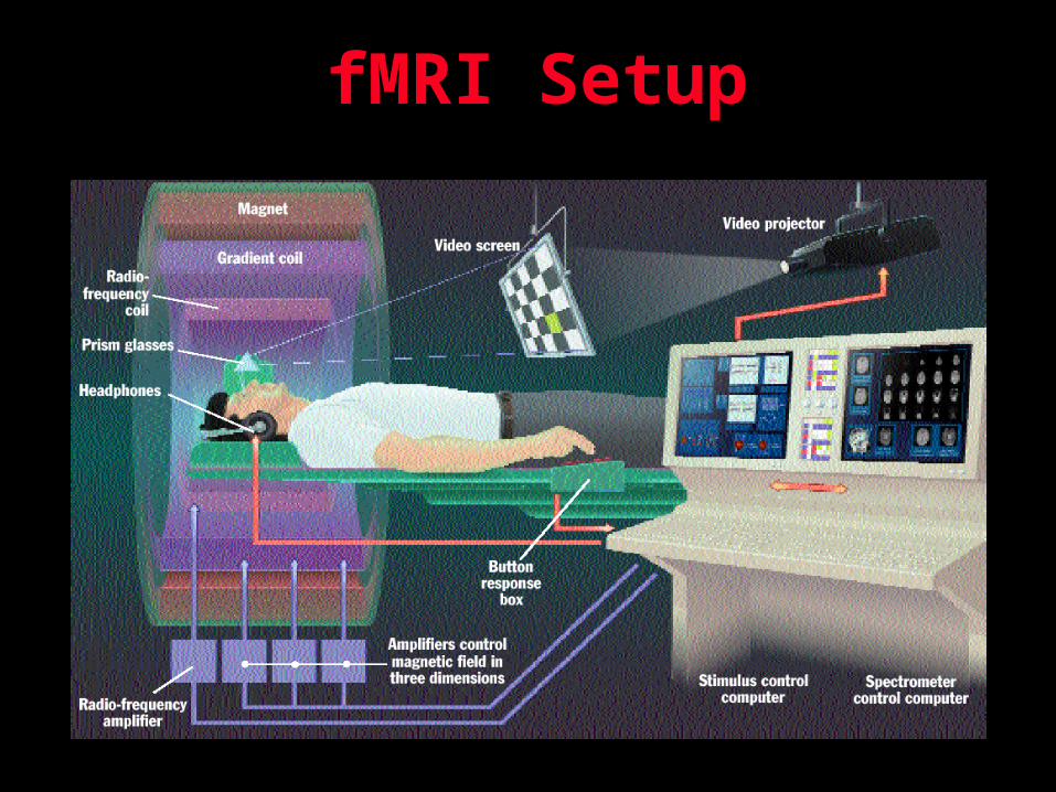
fMRI Setup

Intra-operative visualization and functional mapping

Cortical Mapping Annotation
a: Brain Tag/Event Treeb: Time Stamp & RAS c: Stimulation settingsd: Sensory/ motor/language Events
a b
c
d
• Track location of stimulation and recording points
• Represent gyral anatomy

Diffusion tensor imaging
• Water proton diffusion is facilitated parallel to fibers and restricted perpendicular
• Trajectory and location of white matter tracts can be detected by means of anisotropic diffusion tensor imaging

Intraoperative visualization of white matter anatomy

Automatic Segmentation of Meningioma

Use 3D Slicer to Magnify Tumor Region
Image shows 2nd MR scan of patient while the tumor of first scan is shown in red

Follow Tumor Growth in 3D
Tumor in 1st Scan and 2nd Scan

Surgical Planning and Intra-operative
Decision Making

MRI-Guided thermal tissue
ablation

Functional brain mapping Conclusions and future directions
• fMRI and DTI can provide neurosurgeons with valuable pre-operative information
• Combining multi-modality approach to maximize information
• Pre-operative planning and intra-operative decision making
• Predict post-operative outcomes and avoid neurologic injury