acute and sub-chronic toxicity studies of the aqueous …...... of asian and indian origin is...
TRANSCRIPT

Available online at www.scholarsresearchlibrary.com
Scholars Research Library
Der Pharmacia Lettre, 2012, 4 (5):1619-1629
(http://scholarsresearchlibrary.com/archive.html)
ISSN 0975-5071 USA CODEN: DPLEB4
1619 Scholar Research Library
Acute and Sub-Chronic Toxicity Studies of the aqueous and ethanolleaf extracts of Citrus sinensis(Linnaeus) Osbeck (pro sp.) inWistar rats.
1,2Protus Arrey Tarkang,1Gabriel A. Agbor, 1Tchamgoue DeutouArmelle, 1Tchokouaha
Lauve Rachel Yamthe, 3Kemeta David, 4Y. S. MengueNgadena
1Centre for the Research on Medicinal Plants and Traditional Medicine, Institute of Medical Research and Medicinal Plants Studies (IMPM), P. O. Box 6163, Yaoundé, Cameroon.
2Department of Pharmacology and Pharmacognosy, University of Nairobi, Kenya 3Department of Animal Biology, University of Ngaoundere, Cameroon.
4Department of Animal Biology and Physiology, University of Yaoundé I, Cameroon. _____________________________________________________________________________________________ ABSTRACT Due to the decreasing efficacy and increasing contraindications of synthetic drugs, the use of natural drugs is on the rise and as a result, self-medication is very frequent. Citrus sinensis, of Asian and Indian origin is cultivated all around the world and use of its fruits as food and other parts for treatment of various illnesses is frequent in folk medicine. Acute oral toxicity study of the extracts of CP up to a dose of 5gKg-1BW was studied in mice. Sub-acute (aqueous and ethanol extract) and sub-chronic oral toxicity (aqueous only) were carried out in ratsat graded doses of 0.25gKg-1, .5gKg-1 and 1gKg-1 BW. Control groups received water and corn oil respectively. Assay for hematological, plasma biochemical parameters and histopathological examination were carried out. No signs of acute oral toxicity were recorded. Administration of the ethanol extract showed significant decreases in hematologic parameters and increases in animal body weight, liver, renal, lipid and glycemic parameters as well as vascular and inflammatory changes in liver and kidney, at high doses. The aqueous extract acted like an immune stimulator, with strong antioxidant activity while the ethanol extract presented the risk of artherosclerotic diseases and renal and liver malfunction. Key words: Citrus sinensis, Traditional medicine Africa, toxicity, hematopoiesis, immune stimulator, hyperglycemia, hyperlipidemia _________________________________________________________________________________________
INTRODUCTION
Plants have been the basis of many traditional medicine systems throughout the world for over 5000 years and continue to provide mankind with new remedies [15]. In Sub Sahara Africa where there has been a long tradition of sourcing treatment from herbs, traditionalmedicine remains the first point of healthcare for many people [32]. Phytotherapeutic products from these medicinal plants have become universally popular in primaryhealthcare, and some have been mistakenly regarded as safe simply because they are of natural source. This presumption has led to plant products being widely used as self-medication without compromising health effects. [34]. This continuous interest and perpetual use of medicinal plants has brought about today's modern and sophisticated procedure of their processing and usage [6].Moreover, the decreasing efficacyand increasing contraindications of synthetic drugs has

Protus A Tarkang et al Der Pharmacia Lettre, 2012, 4 (5):1619-1629 _____________________________________________________________________________
1620 Scholar Research Library
seen the use of natural drugs get on the rise [22]. There is the strong need therefore for the safety and efficacy of these plant products to be determined due to self-medication [23]. One of such plants is Citrus sinensis (CS), commonly called sweet orange, of the Rutaceae family. It is a spreading evergreen tree with slender blunt spines which grows to about 1.5 m. Its leaves are narrow and rounded at the base, 2 cm long, ovate-oblong, pointed at the tip, green on the upper surface and deep green on the lower surface. Petioles are short with very narrow wings. Its inflorescence is small and white. It bears sweet smelling rounded fruits which are deep yellow to orange in colour [12]. Though of Asian and Indian origin, cultivation is carried all around the world. CS fruits are used basically as food due to presence of many vitamins. However, extracts obtained from the leaves and fruit peels are used to kill mosquito larva and mites, as antimicrobial and antifungal agents. Leaf extracts have also been used in folk medicine to treat diabetes [25], neurological disorders and to facilitate digestion [16][33]. It is also usedtraditionally in combination with honey to treatment sickle cell disease in children [24] and in combination with other plants for the treatment of malaria [30]. Preliminary phytochemical screening of leaf and fruit peel extracts revealed the presence of alkaloids, phenols, flavonoids, tannins and saponins, with fruit peel extracts showing a higher quantity of all observed phytochemicals than the leave extracts [11]. These have demonstrated strong antioxidant activity.Many substances have been isolated from CS leaves such asglycosides (apigenin and diosmetin), ruteosides (luteolin), caffeine, hydroxyproline, flavonoids [natsudaidaine, HEPTA (3,5,6,7,8,3',4'-hexamethoxyflavone), hesperidin, and diosmin], and the triterpenelinomin [20][19]. Hesperidin and diosmin have been shown to have anti-inflammatory, antihypertensive, diuretic, analgesic and hypolipidemic properties [1].Natsudaidaine and HEPTA showed a positive inotropic effect on the guinea pig right ventricle; hence it could be of importance in the future as pharmacological tools for the treatment of cardiac disorders [13]. Despite the constant and consistentuse of this plant in various forms, there is little or no evidence on its toxicity. This study therefore aims at evaluating the oral acute and chronic toxicity of the aqueous and ethanol leaf extracts of CS.
MATERIALS AND METHODS 1.0 Experimental 2.1 Plant Material Fresh sweet orange leaves were harvested from their natural habitat in the outskirts of Mamfe, Cameroon in the month of August 2011. Plant identification and voucher specimen No. TN6229 referencing was done at the Institute of Medical Research and Medicinal Plants Studies (IMPM) herbarium in Yaoundé, Cameroon. The freshly harvested leaves were then air dried, pulverized and then weighed quantities were immersed in water and ethanol (80%) respectively for 4 h. Each of the macs was then transferred into a conical percolator for 72 h and then the extracts were filtered with a sieve of 80µm pore size. The ethanol filtrate was first concentrated using a rotary evaporator and then both filtrates were concentrated in an air oven at 60oC. The extracts were weighed and stored in sealed plastic containers at 4oC for subsequent [35]. 2.2 Experimental Animals Male and female Swiss albino mice (25 – 30g) and Wistar rats (170 – 210g) obtained from the animal house of IMPM were used for the acute, sub-acute and sub-chronic toxicity studies respectively. They were housed in stainless steel wire mesh cages up to a maximum of 6 per cage, in a well-ventilated room with 12 h light/dark cycle, with free access to clean drinking water and food (standard rat feed). They were allowed to acclimatize for one week before experimentation. Plant extracts were administered orally. All animals had regular supply of clean drinking water and food[26]. 2.3 Acute Toxicity Testing The acute oral toxicity of the aqueous and ethanol extracts of CS leaf was evaluated in Swiss albino mice according to the procedures outlined by the Organization for Economic Co-operation and Development [28]. Following the fasting period, the mice were weighed and the dose was calculated in reference to the body weight. Volume of the

Protus A Tarkang et al Der Pharmacia Lettre, 2012, 4 (5):1619-1629 _____________________________________________________________________________
1621 Scholar Research Library
extracts given to the mice was 10mlKg-1 Body weight (BW) body weight. The crude extract was suspended in a vehicle (distilled water and corn oil for the aqueous and ethanol extracts respectively). Single male and female adult Swiss albino mice (25-30g) were dosed in a stepwise procedure using the fixed doses of 0.005, 0.05, 0.3, 1.2 and 2gKg-1BW of the aqueous and ethanol extracts respectively and animals were observed for signs of toxicity. If there was no mortality or signs of toxicity at the highest dose, then an upper limit dose was used for the main test. For the main test, a single high oral dose of 5gKg-1BWof each crude extract was administered to three male (Test 1) and three female (Test 2) mice in the treatment groups, whereas the control groups received the vehicle. Food was provided to the mice approximately an hour after treatment. The animals were observed 30min after dosing, followed by hourly observation for 8 h and once a day for the next 13 days. All observations were systematically recorded with individual records being maintained for each animal. Surviving animals were weighed and visual observations for mortality, behavioral pattern, changes in physical appearance, injury, pain and signs of illness were conducted daily during the period. 2.4 Sub-acute and Sub-chronic Toxicity Testing Sub-acute and sub-chronic toxicity of the aqueous and ethanol extracts of CS leaf was evaluated in Wistar rats. For the aqueous extract the rats were divided into 4 groups (A, B, C, D) of 12 rats each, while for the ethanol extract the rats were divided into 4 groups (E, F, G, H) of 6 rats each. Groups A and E served as control and received the vehicle only (water and corn oil for aqueous and ethanol extracts respectively), while groups B, C, D and F, G, H served as test groups and were administered graded doses of 0.25, 0.5 and 1gKg-1BW of each extract respectively. At the end of 28 days (sub-acute toxicity), 6 rats in each group of A, B, C, D and all the rats of E, F, G, H were sacrificed after an overnight fast, under diethyl ether anaesthesia, whereas the remaining 6 rats of each of groups A, B, C and D were sacrificed in like manner at the end of 90 days (sub-chronic toxicity). Blood was collected for hematological and biochemical analysis through the jugular vein. The liver, kidney and heart were harvested immediately clean of blood using physiological saline and weighed. The liver and kidney were then fixed in 10% formalin for histopathological examination. White blood cell (WBC), red blood cell (RBC) and platelet (PLT) counts as well as their indices were analyzed using a Hospitex Diagnostics Hema Screen 18 automatic hematology analyzer. Safety endpoints for plasma biochemical analysis included total proteins (TP), aspartate transaminase (AST), alanine transaminase (ALT), alkaline phosphatase (ALP), blood urea nitrogen (BUN), uric acid (URIC), creatinine (CRE), cholesterol (CHOL), triglycerides (TGY), glucose (GLU) and these were evaluated using standard analytical kits from Fortress Diagnostics Ltd, UK. The fixed organs were dehydrated with 100% ethanol solution and embedded in paraffin. They were then processed into 4-5µm thick sections and then stained using hematoxylin-eosin and observed under microscope as earlier described by Gabe [36]. . 2.5 Statistical Analysis All variables were subjected to descriptive data analysis. All continuous variables were expressed as the mean and the standard deviation from the mean. The results were analyzed statistically using one-way ANOVA and two-tailed Student's t-test (IBM SPSS 20 Inc., USA) to identify the differences between treated groups and controls. The data was considered significant at P < 0.05.
RESULTS
3.1 Acute Toxicity Testing No mortality was recorded in both male and female mice administered the aqueous and ethanol leaf extracts of CSup to a dose of 2gKg-1BW. For the main test as well, at a dose of 5gKg-1BW, no mortalities or signs of toxicity were recorded, as shown in Table 1.
Table 1.Potential toxic effects of the crude leaf extracts of Citrus sinensisin mice.
Observation Aqueous Extract Ethanol Extract Control
(Distilled H2O) Test 1 (Male) (5gKg-1BW)
Test 2 (Female) (5gKg-1BW)
Control (Corn oil)
Test 1 (Male) (5gKg-
1BW)
Test 2 (Female) (5gKg-1BW)
Number of Deaths 0/3 0/3 0/3 0/3 0/3 0/3
Protus A Tarkang et al Der Pharmacia Lettre, 2012, 4 (5):1619-1629 _____________________________________________________________________________
1622 Scholar Research Library
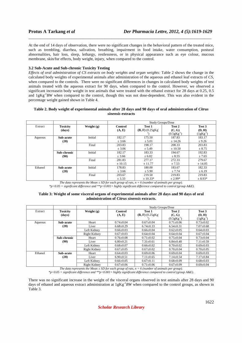
At the end of 14 days of observation, there were no significant changes in the behavioral pattern of the treated mice, such as trembling, diarrhea, salivation, breathing, impairment in food intake, water consumption, postural abnormalities, hair loss, sleep, lethargy, restlessness, or in physical appearance such as eye colour, mucous membrane, skin/fur effects, body weight, injury, when compared to the control. 3.2 Sub-Acute and Sub-chronic Toxicity Testing Effects of oral administration of CS extracts on body weights and organ weights: Table 2 shows the change in the calculated body weights of experimental animals after administration of the aqueous and ethanol leaf extracts of CS, when compared to the controls. There were no significant differences in changes in calculated body weights of test animals treated with the aqueous extract for 90 days, when compared to the control. However, we observed a significant increasein body weight in test animals that were treated with the ethanol extract for 28 days at 0.25, 0.5 and 1gKg-1BW when compared to the control, though this was not dose-dependent. This was also evident in the percentage weight gained shown in Table 4.
Table 2. Body weight of experimental animals after 28 days and 90 days of oral administration of Citrus sinensis extracts
Study Groups/Dose
Extract Toxicity (days)
Weight (g) Control (A, E)
Test 1 (B, F) (0.25gKg-
1)
Test 2 (C, G)
(0.5gKg-1)
Test 3 (D, H)
(1gKg-1) Aqueous Sub-acute
(28) Initial 182.17
± 3.66 175.50 ± 5.01
187.83 ± 14.26
183.17 ± 9.26
Final 203.83 ± 3.06
198.17 ± 5.49
208.33 ± 10.50
203.83 ± 8.75
Sub-chronic (90)
Initial 182.17 ± 3.66
183.33 ± 6.02
194.67 ± 8.55
192.83 ± 7.65
Final 281.83 ± 10.15
277.17 ± 11.77
272.33 ± 7.53
279.67 ± 14.05
Ethanol Sub-acute (28)
Initial 178.83 ± 3.66
180.00 ± 5.90
183.67 ± 7.74
182.33 ± 6.19
Final 203.67 ± 3.33
219.50 ± 10.33*
219.83 ± 2.99*
219.83 ± 8.93*
The data represents the Mean ± SD for each group of rats, n = 6 (number of animals per group). *p<0.05 = significant difference and **p<0.001= highly significant difference compared to control (group A&E).
Table 3: Weight of some visceral organs of experimental animals after 28 days and 90 days of oral administration of Citrus sinensis extracts.
Study Groups/Dose
Extract Toxicity (days)
Weight (g) Control (A, E)
Test 1 (B, F) (0.25gKg-
1)
Test 2 (C, G)
(0.5gKg-1)
Test 3 (D, H)
(1gKg-1) Aqueous Sub-acute
(28) Heart 0.74±0.04 0.67±0.04 0.71±0.06 0.73±0.02 Liver 6.68±0.29 6.74±0.33 6.54±0.31 7.07±0.68
Left Kidney 0.66±0.03 0.66±0.04 0.62±0.05 0.64±0.03 Right Kidney 0.67±0.03 0.64±0.04 0.64±0.04 0.67±0.04
Sub-chronic (90)
Heart 0.76±0.08 0.71±0.02 0.75±0.04 0.73±0.04 Liver 6.80±0.21 7.31±0.61 6.84±0.48 7.11±0.59
Left Kidney 0.68±0.07 0.66±0.02 0.70±0.02 0.69±0.03 Right Kidney 0.67±0.05 0.67±0.02 0.70±0.04 0.70±0.05
Ethanol Sub-acute (28)
Heart 0.64±0.03 0.69±0.06 0.69±0.04 0.69±0.03 Liver 6.90±0.51 7.11±0.65 7.14±0.54 7.17±0.84
Left Kidney 0.66±0.05 0.67±0.11 0.68±0.09 0.68±0.03 Right Kidney 0.67±0.06 0.71±0.06 0.67±0.09 0.69±0.04
The data represents the Mean ± SD for each group of rats, n = 6 (number of animals per group). *p<0.05 = significant difference and **p<0.001= highly significant difference compared to control (group A&E).
There was no significant increase in the weight of the visceral organs observed in test animals after 28 days and 90 days of ethanol and aqueous extract administration at 1gKg-1BW when compared to the control groups, as shown in Table 3.

Protus A Tarkang et al Der Pharmacia Lettre, 2012, 4 (5):1619-1629 _____________________________________________________________________________
1623 Scholar Research Library
The effect of the aqueous and ethanol extracts of CS on the percentage weight gain and relative organ weight (ROW) in experimental animals is presented in Table 4. Administration of CSaqueous extracts to experimental animals induced an increase in animal organ weights in a dose responsive manner. This increase in organ weights corresponds to the decreased values in percentage body weight gained in experimental animals compared to the control. This was not the case with animals that received ethanol extracts where, the percentage body weight gained in experimental animals was significantly (p < 0.05) higher than that of the control.
Table 4.The relative organ weight (ROW) per 100 g body weight recorded at the end of the study from
experimental animals after 28 days and 90 days of oral administration of Citrus sinensis extracts.
Study Groups/Dose Extract Toxicity
(days) Organ
Weight (g) Control (A, E)
Test 1 (B, F) (0.25gKg-
1)
Test 2 (C, G)
(0.5gKg-1)
Test 3 (D, H)
(1gKg-1) Aqueous Sub-acute
(28) Heart 0.36±0.01 0.33±0.01 0.34±0.01 0.36±0.01 Liver 3.27±0.09 3.40±0.06 3.13±0.02 3.46±0.07
Left Kidney 0.32±0.01 0.33±0.01 0.29±0.01 0.31±0.01 Right Kidney 0.32±0.01 0.32±0.01 0.30±0.01 0.32±0.01
% Body wt gained 11.89 12.91 10.91 11.28 Sub-chronic
(90) Heart 0.26±0.01 0.25±0.01 0.27±0.01 0.26±0.01 Liver 2.41±0.02 2.64±0.05 2.51±0.06 2.54±0.04
Left Kidney 0.24±0.01 0.24±0.01 0.26±0.01 0.25±0.01 Right Kidney 0.23±0.01 0.24±0.01 0.26±0.01 0.25±0.01
% Body wt gained 54.70 51.18 39.89 45.03 Ethanol Sub-acute
(28) Heart 0.31±0.01 0.31±0.01 0.31±0.01 0.31±0.01 Liver 3.39±0.15 3.24±0.06 3.24±0.18 3.26±0.09
Left Kidney 0.32±0.02 0.31±0.01 0.31±0.03 0.31±0.01 Right Kidney 0.33±0.02 0.32±0.01 0.30±0.03 0.31±0.01
% Body wt gained 13.89 21.91 19.68 20.56 The data represents the Mean ± SD for each group of rats, n = 6 (number of animals per group).
*p<0.05 = significant difference and **p<0.001= highly significant difference compared to control (group A&E).
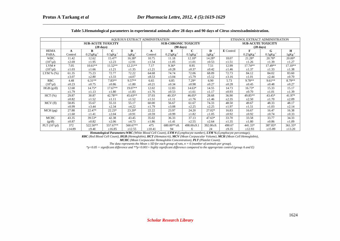
Effects of administration of CS extracts on some hematological parameters: The effects of administration of the aqueous and ethanol leaf extracts of CS in experimental animals is shown on Table 5. After 28 days, there was a significant (p < 0.001) dose-dependent increase in LYM #, RBC, PLT, HCT, HGB, PLT, WBC (p < 0.05) and a significant increase at all doses of MCH in rats treated with the aqueous extract. In rats treated with the ethanol extract, there was a significant increase (p < 0.001) in LYM #, RBC, HCT, WBC (p < 0.05), HGB, which were all dose-dependently decreasing to normal and a significant dose-dependent decrease in PLT. In rats treated with the aqueous extract after 90 days, there was a significant (p < 0.05) dose-dependent increase in WBC. We also observed a significant (p < 0.05) increase in LYM #, RBC, HGB, HCT, PLT at doses below or equal to 0.5gKg-1 which decreased to normal at 1gKg-1BW. There were also significant (p < 0.05) increases of MCH and MCHC at 1gKg-1BW. Effects of administration of CS extracts on some biochemical parameters: The effects of administration of the leaf extracts of CS after 28 days and 90 days on plasma biochemical parameters in experimental rats are presented in Table 6. After 28 days of administration of the aqueous extract, we observed a dose-dependent significant increase (p < 0.001) in TP, TGY (at a dose of 1gKg-1) and a slightly significant increase in BUN. A dose-dependent significant decreased (p < 0.001), was observed in ALT and GLU (p < 0.05). Meanwhile, in rats treated with the ethanol leaf extract, there was a dose-dependent significant increase (p < 0.001) in BUN and significant increases (p < 0.05) at all doses in TGY, CHOL, GLU and in AST,ALT, ALP, URIC at a dose of 1gKg-1BW, when compared to the control. After 90 days of treatment with the aqueous leaf extract, there were significantly low (p < 0.05) values for AST, BUN, TGY, CHOL and GLU, which dose-dependently increased to normal, when compared to the control.
Protus A Tarkang et al Der Pharmacia Lettre, 2012, 4 (5):1619-1629 _____________________________________________________________________________
1624 Scholar Research Library
Table 5.Hematological parameters in experimental animals after 28 days and 90 days of Citrus sinensisadministration.
AQUEOUS EXTRACT ADMINISTRATION ETHANOL EXTRACT ADMINISTRATION SUB-ACUTE TOXICITY
(28 days) SUB-CHRONIC TOXICITY
(90 days) SUB-ACUTE TOXICITY
(28 days) HEMA PARA.
A Control
B 0.25gKg-1
C 0.5gKg-1
D 1gKg-1
A Control
B 0.25gKg-1
C 0.5gKg-1
D 1gKg-1
E Control F 0.25gKg-1
G 0.5gKg-1
H 1gKg-1
WBC (103/µl)
11.42 ±2.69
12.62 ±1.95
15.47* ±2.23
16.38* ±2.91
10.75 ±1.54
11.18 ±1.05
12.18* ±1.01
14.28* ±0.53
18.07 ±1.51
21.28* ±1.26
20.78* ±1.39
20.00* ±1.27
LYM # (103/µl)
7.05 ±1.03
10.61** ±1.66
11.52** ±1.21
12.21** ±1.35
7.17 ±1.21
9.36* ±0.28
8.95 ±0.37
7.51 ±0.42
12.99 ±1.46
17.74** ±1.37
17.49** ±1.33
17.10** ±1.38
LYM % (%) 61.35 ±3.07
75.23 ±2.80
72.77 ±3.33
72.22 ±4.07
64.68 ±8.53
74.74 ±3.04
72.06 ±5.79
68.09 ±5.12
72.73 ±3.16
84.12 ±1.01
84.02 ±2.44
83.60 ±0.70
RBC (106/µl)
4.48 ±0.23
6.31** ±0.75
7.83** ±0.86
9.57** ±1.80
6.65 ±0.21
6.85 ±0.34
7.29* ±0.98
6.90 ±0.27
5.73 ±0.28
9.78** ±0.45
9.61** ±0.48
8.79** ±0.27
HGB (g/dl) 12.68 ±1.79
14.75* ±1.13
17.67** ±1.80
19.07** ±1.83
12.02 ±1.76
12.83 ±0.53
14.63* ±1.61
14.55 ±1.17
14.73 ±0.93
16.73* ±0.70
15.33 ±1.01
15.17 ±1.39
HCT (%) 29.87 ±0.82
30.87 ±1.52
42.78** ±1.11
45.63** ±1.52
37.03 ±1.51
40.35* ±1.11
46.05* ±1.76
28.68 ±1.46
36.90 ±2.35
49.85** ±2.50
43.45* ±1.70
41.97* ±2.09
MCV (fl) 58.85 ±0.99
55.67 ±3.44
55.33 ±2.34
55.17 ±4.22
60.00 ±1.79
56.67 ±3.08
61.67 ±2.25
74.33 ±2.25
48.50 ±1.97
49.67 ±1.51
48.33 ±1.03
48.17 ±2.14
MCH (pg) 27.88 ±1.60
22.47* ±1.41
22.23* ±2.04
23.20* ±0.91
22.22 ±0.59
23.97 ±0.99
24.20 ±1.82
27.02* ±1.05
16.83 ±0.92
16.67 ±0.92
16.47 ±0.74
16.38 ±0.33
MCHC (g/dl)
43.35 ±0.87
39.53* ±0.82
42.38 ±2.06
43.45 ±4.73
35.02 ±1.86
36.33 ±1.41
37.13 ±2.55
47.63* ±2.64
33.70 ±1.35
33.58 ±1.80
33.77 ±0.86
34.33 ±1.09
PLT (103/µl) 372 ±14.89
522.50** ±9.40
557.67** ±16.85
560.67** ±12.55
475 ±10.41
680.00**±8.94
498.00±9.16
392.00±9.24
490.67 ±9.35
441.33* ±12.93
387.83* ±15.89
361.33* ±13.20
Hematological Parameters:WBC (White Blood Cell Count), LYM # (Lymphocyte number), LYM % (Lymphocyte percentage), RBC (Red Blood Cell Count), HGB (Hemoglobin), HCT (Hematocrit), MCV (Mean Corpuscular Volume), MCH (Mean Cell Hemoglobin),
MCHC (Mean Corpuscular Hemoglobin Concentration), PLT (Platelet Count). The data represents the Mean ± SD for each group of rats, n = 6 (number of animals per group).
*p<0.05 = significant difference and **p<0.001= highly significant difference compared to the appropriate control (group A and E)

Protus A Tarkang et al Der Pharmacia Lettre, 2012, 4 (5):1619-1629 _____________________________________________________________________________
1625 Scholar Research Library
Table 6. Plasma biochemical parameters in experimental animals after 28 days and 90 days of Citrus sinensisadministration.
AQUEOUS EXTRACT ADMINISTRATION ETHANOL EXTRACT ADMINISTRATION SUB-ACUTE TOXICITY
(28 days) SUB-CHRONIC TOXICITY
(90 days) SUB-ACUTE TOXICITY
(28 days) BIOCH PARA.
A Control
B 0.25gKg-1
C 0.5gKg-1
D 1gKg-1
A Control
B 0.25gKg-1
C 0.5gKg-1
D 1gKg-1
E Control F 0.25gKg-1
G 0.5gKg-1
H 1gKg-1
T P (g/dl) 6.28 ±0.16
7.37* ±0.40
9.78** ±1.23
9.17** ±1.26
6.75 ±1.10
5.69 ±0.59
5.70 ±0.95
6.75 ±0.58
8.23 ±0.70
9.50 ±0.20
8.60 ±0.86
8.62 ±0.79
AST (U/l) 126.93 ±5.58
95.00** ±6.23
94.21** ±7.64
93.51** ±5.47
72.89 ±3.63
57.98** ±5.03
58.33** ±2.95
70.96 ±8.71
41.49 ±2.14
36.05 ±3.73
45.18 ±5.39
51.58* ±5.55
ALT (U/l) 14.70 ±1.27
10.83* ±2.12
11.08* ±2.86
9.84* ±1.66
11.93 ±0.90
11.56 ±1.21
13.37 ±1.65
12.38 ±0.77
14.93 ±1.36
16.51 ±0.95
16.65 ±1.82
17.92* ±1.84
ALP (U/l)
210.13 ±6.43
184.36* ±5.37
192.11* ±4.92
198.65 ±6.23
108.81 ±4.11
105.72 ±7.34
104.89 ±8.37
106.63 ±6.81
67.49 ±3.98
71.59 ±5.27
72.83 ±6.18
75.14* ±6.82
BUN (mg/dl)
61.92 ±4.48
66.64 ±3.15
67.58* ±7.24
68.09* ±4.73
105.91 ±8.30
88.68* ±5.84
91.85* ±8.18
93.70 ±7.37
49.48 ±3.32
50.86 ±3.29
70.44** ±3.58
71.70** ±3.80
URIC (mg/dl)
5.43 ±0.24
5.63 ±0.46
5.63 ±0.39
5.65 ±0.41
2.55 ±0.17
3.57 ±0.17
3.55 ±0.34
3.75 ±0.32
4.09 ±0.46
4.00 ±0.71
4.22 ±0.80
5.83* ±0.82
CRE (mg/dl) 0.73 ±0.16
0.73 ±0.03
0.53 ±0.02
0.53 ±0.02
0.56 ±0.17
0.44 ±0.01
0.67 ±0.01
0.67 ±0.03
0.47 ±0.16
0.53 ±0.04
0.67 ±0.01
0.67 ±0.04
TGY (mg/dl)
87.75 ±6.29
77.45 ±9.24
81.86 ±9.92
146.57** ±17.19
63.38 ±9.04
38.03** ±5.27
47.42* ±5.47
69.48 ±7.90
67.78 ±5.44
92.22* ±10.68
117.22** ±9.76
97.78* ±6.55
CHOL (mg/dl)
59.32 ±5.46
52.72 ±5.70
56.91 ±5.39
50.19 ±2.26
55.05 ±4.63
37.85** ±2.85
57.74 ±5.47
58.82 ±4.01
65.77 ±3.41
76.50* ±2.80
58.75* ±3.62
58.24* ±2.00
GLU (mg/dl)
144.29 ±10.28
106.30** ±6.68
110.69* ±12.52
110.00* ±8.44
128.84 ±8.31
110.29**±6.16
116.80**±3.61
139.78 ±6.49
115.22 ±3.36
169.39** ±13.58
165.54** ±16.17
164.32* ±8.19
Biochemical parameters: TP (Total Proteins); AST (Aspartate transaminase); ALT (Alanine transaminase); ALP (Alkaline phosphatase); BUN (Blood urea nitrogen); URIC (Uric acid); CRE (Creatinine); TGY (Triglycerides); CHOL (Cholesterol); GLU (Glucose).
The data represents the Mean ± SD for each group of rats, n = 6 (number of animals per group). *p<0.05 = significant difference and **p<0.001= highly significant difference compared to the appropriate control (group A and E).

Protus A Tarkang et al Der Pharmacia Lettre, 2012, 4 (5):1619-1629 _____________________________________________________________________________
1626 Scholar Research Library
Effects of administration of CS on some visceral organs (Histopathological examination):Histopathological examination of the liver and kidneyrevealed that these organs in control groups (A and E) showed normal morphological structures without signs of vascular or inflammatory changes. The same was also observed for those of experimental animals treated with the aqueous leaf extract for 28 and 90 days, when compared to the control. However, in experimental animal treated with the ethanol leaf extract after 28 days, analysis revealed signs of toxicity at a dose of 1gKg-1 BW, when compared to the control. Signs of toxicity in the liver included vascular congestion and leucocyte infiltration and in the kidney, we observed mild tubular clarification and glumerulosclerosis, as shown in Fig 1.
Controlrat Liver: Normal structure Control rat Kidney:Normal structure
Figure 1.Light micrograph plates of tissue sections from the liver and kidney of experimental animals after 28 days of administration of the ethanol leaf extract of Carica papaya, a a dose of 1gKg-1BW
showing vascular and inflammatory changes (H & E × 40).
Experimental rat Liver at 1gKg-1BW: Vascular congestion and leucocyte infiltration.
Experimental rat Kidney at 1gKg-1BW: Mild tubular clarification and glumerulosclerosis.

Protus A Tarkang et al Der Pharmacia Lettre, 2012, 4 (5):1619-1629 _____________________________________________________________________________
1627 Scholar Research Library
DISCUSSION
In the acute toxicity study, there was no mortality or any signs of toxicity recorded in experimental animals after treatment with the aqueous and ethanol leaf extracts of CS up to a dose of 5gKg-1BW. In accordance with the OECD Guidance Document for Acute Oral Toxicity Testing [28], doses higher than 5gKg-1BW are generally not considered as dose related and compounds with LD50 values lower than 2gKg-1BW are generally considered to be relatively safe. In this regard, the aqueous and ethanol leaf extracts of CS can be considered to be non-toxic at acute administration since the extracts were well tolerated and there was no observed adverse effect. There were no significant changes in calculated BWand organ weights of experimental animals treated with the aqueous leaf extract of CS for 28 and 90 days, when compared to that of the control. Theincrease in weight was normal. However, there was a significant increase (p < 0.05) in the calculated BWand no significant change in organ weights of experimental animals treated with the ethanol leaf extract of CS after 28 days, when compared to the control. This was evident in the increased values in percentage BW gained of experimental animals when compared to the control, as shown in Table 4. Comparison of body and organ weights between treated and untreated groups of animals have conventionally been used to evaluate the toxic or adverse effects[21][9] and as an assessment of therapeutic response to test articles or drugs [3]. In this study, the observed increase in organ weights of the experimental animals treated with the aqueous extractcorresponded to a decrease in percentage BW gained when compared to the controls. Therefore, CSaqueous leaf extract had a dose dependent increase on the BWbut did not have any adverse effects on experimental animals that would cause them to loose appetite [27]. The inverse relationship between the ROW of the experimental animals and the percentage BW gained, with that of the controls could be indicative of an adaptive response of the organs to the accumulation of the extracts [14]. This signifies that the organ weights did not indicate any toxic or adverse effects from CSleaf extracts as earlier observed in the acute toxicity. However, in experimental animals treated with the ethanol leaf extract of CS, the percentage BW gained in experimental animals is higher than that of the control. Since there was no significant change in organ weights, this apparent difference could just beBW related and not treatment related [31], proving no observed adverse effects from the extracts. Analysis of full blood count carried out in experimental and control animals enabled us to understand the toxicity of these extracts on the hematopoietic system. In order to understand the risk alterations in the human hematopoietic system upon exposure to drugs, analysis in toxicity studies must be carried out using animal models and then extrapolated to humans [17]. Hematopoiesis is the process of blood cell formation. All blood cells are believed to be derived from the pluripotential stem cell, an immature cell with the capability of becoming an erythrocyte (RBC), a leukocyte (WBC), or a thrombocyte (platelet). In healthy adults, stem cells in hematopoietic sites undergo a series of divisions and maturational changes to form the mature cells found in the blood [7].In this study, there was a significant dose-dependent increase in all hematological parameters in experimental animals treated with the aqueous leaf extract of CS after 28 days when compared to the control. This increase was brought to normal after prolonged administration (90 days). The same trend was observed in experimental animals treated with the ethanol leaf extract after 28 days, apart from the PLT, which showed a significant (p < 0.05) dose-dependent decrease when compared to the control. The hematopoietic system was stimulated by this extract, leading to the over-production of WBC (leukopoiesis), RBC (erythropoiesis) and platelets [2]. The function of the WBC is to protect the body from infection by foreign organisms, while the RBC boosts the immune system by providing nourishment and oxygen and the PLT protect blood vessels from endothelial damage as well as initiate repair of these vessels during trauma. These observations are indicative of a strong immuno-stimulatory, antioxidant and endothelial protection activity of CS extracts. The MCV and MCH give the volume and weight of the HGB in each RBC while the MCHC gives a valuable indicator of HGB deficiency [7]. The increased and/or normal values of these parameters in experimental animals,are a validation of the immune stimulation by these leaf extracts.However, the dose-dependent decrease observed in PLT after the ethanol extract administration, which is supposed to indicate a breakdown in the endothelial protection or repair system, might be due to trauma. Okwuet al [11] had earlier reported strong antioxidant activities of CS extracts and some substances isolated from this plant have been shown to have strong anti-inflammatory and analgesic properties [1], probably a justifiable reason for its inclusion in apolyherbal product commonly used for the treatment of malaria [30].
Protus A Tarkang et al Der Pharmacia Lettre, 2012, 4 (5):1619-1629 _____________________________________________________________________________
1628 Scholar Research Library
In this study, assay of the liver, renal, lipid and glycemic plasma biochemical profiles of experimental compared to control animals was carried out in order to give insight into pathological changes and other effects upon administration ofCS leaf extracts. Lipid peroxidation is induced when drugs are taken and this results in the release of cytosolic enzymes into the blood stream, such as ALT, AST, ALP [4], which when observed in increased quantities in the plasma are indicative of liver and cellular damage. Liver profile parameters assayed (TP, ALT, AST, ALP) revealed that prolonged use of the aqueousCS leaf extracts did not have any adverse effects on the functioning of the liver. After 28 days of treatment with the aqueous extracts, experimental animals showed a highly significant increase in TP, which might be due to dehydration. However, this stabilized after prolonged (90 days) administration, whencompared to the control. The parameters that showed either an increase (ALT and BUN) or a decrease (AST and GLU), dose-dependently came back to normal when compared to the control. This is indicative of the absence of any cellular or liver damage [7], hence a probablehepatoprotective capacity of the aqueous leaf extract [8][29], since the plant has been reported to have strong antioxidantproperties. On the contrary, upon treatment of experimental animals with the ethanol leaf extract of CS, we observed significantly high values of AST, ALT and ALP at a high dose of 1gKg-
1BW. This was indicative of liver or cellular damage of myocardial or kidney tissues [7]. Whereas prolonged administration of the aqueous extract did not present any significant changes in renal parameters in experimental animals, we observed significant increases in BUN and URIC after treatment with ethanol extract for 28 days, when compared to the control. This is indicative of a decreased renal function and destruction respectively. BUN and creatinine are indicators of glomerular filtration rate (GFR), which is an indicator of the renal function [10], but since there is an increase in the BUN while CRE values are normal, this presupposes a non-renal cause. Some of the factors associated with artherosclerotis are blood lipid levels and lipoproteins [18]. The significant increase (p < 0.05) in TGY and CHOL after treatment with the CS ethanol leaf, presents a high risk of hypercholesterolemia and hypertrygliceridemia. This correlates with a significant increase in GLU, which might be due to impaired insulin activity or insufficient secretion caused by the ethanol extract, causing hyperglycemia and consequently activating hormone sensitive lipases in the adipose to release lipids [5]. This is also reflected in the significant increase in weight of the animals, contrary to those treated with the aqueous extract. This is also seen in the vascular and inflammatory changes observed in these set of animals when treated with the ethanol extract at high doses. These could be due to deposition of fat droplets in those tissues.
CONCLUSION
Prolonged administration of the aqueous leaf extract of CS did not show any signs of toxicity in Wistar rats. The extract actually acted like an immune stimulator, an antioxidant and hypoglycemic agent. However, the ethanol extract could be link to hyperglycemic and cardiovascular diseases at high doses, therefore high doses should be discouraged.
REFERENCES
[1] A Gil-Izquierdo, MI Gil, F Ferreres and FA Tomás-Barberán..J. of Agric. and Food Chem., 2001, 49: 1035-1041. [2] AC Guyton and JE Hall.Textbook of Medical Physiology, 11th Ed. Elsevier Saunders, USA.2006; 1152pp. [3] AC Winder, LA Lembke and MD Stephens.Arthritis Rheumatism, 1969, 12: 472 – 482. [4] AG Agbor, JE Oben, B Nkegoum, JP Takala and JY Ngongang. Pak J Biol Sci.,2005, 8: 1397 – 1401. [5] AS Loci et al. J Ethnopharmacol.,1994, 43, 167. [6] BB Petrovska. Phcog Rev2012, 6:1-5. [7] BM Cavanaugh. Nurse’s Manual of Laboratory and Diagnostics Tests. 4th Ed. F. A. Davis Company, Philadelphia. 2003; 688pp. [8] BR Mossallanejad and HN Varzi.Asian J Anim Sci., 2011, 5:213 -218. [9] CJ Pfeiffer. ToxicolApplPharmacol. 1968, 13(2), 220–7. [10] DC Eaton and JP Pooler. Vander’s Physiology.7th Ed. McGraw-Hill Lange, USA.2009; 230pp.

Protus A Tarkang et al Der Pharmacia Lettre, 2012, 4 (5):1619-1629 _____________________________________________________________________________
1629 Scholar Research Library
[11] DE Okwu, AN Awurum and JI Okoronkwo.African Crop Science Conference Proceedings, 2007, 8: 1755-1758. [12] EA Sofowora. Medicinal Plants and Traditional Medicine in Africa 2nd Ed. Spectrum Books Ltd, Ibadan, Nigeria.1993; 289pp. [13] ED Oliveira, TS Leite, BA Silva and EA Conde-Garcia. Brazilian J. of Med. and Biol. Res., 2005, 38: 111-118. [14] FO Jimohet al. Afr J Biotechnol., 2008, 7: 3173 – 3177 [15] G Samuelsson. Drugs of Natural Origin: A Textbook of Pharmacognosy, 5th Swedish Pharmaceutical Press, Stockholm, 2004; 320pp. [16] GL Mwaiko. East Afr. Med. J., 1992, 69: 223-226. [17] H Olson et al. Regul. Toxicol.Pharmacol.2000, 32, 56–67. [18] HP Shaila, SL Udopa and AL Udopa.Fitoterapia, 1997, 5: 405 – 409. [19] I Stewart. Journal of Agric. and Food Chem., 1985, 33: 1163-1165. [20] JA Duke. Handbook of Phytochemical Constituents of Grass Herbs and Other Economic Plants.CRC Press, Boca Raton, FL, USA.1992; 26pp. [21] JM Peters and EM Boyd.J Nutr.,1966,90(4), 354–60. [22] K Kelly. History of medicine. New York: Facts on file; 2009; 29-50. [23] L Rodriguez-Fragoso, J Reyes-Esparza, SW Burchiel, D Herrera-Ruiz and E Torres.Toxicology and Applied Pharmacology, 2008, 227(1), 125-135. [24] ME Bassey and EO Effiong. J. Nat. Prod. Plant Resour.,2011, 1 (3): 33-42. [25] MO Soladoye, EC Chukwuma and FP Owa. J. Nat. Prod. Plant Resour.,2012, 2 (1): 60-72. [26] National Academies Press. Guide for Care and Use of Laboratory Animals. 8th Ed. Nat Res Council of the Nat Academies.2010; 248pp. [27] NI El-Sanusi and S El-Adam.Asian J Anim Vet Adv., 2007, 2: 27-31. [28] OECD. OECD Guidance Document on Acute Oral Toxicity Testing; Organization for EconomicCo-operation and Development: Paris, France, 2001. [29] P Akanitapichatet al.Food and Chemical Toxicology, 2010, 48, 3017–3021 [30] P ArreyTarkanget al. J. Nat. Prod. Plant Resour.,2012, 2 (3):372-380. [31] RS Sellers et al. Toxicol Path.,2007, 35: 751 – 755. [32] WHO/TDR News No. 79. 2007; 8-13. [33] Y Fan, Z Ding, L Yang, L Xu and K Li. ZhongguoZhongYaoZaZhi, 1995, 20: 397-398. [34] YK Vaghasiya, VJ Shukla and SV Chanda. J. Pharmacol. Toxicol.2011, 6, 113-123. [35] SS Handa, SPS Khanuja, G Longo and DD Rakesh.Extraction Technologies for Medicinal and Aromatic Plants.ICS-UNIDO International Centre for Science and High Technology, Trieste, Italy.2008; 266pp. [36] M Gabe. Techniques Histologiques.Mason, 120, Boulevard Saint Germain, Paris.1968; 128-243.