709 d effect of controlled volumetric … tissues, we recognize that there is not a unanimous ......
TRANSCRIPT

709
EFFECT OF CONTROLLED VOLUMETRIC TISSUE HEATINGWITH RADIOFREQUENCY ON CELLULITE AND THE
SUBCUTANEOUS TISSUE OF THE BUTTOCKS AND THIGHSMa. Emilia del Pino MD,a Ramón H. Rosado MD,b Alejandro Azuela MD,a Ma. Graciela Guzmán MD,b
Dinorah Argüelles MD,b Carlos Rodríguez MD,c Gesche M. Rosadoa. Dermatology Surgeons, Hospital Angeles del Pedregal, México
b. Plastic Surgeons, Medlight, Clinicas de Láser, IPL y Radiofrecuencia, Méxicoc. Radiologist, Hospital Angeles del Pedregal, México
d. Study Coordinator, Medlight, Clinicas de Laser, IPL y Radiofrecuencia, México
AbstractBackground: Regardless of diet and exercise, genetics plays an important part in creating puckering skin or dimples, whichare difficult to hide at any age. The demand for a nonsurgical, noninvasive treatment of cellulite has inspired some manufacturers to invest in a new age of sophisticated devices and treatment therapies to repair the skin and improve contours. Although many of these new choices have demonstrated a smoothing effect (following a multitude oftreatments), the objective documentation has in most cases been limited to biopsies, circumference measurements, and photographic evidence. Hypothesis: We believe that the application of noninvasive high-energy radiofrequency (RF) to the skin of the thigh andbuttocks heats the subcutaneous adipose tissue, causing collagen fibers to contract. The resulting impact to the subcutaneoustissue and collagen is expected to improve the skin’s external architecture. Given that the subcutaneous tissue and adiposetissue are difficult to evaluate through histological methods, this investigation seeks to demonstrate the changes that occurwhen applying two treatments of high-energy RF on the subcutaneous tissue of thighs and buttocks utilizing real time ultrasound image scanning. Materials and Methods: Twenty-six healthy female patients (ages 18 to 50) with visible bilateral cellulite (grade 1 to 3) oneither the buttocks and/or thighs received 2 treatment sessions (15 days apart) of unipolar RF using the Accent RF System(Alma Lasers Inc). The system utilizes a unipolar RF applicator that is electrically cooled to aid in patient comfort duringthe treatment. Appropriate energy was set and the treatment was delivered in 3 passes of 30 seconds each. Evaluation of thethickness of the subcutaneous tissue on buttocks and thighs took place before the first treatment, second treatment, and 15 days following the second treatment with a with real-time scanning image ultrasound (Philips Medical Systems). Clinicalimprovement was objectively evaluated through comparative pre- and post-treatment measurements of the distance betweenthe stratum corneum to the Camper’s fascia and from the stratum corneum to the muscle. The study also evaluated the structure and changes of the collagen (thickening and realignment of septae) resulting from 2 treatments of RF. Photographywas used to document contour and superficial changes.Results: From the measurements of the distance between the stratum corneum to the Camper’s fascia and from the stratumcorneum to the muscle we were able to demonstrate that 68% of the patients presented a contraction of the volume ofapproximately 20%.Conclusions: Based on the demonstrated results with real-time ultrasound scanning, we have observed that 2 RF treatmentson the subcutaneous tissue of the buttocks and thighs provide a volumetric contraction effect in the majority of patients. Thisvalidates the primary hypothesis of our protocol and establishes that the RF energy works on the connective tissue of the subcutaneous adipose tissue. This effect should be the same on any other body part.
IntroductionThrough ultrasound we observed the subcutaneous tissue andfat positioned between the skin and the muscle. It is possibleto observe in anatomical views the layer between the subcutaneous tissue and the adipose layer, as well as theintegrity of the fibrous bands that divide them. As people age,the quality of the fibrous bands is lost and deformity appearsthat can be observed with an ultrasound.
BackgroundRadiofrequencyElectric currents have been used in medicine for more than a century. Low-frequency electric current causes spasms in muscle tissue, and in low intensity can be used for
biostimulation, such as in cardioversion for atrial fibrillation.1
High-frequency current in the 0.3 to 100 MHz range isdefined as radiofrequency current (RF). RF only produces a thermal effect on living tissue depending on the electricproperties of the tissue. High-frequency RF current hasdemonstrated its efficiency in heating tissue in electrosurgeryand has recently become an attractive source of energy fordifferent aesthetic and dermatological applications.2,3
Thermal energy has been proposed as a method for contract-ing loose, lax skin through the well-known mechanism of collagen denaturalization. Even though there are numerousin vivo and in vitro experimental studies that have providedevidence for the biology and biomechanics of thermally modified tissues, we recognize that there is not a unanimous
COPYRIGHT © 2006 JOURNAL OF DRUGS IN DERMATOLOGY

RADIOFREQUENCY ON CELLULITE AND THE SUBCUTANEOUSTISSUE OF THE BUTTOCKS AND THIGHS
opinion regarding the optimal therapeutic algorithm, themechanism of action, the final improvement clinical reports,or the long-term follow-up of these thermally modified tissues. However, there appears to be agreement concerningthe basic science of thermal modification of connective tissue, as well as several clinical implications associated withits use.
When the collagen is heated, the bonds that are sensitive toheat begin to break. In a transition process, the protein transforms from a highly organized crystalline structure to adisorganized gel (denaturalization). Collagen contractionoccurs through an unfolding of the triple helix when crossedintermolecular unions, which are sensitive to heat, aredestroyed and the tension of residual crossed intermolecularunions stabilizes to the heat. Collagen denaturalization is usually present at 65°C. The behavior of the heat-inducedconnective tissue and the amount of tissue contractiondepends on various factors, including the highest reachedtemperature (peak temperature), the RF exposure time, and the mechanical stress applied to the tissue during theheating process. Thermal properties of tissue can also varydepending on species, age, pH, the electrolyte concentrationin the environment, the concentration and orientation ofcollagen fibers, and the tissue’s hydration levels.4
The selective electrothermolysis produced by RF is highlyeffective in creating a thermal effect on biologic tissues.Unlike optic energy that depends on chromophore concentration of the skin in order to achieve a selective thermal destruction of target tissue, RF depends on the electrical properties of tissues.5 The RF technology may helpto increase the disruption of adipose tissue, which at the sametime helps to move and eliminate the fat deposits in a noninvasive way without having fat necrosis.
The Accent equipment consists of a base system that generates RF technology (40.68 MHz) delivered through oneof 2 handpiece applicators to induce controlled volumetrictissue heating (CVTH). The individual applicators provide afunctional delivery of energy to different depths. The firsthandpiece delivers bipolar energy and has a penetrationbetween 2 and 6 mm into the tissue to stimulate dermal structural changes. The second handpiece delivers unipolarenergy with a penetration of 20 mm that is designed to reachthe subcutaneous adipose tissue.
In this therapeutic modality of high-frequency RF, the energywaves work on a molecular level oscillating at high speed andcausing a displacement of charged particles. This results inthe rotation of water molecules which will dissipate energydepending on the electrical conductivity of tissue. Bothmodes (bipolar and unipolar) deliver energy to the tissuethrough an electrode tip that is cooled to prevent epidermalheating and provide additional comfort to the patient. Whenheating deep tissues therapeutically, local blood circulationimproves, helping the zones affected by edema to promote thedrainage and replenishment of the retained fluids and thecatabolic products.
RF systems have demonstrated good results correcting irregularities of the cutaneous surface with an efficacy comparable to lasers. RF has the advantage of faster recoverytime and not being influenced by competing chromophores inthe cutaneous surface. Therefore, any skin type can be treated. Although this study did not focus on other areas ofthe body, there are reports of studies of the RF device appliedto the face where there was an improvement of skin tightening with visible results from the first week followingtreatment, but more evident 3 months post-treatment without any complications. Continuing improvement following the discontinuation of treatment has been seen inthis investigation as well. The prior reports have been validated largely through photographic evidence.2,3 To date, itis the authors’ experience that real-time ultrasound imaginghas never been used to document or observe the effect of volumetric RF on the subcutaneous adipose tissue.
Subcutaneous Adipose TissueAnatomic and physiologic studies on the adipose tissue have been focused on in vivo studies of individual adipocytesor in vivo studies with functional and minimally invasive methods.
Traditionally, the subcutaneous adipose tissue has been considered insulation and a source of stored energy. Morerecently, there has been greater interest in the distributionand composition of the adipose tissue in relation to healthand morbidity. The actual concepts of the adipose tissue’sanatomy are derived from the histological studies ofNürnberger and Müller,6 who analyzed samples of healthymen and women’s adipose tissue and of women with cellulite.They reported indentations into the deep adipose tissuethrough the dermis on women, but not in men. They alsodescribed modifications on the fibrous septae architecture oriented perpendicular to the cutaneous surface on womenand in a crisscross pattern on men.
Histological Characteristics of Subcutaneous Adipose TissueThe histology of subcutaneous adipose tissue has been thoroughly investigated by Piérard et al7 who made studies oncorpses of the macroscopic and microscopic characteristics ofthe skin, thighs, and buttocks of men and women without alterations and on women with cellulite. The macroscopicexamination of the specimens of full thickness proved the complexity of the 3-dimensional net formed by the fibrousbands, which are born from the hypodermis. Piérard felt thatthere were no continuous layers of connective tissue that maybe called septae between the lobules of adipose tissue in womenwith cellulite, even though the microscopic examination ofthigh skin in men shows a dermal-hypodermic leveled interfacewithout any clinical signs of cellulite. In contrast, the dermal-hypodermic interface of women’s thigh skin (evenwithout cellulite) demonstrates that the adipose lobules have agranular aspect, which protrudes into the dermis. The lobulesrise as valleys and hills under the dermal surface. In some cases,the sweat glands are trapped in these fat lobules. There is nocorrelation between the extent of this finding and the clinicaltype and severity of cellulite.6,7 A more undulated dermis-hypo-
JOURNAL OF DRUGS IN DERMATOLOGYSEPTEMBER 2006 • VOLUME 5 • ISSUE 8
710

RADIOFREQUENCY ON CELLULITE AND THE SUBCUTANEOUSTISSUE OF THE BUTTOCKS AND THIGHS
dermic interface on women, which corresponds to the fibrousbands observed in the macroscopic studies on corpses, has beenconfirmed using high resolution ultrasound images.7
Recently, the architecture of the fibrous septae net has beenvisualized through a 3-dimensial MRI as well as with a high-resolution ultrasound (Figure 1). The Camper’s fasciacan clearly be observed as a thin flat structure more or lessparallel to the cutaneous surface. Other septae were detectedas thin structures oriented like pillars in 3 directions: perpendicular, parallel, and with a 45° angle. In women withcellulite, there are a higher percentage of perpendicular fibersin comparison with women (and men) that do not have cellulite. As for the fibers in other directions, women withcellulite have a lower percentage of parallel septae to the skinand a higher percentage of angled septae.7,8 Furthermore, anMRI study on adipose tissue comparing young and maturewomen found a higher content of water within the dermis inthe older group. A larger amount of free water between thedermis has been related to collagen architecture degradationduring the aging process, leaving less interaction sitesbetween water and macromolecules (Figures 1).
Skin aging is a process that can be classified into 2 groups:intrinsic aging and photoaging. These are considered differentprocesses with the first caused by the passage of time and thesecond due to continuous exposure to the ultraviolet raysfrom the sun. In both types of aging, the most dramatic histological changes are found in the dermis. Collagen alterations, the main skin component, have been identified asthe cause of the changes observed.
The dermis contains mainly collagen type I (85% to 90%)and less collagen type III (10% to 15%). The dermal
fibroblasts synthesize the individual chains of polypeptideprocollagen I and II, precursors of collagen type II and type IIIthat are formerly polymerized in the carboxylic rings andamino terminals to form the triple helixes. Skin that is notnormally exposed to the sun’s ultraviolet radiation, such asthe thighs and buttocks, mainly goes through the intrinsicaging process. In a study about collagen metabolism in theaging process, it was observed that in the areas not exposed tosun, the synthesis of collagen diminishes as the aging processgo on, maintaining a negative balance between synthesis andcollagen degradation.1 Since the buttocks and thighs undergoa lesser degree of photoaging, they are ideal anatomical areasto observe the effect of RF energy on the chronologically agedcollagen in the adipose tissue.
CelluliteCellulite is a type of lipodystrophy considered by many to bean aesthetic disorder in which the alteration is a morpholog-ical constitutional disposition with no significant histologicalor biological alterations of the adipose tissue. It affects femalesalmost exclusively, and appears around puberty.Approximately 90% of the female population have somedegree of cellulite. It is common to confuse cellulite appear-ance with obesity, even though it is a different condition.Obesity is a generalized condition in which the adipocytesincrease in number and size. Cellulite is localized to specificsites with characteristic structural changes (lipodystrophy).
Cellulite is mainly located on the lateral aspects of the thighsand buttocks and is highly related to hormonal changes infemales. Cellulite differs from the fat on the abdominal wall,which is more dependent on metabolism and diet, and is moreeasily removed. The skin with cellulite is rough to the touch.When it is pinched, it has the appearance of orange skin, andis often associated with a painful sensation.
Cellulite PathogenesisIn the gynoid zones (thighs, hips, and buttocks), women haveadipocytes 5 times greater than in other body zones. The cuta-neous microcirculation has certain special characteristics thatdeposit more fat and retain more interstitial fluids. The fat iskept in the adipocytes that are found between the skin andmuscles and divided by fibrous tissue bands. These fibrousbands give the adipose tissue a wall-like aspect between theskin and muscles which slow down the lymphatic drainage.
Ultrasound in the Study CelluliteIt is complicated to study the RF thermal effect on the subcutaneous tissue in large areas like the thighs and buttocks. A biopsy may cause trauma to the tissue, whichwould modify the next sample by leaving scar tissue thatwould alter the histological morphology of the study zone. Itis technically difficult to take the whole thickness of healthyadipose tissue without causing a deformity during extractionor processing. It was determined that in vivo observation inreal time with noninvasive methods, like the ultrasound,would allow us to register changes on large anatomical zones,quantify them, and keep the records of what could happenwhen heating the tissue with RF. The Real Time ScanningCompound Image (RTSCI) ultrasound has a great variety of
JOURNAL OF DRUGS IN DERMATOLOGYSEPTEMBER 2006 • VOLUME 5 • ISSUE 8
711
Figure 1. Campers Fascia Identified throughHigh-Resolution Ultrasound.

RADIOFREQUENCY ON CELLULITE AND THE SUBCUTANEOUSTISSUE OF THE BUTTOCKS AND THIGHS
medical applications including blood vessels, musculoskeletalsystem, gynecological and abdominal exams, and so forth.However, its use for the study of skin and subcutaneous tissueis not well known.9
We will assume that applying RF energy, which creates heatthrough water molecule rotation and tissue impedance, willmove the trapped interstitial fluids. We believe that this musttrigger a contraction of the collagen fibers and improve theskin’s contour and texture.
HypothesisHigh-energy RF application in a unipolar mode heats the subcutaneous adipose tissue, which will cause contraction ofcollagen fibers, simultaneously improving the alterations of the skin’s external architecture. Given that the subcuta-neous tissue and adipose tissue are difficult to evaluatethrough histological methods, we wish to observe throughreal-time ultrasound the changes produced by the volumetricRF heating on both tissues.
H1: Radiofrequency, when emitting energy at a high energy,contributes to the connective tissue contraction; therefore,with a more compact subcutaneous adipose tissue, skin tightening will be observed.
H2: Radiofrequency, when raising temperature locally, helpsto eliminate the excess liquid in the subcutaneous adipose tissue, which will contribute to a more compressed subcutaneous adipose tissue.
H0: No changes on the architecture of subcutaneous tissuewere observed after the RF treatments.
HA: The RTSCI is useful to evaluate the changes caused bythe RF in the subcutaneous tissue.
Materials and MethodsThe study included 26 healthy, voluntary women between 18 and 50 years old. All volunteers signed an informed consent and a photograph authorization. The high-energy RFsource used was the Accent RF system (Alma Lasers Inc,Ceasaria, Israel; Fort Lauderdale, FL) in the unipolar mode,which emits RF energy from the tip, allowing the energy to penetrate up to 2 cm in the tissue. The applied energy can be100 to 200 watts, designed to produce heat in the dermis andthe hypodermis. A real-time scanning image ultrasound(Philips Medical Systems, The Netherlands) with a multi-frequency linear transducer for soft tissue (2-5 MHz),was used to evaluate the thickness of the subcutaneous tissueon the buttocks and thighs.
Inclusion Criteria
•Adult women between 18 and 50 years old
•Without liposuction or previous surgery on thighsand buttocks
•With normal weight, or not exceeding more than 20% of the anthropometric scales for age and size
Exclusion Criteria
•Pregnancy
•Pacemakers or any other electronic implant
•Skin cancer
•Viral or bacterial infections
•Epilepsy
•Autoimmune disease
•Isotretinoin treatment in the last 12 months
•Scleroderma
•Extensive radiotherapy treatments
Treatment Methodology1) The RF treatment was delivered in 2 sessions 15
days apart.
2) The evaluations with ultrasound were made before thefirst treatment and 15 days after the second treatment.The ultrasound was performed on the right lateral aspectof the thigh and buttock with the patient standing. Onthe buttock the ultrasound was done on the area ofgreatest deformity. On the thigh, it was taken on thelateral aspect at the level of the great femoral trochanter (frequently the zone of most fat accumulation known as the “saddlebags”).
The following measurements were taken during theultrasound:
a. The distance between the deep dermis and the first lineof fibrous tissue (Camper’s fascia), on the larger distanceand on the shorter one.
b. The distance from the deep dermis to the muscle.
c. A longitudinal panoramic take: on the buttocks from themedial point to the gluteal fold on the thigh; and on thethigh, from the great trochanter to the lower third. Thefirst evaluation was used as a treatment control. Thequantity, thickness, and quality of the observed fibrous bands were registered and described as continued straightlines, discontinued straight, continuous undulated, anddiscontinuous undulated. The measurements were alwaystaken on the same anatomical location.
3) Before starting the treatment, the patients filled out the following registers: clinical history, informed consent,photograph authorization.
4) Before each session, the following data were recorded:weight, size, circumference of waist, abdomen, hips, andthighs as well as photographs.
5) Photographs were taken in an ex profeso photographicstand of the posterior, right lateral and left lateral views.
6) The zones to be treated were marked with the patientlying down. The marks were grids of 7 x 7 cm made witha skin marker and numbered on top of the whole area
JOURNAL OF DRUGS IN DERMATOLOGYSEPTEMBER 2006 • VOLUME 5 • ISSUE 8
712
RADIOFREQUENCY ON CELLULITE AND THE SUBCUTANEOUSTISSUE OF THE BUTTOCKS AND THIGHS
affected with cellulite, excluding the bony prominencesand the thighs’ medial aspect.
7) The initial parameters were 150 W x 30 sec = 4,500joules = 91 J/cm2 with 3 consecutive passes on each areaduring each session. Parameters were modified to ahigher or lower energy depending on the patient’stolerance to heat or until the temperature rose tobetween 39°C and 41°C on each treated zone. This was avariable on each patient depending on the amount of fat, thickness of the skin, and the anatomical site.
8) Mineral oil was used to slide the hand piece over the treating area and to avoid friction on the skin.
9) During the treatment, the electrode’s tip was keptperpendicular and in full contact with the skin. Theelectrode was also maintained in constant movement forup to 30 seconds or until the desired temperature wasachieved (±40°C), and before the patient complained of any pain.
10) Patients filled out a treatment survey form.
11) A specially designed temperature register form was filledout with the following information:
a) Chosen parameters for the treatment and record ofchanges.
b) Anatomical schemes of the treated zones weremarked with numbers and correlated with each of theanatomical treated sites.
c) Applied energy and duration of each application.
d) Basal temperature and peak temperature of eachsquare were measured with an infrared thermometer.
e) Any adverse effects.
12) The duration of each treatment was of approximately1.5 hours.
ResultsIndependent VariableA register was made of the peak temperatures reached duringthe RF application. The patients had different tolerances to heat and the peak temperatures ranged between 36°C and 40°C.
Quantitative Dependent VariablesUltrasounds were studied and measurements of the initial andfinal ultrasound were recorded and divided into the thighsand buttocks:
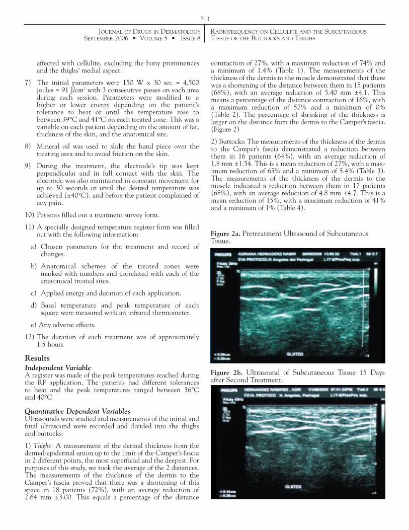
1) Thighs: A measurement of the dermal thickness from thedermal-epidermal union up to the limit of the Camper’s fasciain 2 different points, the most superficial and the deepest. Forpurposes of this study, we took the average of the 2 distances.The measurements of the thickness of the dermis to theCamper’s fascia proved that there was a shortening of thisspace in 18 patients (72%), with an average reduction of 2.64 mm ±3.00. This equals a percentage of the distance
contraction of 27%, with a maximum reduction of 74% and a minimum of 1.4% (Table 1). The measurements of thethickness of the dermis to the muscle demonstrated that therewas a shortening of the distance between them in 15 patients(68%), with an average reduction of 5.40 mm ±4.1. Thismeans a percentage of the distance contraction of 16%, witha maximum reduction of 57% and a minimum of 0% (Table 2). The percentage of shrinking of the thickness islarger on the distance from the dermis to the Camper’s fascia.(Figure 2)
2) Buttocks: The measurements of the thickness of the dermisto the Camper’s fascia demonstrated a reduction betweenthem in 16 patients (64%), with an average reduction of 1.8 mm ±1.54. This is a mean reduction of 27%, with a max-imum reduction of 65% and a minimum of 5.4% (Table 3).The measurements of the thickness of the dermis to the muscle indicated a reduction between them in 17 patients(68%), with an average reduction of 4.8 mm ±4.7. This is amean reduction of 15%, with a maximum reduction of 41%and a minimum of 1% (Table 4).
JOURNAL OF DRUGS IN DERMATOLOGYSEPTEMBER 2006 • VOLUME 5 • ISSUE 8
713
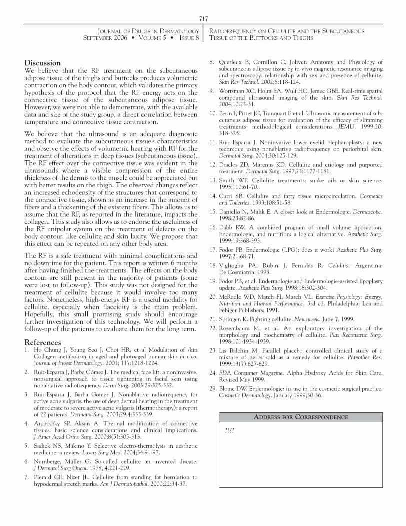
Figure 2a. Pretreatment Ultrasound of SubcutaneousTissue.
Figure 2b. Ultrasound of Subcutaneous Tissue 15 Daysafter Second Treatment.

RADIOFREQUENCY ON CELLULITE AND THE SUBCUTANEOUSTISSUE OF THE BUTTOCKS AND THIGHS
JOURNAL OF DRUGS IN DERMATOLOGYSEPTEMBER 2006 • VOLUME 5 • ISSUE 8
714
Table 2. Thigh Measurement of Dermis to Muscle.
Table 3. Buttocks Measurement of Dermis to Camper’s Fascia.
Difference = difference between the first and last session measurements. Increase = those patients who increased measurementsbetween the first and the last session. Reduction = those patients who reduced measurements between the first and the last session;Avg = average; Count = the number of patients of the sample the total and the number of the different categories of the sample.Min. = minimum. Max = maximum; 1stdv. = 1 standard deviation.
MEASUREMENT PERCENTAGEDifference Increase Decrease Difference Increase Decrease
Average - 0.2980 0.2163 - 0.5400 -8.80% 8.72% -17.04%Count 25 8 17 25 8 17Percentage 32% 68% 32% 68%MIN - 1.3400 0.0400 - -57.02% 1.30% 0.00%MAX 0.5000 0.5000 - 1.3400 20.00% 20.00% -57.02%1 Stdev 0.4958 0.1469 0.4061 16.72% 5.85% 13.37%Min 67% cl - 0.7938 0.0694 0.1339 -25.52% 2.87% -3.67%Max 67% cl 0.1978 0.3631 - 0.9461 7.93% 14.58% -30.42%
MEASUREMENT PERCENTAGEDifference Increase Decrease Difference Increase Decrease
Average - 0.0706 0.1233 - 0.1797 -7.15% 29.57% -27.80%Count 25 9 16 25 9 16Percentage 36% 64% 36% 64%MIN - 0.5000 0.0200 - 0.0150 -64.94% 4.50% -5.036%MAX 0.2850 0.2850 - 0.5000 77.08% 77.08% -64.94%1 Stdev 0.1993 0.0911 0.1545 35.31% 28.47% 17.27%Min 67% cl - 0.2699 0.0322 - 0.0252 -42.46% 1.10% -10.53%Max 67% cl 0.1287 0.2144 - 0.3342 28.16% 58.04% -45.08%
Table 4. Buttocks Dermis to Muscle.MEASUREMENT PERCENTAGE
Difference Increase Decrease Difference Increase DecreaseAverage - 0.2212 0.3263 - 0.4788 -6.80% 11.66% -15.49%Count 25 8 17 25 8 17Percentage 32% 68% 32% 68%MIN - 1.6800 0.0300 - 0.0300 -41.28% 1.30% -.0.95%MAX 0.5100 0.5100 - 1.6800 26.98% 26.98% -41.28%1 Stdev 0.5531 0.1981 0.4706 17.44% 8.88% 13.07%Min 67% cl - 0.7743 0.1282 - 0.0083 -24.24% 2.78% -2.42%Max 67% cl 0.3319 0.5243 - 0.9494 10.63% 20.54% -28.56%
Table 1. Thigh Measurement of Dermis to Camper’s Fascia. MEASUREMENT PERCENTAGE
Difference Increase Decrease Difference Increase DecreaseAverage - 0.1594 0.1107 - 0.2644 -11.55% 27.39% -26.70%Count 25 7 18 25 7 18Percentage 28% 72% 28% 72%MIN - 1.0150 0.0050 - 0.0100 -73.82% 1.69% -1.37%MAX 0.2900 0.2900 - 1.0150 60.47% 60.47% -73.82%1 Stdev 0.3091 0.0950 0.3000 32.87% 23.51% 21.51%Min 67% cl - 0.4685 0.0157 0.0355 -44.43% 3.88% -5.19%Max 67% cl 0.1497 0.2057 - 0.5644 21.32% 50.91% -48.21%

RADIOFREQUENCY ON CELLULITE AND THE SUBCUTANEOUSTISSUE OF THE BUTTOCKS AND THIGHS
To evaluate if there was a correlation between the appliedtemperatures and the amount of tissue contraction obtainedfrom the ultrasounds, a statistical regression was applied andrevealed that the results on the thigh were better than on thebuttocks. (Table 5).
Qualitative Dependent VariablesQualitative dependant variables were defined by the description of echogenesis of the adipose tissue. Regarding themorphological improvement achieved with the Accent RFtreatment, we found that the improvement of the fibrousbands went from curved to straight and from discontinuous tocontinuous, and therefore established a grading of 5 as thehighest digit for the most disorganized fibers and 1 for themost organized. We observed that 50% of the patientsobtained morphological improvement. On the thigh, theimprovement averaged 1.7 digits (scale 5 to 1) and on thebuttocks it averaged 1.3 digits.
When analyzing the changes in the Camper’s fascia betweenthe first session and 45 days later, we repeatedly observed onthe ultrasound a noticeable organization of the fibrous lines,as well as an increase of the fibrous tissue in 53% of the cases,and an increase of the thickness of the fibers in 57% of the cases (Figure 5).
JOURNAL OF DRUGS IN DERMATOLOGYSEPTEMBER 2006 • VOLUME 5 • ISSUE 8
715
Figure 3a. Pretreatment Ultrasound of SubcutaneousTissue.
Figure 3b. Ultrasound of Subcutaneous Tissue 15 Daysafter Second Treatment.
Figure 4a. Pretreatment Ultrasound of SubcutaneousTissue.
Figure 4b. Ultrasound of Subcutaneous Tissue 15 days after Second Treatment.
Table 5. Regression Statistics of the Thigh and Buttocks.
Regression Statistics Thighs Buttocks
Multiple R 0.8300 0.7353
R2 0.6889 0.5407
Adjusted R2 0.6465 0.6465
Standard Error 0.4277 0.5290
Observations 26 26

RADIOFREQUENCY ON CELLULITE AND THE SUBCUTANEOUSTISSUE OF THE BUTTOCKS AND THIGHS
Photographs were not considered in the protocol to observethe effect of the RF; nevertheless they are essential to evaluate the efficacy of the treatment by itself, without itbeing the central objective of this protocol (Figures 6a-h).
Adverse EffectsDuring the treatment, small blisters appeared in some areas in2 patients, and ecchymosis on the thighs of 3 patients the dayfollowing treatment. All of the above adverse effects wereresolved and without complication.
Patient Degree of SatisfactionPatient satisfaction was evaluated by their perception of thebenefits from the treatment on the body contour, the waytheir clothes fit, and skin texture. Most of them were satisfiedwith the results. The most satisfied group were the womenwho had the most accentuated defects.
JOURNAL OF DRUGS IN DERMATOLOGYSEPTEMBER 2006 • VOLUME 5 • ISSUE 8
716
Figure 5. a)Ultrasound of Subcutaneous Tissue priorto Treatment.
Figure 5. b) Ultrasound of Subcutaneous Tissue 15 DaysAfter 2 Treatments.
Figure 6. a) Before Treatment. b) 15 Days after 2 Treatments. Figure 6. c) Before Treatment. d) 15 Days after 2 Treatments.
Figure 6. e) Before Treatment. f) 15 Days after 2 Treatments. Figure 6. g) BeforeTreatment. h) 15 Days after 2 Treatments.
RADIOFREQUENCY ON CELLULITE AND THE SUBCUTANEOUSTISSUE OF THE BUTTOCKS AND THIGHS
DiscussionWe believe that the RF treatment on the subcutaneous adipose tissue of the thighs and buttocks produces volumetriccontraction on the body contour, which validates the primaryhypothesis of the protocol that the RF energy acts on the connective tissue of the subcutaneous adipose tissue.However, we were not able to demonstrate, with the availabledata and size of the study group, a direct correlation betweentemperature and connective tissue contraction.
We believe that the ultrasound is an adequate diagnosticmethod to evaluate the subcutaneous tissue’s characteristicsand observe the effects of volumetric heating with RF for thetreatment of alterations in deep tissues (subcutaneous tissue).The RF effect over the connective tissue was evident in theultrasounds where a visible compression of the entire thickness of the dermis to the muscle could be appreciated butwith better results on the thigh. The observed changes reflectan increased echodensity of the structures that correspond tothe connective tissue, shown as an increase in the amount offibers and a thickening of the existent fibers. This allows us toassume that the RF, as reported in the literature, impacts thecollagen. This study also allows us to endorse the usefulness ofthe RF unipolar system on the treatment of defects on thebody contour, like cellulite and skin laxity. We propose thatthis effect can be repeated on any other body area.
The RF is a safe treatment with minimal complications andno downtime for the patient. This report is written 6 monthsafter having finished the treatments. The effects on the bodycontour are still present in the majority of patients (somewere lost to follow-up). This study was not designed for thetreatment of cellulite because it would involve too many factors. Nonetheless, high-energy RF is a useful modality forcellulite, especially when flaccidity is the main problem.Hopefully, this small promising study should encourage further investigation of this technology. We will perform afollow-up of the patients to evaluate them for the long term.
References1. Ho Chung J, Young Seo J, Choi HR, et al Modulation of skin
Collagen metabolism in aged and photoaged human skin in vivo.Journal of Invest Dermatology. 2001; 117:1218-1224.
2. Ruiz-Ezparza J, Barba Gómez J. The medical face lift: a noninvasive,nonsurgical approach to tissue tightening in facial skin using nonablative radiofrequency. Derm Surg. 2003;29:325-332.
3. Ruiz-Esparza J, Barba Gomez J. Nonablative radiofrequency foractive acne vulgaris: the use of deep dermal heating in the treatmentof moderate to severe active acne vulgaris (thermotherapy): a reportof 22 patients. Dermatol Surg. 2003;29:4:333-339.
4. Arcnoczky SP, Aksan A. Thermal modification of connective tissues: basic science considerations and clinical implications. J Amer Acad Ortho Surg. 2000;8(5):305-313.
5. Sadick NS, Makino Y. Selective electro-thermolysis in aestheticmedicine: a review. Lasers Surg Med. 2004;34:91-97.
6. Nurnberge, Müller G. So-called cellulite an invented disease. J Dermatol Surg Oncol. 1978; 4:221-229.
7. Pierard GE, Nizet JL. Cellulite from standing fat herniation to hypodermal stretch marks. Am J Dermatopathol. 2000;22:34-37.
8. Querleux B, Cornillon C, Jolivet. Anatomy and Physiology of subcutaneous adipose tissue by in vivo magnetic resonance imagingand spectroscopy: relationship with sex and presence of cellulite. Skin Res Technol. 2002;8:118-124.
9. Wortsman XC, Holm EA, Wulf HC, Jemec GBE. Real-time spatialcompound ultrasound imaging of the skin. Skin Res Technol.2004;10:23-31.
10. Perin F, Pittet JC, Tranquart F, et al. Ultrasonic measurement of sub-cutaneus adipose tissue for evaluation of the efficacy of slimmingtreatments: methodological considerations. JEMU. 1999;20:318-325.
11. Ruiz Esparza J. Noninvasive lower eyelid blepharoplasty: a newtechnique using nonablative radiofrequency on periorbital skin.Dermatol Surg. 2004;30:125-129.
12. Draelos ZD, Marenus KD. Cellulite and etiology and purportedtreatment. Dermatol Surg. 1997;23:1177-1181.
13. Smith WP. Cellulite treatments: snake oils or skin science.1995;110:61-70.
14. Curri SB. Cellulite and fatty tissue microcirculation. Cosmetics and Toiletries. 1993;108:51-58.
15. Daniello N, Malik E. A closer look at Endermologie. Dermascope.1998;23:82-86.
16. Dabb RW. A combined program of small volume liposuction,Endermologie, and nutrition: a logical alternative. Aesthetic Surg.1999;19:368-393.
17. Fodor PB. Endermologie (LPG): does it work? Aesthetic Plas Surg.1997;21:68-71.
18. Viglioglia PA, Rubin J, Ferradás R. Celulitis. Argentina: De Cosmiatria; 1993.
19. Fodor PB, et al. Endermologie and Endermologie-assisted lipoplastyupdate. Aesthetic Plas Surg. 1998;18:302-304.
20. McRadle WD, Match FI, Match VL. Exercise Physiology: Energy,Nutrition and Human Performance. 3rd ed. Philadelphia: Lea andFebiger Publishers; 1991.
21. Springen K. Fighting cellulite. Newsweek. June 7, 1999.
22. Rosembaum M, et al. An exploratory investigation of the morphology and biochemistry of cellulite. Plas Reconstruc Surg.1998;101:1934-1939.
23. Lis Balchin M. Parallel placebo controlled clinical study of a mixture of herbs sold as a remedy for cellulite. Phtyother Res.1999;13(7):627-629.
24. FDA Consumer Magazine. Alpha Hydroxy Acids for Skin Care.Revised May 1999.
29. Blome DW. Endermologie: its use in the cosmetic surgical practice.Cosmetic Dermatology. January 1999;30-36.
JOURNAL OF DRUGS IN DERMATOLOGYSEPTEMBER 2006 • VOLUME 5 • ISSUE 8
717
????
ADDRESS FOR CORRESPONDENCE