pdf version - weizmann institute of science
TRANSCRIPT

RESEARCH ARTICLE 3021
Development 139, 3021-3031 (2012) doi:10.1242/dev.080127© 2012. Published by The Company of Biologists Ltd
INTRODUCTIONGenome-encoded miRNAs bind to specific sites on the 3�untranslated region (3�UTR) of their target mRNAs, to impart post-transcriptional silencing (Bartel, 2004). This regulatory layer actsin concert with transcription factors to refine gene expression andconfer robustness to developmental transitions (Stark et al., 2005;Hornstein and Shomron, 2006; Li et al., 2009). Total inactivationof miRNA maturation causes pancreas agenesis (Lynn et al., 2007),indicating that miRNAs are essential for early pancreasdevelopment. Specific miRNAs were shown to act in endocrinetissues; for example, miR-375 is highly expressed in the endocrinepancreas (Poy et al., 2004; Avnit-Sagi et al., 2009), and loss of itsfunction disrupts islet morphogenesis and endocrine celldifferentiation (Kloosterman et al., 2007; Poy et al., 2009).
miR-7 is another miRNA that is highly and specificallyexpressed in the endocrine pancreas of zebrafish, mouse andhuman (Wienholds et al., 2005; Landgraf et al., 2007; Bravo-Eganaet al., 2008; Correa-Medina et al., 2009). miR-7 is an evolutionarilyconserved miRNA, encoded by a single gene in flies and by threedifferent genomic loci in humans and mice (mouse: mmu-mir-7a-1 at Chr13, mmu-mir-7a-2 at Chr7 and mmu-mir-7b at Chr17). Theduplication of the miR-7 gene in vertebrates hampers genetic loss-of-function analysis. As a consequence, the role of miR-7 inendocrine pancreas development is still unclear.
The endocrine differentiation program is initiated by neurogenin3 (Ngn3), which induces a complex network of transcriptionfactors, to specify the different endocrine lineages (Gradwohl et al.,2000; Gu et al., 2002; Martin et al., 2007; Bonal and Herrera, 2008;Lyttle et al., 2008). This process results in the differentiation ofmature hormone-producing cells (Murtaugh and Melton, 2003).
Pax6, a paired-domain transcription factor acting downstream ofNgn3, is pivotal in differentiation of hormone-producing cell types:-, -, - and PP cells. However, Pax6 negatively regulates theformation of ghrelin-expressing -cells (Sander et al., 1997; St-Onge et al., 1997; Ashery-Padan et al., 2004; Heller et al., 2005;Dames et al., 2010). In both humans and mice, two Pax6 alleles arerequired in order to maintain proper glucose homeostasis, i.e. lossof only one allele results in glucose intolerance (Yasuda et al.,2002; Ding et al., 2009). Similarly, the development of other organsis sensitive to Pax6 haplo-insufficiency, including the iris and thelens (van Raamsdonk and Tilghman, 2000; Collinson et al., 2001;Baulmann et al., 2002; Kroeber et al., 2010). However, high levelsof Pax6 are also not tolerated. Thus, Pax6 overexpression in micecauses eye abnormalities (Schedl et al., 1996) and inducesapoptosis in the brain and in the endocrine pancreas (Yamaoka etal., 2000; Berger et al., 2007). Taken together, it appears that Pax6expression is tightly controlled to ensure appropriate levels ofexpression.
In this study, we characterized miR-7 expression and identifiedits functions in endocrine cell differentiation. We show that miR-7directly represses Pax6 mRNA expression and fine-tunes its levels.Together, miR-7 and Pax6 are wired into a network that regulatesendocrine cell differentiation. Our results demonstrate theimportance of miRNAs in refining the development of hormone-producing cells.
MATERIALS AND METHODSAnimalsMice were housed and handled in accordance with protocols approved bythe Institutional Animal Care and Use Committee of Weizmann Instituteof Science. Conditional miR-7 transgenic mice were generated aspreviously described (Srinivas et al., 2001). Briefly, a 500 bp fragmentflanking the miR-7a-1 gene was cloned into the Rosa26 locus downstreamof the PGK promoter and a transcriptional STOP cassette and upstream ofIRES-EGFP-polyadenylation signal. Correct homologous recombinationinto the ROSA26 locus was identified by Southern blot analysis toembryonic stem cell colonies (129/SvEv). Scanning 209 colonies identified29 positive colonies and a mouse line was derived through blastocystinjection (C57BL6/J background). Rosa-miR-7 mice were crossed to a
1Department of Molecular Genetics, Weizmann Institute of Science, Rehovot 76100,Israel. 2Integrated DNA Technologies, 1710 Commercial Park Coralville, IA 52241,USA.
*Author for correspondence ([email protected])
Accepted 1 June 2012
SUMMARYGenome-encoded microRNAs (miRNAs) provide a post-transcriptional regulatory layer that is important for pancreas development.However, how specific miRNAs are intertwined into the transcriptional network, which controls endocrine differentiation, is notwell understood. Here, we show that microRNA-7 (miR-7) is specifically expressed in endocrine precursors and in mature endocrinecells. We further demonstrate that Pax6 is an important target of miR-7. miR-7 overexpression in developing pancreas explants orin transgenic mice led to Pax6 downregulation and inhibition of - and -cell differentiation, resembling the molecular changescaused by haploinsufficient expression of Pax6. Accordingly, miR-7 knockdown resulted in Pax6 upregulation and promoted - and-cell differentiation. Furthermore, Pax6 downregulation reversed the effect of miR-7 knockdown on insulin promoter activity. Thesedata suggest a novel miR-7-based circuit that ensures precise control of endocrine cell differentiation.
KEY WORDS: -Cell, microRNA, miR-7, Pax6, Pancreas development, Mouse
Pancreas-enriched miRNA refines endocrine celldifferentiationSharon Kredo-Russo1, Amitai D. Mandelbaum1, Avital Ness1, Ilana Alon1, Kim A. Lennox2, Mark A. Behlke2
and Eran Hornstein1,*
DEVELO
PMENT

3022
Pdx1-Cre transgene (Gu et al., 2002) and mated to homozygosity. Othermouse strains used in this study were Ngn3-CreER, serving as Ngn3 nulls(Wang et al., 2010), and a Pax6 null allele (St-Onge et al., 1997).
Organ cultureDorsal pancreatic rudiments of E12.5 ICR mouse embryos were dissectedfrom the adjacent mesenchyme, using a tungsten needle. The explants werecultured in M199 medium supplemented with 10% fetal bovine serum(Gibco), 2 mM L-glutamine and 100 U/ml penicillin/streptomycin.Individual explants were plated in 30 l inverted ‘hanging drops’ on a 35-mm Petri dish cover (NUNC), with medium containing either anti-microRNA antagomirs (Dharmacon) or cholesterol-conjugated miRNAmimics (IDT) at 1 M concentration. The exact oligo sequences are insupplementary material Table S2. Explants were further grown for up to48 hours at 37°C with a 5% CO2 in a humidified incubator as previouslydescribed (Kredo-Russo and Hornstein, 2011). BrdU (3 g/ml) was addedto the medium 1 hour before harvest for analysis of proliferation.
Pancreas histology and quantification analysisImmunofluorescence protocols for paraffin sections have been describedpreviously (Melkman-Zehavi et al., 2011) and whole-explant staining hasbeen described previously (Kredo-Russo and Hornstein, 2011). Theprimary antibodies used were rabbit anti-Pax6 (1:300, Covance), guineapig anti-insulin (1:200, Dako), rabbit anti-glucagon (1:200, Dako), rabbitanti-Cpa1 (1:100, Sigma), mouse anti-Ngn3 [1:500, Developmental StudiesHybridoma Bank (DSHB)] goat anti-ghrelin (1:100, Santa Cruz) and goatanti-Hnf1b (1:200, Santa-Cruz). Secondary antibodies were Cy2-, Cy3- orCy5-conjugated donkey anti-guinea pig, anti-mouse and anti-rabbit IgG(1:200, Jackson ImmunoResearch). Nuclei were stained with DAPI(1:10,000, Molecular Probes). Whole-mount BrdU analysis that includes a2 hour DNase I treatment was carried out as previously described(Tkatchenko, 2006).
Fluorescent confocal images were captured with a Zeiss LSM 510microscope, using an optical depth of 1 m, with at least six to eight opticalsections at 5 m intervals throughout the whole organ.
Morphometry of the explants was performed by quantification of theimmunostained area from the entire explant sections from a minimum ofthree mutants and three wild-type matched littermates,. Total tissue areaand total hormone-positive area, were calculated using ‘Niss-elements’software (Nikon).
For cell number quantification at E15.5, hormone-positive cells weremanually counted every fifth section throughout the whole pancreasanlagen. Data are the average number of cells/section in multiple sectionsand were analyzed for four or more individual animals per genotype. Cellnumber analysis of total hormone-positive cells in whole E12.5 explantswas performed manually, by counting cells in six stacked z-sectionconfocal images, spanning the whole explants.
Quantitative PCR for miRNA and mRNAExtraction of total RNA was carried out using the miRNeasy Mini Kit(QIAGEN). Synthesis of cDNA obtained by using an oligo d(T) primer(Promega) and SuperScript II reverse transcriptase (Invitrogen).Synthesis of cDNA of miRNA obtained by using Taqman MicroRNAqPCR Assays (Applied Biosystems). qPCR analysis of mRNA wasperformed on LightCycler 480 System (Roche) using Kapa SYBR GreenqPCR kit (Finnzymes). miRNA and mRNA levels were normalized tothe expression of small RNAs (sno234 and U6) or mRNA (Gapdh andHprt), respectively. Primer sequences are described in supplementarymaterial Table S2.
miRNA in situ hybridizationParaffin sections of E12.5-E15.5 pancreata were hybridized with DIG-labeled LNA probes (Exiqon) overnight at 48°C (miR-7), 54°C (U6,control) or 60°C (miR-375) as previously described (Pena et al., 2009) anddeveloped with TSA kit (PerkinElmer) as previously described(Silahtaroglu et al., 2007). When in situ hybridization was combined withimmunofluorescence, primary antibodies were added to the anti-Dig-PODincubation (1:500, Roche).
Cell culture, luciferase reporter assay and western blottingHEK-293T cells (American Type Culture Collection) and MIN6 cells (agift from Jun-ichi Miyazaki, Osaka University, Japan) were grown inDulbecco’s modified Eagle medium (DMEM) with 10% FBS, 2 mM L-glutamine, 100 U/ml penicillin/streptomycin at 37°C; 5% CO2 in ahumidified incubator. Experiments on MIN6 cells were performed betweenpassages 18 to 28.
A 742 bp fragment of the mouse Pax6 3�UTR sequence (chr2105536551-105537201) was subcloned into psiCHECK-2 Vector(Promega) and transfected into HEK-293T cells using jetPEI (PolyplusTransfection, Illkirch, France), following manufacturers’ instructions. Dual-Reporter luciferase assay was performed 48 hours later, according to themanufacturer’s instructions (Promega). miR-7 overexpression wasachieved using expression vectors miRVec-miR-7 or miRVec control (akind gift from Reuben Agami, The Netherlands Cancer Institute,Amsterdam). miR-7 knockdown was carried out using oligos against miR-7 or against scrambled sequence, as negative control oligos (50 nM,Ambion), using Lipofectamine 2000 Reagent (Invitrogen). For westernblots, cellular lysate was subject to 10% SDS-PAGE and immunoblottedwith rabbit anti-Pax6 (1:5000, Chemicon), mouse anti-GAPDH (1:10,000,Ambion) and quantified with ImageJ software.
For analysis of insulin transcription, firefly luciferase reporter driven bythe rat insulin promoter and an A20-Renilla luciferase construct (gift ofMichael Walker), were transfected using Lipofectamine 2000 Reagent(Invitrogen) to MIN6 cells. Anti miR-7 oligo (100 nM) and Pax6 siRNA(10 nM) are from IDT; overexpression of miR-7 was achieved using amiRVec vector.
Statistical analysisAnalysis was performed using either Student’s t-test or two-way ANOVAby the JMP software. Results are given as mean±s.e.m. The null hypothesiswas rejected at the 0.05 level (**) or 0.01 (*). Gene Ontology analysis wasperformed using DAVID (Dennis et al., 2003).
RESULTSmiR-7 is expressed in endocrine cells of thepancreasTo identify the spatial expression pattern of miR-7, we establisheda method for fluorescent miRNA in situ hybridization combinedwith immunofluorescent protein detection in mammalian pancreata.In situ hybridization on E12.5-E15.5 pancreatic sections wascarried out using a digoxigenin (DIG)-labeled LNA probe. Aspositive and negative controls we used ubiquitously expressed U6and scrambled oligos, respectively (supplementary material Fig.S1A-D). Our analysis uncovered miR-7 expression in a subset ofclustered epithelial cells, within the ‘trunk’ compartment (Zhou etal., 2007) of the developing pancreas (Fig. 1A-E; supplementarymaterial Fig. S1A,B). Furthermore, miR-7 was colocalized withinsulin and glucagon in differentiating - and -cells, respectively(E13.5-E15.5; Fig. 1A-A�; supplementary material Fig. S1E,F).miR-7 and Cpa1 expression domains were mutually exclusive atE15.5 (Fig. 1B-B�), as were miR-7 and Hnf1 at E14.5 (Fig. 1C-C�). These data indicate that miR-7 is not expressed indifferentiated acinar or duct cells. To examine miR-7 expression inendocrine precursor cells, we performed immunostaining of Ngn3.At E12.5, E13.5 and E14.5, miR-7 was colocalized with manyNgn3-positive cells (Fig. 1D-E�; supplementary material Fig. S1G),suggesting that miR-7 is induced in newly born endocrine cells.Independent genetic support to this study came from the analysisof Ngn3-null pancreata. It was previously shown that Ngn3-deficient embryos completely lack endocrine hormone-producingcells (Gradwohl et al., 2000). Consistent with this, the expressionof endocrine markers, such as Pax6 and insulin, was downregulatedin Ngn3-null pancreata (Fig. 1F). As miR-7 expression was alsoabrogated in E14.5 Ngn3-null pancreas, we conclude that this
RESEARCH ARTICLE Development 139 (16)
DEVELO
PMENT

miRNA is specifically expressed within the endocrine lineage.Furthermore, this regulation was specific to miR-7, as theexpression of miR-17 and Let-7b was not changed. Notably, miR-375, another pancreatic miRNA (Poy et al., 2004; Avnit-Sagi et al.,2009; Poy et al., 2009), was also downregulated in Ngn3-nullpancreata, yet some residual expression was maintained, unlikemiR-7. Altogether, this analysis reveals the endocrine-specificexpression pattern of miR-7, wherein miR-7 is induced in Ngn3+
precursors and is maintained in the differentiated endocrine cells.
Pax6 is a miR-7 targetTo identify potential miR-7 targets that play a role in pancreasdevelopment, we employed two unbiased bioinformaticapproaches. First, we analyzed ‘gene ontology’ (GO) terms relatedto miR-7 targets [DAVID (Dennis et al., 2003)]. Among the 237predicted miR-7 targets [TargetScan (Lewis et al., 2005)], we foundthat GO term ‘Regulation of transcription’ is the most significantlyenriched (52 genes, P<2.35E–7). Intriguingly, within this list, Pax6was the only established pancreatic transcription factor
3023RESEARCH ARTICLEmiR-7 regulates Pax6 expression
Fig. 1. miR-7 is expressed in the endocrine cells of thepancreas. (A-C�) miR-7 fluorescent in situ hybridizationcombined with protein immunofluorescence analysis of E13.5-E15.5 pancreas sections. (A-A�) miR-7 (red) colocalization withinsulin (Ins; green, A�) or glucagon (Gcg; white, A�). Blue, nuclei.Insets are higher magnifications of the field marked by thedashed square. (B-B�) miR-7 (red) is not expressed in acinar cellsmarked by Cpa1 at E15.5 (green, B� and B�) or in duct cellsmarked by HNF1b at E14.5 (green, C�). C� is a highermagnification of the field marked by the dashed square in C’. (D-E�) miR-7 is expressed in many Ngn3-positive cells at E13.5(white, D�,D�) as well as at E14.5 (E�,E�). Higher magnificationinsets with arrowheads indicating representative cells that co-express miR-7 and Ngn3. Scale bars: 50 m. (F) miR-7 expressionis dependent on Ngn3. qPCR analysis of miR-7, miR-375, miR-17,let-7b in E14.5 Ngn3 knockout (KO) pancreatic buds, relative toNgn3 heterozygous controls (WT). Data are normalized tosno234. qPCR data of Pax6 and insulin expression in the samesamples, normalized to Hprt and Gapdh, and then presentedrelative to control. Error bars represent ±s.e.m. **P<0.05.
DEVELO
PMENT

3024
(supplementary material Table S1 and Fig. 2A). Furthermore, thebinding site for miR-7 at the Pax6 mRNA 3� untranslated region(3�UTR) is predicted to be strong and conserved (Fig. 2B).Independently, we built an interaction map of miRNAs with the3�UTRs of transcription factors that are known to control pancreasdevelopment, including Pdx1, Ngn3, Nkx2.2, Nkx6.1, MafB, Pax4,Pax6, Arx, Hnf1b and Hnf6 (for a comprehensive list seesupplementary material Table S1). This approach provided a wealthof potential interactions; however, Pax6 was the only miR-7predicted target. As Pax6 expression is known to be tightlyregulated in many organs (van Raamsdonk and Tilghman, 2000;Baulmann et al., 2002; Yasuda et al., 2002; Ding et al., 2009), wehypothesized that miR-7 may be a new endocrine regulatory geneupstream of Pax6.
To determine whether miR-7 directly targets Pax6 3�UTR, weperformed a heterologous reporter assay. The whole 3�UTR ofPax6 (742 bp) was cloned into a dual-luciferase reporter vector andintroduced into HEK-293T cells along with expression vector formiR-7 (miRvec-7), or a miRNA control vector (harboring arandom and non-targeting miRNA-like sequence, ‘Ctrl’).Overexpression of miR-7 (‘miR-7 OE’) significantly decreased
luciferase activity, relative to the negative control (Fig. 2C; to62%). The addition of an anti-miRNA oligo partially blocked thisrepression, supporting the functionality of the predicted miR-7binding site. Moreover, introduction of a mutation into the 3�UTRsequence, in which six nucleotides of the potential ‘seed’-bindingsite were deleted (marked in red, Fig. 2B), completely abolishedmiR-7-dependent repression (Fig. 2C). To determine whether miR-7 represses the expression of endogenous Pax6, we transfected a -cell line (MIN6) with miRvec-7. Overexpression of miR-7 resultedin a decrease in PAX6 protein level to 60% of its level in untreatedwild-type cells (‘WT’) or control miRvec-treated cells (‘Ctrl’), asmeasured by western blots. Conversely, inhibition of miR-7 byanti-miR-7 oligos (termed ‘miR-7-KD’) significantly upregulatedPAX6 protein levels, relative to untreated cells (‘WT’), or tonegative control of scrambled oligos (‘Ctrl’, Fig. 2D). Takentogether, these results indicate that Pax6 is a bona fide target ofmiR-7 in -cells. To test the possible existence of a reciprocalfeedback loop, in which Pax6 regulates miR-7 expression, wequantified miR-7 levels in E14.5 Pax6-null embryos (‘Pax6 KO’).However, qPCR analysis revealed comparable miR-7 levelsbetween Pax6 KO and littermate controls (supplementary material
RESEARCH ARTICLE Development 139 (16)
Fig. 2. Pax6 is a miR-7 target.(A) Gene ontology analysis of miR-7predicted targets depicts the term‘Regulation of transcription’ assignificantly enriched (P<2.35E–7).Within this list, Pax6 is the onlycharacterized factor known tocontrol pancreas development (seealso supplementary material TableS1). (B) Predicted base pairing ofmature miR-7 sequence and Pax63�UTR. Seed-sequence is coded inred. (C) The relative luciferase activityof a reporter that harbors the Pax63�UTR (742 bp). The luciferasereporter expression is repressed bymiR-7 overexpression (miR-7 OE),whereas introduction of ‘anti-miR’oligos (miR-7 KD) partially abrogatesthis repression. A reporter thatharbors Pax6 3�UTR with deletedmiR-7 seed sequence is completelyinsensitive to miR-7OE (‘mut UTR’).Data are normalized to the activity offirefly luciferase co-expressed fromthe dual reporter and to a negativecontrol miRNA vector (‘Ctrl’). n3independent experiments, intriplicates each. **P<0.05.(D) Representative western blots ofPax6 detection in MIN6 cells treatedwith miR-7 KD oligos or miR-7 OEplasmid, relative to untreated cells ornegative controls. Quantification ofband densitometry of fourindependent experiments induplicates (ANOVA test, **P<0.05).(E) Colocalization of miR-7 in situhybridization (red) and Pax6immunofluorescence (green) inE15.5 pancreas sections. Blue,nuclei. Scale bar: 50 m.
DEVELO
PMENT
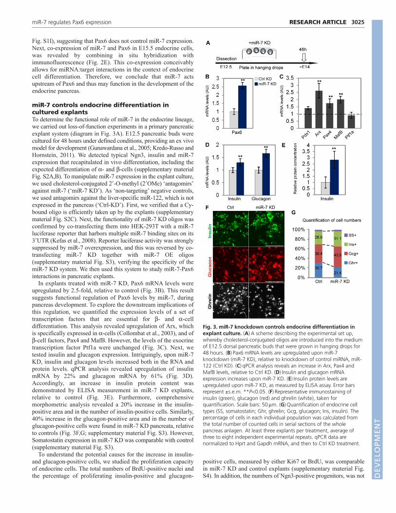
Fig. S1I), suggesting that Pax6 does not control miR-7 expression.Next, co-expression of miR-7 and Pax6 in E15.5 endocrine cells,was revealed by combining in situ hybridization withimmunofluorescence (Fig. 2E). This co-expression conceivablyallows for miRNA:target interactions in the context of endocrinecell differentiation. Therefore, we conclude that miR-7 actsupstream of Pax6 and thus may function in the development of theendocrine pancreas.
miR-7 controls endocrine differentiation incultured explantsTo determine the functional role of miR-7 in the endocrine lineage,we carried out loss-of-function experiments in a primary pancreaticexplant system (diagram in Fig. 3A). E12.5 pancreatic buds werecultured for 48 hours under defined conditions, providing an ex vivomodel for development (Gunawardana et al., 2005; Kredo-Russo andHornstein, 2011). We detected typical Ngn3, insulin and miR-7expression that recapitulated in vivo differentiation, including theexpected differentiation of - and -cells (supplementary materialFig. S2A,B). To manipulate miR-7 expression in the explant culture,we used cholesterol-conjugated 2�-O-methyl (2�OMe) ‘antagomirs’against miR-7 (‘miR-7 KD’). As ‘non-targeting’ negative controls,we used antagomirs against the liver-specific miR-122, which is notexpressed in the pancreas (‘Ctrl-KD’). First, we verified that a Cy-bound oligo is efficiently taken up by the explants (supplementarymaterial Fig. S2C). Next, the functionality of miR-7 KD oligos wasconfirmed by co-transfecting them into HEK-293T with a miR-7luciferase reporter that harbors multiple miR-7 binding sites on its3�UTR (Kefas et al., 2008). Reporter luciferase activity was stronglysuppressed by miR-7 overexpression, and this was reversed by co-transfecting miR-7 KD together with miR-7 OE oligos(supplementary material Fig. S3), verifying the specificity of themiR-7 KD system. We then used this system to study miR-7-Pax6interactions in pancreatic explants.
In explants treated with miR-7 KD, Pax6 mRNA levels wereupregulated by 2.5-fold, relative to control (Fig. 3B). This resultsuggests functional regulation of Pax6 levels by miR-7, duringpancreas development. To explore the downstream implications ofthis regulation, we quantified the expression levels of a set oftranscription factors that are essential for - and -celldifferentiation. This analysis revealed upregulation of Arx, whichis specifically expressed in -cells (Collombat et al., 2003), and of-cell factors, Pax4 and MafB. However, the levels of the exocrinetranscription factor Ptf1a were unchanged (Fig. 3C). Next, wetested insulin and glucagon expression. Intriguingly, upon miR-7KD, insulin and glucagon levels increased both in the RNA andprotein levels. qPCR analysis revealed upregulation of insulinmRNA by 22% and glucagon mRNA by 61% (Fig. 3D).Accordingly, an increase in insulin protein content wasdemonstrated by ELISA measurement in miR-7 KD explants,relative to control (Fig. 3E). Furthermore, comprehensivemorphometric analysis revealed a 20% increase in the insulin-positive area and in the number of insulin-positive cells. Similarly,40% increase in the glucagon-positive area and in the number ofglucagon-positive cells were found in miR-7 KD pancreata, relativeto controls (Fig. 3F,G; supplementary material Fig. S3). However,Somatostatin expression in miR-7 KD was comparable with control(supplementary material Fig. S3).
To understand the potential causes for the increase in insulin-and glucagon-positive cells, we studied the proliferation capacityof endocrine cells. The total numbers of BrdU-positive nuclei andthe percentage of proliferating insulin-positive and glucagon-
positive cells, measured by either Ki67 or BrdU, was comparablein miR-7 KD and control explants (supplementary material Fig.S4). In addition, the numbers of Ngn3-positive progenitors, was not
3025RESEARCH ARTICLEmiR-7 regulates Pax6 expression
Fig. 3. miR-7 knockdown controls endocrine differentiation inexplant culture. (A) A scheme describing the experimental set up,whereby cholesterol-conjugated oligos are introduced into the mediumof E12.5 dorsal pancreatic buds that were grown in hanging drops for48 hours. (B) Pax6 mRNA levels are upregulated upon miR-7knockdown (miR-7 KD), relative to knockdown of control miRNA, miR-122 (Ctrl KD). (C) qPCR analysis reveals an increase in Arx, Pax4 andMafB levels, relative to Ctrl KD. (D) Insulin and glucagon mRNAexpression increases upon miR-7 KD. (E) Insulin protein levels areupregulated upon miR-7 KD, as measured by ELISA assay. Error barsrepresent ±s.e.m. **P<0.05. (F) Representative immunostaining ofinsulin (green), glucagon (red) and ghrelin (white), taken forquantification. Scale bars: 50 m. (G) Quantification of endocrine celltypes (SS, somatostatin; Ghr, ghrelin; Gcg, glucagon; Ins, insulin). Thepercentage of cells in each individual population was calculated fromthe total number of counted cells in serial sections of the wholepancreas anlagen. At least three explants per treatment, average ofthree to eight independent experimental repeats. qPCR data arenormalized to Hprt and Gapdh mRNA, and then to Ctrl KD treatment.
DEVELO
PMENT
3026
affected by miR-7 KD (supplementary material Fig. S3),suggesting that the increase in insulin- and glucagon-positive cellsemerges neither from enhanced proliferation, nor from changes inthe size of the Ngn3 precursor pool.
Although Pax6 positively regulates insulin and glucagonexpression, it negatively regulates ghrelin expression anddifferentiation of -cells (Heller et al., 2005; Dames et al., 2010).Accordingly, miR-7 KD resulted in reduced numbers of ghrelin-positive cells (Fig. 3F,G; supplementary material Fig. S3),
supporting the view that miR-7 knockdown acts upstream of Pax6to promote differentiation into insulin- and glucagon-positive cellsat the expense of ghrelin-positive cells.
miR-7 overexpression ex vivo resembles Pax6haploinsufficiencyTo determine the effect of miR-7 overexpression in the explantsystem, synthetic ‘mimic’ oligos of miR-7 were used (Fig. 4A, ‘miR-7 OE’). As a control, we used a C. elegans miRNA, which is not
RESEARCH ARTICLE Development 139 (16)
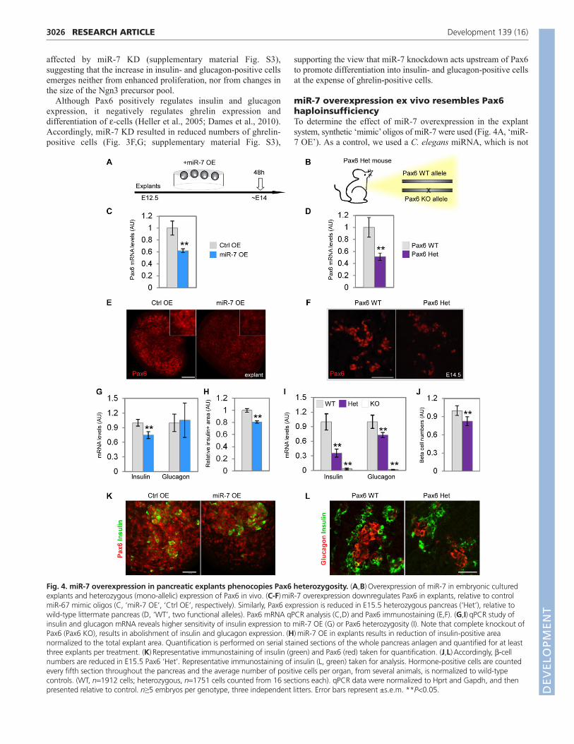
Fig. 4. miR-7 overexpression in pancreatic explants phenocopies Pax6 heterozygosity. (A,B) Overexpression of miR-7 in embryonic culturedexplants and heterozygous (mono-allelic) expression of Pax6 in vivo. (C-F) miR-7 overexpression downregulates Pax6 in explants, relative to controlmiR-67 mimic oligos (C, ‘miR-7 OE’, ‘Ctrl OE’, respectively). Similarly, Pax6 expression is reduced in E15.5 heterozygous pancreas (‘Het’), relative towild-type littermate pancreas (D, ‘WT’, two functional alleles). Pax6 mRNA qPCR analysis (C,D) and Pax6 immunostaining (E,F). (G,I) qPCR study ofinsulin and glucagon mRNA reveals higher sensitivity of insulin expression to miR-7 OE (G) or Pax6 heterozygosity (I). Note that complete knockout ofPax6 (Pax6 KO), results in abolishment of insulin and glucagon expression. (H) miR-7 OE in explants results in reduction of insulin-positive areanormalized to the total explant area. Quantification is performed on serial stained sections of the whole pancreas anlagen and quantified for at leastthree explants per treatment. (K) Representative immunostaining of insulin (green) and Pax6 (red) taken for quantification. (J,L) Accordingly, -cellnumbers are reduced in E15.5 Pax6 ‘Het’. Representative immunostaining of insulin (L, green) taken for analysis. Hormone-positive cells are countedevery fifth section throughout the pancreas and the average number of positive cells per organ, from several animals, is normalized to wild-typecontrols. (WT, n1912 cells; heterozygous, n1751 cells counted from 16 sections each). qPCR data were normalized to Hprt and Gapdh, and thenpresented relative to control. n≥5 embryos per genotype, three independent litters. Error bars represent ±s.e.m. **P<0.05. D
EVELO
PMENT

expressed in mammals (miR-67, ‘Ctrl OE’). In explants treated withmiR-7 OE, Pax6 mRNA levels decreased to 60% of control levels(Fig. 4C). Additionally, immunofluorescent analysis revealedreduction of PAX6 protein levels (Fig. 4E). Furthermore, miR-7 OEresulted in downregulation of insulin mRNA (Fig. 4G) and of MafBmRNA (supplementary material Fig. S5A), two genes that aredirectly controlled by Pax6 (Sander et al., 1997; Nishimura et al.,2008). Accordingly, a decrease in insulin content was demonstratedin miR-7 OE explants by ELISA measurement, relative to controls(supplementary material Fig. S5C). Consistently, a reduction in miR-7 OE explant insulin-positive area was detected by immunostainingquantification (Fig. 4H,K). By contrast, ghrelin expression wasupregulated (supplementary material Fig. S5B), consistent withsignificant ghrelin expansion in Pax6 knockout (Heller et al., 2005;Dames et al., 2010). However, glucagon levels were not significantlychanged. Thus, gain-of-function analysis substantiates ourconclusions that miR-7 regulates Pax6 and downstream endocrinegenes, including insulin and ghrelin.
We complemented this study by analyzing an in vivo model forgenetic downregulation of Pax6 expression. Homozygous loss ofPax6 causes complete loss of insulin and glucagon expression andupregulation of ghrelin expression (Sander et al., 1997; St-Onge etal., 1997; Heller et al., 2005; Dames et al., 2010). We hypothesizedthat knockout of a single Pax6 allele may downregulate Pax6expression to intermediate levels, by a mechanism that isindependent of miR-7. We therefore analyzed previouslyuncharacterized Pax6 heterozygous pancreata (‘Het’, Fig. 4B). Thisanalysis revealed a 50% reduction in Pax6 mRNA and protein levelsin E14.5 Pax6 Het embryos, relative to wild-type littermate controls(Fig. 4D,F). Similar to miR-7 OE, MafB levels were downregulatedby 20% in Pax6 Het pancreata relative to control (supplementarymaterial Fig. S5B). Furthermore, insulin and glucagon mRNA levelswere decreased by 65% and 30%, respectively, in Pax6 Hetpancreata, while ghrelin expression was increased by 30% (Fig. 4I;supplementary material Fig. S5B). In addition, serial sectionquantification revealed a 22% decrease in -cell numbers (Fig. 4J,L),while -cell numbers were comparable between Het and wild-typepancreata. These data demonstrate the sensitivity of -cells tointermediate Pax6 levels and reveal a surprising similarity betweenthe molecular phenotype gained by miR-7 overexpression and theone caused by Pax6 haploinsufficiency.
Overexpression of miR-7 in vivo reducedexpression of endocrine genesTo examine the consequences of miR-7 overexpression in vivo, wegenerated a conditional miR-7 transgenic mouse line. miR-7a-1genomic sequence was inserted by homologous recombination intothe ubiquitously expressed Rosa26 locus (Fig. 5A) as previouslydescribed (Srinivas et al., 2001). In this knock-in model, theexpression of miR-7 is coupled to the expression of enhanced greenfluorescent protein (EGFP) and both are conditionally blocked bya ‘Neo-STOP’ cassette, flanked by LoxP sites. Thus, miR-7 andEGFP expression are induced only in tissues that express the Cretransgene. To validate the inducible expression of miR-7, weisolated mouse embryonic fibroblasts (MEFs) from Rosa26-miR-7 mice. Upon adenoviral introduction of Cre recombinase, theexpression of miR-7 was specifically upregulated by twofold,relative to control infection, whereas the level of an unrelatedmiRNA, miR-199, remained unchanged (Fig. 5B).
To stimulate the production of miR-7 early in the pancreaticlineage, we crossed the Rosa26-miR-7 allele to a Pdx1-Cretransgene (Gu et al., 2002). Pdx1-Cre;Rosa26-miR-7 mice
specifically expressed GFP protein in E13.5 pancreatic cells (Fig.5C), while littermates that did not harbor the Pdx1-Cre transgenedid not express GFP (‘Ctrl’).
We next characterized E15.5 pancreata of the Pdx1-Cre;Rosa26-miR-7 mice by qPCR. Expression of the mature endocrine cellmarkers, insulin and glucagon, was downregulated (to 72% and68% of control, respectively). Quantification of the expression ofdifferent transcription factors revealed a reduction in Arx (to 63%)and Pax4 (to 62%), and in the miR-7 target gene Pax6 (to 49%,Fig. 5D). Notably, Cpa1 and Ptf1a levels were unchanged,indicating that the exocrine lineage was unaffected by miR-7 mis-expression. A decrease in the expression of endocrine markers wasalso seen by immunostaining for insulin and glucagon at E15.5(Fig. 5E). Therefore, when miR-7 was overexpressed in Pdx1-Cre;Rosa26-miR-7 embryos, Pax6 levels decreased and theexpression of insulin and glucagon was downregulated, providingin vivo evidence for control of Pax6 by miR-7.
miR-7;Pax6 interactions upstream of insulinpromoter activationFinally, to elucidate the genetic interactions of miR-7 and Pax6, weused a luciferase reporter, driven by the insulin promoter(Melkman-Zehavi et al., 2011). As the insulin promoter is directlyactivated by Pax6 (Sander et al., 1997), it provides a model systemfor monitoring the effect of miR-7; Pax6 axis on insulin expression.Transfecting MIN6 cells with either siRNA against Pax6 (‘siPax6’)or miR-7 overexpression vector (‘miR-7 OE’), resulted indownregulation of insulin promoter activity, relative to scrambledsiRNA, or empty miRvec, respectively (Fig. 5F). These resultssupport the observed decrease in insulin mRNA expression bymiR-7 OE in explants (Fig. 4G) and in miR-7 transgenic model(Fig. 5D). The combined effect of siPax6 together with miR-7 OE,significantly repressed the activity of the insulin promoter.Furthermore, transfecting MIN6 cells with knockdown oligosagainst miR-7 (‘miR-7 KD’) resulted in upregulation of insulinpromoter activity, relative to scrambled oligos (‘Ctrl KD’),consistent with insulin mRNA upregulation in miR-7 KD-treatedexplants (Fig. 3D). However, when inhibited simultaneously,siPax6 reversed the effect of miR-7 KD, suggesting that Pax6mediates miR-7 regulation of insulin expression (Fig. 5F, rightpanel). These results stress the epistatic relationship of miR-7upstream of Pax6, in the control of insulin expression. Altogether,our results suggest that miR-7 acts to limit Pax6 expression levels,allowing precise endocrine cell maturation in the development ofthe pancreas.
DISCUSSIONThe role of Pax6 in development has been extensively studied.However, how Pax6 levels are fine-tuned in tissues is not wellunderstood (Baulmann et al., 2002; Ding et al., 2009). Here, wediscover a miR-7-based mechanism that controls and refines Pax6levels in the endocrine pancreas, through a conserved binding site inthe 3�UTR of Pax6 mRNA. This mechanism enables accurate insulinand glucagon expression and proper - and -cell differentiation.
miR-7 is a new component in the regulation of theendocrine lineageWe show that miR-7 is expressed specifically in the endocrinelineage. miR-7 is expressed relatively early in newly born endocrineprecursor cells that express Ngn3, probably coinciding with earlyexpression of endocrine transcription factors such as NeuroD/2(Jensen et al., 2000). The expression of miR-7 is maintained in
3027RESEARCH ARTICLEmiR-7 regulates Pax6 expression
DEVELO
PMENT

3028
endocrine cells during their specification to - and -cells.Intriguingly, miR-7 is not detected at E15.5 in Cpa1+ exocrine cellsor in Hnf1b+ duct cells, in accordance with its specific endocrineexpression. Independent evidence for the endocrine-specificexpression of miR-7 emerges from genetic studies, whereinnullification of Ngn3 completely abrogates miR-7 expression.Intriguingly, miR-375 expression is only partially reduced in Ngn3knockout embryos, suggesting differences in the control of miR-375and miR-7 expression. In agreement with our observations, in humanfetuses, miR-7 has been shown to be expressed from around theninth week of gestation and reaches maximal expression at the peak
of pancreatic endocrine development (Correa-Medina et al., 2009).Thus, miR-7 expression and presumably its function, appear to beconserved in both mouse and human development.
Pax6 is an important target of miR-7 in thedeveloping pancreasThe endocrine expression of miR-7 suggests that it plays importantroles in differentiation of the endocrine pancreas. Bioinformaticspredictions suggest that miR-7 has a battery of targets, which playa role in pancreas biology, including recently studied IRS2 (Lewiset al., 2005; Kefas et al., 2008). We focused on regulation of Pax6
RESEARCH ARTICLE Development 139 (16)
Fig. 5. Overexpression of miR-7 in vivo reduces theexpression of endocrine genes. (A) Schematic of thetargeting construct for conditional miR-7-IRES-GFPmisexpression. LoxP sites are indicated by arrowheads.(B) miR-7 upregulation is Cre dependent. qPCR analysis ofmiR-7 and miR-199 levels in primary embryonic fibroblastsharvested from Rosa-miR-7 transgenic mice and infectedwith an adenovirus that expresses either Cre-GFP (‘Ad-Cre’) or GFP alone (‘Ad-GFP’). Data are normalized tosno234 (three independent MEF lines, each in triplicate).(C) GFP is specifically expressed in E13.5 Pdx1-Cre;Rosa-miR7 pancreatic epithelium but not in littermates that donot harbor the Cre recombinase (‘Ctrl’). Asterisks depicterythrocyte autofluorescence. (D,E) miR-7 mis-expressionrepresses endocrine genes. (D) qPCR analysis of pancreaticgenes in E15.5 Pdx1-Cre;Rosa-miR-7 samples, asindicated. Data are normalized to Hprt and Gapdh mRNA,and then to the expression levels in littermate controls(n6 each genotype, three litters). Error bars represent±s.e.m. (**P<0.05, *P<0.01). (E) Immunostaining forinsulin (green) and glucagon (red) in sections of Pdx1-Cre;Rosa-miR-7 pancreas and control littermates. Blue,nuclei. Scale bar: 50 m. (F) Pax6;miR-7 interactionupstream of insulin promoter activation in MIN6 cells.Insulin promoter activity is downregulated by miR-7overexpression, relative to control (‘miR-7 OE’, ‘Ctrl OE’,respectively) and is suppressed by siRNA against Pax6(‘siPax6). Combining miR-7 OE with siPax6 enhanced thesuppressions of the insulin promoter (left panel). Insulinpromoter activity is upregulated by miR-7 knockdown,relative to negative control scrambled oligo (‘miR-7 KD’,‘Ctrl KD’, respectively). Concomitant introduction of miR-7KD with siPax6 restores insulin promoter activity (rightpanel). All firefly luciferase data is normalized to theactivity of Renilla luciferase co-transfected and presentedrelative to the control experiment. n3 independentexperiments. Error bars represent ±s.e.m. **P<0.05.
DEVELO
PMENT

by miR-7 because of our bioinformatic analysis and because Pax6levels are known to be tightly regulated. For example, continuousPax6 overexpression may be harmful to endocrine cells (Yamaokaet al., 2000), whereas complete knockout results in loss of matureendocrine markers, including insulin and glucagon (Sander et al.,1997; St-Onge et al., 1997). Furthermore, loss of even a singlePax6 allele causes glucose intolerance in humans and mice (Yasudaet al., 2002; Ding et al., 2009). Plausibly, Pax6 dose sensitivity inthe endocrine pancreas and in other developing organs (vanRaamsdonk and Tilghman, 2000) provides a rationale for therequired control by miR-7. Our experimental data suggest that,indeed, miR-7 provides another mechanism for refining Pax6expression. Plausibly, miR-7 has additional targets in pancreasdevelopment and homeostasis that are yet to be discovered.
The Pax6-miR-7 pathway affects endocrinedifferentiationWe report that heterozygous expression of Pax6 is haploinsufficientand leads to defined changes in the expression of endocrine genes,including MafB, insulin and glucagon. These molecularphenotypes are also observed upon miR-7 overexpression, in bothexplant and transgenic models, supporting molecular interaction ofmiR-7 and Pax6. Furthermore, miR-7 knockdown alleviates theeffect of Pax6 downregulation in the control of the insulinpromoter. Taken together, these analyses provide substantiatingevidence for miR-7 activity upstream of Pax6 and its effector MafB(Nishimura et al., 2008) during development. Pax6 is a negativeregulator of Ghrelin-positive -cell fate (Heller et al., 2005; Dameset al., 2010). Lower numbers of ghrelin-positive -cells and higherinsulin-positive and glucagon-positive cell numbers in miR-7 KD,are consistent with miR-7 function in the differentiation ofendocrine cell types, upstream of Pax6. Intriguingly, - and -cellsexhibit differential sensitivity to reduction in Pax6 levels, either ina heterozygous Pax6 model or when miR-7 is overexpressed. Bycontrast, Pax6 upregulation predominantly causes an increase inglucagon and Arx levels, consistent with observations by others(Sander et al., 1997; Heller et al., 2004; Nishimura et al., 2008).The mechanisms underlying differential Pax6 sensitivity may berelated to Pax4 expression in -cells (Smith et al., 1999) or toadditional molecular components that are not yet known.
Network of miR-7 and pancreatic transcriptionfactorsThe overall picture emerging from our analyses is that duringdevelopment, endocrine transcription factors direct endocrine celldifferentiation. Unexpectedly, miR-7 partially counteracts theactivity of these transcription factors, acting as an inhibitory geneticelement in endocrine cell maturation. Intriguingly, miR-7 hasalready been shown to act downstream of the neurogenin homologAtonal in Drosophila retinal development (Li et al., 2009). Wetherefore suggest that miR-7 may act similarly in mammals tominimize unwanted changes in the endocrine developmentalprogram and to control Pax6 levels (see Fig. 6). In so doing, miR-7 may convey robustness to pancreas developmental, like itshomolog in the fly. In future studies, it may be important toconsider cases wherein pathological miR-7 expression causesabnormal Pax6 levels in humans. Another interesting direction forfuture studies is to explore potential Pax6 3�UTR isoforms, asexpression may be differentially regulated in cis by such alternativemRNA variants (Jan et al., 2011). Potentially, some Pax6 3�UTRisoforms would not harbor binding sites for endocrine-enrichedmiRNAs, thus avoiding post-transcriptional regulation.
In summary, our analysis characterizes miR-7 as a novelcomponent in the regulation of endocrine cell differentiation. Betterunderstanding of the compound miRNA-transcription factornetwork in pancreas development may pave the way to moreeffective maturation of -cells in culture towards cell-based therapyfor diabetes.
AcknowledgementsWe thank Yuval Dor, Judith Magenheim, Ruth Ashery-Padan, Michael Walkerand Tal Melkman-Zehavi for protocols, reagents and productive discussions.We thank Jun-ichi Miyazaki for MIN6 cells; Benjamin Purow for miR-7 reporterplasmid; and Reuven Agami for miRvec-7.
FundingThis work was supported by a grant from the Juvenile Diabetes ResearchFoundation [#99-2007-71 to E.H.]; the German-Israeli Foundation (GIF) YoungInvestigator Award; the European Foundation for the Study of Diabetes(EFSD)/D-Cure Young Investigator Award; EFSD-Lilly; and the Israel ScienceFoundation. Additional funding was provided by the Wolfson family charitabletrust; the Celia Benattar Memorial Fund for Juvenile Diabetes; CarolitoStiftung; the Charlene Vener New Scientist Fund; the Kimmel Institute for StemCell Research; the estates of F. Sherr, L. Asse and N. Baltor; the CharlesteinFoundation; and the Fraida Foundation. E.H. is the incumbent of the Helenand Milton A. Kimmelman Career Development Chair, and research in this labis supported by Dr Sydney Brenner and friends. The funders had no role instudy design, data collection and analysis, decision to publish or preparation ofthe manuscript.
Competing interests statementThe authors declare no competing financial interests.
3029RESEARCH ARTICLEmiR-7 regulates Pax6 expression
Fig. 6. A schematic model for miR-7-Pax6 interactions in pancreasdevelopment. The regulation of Pax6 levels (blue) by miR-7, regulatesthe differentiation of hormone-expressing endocrine cells. miR-7knockdown de-represses Pax6 and results in reduced ghrelin (Ghr)expression and preference towards insulin and glucagon-positive cells(Ins and Gcg). Similarly, miR-7 overexpression or heterozygous (Het)expression of Pax6 results in reduced Pax6 and reciprocal changes in theexpression of hormones.
DEVELO
PMENT

3030
Author contributionsS.K.-R., A.N., A.D.M. and E.H. designed the experiments; E.H. and S.K.-R.wrote the manuscript; M.A.B. and K.A.L. designed and provided reagents;S.K.-R., A.D.M., I.A. and A.N. conducted all experiments and analyses in thiswork.
Supplementary materialSupplementary material available online athttp://dev.biologists.org/lookup/suppl/doi:10.1242/dev.080127/-/DC1
ReferencesAshery-Padan, R., Zhou, X., Marquardt, T., Herrera, P., Toube, L., Berry, A.
and Gruss, P. (2004). Conditional inactivation of Pax6 in the pancreas causesearly onset of diabetes. Dev. Biol. 269, 479-488.
Avnit-Sagi, T., Kantorovich, L., Kredo-Russo, S., Hornstein, E. and Walker, M.D. (2009). The promoter of the pri-miR-375 gene directs expression selectively tothe endocrine pancreas. PLoS ONE 4, e5033.
Bartel, D. P. (2004). MicroRNAs: genomics, biogenesis, mechanism, and function.Cell 116, 281-297.
Baulmann, D. C., Ohlmann, A., Flugel-Koch, C., Goswami, S., Cvekl, A. andTamm, E. R. (2002). Pax6 heterozygous eyes show defects in chamber angledifferentiation that are associated with a wide spectrum of other anterior eyesegment abnormalities. Mech. Dev. 118, 3-17.
Berger, J., Berger, S., Tuoc, T. C., D’Amelio, M., Cecconi, F., Gorski, J. A.,Jones, K. R., Gruss, P. and Stoykova, A. (2007). Conditional activation of Pax6in the developing cortex of transgenic mice causes progenitor apoptosis.Development 134, 1311-1322.
Bonal, C. and Herrera, P. L. (2008). Genes controlling pancreas ontogeny. Int. J.Dev. Biol. 52, 823-835.
Bravo-Egana, V., Rosero, S., Molano, R. D., Pileggi, A., Ricordi, C.,Dominguez-Bendala, J. and Pastori, R. L. (2008). Quantitative differentialexpression analysis reveals miR-7 as major islet microRNA. Biochem. Biophys.Res. Commun. 366, 922-926.
Collinson, J. M., Quinn, J. C., Buchanan, M. A., Kaufman, M. H., Wedden, S.E., West, J. D. and Hill, R. E. (2001). Primary defects in the lens underliecomplex anterior segment abnormalities of the Pax6 heterozygous eye. Proc.Natl. Acad. Sci. USA 98, 9688-9693.
Collombat, P., Mansouri, A., Hecksher-Sorensen, J., Serup, P., Krull, J.,Gradwohl, G. and Gruss, P. (2003). Opposing actions of Arx and Pax4 inendocrine pancreas development. Genes Dev. 17, 2591-2603.
Correa-Medina, M., Bravo-Egana, V., Rosero, S., Ricordi, C., Edlund, H., Diez,J. and Pastori, R. L. (2009). MicroRNA miR-7 is preferentially expressed inendocrine cells of the developing and adult human pancreas. Gene Expr.Patterns 9, 193-199.
Dames, P., Puff, R., Weise, M., Parhofer, K. G., Goke, B., Gotz, M., Graw, J.,Favor, J. and Lechner, A. (2010). Relative roles of the different Pax6 domainsfor pancreatic alpha cell development. BMC Dev. Biol. 10, 39.
Dennis, G., Jr, Sherman, B. T., Hosack, D. A., Yang, J., Gao, W., Lane, H. C.and Lempicki, R. A. (2003). DAVID: database for annotation, visualization, andintegrated discovery. Genome Biol. 4, P3.
Ding, J., Gao, Y., Zhao, J., Yan, H., Guo, S. Y., Zhang, Q. X., Li, L. S. and Gao,X. (2009). Pax6 haploinsufficiency causes abnormal metabolic homeostasis bydown-regulating glucagon-like peptide 1 in mice. Endocrinology 150, 2136-2144.
Gradwohl, G., Dierich, A., LeMeur, M. and Guillemot, F. (2000). neurogenin3is required for the development of the four endocrine cell lineages of thepancreas. Proc. Natl. Acad. Sci. USA 97, 1607-1611.
Gu, G., Dubauskaite, J. and Melton, D. A. (2002). Direct evidence for thepancreatic lineage: NGN3+ cells are islet progenitors and are distinct from ductprogenitors. Development 129, 2447-2457.
Gunawardana, S. C., Hara, M., Bell, G. I., Head, W. S., Magnuson, M. A. andPiston, D. W. (2005). Imaging Beta cell development in real-time usingpancreatic explants from mice with green fluorescent protein-labeled pancreaticBeta cells. In Vitro Cell. Dev. Biol. Anim. 41, 7-11.
Heller, R. S., Stoffers, D. A., Liu, A., Schedl, A., Crenshaw, E. B., 3rd, Madsen,O. D. and Serup, P. (2004). The role of Brn4/Pou3f4 and Pax6 in forming thepancreatic glucagon cell identity. Dev. Biol. 268, 123-134.
Heller, R. S., Jenny, M., Collombat, P., Mansouri, A., Tomasetto, C., Madsen,O. D., Mellitzer, G., Gradwohl, G. and Serup, P. (2005). Genetic determinantsof pancreatic epsilon-cell development. Dev. Biol. 286, 217-224.
Hornstein, E. and Shomron, N. (2006). Canalization of development bymicroRNAs. Nat. Genet. 38 Suppl., S20-S24.
Jan, C. H., Friedman, R. C., Ruby, J. G. and Bartel, D. P. (2011). Formation,regulation and evolution of Caenorhabditis elegans 3�UTRs. Nature 469, 97-101.
Jensen, J., Heller, R. S., Funder-Nielsen, T., Pedersen, E. E., Lindsell, C.,Weinmaster, G., Madsen, O. D. and Serup, P. (2000). Independentdevelopment of pancreatic alpha- and beta-cells from neurogenin3-expressingprecursors: a role for the notch pathway in repression of prematuredifferentiation. Diabetes 49, 163-176.
Kefas, B., Godlewski, J., Comeau, L., Li, Y., Abounader, R., Hawkinson, M.,Lee, J., Fine, H., Chiocca, E. A., Lawler, S. et al. (2008). microRNA-7 inhibitsthe epidermal growth factor receptor and the Akt pathway and is down-regulated in glioblastoma. Cancer Res. 68, 3566-3572.
Kloosterman, W. P., Lagendijk, A. K., Ketting, R. F., Moulton, J. D. andPlasterk, R. H. (2007). Targeted inhibition of miRNA maturation withmorpholinos reveals a role for miR-375 in pancreatic islet development. PLoSBiol. 5, e203.
Kredo-Russo, S. and Hornstein, E. (2011). MicroRNA knock down bycholesterol-conjugated antisense oligos in mouse organ culture. Methods Mol.Biol. 732, 89-97.
Kroeber, M., Davis, N., Holzmann, S., Kritzenberger, M., Shelah-Goraly, M.,Ofri, R., Ashery-Padan, R. and Tamm, E. R. (2010). Reduced expression ofPax6 in lens and cornea of mutant mice leads to failure of chamber angledevelopment and juvenile glaucoma. Hum. Mol. Genet. 19, 3332-3342.
Landgraf, P., Rusu, M., Sheridan, R., Sewer, A., Iovino, N., Aravin, A., Pfeffer,S., Rice, A., Kamphorst, A. O., Landthaler, M. et al. (2007). A mammalianmicroRNA expression atlas based on small RNA library sequencing. Cell 129,1401-1414.
Lewis, B. P., Burge, C. B. and Bartel, D. P. (2005). Conserved seed pairing, oftenflanked by adenosines, indicates that thousands of human genes are microRNAtargets. Cell 120, 15-20.
Li, X., Cassidy, J. J., Reinke, C. A., Fischboeck, S. and Carthew, R. W. (2009). AmicroRNA imparts robustness against environmental fluctuation duringdevelopment. Cell 137, 273-282.
Lynn, F. C., Skewes-Cox, P., Kosaka, Y., McManus, M. T., Harfe, B. D. andGerman, M. S. (2007). MicroRNA expression is required for pancreatic islet cellgenesis in the mouse. Diabetes 56, 2938-2945.
Lyttle, B. M., Li, J., Krishnamurthy, M., Fellows, F., Wheeler, M. B., Goodyer,C. G. and Wang, R. (2008). Transcription factor expression in the developinghuman fetal endocrine pancreas. Diabetologia 51, 1169-1180.
Martin, M., Hauer, V., Messmer, M., Orvain, C. and Gradwohl, G. (2007).Transcription factors in pancreatic development. Animal models. Endocr. Dev.12, 24-32.
Melkman-Zehavi, T., Oren, R., Kredo-Russo, S., Shapira, T., Mandelbaum, A.D., Rivkin, N., Nir, T., Lennox, K. A., Behlke, M. A., Dor, Y. et al. (2011).miRNAs control insulin content in pancreatic beta-cells via downregulation oftranscriptional repressors. EMBO J. 30, 835-845.
Murtaugh, L. C. and Melton, D. A. (2003). Genes, signals, and lineages inpancreas development. Annu. Rev. Cell Dev. Biol. 19, 71-89.
Nishimura, W., Rowan, S., Salameh, T., Maas, R. L., Bonner-Weir, S., Sell, S.M. and Sharma, A. (2008). Preferential reduction of beta cells derived fromPax6-MafB pathway in MafB deficient mice. Dev. Biol. 314, 443-456.
Pena, J. T., Sohn-Lee, C., Rouhanifard, S. H., Ludwig, J., Hafner, M.,Mihailovic, A., Lim, C., Holoch, D., Berninger, P., Zavolan, M. et al. (2009).miRNA in situ hybridization in formaldehyde and EDC-fixed tissues. Nat.Methods 6, 139-141.
Poy, M. N., Eliasson, L., Krutzfeldt, J., Kuwajima, S., Ma, X., Macdonald, P.E., Pfeffer, S., Tuschl, T., Rajewsky, N., Rorsman, P. et al. (2004). Apancreatic islet-specific microRNA regulates insulin secretion. Nature 432, 226-230.
Poy, M. N., Hausser, J., Trajkovski, M., Braun, M., Collins, S., Rorsman, P.,Zavolan, M. and Stoffel, M. (2009). miR-375 maintains normal pancreaticalpha- and beta-cell mass. Proc. Natl. Acad. Sci. USA 106, 5813-5818.
Sander, M., Neubuser, A., Kalamaras, J., Ee, H. C., Martin, G. R. and German,M. S. (1997). Genetic analysis reveals that PAX6 is required for normaltranscription of pancreatic hormone genes and islet development. Genes Dev.11, 1662-1673.
Schedl, A., Ross, A., Lee, M., Engelkamp, D., Rashbass, P., van Heyningen, V.and Hastie, N. D. (1996). Influence of PAX6 gene dosage on development:overexpression causes severe eye abnormalities. Cell 86, 71-82.
Silahtaroglu, A. N., Nolting, D., Dyrskjot, L., Berezikov, E., Moller, M.,Tommerup, N. and Kauppinen, S. (2007). Detection of microRNAs in frozentissue sections by fluorescence in situ hybridization using locked nucleic acidprobes and tyramide signal amplification. Nat. Protoc. 2, 2520-2528.
Smith, S. B., Ee, H. C., Conners, J. R. and German, M. S. (1999). Paired-homeodomain transcription factor PAX4 acts as a transcriptional repressor inearly pancreatic development. Mol. Cell. Biol. 19, 8272-8280.
Srinivas, S., Watanabe, T., Lin, C. S., William, C. M., Tanabe, Y., Jessell, T. M.and Costantini, F. (2001). Cre reporter strains produced by targeted insertionof EYFP and ECFP into the ROSA26 locus. BMC Dev. Biol. 1, 4.
St-Onge, L., Sosa-Pineda, B., Chowdhury, K., Mansouri, A. and Gruss, P.(1997). Pax6 is required for differentiation of glucagon-producing alpha-cells inmouse pancreas. Nature 387, 406-409.
Stark, A., Brennecke, J., Bushati, N., Russell, R. B. and Cohen, S. M. (2005).Animal MicroRNAs confer robustness to gene expression and have a significantimpact on 3�UTR evolution. Cell 123, 1133-1146.
Tkatchenko, A. V. (2006). Whole-mount BrdU staining of proliferating cells byDNase treatment: application to postnatal mammalian retina. Biotechniques 40,29-30, 32.
RESEARCH ARTICLE Development 139 (16)
DEVELO
PMENT

van Raamsdonk, C. D. and Tilghman, S. M. (2000). Dosage requirement andallelic expression of PAX6 during lens placode formation. Development 127,5439-5448.
Wang, S., Yan, J., Anderson, D. A., Xu, Y., Kanal, M. C., Cao, Z., Wright, C. V. and Gu, G. (2010). Neurog3 gene dosage regulates allocation ofendocrine and exocrine cell fates in the developing mouse pancreas. Dev. Biol.339, 26-37.
Wienholds, E., Kloosterman, W. P., Miska, E., Alvarez-Saavedra, E.,Berezikov, E., de Bruijn, E., Horvitz, H. R., Kauppinen, S. and Plasterk, R.H. (2005). MicroRNA expression in zebrafish embryonic development. Science309, 310-311.
Yamaoka, T., Yano, M., Yamada, T., Matsushita, T., Moritani, M., Ii, S.,Yoshimoto, K., Hata, J. and Itakura, M. (2000). Diabetes and pancreatictumours in transgenic mice expressing Pax 6. Diabetologia 43, 332-339.
Yasuda, T., Kajimoto, Y., Fujitani, Y., Watada, H., Yamamoto, S., Watarai, T.,Umayahara, Y., Matsuhisa, M., Gorogawa, S., Kuwayama, Y. et al. (2002).PAX6 mutation as a genetic factor common to aniridia and glucose intolerance.Diabetes 51, 224-230.
Zhou, Q., Law, A. C., Rajagopal, J., Anderson, W. J., Gray, P. A. and Melton,D. A. (2007). A multipotent progenitor domain guides pancreaticorganogenesis. Dev. Cell 13, 103-114.
3031RESEARCH ARTICLEmiR-7 regulates Pax6 expression
DEVELO
PMENT