partha varanashi - university of adelaide · 2 declaration i declare that this thesis is a record...
TRANSCRIPT
1
BARLEY CELLULOSE SYNTHASES INVOLVED IN SECONDARY
CELL WALL FORMATION AND STEM STRENGTH: GENERATION
OF cDNA CONSTRUCTS FOR FUNCTIONAL ANALYSIS
by
Partha Varanashi
A thesis submitted for the partial fulfillment of the requirements of the
Masters of Biotechnology (Plant Biotechnology)
The University of Adelaide
Faculty of Sciences
School of Agriculture, Food and Wine
Waite Campus
2008
2
DECLARATION
I declare that this thesis is a record of original work and contains no material which has been
accepted for the award of any other degree or diploma in any university. To the best of my
knowledge and belief, this thesis contains no material previously published or written by another
person, except where due reference is made in the text.
Partha Varanashi
Associate Professor Maria Hrmova
3
PREFACE
This research was performed over 10 months as part of a Masters in
Biotechnology (Plant Biotechnology). The literature review was previously assessed in
accordance with the correction suggested by the examiners. The main focus of the project
remains very similar to that of the research proposal. However the goals were not
achieved according to the time deadline stated in the research proposal. Hence protein
purification was could not be carried out.
Although the research manuscript contained herein will provide the first draft of a
future publication to be submitted to Plant journal, due to time constraint, all data
relevant to that publication has not been collected. However, additional data which was
not conclusive was collected and this is provided within the appendices. The research
manuscript outlines stages involved in the construction and heterologus expression of
barley CesA4 cDNA. While the appendices contain additional data from HvCesA4
protein structure prediction, media recipes, in-silico representation of the HvCesA4
constructs with respective vectors.
4
Title: BARLEY CELLULOSE SYNTHASES INVOLVED IN SECONDARY CELL
WALL FORMATION AND STEM STRENGTH: GENERATION OF cDNA
CONSTRUCTS FOR FUNCTIONAL ANALYSIS
Author:
Partha Varanashi
The University of Adelaide
Plant Genomics Centre
Glen Osmond 5064, Australia
Email [email protected]
Corresponding author:
Dr Maria Hrmova
Associate Professor
The University of Adelaide
Plant Genomics Centre
Glen Osmond 5064, Australia
T: +61 8 8303 7280
F: +61 8 8303 7102
Email: [email protected]

5
Table of Contents
PART I- LITERATURE REVIEW & RESEARCH PLAN
Introduction....................................................................................................................... 7
Literature Review ............................................................................................................. 8
Introduction................................................................................................................ 8
Cellulose .................................................................................................................... 8
Cellulose Biosynthesis ............................................................................................... 9
Cellulose synthase gene family................................................................................ 10
Conclusions.............................................................................................................. 12
The Research Plan .......................................................................................................... 12
Overview.................................................................................................................. 12
Bioinformatics.......................................................................................................... 13
Design of HvCesA4 constructs................................................................................ 14
Primer design ........................................................................................................... 16
Cloning into pPICZ and pPICZα vectors:................................................................ 17
Transformation into Pichia pastoris ......................................................................... 18
Pichia pastoris expression system............................................................................ 18
Time Table............................................................................................................... 19
Conclusions.............................................................................................................. 20
Appendix.......................................................................................................................... 21
References........................................................................................................................ 21
Acknowledgements ......................................................................................................... 24
Summary.......................................................................................................................... 25
Introduction..................................................................................................................... 25
Results .............................................................................................................................. 27
Prediction of membrane helices and catalytic module of HvCesA4 ....................... 27
Cloning into Entry Vector Systems ......................................................................... 28
Cloning into destination vector system.................................................................... 29
Protein expression.................................................................................................... 29
Discussion......................................................................................................................... 30
Experimental Procedures............................................................................................... 33
Bioinformatics and predictions of membrane helices.............................................. 33
Molecular cloning and primers for HvCesA4 constructs ........................................ 34
DNA sequencing...................................................................................................... 37

6
Transformation into Pichia pastoris ......................................................................... 38
Protein expression.................................................................................................... 38
Protein extraction, dot blot, Western blot and SDS-PAGE analyses....................... 39
Acknowledgements ......................................................................................................... 39
References........................................................................................................................ 40
Figures Legends .............................................................................................................. 44
Supplementary data........................................................................................................ 50
Appendices....................................................................................................................... 56
7
Introduction Barley is ranked as the fourth largest crop grown in the world based on quantity of
production. One of the major problems farmers face during barley cultivation is crop
lodging. Owing to weak stems, barley crops cannot withstand heavy winds and hence fall
over. Crop lodging may result in a loss of crop production or in difficulties during
harvesting. The stem strength depends on a type of cells present in the stem region of the
plant. Secondary plant cell walls present in each cell are known to provide the stem
strength. These secondary cell walls are made up of cellulosic microfibrils embedded in a
matrix containing complex non-cellulosic polysaccharides; usually these are pectic
polysaccharides, xyloglucans and heteroxylans as major constituents, and heteromannans
as minor constituents. In vascular plants the major constituent of cell walls are found to
be cellulose, which constitutes (1-4)-β-D-glucan microfibrils. In recent discoveries
candidate genes for biosynthesis of key components of secondary cell walls have been
tracked down to cellulose synthase (CesA) and cellulose synthase like (Csl) gene families
(Richmond 2000).
It is now well known that assembly of cellulose or (1-4)-β-D-glucan microfibrils are
likely to be carried out by a certain set of enzymes (Doblin et al., 2002). Among the
cellulose synthase gene family, cellulose synthase A (CesA) genes in plants are thought to
be responsible for encoding glycosyl transferases, which plays a key roles in biosynthesis
of cellulose (Delmer, 1999). So far in barley nine HvCesA genes have been designated
(Burton et al., 2004). These represent HvCesA1 to HvCesA8. In barley, however, the
functional analyses of the HvCesA genes are incomplete. Thus functional analysis of
8
these proteins is important for understanding of a complete process of cellulose
biosynthesis. Complete information generated on the enzymes encoded by the HvCesA
gene would aid in manipulation of genes responsible for cellulose biosynthesis.
Manipulations would be aimed at improving stem strength of barley plants, thus helping
in reducing crop lodging and undoubtedly increasing the yields.
Literature Review
Introduction
One of the most important distinguishing features of plant cells is the presence of a cell
wall on their surfaces. Plant cell walls are bound by rigid walls, which are mainly made
up of cellulosic, non cellulosic and pectic compounds. The amount of each compound
varies from plant to plant. These cell walls provide the exoskeletal structure for the plant
and are crucial to the structure and growth of the plant. They also contribute to the shape
and morphology of the plant. Cell walls also take part in regulating cell growth along
with providing the structural and mechanical support for the plant (Bacic et al., 1988).
Cellulose holds the key role in the above activities of cell wall, thus attracting the interest
of researchers. This literature review mainly focuses on the biosynthesis of cellulose,
which involves cellulose synthase (Ces) genes. Few genes are found in the cellulose
synthase gene family. They are very similar to each other and are conserved in most of
the higher plants.
Cellulose
Among polymers, cellulose is found to be the most plentiful and is a major constituent of
vascular plants (Brown, 2004). Cellulose also contributes to growth and cell division.

9
Plant cell walls are made up of cellulose and other non-cellulosic components. Cellulose
microfibrils are insoluble, cable-like structures composed of 36 hydrogen bonded (1-4)-β-
D-glucan chains (Fig. 1) (Finaev, 2007). These 36 chains are arranged in parallel arrays to
form para-crystalline microfibrils. The microfibrils vary in diameter from primary cell
wall to secondary cell wall. In primary cell wall the diameter of paracrystalline
microfibrils is found to be 3 nm and in secondary cell walls the diameter is found to be 5-
10 nm (Carpita and Gibeaut, 1993).
Figure 1. Structure of cellulose showing the (1-4)-β-D glucan chain.
In order to study the structure of cellulose, chemists have extracted and purified the
polymer from plant cell walls. However, cellulose thus obtained may have different
properties than native cellulose (Brett, 2000). Hence, studies on cellulose biosynthesis at
molecular levels using purified or recombinant proteins are preferable, and the results
obtained from these studies would aid to a better understanding of cellulose biosynthesis
and structure.
Cellulose Biosynthesis
Cellulose biosynthesis in vascular plants is found to be co-localised within plasma
membranes. Freeze fractured plasma membranes of vascular plants have shown that
cellulose synthase forms complexes and appear as a rosette of six globular particles
arranged symmetrically. These six globular particles seems to produce (1-4)-β-D-
glucan molecules, which co-crystallize into cellulosic microfibrils (Roelofsen,1958;
Mueller and Brown 1980; Kimura at al. 1999; Kimura, 2002). This rosette terminal
complex (Fig. 2) was also seen at the ends of cellulose microfibrils, which were
synthesized in-vitro using membrane extracts of suspension-cultured cells of Rubus
fructicosus (Lai-Kee-Him et al., 2002). Once the cellulose microfibrils are ordered in
a specific orientation, the direction of cell elongation is essentially decided. These
microfibrils, which are long and inelastic are found in the secondary cell wall region
and provide the mechanical strength for the cell
Figure 2. Biosynthesis of cellulose in plama membrane of a vascular plant (Somerville, 2006). Cellulose synthase gene family A gene family responsible for the biosynthesis of (1-4)-β-D-glucan molecules, or
cellulose, have been designated cellulose synthases (Ces) (Pear et al., 1996). The
10
NOTE: This figure is included on page 10 of the print copy of the
thesis held in the University of Adelaide Library.
nucleotide sizes of barley CesA genes vary between 3.5 to 5.5 kb and these genes are
known to have 8 to 9 introns (Finaev, 2007).
Figure 3. A schematic model of CesA showing its organization within plasma membrane (http://www.unimuenster.de/Biologie. Botanik/agschaewen/ forschung/cesa/e_cesa.html). Cellulose synthase genes from barley are translated into approximately 1,100 amino
acid residues. The protein sequences of CesAs have a large central domain of
approximately 530 amino acids (Pear et al., 1996). This catalytic centre lies between
the two regions bound by trans-membrane helices or domains and is thought to be
localized in the cytoplasm. The trans-membrane domains on the other hand are
thought to be positioned in membrane regions. In-silico studies of barley CesAs, using
the protein topology prediction tools predict that their NH2-termini co-locate in
cytoplasm (Fig. 3). Interestingly, the central catalytic domain or the catalytic centre
among all CesA proteins, which have been studied so far, seems to be highly
conserved. Further, in barley cellulose synthase proteins (HvCesAs) the motif
QXXRW and catalytic residues SDD and TED
11
NOTE: This figure is included on page 11 of the print copy of the
thesis held in the University of Adelaide Library.
12
are highly conserved among all HvCesAs. The cellulose synthase proteins are classified
within glycosyl transferases, GT2 group (Coutinho and Henrissat, 1999), where also
chitin synthases are listed. Experiments on chitin synthase 2 of yeasts proved the
QVLRW motif is required for chitin synthase activity (Nagahashi et al., 1995). One of
the barley CesA genes, designated HvCesA4 is 3,129 kb long and the HvCesA4 gene
translates into 1,042 amino acid residues in length. Both NH2- and COOH- termini are
predicted to be localized in the cytoplasm.
Conclusions
The HvCesA4 gene that is the subject of my study shows a high similarity to the other
cellulose synthase genes in barley. Functional analyses of HvCesA4 protein will take us
one step closer in understanding the biosynthesis of cellulose in vascular plants.
The Research Plan
Overview
A goal of my project is to design and generate a series of DNA constructs of the
HvCesA4 isoenzyme through molecular cloning and express these DNA constructs in
heterologous system of Pichia pastoris. The vCesA4 gene has not yet been sequenced
completely, however partial cDNAs of HvCesA4 gene were obtained from the National
Center for Biotechnology Information (NCBI) Database (Burton et al., 2004). Further, a
RNA transcript of HvCesA4 gene was obtained (Burton et al. 2008, unpublished data).
The introns were removed from these sequences to obtain an open-reading-frame of the
HvCesA4 gene. The gene thus obtained was analysed through BLAST searches provided

13
by the NCBI. These searches confirmed that the HvCesA4 gene is very similar to the
other members of cellulose synthase gene family in barley.
Bioinformatics
Information Technology has been playing a major role in modern research.
Bioinformatics tools helps to improve the accuracy of research. During this project we
have used various in-silico protein prediction tools in order to get insights into the
organization of the HvCesA4 gene. These tools predict the secondary structure and
topology of proteins based on their amino acid sequences. Along with the above
predictions we have also estimated potential N- and O-glycosylation and phosphorylation
sites. The results obtained from these protein prediction tools aided us in designing the
constructs of the HvCesA4 gene for cloning and expression in Pichia pastoris. The sites
to be used for prediction of protein topology and post-translational modifications include
TOPRED, HMMTOP, SOSUI, DAS, etc. (the list of these sites is summarized in
References). However, it is important to note that these results represent just predictions
and often they vary from one tool to another. Thus, it is important to re-check the results
with different sites and conclude a prediction consensus.
Table 1 Predictions of membrane helices for HvCesA4 by SOSUI (http://bp.nuap.nagoyau.ac.jp/sosui/sosui_submit.html). Eight membrane helices were predicted. Design of HvCesA4 constructs
The partial cDNA sequence of the barley HvCesA4 gene was obtained from the NCBI
database (Burton et al., 2004). Rest of the sequence is obtained by removing introns
from the open reading frame of HvCesA4 gene. In order to study the functions of this
gene it is essential to design a series of DNA constructs that could then suggest
possible functions of individual segments of the gene. Hence, the entire HvCesA4
gene needs to be truncated into segments and these truncations need to be very
carefully designed. In this project four constructs of HvCesA4 will be designed.
According to the topology prediction the HvCesA4, the protein has its NH2-terminus
inside the cytoplasm (Fig. 4). Eight transmembrane helices were predicted. According
to several prediction tools the first helix
14
NOTE: This table is included on page 14 of the print copy of the
thesis held in the University of Adelaide Library.

15
starts at the 197th amino acid, followed by the second trans-membrane helix at the 227th
amino acid residue. A catalytic domain of 570 amino acid residues was predicted, and
this domain extends until the third trans-membrane helix, which was predicted to start at
the 818th amino acid residue. Five more trans-membrane helices were predicted after the
third helix, whereby the sequence ends at 1042 amino acid residue (Table 1).
Based on this topology prediction, we decided on a series of truncation sites that will
produce three DNA constructs designated a truncated catalytic module (long and short),
and a catalytic module with flanking membrane-anchoring helices. The following four
constructs will be prepared for cloning into the yeast expression vectors:
1. Full length HvCesA4
2. Truncated catalytic module of HvCesA4 with flanking membrane-anchoring helices
3. A short truncated catalytic module of HvCesA4 without the QVLRW region
4. A long truncated catalytic module of HvCesA4 with the QVLRW region.

16
Figure 4. Topology of HvCesA4 enzyme predicted with the HMMTOP algorithm reveals
eight trans-membrane helices (Tusnady and Simon, 2001). The graphics was visualized
with TMRpres2D software (Spyropoulos et al., 2004).
Primer design
Primers need to be designed for each of the four constructs, thus a total of eight primers is
required. The full length construct and the construct with flanking membrane-anchoring
helices would also include a His tag and TEV sites. Further, according to Pichia
Expression Kit Manual, each primer should begin with A/YAA/TAATGTCT yeast
consensus sequence, when the pPICZ vector is used. However, recent experiments with
the pPICZ vector indicated that expression was also successful when CAAA sequence
was used instead of A/YAA/TAATGTCT yeast consensus sequence . Each primer will
beneeds to be validated using the Netprimer tool provided by Primer Biosoft
International.
Cytoplasmic
Extracellular

17
Cloning into pPICZ and pPICZα vectors:
The full length HvCesA4 cDNA and the remaining three truncated cDNA segments will
be cloned into pPICZ vector for expression in the yeast expression system Pichia pastoris
(Figure 5). EcoRI is to be used as the restriction site at the 5’ primers and Xba1 is to be
used as the restriction site at 3’ primers.
.
Figure 5. Representation of pPICZ vector with AOX region and multiple cloning sites
The two remaining primers, which are designed for long and short truncation segments of
the catalytic module of the HvCesA4 cDNA, will be cloned into pPICZα vector. The
restriction sites to be used are again EcoRI and XbaI. The pPICZ vector contains a 942
bp fragment containing the AOX1 promoter that allows methanol-inducible, high-level
expression in Pichia (Figure 4). Standard cloning procedures that will be used throughout
my project are specified in the Easy Select Manual iew (Invitrogen Technologies).

18
Transformation into Pichia pastoris
After the cDNA is cloned in the pICZ vectors, the vector needs to be linearized to
stimulate recombination, when it is transformed into the Pichia cells. The pPICZ vectors
along with four different gene constructs are then transformed into Pichia pastoris
SMD1168H cells, and the ransformation will be performed by electroporation.
Pichia pastoris expression system
The Pichia Expression System Kit is based on the yeast Pichia pastoris. Pichia pastoris
was developed into an expression system by Salk Institute Biotechnology/Industry
Association (SIBIA) for high-level expression of recombinant proteins. As a eukaryote,
Pichia pastoris has many of the advantages of higher eukaryotic expression system such
as it allows protein folding, protein processing and post-translational modifications.
Along with these advantages the Pichia expression system is easy to manipulate, less
expensive and relatively faster than other eukaryotic expression systems. Also Pichia
expression system is known to give higher expression levels. Good amount of HvCesA2
iso-enzyme was recently expressed in Pichia (Hrmova M, Peng C, Fincher G.,
unpublished data). The yeast Pichia pastoris shares the advantages of molecular and
genetic manipulations with Saccharomyces, and it has the added advantages of
producing10-100 fold higher protein expression levels. These features make Pichia one
of the most suitable expression systems for eukaryotic genes.
19
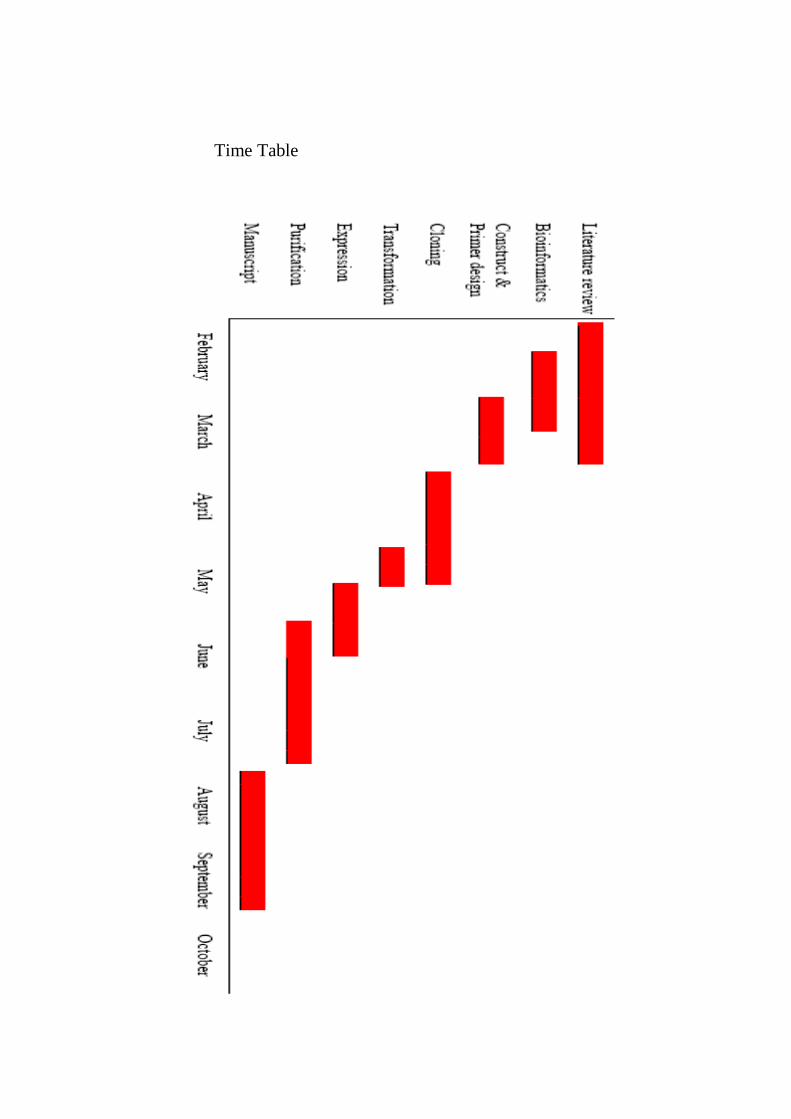
Time Table
20
Conclusions
Study of cellulose synthesis has been one of the most interesting fields in bioscience
industry today due to its promising involvement not only in agriculture but also in bio-
ethanol production and a number of other industries.
My research project will be focusing on the functional analysis of HvCesA4 enzyme and
will contribute to a better understanding of cellulose biosynthesis in barley. The results
will define the importance of specific HvCesA4protein motifs, which are found to be
conserved in the cellulose synthase gene family. A successfully expressed and purified
HvCesA4 protein will help in determining two- and/or three-dimensional structure of the
enzyme. Further, production of highly purified protein will be useful in production of
antibodies that could determine sub-cellular localization of the HvCesA4 protein by
immuno-histochemistry and electron microscopy. Finally, the findings would help in
characterizing the biochemical and biophysical properties of the HvCesA4 protein.
21
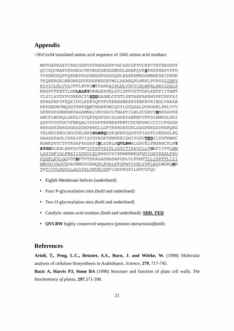
Appendix
>HvCesA4 translated amino acid sequence of 1042 amino acid residues: MDTGEPKAKVCRACGDDVGTREDGSPFVACAECGFPVCRPCYEYERSDGT
QCCPQCNARYKRHKGCPRVEGDDEDGDMDDLEEEFQVKSPKKPHEPVPFD
VYSENGEQPPQKWRPGGPAMSSFGGSGQELEAEREMEGSMEWKDRIDKWK
TKQEKRGKLNRDNSDDDDDDKNDDEYMLLAEARQPLWRKLPVPSSQINPY
RIVIVLRLVVLCFFLRFRIMTPANDAIPLWLVSVICELWFALSWILDQLP
KWSPVTRETYLDRLALRYDREGEPSRLSPIDFFVSTVDPLKEPPIITANT
VLSILAVDYPVDRNSCYVSDDGASMLCFDTLSETAEFARRWVPFCKKFAI
EPRAPEFYFSQKIDYLKDKVQPTFVKERRAMKREYEEFKVRINGLVAKAE
KKPEEGWVMQDGTPWPGNNTRDHPGMIQVYLGSQGALDVEGHELPRLVYV
SREKRPGHNHHKKAGAMNALVRVSAVLTNAPFILNLDCDHYVNNSKAVRE
AMCFLMDPQLGKKLCYVQFPQGFDGIDLHDRYANRNVVFFDINMKGLDGI
QGPVYVGTGCVFNRQALYGYDPPRPEKRPKMTCDCWPSWCCCCCCFGGGK
HRKSSKDKKGGGGGDDEPRRGLLGFYKKRGKKDKLGGGPKKGSYRKRQRG
YELEEIEEGIEGYDELERSSLMSQKSFQKRFGQSPVFIASTLVEDGGLPQ
GAAADPAGLIKEAIHVISCGYEGKTEWGKEIGWIYGSVTEDILTGFKMHC
RGWKSVYCTPTRPAFKGSAPINLSDRLHQVLRWALGSVEIFMSRHCPLWY
AYGGRLKWLERFAYTNTIVYPFTSIPLIAYCTIPAVCLLTGKFIIPTLNN
LASIWFIALFMSIIATGVLELRWSGVSIEDWWRNEQFWVIGGVSAHLFAV
FQGFLKVLGGVDTNFTVTSKAGADEADAFGDLYLFKWTTLLIPPTTLIII
NMVGIVAGVSDAVNNGYGSWGPLFGKLFFSFWVIVHLYPFLKGLMGRQNR
TPTIVVLWSVLLASIFSLVWVRIDPFIAKPKGPILKPCGVQC
• Eighth Membrane helices (underlined)
• Four N-glycosylation sites (bold and underlined)
• Two O-glycosylation sites (bold and underlined)
• Catalytic amino acid residues (bold and underlined): SDD, TED
• QVLRW highly conserved sequence (protein interactions)(bold)
References
Arioli, T., Peng, L.C., Betzner, A.S., Burn, J. and Wittke, W. (1998) Molecular
analysis of cellulose biosynthesis in Arabidopsis. Science, 279, 717-745.
Bacic A, Harris PJ, Stone BA (1998) Structure and function of plant cell walls. The
biochemistry of plants, 297,371-398.

22
Brett, C.T. (2000) Cellulose microfibrils in plants: biosynthesis, deposition, and
integration into the cell wall. Int. Rev. Cytol, 199, 161-189.
Brown, R.M. (2004) Cellulose structure and biosynthesis: What is in store for the 21st
century. J. Polymer. Sci. A, 42, 487-511.
Burton, R.A., Shirley, N.J., King, B.J., Harvey, A.J. and Fincher, G.B. (2004) The
CesA Gene Family of Barley. Quantitative Analysis of Transcripts Reveals Two Groups
of Co-Expressed Genes. Plant Physiol., 134, 224-236.
Carpita, N.C. and Gibeaut, D.M. (1993) Structural models of primary cell walls in
flowering plants: consistency of molecular structure with the physical properties of the
walls during growth. The Plant Journal, 3, 1-30.
Coutinho, P. and Henrissat, B. (1999). Carbohydrate-active enzymes: an integrated database approach. Royal Society of Chemistry.,. 3–12. Delmer, D.P. (1999) Cellulose biosynthesis: exciting times for a difficult field of study.
Annu. Rev. Plant Physiol. Plant Mol. Biol., 50, 245-265.
Doblin, M.S., Kurek, I., Jacob-Wilk, D. and Delmer, D.P. (2002) Cellulose
Biosynthesis in Plants: from Genes to Rosettes. Plant Cell Physiol., 43, 1407-1420.
Finaev, D. (2007) Some aspects of cellulose biosynthesis. Biologia Plantarum, 51, 407-
413.
Herth, W. (1983) Arrays of plasma membrane rosettes in cellulose microfibril formation
of Spirogyra. Planta, 159, 347-360.
Kimura, S., Laosinchai, W., Itoh, T., Cui, X.J., Linder, C.R. and Brown, R.M. (1999)
Immunogold labeling of rosette terminal cellulose-synthesizing complexes in the vascular
plant Vigna angularis. Plant Cell, 11, 2075-2087.
Lai-Kee-Him, J., Chanzy. H., Muller. M, Putaux, J.L., Imai, T. and Bulone, V.
(2002) In vitro versus in vivo cellulose microfibrils from plant primary wall synthases:
structural differences. J. Biol. Chem., 277, 36931-36955.
Mueller. S. C, Brown RM Jr (1980) Evidence for an intramembranous component
associated
with a cellulose microfibril synthesizing complex in higher plants. J Cell Biol 84: 315–
326.
Nagahashi, S., Sudoh, M., Ono, N., Sawada, R. and Yamaguchi, E. (1995)
Characterization of chitin synthase 2 of Saccharomyces cerevisiae: implication of two

23
highly conserved domains as possible catalytic sites. J. Biol. Chem., 270, 13961-13977.
Pear, J.R., Kawagoe, Y., Schreckengost, W.E., Delmer, D.P. and Stalker, D.M. (1996)
Higher plants contain homologs of the bacterial celA genes encoding the catalytic subunit
of cellulose synthase. PNAS, 93, 12637-12642.
Roelofsen A (1958) Cell wall structure as related to surface growth. Acta Bot Neerl, 7,
77–89.
Richmond, T. (2000) Higher plant cellulose synthases. Genome Biol., 4, 30011-30033.
Somerville C (2006) Cellulose Synthesis in Higher Plants. Annual Review of Cell and
Developmental Biology, 22, 53-78
Spyropoulos, I.C., Liakopoulos, T.D., Bagos, P.G. and Hamodrakas, S.J. (2004)
TMRPres2D: high quality visual representation of transmembrane protein models.
Bioinformatics, 20, 3258-3260.
Tusnady, G.E. and Simon, I. (2001) The HMMTOP transmembrane topology
prediction server. Bioinformatics, 17, 849-850.
World Wide Web sites:
NCBI Nucleotide database: http://www.ncbi.nlm.nih.gov/ Expasy prediction: http://expasy.org/ HMMTOP prediction: http://www.enzim.hu/hmmtop/html/submit.html NetNgly prediction: http://www.cbs.dtu.dk/services/NetNGlyc/ PSIPRED prediction: http://bioinf.cs.ucl.ac.uk/psipred/psiform.html TopPred prediction: http://bioweb.pasteur.fr/seqanal/interfaces/toppred.html SOSUI prediction: http://bp.nuap.nagoya-u.ac.jp/sosui/sosui_submit.html

24
Acknowledgements
I thank Associate Professor Maria Hrmova for supervision and Professor Geoffrey B.
Fincher for being a co-supervisor and for giving me the opportunity to work on the
project in his laboratory. I also thank Dr. Andrew J. Harvey for his guidance and for being
my internal examiner. Finally, I thank Professor Tony Bacic for being my external
examiner. I would also like to extend my thanks to all the ACPFG colleagues for their
support.

25
PART II- THE MANUSCRIPT
Summary
Cellulose is the most abundant polysaccharide polymer on earth that contributes
significantly to the structure and development of flora. A part of the biosynthesis of
cellulose has been tracked to two gene families, namely cellulose synthase (CesA) and
cellulose synthase like (Csl) gene families. CesA genes are known to encode a catalytic
subunit of cellulose synthase. It has been suggested that cellulose synthase forms a
protein complex, which is believed to be located in the plasma membrane of plant cells
and to contribute to the production of cellulosic micro-fibrils. These micro-fibrils co-
crystallize to form cellulose. Up to now, nine CesA genes have been assigned in barley,
which are designated HvCesA1 to HvCesA9 (Rachel Burton unpublished data). Out of
these nine genes, it was found that HvCesA4, HvCesA7 and HvCesA8 were expressed
abundantly during stem and root development in barley. To our knowledge, none of these
protein sequences have so far expressed heterologously. We believe that heterologous
expression of these proteins would be beneficial to our understanding of protein
properties, and to structural studies that would lead to our better understanding of
cellulose synthesis. In this study four constructs of the HvCesA4 isoenzyme from barley
have been cloned into the pPICZ vector system. In addition, protein expression of
HvCesA4 constructs was attempted in a heterologous host of Pichia pastoris.
Introduction
The majority of plant cell walls contain cellulose as a major structural component, where
cellulosic microfibrils with xyloglucan represent a major load-bearing components (Pear

26
et al., 1996). Cellulose is the most abundant polysaccharide in the biosphere, and is
involved in key cellular and physiological processes of plants, but the detailed
mechanism of its biosyntheses at molecular level is not understood. Cellulose is a linear
β-D-glucan polymer composed of β-linked glucosyl residues, and linkage configuration
between the β-glucosyl residues in cellulose has decisive effects on its conformation and
hence biological function (Stone et al., 2008). Cellulose synthase occurs in large protein
families with more than 9,000 members and are classified in the glycosyl transferase GT2
group of enzymes (CAZy; http://www.cazy.org/fam/acc_GH.html; Coutinho and
Henrissat, 1999). Their cDNAs, e.g. barley cellulose synthase HvCesA4, are translated
into proteins with 1082 amino acid residues (Burton et al., 2004). The topology
predictions show 7-8 membrane-spanning helices positioned at HvCesA4 NH2- and
COOH-termini, while single large cytoplasmic regions encompass the catalytic module.
CesA genes were found to be involved in synthesis of polysaccharides that form primary
and secondary cell walls (Arioli et al., 1998; Fagard et al., 2000; Burn et al., 2002;
Somerville, 2006). It has been shown in Arabidopsis that a CesA gene knockout mutant
showed cellulose deficiency (Arioli et al., 1998; Taylor et al., 1999; Fagard et al., 2000;
Taylor et al., 2000; Scheible et al., 2001; Beeckman et al., 2002; Burn et al., 2002;
Gardiner et al., 2003; Taylor et al., 2003). Out of eight CesA gene so far identified in
barley (Burton et al., 2004), few have been studied.
The plant glucan synthases are predicted to form oligomeric assemblies in the plasma
membrane (hexameric rosettes) (Somerville, 2006), and when purified, they are rapidly

27
inactivated (Brown, 1996; Kimura et al., 1999; Lai-Kee-Him et al., 2002). The only
known three-dimensional (3D) structure of a GT2 enzyme is for a 256-residue spore-coat
protein from Bacillus subtilis; this offers insights into the geometry of the active site of
these GTs (Charnock and Davies, 1999), but the function of the protein is unknown.
The focus of this project is on cellulose synthase that mediates synthesis of cellulose or
(1,4)-β-D-glucan, which represents the key component of plant cell walls of economically
important cereals and grasses (Stone et al., 2008). In this project effort was put towards
constructing expression vectors containing cDNA sequence of HvCesA4 and towards
expressing some of these proteins heterologously. It has been reported that CesA genes
have unique and conserved motifs such as QXRLW and TED (Burton et al.,
2004(Holland et al., 2000). The HvCesA4 isoenzyme also contains QVLRW and TED
motifs in its catalytic region. For the better understanding of the HvCesA4 protein
structure, we have conducted in-silico protein sequence investigations, before attempting
expressing the protein in Pichia pastoris. In summary, three types of vectors have been
successfully constructed and tested for protein expression
Results
Prediction of membrane helices and catalytic module of HvCesA4
Several web-based applications were used for predicting the topology, secondary
structure dispositions and locations of the membrane helices in the HvCesA4 protein.
These tools revealed that several locations of these helices were possible. In the end the
consensus was reached that HvCesA4 contains eight membrane helices (Fig. 1). The N-

28
terminus of HvCesA4 was identified to be located on the intracellular side of the plasma
membrane, from where the protein sequence traverses the membrane to extra-cellular
space, before the sequence enters back to the cytoplasm and forms a large catalytic region
(Fig. 1). According to the topology prediction server TopPred, two membrane helices
were found at the N-terminus, while six membrane helices were found at the C-terminus
(Fig. 1). A 570 amino acid residues long catalytic region was predicted to be located
inside the cytoplasm, which contains TED, LALRY and QVLRW conserved motifs. The
eight membrane helices were predicted between 197-216, 226-248, 818-840, 849-871,
888-910, 938-960, 971-993, and 1003-1025 amino acid residues. Surprisingly only four
N-glycosilation site and two O-glycosylation sites were predicted in the sequence. It is of
interest that various protein topology and secondary structure prediction servers revealed
slightly different possibilities for positions of membrane helices (data not shown).
However, the consensus from these predictions supports the view that HvCesA4 contains
8 membrane helices (SOSUI server at http://bp.nuap.nagoya-
u.ac.jp/sosui/sosui_submit.html).
Cloning into Entry Vector Systems
Ligation was carried out with four constructs of HvCesA4. As an entry vector, primarily
pGEM-T vector system was used throughout. However, when the clones were
transformed into the DH5α competent cells, no colonies were observed. For this reason,
the cloning procedure was repeated with pCR8-TOPO vector system. In this case, the
colonies were observed, which were screened to be positive by colony PCR screening
technique.
29
Cloning into destination vector system
The three of four constructs (constructs 2-4) were successfully ligated into the destination
pPICZ vector system that was in-frame with the yeast α-factor secretion sequence and
under the transcriptional control of the alcohol oxidase promoter AOX1. All three
constructs were sequenced in both directions and have not contained any errors in their
open reading frames.
Protein expression
Protein expression was attempted with the three HvCesA4 constructs (constructs 2-4) in
Pichia pastoris SMD1168H cells and the expression was monitored by SDS-PAGE and
Western blot analyses. The preliminary experiment revealed that catalytic module of
HvCesA4 with flanking helices has been successfully expressed, where 70 kDa His-
tagged protein was clearly visible (Fig.2).
As for the expression of the two truncated sequences (constructs 2 and 3) corresponding
to the two versions of the catalytic module of HvCesA4, the dot blot analyses have not
revealed the presence of these proteins in soluble forms in the cultivation media (data
shown in supplementary material). The most likely explanation for this observation is the
fact that protein concentration might have been low for SDS-PAGE and Western blot
analyses.
30
Discussion
It has been concluded that at least three barley genes, encoding cellulose-synthases, are
coordinately expressed out of nine barley HvCesA genes, during cellulose synthesis
(Appenzeller et al., 2004; Burton et al., 2004). It has been suggested that at least three
different genes are essential for making a planar rosette complex that typically contains
up to 30-36 subunits (Perrin, 2001; Scheible et al., 2001). The studies have also shown
that HvCesA1, HvCesA2 and HvCesA6 isoenzymes are predominantly and coordinately
transcribed mostly in young tissues (Burton et al., 2004). Another group of HvCesAs
contains HvCesA4, HvCesA7 and HvCesA8 isoenzymes and these were found to be
coordinately transcribed in the mature tissues. With this information it could be
concluded that the group containing HvCesA1, HvCesA2 and HvCesA6 is most likely to
contribute for primary cell wall synthesis and the second group containing HvCesA4,
HvCesA7 and HvCesA8, is likely to take part in secondary cell wall synthesis. For this
reason, as outlined in previous sections of my thesis, my aim was to generate DNA
vectors of HvCesA genes that are involved in secondary cell wall formation, in particular
of the HvCesA8 isoform.
Bioinformatical analysis of the 1042-amino acid residue long HvCesA4 protein sequence
(Figure 1; this Figure should be the sequence of HvCesA4) has shown that the full-length
protein contains eighth membrane helices (Figure 2) (and a long catalytic centre region,
which was about 570 amino acid residue long. A highly conserved motif QVLRW was
found within this catalytic region, alongside with four N-glycosylation sites and two O-
glycosylation sites (Figure 1). Apart of catalytic motifs NED and SDD (Figure 1), all the

31
above HvCesA genes carry a common sequence such as QXXLRW motif, which is
highly conserved in plant cellulose synthases (Pear et al., 1996; Delmer, 1999). It is of
interest that the QXXLRW motif is also known to be associated with bacterial cellulose
synthases and other processive glycosyltransferases such as chitin and hyaluronan
synthases (Marks et al., 2001). Thus, we were interested in constructing expression
vectors encoding soluble catalytic modules of HvCesA4 with and without the QXXLRW
motifs, in addition to constructing vectors containing its full-length sequence and the
sequences of the catalytic module with flanking helices on each side of the catalytic
module.
Due to a high GC content of the HvCesA4 sequence, the primer design and amplification
of DNA by PCR proved to be challenging. The amounts of PCR products obtained
specific primers and HvCesA4 template were low and these amounts were not sufficient
for further cloning steps. To overcome these problem three new primers were designed
for a full-length sequence of HvCesA4 with longer priming sites that would avoid GC
rich segments in the sequence. Using this approach, all four HvCesA4 constructs were
successfully cloned into the entry vector pCR8. An attempt was also made to clone the
four HvCesA4 constructs into the entry vector pGEM-Teasy, however they were
unsuccessful. Further cloning of DNA sequences into the destination vector pPICZ was
carried out successfully for three HvCesA4 constructs containing truncated sequences.
The cloning of the full-length sequence of HvCesA4 into pPICZ was unsuccessful. The
3164 bp long sequence of HvCesA4 could not be cloned into pPICZ vector for a variety
32
of reasons, such as e.g. forming secondary DNA structures that could occlude a steric
availability of restriction sites.
Pichia pastoris expression system was chosen over the E.coli expression system for
expressing the HvCesA4 proteins, because the HvCesA4 sequence showed the presence
of several N- and O-glycosylation sites, as specified above. As an eukaryote, Pichia
pastoris possesses many advantages of higher eukaryotic expression systems, such as
protein processing, protein folding, and the availability of post-translational
modifications, while it could not be manipulated as easily as the E. coli expression
system. Due to all these advantages, Pichia pastoris expression system was chosen over
the E.coli one. From the three successfully cloned sequences, HvCesA4_T1, a truncated
construct containing the central catalytic module flanked by two membrane helices on
each side, was chosen to be tested for protein expression. The HvCesA4-pPICZ vector
was transformed into the Pichia cells and successfully expressed. A 70 kDa His-tagged
protein was observed by Western analysis by blotting the protein produced by the
transformed Pichia cells with His-tag antibodies. Based on this detection it can be
concluded that the expressed protein was encoded by HvCesA4_T1 construct carrying
the 6x-His epitope. On the other hand, no expressed protein was observed with the two
other successfully cloned constructs. Various reasons were implicated including probable
contamination of Pichia cell cultures with bacteria. Due to the lack of time, the
experiments could not be repeated hence further conclusions cannot be drawn. Yet, these
experiments back up the idea of successful use of Pichia expression system for
production the HvCesA proteins in general. It is very likely that all the constructs
33
designed for HvCesA4 production could be expressed successfully in Pichia pastoris
expression system.
As for the future prospects of these studies to investigate HvCes4 proteins, the
heterologus expression of the above gene followed by purification and characterization
could yield significant data for understanding of secondary cell wall synthesis and stem
strength in barley. This project has taken us a step closer in the above process, where the
three constructs of HvCesA4 were cloned into pPICZ vector system for expression in
Pichia pastoris. In the future, the full-length sequence of HvCesA4 needs to be cloned
into pPICZ vector using different techniques such as a direct PCR product cloning into
destination vectors. It is projected that successful expressions of these HvCesA4 proteins
should be followed be purification of the proteins and subjecting them These to activity
assays, followed by complete bio-chemical, bio-physical and structural studies needs
Experimental Procedures
Bioinformatics and predictions of membrane helices
Barley HvCesA4 cDNA (GenBank accession number AY483154) ((Burton et al., 2004)
was retrieved from the NCBI database (http://www.ncbi.nlm.nih.gov/). HvCesA4 cDNA
was translated and the amino acid sequence was fed into the following protein secondary
structure and topology prediction web-based applications (Expasy http://expasy.org/;
HMMTOP http://www.enzim.hu/hmmtop/html/submit.html; NetNgly -
ttp://www.cbs.dtu.dk/services/NetNGlyc/;PSIPRED,http://bioinf.cs.ucl.ac.uk/psipred/psif
orm.html, TopPred, http://bioweb.pasteur.fr/seqanal/interfaces/toppred.html, SOSUI,
http://bp.nuap.nagoyau.ac.jp/sosui/sosui_submit.html). Based on the bioinformatical

34
analyses, the four constructs were designed and designated as HvCesA4 constructs 1-4.
The first construct (HvCesA4 construct 1) represents the full-length sequence. The second
construct represents the catalytic subunit and two flanking membrane helices on each side
(HvCesA4 construct 2). The third construct (HvCesA4 construct 3) contained the central
catalytic subunit with QVLRW motif. The forth construct (HvCesA4 construct 4) is the
shortest of all, and contained the partial catalytic module excluding the QVLRW motif.
Molecular cloning and primers for HvCesA4 constructs
Initially seven primers were designed based on the HvCesA4 protein topology and
membrane helical predictions. For all the constructs, the primers with restriction sites,
yeast consensus sequence and poly-histidine sequences were designed. For all 5’ primers
EcoR1 restriction digestion sites were chosen and for all 3’ primers Xba1 restriction sites
were incorporated. The sequences of the primers are as follows.
HvCesA4 construct 1:
Forward primer GTCAGAATTCAAACATCATCATCATCATCATGAAAATCTGTACTTTCAAGGTGACACCGGCGAGCCCAAGGCC
Reverse primer GCCAAGCCCAAGGGACCCATTCTTAAGCCGTGTGGAGTACAGTGCTGAGGATAATCTAGA
HvCesA4 construct 2:
Forward primer AGTCGAATTCAAAATGGGTCATCATCATCATCATCATGAAAATCTGTACTTTCAAGGTACCCCGGCCAACGACGCCATC
Reverse primer GGGGTGCTGGAGCTGCGGTGGAGCGGGGTGAGCTGAGGATAATCTAGA
35
For HvCesA4 construct 3 and 4, primers were designed with restriction sites EcoR1 and
Xba1 on 5’ and 3’ ends respectively, where also a TEV (Tobacco Etched Virus) site was
added at the 3’ end.
HvCesA4 constructs 3 and 4:
Forward primer CCGGAATTCAGACTCGCCCTGCGCTACGACCGCGAG
HvCesA4 construct 3:
Reverse primer TCTGGTACGCCTACGGCGGCCACCTTGAAAGTACAGATTTTCTCTAGA
HvCesA4 construct 4:
Reverse primer CGCTCCTCGCTCATGTCACAGACCTTGAAAGTACAGATTTTCTCTAGAAGG
Due to high GC content in the primers designed for HvCesA4 construct 1, two additional
forward primers and one additional reverse primer were designed.
Forward primer 2 GAATTCAAACATCATCATCATCATCATGAAAATCTGTACTTTCAAGGTGACACCGGCGAGCCCAA
Forward primer 3 GAATTCAAACATCATCATCATCATCATGAAAATCTGTACTTTCAAGGTGAGCCCAAGGCCAAGGT
Reverse primer 2 ATTCTTAAGCCGTGTGGAGTACAGTGCTGAGGATAATCTAGA.
36
The expected size of PCR products with the primers for HvCesA4_FL is 3162bp,
HvCesA4_T1 is 2044bp, HvCesA4_T2 is 1669bp and for HvCesA4_T3 is 1273bp.
Cloning into entry vector
The constructs are produced and amplified with respective primers using full length
HvCesA4 (kindly donated by Dr. Rachel Burton).The sticky end PCR products of the four
constructs HvCesA4 (constructs 1-4) were purified using nucleospin extract II kit and the
fresh DNA were ligated into pCR8/GW-TOPo vector (Invitrogen), which served as an
entry vector. Cloned vector was transformed into One Shot® Mach1™ T1 phage-
resistant chemically competent E. coli cells (Invitrogen). Antibiotic zeocin was used for
clone selection at 100 µg/ml. Successfully transformed colonies were picked and grown
overnight on Luria-Bretani medium along with the zeocin at 30 µg/ml. DNA was
extracted from the cells using alkaline lysis extraction method. Extracted DNA was
subjected to digestion with Xba1 and EcoR1 restriction enzymes. The constructs from the
entry vector pCR8 were gel purified and stored at -20 C for further cloning into the
destination vector.
Preparation of destination vectors
Glycerol stocks of E. coli cells containing the destination vector pPICZB and pPICZαA
were obtained from Mrs. Margaret Buchanan. The cells were inoculated into the Luria-
Bertani agar media plates with zeocin at 30 µg/ml. The plates were incubated overnight at
37º C. The positive colonies are inoculated on to LB medium with Zeocin (30µg/ml).
Cell culture is incubated overnight at 37ºC. Plasmids from the cell suspensions were
37
extracted using alkaline lysis DNA-extraction method. The plasmids were subjected to
restriction digestion with Xba1 and EcoR1 restriction enzymes. The digested products
were run on 1% (w/v) agarose gels and the small fragments released by restriction
digestion were eliminated from the vectors. Linearised vectors were cut off the gels,
purified by Nucleo-spin DNA purification kit (Macherey-Nagel) and stored at -20º C.
Cloning into destination vectors
The four constructs with EcoR1 restriction sites on 5’end and Xba1 restriction sites on 3’
end were ligated into the destination pPICZ vector that was in-frame with the yeast α-
factor secretion sequence and under the transcriptional control of the alcohol oxidase
promoter AOX1. Two different variants of pPICZ were used. Constructs 1 and 2 and
were cloned on to pPICZB, while constructs 3 and 4 were ligated into pPICZαA. The
cloned vectors were transformed into One Shot® Mach1™ T1 phage-resistant chemically
competent E. coli cells (Invitrogen) as recommended by in the manufacturer (EasySelect
™ Pichia Expression Kit, version G). The transformed cells were incubated overnight at
37 C. DNA was extracted from the E. coli cells using alkaline lysis method.
DNA sequencing
All constructs were sequenced using AOX1 forward and reverse primers (EasySelect ™
Pichia Expression Kit, version G), and by using HvCesA4 gene specific internal primers
kindly provided by Dr. Rachel Burton.

38
Transformation into Pichia pastoris
About 10 ml of competent Pichia pastoris cells were prepared using the protocol from
the EasySelect ™ Pichia Expression Kit version G. The HvCesA4 constructs 2-4 were
linearised with the restriction enzyme Pme1. The enzyme mixture was heat inactivated at
60º C for 20min. The linearised plasmids were purified using NucleoSpin® Extract II Kit
from Macherey-Nagel kit. About 5-10 µg of the purified DNA was used for
transformation of Pichia pastoris SMD1168H competent cells, using electroporation
technique. Approximately 80 µl of Pichia pastoris SMD1168H competent cells were
deposited in a 0.2cm electroporation cell and 10 µl of purified and linearised plasmid was
added. The electroporation was carried out with 1500 V, 400Ω and 25µF. The
transformed mixture was platted onto the YPD media with zeocin antibiotic at 100 µg/ml.
Plates were incubated at 28ºC for 4-6 days, after which colonies were observed.
Protein expression
Successfully transformed Pichia pastoris cells were inoculated into the BMGY media
after 4-6 days of growth on YPD media at 28ºC. Cells were incubated at 28ºC overnight.
Cells were washed by centrifugation (1500g, 10 min) and transferred to the BMMY
medium, where they were incubated for 5 days, during which 0.5% (v/v) methanol
concentration was maintained. The cells were incubated with constant shaking at 220
rpm, and harvested after 5 days of growth.

39
Protein extraction, dot blot, Western blot and SDS-PAGE analyses
After 5 days of incubation of cell expressing construct 1, proteins were extracted from the
cell suspension using alkaline lysis method (Tobais von der Haar, 2007) and fractionated
into three fractions. The samples were spotted onto a nitrocellulose membrane, the
membrane was blocked with non-fat milk for 3 hours, washed with phosphate buffered
saline and again blocked with 1:2000 dilution of monoclonal anti-polyhistidine, alkaline
phosphatase conjugate clone His-1 antibodies overnight at 4 oC. The following day the
nitrocellulose membrane was developed with BCIP\NBT reagent. For the constructs 3
and 4, the cell cultures after 5 days of incubation were spun (1500g, 10 min) and the
culture media were analysed for the protein expression. SDS-PAGE analyses proceeded
according to the standard protocol (Hrmova et al., 1996)
Acknowledgements
I thank Associate Professor Maria Hrmova for supervision and Professor Geoffrey B.
Fincher for giving me the opportunity to work on the project in his laboratory. I also
thank Dr. Andrew J. Harvey for being my internal examiner and to Professor Tony Bacic
for being my external examiner. I would also like to extend my thanks to Dr. Rachel
Burton, Dr. Andrew Jacob for their support. Also I thank Ms. Jessica Smith, Ms.Maria
Lombardi and Mrs. Jodie Kretschmer Jodie and all my colleagues at Plant Genomics
Centre for their support.

40
References
Appenzeller L, Doblin M, Barreiro R, Wang HY, Niu XM (2004) Cellulose synthesis in maize: isolation and expression analysis of the cellulose synthase (CesA) gene family. Cellulose 11: 287
Arioli T, Peng LC, Betzner AS, Burn J, Wittke W (1998) Molecular analysis of cellulose biosynthesis in Arabidopsis. Science 279: 717
Beeckman T, Przemeck GKH, Stamatiou G, Lau R, Terryn N (2002) Genetic complexity of cellulose synthase A gene function in Arabidopsis embryogenesis. Plant Physiol. 130: 1883
Brett CT (2000) Cellulose microfibrils in plants: biosynthesis, deposition, and integration into the cell wall. Int. Rev. Cytol 199: 161
Brown RM (1996) The biosynthesis of cellulose. J. Macromol. Sci. A 33: 1345 Brown RM (2004) Cellulose structure and biosynthesis: What is in store for the 21st
century. J. Polymer. Sci. A 42: 487 Burn JE, Hurley UA, Birch RJ, Arioli T, Cork A, Williamson RE (2002) The
cellulose-deficient Arabidopsis mutant rsw3 is defective in a gene encoding a putative glucosidase II, an enzyme processing N-glycans during ER quality control. Plant J. 32: 949
Burton RA, Shirley NJ, King BJ, Harvey AJ, Fincher GB (2004) The CesA Gene Family of Barley. Quantitative Analysis of Transcripts Reveals Two Groups of Co-Expressed Genes. Plant Physiol. 134: 224-236
Burton RA, Shirley NJ, King BJ, Harvey AJ, Fincher GB (2004) The cesa gene family of barley. Quantitative analysis of transcripts reveals two groups of co-expressed genes. Plant Physiol. 134: 224
Carpita NC, Gibeaut DM (1993) Structural models of primary cell walls in flowering plants: consistency of molecular structure with the physical properties of the walls during growth. The Plant Journal 3: 1-30
Delmer DP (1999) Cellulose biosynthesis: exciting times for a difficult field of study. Annu. Rev. Plant Physiol. Plant Mol. Biol. 50: 245
Doblin MS, Kurek I, Jacob-Wilk D, Delmer DP (2002) Cellulose Biosynthesis in Plants: from Genes to Rosettes. Plant Cell Physiol. 43: 1407-1420
Fagard M, Desnos T, Desprez T, Goubet F, Refregier G (2000) PROCUSTE1 encodes a cellulose synthase required for normal cell elongation specifically in roots and dark-grown hypocotyls of Arabidopsis. Plant Cell 12: 2409
Finaev D (2007) Some aspects of cellulose biosynthesis. Biologia Plantarum 51: 407-413 Gardiner JC, Taylor NG, Turner SR (2003) Control of cellulose synthase complex
localization in developing xylem. Plant Cell 15: 1740 Herth W (1983) Arrays of plasma membrane rosettes in cellulose microfibril formation
of Spirogyra. Planta 159: 347 Holland N, Holland D, Helentjaris T, Dhugga KS, Xoconostle-Cazares B, Delmer
DP (2000) A Comparative Analysis of the Plant Cellulose Synthase (CesA) Gene Family. Plant Physiol. 123: 1313-1324
Kimura S, Laosinchai W, Itoh T, Cui XJ, Linder CR, Brown RM (1999) Immunogold labeling of rosette terminal cellulose-synthesizing complexes in the vascular plant Vigna angularis. Plant Cell 11: 2075

41
Lai-Kee-Him J, Chanzy H, Muller M, Putaux JL, Imai T, Bulone V (2002) In vitro versus in vivo cellulose microfibrils from plant primary wall synthases: structural differences. J. Biol. Chem. 277: 36931
Marks DL, Dominguez M, Wu KJ, Pagano RE (2001) Identification of active site residues in glucosylceramide synthase: a nucleotide-binding/catalytic motif conserved with processive beta-glycosyltransferases. J. Biol. Chem. 276: 26492
Nagahashi S, Sudoh M, Ono N, Sawada R, Yamaguchi E (1995) Characterization of chitin synthase 2 of Saccharomyces cerevisiae: implication of two highly conserved domains as possible catalytic sites. J. Biol. Chem. 270: 13961
Pear JR, Kawagoe Y, Schreckengost WE, Delmer DP, Stalker DM (1996) Higher plants contain homologs of the bacterial celA genes encoding the catalytic subunit of cellulose synthase. Proc. Natl. Acad. Sci. USA 93: 12637
Pear JR, Kawagoe Y, Schreckengost WE, Delmer DP, Stalker DM (1996) Higher plants contain homologs of the bacterial celA genes encoding the catalytic subunit of cellulose synthase. PNAS 93: 12637-12642
Perrin RM (2001) Cellulose: how many cellulose synthases to make a plant. Curr. Biol. 11: R213
Scheible WR, Eshed R, Richmond T, Delmer D, Somerville CR (2001) Modifications of cellulose synthase confer resistance to isoxaben and thiazolidinone herbicides in Arabidopsis Ixr1 mutants. Proc. Natl. Acad. Sci. USA 98: 10079
Somerville C (2006) Cellulose Synthesis in Higher Plants. Annual Review of Cell and Developmental Biology 22: 53-78
Taylor NG, Howells RM, Huttly AK, Vickers K, Turner SR (2003) Interactions among three distinct CesA proteins essential for cellulose synthesis. Proc. Natl. Acad. Sci. U.S.A 100: 1450
Taylor NG, Laurie S, Turner SR (2000) Multiple cellulose synthase catalytic subunits are required for cellulose synthesis in Arabidopsis. Plant Cell 12: 2529
Taylor NG, Scheible WR, Cutler S, Somerville CR, Turner SR (1999) The irregular xylem3 locus of Arabidopsis encodes a cellulose synthase required for secondary cell wall synthesis. Plant Cell 11: 769

42
Table 1. SOSUI protein topology prediction of HvCesA4 protein sequence. The presence
of eight membrane helices is indicated, where the first two helices are positioned at the
N-terminus, while the next six helices are located at the C-terminus of the protein. The
average lengths of the membrane are 20-23 residues, which are ideal as anchor helices.
43
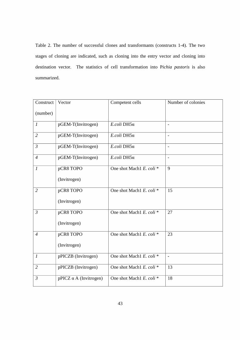
Table 2. The number of successful clones and transformants (constructs 1-4). The two
stages of cloning are indicated, such as cloning into the entry vector and cloning into
destination vector. The statistics of cell transformation into Pichia pastoris is also
summarized.
Construct
(number)
Vector Competent cells Number of colonies
1 pGEM-T(Invitrogen) E.coli DH5α -
2 pGEM-T(Invitrogen) E.coli DH5α -
3 pGEM-T(Invitrogen) E.coli DH5α -
4 pGEM-T(Invitrogen) E.coli DH5α -
1 pCR8 TOPO
(Invitrogen)
One shot Mach1 E. coli * 9
2 pCR8 TOPO
(Invitrogen)
One shot Mach1 E. coli * 15
3 pCR8 TOPO
(Invitrogen)
One shot Mach1 E. coli * 27
4 pCR8 TOPO
(Invitrogen)
One shot Mach1 E. coli * 23
1 pPICZB (Invitrogen) One shot Mach1 E. coli * -
2 pPICZB (Invitrogen) One shot Mach1 E. coli * 13
3 pPICZ α A (Invitrogen) One shot Mach1 E. coli * 18
44
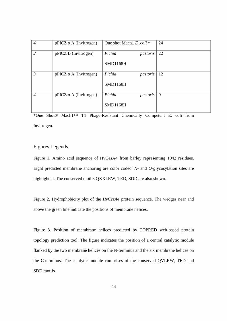
*One Shot® Mach1™ T1 Phage-Resistant Chemically Competent E. coli from
Invitrogen.
Figures Legends
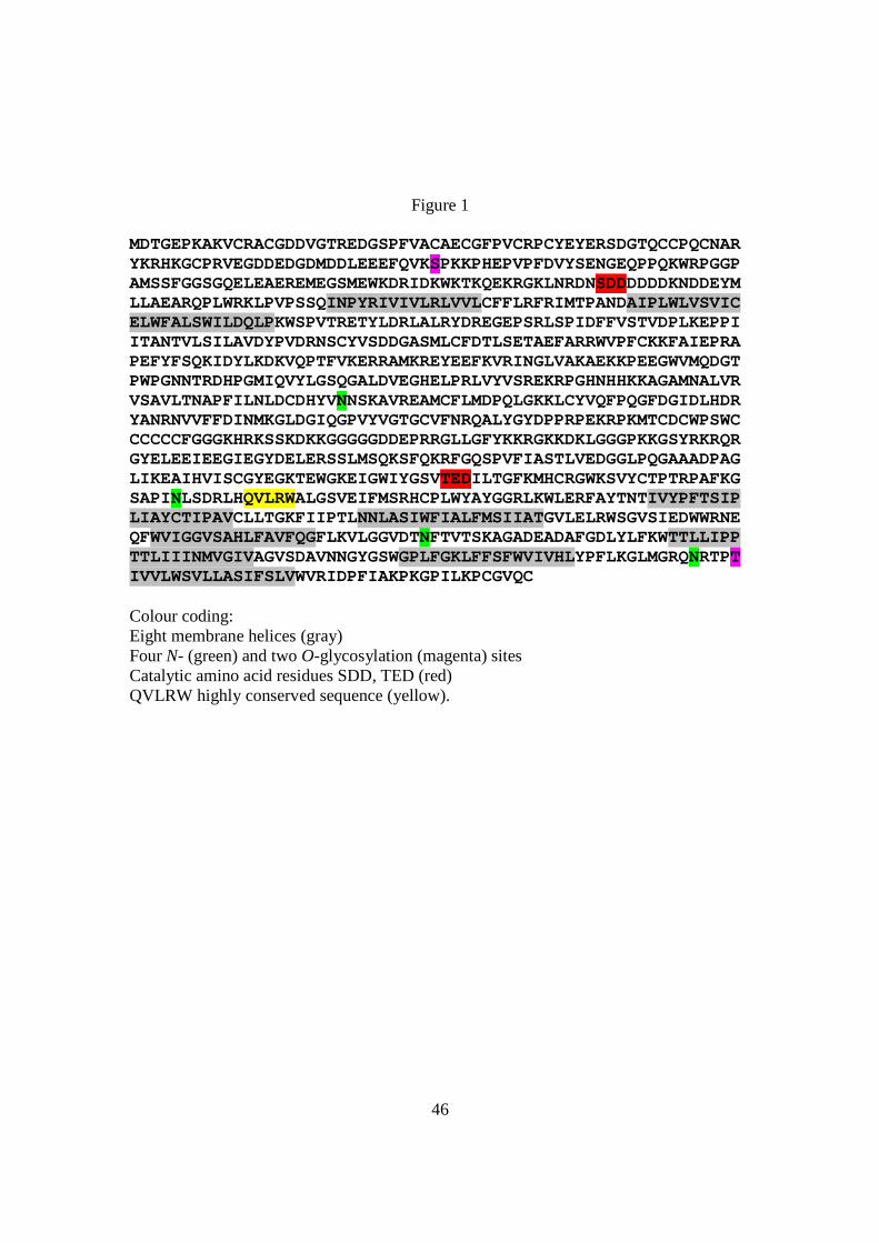
Figure 1. Amino acid sequence of HvCesA4 from barley representing 1042 residues.
Eight predicted membrane anchoring are color coded, N- and O-glycosylation sites are
highlighted. The conserved motifs QXXLRW, TED, SDD are also shown.
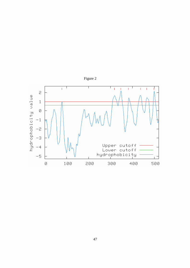
Figure 2. Hydrophobicity plot of the HvCesA4 protein sequence. The wedges near and
above the green line indicate the positions of membrane helices.
Figure 3. Position of membrane helices predicted by TOPRED web-based protein
topology prediction tool. The figure indicates the position of a central catalytic module
flanked by the two membrane helices on the N-terminus and the six membrane helices on
the C-terminus. The catalytic module comprises of the conserved QVLRW, TED and
SDD motifs.
4 pPICZ α A (Invitrogen) One shot Mach1 E .coli * 24
2 pPICZ B (Invitrogen) Pichia pastoris
SMD1168H
22
3 pPICZ α A (Invitrogen) Pichia pastoris
SMD1168H
12
4 pPICZ α A (Invitrogen) Pichia pastoris
SMD1168H
9
45
Figure 4. Western blot (left) and SDS-PAGE (right) analyses of construct 2 reveal the
presence of the 70 kDa His-tagged proteins that was expressed in Pichia pastoris cells.
Lanes 1 and 2 indicate protein that was isolated from colonies number 7 and 9 (fraction 3
after alkaline lysis). Lane 3 contains a crude total protein from the colony number 7. St
indicates protein standards ranging from 220 to 20 kDa.
46
Figure 1
MDTGEPKAKVCRACGDDVGTREDGSPFVACAECGFPVCRPCYEYERSDGTQCCPQCNARYKRHKGCPRVEGDDEDGDMDDLEEEFQVKSPKKPHEPVPFDVYSENGEQPPQKWRPGGPAMSSFGGSGQELEAEREMEGSMEWKDRIDKWKTKQEKRGKLNRDNSDDDDDDKNDDEYMLLAEARQPLWRKLPVPSSQINPYRIVIVLRLVVLCFFLRFRIMTPANDAIPLWLVSVICELWFALSWILDQLPKWSPVTRETYLDRLALRYDREGEPSRLSPIDFFVSTVDPLKEPPIITANTVLSILAVDYPVDRNSCYVSDDGASMLCFDTLSETAEFARRWVPFCKKFAIEPRAPEFYFSQKIDYLKDKVQPTFVKERRAMKREYEEFKVRINGLVAKAEKKPEEGWVMQDGTPWPGNNTRDHPGMIQVYLGSQGALDVEGHELPRLVYVSREKRPGHNHHKKAGAMNALVRVSAVLTNAPFILNLDCDHYVNNSKAVREAMCFLMDPQLGKKLCYVQFPQGFDGIDLHDRYANRNVVFFDINMKGLDGIQGPVYVGTGCVFNRQALYGYDPPRPEKRPKMTCDCWPSWCCCCCCFGGGKHRKSSKDKKGGGGGDDEPRRGLLGFYKKRGKKDKLGGGPKKGSYRKRQRGYELEEIEEGIEGYDELERSSLMSQKSFQKRFGQSPVFIASTLVEDGGLPQGAAADPAGLIKEAIHVISCGYEGKTEWGKEIGWIYGSVTEDILTGFKMHCRGWKSVYCTPTRPAFKGSAPINLSDRLHQVLRWALGSVEIFMSRHCPLWYAYGGRLKWLERFAYTNTIVYPFTSIPLIAYCTIPAVCLLTGKFIIPTLNNLASIWFIALFMSIIATGVLELRWSGVSIEDWWRNEQFWVIGGVSAHLFAVFQGFLKVLGGVDTNFTVTSKAGADEADAFGDLYLFKWTTLLIPPTTLIIINMVGIVAGVSDAVNNGYGSWGPLFGKLFFSFWVIVHLYPFLKGLMGRQNRTPTIVVLWSVLLASIFSLVWVRIDPFIAKPKGPILKPCGVQC Colour coding: Eight membrane helices (gray) Four N- (green) and two O-glycosylation (magenta) sites Catalytic amino acid residues SDD, TED (red) QVLRW highly conserved sequence (yellow).
47
Figure 2

48
Figure 3

49
Figure 4
lane1 lane2 lane3 St St lane3 lane2 lane1

50
Supplementary data
Standard FL T1 T1 T3 T2 T2
Figure 1. The plasmid pCR8+HvCesA4 constructs subjected to restriction digestion. The
HvCes4 constructs are extracted from the gel, purified and cloned in to the respective
destination vector.

51
pCR8/GW-TOPO with HvCesA4 FL
5988 bpSpnR
M 13 (-20) forw ard primer
M 13 rev erse primer
GW1 primer
GW2 primer
Spn promoter
TOPO Cloning s ite
TOPO Cloning s ite
pUC origin
rrnB T1 transcription term inator
rrnB T2 transcription term inator
a ttL1
a ttL2
3 '-T ov erhang
3 '-T ov erhang
XbaI (3167)
XbaI (3414)
EcoRI (2)
EcoRI (3179)
EcoRI (5978)
Figure 2. Entry vector pCR8/GW-TOPO containing HvCesA4_FL DNA.
52
pPICZ B HvCesA4 T1
5303 bp
6xHis
Zeo(R)
3 ' AOX1 primer
5 ' AOX1 primer
TEF1 promoter
AOX1 promoter
EM 7 promoter
pUC origin
AOX1 transcription te rm ina tor
CYC1 transcription te rmina tor
PmeI (414)
XbaI (2982)
EcoRI (948)
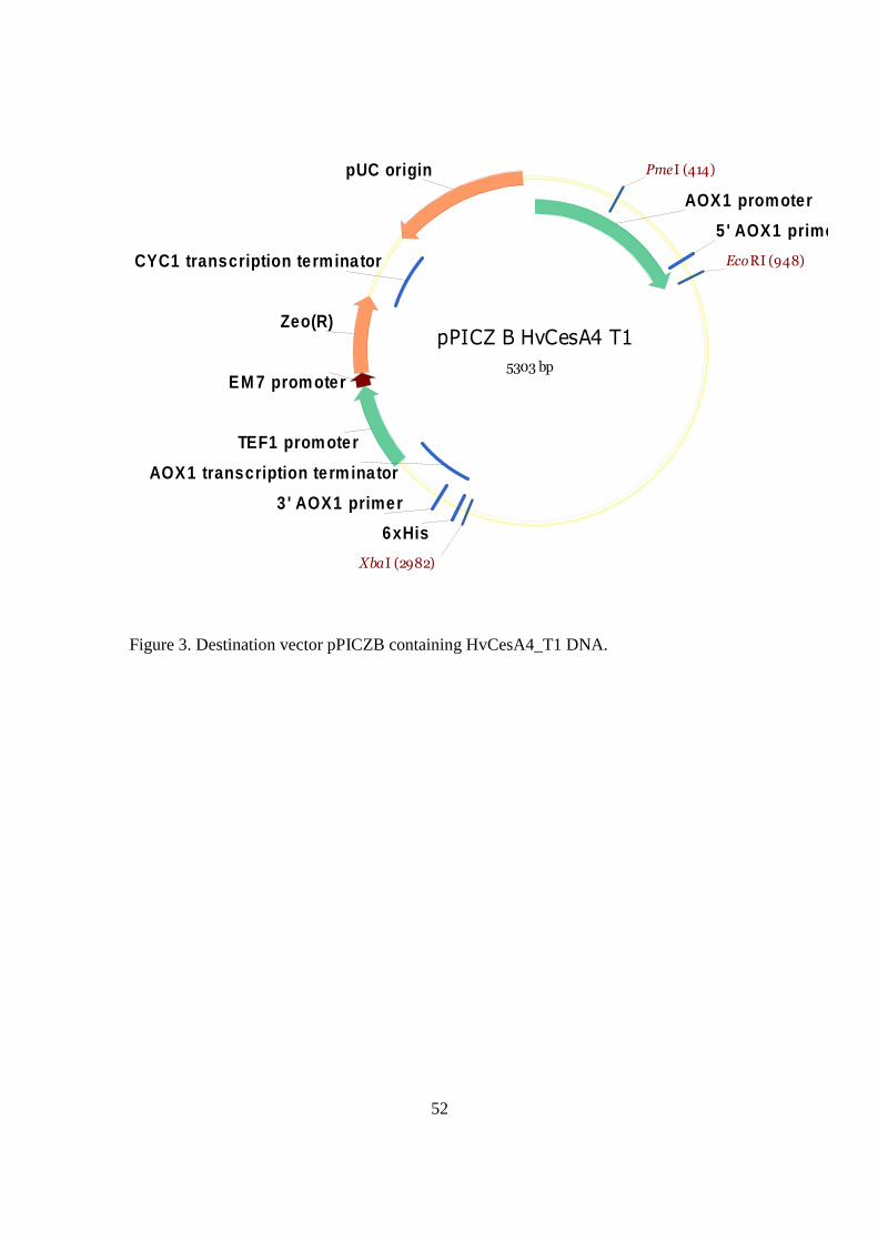
Figure 3. Destination vector pPICZB containing HvCesA4_T1 DNA.
53
pPICZalpha A HvCesA4 T2
5196 bp
6xHis
a lpha-factor s igna l peptide
Zeo(R)
3 ' AOX1 primer
5 ' AOX1 primer
a lpha-factor primer
TEF1 promoter
AOX1 promote r
EM 7 promote r
pUC origin
AOX1 transcription terminator
CYC1 transcription terminator
EcoRI (1212)
XbaI (2875)
PmeI (414)
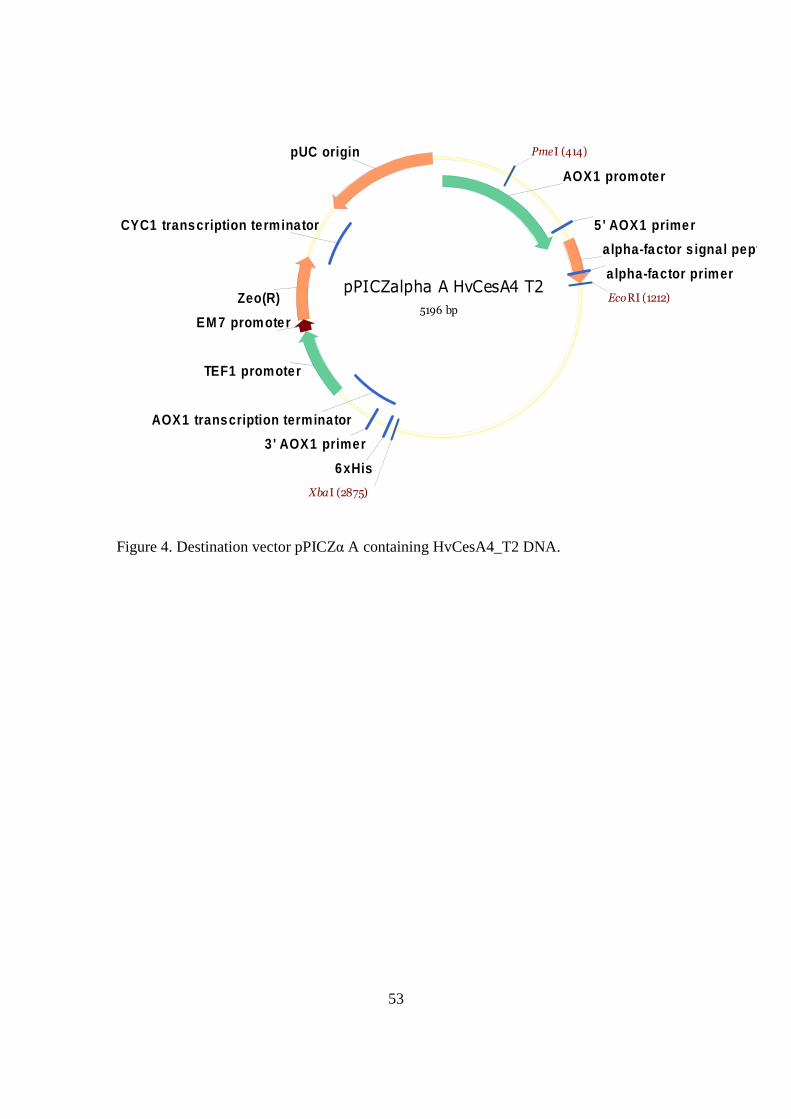
Figure 4. Destination vector pPICZα A containing HvCesA4_T2 DNA.

54
pPICZalpha A HvCesA4 T3
4799 bp
6xHis
a lpha-factor s igna l peptide
Zeo(R)
3 ' AOX1 primer
5 ' AOX1 primer
a lpha-factor primer
TEF1 promoter
AOX1 promoter
EM 7 promoter
pUC origin
AOX1 transcription term ina tor
CYC1 transcription term ina tor
EcoRI (1212)
XbaI (2478)
PmeI (414)
Figure 5. Destination vector pPICZα A containing HvCesA4_T3 DNA.

55
Figure 6. Dot blot of HvCesA4_T2 and HvCesA4_T3 reveals the presence of 6x-His-
tagged protein in only fraction 2 and fraction 3 of the samples.

56
Appendices
HvCesA4 cDNA constructs without restriction sites >HvCesA4 full-length sequence (3128 bp) ATGGACACCGGCGAGCCCAAGGCCAAGGTGTGCCGCGCGTGCGGCGACGATGTCGGGACGCGGGAGGATGGCAGCCCCTTCGTGGCCTGCGCCGAGTGCGGGTTCCCGGTGTGCCGGCCGTGCTACGAGTACGAGCGCAGCGACGGCACGCAGTGCTGCCCCCAGTGCAACGCCCGCTACAAGCGCCACAAAGGGTGCCCGAGGGTGGAAGGGGACGACGAGGACGGCGACATGGACGACTTAGAAGAGGAGTTCCAGGTCAAGAGCCCCAAGAAGCCTCACGAGCCCGTTCCCTTCGACGTCTACTCGGAGAACGGCGAGCAGCCGCCGCAGAAGTGGCGCCCCGGTGGCCCGGCCATGTCCTCCTTCGGTGGAAGCGGTCAGGAGCTTGAGGCGGAGCGGGAGATGGAGGGGAGCATGGAGTGGAAGGACAGGATCGACAAGTGGAAGACCAAGCAGGAGAAGAGGGGCAAGCTCAACCGCGACAACAGCGACGACGACGACGACGACAAGAACGACGACGAGTACATGCTGCTGGCTGAGGCGCGGCAGCCGCTGTGGCGCAAGCTGCCGGTGCCGTCGAGCCAGATCAACCCGTACCGCATCGTCATCGTGCTCCGCCTGGTGGTGCTCTGCTTCTTCCTCCGCTTCCGGATCATGACCCCGGCCAACGACGCCATCCCGCTGTGGCTGGTGTCCGTCATCTGCGAGCTCTGGTTCGCGCTCTCCTGGATCCTGGACCAGCTGCCCAAGTGGTCGCCGGTGACCCGGGAGACGTACCTGGACCGCCTCGCCCTGCGCTACGACCGCGAGGGCGAGCCTTCCCGGCTGTCCCCCATCGACTTCTTCGTGAGCACGGTGGACCCGCTCAAGGAGCCCCCCATCATCACGGCCAACACCGTGCTGTCCATCCTCGCCGTCGACTACCCCGTGGACCGCAACAGCTGCTACGTCTCCGACGACGGCGCCTCCATGCTCTGTTTCGACACCCTCTCCGAGACGGCCGAGTTCGCGCGCCGCTGGGTGCCCTTCTGCAAGAAGTTCGCCATCGAGCCCCGTGCCCCAGAGTTCTACTTCTCACAGAAGATCGACTACCTCAAGGACAAGGTGCAGCCGACGTTCGTCAAGGAGCGGCGCGCCATGAAGCGCGAGTACGAGGAGTTCAAGGTGCGGATCAACGGGCTGGTGGCCAAGCCGAGAAGAAGCCCGAGGAAGGGTGGGTGATGCAGGACGGCACGCCGTGGCCCGGGAACAACACCAGAGACCACCCCGGGATGATACAGGTGTATTTGGGCAGCCAGGGCGCGCTGGACGTGGAGGGCCACGAGCTGCCGCGGTTGGTGTACGTGTCCCGAGAGAAGAGGCCAGGACACAACCACCACAAGAAGGCCGGCGCCATGAACGCGCTGGTGCGGGTGTCGGCGGTGCTCACCAACGCGCCCTTCATCCTCAACCTCGACTGCGACCACTACGTGAACAACAGCAAGGCCGTCCGCGAGGCCATGTGCTTCCTCATGGACCCCCAGCTCGGCAAGAAACTCTGCTACGTCCAGTTCCCGCAGGGCTTCGACGGCATCGACCTCCACGATCGATACGCCAACCGCAACGTCGTCTTCTTCGACATCAACATGAAGGGGCTAGACGGCATACAGGGCCCGGTGTACGTGGGCACGGGGTGCGTGTTCAACAGGCAGGCGCTGTACGGGTACGACCCGCCGCGGCCGGAGAAGAGGCCCAAGATGACGTGCGACTGCTGGCCATCGTGGTGCTGCTGCTGCTGCTGCTTCGGCGGAGGGAAGCACCGCAAATCGAGCAAGGACAAGAAGGGCGGCGGCGGCGGCGACGACGAGCCCCGGCGCGGGCTCCTTGGGTTCTACAAGAAGCGGGGCAAGAAGGACAAGCTCGGCGGCGGGCCGAAGAAGGGGTCGTACAGGAAGCGGCAGCGCGGGTACGAGCTGGAGGAGATCGAGGAGGGCATCGAGGGGTACGACGAGCTGGAGCGCTCCTCGCTCATGTCACAGAAGAGCTTCCAGAAGCGGTTCGGCCAGTCGCCGGTGTTCATCGCCTCCACGCTCGTCGAGGACGGCGGGCTGCCGCAGGGCGCTGCCGCCGACCCCGCAGGCCTCATCAAGGAGGCTATCCACGTCATCAGCTGTGGGTACGAAGGGAAGACCGAGTGGGGCAAGGAGATTGGGTGGATCTACGGGTCGGTGACAGAGGACATCCTGACGGGCTTCAAGATGCACTGCCGTGGGTGGAAGTCCGTCTACTGCACGCCCACACGGCCAGCGTTCAAGGGATCGGCGCCCATCAACTTGTCGGACAGGCTTCACCAGGTGCTTCGTTGGGCGCTCGGTTCCGTCGAGATCTTCATGAGCCGTCATTGCCCGCTCTGGTACGCCTACGGCGGCCGTCTCAAGTGGCTCGAGCGCTTCGCCTACACCAACACCATCGTCTACCCCTTCACCTCCATTCCCCTCATCGCCTACTGCACCATCCCCGCCGTCTGCCTCCTCACCGGCAAGTTCATAATCCCCACGCTCAACAACCTGGCGAGCATTTGGTTCATCGCCTTGTTCATGTCCATCATCGCGACGGGGGTGCTGGAGCTGCGGTGGAGCGGGGTGAGCATCGAGGACTGGTGGCGTAACGAGCAGTTCTGGGTCATAGGTGGCGTGTCCGCGCATCTGTTCGCCGTCTTCCAGGGCTTCCTCAAGGTGTTGGGCGGAGTGGACACCAACTTCACTGTCACCTCCAAGGCGGGCGCTGATGAGGCCGATGCATTCGGTGACCTCTACCTCTTCAAGTGGACAACCCTGCTGATCCCGCCCACCACGCTCATCATTATCAACATGGTCGGCATCGTCGCCGGCGTGTCCGACGCCGTCAACAATGGGTACGGGTCCTGGGGCCCGCTCTTCGGGA

57
AGCTCTTCTTCTCCTTCTGGGTCATCGTTCACCTCTACCCGTTCCTCAAGGGGCTCATGGGGAGGCAGAACCGGACGCCCACCATCGTTGTGCTCTGGTCTGTCCTGCTCGCCTCCATCTTCTCCCTTGTGTGGGTCAGGATCGACCCCTTCATTGCCAAGCCCAAGGGACCCATTCTTAAGCCGTGTGGAGTACAGTGCTGA >HvCesA4_T1 (2044 bp) AGTCGAATTCAAAATGGGTCATCATCATCATCATCATGAAAATCTGTACTTTCAAGGTACCCCGGCCAACGACGCCATCCCGCTGTGGCTGGTGTCCGTCATCTGCGAGCTCTGGTTCGCGCTCTCCTGGATCCTGGACCAGCTGCCCAAGTGGTCGCCGGTGACCCGGGAGACGTACCTGGACCGCCTCGCCCTGCGCTACGACCGCGAGGGCGAGCCTTCCCGGCTGTCCCCCATCGACTTCTTCGTGAGCACGGTGGACCCGCTCAAGGAGCCCCCCATCATCACGGCCAACACCGTGCTGTCCATCCTCGCCGTCGACTACCCCGTGGACCGCAACAGCTGCTACGTCTCCGACGACGGCGCCTCCATGCTCTGTTTCGACACCCTCTCCGAGACGGCCGAGTTCGCGCGCCGCTGGGTGCCCTTCTGCAAGAAGTTCGCCATCGAGCCCCGTGCCCCAGAGTTCTACTTCTCACAGAAGATCGACTACCTCAAGGACAAGGTGCAGCCGACGTTCGTCAAGGAGCGGCGCGCCATGAAGCGCGAGTACGAGGAGTTCAAGGTGCGGATCAACGGGCTGGTGGCCAAGGCCGAGAAGAAGCCCGAGGAAGGGTGGGTGATGCAGGACGGCACGCCGTGGCCCGGGAACAACACCAGAGACCACCCCGGGATGATACAGGTGTATTTGGGCAGCCAGGGCGCGCTGGACGTGGAGGGCCACGAGCTGCCGCGGTTGGTGTACGTGTCCCGAGAGAAGAGGCCAGGACACAACCACCACAAGAAGGCCGGCGCCATGAACGCGCTGGTGCGGGTGTCGGCGGTGCTCACCAACGCGCCCTTCATCCTCAACCTCGACTGCGACCACTACGTGAACAACAGCAAGGCCGTCCGCGAGGCCATGTGCTTCCTCATGGACCCCCAGCTCGGCAAGAAACTCTGCTACGTCCAGTTCCCGCAGGGCTTCGACGGCATCGACCTCCACGATCGATACGCCAACCGCAACGTCGTCTTCTTCGACATCAACATGAAGGGGCTAGACGGCATACAGGGCCCGGTGTACGTGGGCACGGGGTGCGTGTTCAACAGGCAGGCGCTGTACGGGTACGACCCGCCGCGGCCGGAGAAGAGGCCCAAGATGACGTGCGACTGCTGGCCATCGTGGTGCTGCTGCTGCTGCTGCTTCGGCGGAGGGAAGCACCGCAAATCGAGCAAGGACAAGAAGGGCGGCGGCGGCGGCGACGACGAGCCCCGGCGCGGGCTCCTTGGGTTCTACAAGAAGCGGGGCAAGAAGGACAAGCTCGGCGGCGGGCCGAAGAAGGGGTCGTACAGGAAGCGGCAGCGCGGGTACGAGCTGGAGGAGATCGAGGAGGGCATCGAGGGGTACGACGAGCTGGAGCGCTCCTCGCTCATGTCACAGAAGAGCTTCCAGAAGCGGTTCGGCCAGTCGCCGGTGTTCATCGCCTCCACGCTCGTCGAGGACGGCGGGCTGCCGCAGGGCGCTGCCGCCGACCCCGCAGGCCTCATCAAGGAGGCTATCCACGTCATCAGCTGTGGGTACGAAGGGAAGACCGAGTGGGGCAAGGAGATTGGGTGGATCTACGGGTCGGTGACAGAGGACATCCTGACGGGCTTCAAGATGCACTGCCGTGGGTGGAAGTCCGTCTACTGCACGCCCACACGGCCAGCGTTCAAGGGATCGGCGCCCATCAACTTGTCGGACAGGCTTCACCAGGTGCTTCGTTGGGCGCTCGGTTCCGTCGAGATCTTCATGAGCCGTCATTGCCCGCTCTGGTACGCCTACGGCGGCCGTCTCAAGTGGCTCGAGCGCTTCGCCTACACCAACACCATCGTCTACCCCTTCACCTCCATTCCCCTCATCGCCTACTGCACCATCCCCGCCGTCTGCCTCCTCACCGGCAAGTTCATAATCCCCACGCTCAACAACCTGGCGAGCATTTGGTTCATCGCCTTGTTCATGTCCATCATCGCGACGGGGGTGCTGGAGCTGCGGTGGAGCGGGGTGAGCTGAGGATAATCTAGA >HvCesA4_T2 (1631 bp) CTCGCCCTGCGCTACGACCGCGAGGGCGAGCCTTCCCGGCTGTCCCCCATCGACTTCTTCGTGAGCACGGTGGACCCGCTCAAGGAGCCCCCCATCATCACGGCCAACACCGTGCTGTCCATCCTCGCCGTCGACTACCCCGTGGACCGCAACAGCTGCTACGTCTCCGACGACGGCGCCTCCATGCTCTGTTTCGACACCCTCTCCGAGACGGCCGAGTTCGCGCGCCGCTGGGTGCCCTTCTGCAAGAAGTTCGCCATCGAGCCCCGTGCCCCAGAGTTCTACTTCTCACAGAAGATCGACTACCTCAAGGACAAGGTGCAGCCGACGTTCGTCAAGGAGCGGCGCGCCATGAAGCGCGAGTACGAGGAGTTCAAGGTGCGGATCAACGGGCTGGTGGCCAAGGCCGAGAAGAAGCCCGAGGAAGGGTGGGTGATGCAGGACGGCACGCCGTGGCCCGGGAACAACACCAGAGACCACCCCGGGATGATACAGGTGTATTTGGGCAGCCAGGGCGCGCTGGACGTGGAGGGCCACGAGCTGCCGCGGTTGGTGTACGTGTCCCGAGAGAAGAGGCCAGGACACAACCACCACAAGAAGGCCGGCGCCATGAACGCGCTGGTGCGGGTGTCGGCGGTGCTCACCAACGCGCCCTTCATCCTCAACCTCGACTGCGACCACTACGTGAACAACAGCAAGGCCGTCCGCGAGGCCA

58
TGTGCTTCCTCATGGACCCCCAGCTCGGCAAGAAACTCTGCTACGTCCAGTTCCCGCAGGGCTTCGACGGCATCGACCTCCACGATCGATACGCCAACCGCAACGTCGTCTTCTTCGACATCAACATGAAGGGGCTAGACGGCATACAGGGCCCGGTGTACGTGGGCACGGGGTGCGTGTTCAACAGGCAGGCGCTGTACGGGTACGACCCGCCGCGGCCGGAGAAGAGGCCCAAGATGACGTGCGACTGCTGGCCATCGTGGTGCTGCTGCTGCTGCTGCTTCGGCGGAGGGAAGCACCGCAAATCGAGCAAGGACAAGAAGGGCGGCGGCGGCGGCGACGACGAGCCCCGGCGCGGGCTCCTTGGGTTCTACAAGAAGCGGGGCAAGAAGGACAAGCTCGGCGGCGGGCCGAAGAAGGGGTCGTACAGGAAGCGGCAGCGCGGGTACGAGCTGGAGGAGATCGAGGAGGGCATCGAGGGGTACGACGAGCTGGAGCGCTCCTCGCTCATGTCACAGAAGAGCTTCCAGAAGCGGTTCGGCCAGTCGCCGGTGTTCATCGCCTCCACGCTCGTCGAGGACGGCGGGCTGCCGCAGGGCGCTGCCGCCGACCCCGCAGGCCTCATCAAGGAGGCTATCCACGTCATCAGCTGTGGGTACGAAGGGAAGACCGAGTGGGGCAAGGAGATTGGGTGGATCTACGGGTCGGTGACAGAGGACATCCTGACGGGCTTCAAGATGCACTGCCGTGGGTGGAAGTCCGTCTACTGCACGCCCACACGGCCAGCGTTCAAGGGATCGCGCCCATCAACTTGTCGGACAGGCTTCACCAGGTGCTTCGTTGGGCGCTCGGTTCCGTCGAGATCTTCATGAGCCGTCATTGCCCGCTCTGGTACGCCTACGGCGGCCGTCTCAAG >HvCesA4_T3 (1233 bp) CTCGCCCTGCGCTACGACCGCGAGGGCGAGCCTTCCCGGCTGTCCCCCATCGACTTCTTCGTGAGCACGGTGGACCCGCTCAAGGAGCCCCCCATCATCACGGCCAACACCGTGCTGTCCATCCTCGCCGTCGACTACCCCGTGGACCGCAACAGCTGCTACGTCTCCGACGACGGCGCCTCCATGCTCTGTTTCGACACCCTCTCCGAGACGGCCGAGTTCGCGCGCCGCTGGGTGCCCTTCTGCAAGAAGTTCGCCATCGAGCCCCGTGCCCCAGAGTTCTACTTCTCACAGAAGATCGACTACCTCAAGGACAAGGTGCAGCCGACGTTCGTCAAGGAGCGGCGCGCCATGAAGCGCGAGTACGAGGAGTTCAAGGTGCGGATCAACGGGCTGGTGGCCAAGGCCGAGAAGAAGCCCGAGGAAGGGTGGGTGATGCAGGACGGCACGCCGTGGCCCGGGAACAACACCAGAGACCACCCCGGGATGATACAGGTGTATTTGGGCAGCCAGGGCGCGCTGGACGTGGAGGGCCACGAGCTGCCGCGGTTGGTGTACGTGTCCCGAGAGAAGAGGCCAGGACACAACCACCACAAGAAGGCCGGCGCCATGAACGCGCTGGTGCGGGTGTCGGCGGTGCTCACCAACGCGCCCTTCATCCTCAACCTCGACTGCGACCACTACGTGAACAACAGCAAGGCCGTCCGCGAGGCCATGTGCTTCCTCATGGACCCCCAGCTCGGCAAGAAACTCTGCTACGTCCAGTTCCCGCAGGGCTTCGACGGCATCGACCTCCACGATCGATACGCCAACCGCAACGTCGTCTTCTTCGACATCAACATGAAGGGGCTAGACGGCATACAGGGCCCGGTGTACGTGGGCACGGGGTGCGTGTTCAACAGGCAGGCGCTGTACGGGTACGACCCGCCGCGGCCGGAGAAGAGGCCCAAGATGACGTGCGACTGCTGGCCATCGTGGTGCTGCTGCTGCTGCTGCTTCGGCGGAGGGAAGCACCGCAAATCGAGCAAGGACAAGAAGGGCGGCGGCGGCGGCGACGACGAGCCCCGGCGCGGGCTCCTTGGGTTCTACAAGAAGCGGGGCAAGAAGGACAAGCTCGGCGGCGGGCCGAAGAAGGGGTCGTACAGGAAGCGGCAGCGCGGGTACGAGCTGGAGGAGATCGAGGAGGGCATCGAGGGGTACGACGAGCTGGAGCGCTCCTCGCTCATGTCACAG
59
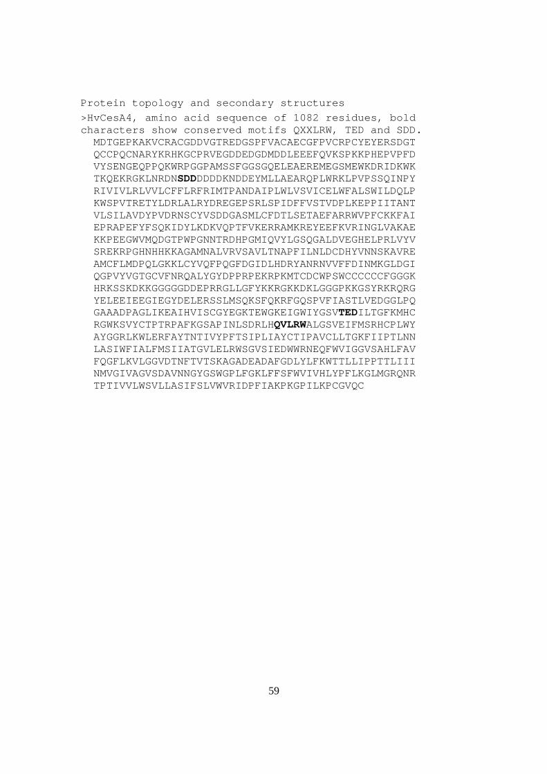
Protein topology and secondary structures
>HvCesA4, amino acid sequence of 1082 residues, bold characters show conserved motifs QXXLRW, TED and SDD. MDTGEPKAKVCRACGDDVGTREDGSPFVACAECGFPVCRPCYEYERSDGT QCCPQCNARYKRHKGCPRVEGDDEDGDMDDLEEEFQVKSPKKPHEPVPFD VYSENGEQPPQKWRPGGPAMSSFGGSGQELEAEREMEGSMEWKDRIDKWK TKQEKRGKLNRDNSDDDDDDKNDDEYMLLAEARQPLWRKLPVPSSQINPY RIVIVLRLVVLCFFLRFRIMTPANDAIPLWLVSVICELWFALSWILDQLP KWSPVTRETYLDRLALRYDREGEPSRLSPIDFFVSTVDPLKEPPIITANT VLSILAVDYPVDRNSCYVSDDGASMLCFDTLSETAEFARRWVPFCKKFAI EPRAPEFYFSQKIDYLKDKVQPTFVKERRAMKREYEEFKVRINGLVAKAE KKPEEGWVMQDGTPWPGNNTRDHPGMIQVYLGSQGALDVEGHELPRLVYV SREKRPGHNHHKKAGAMNALVRVSAVLTNAPFILNLDCDHYVNNSKAVRE AMCFLMDPQLGKKLCYVQFPQGFDGIDLHDRYANRNVVFFDINMKGLDGI QGPVYVGTGCVFNRQALYGYDPPRPEKRPKMTCDCWPSWCCCCCCFGGGK HRKSSKDKKGGGGGDDEPRRGLLGFYKKRGKKDKLGGGPKKGSYRKRQRG YELEEIEEGIEGYDELERSSLMSQKSFQKRFGQSPVFIASTLVEDGGLPQ GAAADPAGLIKEAIHVISCGYEGKTEWGKEIGWIYGSVTEDILTGFKMHC RGWKSVYCTPTRPAFKGSAPINLSDRLHQVLRWALGSVEIFMSRHCPLWY AYGGRLKWLERFAYTNTIVYPFTSIPLIAYCTIPAVCLLTGKFIIPTLNN LASIWFIALFMSIIATGVLELRWSGVSIEDWWRNEQFWVIGGVSAHLFAV FQGFLKVLGGVDTNFTVTSKAGADEADAFGDLYLFKWTTLLIPPTTLIII NMVGIVAGVSDAVNNGYGSWGPLFGKLFFSFWVIVHLYPFLKGLMGRQNR TPTIVVLWSVLLASIFSLVWVRIDPFIAKPKGPILKPCGVQC

60
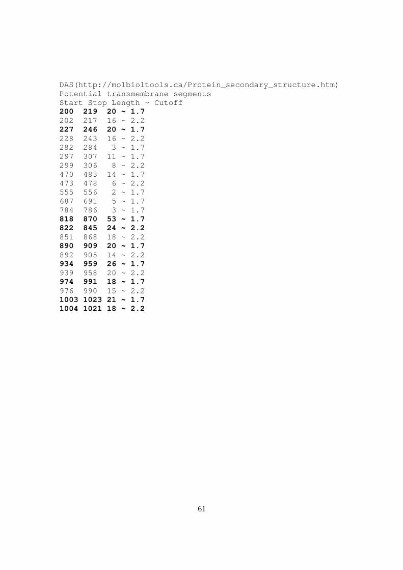
Predicted membrane helices TMpred(http://www.ch.embnet.org/software/TMPRED_form.html) The sequence positions in brackets denote the positions of helices. Only scores above 500 are considered to be significant. Inside to outside helices: 10 found From to score center 200 ( 204) 223 ( 220) 1187 212 226 ( 226) 246 ( 244) 1471 236 466 ( 466) 484 ( 482) 643 474 581 ( 581) 597 ( 597) 922 589 812 ( 820) 839 ( 836) 1310 828 851 ( 851) 869 ( 869) 2435 859 886 ( 886) 905 ( 905) 1331 895 939 ( 939) 959 ( 957) 1339 949 973 ( 973) 991 ( 989) 1727 981 1001 (1002)1020 (1020) 2340 1011 Outside to inside helices: 13 found from to score center 199 ( 199) 215 ( 215) 1562 207 226 ( 228) 246 ( 244) 1584 236 292 ( 292) 310 ( 310) 109 300 469 ( 469) 486 ( 486) 563 478 546 ( 549) 570 ( 567) 396 559 581 ( 581) 600 ( 600) 285 591 777 ( 784) 803 ( 803) 161 793 822 ( 822) 840 ( 840) 1421 830 851 ( 851) 869 ( 869) 2364 861 886 ( 889) 905 ( 905) 1401 897 942 ( 942) 960 ( 960) 1511 950 970 ( 970) 991 ( 988) 1437 978 1005 (1005)1021 (1021) 2285 1013 Toppred (http://cbi.labri.fr/outils/Pise/toppred.html) Helix Begin - End Score Certainity 1 226 – 246 1.583 Certain 2 291 – 311 0.625 Putative 3 581 – 601 0.998 Putative 4 821 - 841 1.699 Certain 5 850 - 870 2.191 Certain 6 885 - 905 1.419 Certain 7 940 - 960 1.697 Certain 8 968 - 988 1.447 Certain 9 1001 – 1021 2.175 Certain
61
DAS(http://molbioltools.ca/Protein_secondary_structure.htm) Potential transmembrane segments Start Stop Length ~ Cutoff 200 219 20 ~ 1.7 202 217 16 ~ 2.2 227 246 20 ~ 1.7 228 243 16 ~ 2.2 282 284 3 ~ 1.7 297 307 11 ~ 1.7 299 306 8 ~ 2.2 470 483 14 ~ 1.7 473 478 6 ~ 2.2 555 556 2 ~ 1.7 687 691 5 ~ 1.7 784 786 3 ~ 1.7 818 870 53 ~ 1.7 822 845 24 ~ 2.2 851 868 18 ~ 2.2 890 909 20 ~ 1.7 892 905 14 ~ 2.2 934 959 26 ~ 1.7 939 958 20 ~ 2.2 974 991 18 ~ 1.7 976 990 15 ~ 2.2 1003 1023 21 ~ 1.7 1004 1021 18 ~ 2.2