pancreatic somatostatin expressing-cells: an untapped
TRANSCRIPT
HAL Id: tel-01942168https://tel.archives-ouvertes.fr/tel-01942168
Submitted on 3 Dec 2018
HAL is a multi-disciplinary open accessarchive for the deposit and dissemination of sci-entific research documents, whether they are pub-lished or not. The documents may come fromteaching and research institutions in France orabroad, or from public or private research centers.
L’archive ouverte pluridisciplinaire HAL, estdestinée au dépôt et à la diffusion de documentsscientifiques de niveau recherche, publiés ou non,émanant des établissements d’enseignement et derecherche français ou étrangers, des laboratoirespublics ou privés.
Pancreatic somatostatin expressing-cells : an untappedsource for β-cell regeneration ?
Noemie Druelle
To cite this version:Noemie Druelle. Pancreatic somatostatin expressing-cells : an untapped source for β-cell regener-ation ?. Agricultural sciences. Université Côte d’Azur, 2016. English. �NNT : 2016AZUR4106�.�tel-01942168�
UNIVERSITE COTE D’AZUR – UFR SCIENCES
Ecole Doctorale des Sciences de la Vie et de la Santé (ED85)
THESE
Pour l’obtention du titre de
Docteur en Sciences de la Vie et de la Santé
Spécialité : Recherche Clinique et Thérapeutique
Présentée et soutenue par
Noémie DRUELLE
Pancreatic somatostatin expressing-cells:
an untapped source for β-cell regeneration?
Soutenue publiquement le 2 décembre 2016 devant le jury composé de :
Prof. François Cuzin
Dr. Miriam Cnop
Dr. Jacob Hecksher-Sørensen
Prof. Ahmed Mansouri
Dr. Patrick Collombat
Président
Rapporteur
Rapporteur
Examinateur
Directeur de thèse


ACKNOWLEDGEMENTS
Soyons reconnaissants aux personnes qui nous donnent du bonheur ; elles sont les charmants jardiniers par qui nos âmes sont fleuries.
Marcel Proust
Mes premiers remerciements s’adressent à mon directeur de thèse Patrick Collombat, sans qui ce joli projet n’aurait pas vu le jour. Merci de m’avoir fait confiance et de m’avoir accueillie dans ton équipe ! Merci aussi de t’être toujours battu pour nous offrir les meilleures conditions de travail. Je remercie également l’équipe 7 du C3M et plus particulièrement Sophie Giorgetti-Peraldi, avec qui j’ai fait mes premiers pas dans la recherche.
I would also wish to thank Prof. François Cuzin, Prof. Ahmed Mansouri, Dr. Miriam Cnop and Dr. Jacob Hecksher-Sørensen who accepted to be part of my jury, it is a real honor to have my work reviewed by such experts. Thanks a lot to Dr. Minoo Rassoulzadegan for sharing her knowledge on so many subjects, and for her precious help with cloning experiments.
Pr. Frédérique Vidal, merci d’avoir enseigné avec autant de patience et de passion - vous m’avez transmis l’amour de la biologie, sans lequel ce travail n’aurait pas été réalisable. Merci également de m’avoir fait l’honneur d’accepter l’invitation à ma défense de thèse.
Thank you to all the people of the Diabetes Genetics Team, past and present, for all the scientific discussions and all the fun in the lab we had. Elisabet, a big thanks for your patience and everything you taught me. Bibi, thanks for having been such a good mouse- and lab- manager. Grazie to the Italians invaders Tiziana, Serena, Fabio and muchas gracias to Sergi for their help with this project. I will leave the team with a lot of memories! Aidin, thank you for being the strangest person I’ve ever met.
Thanks to all the present and past iBV members I had the chance to meet - Yas, Ana, Filippo, Sandrine, Pierre, Anthony, Seb, Rohan, the Shedl’s and Chaboissier’s teams for great technical advices, happy hour times, and crazy moments at the Pompei or wherever. Un grand merci également à nos fantastiques gestionnaires Mireille et Cécile, qui s’occupent de nous à merveille.
A mes chers parents, merci de m’avoir suivie dans cette aventure et dans tous mes projets fous ! Un immense merci pour votre soutien, qui m’a permis de passer ces années à Nice dans les meilleures conditions. Ces remerciements seraient incomplets si je n’en adressais pas à Alexia et Sébastien, Simon et Nadège, Christopher et Hakima, vous êtes les meilleurs frères/sœur/beau-frère/belles-sœurs qu’il soit, merci d’avoir toujours été là. Egalement une immense pensée pour mes merveilleux grands parents qui m’ont constamment soutenue ! Un énorme merci à Martine pour son inconditionnel support ces deux dernières années, pour ses conseils et son accueil toujours chaleureux. Bilette, Didi, merci pour votre patience dans les moments de crise ou de doute, pour le café rituel du matin, votre bienveillance et tous ces encouragements. Bilette, merci pour tout le temps que tu as consacré à ce travail. Merci à vous pour tous ces moments de rire et de qPCR. Mag, Maé, merci pour votre écoute, vos conseils toujours précieux et toute l’aide que vous m’avez apportée pour ce projet - Bisous cœur ! Alizée, Laura - mes Bellas - merci d’avoir été là toutes ces années, dans les bons et les mauvais moments. Merci à Louis, Pierre Jean et Clémence d’avoir toujours été présents pour moi. A vous tous, merci pour tous ces moments agréables et ces beaux souvenirs, vous êtes de loin les meilleurs amis du monde, encore merci à vous ! Last but not least, je remercie tendrement Carine d’avoir inconditionnellement cru en moi, d’avoir eu la patience de me supporter et de m’encourager à chaque instant. Merci aussi pour tous ces jolis moments et ceux à venir. Merci infiniment à vous tous, et à tous ceux que j’aurais pu oublier.

TABLE OF CONTENT
INTRODUCTION ..................................................................................... 1
I. Diabetes Mellitus ............................................................................................ 2
Type 1 Diabetes Mellitus (T1DM) ............................................................ 3
Type 2 Diabetes Mellitus (T2DM) ............................................................ 5
Gestational diabetes ................................................................................ 6
Monogenic diabetes ................................................................................ 7
II. Pancreas anatomy and function ................................................................... 7
Gross anatomy of the pancreas ............................................................... 8
Organization and function of the exocrine tissue ..................................... 9
a. Acinar cells ............................................................................................ 9
b. Duct cells ............................................................................................. 10
Organization and function of the endocrine tissue ................................. 10
a. Insulin-secreting -cells ....................................................................... 13
b. Glucagon-secreting -cells .................................................................. 14
c. Somatostatin-secreting -cells ............................................................. 15
d. Pancreatic polypeptide-secreting PP-cells .......................................... 16
e. Ghrelin-secreting -cells ...................................................................... 17
III. Mouse pancreatic development .................................................................. 18
Early development ................................................................................. 19
a. Endoderm patterning ........................................................................... 19
b. Gut tube patterning and pancreas budding ......................................... 19
Pancreas specification ........................................................................... 21
a. Primary transition ................................................................................. 22
b. Secondary transition ............................................................................ 24
Exocrine specification ............................................................................ 25
a. Acinar cell fate ..................................................................................... 25
b. Ductal cell fate ..................................................................................... 25
Endocrine specification .......................................................................... 27
Differentiation of endocrine cells ............................................................ 28
a. -cells – glucagon ............................................................................... 29
b. -cells – insulin .................................................................................... 29
c. -cells – somatostatin .......................................................................... 30
IV. T1DM actual therapies & current research ................................................ 32
Management of T1DM ........................................................................... 32
a. Exogenous insulin administration ........................................................ 32
b. Total pancreas and islets transplantation ............................................ 33
Current axes of research ....................................................................... 34
a. -cell mass replacement from stem cells ............................................. 35
1) Pluripotent stem cells (PSCs) ........................................................ 36
2) Tissue stem cells ........................................................................... 38
b. -cell mass replacement from pancreatic cells .................................... 39
1) From acinar cells ........................................................................... 39
2) From ductal cells ........................................................................... 40
3) From endocrine cells ..................................................................... 41
V. Pax4-mediated -cell regeneration ............................................................. 42
MATERIAL AND METHODS ................................................................ 45
I. Animal procedures ...................................................................................... 46
Generation of transgenic mouse line ..................................................... 46
Genotyping ............................................................................................ 47
II. Mice treatments ............................................................................................ 48
Proliferation assessment ....................................................................... 48
Induction of streptozotocin-mediated hyperglycemia ............................. 49
III. Tolerance tests and blood glucose level measurements ......................... 49
IV. RNA analyses ............................................................................................... 50
RNA extraction from total pancreas ....................................................... 50
cDNA synthesis ..................................................................................... 50
qPCR analyzes ...................................................................................... 51
V. Pancreatic tissue processing and sectioning ........................................... 52
Paraffin embedding – Microtome sectioning .......................................... 52
OCT embedding – Cryostat sectioning .................................................. 52
VI. Staining of pancreatic sections .................................................................. 53
Immunohistochemistry (IHC) ................................................................. 53
IHC on cryosections .............................................................................. 55
VII. Quantification analyses ............................................................................... 56
VIII. Data analyses and statistics ....................................................................... 57
RESULTS ............................................................................................. 58
I. Sst::Cre-Pax4-OE double transgenic mouse line generation and
phenotype ............................................................................................................. 59
II. Pax4 misexpression in -cells results in progressive islet hypertrophy
and insulin-producing cell hyperplasia .............................................................. 61
III. Pax4 misexpression in somatostatin+ cells induces their conversion into
-like cells ............................................................................................................. 65
IV. Pax4 misexpression in -cells results in increased proliferation within
the ductal epithelium ............................................................................................ 69
V. Reactivation of EMT and Neurog3-controlled endocrine differentiation
program in Sst-Cre::Pax4-OE pancreata ............................................................ 73
VI. Supplementary insulin-expressing cells in Sst-Cre::Pax4-OE pancreata
display a -cell phenotype and are functional ................................................... 76
VII. Adult somatostatin+ cells convert into insulin-producing cells upon Pax4
misexpression following streptozotocin treatment ........................................... 79
DISCUSSION ........................................................................................ 82
I. Pax4 misexpression in -cells induces their conversion into -cells ..... 83
II. Recruitment of Neurog3 expressing ductal cells and EMT reawakening 85
CONCLUSION / PERSPECTIVES ........................................................ 87
I. Somatostatin: a signal inducing -cell regeneration? .............................. 88
II. Model proposed ........................................................................................... 89
BIBLIOGRAPHY................................................................................... 92
FIGURE INDEX
Figure 1: The two main types of diabetes – Type 1 and Type 2. ................................ 4
Figure 2: Human pancreas anatomy and histology .................................................... 8
Figure 3: Microarchitecture of human and mouse islets. .......................................... 11
Figure 4: Endocrine cell proportions of human and mouse islets. ............................ 12
Figure 5: Insulin release from pancreatic -cells ...................................................... 14
Figure 6: Gut tube patterning and endoderm-derived organs ................................... 20
Figure 7: Ventral and dorsal pancreas specification ................................................. 21
Figure 8: Primary and secondary transitions ............................................................ 23
Figure 9: Simplified model of pancreatic cell fate determination ............................... 26
Figure 10: Cell sources used in the replacement of -cells ...................................... 35
Figure 11: Pax4 misexpression in adult or embryonic -cells induce their neogenesis
and conversion into -like cells. ................................................................................ 44
Figure 12: Transgenic mouse line allowing Pax4 misexpression in somatostatin-
expressing cells ........................................................................................................ 46
Figure 13: Cldu and Idu labelling. ............................................................................. 48
Figure 14: Transgenic mouse line allowing Pax4 misexpression in somatostatin-
expressing cells. ....................................................................................................... 60
Figure 15: Pax4 misexpression in -cells leads to islet hypertrophy and -cell
hyperplasia. .............................................................................................................. 62
Figure 16: Abnormal positioning of non--cells within the islets of Sst-Cre::Pax4-OE
transgenic mice. ....................................................................................................... 64
Figure 17: Conversion of adult -cells into insulin-producing cells upon Pax4
misexpression. .......................................................................................................... 66
Figure 18: Additional examples illustrating the conversion of -cells to insulin-
producing cells upon Pax4 misexpression. ............................................................... 68
Figure 19: Increased cell proliferation within the ductal epithelium of Sst-Cre::Pax4-
OE pancreata. .......................................................................................................... 70
Figure 20: Increased proliferation within the ductal epithelium of Sst-Cre::Pax4-OE
pancreata occurring before 2 month of age. ............................................................. 72
Figure 21: Re-expression of Neurog3 in duct-associated cells upon Pax4
misexpression in -cells and reawakening of epithelial-to-mesenchymal transition. . 74
Figure 22: Sst-Cre::Pax4-OE endogenous and supplementary insulin+ cells display a
-cell phenotype. ...................................................................................................... 77
Figure 23: Improved -cell function in transgenic Sst-Cre::Pax4-OE mice. .............. 78
Figure 24: Pax4 misexpression in -cells can induce functional -like cell neogenesis
upon chemically-induced -cell ablation. .................................................................. 80
Figure 25: Neurog3-mediated EMT reawakening upon Pax4 misexpression in -cells.
................................................................................................................................. 90
TABLE INDEX
Table 1: Blood glucose levels for non-diabetic, prediabetic and diabetic patients ..... 12
Table 2: Primer sequences used for genotyping ....................................................... 47
Table 3: RT PCR program used ................................................................................ 51
Table 4: Primary antibodies used for IHC .................................................................. 54
Table 5: Secondary antibodies used for IHC ............................................................. 55

1
INTRODUCTION
Introduction
2
I. Diabetes Mellitus
Affecting approximately 422 million people worldwide (“Worldwide trends in
diabetes since 1980,” 2016), diabetes mellitus figures among the most common
metabolic conditions. The prevalence of diabetes mellitus is increasing and predicted
to rise to 552 million affected people by 2030 (Whiting et al., 2011). Despite therapies
and prevention strategies, diabetes is one of the four main chronic diseases and was
the direct cause of approximately 1.5 million deaths in 2012 (“WHO | Global report on
diabetes”).
Due to a failure of insulin production or action, diabetes is responsible for long-
lasting elevated blood glucose levels, such chronic hyperglycemia being associated
with various complications. Indeed, diabetes is reported to be the main cause of
blindness and increases the risk of cardiovascular diseases, nephropathy, stroke and
amputation (Alwan, 2010; Morrish et al., 2001; Pascolini and Mariotti, 2012).
Diabetes patients are also at risk of dying prematurely (Manuel and Schultz, 2004;
Narayan et al., 2003). In addition to health concerns, diabetes care represents a
substantial financial burden with an annual worldwide cost of $825 billion (“WHO |
Global report on diabetes”).
With chronic hyperglycemia as a common feature, diabetes mellitus is
classified into different forms, described hereafter.
Introduction
3
Type 1 Diabetes Mellitus (T1DM)
Formerly known as juvenile diabetes or insulin-dependent diabetes, Type 1
Diabetes Mellitus (T1DM) represents approximately 10% of all diabetic patients. Two
peaks of disease onset have been reported: the first between 6 and 15 years of age
and the second later in adolescence, typically before 30 years (Herold et al., 2013).
T1DM is a polygenic disorder characterized by a cytolytic T-cell mediated
autoimmune-destruction of the insulin-producing -cells (Bottazzo GF, Florin-
Christensen A, 1974; Gepts, 1965; Kolb et al., 1995), such selective loss leading to a
lack of insulin secretion subsequently resulting in high blood glucose levels (Figure
1A-B).
While the exact causes for this autoimmune-mediated cell destruction are not
yet known, a genetic component in the development of T1DM is well documented.
The first described susceptibility locus associated with T1DM was the human
leukocyte antigen (HLA) gene (Nerup et al., 1974; Singal and Blajchman, 1973).
Other genes outside the HLA locus and strongly associated with T1DM have also
been identified. Indeed, polymorphisms in the insulin (Rotwein et al., 1986), PTPN22
(Bottini et al., 2004), CTLA-4 (Ueda et al., 2003) and IL-2RA (Maier et al., 2009)
genes are frequently associated with T1DM. In addition to genetic influences, the
rapid rise in T1DM incidence suggests the contribution of environmental (Atkinson et
al., 1994; Gillespie et al., 2004; Ziegler et al., 2011) or viral (Ginsberg-Fellner et al.,
1985; Honeyman et al., 2000) stimuli in the development of this metabolic disease.
The progressive destruction of the -cells by the immune system of T1DM
patients leads to the production of autoantibodies directed towards several antigens,
including GAD65 (Baekkeskov et al., 1990; Solimena et al., 1988; Towns and

Introduction
4
Pietropaolo, 2011), insulin (Palmer et al., 1983; Wegmann et al., 1994) and proinsulin
(Kuglin et al., 1988), ICA512 (Rabin et al., 1992) and ZNT8 (Wenzlau et al., 2007).
Easily detectable in the serum, autoantibodies represent robust hallmarks for T1DM
diagnosis (Bingley et al., 1997; Riley et al., 1990; Verge et al., 1996).
Figure 1: The two main types of diabetes – Type 1 and Type 2. In healthy subjects,
insulin is released from pancreatic -cells, leading to glucose uptake by target tissues and
normoglycemia (A). In type 1 diabetic patients, the loss of -cells in pancreatic islets induces
a deficiency in insulin secretion and a lack of glucose uptake, further resulting in high blood
glucose levels (B). In type 2 diabetic patients, insulin is secreted but a defect in its action on
glucose uptake by target tissues results in hyperglycemia (C).
Introduction
5
Even though symptoms usually do not appear before at least 80% of the -cell mass
has been destroyed (Gillespie, 2006), the final absolute destruction of these cells
leads to the dependence on exogenous insulin administration for T1DM patient
survival.
Type 2 Diabetes Mellitus (T2DM)
Type 2 Diabetes Mellitus (T2DM), previously known as adult-onset diabetes or
non-insulin dependent diabetes, is the most common type of diabetes. Traditionally
associated with adults, T2DM is a progressive condition characterized by insulin
resistance due to defective insulin action on peripheral target tissues (liver, adipose
tissue, muscles - Figure 1A, C). This sustained insulin resistance is accompanied by
a -cell compensation in order to increase insulin secretion. However, such
adjustments can result, in time, in a failure in -cell function (Kahn, 2000), further
contributing to the loss of -cells and impairment of insulin secretion. The subsequent
reduced -cell mass in T2DM patients highlights such failure in -cell compensation
(Klöppel et al., 1985).
T2DM is a complex disease caused by genetic variations acting in concert
with environmental factors. Indeed, the IRS-1 and PPAR genes are highly
polymorphic in patients suffering from T2DM (Andrulionytè et al., 2004; Kahn et al.,
1996). Furthermore, several environmental factors influence T2DM prevalence (Hu
FB, Li TY, Colditz GA, Willett WC, 2003; Murea et al., 2012), among which obesity-
triggered inflammation, which was proposed to be the major risk factor in this disease
(Haffner, 1998). In addition, endoplasmic reticulum stress, due to high fat diet, has
Introduction
6
been shown to have an important impact in insulin signaling impairment and in -cell
dysfunction (Cnop et al., 2012; Eizirik and Cnop, 2010; Eizirik et al., 2008).
Treatment of T2DM mostly focuses on lowering blood glucose levels by
lifestyle adjustments, such as an adapted diet combined with increased physical
activity (Eriksson and Lindgärde, 1991; Pan et al., 1997). Although less effective than
lifestyle changes, oral hypoglycemic agents can also be used to manage T2DM
hyperglycemia (Knowler et al., 2002; Ripsin et al., 2009). Insulin therapy can
eventually be considered, if previously mentioned strategies are not effective or in
advanced stages of this disease.
Gestational diabetes
Gestational diabetes is a metabolic disorder with a prevalence of 9.2%
(DeSisto et al., 2014) during the 3rd trimester of pregnancy. Reported risk factors
include maternal age and obesity (Solomon, 1997). During gestation, the action of
human placental lactogen and other hormones, such as prolactin, may interfere with
insulin signaling (Carr and Gabbe, 1998; Sorenson and Brelje, 1997) leading to
insulin resistance. Moreover, although insulin does not pass through the placental
barrier, glucose does and can stimulate the fetus to produce the required insulin. This
excess of insulin can result in neonatal hypoglycemia while glucose surplus can lead
to macrosomia (Wendland et al., 2012; Wong et al., 2013). In addition, the incidence
of obesity and T2DM is increased in these children (Dabelea et al., 2008).
Although gestational diabetes is reversed within 48 hours after delivery for
90% of women (Kahn, 1998), it increases the risk of the mother developing T2DM
(Bellamy et al., 2009). Moderate physical exercise, improved diet and eventually,
Introduction
7
exogenous insulin injections can be used to prevent and control gestational diabetes
(Alwan et al., 2009).
Monogenic diabetes
As described above, Type 1, Type 2 and gestational diabetes are polygenic
diseases. However, rare monogenic forms of diabetes exist and represent 1-2% of
diabetes cases (Eide et al., 2008). MODY (Maturity Onset Diabetes of the Young)
represents the main subtype of monogenic diabetes and generally occurs before 25
years of age (Tattersall and Fajans, 1975). This condition can be due to inherited or
spontaneous mutations in different genes, such as HNF-4, HNF-1 or HNF-1
(Ellard, 2000).
Neonatal diabetes mellitus (NDM), another monogenic form of diabetes,
commonly occurs before 6 months of age (Schwitzgebel, 2014). It can be transient or
permanent and is generally due to mutations in the ABCC8 and KCNJ11 genes,
involved in insulin signaling pathway (Flanagan et al., 2007).
II. Pancreas anatomy and function
Apparently initially discovered by Herophilus, a Greek anatomist, in
approximately 300 BC, the organ was named “pancreas” by Ruphos of Ephesus
around the first century AD. Translated “all flesh” from the Greek [pan: all and kreas:
flesh], probably due to the consistency of the tissue (Busnardo et al., 1983), the
pancreas has also been called “the hermit organ” because of its hidden location
(Gavaghan, 2002).
Introduction
8
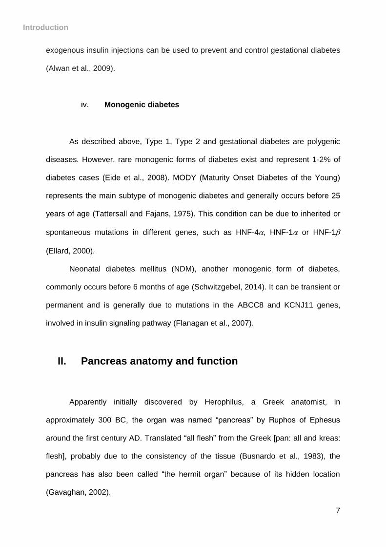
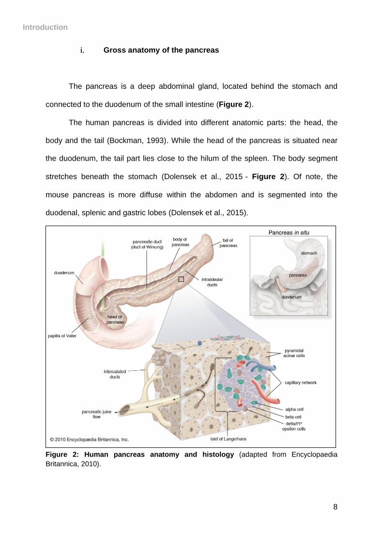
Gross anatomy of the pancreas
The pancreas is a deep abdominal gland, located behind the stomach and
connected to the duodenum of the small intestine (Figure 2).
The human pancreas is divided into different anatomic parts: the head, the
body and the tail (Bockman, 1993). While the head of the pancreas is situated near
the duodenum, the tail part lies close to the hilum of the spleen. The body segment
stretches beneath the stomach (Dolensek et al., 2015)- Figure 2). Of note, the
mouse pancreas is more diffuse within the abdomen and is segmented into the
duodenal, splenic and gastric lobes (Dolensek et al., 2015).
Figure 2: Human pancreas anatomy and histology (adapted from Encyclopaedia
Britannica, 2010).
Introduction
9
The pancreas is an amphicrine gland, thus composed of two compartments: the
exocrine tissue and the endocrine tissue, each with distinct structures and functions
(Figure 2). Responsible for the secretion of various enzymes and hormones, it has a
preponderant role in food digestion and glucose homeostasis (Cano et al., 2007;
Slack, 1995).
Organization and function of the exocrine tissue
Representing the major component of the gland, the exocrine compartment
accounts for approximately 95% of the total organ (Rahier et al., 1981). The exocrine
pancreas is involved in two physiological processes: the regulation of gastric acid
and chemical digestion. It is mostly composed of serous acinar cells and ductal cells
forming a branched network throughout the gland (Edlund, 2002).
a. Acinar cells
Pancreatic acini are made of pyramidal acinar cells containing zymogen
granules within secretory vesicles. These exocrine acinar cells produce and secrete
a juice composed of several enzymes (i.e. lipases, amylase, trypsin) involved in food
digestion (Tan, 2005). Combined, these enzymes hydrolyze complex nutrients, such
as polysaccharides, triglycerides and proteins (Whitcomb and Lowe, 2007).
Pancreatic enzymes are released as pancreatic juice into the duodenum through the
branched ductal network.
Introduction
10
b. Duct cells
Pancreatic ductal cells are organized into a complex network running
throughout the entire pancreas. Acinar cell secretions are collected by intercalated
ducts and siphoned into intralobular ducts; this pancreatic juice is drained into the
duodenum at the papilla of Vater through the main pancreatic duct also called duct of
Wirsung (Reichert and Rustgi, 2011)- Figure 2).
To regulate duodenal acidity, ductal cells produce water and bicarbonate
which is added to the enzyme mixture, thus providing optimal conditions for enzyme
activity (Githens, 1988).
Organization and function of the endocrine tissue
The endocrine compartment of the pancreas is composed of small functional
units named islets of Langerhans. Originally described by Paul Langerhans in his
thesis in 1869 (Langerhans, 1869), the islets represent about 4-5% of the pancreatic
mass and are found scattered throughout the exocrine tissue (Ionescu-Tirgoviste et
al., 2015; Rahier et al., 1981). However, regional islet distribution in the different
parts of the pancreas is subject to controversy. Indeed, while reports suggest the
highest number of mouse or human islets in the pancreatic tail (Wang et al., 2013),
other studies point to a similar enrichment in the head of the pancreas (Hörnblad et
al., 2011).
Islets of Langerhans are arranged into clusters containing five hormone-
secreting cell types: -cells, -cells, -cells, PP-cells and -cells; respectively
responsible for the secretion of insulin, glucagon, somatostatin, pancreatic
Introduction
11
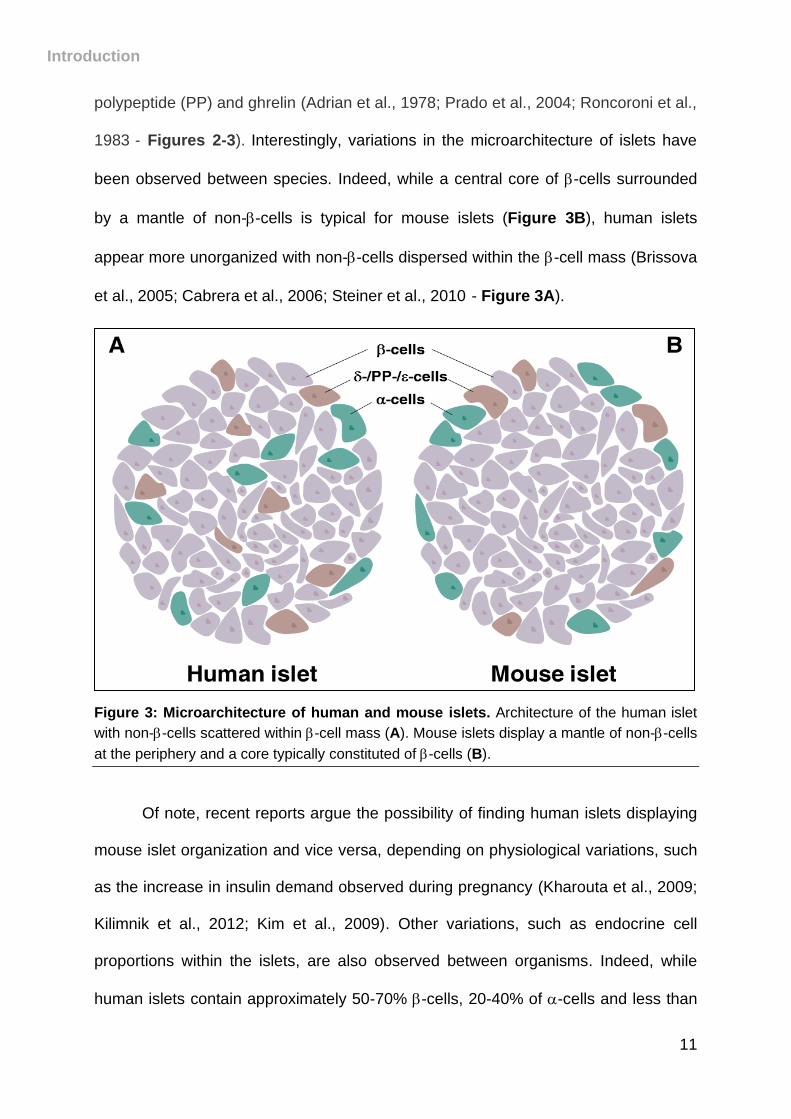
polypeptide (PP) and ghrelin (Adrian et al., 1978; Prado et al., 2004; Roncoroni et al.,
1983)- Figures 2-3). Interestingly, variations in the microarchitecture of islets have
been observed between species. Indeed, while a central core of -cells surrounded
by a mantle of non--cells is typical for mouse islets (Figure 3B), human islets
appear more unorganized with non--cells dispersed within the -cell mass (Brissova
et al., 2005; Cabrera et al., 2006; Steiner et al., 2010)- Figure 3A).
Figure 3: Microarchitecture of human and mouse islets. Architecture of the human islet
with non--cells scattered within -cell mass (A). Mouse islets display a mantle of non--cells
at the periphery and a core typically constituted of -cells (B).
Of note, recent reports argue the possibility of finding human islets displaying
mouse islet organization and vice versa, depending on physiological variations, such
as the increase in insulin demand observed during pregnancy (Kharouta et al., 2009;
Kilimnik et al., 2012; Kim et al., 2009). Other variations, such as endocrine cell
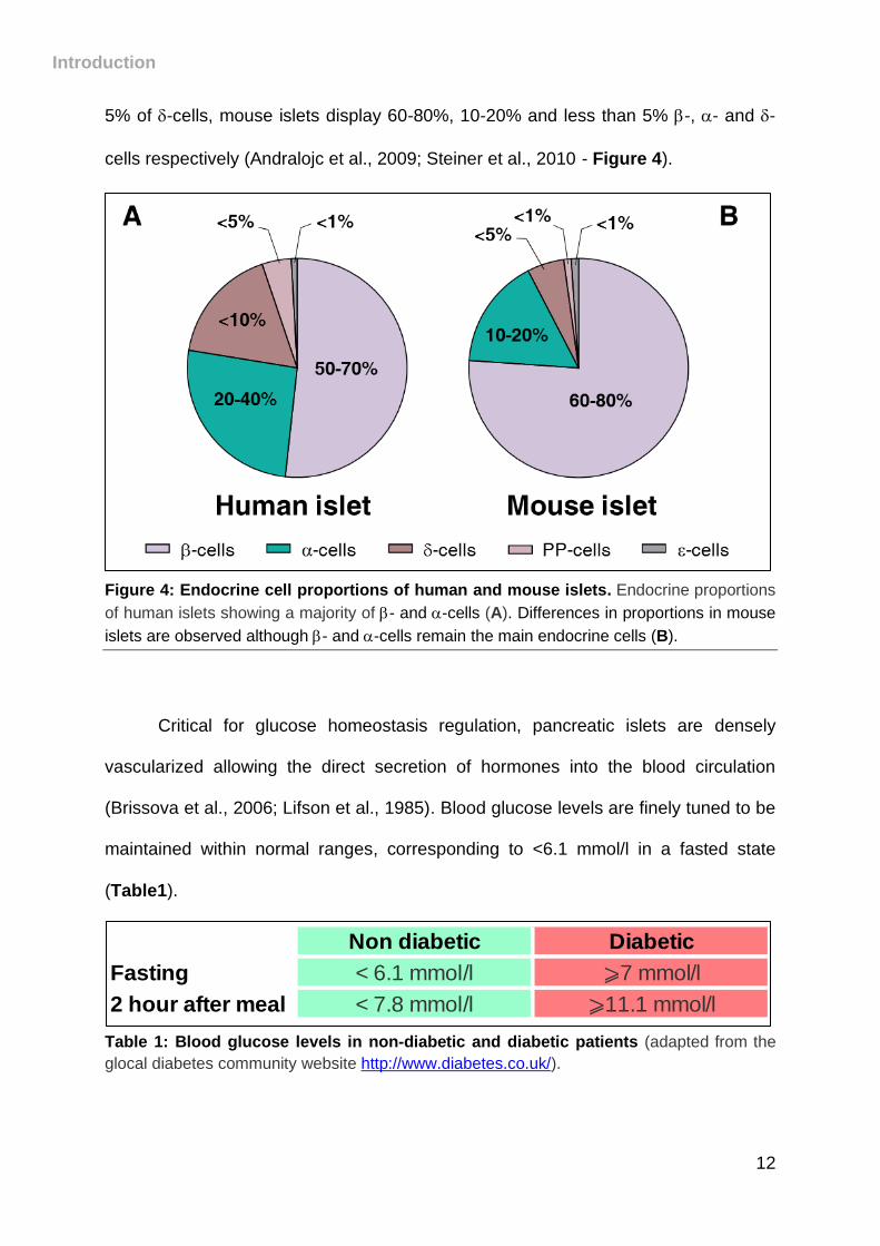
proportions within the islets, are also observed between organisms. Indeed, while
human islets contain approximately 50-70% -cells, 20-40% of -cells and less than
Introduction
12
5% of -cells, mouse islets display 60-80%, 10-20% and less than 5% -, - and -
cells respectively (Andralojc et al., 2009; Steiner et al., 2010)- Figure 4).
Figure 4: Endocrine cell proportions of human and mouse islets. Endocrine proportions
of human islets showing a majority of - and -cells (A). Differences in proportions in mouse
islets are observed although - and -cells remain the main endocrine cells (B).
Critical for glucose homeostasis regulation, pancreatic islets are densely
vascularized allowing the direct secretion of hormones into the blood circulation
(Brissova et al., 2006; Lifson et al., 1985). Blood glucose levels are finely tuned to be
maintained within normal ranges, corresponding to <6.1 mmol/l in a fasted state
(Table1).
Table 1: Blood glucose levels in non-diabetic and diabetic patients (adapted from the
glocal diabetes community website http://www.diabetes.co.uk/).
Non diabetic Diabetic
Fasting < 6.1 mmol/l ⩾7 mmol/l
2 hour after meal < 7.8 mmol/l ⩾11.1 mmol/l
Introduction
13
a. Insulin-secreting -cells
Pancreatic -cells represent the most abundant cell type in islets with 50-70%
in humans and 60-80% in mice (Dolensek et al., 2015)- Figure 4). Synthesizing and
secreting the hypoglycemic hormone insulin, they play a key role in regulating
glucose homeostasis. Insulin is a protein of 51 amino acids, composed of two
polypeptide chains linked by disulfide bonds resulting from the cleavage of proinsulin
by the action of prohormone convertases PC1/3 and PC2 (Steiner et al., 1967; Weiss
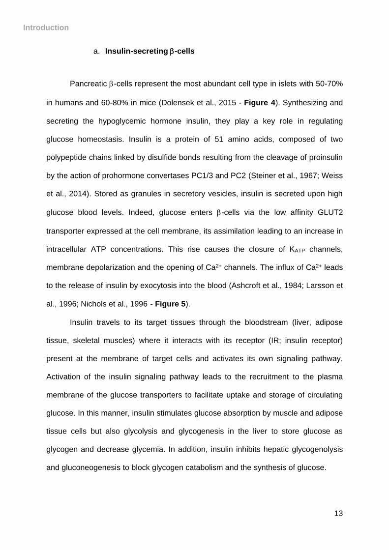
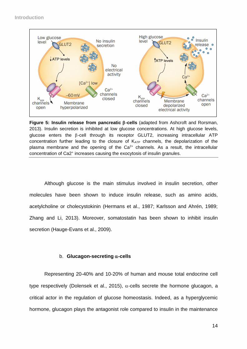
et al., 2014). Stored as granules in secretory vesicles, insulin is secreted upon high
glucose blood levels. Indeed, glucose enters -cells via the low affinity GLUT2
transporter expressed at the cell membrane, its assimilation leading to an increase in
intracellular ATP concentrations. This rise causes the closure of KATP channels,
membrane depolarization and the opening of Ca2+ channels. The influx of Ca2+ leads
to the release of insulin by exocytosis into the blood (Ashcroft et al., 1984; Larsson et
al., 1996; Nichols et al., 1996)- Figure 5).
Insulin travels to its target tissues through the bloodstream (liver, adipose
tissue, skeletal muscles) where it interacts with its receptor (IR; insulin receptor)
present at the membrane of target cells and activates its own signaling pathway.
Activation of the insulin signaling pathway leads to the recruitment to the plasma
membrane of the glucose transporters to facilitate uptake and storage of circulating
glucose. In this manner, insulin stimulates glucose absorption by muscle and adipose
tissue cells but also glycolysis and glycogenesis in the liver to store glucose as
glycogen and decrease glycemia. In addition, insulin inhibits hepatic glycogenolysis
and gluconeogenesis to block glycogen catabolism and the synthesis of glucose.
Introduction
14
Figure 5: Insulin release from pancreatic -cells (adapted from Ashcroft and Rorsman,
2013). Insulin secretion is inhibited at low glucose concentrations. At high glucose levels,
glucose enters the -cell through its receptor GLUT2, increasing intracellular ATP
concentration further leading to the closure of KATP channels, the depolarization of the
plasma membrane and the opening of the Ca2+ channels. As a result, the intracellular
concentration of Ca2+ increases causing the exocytosis of insulin granules.
Although glucose is the main stimulus involved in insulin secretion, other
molecules have been shown to induce insulin release, such as amino acids,
acetylcholine or cholecystokinin (Hermans et al., 1987; Karlsson and Ahrén, 1989;
Zhang and Li, 2013). Moreover, somatostatin has been shown to inhibit insulin
secretion (Hauge-Evans et al., 2009).
b. Glucagon-secreting -cells
Representing 20-40% and 10-20% of human and mouse total endocrine cell
type respectively (Dolensek et al., 2015), -cells secrete the hormone glucagon, a
critical actor in the regulation of glucose homeostasis. Indeed, as a hyperglycemic
hormone, glucagon plays the antagonist role compared to insulin in the maintenance
Introduction
15
of blood glucose levels within normal ranges (Unger, 1971). In accordance with a
common feature of hormones, glucagon is originally synthesized as a precursor
named proglucagon. This precursor will be processed in pancreatic -cells to
produce the 29-amino acid peptide glucagon (Furuta et al., 2001), stored as granules
in secretory vesicles.
The exocytosis of glucagon is induced when blood sugar levels drop, during
fasting or increased energy expenditure, to promote target tissues to mobilize
glucose and therefore increase glycemic levels. In the liver, glucagon interacts with
its membrane-bound receptor - a G-protein coupled receptor - (Authier and
Desbuquois, 2008; Brubaker and Drucker, 2002) to promote hepatic
gluconeogenesis and glycogenolysis, therefore stimulating the breakdown of
glycogen (Habegger et al., 2010).
Regulation of glucagon secretion is driven by various signals. Glucagon
release is inhibited by glucose, insulin and somatostatin (Hamaguchi et al., 1991;
Hauge-Evans et al., 2009; Kisanuki et al., 1995). Moreover, other stimuli such as
circulating amino acids (Pagliara et al., 1974) and fatty acids (Bollheimer et al., 2015)
have been shown to impact the secretion of glucagon.
c. Somatostatin-secreting -cells
Pancreatic -cells make up less than 10% and 5% of endocrine cell types in
human and mouse islets, respectively (Dolensek et al., 2015). -cells secrete
somatostatin (SST), also known as growth hormone-inhibiting hormone (GHIH) or
somatotropin release-inhibiting factor (SRIF).
Introduction
16
Somatostatin exists as two isoforms, somatostatin-14 (SST-14) and
somatostatin-28 (SST-28), resulting from different cleavage of the prosomatostatin
(Andrews and Dixon, 1986). Binding somatostatin receptors (members of the G-
protein coupled receptor family which comprise five subtypes - SSTR1-5 - (Yamada
et al., 1993), SST-14 and SST-28 are able to exert a wide range of effects.
Typically, somatostatin is described as a paracrine and endocrine peptide as it
can induce the inhibition of the secretion of numerous hormones of the
gastrointestinal tract, the nervous system and the pancreas (Dollinger et al., 1976). In
addition to pancreatic -cells, somatostatin is synthetized by cells in the central
nervous and gastrointestinal system (Reichlin, 1983). While tissue distribution of
SST-14 and SST-28 is still debated, it has been suggested that the two peptides
have different inhibitory potentials. SST-14 strongly inhibits glucagon secretion
through SSTR2 while SST-28 mostly blocks insulin release through SSTR5
(D’Alessio et al., 1989; Mandarino et al., 1981; Mitra et al., 1999). However, recent
analyses suggested that SSTR5 is not expressed in islet endocrine cells (DiGruccio
et al., 2016). Various stimuli induce somatostatin secretion, such as low
concentrations of glucose, fatty acids, HCL and urocortin 3 (Rouiller et al., 1980;
Schusdziarra et al., 1978; van der Meulen et al., 2015).
d. Pancreatic polypeptide-secreting PP-cells
Representing less than 5 and 1% of human and mouse total endocrine cells
respectively, PP-cells are more abundant in the islets located in head region of the
pancreas (Baetens et al., 1979; Gingerich et al., 1978). PP-cells secrete pancreatic
polypeptide (PP), a peptide of 36 amino acids and a member of the neuropeptide Y
Introduction
17
(NPY) family (Holzer et al., 2012). Pancreatic polypeptide is initially synthesized as a
precursor named pre-propancreatic polypeptide before further processing to produce
the hormone (Boel et al., 1984; Leiter et al., 1984).
Principally secreted postprandially (Batterham et al., 2003), PP is involved in
gastrointestinal motility, food intake, energy metabolism and gastric secretions
(Asakawa et al., 2003; Katsuura et al., 2002; Lin et al., 1977). The precise role of PP
in the pancreas is not yet evident, although its implication in the regulation of
pancreatic exocrine secretions is well documented (Lonovics et al., 1981; Louie et
al., 1985; Putnam et al., 1989). Moreover, it has been suggested a role for PP in the
stimulation of insulin secretion via the inhibition of somatostatin release (Kim et al.,
2014).
e. Ghrelin-secreting -cells
Pancreatic -cells are abundantly present in the fetal pancreas (Rindi et al.,
2002; Wierup et al., 2004) but constitute less than 1% of the endocrine cells in adult
human and mouse islets (Andralojc et al., 2009; Dolensek et al., 2015). Ghrelin, the
hormone secreted by the pancreatic -cells, is a 28 amino acid peptide resulting from
two successive cleavages of the precursor pre-proghrelin and is mainly produced
and secreted by gastrointestinal cells upon food intake (Cummings et al., 2001). This
hormone is involved in numerous physiological processes, such as regulation of
appetite, body weight, gastrointestinal motility, gastric secretions and control of
anxiety (Müller et al., 2015).
The role of ghrelin in the pancreas and on insulin secretion is still debated as it
has been shown to stimulate or suppress insulin release depending on its plasma
Introduction
18
concentration (Granata et al., 2010; Salehi et al., 2004). Subject to controversy is the
hypothesis that ghrelin could influence somatostatin, glucagon and PP secretion
(Arosio et al., 2003; Egido et al., 2002; Salehi et al., 2004). However, a recent report
suggested a role of ghrelin in potentiating glucose-stimulated somatostatin secretion
in mouse and human islets (DiGruccio et al., 2016).
III. Mouse pancreatic development
Our current knowledge of pancreatic development mostly results from
information gleamed using loss- and/or gain-of-function mouse models and genetic
lineage-tracing experiments. It is important to mention that, while sharing similarities,
rodent and human pancreas development also show divergences (Sarkar et al.,
2008). Pancreas development is a highly complex process, requiring diverse
interactions between tissues, genes and soluble factors. An elegant approach using
whole-mount immunofluorescence experiments and global gene expression analyses
on endodermal domains suggested that pancreas organogenesis is mostly
dependent on combinations of large cohort of transcription factors (TFs) and
signaling pathways (Sherwood et al., 2009). Of note, epigenetic modifications
collaborate with transcription factors to determine pancreatic cell fates (Haumaitre et
al., 2008; Xu et al., 2011), although this aspect will not be discussed in this
manuscript.
Here, we will describe key events and TFs involved in mouse pancreas
organogenesis.
Introduction
19
Early development
a. Endoderm patterning
During embryogenesis, following fertilization, the zygote undergoes multiple
cycles of mitotic divisions until the formation of the blastocyst. The blastocyst will
implant into the uterine wall at which point the process of gastrulation begins. In
vertebrates, gastrulation leads to the formation of three germ layers identified as
ectoderm, mesoderm and endoderm, each layer giving rise to distinct organs. In
mice, at embryonic day (E) 6-7.5, the endoderm initially appears as a flat single-cell
layer epithelium which will then, 2 days later (E8.5), internalize and fold to form the
primitive gut tube.
b. Gut tube patterning and pancreas budding
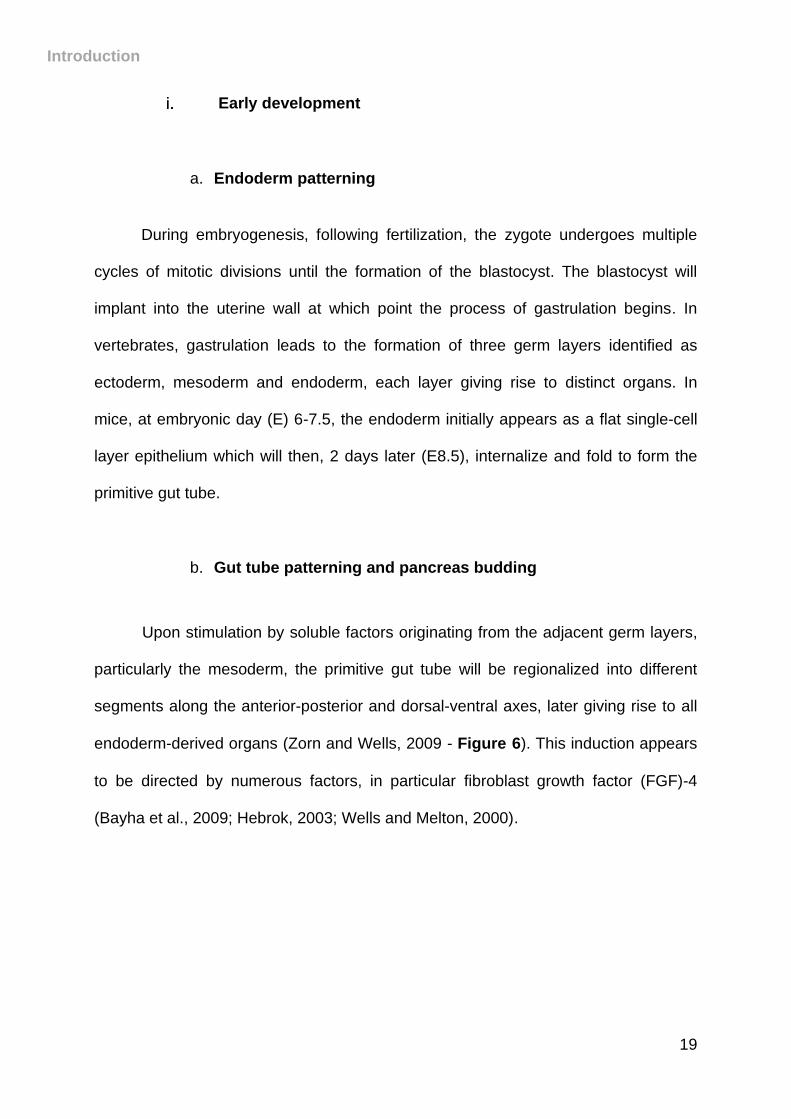
Upon stimulation by soluble factors originating from the adjacent germ layers,
particularly the mesoderm, the primitive gut tube will be regionalized into different
segments along the anterior-posterior and dorsal-ventral axes, later giving rise to all
endoderm-derived organs (Zorn and Wells, 2009)- Figure 6). This induction appears
to be directed by numerous factors, in particular fibroblast growth factor (FGF)-4
(Bayha et al., 2009; Hebrok, 2003; Wells and Melton, 2000).
Introduction
20
Figure 6: Gut tube patterning and endoderm-derived organs (adapted from Zorn and
Wells, 2009). Schematic representation of the gut tube of a mouse embryo at E10.5. The gut
tube is divided into Foregut, Midgut and Hindgut segments through anterior-posterior and
dorsal-ventral axes and will give rise to digestive and respiratory tracts and associated
organs.
In vertebrates, the pancreas emerges from ventral and dorsal domains of the
foregut endoderm (E9.5) (Slack, 1995). As a result of their localization along the gut
tube, the two buds are not surrounded by the same tissues and are therefore
subjected to different signals (Figure 7). The dorsal outgrowth of the pancreas
receive permissive signals from the notochord and the dorsal aorta (M. Hebrok et al.,
1998), whereas the ventral bud is subjected to cardiac mesoderm and septum
transversum mesenchyme signals (Kumar et al., 2003).
Signals from the notochord, such as FGF, activin ligands and retinoic acid
(RA), direct the adjacent endoderm to develop into the pancreatic dorsal bud (Martín
et al., 2005), by repressing sonic hedgehog (Shh) expression (Kim et al., 1997), an
activator of hedgehog (Hh) pathway (Figure 7A). Repression of the signaling factor
Shh results in the expression of Pdx1 (Hebrok et al., 1998), a key regulator of
pancreas development. In addition, FGFs and BMPs signals from the cardiac
Introduction
21
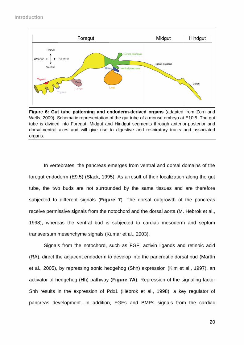
mesoderm and the septum transversum mesenchyme are required for Pdx1
expression and ventral pancreatic bud specification (Kumar et al., 2003; Wandzioch
and Zaret, 2009)- Figure 7B).
Figure 7: Ventral and dorsal pancreas specification (adapted from Mastracci and Sussel,
2012). Most signals received by the dorsal pancreas originate from the notochord (A), while
the ventral pancreas is subjected to septum transversum mesenchyme and cardiac
mesoderm signals (B).
Pancreas specification
Ventral and dorsal buds are morphologically detectable around E9.5 in mice.
However, as early as E8.5 and prior the outgrowth of these two primordia, the ventral
and dorsal prepancreatic regions are specified. Indeed, Wessells and Cohen
demonstrated the ability of cells in the foregut endoderm to differentiate into all
pancreatic lineages (Wessells and Cohen, 1967). It has also been suggested that
such prepancreatic epithelium contains multipotent pancreatic progenitors (MPCs)
able to differentiate into all pancreatic cells (Gu et al., 2002). Of note, pancreatic
anlage specification is a dynamic process involving several transcription factors
acting in coordination (Figure 9).
Introduction
22
Pdx1 (pancreatic and duodenal homeobox 1) is a key regulator of pancreas
development, its expression in progenitor cells (at E8.5) driving the differentiation of
all exocrine and endocrine pancreatic cells. Mice lacking Pdx1 display bud formation
but pancreas agenesis, demonstrating that Pdx1 is necessary for bud growth but not
for their initial induction (Jonsson et al., 1994; Offield et al., 1996; Stoffers et al.,
1997), suggesting that pancreatic development is initiated before Pdx1 expression.
Motor neuron and pancreas homeobox 1 (Mnx1), also known as Homeobox HB9
(HLXB9) precedes Pdx1 in the dorsal pancreatic endoderm as it is expressed on
embryonic day 8 (Harrison et al., 1999; Li et al., 1999). Its expression is necessary
for dorsal bud induction, as HLXB9-deficient mice displayed dorsal lobe agenesis
due to a lack of Pdx1 induction. In addition to Pdx1 and Mnx1 expression, early
pancreatic specification requires numerous additional transcription factors, including
Ptf1a (Burlison et al., 2008; Kawaguchi et al., 2002; Krapp et al., 1998), Sox9
(Seymour et al., 2007) and Hnf1 (De Vas et al., 2015; Haumaitre et al., 2005).
Indeed, the mice deficient for each of these genes display varying degrees of
pancreas hypoplasia or agenesis.
Importantly, mouse pancreas organogenesis is separated into two major
waves : a primary and a secondary transitions (Pictet et al., 1972; Rutter et al.,
1968).
a. Primary transition
The primary transition begins around E9.5 with the thickening of the foregut
endoderm, which mainly contains MPCs. The shift from a monolayer to a multilayer
Introduction
23
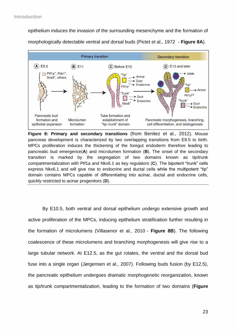
epithelium induces the invasion of the surrounding mesenchyme and the formation of
morphologically detectable ventral and dorsal buds (Pictet et al., 1972) - Figure 8A).
Figure 8: Primary and secondary transitions (from Benitez et al., 2012). Mouse
pancreas development is characterized by two overlapping transitions from E9.5 to birth.
MPCs proliferation induces the thickening of the foregut endoderm therefore leading to
pancreatic bud emergence(A) and microlumen formation (B). The onset of the secondary
transition is marked by the segregation of two domains known as tip/trunk
compartmentalization with Ptf1a and Nkx6.1 as key regulators (C). The bipotent “trunk” cells
express Nkx6.1 and will give rise to endocrine and ductal cells while the multipotent “tip”
domain contains MPCs capable of differentiating into acinar, ductal and endocrine cells,
quickly restricted to acinar progenitors (D).
By E10.5, both ventral and dorsal epithelium undergo extensive growth and
active proliferation of the MPCs, inducing epithelium stratification further resulting in
the formation of microlumens (Villasenor et al., 2010)- Figure 8B). The following
coalescence of these microlumens and branching morphogenesis will give rise to a
large tubular network. At E12.5, as the gut rotates, the ventral and the dorsal bud
fuse into a single organ (Jørgensen et al., 2007). Following buds fusion (by E12.5),
the pancreatic epithelium undergoes dramatic morphogenetic reorganization, known
as tip/trunk compartmentalization, leading to the formation of two domains (Figure
Introduction
24
8C). Of note, Notch signaling appears to play a key role in the tip/trunk segregation
(Qu et al., 2013).
b. Secondary transition
The secondary transition begins around E12.5-E13.5 and is marked by an
extensive reorganization due to a massive growth and branching of the pancreatic
epithelium, concomitant to a major wave of endocrine and exocrine cell
differentiation.
The bipotent “trunk” region contains cells that will give rise to endocrine and
ductal cells while the multipotent “tip” domain contains MPCs capable of
differentiating into acinar, ductal and endocrine cells. The multipotent tip progenitors
undergo a developmental switch at around E13-E14 and are then restricted to acinar
progenitors (Zhou et al., 2007). The specification of the different cell fates is driven by
numerous genes and transcription factors, with Ptf1a and Nkx6.1 as key regulators of
tip/trunk segregation (Figure 8D). Ptf1a is expressed in the tip cells while Nkx6.1 is
exclusively expressed in trunk bipotent cells. Importantly, these two transcription
factors mutually inhibit each other, favoring one or the other domain. It has been
suggested that Ptf1a represses Nkx6.1 to block endocrine differentiation at the
expense of acinar development (Schaffer et al., 2010).
Introduction
25
Exocrine specification
a. Acinar cell fate
Acinar cells arise from MPCs located in the tip cells of the pancreatic
epithelium. This differentiation occurs between E14.5 and E15.5 in mice and acini are
morphologically visible by E15.5.
Acinar cell specification is regulated by several transcription factors (Figure
9), including Ptf1a and Nr5a2 (Esni, 2004; Rose et al., 2001). As previously
mentioned, Ptf1a-null mice display a pancreas agenesis. Interestingly, a total loss of
the exocrine tissue is observed in these mice, while few endocrine cells remain
(Krapp et al., 1996). In addition, the loss of Nr5a2 in adult mice results in impaired
exocrine enzymes production and secretion (Holmstrom et al., 2011). Moreover,
Mist1 is required for acinar fate maintenance and complete differentiation. Although
Mist1-deficient mice display acinar cells, these cells present defective apical-basal
polarity leading to a massive disorganization of the exocrine tissue (Direnzo et al.,
2012; Pin et al., 2001). Interestingly, carbopeptidase (Cpa) is expressed by the
forming acini at E12.5 and is considered to be an early exocrine marker (Jørgensen
et al., 2007). Finally, recent studies described c-Myc as another acinar differentiation
factor, as its inactivation in pancreatic progenitors leads to acinar cell hypoplasia
(Bonal et al., 2009; Nakhai et al., 2008).
b. Ductal cell fate
The mechanisms involved in ductal cell fate specification are poorly
understood. However, as previously mentioned, the “trunk” region contains bipotent
Introduction
26
cells able to give rise to endocrine and ductal cells. It has been suggested that the
switch that induces trunk cells to derive into duct cells is mostly driven by Notch
pathway (Figure 9).
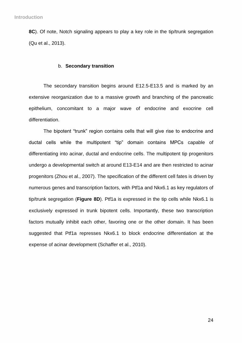
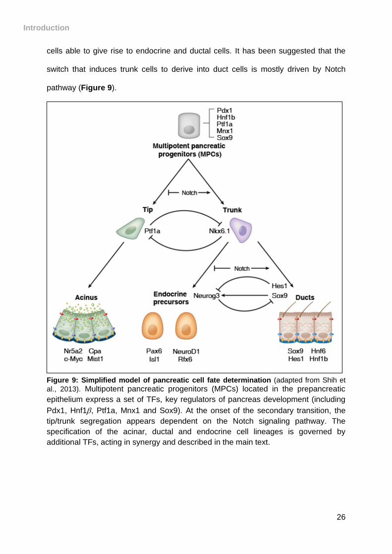
Figure 9: Simplified model of pancreatic cell fate determination (adapted from Shih et
al., 2013). Multipotent pancreatic progenitors (MPCs) located in the prepancreatic
epithelium express a set of TFs, key regulators of pancreas development (including
Pdx1, Hnf1, Ptf1a, Mnx1 and Sox9). At the onset of the secondary transition, the
tip/trunk segregation appears dependent on the Notch signaling pathway. The
specification of the acinar, ductal and endocrine cell lineages is governed by
additional TFs, acting in synergy and described in the main text.
Introduction
27
The activation of the Notch signaling pathway induces the expression of a
target gene Hes1. Importantly, Hes1 has been found to act as a repressor of the pro-
endocrine gene Neurogenin 3 (Neurog3). Indeed, Neurog3-deficiency is proposed to
induce ductal cell specification (Jensen et al., 2000; Magenheim et al., 2011) while
Hes1 mutant mice display an augmented differentiation of endocrine cells
(Jørgensen et al., 2007). Moreover, the ductal epithelium expresses Sox9 (Delous et
al., 2012), Hnf6 (Pierreux et al., 2006) and Hnf1 (De Vas et al., 2015), these
transcription factors forming a transcriptional network that has been involved in duct
cell specification. Of note, the deletion of the corresponding genes leads to the
impairment of duct morphogenesis.
Endocrine specification
Endocrine allocation and maintenance of the different endocrine cell lineages
requires the sequential and stage-dependent activation or repression of specific
genes. Indeed, endocrine cell specification is controlled by a network of transcription
factors acting in specific combinations (Figure 9). The master gene involved in
endocrine fate determination is Neurog3. Neurog3 is a proendocrine gene required
for the development of all the endocrine cells, as Neurog3-deficient mice do not
display any hormone-expressing endocrine cells (Gradwohl et al., 2000). Both
activation and repression of Neurog3 expression have been suggested to be
dependent on Notch signaling. A recent study demonstrated that high Notch activity
induces the expressions of Hes1 and Sox9, leading to Neurog3 inhibition and the
subsequent blockade of endocrine differentiation. However, intermediate Notch

Introduction
28
activity leads to the expression of Sox9 alone and the derepression of Neurog3
expression (Shih et al., 2012).
Neurog3 expression begins at E8.5 and peaks at E15.5, corresponding to the
major wave of endocrine cell differentiation with little or no of this gene in hormones-
expressing cells (Schwitzgebel et al., 2000). Previous reports demonstrated that
Neurog3 initiates endocrine specification but acts upstream of numerous transcription
factors important for endocrine cell differentiation/maintenance. Indeed, using gene-
deficient mouse models and further genetic analyses, it has been shown that Insm1,
Rfx6, Isl1, NeuroD1, Pax6, Myt1 are required for proper endocrine-cell differentiation.
The deletion of one of these gene impact in different manner final islet-cell
differentiation (Ahlgren et al., 1997; Mellitzer et al., 2006; Naya et al., 1997; Sander
et al., 1997; Smith et al., 2010; Soyer et al., 2010; Wang et al., 2008)- Figure 9).
Differentiation of endocrine cells
As previously mentioned, Neurog3-expressing cells generate all the five
endocrine cell types: -, -, -, PP- and -cells. While early expression of Neurog3
exclusively gives rise to -cells (E8.5), -, -, PP- and -cells appear at E10.5, E11.5,
E12.5 and E14.5 respectively (Johansson et al., 2007; Prado et al., 2004). Endocrine
cells subsequently migrate and emerge from the ductal epithelium and aggregate to
eventually form the islets of Langerhans around E18.5 (Bouwens and De Blay,
1996).
The specification of the different hormone-secreting cells largely depends on
the sequential activation/repression of TFs. Importantly, these TFs control the
Introduction
29
differentiation, as well as the maintenance and the function of endocrine cell
subtypes.
Here we will focus on -, - and -cell determination/maintenance.
a. -cells – glucagon
Arx (aristaless-related homeobox) plays a key role in the determination of the
- and PP-cell lineages, later being restricted to mature glucagon-expressing cells
where it is involved in maintaining their identity. Indeed, Arx mutant mice display
severe hypoglycemia caused by a loss of -cells associated with a proportional
increase in - and -cell counts (Collombat et al., 2003).
Although Pax6, NeuroD1 and Nkx2.2 are not specific to -cells, their deletion
induces glucagon-expressing cell deficiency (Naya et al., 1997; Sander et al., 1997;
Sussel et al., 1998). Brn4 (also known as POU3F4) is exclusively expressed by -
cells. Although not necessary for -cell development, Brn4 acts as a transcriptional
regulator of glucagon expression (Heller et al., 2004; Hussain et al., 1997).
b. -cells – insulin
A number of TFs involved in early pancreatic development are also found to
be important in -cell identity maintenance.
Pax4 is critical for -cell determination and is exclusively expressed in -cells
in the adult pancreas. Accordingly, Pax4-deficient mice develop severe
hyperglycemia and die shortly after birth. Analysis of their pancreas revealed the
absence of - and -cells, concomitant with an increase in -cell numbers (Sosa-
Introduction
30
Pineda et al., 1997). Together with Pax4, Pdx1 and Nkx6.1 are major -cell
determinants. Indeed, -cell-specific inactivation of Nkx6.1 or Pdx1 leads to their
reprogramming into - and -cells (Gao et al., 2014; Schaffer et al., 2013). Moreover,
the loss of one allele of Pdx1 results in impaired -cell function and glucose
intolerance (Brissova et al., 2002).
MafA is expressed exclusively in -cells and regulates the expression of
genes involved in insulin synthesis and secretion. Its deletion causes impaired insulin
secretion and abnormal islet architecture (Olbrot et al., 2002; Zhang et al., 2005).
Interestingly, MafA was found to regulate insulin gene expression together with Pdx1
and NeuroD1 (Zhao et al., 2005). While not expressed exclusively in -cells, Nkx2.2
plays a role in the differentiation and maturation of insulin-producing cells, as its
deletion induces severe hyperglycemia in mice (Sussel et al., 1998). Similarly,
NeuroD1 and Pax6 appear to be involved in establishing and maintaining -cells as
their loss results in reduced -cell numbers (Gu et al., 2010; Naya et al., 1997;
Sander et al., 1997).
c. -cells – somatostatin
Somatostatin-producing -cell specification during pancreas development is
not well understood, mostly due to a lack of identification of -cell-specific
transcription factors.
As previously mentioned, total depletion of Pax4 expression leads to an
absence -cells (Sosa-Pineda et al., 1997). Conversely, Ganon et al. observed
increased numbers of -cells after specific the deletion of Pdx1 in -cells, such
supplementary somatostatin-expressing cells not arising from the conversion of
Introduction
31
Pdx1-null--cells. The authors therefore suggested that the increased -cell number
is due to either their augmented proliferation or the reallocation of endocrine
progenitors toward a -cell fate (Gannon et al., 2008).
In addition, an increase in somatostatin-positive cells was observed in mice
deficient for Arx (Collombat et al., 2003), as well as in Arx/Pax4 double mutant mice
(Collombat et al., 2005). Moreover, pancreata of mice lacking Arx and Nkx2.2
displayed an increase in -cell numbers, a significant number of such cells
expressing the hormone ghrelin (Kordowich et al., 2011). These studies strongly
suggest an antagonist effect of Arx on somatostatin-expressing cell development.
The first transcription factor involved in pancreatic somatostatin-cell
development has been recently characterized. In addition to its critical role in ventral
pancreas organogenesis (Bort et al., 2004), it has been shown that Hhex is essential
for the differentiation and the maintenance of -cells (Zhang et al., 2014). In this
elegant study, Hhex deficiency in mouse pancreas was found to induce a massive
loss of -cell numbers concomitant with a decrease in somatostatin secretion.
Interestingly, a recent study showed a -to- cell conversion upon the inactivation of
Mnx1 in endocrine precursors. The authors further suggested that Mnx1 acts as a
repressor of δ-cell specification through the direct or indirect inhibition of Hhex
expression (Pan et al., 2015).
Finally, -endosulfine, PCSK9 and Ptch1 receptor were found to be
specifically expressed in -cells in rat-, human- and mouse-islets; but no investigation
concerning their role in -cell fate specification has been undertaken (Grieco et al.,
2011; Gros et al., 2002; Langhi et al., 2009).
Introduction
32
IV. T1DM actual therapies & current research
As previously mentioned, diabetes patients are exposed to various
cardiovascular complications due to chronic increased blood glucose levels and are
at risk of dying prematurely. Thereby, doctors and researchers are acting collectively
in an attempt to manage or even reverse this hyperglycemia.
Management of T1DM
The priority in the management of T1DM is to monitor blood glucose levels to
strictly maintain glycemia within normal ranges (Table 1), thereby preventing
hyperglycemia- and hypoglycemia-associated complications.
a. Exogenous insulin administration
To compensate the inability of the pancreas to secrete insulin, type 1 diabetic
patients are dependent on exogenous insulin administration, such compensation
being indispensable for their survival. Although efficient, chronic insulin
administration through subcutaneous daily injections results in difficulties to strictly
regulate glycemic levels (due to fluctuations in diet, physical exercises or age) and
induces glycemic variability (alternations between hyper- and hypoglycemia) (Group.,
1993). The more recent implementation of subcutaneous pumps allows sustained
delivery of insulin and appears to be more efficient at maintaining normoglycemia
(Shetty and Wolpert, 2010).
Introduction
33
Another or an additive strategy involves the use of a combination of different
types of insulin with distinct timing of actions in lowering glucose levels. Mixed
preparations with long-, regular or short-acting and rapid-acting insulin can be used
for an improved delivery of insulin and therefore glycemic regulation. Long-acting
insulin acts several hours after injection and for a period of 24 hours while regular or
short-acting insulin acts within 30 minutes and is effective for 3 to 6 hours. Finally,
rapid-acting insulin requires only 15 min to lower blood glucose levels and is efficient
for up to 4 hours (McCall and Farhy, 2013). Such preparations afford a better
glycemic control, depending on the need and eating habits of patients, and can be
used through injections or via pumps.
Recently developed, the artificial pancreas (AP) is based on a closed-loop
control system consisting of a continuous subcutaneous glucose sensor and insulin
pump. This allows constant glucose-monitoring and insulin delivery. Although not yet
available, clinical trials demonstrate that this approach offers significant advantages
as it manages blood glucose levels with less variations and presents less daily stress
for the patients (Kovatchev et al., 2016; Kropff et al., 2015). However, hypoglycemia
remains a risk that should be dealt prior to a putative application.
b. Total pancreas and islets transplantation
As previously mentioned, exogenous insulin administration represents an
imperfect treatment for diabetes due to its inability to finely regulate blood glucose
levels. Thus, two alternatives aiming at replacing the damaged -cell mass in diabetic
patients exist: whole pancreas or islets transplantation. Whole pancreas
transplantation often also includes kidney transplants, as the organs could have been
Introduction
34
damaged due to hyperglycemia-associated complications. However, pancreas
transplantation is a heavy burden for diabetic patients, which is why islets
transplantation has been favored for the past decade. The Edmonton protocol
(Shapiro et al., 2000) has played a key role in islet transplantation from cadaveric
donors. It is of importance to note that these grafts involve lifelong
immunosuppressant treatment to avoid rejection and new -cell destruction, such
treatment rendering the patients susceptible to other infections.
Nonetheless, total pancreas or islet transplantation normally result in a rapid
glycemia normalization and exogenous insulin independence for up to 5 years,
further improving the quality of life of the patients (Domínguez-Bendala et al., 2016;
Sureshkumar et al., 2006). However, these medical procedures are mostly used for
type 1 diabetic patients with serious complications.
Although efficient, these therapies face the shortage of organ donors and the
associated side-effects of immunosuppressive drugs (Bruni et al., 2014).
Current axes of research
Despite the help offered by the different strategies available to physiologically
mimic the action of insulin, the difficulties in strictly regulating glycemic levels,
discomfort due to daily injections or pump replacement and financial limitations, have
encourage researchers to investigate alternatives. Consequently, current research
focuses on the replacement of the injured--cell-mass in diabetic patients using
several approaches and cell sources. The common goal of these diverse strategies is
to generate cells synthetizing and secreting appropriate amounts of insulin under
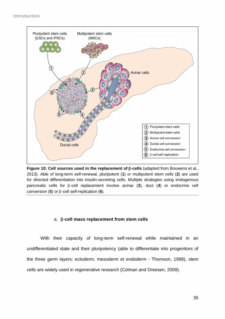
physiological conditions (Figure 10).
Introduction
35
Figure 10: Cell sources used in the replacement of -cells (adapted from Bouwens et al.,
2013). Able of long-term self-renewal, pluripotent (1) or multipotent stem cells (2) are used
for directed differentiation into insulin-secreting cells. Multiple strategies using endogenous
pancreatic cells for -cell replacement involve acinar (3), duct (4) or endocrine cell
conversion (5) or -cell self-replication (6).
a. -cell mass replacement from stem cells
With their capacity of long-term self-renewal while maintained in an
undifferentiated state and their pluripotency (able to differentiate into progenitors of
the three germ layers: ectoderm, mesoderm et endoderm -(Thomson, 1998), stem
cells are widely used in regenerative research (Colman and Dreesen, 2009).
Introduction
36
Several sources of stem cells exist and are used in diabetes research for
directed differentiation into insulin-secreting cells in vitro and subsequently in vivo.
1) Pluripotent stem cells (PSCs)
Stem cells are pluripotent, thus, under specific conditions, they are able to
specialize into progenitors of almost all cell lineages.
Embryonic stem cells (ESCs) are isolated from the inner cell mass (ICM) of a
developing blastocyst in early embryogenesis. The induction of their differentiation
into pancreatic progenitors and insulin-secreting cells is mainly based on
developmental studies and therefore aims at using strategies to modulate signaling
pathways and genes involved in pancreatic development. Numerous studies have
reported the differentiation of ESCs into insulin-producing cells. In 2000, Soria et al.
reported the restoration of chemically-induced diabetes by the injection of genetically
reprogrammed ESCs into -cells in mice. The construct used allowed the expression
of an antibiotic resistance gene under the control of human insulin gene (Soria et al.,
2000). Later, D’Amour et al. derived human embryonic stem cells (hESCs) into
pancreatic progenitors after a five-step protocol using sequential exposure of various
inducing agents and growth factors (D’Amour et al., 2006). Although this study was
the first demonstration of human-derived--cells, the resulting insulin-secreting cells
were glucose unresponsive. Later, the optimization of the previous protocol resulted
in hESC-derived-insulin-producing cells able to respond to glucose following their
transplantation and further differentiation in mice (Kroon et al., 2008). However, all
these protocols led to cell producing too low amounts of secreted insulin mostly due
to incomplete maturation. Recent studies have developed protocols that have had
Introduction
37
more success. Indeed, Rezania et al published a seven-stage protocol leading to the
differentiation of hESCs into -like cells, which were responsive to glucose and able
to restore chemically-induced diabetes in vivo after transplantation into
immunodeficient mice (Rezania et al., 2014). In addition, Pagliuca et al. described a
promising protocol allowing the production of large numbers of insulin-secreting cells
from human pluripotent stem cells, such cells preventing hyperglycemia following
transplantation into Akita immunodeficient mice (Pagliuca et al., 2014). In 2015, a
further improved protocol was published, leading the production of glucose
responsive -cells from hESCs in vitro and in vivo (Russ et al., 2015).
Although representing a promising therapy with clinical trials currently ongoing
(Viacyte, Inc.), the use of ESCs faces several obstacles. Indeed, the use of hESCs
requires embryos at the blastocyst stage to create ESC lines, therefore leading to
ethical concerns with the use of human embryonic cells (De Wert and Mummery,
2003; Mclaren, 2001). Another barrier is that transplanted exogenous ESCs can
trigger host immune rejection, thus requiring lifelong immunosuppressive treatments.
Induced pluripotent stem cells (iPSCs) represent a second type of PSCs,
exhibiting similar properties to ESCs. They are obtained from the reprogramming of
somatic cells into pluripotent cells able to self-renew. By expressing 4 specific
transcription factors, Oct4, Sox2, Klf4 and c-Myc, fibroblasts were reprogrammed into
pluripotent stem cells (Takahashi and Yamanaka, 2006; Yu et al., 2007). Later,
iPSCs were obtained by reprogramming other cell types, such as neuronal progenitor
cells (Kim et al., 2008), keratinocytes (Aasen et al., 2008), hepatocytes and gastric
epithelial cells (Aoi et al., 2008). Recently, pluripotent stem cells were induced from
mouse somatic cells using chemicals that could improve reprogramming efficiency or
replace one or all previously mentioned transcription factors (Bar-Nur et al., 2014;
Introduction
38
Hou et al., 2013; Ye et al., 2016). Using different strategies, such as chemically-
induced differentiation, iPSCS can be directed to differentiate into any cell type
including -cells (Alipio et al., 2010; Zhang et al., 2009). The use of iPSCs in diabetes
therapy therefore presents advantages when compare to hESCs. Indeed, the
reprogrammed cells avoid ethical issues of hESCs and graft rejection as they are
obtained from the differentiation of adult patients somatic cells.
Although promising, the use of embryonic stem cells and induced pluripotent
stem cells in diabetes therapy still needs further investigation. It is important to note
that ESCs and iPSCs possess oncogenic properties, as undifferentiated or not fully
differentiated stem cells transplanted can form teratomas (tumors containing cells
from 3 germ layers) (Hentze et al., 2009). In addition, autoimmunity in type 1 diabetic
patients can trigger the destruction of newly-implanted -cells.
2) Tissue stem cells
Tissue stem cells are found in various tissues including bone marrow (Jiang
et al., 2002), gastric epithelium (Bjerknes and Cheng, 2002), liver (Overturf et al.,
1997), brain (Reynolds and Weiss, 1992), adipose tissue (Zuk et al., 2002) and
umbilical cord (Prindull et al., 1978). Most stem cells found in post-natal tissues are
multipotent, therefore able to develop into different cell types. Although highly
debated, they represent another putative source for -cell regeneration.
Reports have shown the differentiation of bone marrow stem cells into
insulin-producing cells (Ianus et al., 2003; Karnieli et al., 2007), whereas a following
study failed to reproduce these findings (Taneera et al., 2006). Stem cells from
human umbilical cord were also suggested to be able to derive into islet cells
Introduction
39
(Pessina et al., 2004; Sun et al., 2007). In addition, human adipose tissue stem cells
were induced to differentiate into pancreatic endocrine cells (Timper et al., 2006) and
into functional insulin-producing cells in mice (Kajiyama et al., 2010). Liver stem cells
have also been suggested to possess the ability to differentiate into insulin-secreting
cells (Ferber et al., 2000; Yang et al., 2002).
Excluding ethical concerns and risk of immune rejection, the use of tissue
stem cells offers promising perspectives for diabetes therapy, although more
investigation is required to fully restore -cell mass function in diabetic patients using
these cell sources.
b. -cell mass replacement from pancreatic cells
With a rising number of reports, reprogramming/transdifferentiation of adult
pancreatic cells into functional -cells is an exciting topic but still highly debated.
Even though pancreas plasticity has mostly been described in experimental diabetes
models in mice, several approaches including genetic or chemical manipulations,
have been used to induce acinar-, ductal- or endocrine-cell conversion into insulin-
producing cells. Also, the development of lineage tracing tools has resulted in large
progress in this field.
1) From acinar cells
Abundant in the pancreatic tissue, exocrine acinar cells represent more than
95% of the gland, thus they constitute an interesting source for -cell replacement.
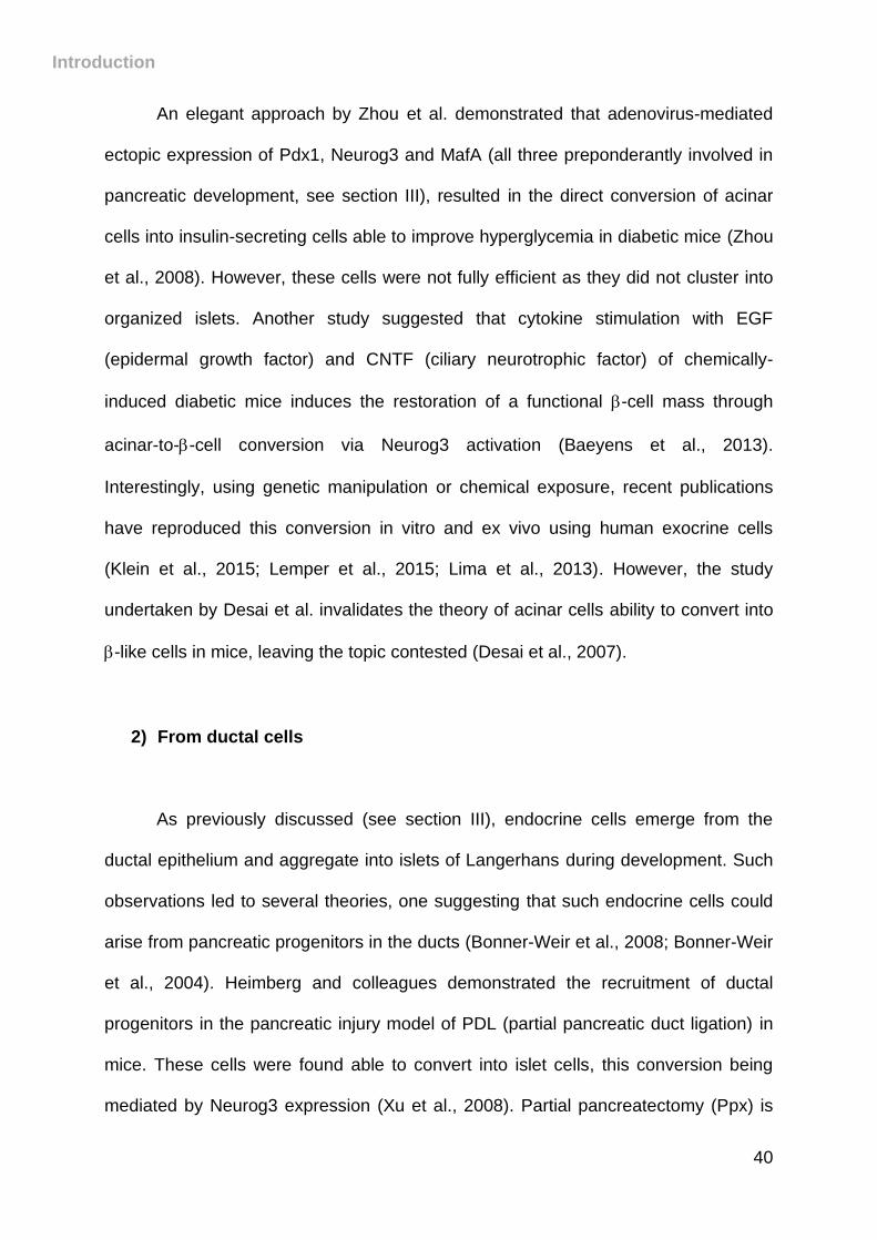
Introduction
40
An elegant approach by Zhou et al. demonstrated that adenovirus-mediated
ectopic expression of Pdx1, Neurog3 and MafA (all three preponderantly involved in
pancreatic development, see section III), resulted in the direct conversion of acinar
cells into insulin-secreting cells able to improve hyperglycemia in diabetic mice (Zhou
et al., 2008). However, these cells were not fully efficient as they did not cluster into
organized islets. Another study suggested that cytokine stimulation with EGF
(epidermal growth factor) and CNTF (ciliary neurotrophic factor) of chemically-
induced diabetic mice induces the restoration of a functional -cell mass through
acinar-to--cell conversion via Neurog3 activation (Baeyens et al., 2013).
Interestingly, using genetic manipulation or chemical exposure, recent publications
have reproduced this conversion in vitro and ex vivo using human exocrine cells
(Klein et al., 2015; Lemper et al., 2015; Lima et al., 2013). However, the study
undertaken by Desai et al. invalidates the theory of acinar cells ability to convert into
-like cells in mice, leaving the topic contested (Desai et al., 2007).
2) From ductal cells
As previously discussed (see section III), endocrine cells emerge from the
ductal epithelium and aggregate into islets of Langerhans during development. Such
observations led to several theories, one suggesting that such endocrine cells could
arise from pancreatic progenitors in the ducts (Bonner-Weir et al., 2008; Bonner-Weir
et al., 2004). Heimberg and colleagues demonstrated the recruitment of ductal
progenitors in the pancreatic injury model of PDL (partial pancreatic duct ligation) in
mice. These cells were found able to convert into islet cells, this conversion being
mediated by Neurog3 expression (Xu et al., 2008). Partial pancreatectomy (Ppx) is
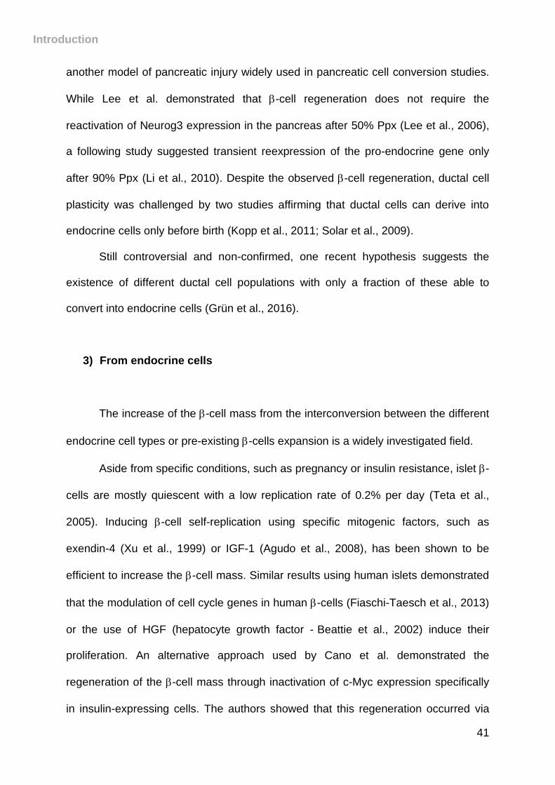
Introduction
41
another model of pancreatic injury widely used in pancreatic cell conversion studies.
While Lee et al. demonstrated that -cell regeneration does not require the
reactivation of Neurog3 expression in the pancreas after 50% Ppx (Lee et al., 2006),
a following study suggested transient reexpression of the pro-endocrine gene only
after 90% Ppx (Li et al., 2010). Despite the observed -cell regeneration, ductal cell
plasticity was challenged by two studies affirming that ductal cells can derive into
endocrine cells only before birth (Kopp et al., 2011; Solar et al., 2009).
Still controversial and non-confirmed, one recent hypothesis suggests the
existence of different ductal cell populations with only a fraction of these able to
convert into endocrine cells (Grün et al., 2016).
3) From endocrine cells
The increase of the -cell mass from the interconversion between the different
endocrine cell types or pre-existing -cells expansion is a widely investigated field.
Aside from specific conditions, such as pregnancy or insulin resistance, islet -
cells are mostly quiescent with a low replication rate of 0.2% per day (Teta et al.,
2005). Inducing -cell self-replication using specific mitogenic factors, such as
exendin-4 (Xu et al., 1999) or IGF-1 (Agudo et al., 2008), has been shown to be
efficient to increase the -cell mass. Similar results using human islets demonstrated
that the modulation of cell cycle genes in human -cells (Fiaschi-Taesch et al., 2013)
or the use of HGF (hepatocyte growth factor -(Beattie et al., 2002) induce their
proliferation. An alternative approach used by Cano et al. demonstrated the
regeneration of the -cell mass through inactivation of c-Myc expression specifically
in insulin-expressing cells. The authors showed that this regeneration occurred via
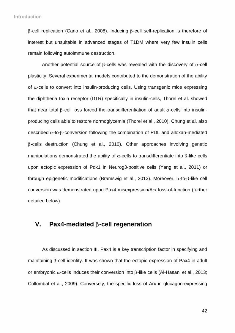
Introduction
42
-cell replication (Cano et al., 2008). Inducing -cell self-replication is therefore of
interest but unsuitable in advanced stages of T1DM where very few insulin cells
remain following autoimmune destruction.
Another potential source of -cells was revealed with the discovery of -cell
plasticity. Several experimental models contributed to the demonstration of the ability
of -cells to convert into insulin-producing cells. Using transgenic mice expressing
the diphtheria toxin receptor (DTR) specifically in insulin-cells, Thorel et al. showed
that near total -cell loss forced the transdifferentiation of adult -cells into insulin-
producing cells able to restore normoglycemia (Thorel et al., 2010). Chung et al. also
described -to--conversion following the combination of PDL and alloxan-mediated
-cells destruction (Chung et al., 2010). Other approaches involving genetic
manipulations demonstrated the ability of -cells to transdifferentiate into -like cells
upon ectopic expression of Pdx1 in Neurog3-positive cells (Yang et al., 2011) or
through epigenetic modifications (Bramswig et al., 2013). Moreover, -to--like cell
conversion was demonstrated upon Pax4 misexpression/Arx loss-of-function (further
detailed below).
V. Pax4-mediated -cell regeneration
As discussed in section III, Pax4 is a key transcription factor in specifying and
maintaining -cell identity. It was shown that the ectopic expression of Pax4 in adult
or embryonic -cells induces their conversion into -like cells (Al-Hasani et al., 2013;
Collombat et al., 2009). Conversely, the specific loss of Arx in glucagon-expressing
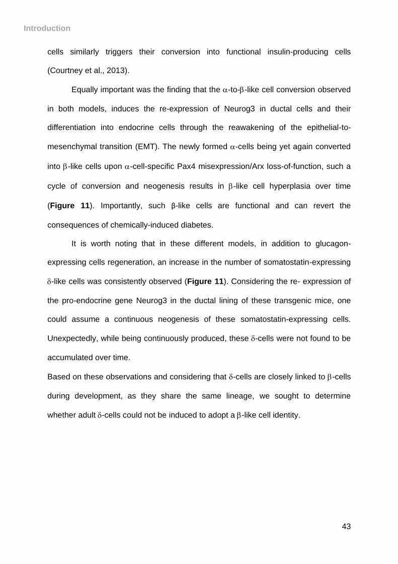
Introduction
43
cells similarly triggers their conversion into functional insulin-producing cells
(Courtney et al., 2013).
Equally important was the finding that the -to--like cell conversion observed
in both models, induces the re-expression of Neurog3 in ductal cells and their
differentiation into endocrine cells through the reawakening of the epithelial-to-
mesenchymal transition (EMT). The newly formed -cells being yet again converted
into -like cells upon -cell-specific Pax4 misexpression/Arx loss-of-function, such a
cycle of conversion and neogenesis results in -like cell hyperplasia over time
(Figure 11). Importantly, such β-like cells are functional and can revert the
consequences of chemically-induced diabetes.
It is worth noting that in these different models, in addition to glucagon-
expressing cells regeneration, an increase in the number of somatostatin-expressing
-like cells was consistently observed (Figure 11). Considering the re- expression of
the pro-endocrine gene Neurog3 in the ductal lining of these transgenic mice, one
could assume a continuous neogenesis of these somatostatin-expressing cells.
Unexpectedly, while being continuously produced, these -cells were not found to be
accumulated over time.
Based on these observations and considering that -cells are closely linked to -cells
during development, as they share the same lineage, we sought to determine
whether adult -cells could not be induced to adopt a -like cell identity.
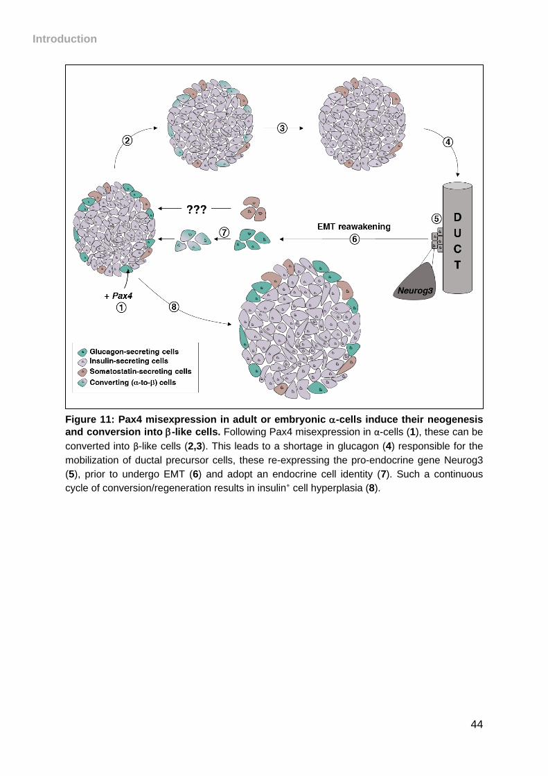
Introduction
44
Figure 11: Pax4 misexpression in adult or embryonic -cells induce their neogenesis
and conversion into -like cells. Following Pax4 misexpression in α-cells (1), these can be
converted into β-like cells (2,3). This leads to a shortage in glucagon (4) responsible for the
mobilization of ductal precursor cells, these re-expressing the pro-endocrine gene Neurog3
(5), prior to undergo EMT (6) and adopt an endocrine cell identity (7). Such a continuous
cycle of conversion/regeneration results in insulin+ cell hyperplasia (8).

45
MATERIAL AND METHODS
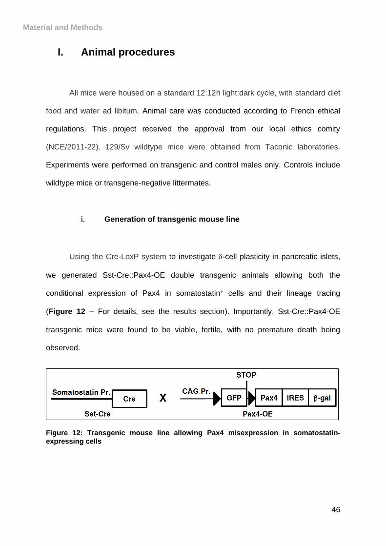
Material and Methods
46
I. Animal procedures
All mice were housed on a standard 12:12h light:dark cycle, with standard diet
food and water ad libitum. Animal care was conducted according to French ethical
regulations. This project received the approval from our local ethics comity
(NCE/2011-22). 129/Sv wildtype mice were obtained from Taconic laboratories.
Experiments were performed on transgenic and control males only. Controls include
wildtype mice or transgene-negative littermates.
Generation of transgenic mouse line
Using the Cre-LoxP system to investigate -cell plasticity in pancreatic islets,
we generated Sst-Cre::Pax4-OE double transgenic animals allowing both the
conditional expression of Pax4 in somatostatin+ cells and their lineage tracing
(Figure 12 – For details, see the results section). Importantly, Sst-Cre::Pax4-OE
transgenic mice were found to be viable, fertile, with no premature death being
observed.
Figure 12: Transgenic mouse line allowing Pax4 misexpression in somatostatin-
expressing cells
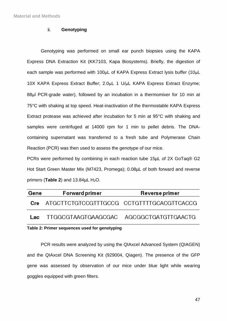
Material and Methods
47
Genotyping
Genotyping was performed on small ear punch biopsies using the KAPA
Express DNA Extraction Kit (KK7103, Kapa Biosystems). Briefly, the digestion of
each sample was performed with 100μL of KAPA Express Extract lysis buffer (10μL
10X KAPA Express Extract Buffer; 2.0μL 1 U/μL KAPA Express Extract Enzyme;
88μl PCR-grade water), followed by an incubation in a thermomixer for 10 min at
75°C with shaking at top speed. Heat-inactivation of the thermostable KAPA Express
Extract protease was achieved after incubation for 5 min at 95°C with shaking and
samples were centrifuged at 14000 rpm for 1 min to pellet debris. The DNA-
containing supernatant was transferred to a fresh tube and Polymerase Chain
Reaction (PCR) was then used to assess the genotype of our mice.
PCRs were performed by combining in each reaction tube 15µL of 2X GoTaq® G2
Hot Start Green Master Mix (M7423, Promega); 0.08µL of both forward and reverse
primers (Table 2) and 13.84µL H2O.
Table 2: Primer sequences used for genotyping
PCR results were analyzed by using the QIAxcel Advanced System (QIAGEN)
and the QIAxcel DNA Screening Kit (929004, Qiagen). The presence of the GFP
gene was assessed by observation of our mice under blue light while wearing
goggles equipped with green filters.
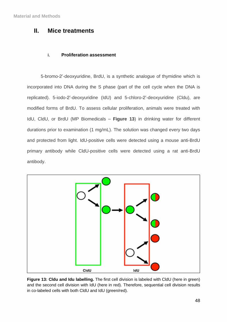
Material and Methods
48
II. Mice treatments
Proliferation assessment
5-bromo-2’-deoxyuridine, BrdU, is a synthetic analogue of thymidine which is
incorporated into DNA during the S phase (part of the cell cycle when the DNA is
replicated). 5-iodo-2’-deoxyuridine (IdU) and 5-chloro-2’-deoxyuridine (Cldu), are
modified forms of BrdU. To assess cellular proliferation, animals were treated with
IdU, CldU, or BrdU (MP Biomedicals – Figure 13) in drinking water for different
durations prior to examination (1 mg/mL). The solution was changed every two days
and protected from light. IdU-positive cells were detected using a mouse anti-BrdU
primary antibody while CldU-positive cells were detected using a rat anti-BrdU
antibody.
Figure 13: Cldu and Idu labelling. The first cell division is labeled with CldU (here in green)
and the second cell division with IdU (here in red). Therefore, sequential cell division results
in co-labeled cells with both CldU and IdU (green/red).
Material and Methods
49
Induction of streptozotocin-mediated hyperglycemia
Streptozotocin is a glucosamine-nitrosourea compound causing DNA damage.
Its chemical structure is similar to glucose which allows it to be transported
specifically into the -cell via the glucose transporter GLUT-2. To induce
hyperglycemia, streptozotocin, (STZ; S0130, Sigma) was dissolved in 0.1M sodium
citrate buffer (pH 4.5), and a single dose was administered intraperitoneally (115
mg/kg) within 10 min after dissolution. Diabetes progression was assessed by
monitoring the blood glucose levels and/or survival rates of mice. Glycemia was
measured with a ONETOUCH glucometer (Life Scan, Inc., CA).
III. Tolerance tests and blood glucose level
measurements
For intraperitoneal glucose tolerance tests (ipGTT) and intraperitoneal insulin
tolerance tests (ipITT), animals were fasted for 16-18 hours and injected
intraperitoneally with 2g/kg of bodyweight of D-(+)-glucose (G7201, Sigma) or
0.75U/kg of insulin (Novo Nordisk A/S). Blood glucose levels were measured at the
indicated time points post-injection with a ONETOUCH glucometer (Life Scan, Inc.,
CA).
Material and Methods
50
IV. RNA analyses
RNA extraction from total pancreas
Mice were euthanized by cervical dislocation and the pancreatic tissue was
isolated and placed in cold RNA later solution (AM7024, Fisher Scientific) for 48
hours at 4°C. A small piece of pancreas was taken and homogenized in lysis buffer
(10L of -mercaptoethanol in 10mL of RLT buffer from Qiagen) with a tissue
disruptor. Total pancreas RNA was extracted using Rneasy Mini Kit (Qiagen)
according to the manufacturer’s instructions.
cDNA synthesis
RNA concentration and quality were determined using an Agilent 2100
Bioanalyzer system (Agilent Technologies). 10µL of H2O containing 1g of RNA was
then processed for first strand synthesis mixing 1µL 10mM dNTP and 1µL 50mM
oligodT. This reaction was then incubated 5’ at 65°C, then on ice for at least 1 min.
For cDNA synthesis, 10µL of DNA synthesis mix (2µL 10x RT buffer; 4µL 25 mM
MgCl2; 2µL 0.1 M DTT; 1µL Rnase out; 1µL Superscript III) was added to each
sample and incubated for 50 min at 50°C, 5 min at 85°C then chilled on ice. Finally,
1µL of Rnase H was added and samples were incubated for 20 min at 37°C.
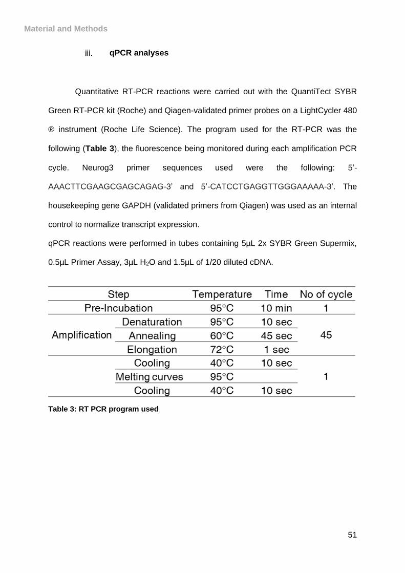
Material and Methods
51
qPCR analyses
Quantitative RT-PCR reactions were carried out with the QuantiTect SYBR
Green RT-PCR kit (Roche) and Qiagen-validated primer probes on a LightCycler 480
® instrument (Roche Life Science). The program used for the RT-PCR was the
following (Table 3), the fluorescence being monitored during each amplification PCR
cycle. Neurog3 primer sequences used were the following: 5’-
AAACTTCGAAGCGAGCAGAG-3’ and 5’-CATCCTGAGGTTGGGAAAAA-3’. The
housekeeping gene GAPDH (validated primers from Qiagen) was used as an internal
control to normalize transcript expression.
qPCR reactions were performed in tubes containing 5µL 2x SYBR Green Supermix,
0.5µL Primer Assay, 3µL H2O and 1.5µL of 1/20 diluted cDNA.
Table 3: RT PCR program used
Material and Methods
52
V. Pancreatic tissue processing and sectioning
Paraffin embedding – Microtome sectioning
Mice were euthanized by cervical dislocation and the pancreatic tissue was
isolated and placed in cold PBS. Tissue was fixed for 30 min at 4°C in Antigenfix
(paraformaldehyde solution pH 7,2-7,4; Microm Microtech France), washed 5 x 20
min in cold PBS and incubated 1 hour in 0.86% saline. Following dehydration through
a series of increasing ethanol dilutions (50%, 70%, 80%, 90%, and 100%), the
pancreas was treated with isopropanol and toluene, a hydrophobic agent, to remove
alcohol from the tissue. Finally, the organ was incubated in clean paraffin baths (1
hour, 3 hours and overnight) and embedded in paraffin in metal molds on a cold
plate. Paraffin blocks were stored at 4°C prior to sectioning.
8m sections were cut using a rotary microtome (Leica) and placed on
polarized glass slides (Fisher Scientific). Paraffin sections were allowed to dry 45 min
at 42°C into a heating block and kept at 37°C overnight. Slides were subsequently
stored at 4°C until staining.
OCT embedding – Cryostat sectioning
Mice were euthanized by cervical dislocation and the pancreatic tissue was
isolated and placed in cold PBS. Tissue was fixed for 30 min at 4°C in a fixative
solution (1.35mL 37% paraformaldehyde; 400L 25% glutaraldehyde; 100L 10X
NP40; 5mL 10X PBS; H2O to 50mL) and washed 6 x 20 min in cold PBS. To remove
the water and protect the tissue from freezing damage, the sample was incubated
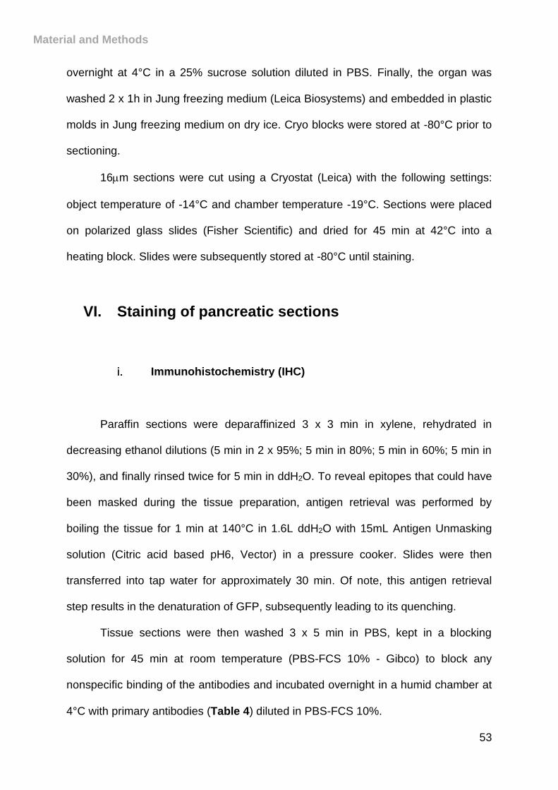
Material and Methods
53
overnight at 4°C in a 25% sucrose solution diluted in PBS. Finally, the organ was
washed 2 x 1h in Jung freezing medium (Leica Biosystems) and embedded in plastic
molds in Jung freezing medium on dry ice. Cryo blocks were stored at -80°C prior to
sectioning.
16m sections were cut using a Cryostat (Leica) with the following settings:
object temperature of -14°C and chamber temperature -19°C. Sections were placed
on polarized glass slides (Fisher Scientific) and dried for 45 min at 42°C into a
heating block. Slides were subsequently stored at -80°C until staining.
VI. Staining of pancreatic sections
Immunohistochemistry (IHC)
Paraffin sections were deparaffinized 3 x 3 min in xylene, rehydrated in
decreasing ethanol dilutions (5 min in 2 x 95%; 5 min in 80%; 5 min in 60%; 5 min in
30%), and finally rinsed twice for 5 min in ddH2O. To reveal epitopes that could have
been masked during the tissue preparation, antigen retrieval was performed by
boiling the tissue for 1 min at 140°C in 1.6L ddH2O with 15mL Antigen Unmasking
solution (Citric acid based pH6, Vector) in a pressure cooker. Slides were then
transferred into tap water for approximately 30 min. Of note, this antigen retrieval
step results in the denaturation of GFP, subsequently leading to its quenching.
Tissue sections were then washed 3 x 5 min in PBS, kept in a blocking
solution for 45 min at room temperature (PBS-FCS 10% - Gibco) to block any
nonspecific binding of the antibodies and incubated overnight in a humid chamber at
4°C with primary antibodies (Table 4) diluted in PBS-FCS 10%.
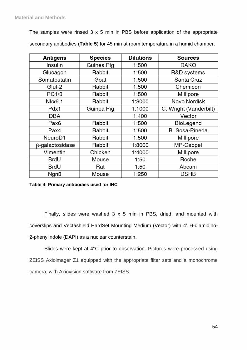
Material and Methods
54
The samples were rinsed 3 x 5 min in PBS before application of the appropriate
secondary antibodies (Table 5) for 45 min at room temperature in a humid chamber.
Table 4: Primary antibodies used for IHC
Finally, slides were washed 3 x 5 min in PBS, dried, and mounted with
coverslips and Vectashield HardSet Mounting Medium (Vector) with 4’, 6-diamidino-
2-phenylindole (DAPI) as a nuclear counterstain.
Slides were kept at 4°C prior to observation. Pictures were processed using
ZEISS Axioimager Z1 equipped with the appropriate filter sets and a monochrome
camera, with Axiovision software from ZEISS.
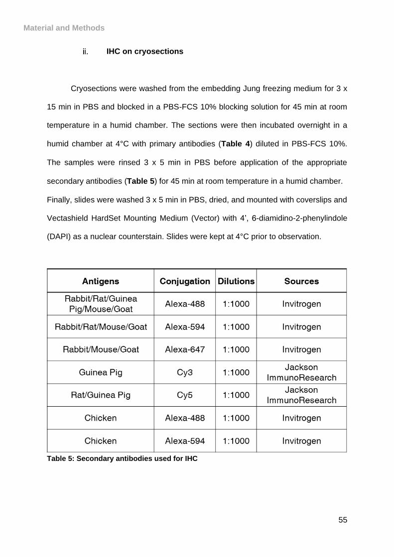
Material and Methods
55
IHC on cryosections
Cryosections were washed from the embedding Jung freezing medium for 3 x
15 min in PBS and blocked in a PBS-FCS 10% blocking solution for 45 min at room
temperature in a humid chamber. The sections were then incubated overnight in a
humid chamber at 4°C with primary antibodies (Table 4) diluted in PBS-FCS 10%.
The samples were rinsed 3 x 5 min in PBS before application of the appropriate
secondary antibodies (Table 5) for 45 min at room temperature in a humid chamber.
Finally, slides were washed 3 x 5 min in PBS, dried, and mounted with coverslips and
Vectashield HardSet Mounting Medium (Vector) with 4’, 6-diamidino-2-phenylindole
(DAPI) as a nuclear counterstain. Slides were kept at 4°C prior to observation.
Table 5: Secondary antibodies used for IHC
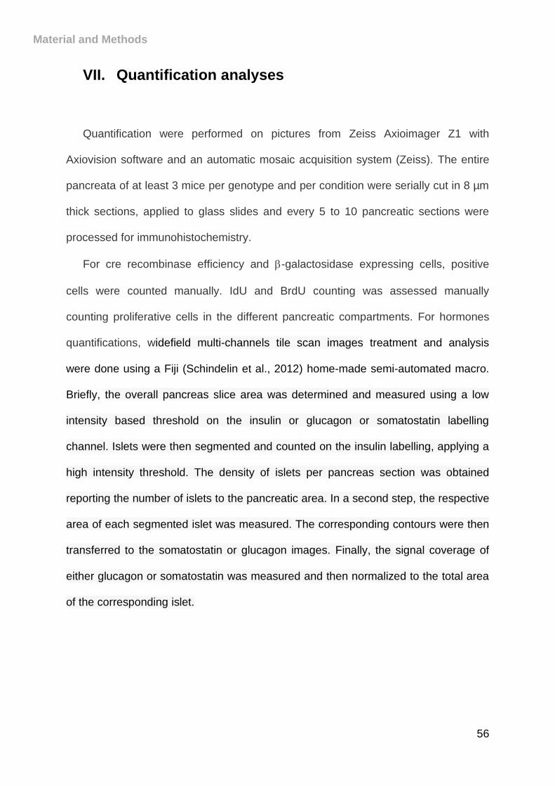
Material and Methods
56
VII. Quantification analyses
Quantification were performed on pictures from Zeiss Axioimager Z1 with
Axiovision software and an automatic mosaic acquisition system (Zeiss). The entire
pancreata of at least 3 mice per genotype and per condition were serially cut in 8 µm
thick sections, applied to glass slides and every 5 to 10 pancreatic sections were
processed for immunohistochemistry.
For cre recombinase efficiency and -galactosidase expressing cells, positive
cells were counted manually. IdU and BrdU counting was assessed manually
counting proliferative cells in the different pancreatic compartments. For hormones
quantifications, widefield multi-channels tile scan images treatment and analysis
were done using a Fiji (Schindelin et al., 2012) home-made semi-automated macro.
Briefly, the overall pancreas slice area was determined and measured using a low
intensity based threshold on the insulin or glucagon or somatostatin labelling
channel. Islets were then segmented and counted on the insulin labelling, applying a
high intensity threshold. The density of islets per pancreas section was obtained
reporting the number of islets to the pancreatic area. In a second step, the respective
area of each segmented islet was measured. The corresponding contours were then
transferred to the somatostatin or glucagon images. Finally, the signal coverage of
either glucagon or somatostatin was measured and then normalized to the total area
of the corresponding islet.
Material and Methods
57
VIII. Data analyses and statistics
All values are depicted as mean ± SEM of at least data from 3 animals. Data
were analyzed using GraphPrism 6 software. Normality was tested using D’Agostino-
Pearson omnibus normality test and appropriate statistical tests were performed.
Results are considered significant if p < 0.0001 (****), p < 0.001 (***), p < 0.01 (**),
and p < 0.05 (*).

58
RESULTS
59
I. Sst::Cre-Pax4-OE double transgenic mouse line
generation and phenotype
Aiming to determine whether the sole misexpression of Pax4 in -cells could
alter their phenotype/identity in vivo, we first crossed Sst-Cre animals (harboring a
transgene encompassing the somatostatin promoter driving the expression of the
phage P1 Cre recombinase - Taniguchi et al., 2011 - Figure 14A) with the ROSA26-
-gal mouse line (harboring a transgene containing the Rosa26 promoter upstream
of a loxP-Neomycin resistance-Stop-loxP--galactosidase cassette - Soriano, 1999
Figure 14A). Our analyses of the pancreata from the resulting Sst-Cre::ROSA26--
gal double transgenic mice validated the specificity of Cre expression solely in
somatostatin-producing cells (Figure 14C).
Subsequently, Sst-Cre animals were mated with Pax4-OE mice (whose
transgene encompasses the ubiquitous CAG promoter upstream of a “GFP-STOP”
cassette flanked by LoxP sites and followed by the Pax4 cDNA in front of an IRES
and the -galactosidase reporter - Collombat et al., 2009 - Figure 14B). In the
resulting Sst-Cre::Pax4-OE double transgenic animals, Pax4 misexpression was
solely detected in Cre-expressing somatostatin+ cells. Accordingly, quantitative
immunohistochemical analyses confirmed such specificity with an ectopic expression
of Pax4 in 663.09% of somatostatin-expressing cells (Figure 14D-E). Importantly,
Sst-Cre::Pax4-OE transgenic mice were found to be viable, fertile, with no premature
death being observed. Along the same line, no statistical difference was observed in
the glycemia of control and transgenic animals of matching ages (Figure 14F),
demonstrating that Pax4 misexpression in somatostatin+ cells does not impact basal
glycemia levels.
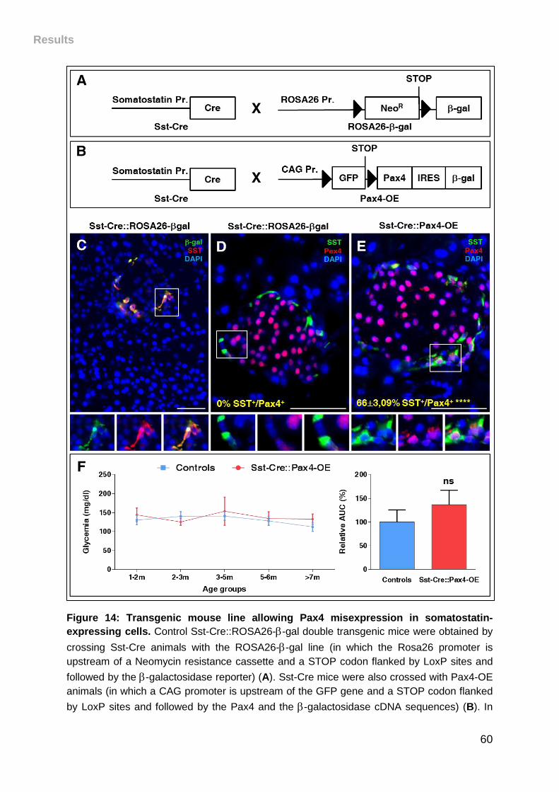
Results
60
Figure 14: Transgenic mouse line allowing Pax4 misexpression in somatostatin-
expressing cells. Control Sst-Cre::ROSA26--gal double transgenic mice were obtained by
crossing Sst-Cre animals with the ROSA26--gal line (in which the Rosa26 promoter is
upstream of a Neomycin resistance cassette and a STOP codon flanked by LoxP sites and
followed by the -galactosidase reporter) (A). Sst-Cre mice were also crossed with Pax4-OE
animals (in which a CAG promoter is upstream of the GFP gene and a STOP codon flanked
by LoxP sites and followed by the Pax4 and the -galactosidase cDNA sequences) (B). In
Results
61
the resulting Sst-Cre::Pax4-OE bitransgenic mouse line, somatostatin expression drives the
expression of the Cre recombinase and allows the excision of the region between the two
LoxP sites thereby promoting the expression of Pax4 and -galactosidase. -galactosidase
and Pax4 immunodetection in Sst-Cre::ROSA26--gal double transgenic mice confirmed Cre
expression specifically in somatostatin-expressing cells (C-D). In Sst-Cre::Pax4-OE islets,
Pax4 is detected in 663.09% of the somatostatin-expressing cells (E). Glycemia of non-
fasted control and transgenic mice of different ages (F). The area under the curve (AUC) was
measured and demonstrated no statistical differences between both groups. For Cre
recombinase efficiency, the p-value was calculated by one sample t-test. Statistics for AUC
were determined using Mann-Whitney test. **** p<0.0001, ns p>0.05. Scale bars, 20m.
II. Pax4 misexpression in -cells results in progressive
islet hypertrophy and insulin-producing cell
hyperplasia
The pancreata of Sst-Cre::Pax4-OE animals and age-/sex-matched controls
were analyzed by immunohistochemistry at 2-, 5-, and 8-months of age. Importantly,
an increase in islet number and size was demonstrated already in 2-month old Sst-
Cre::Pax4-OE animals compared to controls (Figure 15A-B). As interesting were the
further augmentations in both islet number and size observed in 5- and 8-month old
Sst-Cre::Pax4-OE pancreata relative to controls (Figure 15C-D), suggestive of
progressive processes. Quantitative analyses corroborated these results with the
demonstration of a significant (2.380.17-fold) increase in islet counts and a
1.480.07-fold augmentation in islet size when comparing 2-month old Sst-
Cre::Pax4-OE pancreata with controls (Figure 15E-F). Importantly, quantification of
insulin+ area per pancreas demonstrated that islet hypertrophy mostly resulted from a
significant insulin+ cell hyperplasia (Figure 15G).
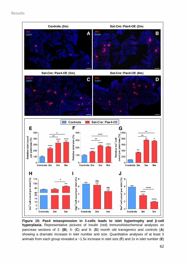
Results
62
Figure 15: Pax4 misexpression in -cells leads to islet hypertrophy and -cell
hyperplasia. Representative pictures of insulin (red) immunohistochemical analyses on
pancreas sections of 2- (B), 5- (C) and 8- (D) month old transgenics and controls (A)
showing a dramatic increase in islet number and size. Quantitative analyses of at least 3
animals from each group revealed a ~1.5x increase in islet size (F) and 2x in islet number (E)
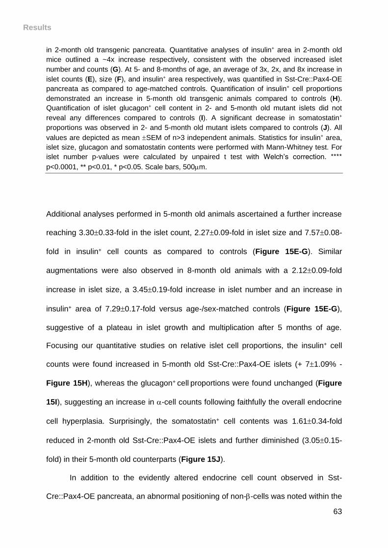
Results
63
in 2-month old transgenic pancreata. Quantitative analyses of insulin+ area in 2-month old
mice outlined a ~4x increase respectively, consistent with the observed increased islet
number and counts (G). At 5- and 8-months of age, an average of 3x, 2x, and 8x increase in
islet counts (E), size (F), and insulin+ area respectively, was quantified in Sst-Cre::Pax4-OE
pancreata as compared to age-matched controls. Quantification of insulin+ cell proportions
demonstrated an increase in 5-month old transgenic animals compared to controls (H).
Quantification of islet glucagon+ cell content in 2- and 5-month old mutant islets did not
reveal any differences compared to controls (I). A significant decrease in somatostatin+
proportions was observed in 2- and 5-month old mutant islets compared to controls (J). All
values are depicted as mean SEM of n>3 independent animals. Statistics for insulin+ area,
islet size, glucagon and somatostatin contents were performed with Mann-Whitney test. For
islet number p-values were calculated by unpaired t test with Welch’s correction. ****
p<0.0001, ** p<0.01, * p<0.05. Scale bars, 500m.
Additional analyses performed in 5-month old animals ascertained a further increase
reaching 3.300.33-fold in the islet count, 2.270.09-fold in islet size and 7.570.08-
fold in insulin+ cell counts as compared to controls (Figure 15E-G). Similar
augmentations were also observed in 8-month old animals with a 2.120.09-fold
increase in islet size, a 3.450.19-fold increase in islet number and an increase in
insulin+ area of 7.290.17-fold versus age-/sex-matched controls (Figure 15E-G),
suggestive of a plateau in islet growth and multiplication after 5 months of age.
Focusing our quantitative studies on relative islet cell proportions, the insulin+ cell
counts were found increased in 5-month old Sst-Cre::Pax4-OE islets (+ 71.09% -
Figure 15H), whereas the glucagon+ cell proportions were found unchanged (Figure
15I), suggesting an increase in -cell counts following faithfully the overall endocrine
cell hyperplasia. Surprisingly, the somatostatin+ cell contents was 1.610.34-fold
reduced in 2-month old Sst-Cre::Pax4-OE islets and further diminished (3.050.15-
fold) in their 5-month old counterparts (Figure 15J).
In addition to the evidently altered endocrine cell count observed in Sst-
Cre::Pax4-OE pancreata, an abnormal positioning of non--cells was noted within the
Results
64
islets. Indeed, while glucagon- and somatostatin-expressing cells were predictably
found uniformly distributed at the periphery of control islets (Figure 16A-B), a
preferential localization at a pole of the islets, close to adjacent ducts, was evidenced
for these non--cells in 2-month old Sst-Cre::Pax4-OE islets (Figure 16C-D).
Figure 16: Abnormal positioning of non--cells within the islets of Sst-Cre::Pax4-OE
transgenic mice. Representative photographs of somatostatin-, glucagon- and insulin-
expressing cells within control islets (A-B) or 2-month old transgenic islets (C-D). While
somatostatin- and glucagon-positive cells were found distributed in the mantle of control islet
(A-B), a preferential localization close to adjacent ducts of such cells was observed in Sst-
Cre::Pax4-OE islets (C-D). For the purpose of clarity, pancreatic ducts are outlined in dashed
orange lines. Scale bars, 50m.
Results
65
Together, these results suggest that the misexpression of Pax4 in -cells
promotes both an increase in islet numbers and size. Interestingly, the islet
hypertrophy appears to result from an insulin+ cell hyperplasia and, to a lesser extent,
from an increase in glucagon+ cell counts, indicative of neogenesis processes.
However, somatostatin+ cells are found underrepresented, suggesting that the
neogenesis processes observed in Sst-Cre::Pax4-OE pancreata could involve their
conversion into alternative cell subtypes.
III. Pax4 misexpression in somatostatin+ cells induces
their conversion into -like cells
To investigate the fate/destiny of Pax4 misexpressing -cells in Sst-Cre::Pax4-
OE pancreata, further immunohistochemical analyses were undertaken. Interestingly,
a fraction of somatostatin-positive cells were found to ectopically express insulin in 2-
month old Sst-Cre::Pax4-OE islets (Figures 17B, 18A-B), such bi-hormonal cells
being expectedly absent in control pancreata (Figure 17A). As these results
suggested a putative conversion of somatostatin+ cells into insulin+ cells, we
investigated the consequences of Pax4 misexpression in -cells using lineage
tracing. The expression of the -galactosidase tracer was therefore monitored
comparing Sst-Cre::ROSA26--gal double transgenics with Sst-Cre::Pax4-OE
animals.
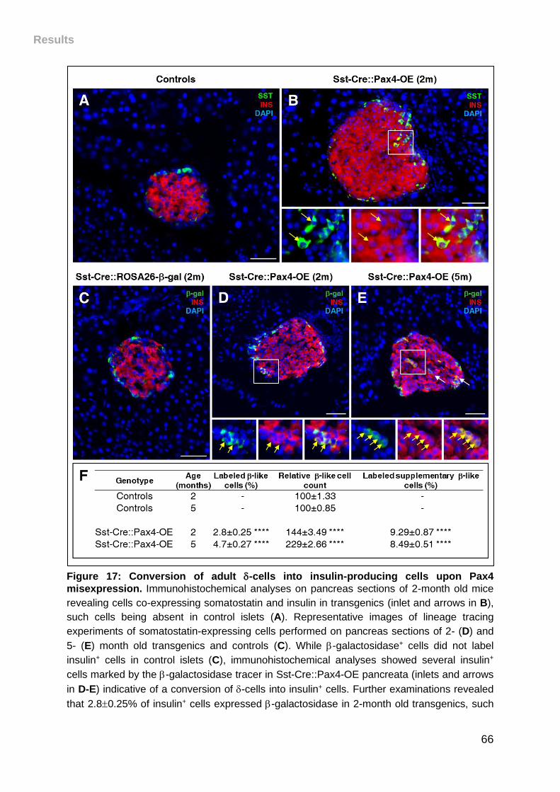
Results
66
Figure 17: Conversion of adult -cells into insulin-producing cells upon Pax4
misexpression. Immunohistochemical analyses on pancreas sections of 2-month old mice
revealing cells co-expressing somatostatin and insulin in transgenics (inlet and arrows in B),
such cells being absent in control islets (A). Representative images of lineage tracing
experiments of somatostatin-expressing cells performed on pancreas sections of 2- (D) and
5- (E) month old transgenics and controls (C). While -galactosidase+ cells did not label
insulin+ cells in control islets (C), immunohistochemical analyses showed several insulin+
cells marked by the -galactosidase tracer in Sst-Cre::Pax4-OE pancreata (inlets and arrows
in D-E) indicative of a conversion of -cells into insulin+ cells. Further examinations revealed
that 2.80.25% of insulin+ cells expressed -galactosidase in 2-month old transgenics, such
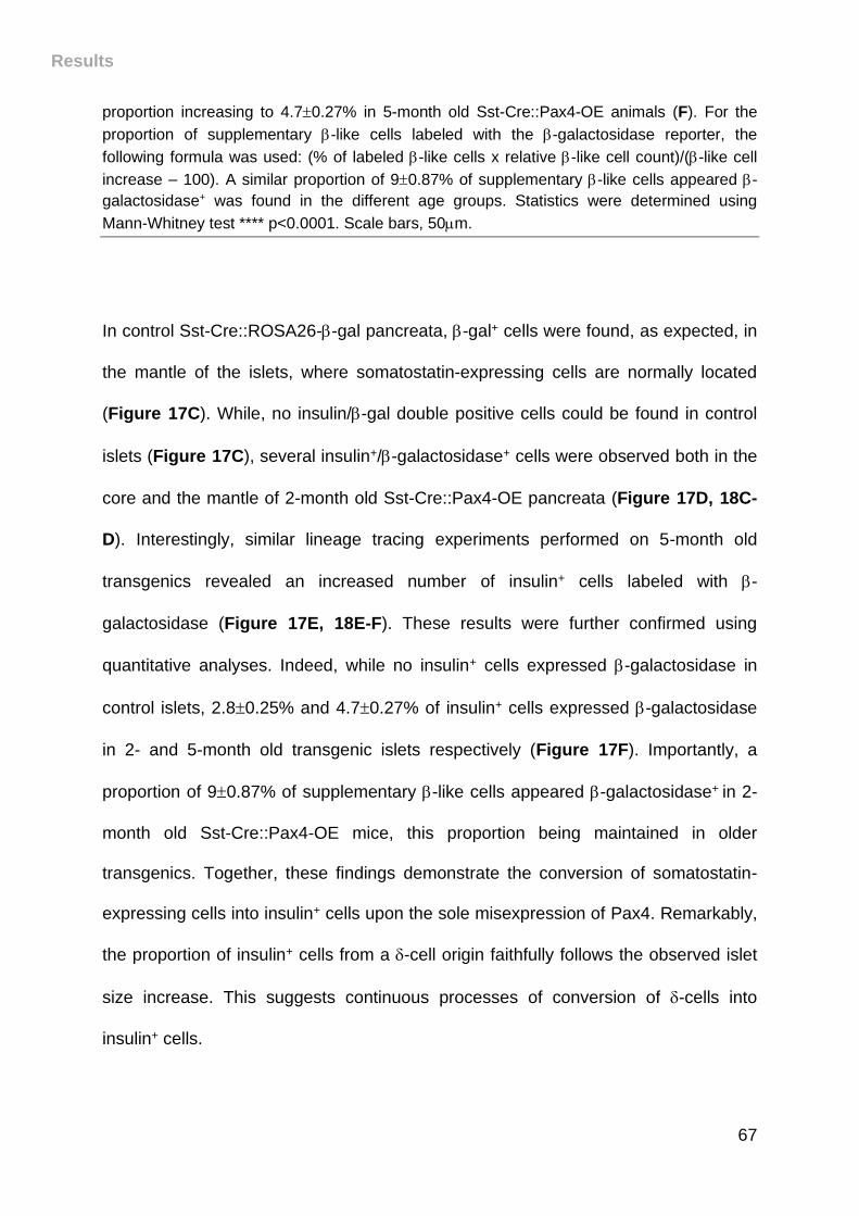
Results
67
proportion increasing to 4.70.27% in 5-month old Sst-Cre::Pax4-OE animals (F). For the
proportion of supplementary -like cells labeled with the -galactosidase reporter, the
following formula was used: (% of labeled -like cells x relative -like cell count)/(-like cell
increase – 100). A similar proportion of 90.87% of supplementary -like cells appeared -
galactosidase+ was found in the different age groups. Statistics were determined using
Mann-Whitney test **** p<0.0001. Scale bars, 50m.
In control Sst-Cre::ROSA26--gal pancreata, -gal+ cells were found, as expected, in
the mantle of the islets, where somatostatin-expressing cells are normally located
(Figure 17C). While, no insulin/-gal double positive cells could be found in control
islets (Figure 17C), several insulin+/-galactosidase+ cells were observed both in the
core and the mantle of 2-month old Sst-Cre::Pax4-OE pancreata (Figure 17D, 18C-
D). Interestingly, similar lineage tracing experiments performed on 5-month old
transgenics revealed an increased number of insulin+ cells labeled with -
galactosidase (Figure 17E, 18E-F). These results were further confirmed using
quantitative analyses. Indeed, while no insulin+ cells expressed -galactosidase in
control islets, 2.80.25% and 4.70.27% of insulin+ cells expressed -galactosidase
in 2- and 5-month old transgenic islets respectively (Figure 17F). Importantly, a
proportion of 90.87% of supplementary -like cells appeared -galactosidase+ in 2-
month old Sst-Cre::Pax4-OE mice, this proportion being maintained in older
transgenics. Together, these findings demonstrate the conversion of somatostatin-
expressing cells into insulin+ cells upon the sole misexpression of Pax4. Remarkably,
the proportion of insulin+ cells from a -cell origin faithfully follows the observed islet
size increase. This suggests continuous processes of conversion of -cells into
insulin+ cells.
Results
68
Figure 18: Additional examples illustrating the conversion of -cells to insulin-
producing cells upon Pax4 misexpression. Immunohistochemical analyses on pancreas
sections of 2-month old mice revealing cells co-expressing somatostatin and insulin-cells in
transgenics (inlets in A, B), such cells being absent in control islets. Representative images
of lineage tracing experiments of somatostatin-expressing cells performed on pancreas
sections of 2- (C-D) and 5- (E-F) month old transgenics, suggesting a -to-insulin+ cell
conversion. Scale bars, 50m.
Results
69
IV. Pax4 misexpression in -cells results in increased
proliferation within the ductal epithelium
Our results demonstrate that the misexpression of the Pax4 gene in -cells is
sufficient to induce their reprogramming into cells displaying a -like cell identity.
However, this conversion can clearly not account for the massive endocrine cell
hyperplasia observed in Sst-Cre::Pax4-OE animals, suggestive of the involvement of
additional processes.
Thus, to investigate whether the latter could also be caused by an increase in
cell replication, cell proliferation rates were assayed in Sst-Cre::Pax4-OE transgenics
and age-matched controls treated with either Idu or BrdU for different durations.
Importantly, after short IdU pulse (3 days), a significant augmentation in replicating
cell numbers was observed in the pancreas of 2-month old transgenic mice as
indicated by increased IdU+ cell numbers (Figure 19A, D). Strikingly, most
proliferating cells were not located within the islets of Langerhans but rather within
the ductal epithelium and lining (Figure 19D). Interestingly, longer IdU treatment (10
days) revealed numerous IdU+ cells again in the ductal epithelium and lining (Figure
19B-C, E), such IdU+ cells being then also found within the islets of Sst-Cre::Pax4-
OE mice (Figure 19B-C, F), suggesting that proliferating cells progress from the
ductal compartment to the islets of Langerhans. Quantitative analyses verified these
observations and outlined a 5.730.38-fold increase in the numbers of IdU+ cells
within the ductal lining or epithelium after short-term IdU treatment comparing Sst-
Cre::Pax4-OE pancreata versus their control counterparts (Figure 19G).

Results
70
Figure 19: Increased cell proliferation within the ductal epithelium of Sst-Cre::Pax4-OE
pancreata. Cell proliferation was assessed evaluating IdU incorporation (detected with a
BrdU antibody) in 2 month-old transgenics and controls after short- (3 days) or long- (10
days) term IdU pulse. Representative images of immunohistochemical analyses on pancreas
sections of control (A-C) and Sst-Cre::Pax4-OE (D-F) mice. An increase in proliferation was
observed in the ductal lining of 2-month old transgenics after a short-term IdU pulse (A, D).
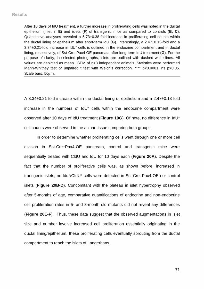
Results
71
After 10 days of IdU treatment, a further increase in proliferating cells was noted in the ductal
epithelium (inlet in E) and islets (F) of transgenic mice as compared to controls (B, C).
Quantitative analyses revealed a 5.730.38-fold increase in proliferating cell counts within
the ductal lining or epithelium after short-term IdU (G). Interestingly, a 2.470.13-fold and a
3.340.21-fold increase in IdU+ cells is outlined in the endocrine compartment and in ductal
lining, respectively, of Sst-Cre::Pax4-OE pancreata after long-term IdU treatment (G). For the
purpose of clarity, in selected photographs, islets are outlined with dashed white lines. All
values are depicted as mean SEM of n>3 independent animals. Statistics were performed
Mann-Whitney test or unpaired t test with Welch’s correction. **** p<0.0001, ns p>0.05.
Scale bars, 50m.
A 3.340.21-fold increase within the ductal lining or epithelium and a 2.470.13-fold
increase in the numbers of IdU+ cells within the endocrine compartment were
observed after 10 days of IdU treatment (Figure 19G). Of note, no difference in IdU+
cell counts were observed in the acinar tissue comparing both groups.
In order to determine whether proliferating cells went through one or more cell
division in Sst-Cre::Pax4-OE pancreata, control and transgenic mice were
sequentially treated with CldU and IdU for 10 days each (Figure 20A). Despite the
fact that the number of proliferative cells was, as shown before, increased in
transgenic islets, no Idu+/CIdU+ cells were detected in Sst-Cre::Pax4-OE nor control
islets (Figure 20B-D). Concomitant with the plateau in islet hypertrophy observed
after 5-months of age, comparative quantifications of endocrine and non-endocrine
cell proliferation rates in 5- and 8-month old mutants did not reveal any differences
(Figure 20E-F). Thus, these data suggest that the observed augmentations in islet
size and number involve increased cell proliferation essentially originating in the
ductal lining/epithelium, these proliferating cells eventually sprouting from the ductal
compartment to reach the islets of Langerhans.
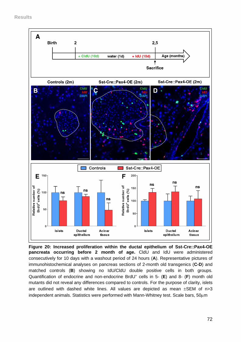
Results
72
Figure 20: Increased proliferation within the ductal epithelium of Sst-Cre::Pax4-OE
pancreata occurring before 2 month of age. CldU and IdU were administered
consecutively for 10 days with a washout period of 24 hours (A). Representative pictures of
immunohistochemical analyses on pancreas sections of 2-month old transgenics (C-D) and
matched controls (B) showing no IdU/CldU double positive cells in both groups.
Quantification of endocrine and non-endocrine BrdU+ cells in 5- (E) and 8- (F) month old
mutants did not reveal any differences compared to controls. For the purpose of clarity, islets
are outlined with dashed white lines. All values are depicted as mean SEM of n>3
independent animals. Statistics were performed with Mann-Whitney test. Scale bars, 50m
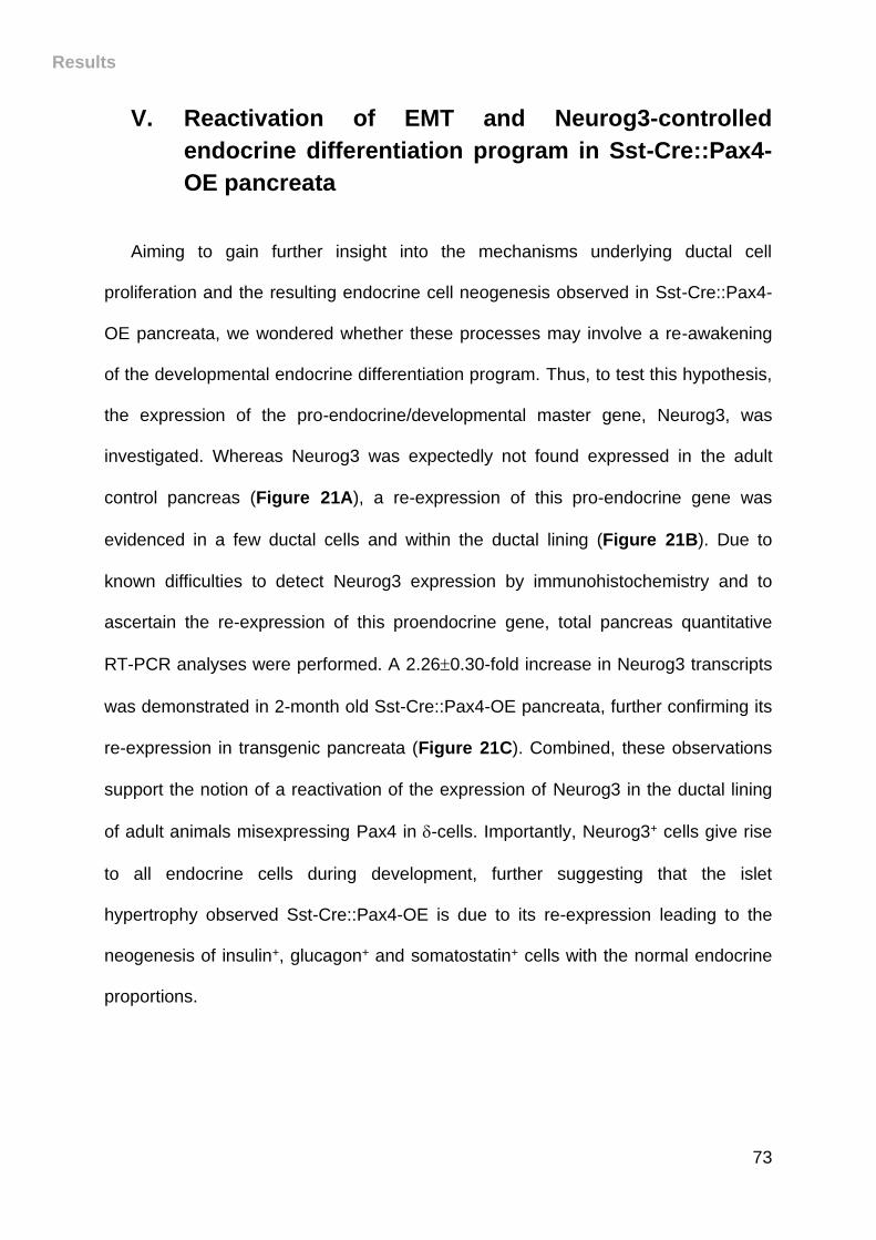
Results
73
V. Reactivation of EMT and Neurog3-controlled
endocrine differentiation program in Sst-Cre::Pax4-
OE pancreata
Aiming to gain further insight into the mechanisms underlying ductal cell
proliferation and the resulting endocrine cell neogenesis observed in Sst-Cre::Pax4-
OE pancreata, we wondered whether these processes may involve a re-awakening
of the developmental endocrine differentiation program. Thus, to test this hypothesis,
the expression of the pro-endocrine/developmental master gene, Neurog3, was
investigated. Whereas Neurog3 was expectedly not found expressed in the adult
control pancreas (Figure 21A), a re-expression of this pro-endocrine gene was
evidenced in a few ductal cells and within the ductal lining (Figure 21B). Due to
known difficulties to detect Neurog3 expression by immunohistochemistry and to
ascertain the re-expression of this proendocrine gene, total pancreas quantitative
RT-PCR analyses were performed. A 2.260.30-fold increase in Neurog3 transcripts
was demonstrated in 2-month old Sst-Cre::Pax4-OE pancreata, further confirming its
re-expression in transgenic pancreata (Figure 21C). Combined, these observations
support the notion of a reactivation of the expression of Neurog3 in the ductal lining
of adult animals misexpressing Pax4 in -cells. Importantly, Neurog3+ cells give rise
to all endocrine cells during development, further suggesting that the islet
hypertrophy observed Sst-Cre::Pax4-OE is due to its re-expression leading to the
neogenesis of insulin+, glucagon+ and somatostatin+ cells with the normal endocrine
proportions.
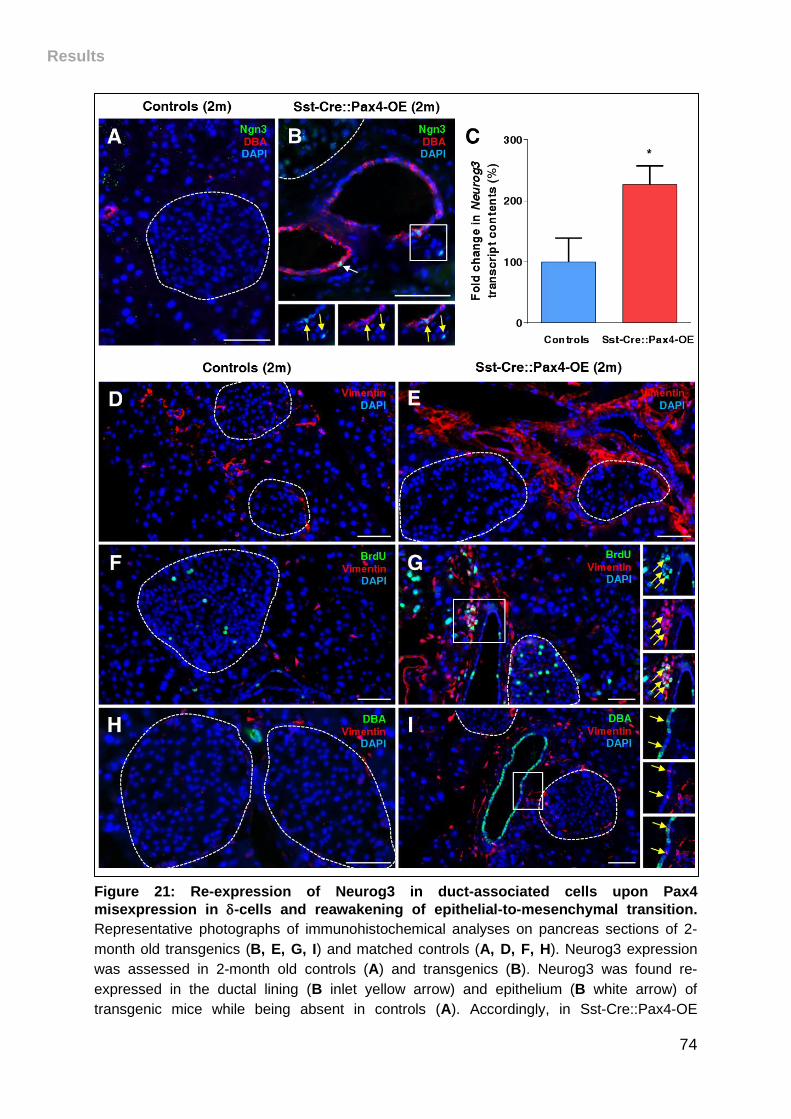
Results
74
Figure 21: Re-expression of Neurog3 in duct-associated cells upon Pax4
misexpression in -cells and reawakening of epithelial-to-mesenchymal transition.
Representative photographs of immunohistochemical analyses on pancreas sections of 2-
month old transgenics (B, E, G, I) and matched controls (A, D, F, H). Neurog3 expression
was assessed in 2-month old controls (A) and transgenics (B). Neurog3 was found re-
expressed in the ductal lining (B inlet yellow arrow) and epithelium (B white arrow) of
transgenic mice while being absent in controls (A). Accordingly, in Sst-Cre::Pax4-OE
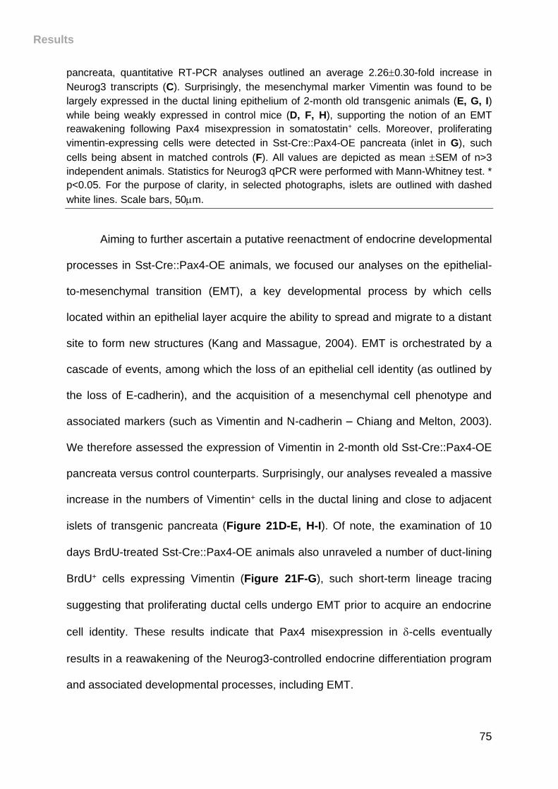
Results
75
pancreata, quantitative RT-PCR analyses outlined an average 2.260.30-fold increase in
Neurog3 transcripts (C). Surprisingly, the mesenchymal marker Vimentin was found to be
largely expressed in the ductal lining epithelium of 2-month old transgenic animals (E, G, I)
while being weakly expressed in control mice (D, F, H), supporting the notion of an EMT
reawakening following Pax4 misexpression in somatostatin+ cells. Moreover, proliferating
vimentin-expressing cells were detected in Sst-Cre::Pax4-OE pancreata (inlet in G), such
cells being absent in matched controls (F). All values are depicted as mean SEM of n>3
independent animals. Statistics for Neurog3 qPCR were performed with Mann-Whitney test. *
p<0.05. For the purpose of clarity, in selected photographs, islets are outlined with dashed
white lines. Scale bars, 50m.
Aiming to further ascertain a putative reenactment of endocrine developmental
processes in Sst-Cre::Pax4-OE animals, we focused our analyses on the epithelial-
to-mesenchymal transition (EMT), a key developmental process by which cells
located within an epithelial layer acquire the ability to spread and migrate to a distant
site to form new structures (Kang and Massague, 2004). EMT is orchestrated by a
cascade of events, among which the loss of an epithelial cell identity (as outlined by
the loss of E-cadherin), and the acquisition of a mesenchymal cell phenotype and
associated markers (such as Vimentin and N-cadherin – Chiang and Melton, 2003).
We therefore assessed the expression of Vimentin in 2-month old Sst-Cre::Pax4-OE
pancreata versus control counterparts. Surprisingly, our analyses revealed a massive
increase in the numbers of Vimentin+ cells in the ductal lining and close to adjacent
islets of transgenic pancreata (Figure 21D-E, H-I). Of note, the examination of 10
days BrdU-treated Sst-Cre::Pax4-OE animals also unraveled a number of duct-lining
BrdU+ cells expressing Vimentin (Figure 21F-G), such short-term lineage tracing
suggesting that proliferating ductal cells undergo EMT prior to acquire an endocrine
cell identity. These results indicate that Pax4 misexpression in -cells eventually
results in a reawakening of the Neurog3-controlled endocrine differentiation program
and associated developmental processes, including EMT.
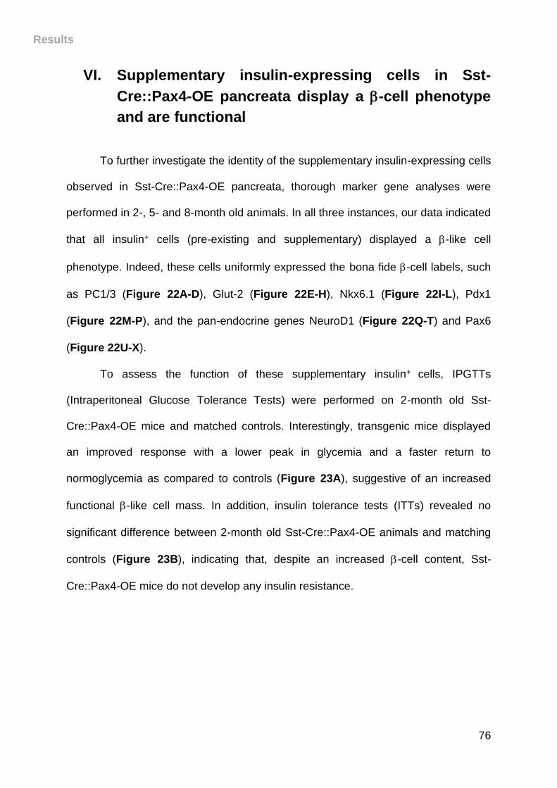
Results
76
VI. Supplementary insulin-expressing cells in Sst-
Cre::Pax4-OE pancreata display a -cell phenotype
and are functional
To further investigate the identity of the supplementary insulin-expressing cells
observed in Sst-Cre::Pax4-OE pancreata, thorough marker gene analyses were
performed in 2-, 5- and 8-month old animals. In all three instances, our data indicated
that all insulin+ cells (pre-existing and supplementary) displayed a -like cell
phenotype. Indeed, these cells uniformly expressed the bona fide -cell labels, such
as PC1/3 (Figure 22A-D), Glut-2 (Figure 22E-H), Nkx6.1 (Figure 22I-L), Pdx1
(Figure 22M-P), and the pan-endocrine genes NeuroD1 (Figure 22Q-T) and Pax6
(Figure 22U-X).
To assess the function of these supplementary insulin+ cells, IPGTTs
(Intraperitoneal Glucose Tolerance Tests) were performed on 2-month old Sst-
Cre::Pax4-OE mice and matched controls. Interestingly, transgenic mice displayed
an improved response with a lower peak in glycemia and a faster return to
normoglycemia as compared to controls (Figure 23A), suggestive of an increased
functional -like cell mass. In addition, insulin tolerance tests (ITTs) revealed no
significant difference between 2-month old Sst-Cre::Pax4-OE animals and matching
controls (Figure 23B), indicating that, despite an increased -cell content, Sst-
Cre::Pax4-OE mice do not develop any insulin resistance.
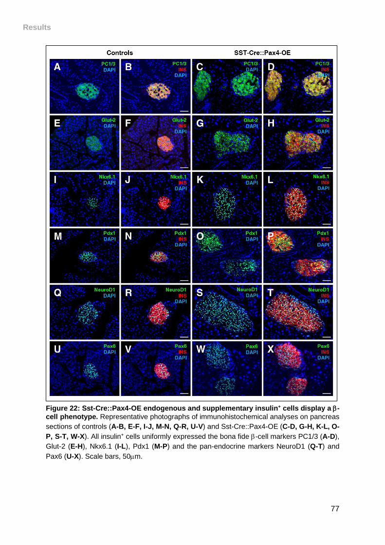
Results
77
Figure 22: Sst-Cre::Pax4-OE endogenous and supplementary insulin+ cells display a -
cell phenotype. Representative photographs of immunohistochemical analyses on pancreas
sections of controls (A-B, E-F, I-J, M-N, Q-R, U-V) and Sst-Cre::Pax4-OE (C-D, G-H, K-L, O-
P, S-T, W-X). All insulin+ cells uniformly expressed the bona fide -cell markers PC1/3 (A-D),
Glut-2 (E-H), Nkx6.1 (I-L), Pdx1 (M-P) and the pan-endocrine markers NeuroD1 (Q-T) and
Pax6 (U-X). Scale bars, 50m.
Results
78
These analyses led us to conclude that the hyperplastic insulin-expressing cells
observed following Pax4 misexpression in somatostatin+ cells, exhibit a -cell
phenotype and are functional.
Figure 23: Improved -cell function in transgenic Sst-Cre::Pax4-OE mice. 2 month-old
transgenic and controls were challenged with glucose. Transgenic mice displayed an
improved glucose clearance with a lower peak in glycemia and a faster recovery to
euglycemia (A). The relative area under the curve (AUC) confirmed this improved glucose
tolerance in 2 month-old Sst-Cre::Pax4-OE animals. Upon insulin tolerance tests (ITT),
glycemia levels of 2 month-old transgenic and matched controls were monitored. Transgenic
mice exhibited unaltered glycemia levels, indicative of a standard insulin sensitivity (B). AUC
were indeed similar in both groups. The relative AUC are expressed as a percentage relative
to the average AUC of the controls. All values are depicted as mean SEM of n>3
independent animals. Statistics were determined using Mann-Whitney test ** p<0.01, *
p<0.05, ns p>0.05.
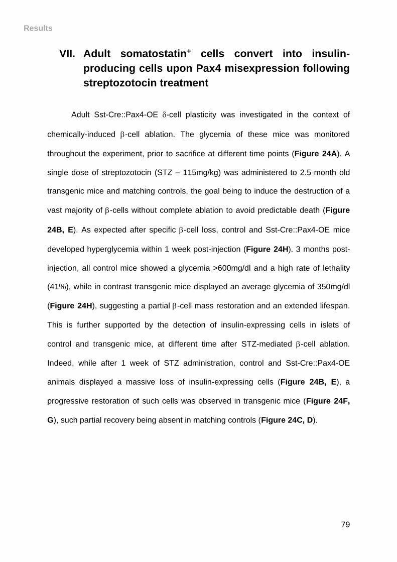
Results
79
VII. Adult somatostatin+ cells convert into insulin-
producing cells upon Pax4 misexpression following
streptozotocin treatment
Adult Sst-Cre::Pax4-OE -cell plasticity was investigated in the context of
chemically-induced -cell ablation. The glycemia of these mice was monitored
throughout the experiment, prior to sacrifice at different time points (Figure 24A). A
single dose of streptozotocin (STZ – 115mg/kg) was administered to 2.5-month old
transgenic mice and matching controls, the goal being to induce the destruction of a
vast majority of -cells without complete ablation to avoid predictable death (Figure
24B, E). As expected after specific -cell loss, control and Sst-Cre::Pax4-OE mice
developed hyperglycemia within 1 week post-injection (Figure 24H). 3 months post-
injection, all control mice showed a glycemia >600mg/dl and a high rate of lethality
(41%), while in contrast transgenic mice displayed an average glycemia of 350mg/dl
(Figure 24H), suggesting a partial -cell mass restoration and an extended lifespan.
This is further supported by the detection of insulin-expressing cells in islets of
control and transgenic mice, at different time after STZ-mediated -cell ablation.
Indeed, while after 1 week of STZ administration, control and Sst-Cre::Pax4-OE
animals displayed a massive loss of insulin-expressing cells (Figure 24B, E), a
progressive restoration of such cells was observed in transgenic mice (Figure 24F,
G), such partial recovery being absent in matching controls (Figure 24C, D).

Results
80
Figure 24: Pax4 misexpression in -cells can induce functional -like cell neogenesis
upon chemically-induced -cell ablation. 2.5 month-old mice were treated with
streptozotocin and sacrificed at different time points (A). Immunohistochemical analyses of
insulin-expressing cells in control (B, D) or transgenic (E, G) islets 1 week, 1 month and 3
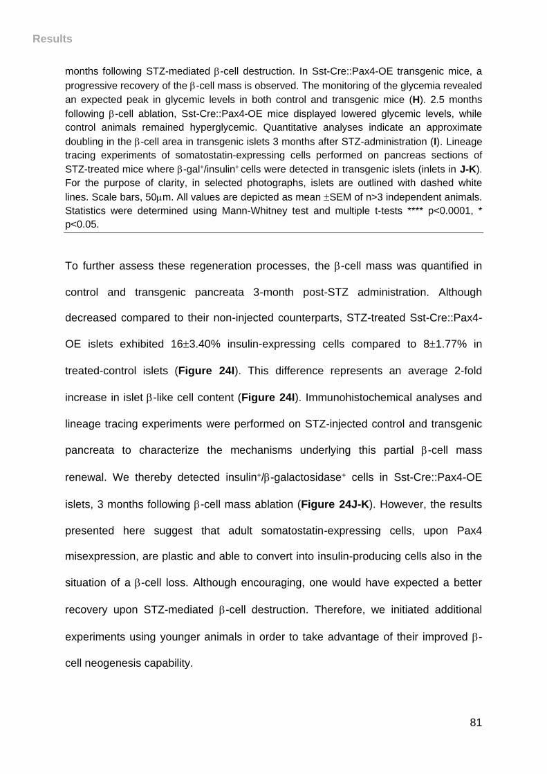
Results
81
months following STZ-mediated -cell destruction. In Sst-Cre::Pax4-OE transgenic mice, a
progressive recovery of the -cell mass is observed. The monitoring of the glycemia revealed
an expected peak in glycemic levels in both control and transgenic mice (H). 2.5 months
following -cell ablation, Sst-Cre::Pax4-OE mice displayed lowered glycemic levels, while
control animals remained hyperglycemic. Quantitative analyses indicate an approximate
doubling in the -cell area in transgenic islets 3 months after STZ-administration (I). Lineage
tracing experiments of somatostatin-expressing cells performed on pancreas sections of
STZ-treated mice where -gal+/insulin+ cells were detected in transgenic islets (inlets in J-K).
For the purpose of clarity, in selected photographs, islets are outlined with dashed white
lines. Scale bars, 50m. All values are depicted as mean SEM of n>3 independent animals.
Statistics were determined using Mann-Whitney test and multiple t-tests **** p<0.0001, *
p<0.05.
To further assess these regeneration processes, the -cell mass was quantified in
control and transgenic pancreata 3-month post-STZ administration. Although
decreased compared to their non-injected counterparts, STZ-treated Sst-Cre::Pax4-
OE islets exhibited 163.40% insulin-expressing cells compared to 81.77% in
treated-control islets (Figure 24I). This difference represents an average 2-fold
increase in islet -like cell content (Figure 24I). Immunohistochemical analyses and
lineage tracing experiments were performed on STZ-injected control and transgenic
pancreata to characterize the mechanisms underlying this partial -cell mass
renewal. We thereby detected insulin+/-galactosidase+ cells in Sst-Cre::Pax4-OE
islets, 3 months following -cell mass ablation (Figure 24J-K). However, the results
presented here suggest that adult somatostatin-expressing cells, upon Pax4
misexpression, are plastic and able to convert into insulin-producing cells also in the
situation of a -cell loss. Although encouraging, one would have expected a better
recovery upon STZ-mediated -cell destruction. Therefore, we initiated additional
experiments using younger animals in order to take advantage of their improved -
cell neogenesis capability.

82
DISCUSSION
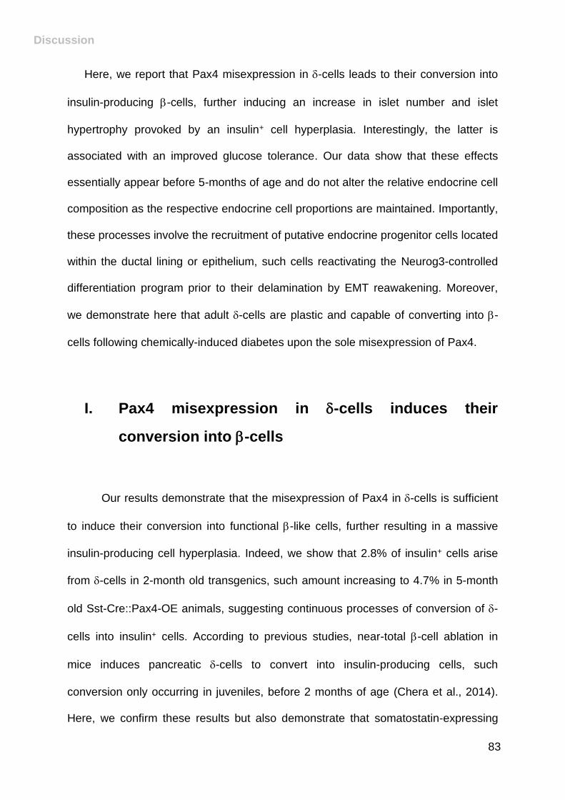
Discussion
83
Here, we report that Pax4 misexpression in -cells leads to their conversion into
insulin-producing -cells, further inducing an increase in islet number and islet
hypertrophy provoked by an insulin+ cell hyperplasia. Interestingly, the latter is
associated with an improved glucose tolerance. Our data show that these effects
essentially appear before 5-months of age and do not alter the relative endocrine cell
composition as the respective endocrine cell proportions are maintained. Importantly,
these processes involve the recruitment of putative endocrine progenitor cells located
within the ductal lining or epithelium, such cells reactivating the Neurog3-controlled
differentiation program prior to their delamination by EMT reawakening. Moreover,
we demonstrate here that adult -cells are plastic and capable of converting into -
cells following chemically-induced diabetes upon the sole misexpression of Pax4.
I. Pax4 misexpression in -cells induces their
conversion into -cells
Our results demonstrate that the misexpression of Pax4 in -cells is sufficient
to induce their conversion into functional -like cells, further resulting in a massive
insulin-producing cell hyperplasia. Indeed, we show that 2.8% of insulin+ cells arise
from -cells in 2-month old transgenics, such amount increasing to 4.7% in 5-month
old Sst-Cre::Pax4-OE animals, suggesting continuous processes of conversion of -
cells into insulin+ cells. According to previous studies, near-total -cell ablation in
mice induces pancreatic -cells to convert into insulin-producing cells, such
conversion only occurring in juveniles, before 2 months of age (Chera et al., 2014).
Here, we confirm these results but also demonstrate that somatostatin-expressing
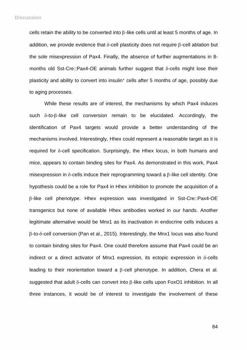
Discussion
84
cells retain the ability to be converted into -like cells until at least 5 months of age. In
addition, we provide evidence that -cell plasticity does not require -cell ablation but
the sole misexpression of Pax4. Finally, the absence of further augmentations in 8-
months old Sst-Cre::Pax4-OE animals further suggest that -cells might lose their
plasticity and ability to convert into insulin+ cells after 5 months of age, possibly due
to aging processes.
While these results are of interest, the mechanisms by which Pax4 induces
such -to--like cell conversion remain to be elucidated. Accordingly, the
identification of Pax4 targets would provide a better understanding of the
mechanisms involved. Interestingly, Hhex could represent a reasonable target as it is
required for -cell specification. Surprisingly, the Hhex locus, in both humans and
mice, appears to contain binding sites for Pax4. As demonstrated in this work, Pax4
misexpression in -cells induce their reprogramming toward a -like cell identity. One
hypothesis could be a role for Pax4 in Hhex inhibition to promote the acquisition of a
-like cell phenotype. Hhex expression was investigated in Sst-Cre::Pax4-OE
transgenics but none of available Hhex antibodies worked in our hands. Another
legitimate alternative would be Mnx1 as its inactivation in endocrine cells induces a
-to--cell conversion (Pan et al., 2015). Interestingly, the Mnx1 locus was also found
to contain binding sites for Pax4. One could therefore assume that Pax4 could be an
indirect or a direct activator of Mnx1 expression, its ectopic expression in -cells
leading to their reorientation toward a -cell phenotype. In addition, Chera et al.
suggested that adult -cells can convert into -like cells upon FoxO1 inhibition. In all
three instances, it would be of interest to investigate the involvement of these
Discussion
85
putative targets in the processes underlying the reprogramming of -cells into -like
cells.
II. Recruitment of Neurog3 expressing ductal cells and
EMT reawakening
In this study, we demonstrated that a proportion of 9% of supplementary -like
cells appeared to arise from -cells in 2-month old Sst-Cre::Pax4-OE mice, this
proportion being maintained in older transgenics. Therefore, the observed conversion
rate of -cells misexpressing Pax4 cannot explain such increase in islet count and
size, suggestive of the involvement of additional processes.
As previously mentioned, Neurog3 is a pro-endocrine gene only expressed in
endocrine progenitors during development, and is responsible for the generation of
the main endocrine cell types. Here, we demonstrate the re-expression of Neurog3
within or close to the ductal lining epithelium upon Pax4 misexpression in
somatostatin-expressing cells. Emphasizing that Neurog3 is responsible for the
specification of all the endocrine cell lineages following specific ratios, the generation
of endocrine cell types from the adult ductal epithelium would respect the standard
endocrine proportions (80% of -cells/10% of -cells/10% of -cells). Our analyses of
islets from animals misexpressing Pax4 in -cells support this hypothesis with a clear
favoring toward -like cells neogenesis. Accordingly, our results demonstrate an
increase in -cell contents perfectly matching the progressive islet hypertrophy.
Moreover, it has been shown that during mouse pancreas morphogenesis,
Neurog3 induces the delamination of progenitors from the ductal epithelium through
Discussion
86
EMT processes (Gouzi et al., 2011). Interestingly, our data show a massive increase
in the expression of mesenchymal markers within the ductal lining of Sst-Cre::Pax4-
OE transgenics, further supporting the notion of an endocrine cell neogenesis from
the ductal epithelium.
Finally, the proliferation of ductal cells and the re-expression of Neurog3 were
previously noted in regeneration processes (Criscimanna et al., 2011). While ductal
cells exhibit low proliferation rates in normal conditions (Kopp et al., 2016), our
analyses reveal a significant augmentation in cell proliferation within the ductal
epithelium, a number of these proliferating cells eventually acquiring a mesenchymal
phenotype. Together, these results clearly suggest that Pax4 misexpression in -cells
induces their conversion into -like cells and a subsequent cycle of endocrine cell
neogenesis through recapitulation of developmental processes.
Our analyses also demonstrate that Pax4 misexpression in -cells induces their
conversion into -like cells following chemically-induced -cell ablation, resulting in a
partial -cell mass restoration. Although not exhibiting euglycemia as expected, Sst-
Cre::Pax4-OE transgenics displayed an average glycemia of 350mg/dl and an
extended lifespan. Therefore, as -cells plasticity seems to decline with age, it would
be of interest to induce -cell ablation before 2 months of age. Accordingly, additional
experiments putting to the test -cells plasticity at 1 month of age are currently
ongoing.
Moreover, to definitively ascertain the requirement of Neurog3 for the
endocrine cell neogenesis observed in Sst-Cre::Pax4-OE transgenics, it would be of
interest to use an inducible knock down of Neurog3 within the ductal epithelium.

87
CONCLUSION / PERSPECTIVES
Conclusion/Perspectives
88
I. Somatostatin: a signal inducing -cell regeneration?
We hypothesized that the Pax4-mediated conversion of somatostatin-
expressing cells induces a disruption of islet homeostasis, further leading to the
recruitment of Neurog3-reexpressing ductal precursor cells adopting an endocrine
cell identity to counteract the loss of -cells. However, further investigations are
required.
Thus, the treatment of Sst-Cre::Pax4-OE mice with a somatostatin agonist or
antagonist would allow for a better comprehension of such mechanisms. More
specifically, the supplementation of Sst-Cre::Pax4-OE transgenics with a
somatostatin agonist is expected to prevent Neurog3 re-expression and therefore -
like cell hyperplasia. In this context, one could wonder whether somatostatin
antagonist administration to wild-type mice would induce such regeneration.
However, somatostatin exerts a wide-range of effects and its inhibition could lead to
undesirable side effects. In addition, the precise expression of the different SST
receptors within endocrine cells remains controversial. Therefore, inhibition of one or
several of these receptors could have an effect on other islet cells, provoking
uncontrolled side-effects. Several studies focusing on the effects of somatostatin
receptor deficiency demonstrated changes in insulin secretion and alterations in islet
architecture depending on the isoform deleted. SSTR2 loss leads to increased
glucagon secretion while no difference in insulin levels was observed (Strowski et al.,
2000). Ablation of SSTR1 or SSTR5 mostly results in glucose intolerance with
decreased numbers of somatostatin-expressing cells being observed in SSTR1
deficient mice (Wang et al., 2006; X. P. Wang et al., 2005). Remarkably, double
mutant mice for SSTR1 and 5 show improved glucose tolerance, increased insulin
Conclusion/Perspectives
89
secretion and islet cell hyperplasia (Wang et al., 2004). Although intriguing,
understanding such results is challenging due to possible compensation in the
expression of the different receptor subtypes. Moreover, the ability for somatostatin
receptor to heterodimerize adds further difficulties in the interpretation (Pfeiffer et al.,
2001; Wang et al., 2006; X. P. Wang et al., 2005).
In addition, the production of SST-28 was found increased in type 1 diabetic
patients and a -cell hyperplasia was observed (Xaio Ping Wang et al., 2005).
Understanding the role of SST and its receptors in the context of type 1 diabetes
would therefore be of great interest. Importantly, we strongly believe that islet
homeostasis is not only preserved by the hormones secreted by the different
endocrine cell types, but also by cellular interactions, integrity and cohesion.
II. Model proposed
The aim of this study was to investigate the plasticity of -cells, aiming to
eventually determine whether the sole misexpression of Pax4 in -cells could induce
their conversion into insulin-producing cells in vivo. To address this question, we
generated Sst-Cre::Pax4-OE double transgenic animals allowing the misexpression
of Pax4 specifically in -cells. In this model, we demonstrated the conversion of adult
somatostatin-expressing cells into insulin+ cells upon the sole misexpression of Pax4.
Our data suggest that such conversion induces a disruption of islet homeostasis,
leading to a reawakening of Neurog3-controlled endocrine differentiation program
and associated developmental processes, including EMT (Figure 25). Importantly,
we observed a progressive islet neogenesis and islet hypertrophy together with an
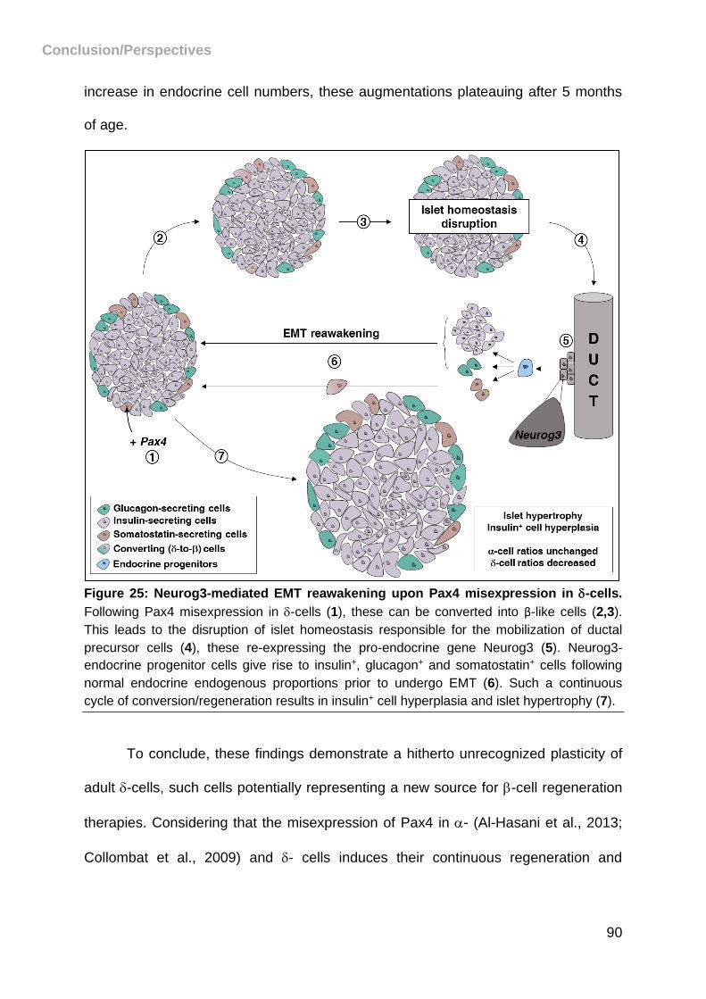
Conclusion/Perspectives
90
increase in endocrine cell numbers, these augmentations plateauing after 5 months
of age.
Figure 25: Neurog3-mediated EMT reawakening upon Pax4 misexpression in -cells.
Following Pax4 misexpression in -cells (1), these can be converted into β-like cells (2,3).
This leads to the disruption of islet homeostasis responsible for the mobilization of ductal
precursor cells (4), these re-expressing the pro-endocrine gene Neurog3 (5). Neurog3-
endocrine progenitor cells give rise to insulin+, glucagon+ and somatostatin+ cells following
normal endocrine endogenous proportions prior to undergo EMT (6). Such a continuous
cycle of conversion/regeneration results in insulin+ cell hyperplasia and islet hypertrophy (7).
To conclude, these findings demonstrate a hitherto unrecognized plasticity of
adult -cells, such cells potentially representing a new source for -cell regeneration
therapies. Considering that the misexpression of Pax4 in - (Al-Hasani et al., 2013;
Collombat et al., 2009) and - cells induces their continuous regeneration and

Conclusion/Perspectives
91
conversion into -like cells, this defines Pax4 as a considerable target for diabetes
therapies.

92
BIBLIOGRAPHY
Bibliography
93
Aasen, T., Raya, A., Barrero, M.J., Garreta, E., Consiglio, A., Gonzalez, F., Vassena, R., Bilic, J., Pekarik, V., Tiscornia, G., Edel, M., Boue, S., Izpisua Belmonte, J.C., 2008. Efficient and rapid generation of induced pluripotent stem cells from human keratinocytes. Nat Biotechnol 26, 1276–1284. doi:nbt.1503 [pii]\r10.1038/nbt.1503
Adrian, T.E., Bloom, S.R., Hermansen, K., Iversen, J., 1978. Pancreatic polypeptide, glucagon and insulin secretion from the isolated perfused canine pancreas. Diabetologia 14, 413–7.
Agudo, J., Ayuso, E., Jimenez, V., Salavert, A., Casellas, A., Tafuro, S., Haurigot, V., Ruberte, J., Segovia, J.C., Bueren, J., Bosch, F., 2008. IGF-I mediates regeneration of endocrine pancreas by increasing beta cell replication through cell cycle protein modulation in mice. Diabetologia 51, 1862–1872. doi:10.1007/s00125-008-1087-8
Ahlgren, U., Pfaff, S.L., Jessell, T.M., Edlund, T., Edlund, H., 1997. Independent requirement for ISL1 in formation of pancreatic mesenchyme and islet cells. Nature. doi:10.1038/385257a0
Al-Hasani, K., Pfeifer, A., Courtney, M., Ben-Othman, N., Gjernes, E., Vieira, A., Druelle, N., Avolio, F., Ravassard, P., Leuckx, G., Lacas-Gervais, S., Ambrosetti, D., Benizri, E., Hecksher-Sørensen, J., Gounon, P., Ferrer, J., Gradwohl, G., Heimberg, H., Mansouri, A., Collombat, P., 2013. Adult duct-lining cells can reprogram into β-like cells able to counter repeated cycles of toxin-induced diabetes. Dev. Cell 26, 86–100. doi:10.1016/j.devcel.2013.05.018
Alipio, Z., Liao, W., Roemer, E.J., Waner, M., Fink, L.M., Ward, D.C., Ma, Y., 2010. Reversal of hyperglycemia in diabetic mouse models using induced-pluripotent stem (iPS)-derived pancreatic beta-like cells. Proc. Natl. Acad. Sci. U. S. A. 107, 13426–13431. doi:10.1073/pnas.1007884107
Alwan, A., 2010. Global status report on noncommunicable diseases. World Health 176. doi:978 92 4 156422 9
Alwan, N., Tuffnell, D.J., West, J., 2009. Treatments for gestational diabetes. Cochrane Database Syst. Rev. doi:10.1002/14651858.CD003395.pub2
Andralojc, K.M., Mercalli, A., Nowak, K.W., Albarello, L., Calcagno, R., Luzi, L., Bonifacio, E., Doglioni, C., Piemonti, L., 2009. Ghrelin-producing epsilon cells in the developing and adult human pancreas. Diabetologia 52, 486–493. doi:10.1007/s00125-008-1238-y
Andrews, P.C., Dixon, J.E., 1986. Biosynthesis and processing of the somatostatin family of peptide hormones. Scand J Gastroenterol Suppl 119, 22–28. doi:10.3109/00365528609087428
Andrulionytè, L., Zacharova, J., Chiasson, J.-L., Laakso, M., 2004. Common polymorphisms of the PPAR-gamma2 (Pro12Ala) and PGC-1alpha (Gly482Ser) genes are associated with the conversion from impaired glucose tolerance to type 2 diabetes in the STOP-NIDDM trial. Diabetologia 47, 2176–84. doi:10.1007/s00125-004-1577-2
Aoi, T., Yae, K., Nakagawa, M., Ichisaka, T., Okita, K., Takahashi, K., Chiba, T., Yamanaka, S., 2008. Generation of pluripotent stem cells from adult mouse liver and stomach cells. Science 321, 699–702. doi:10.1126/science.1154884
Arosio, M., Ronchi, C.L., Gebbia, C., Cappiello, V., Beck-Peccoz, P., Peracchi, M., 2003. Stimulatory effects of ghrelin on circulating somatostatin and pancreatic polypeptide levels. J. Clin. Endocrinol. Metab. 88, 701–704. doi:10.1210/jc.2002-021161
Asakawa, A., Inui, A., Yuzuriha, H., Ueno, N., Katsuura, G., Fujimiya, M., Fujino, M.A., Niijima, A., Meguid, M.M., Kasuga, M., 2003. Characterization of the effects of pancreatic polypeptide in the regulation of energy balance. Gastroenterology 124,
Bibliography
94
1325–1336. doi:10.1016/S0016-5085(03)00216-6 Ashcroft, F.M., Harrison, D.E., Ashcroft, S.J., 1984. Glucose induces closure of single
potassium channels in isolated rat pancreatic beta-cells. Nature 312, 446–8. doi:10.1038/312446a0
Ashcroft, F.M., Rorsman, P., 2013. K ATP channels and islet hormone secretion : new insights and controversies. Nat. Publ. Gr. 9, 660–669. doi:10.1038/nrendo.2013.166
Atkinson, M.A., Bowman, M.A., Campbell, L., Darrow, B.L., Kaufman, D.L., Maclaren, N.K., 1994. Cellular immunity to a determinant common to glutamate decarboxylase and Coxsackie virus in insulin-dependent diabetes. J. Clin. Invest. 94, 2125–2129. doi:10.1172/JCI117567
Authier, F., Desbuquois, B., 2008. Glucagon receptors. Cell. Mol. Life Sci. doi:10.1007/s00018-008-7479-6
Baekkeskov, S., Aanstoot, H.J., Christgau, S., Reetz, A., Solimena, M., Cascalho, M., Folli, F., Richter-Olesen, H., De Camilli, P., 1990. Identificationof the 64K autoantigen in insulin-dependent diabetes as the GABA-synthesizing enzyme glutamic acid decarboxylase. NatureNature 347, 151–156. doi:10.1038/347151a0
Baetens, D., Malaisse-Lagae, F., Perrelet, a, Orci, L., 1979. Endocrine pancreas: three-dimensional reconstruction shows two types of islets of langerhans. Science 206, 1323–5. doi:10.1126/science.390711
Baeyens, L., Lemper, M., Leuckx, G., Groef, S. De, Bonfanti, P., Stangé, G., Shemer, R., Nord, C., Scheel, D.W., Pan, F.C., Ahlgren, U., Gu, G., Stoffers, D.A., Dor, Y., Ferrer, J., Gradwohl, G., Wright, C.V.E., Casteele, M. Van De, German, M.S., Bouwens, L., Heimberg, H., 2013. Transient cytokine treatment induces acinar cell reprogramming and regenerates functional beta cell mass in diabetic mice. Nat. Biotechnol. 32, 76–83. doi:10.1038/nbt.2747
Bar-Nur, O., Brumbaugh, J., Verheul, C., Apostolou, E., Pruteanu-Malinici, I., Walsh, R.M., Ramaswamy, S., Hochedlinger, K., 2014. Small molecules facilitate rapid and synchronous iPSC generation. Nat. Methods 11, 1170–6. doi:10.1038/nmeth.3142
Batterham, R.L., Le Roux, C.W., Cohen, M.A., Park, A.J., Ellis, S.M., Patterson, M., Frost, G.S., Ghatei, M.A., Bloom, S.R., 2003. Pancreatic Polypeptide Reduces Appetite and Food Intake in Humans. J. Clin. Endocrinol. Metab. 88, 3989–3992. doi:10.1210/jc.2003-030630
Bayha, E., Jorgensen, M.C., Serup, P., Grapin-Botton, A., 2009. Retinoic acid signaling organizes endodermal organ specification along the entire antero-posterior axis. PLoS One 4. doi:10.1371/journal.pone.0005845
Beattie, G.M., Montgomery, A.M.P., Lopez, A.D., Hao, E., Perez, B., Just, M.L., Lakey, J.R.T., Hart, M.E., Hayek, A., 2002. A novel approach to increase human islet cell mass while preserving beta-cell function. Diabetes 51, 3435–3439. doi:10.2337/diabetes.51.12.3435
Bellamy, L., Casas, J.-P., Hingorani, A.D., Williams, D., 2009. Type 2 diabetes mellitus after gestational diabetes: a systematic review and meta-analysis. Lancet 373, 1773–1779. doi:10.1016/S0140-6736(09)60731-5
Benitez, C.M., Goodyer, W.R., Kim, S.K., 2012. Deconstructing pancreas developmental biology. Cold Spring Harb. Perspect. Biol. 4, 1–17. doi:10.1101/cshperspect.a012401
Bingley, P.J., Bonifacio, E., Williams, A.J.K., Genovese, S., Bottazzo, G.F., Gale, E.A.M., 1997. Prediction of IDDM in the general population: Strategies based on combinations of autoantibody markers. Diabetes 46, 1701–1710. doi:10.2337/diabetes.46.11.1701
Bjerknes, M., Cheng, H., 2002. Multipotential stem cells in adult mouse gastric
Bibliography
95
epithelium. Am. J. Physiol. Gastrointest. Liver Physiol. 283, G767–G777. doi:10.1152/ajpgi.00415.2001
Bockman, D.E., 1993. Anatomy of the pancreas, in: The Pancreas: Biology, Pathobiology and Disease. p. 8.
Boel, E., Schwartz, T.W., Norris, K.E., Fiil, N.P., 1984. A cDNA encoding a small common precursor for human pancreatic polypeptide and pancreatic icosapeptide. EMBO J. 3, 909–12.
Bollheimer, L.C., Landauer, H.C., Troll, S., Schweimer, J., Wrede, C.E., Schölmerich, J., Buettner, R., 2015. Stimulatory short-term effects of free fatty acids on glucagon secretion at low to normal glucose concentrations. Metab. - Clin. Exp. 53, 1443–1448. doi:10.1016/j.metabol.2004.06.011
Bonal, C., Thorel, F., Ait-Lounis, A., Reith, W., Trumpp, A., Herrera, P.L., 2009. Pancreatic Inactivation of c-Myc Decreases Acinar Mass and Transdifferentiates Acinar Cells Into Adipocytes in Mice. Gastroenterology 136. doi:10.1053/j.gastro.2008.10.015
Bonner-Weir, S., Baxter, L.A., Schuppin, G.T., Smith, F.E., 1993. A second pathway for regeneration of adult exocrine and endocrine pancreas. A possible recapitulation of embryonic development. Diabetes 42, 1715–20. doi:10.2337/diab.42.12.1715
Bonner-Weir, S., Inada, A., Yatoh, S., Li, W.-C., Aye, T., Toschi, E., Sharma, A., 2008. Transdifferentiation of pancreatic ductal cells to endocrine β-cells. Biochem. Soc. Trans. 36, 353–356. doi:10.1042/BST0360353
Bonner-Weir, S., Li, W., Ouziel-yahalom, L., Guo, L., Weir, G.C., Sharma, A., 2004. B-Cell Growth and Regeneration : Replication Is Only Part of the Story 2004. doi:10.2337/db10-0084
Bort, R., Martinez-barbera, J.P., Beddington, R.S.P., Zaret, K.S., 2004. Hex homeobox gene-dependent tissue positioning is required for organogenesis of the ventral pancreas. Development 131, 797–806. doi:10.1242/dev.00965
Bottazzo GF, Florin-Christensen A, D.D., 1974. Islet-cell antibodies in diabetes mellitus with autouimmne polyendocrine deficiencies. Lancet 304, 1279–83.
Bottini, N., Musumeci, L., Alonso, A., Rahmouni, S., Nika, K., Rostamkhani, M., MacMurray, J., Meloni, G.F., Lucarelli, P., Pellecchia, M., Eisenbarth, G.S., Comings, D., Mustelin, T., 2004. A functional variant of lymphoid tyrosine phosphatase is associated with type I diabetes. Nat. Genet. 36, 337–338. doi:10.1038/ng1323
Bouwens, L., De Blay, E., 1996. Islet morphogenesis and stem cell markers in rat pancreas. J. Histochem. Cytochem. 44, 947–951. doi:10.1177/44.9.8773559
Bouwens, L., Houbracken, I., Mfopou, J.K., 2013. The use of stem cells for pancreatic regeneration in diabetes mellitus. Nat. Rev. Endocrinol. 9, 598–606. doi:10.1038/nrendo.2013.145
Bramswig, N.C., Everett, L.J., Schug, J., Dorrell, C., Liu, C., Luo, Y., Streeter, P.R., Naji, A., Grompe, M., Kaestner, K.H., 2013. Epigenomic plasticity enables human pancreatic alpha to beta cell reprogramming. J Clin Invest 123, 1275–1284. doi:10.1172/jci66514
Brissova, M., Fowler, M.J., Nicholson, W.E., Chu, A., Hirshberg, B., Harlan, D.M., Powers, A.C., 2005. Assessment of Human Pancreatic Islet Architecture and Composition by Laser Scanning Confocal Microscopy. J. Histochem. Cytochem. 53, 1087–1097. doi:10.1369/jhc.5C6684.2005
Brissova, M., Shiota, M., Nicholson, W.E., Gannon, M., Knobel, S.M., Piston, D.W., Wright, C.V.E., Powers, A.C., 2002. Reduction in pancreatic transcription factor PDX-1 impairs glucose-stimulated insulin secretion. J. Biol. Chem. 277, 11225–11232. doi:10.1074/jbc.M111272200
Bibliography
96
Brissova, M., Shostak, A., Shiota, M., Wiebe, P.O., Poffenberger, G., Kantz, J., Chen, Z., Carr, C., Jerome, W.G., Chen, J., Baldwin, H.S., Nicholson, W., Bader, D.M., Jetton, T., Gannon, M., Powers, A.C., 2006. Pancreatic islet production of vascular endothelial growth factor-A is essential for islet vascularization, revascularization, and function. Diabetes 55, 2974–2985. doi:10.2337/db06-0690
Brubaker, P.L., Drucker, D.J., 2002. Structure-function of the glucagon receptor family of G protein-coupled receptors: the glucagon, GIP, GLP-1, and GLP-2 receptors. Receptors Channels 8, 179–188. doi:10.1080/10606820213687
Bruni, A., Gala-Lopez, B., Pepper, A.R., Abualhassan, N.S., James Shapiro, a. M., Shapiro, A.J., 2014. Islet cell transplantation for the treatment of type 1 diabetes: Recent advances and future challenges. Diabetes, Metab. Syndr. Obes. Targets Ther. 7, 211–223. doi:10.2147/DMSO.S50789
Burlison, J.S., Long, Q., Fujitani, Y., Wright, C.V.E., Magnuson, M.A., 2008. Pdx-1 and Ptf1a concurrently determine fate specification of pancreatic multipotent progenitor cells. Dev. Biol. 316, 74–86. doi:10.1016/j.ydbio.2008.01.011
Busnardo, A.C., DiDio, L.J. a., Tidrick, R.T., Thomford, N.R., 1983. History of the pancreas. Am. J. Surg. 146, 539–550. doi:10.1016/0002-9610(83)90286-6
Cabrera, O., Berman, D.M., Kenyon, N.S., Ricordi, C., Berggren, P.-O.O., Caicedo, A., 2006. The unique cytoarchitecture of human pancreatic islets has implications for islet cell function. Proc. Natl. Acad. Sci. U. S. A. 103, 2334–9. doi:10.1073/pnas.0510790103
Cano, D.A., Hebrok, M., Zenker, M., 2007. Pancreatic Development and Disease. Gastroenterology 132, 745–762. doi:10.1053/j.gastro.2006.12.054
Cano, D.A., Murcia, N.S., Pazour, G.J., Hebrok, M., 2004. Orpk mouse model of polycystic kidney disease reveals essential role of primary cilia in pancreatic tissue organization. Development 131, 3457–3467. doi:10.1242/dev.01189\r131/14/3457 [pii]
Cano, D.A., Rulifson, I.C., Heiser, P.W., Swigart, L.B., Pelengaris, S., German, M., Evan, G.I., Bluestone, J.A., Hebrok, M., 2008. Regulated beta-cell regeneration in the adult mouse pancreas. Diabetes 57, 958–66. doi:10.2337/db07-0913
Cano, D.A., Sekine, S., Hebrok, M., 2006. Primary Cilia Deletion in Pancreatic Epithelial Cells Results in Cyst Formation and Pancreatitis. Gastroenterology 131, 1856–1869. doi:10.1053/j.gastro.2006.10.050
Carr, D.B., Gabbe, S., 1998. Gestational Diabetes: Detection, Management, and Implications. Clin Diabetes 16.
Chera, S., Baronnier, D., Ghila, L., Cigliola, V., Jensen, J.N., Gu, G., Furuyama, K., Thorel, F., Gribble, F.M., Reimann, F., Herrera, P.L., 2014. Diabetes recovery by age-dependent conversion of pancreatic δ-cells into insulin producers. Nature 514, 503–507. doi:10.1038/nature13633
Chiang, M.K., Melton, D.A., 2003. Single-cell transcript analysis of pancreas development. Dev Cell 4, 383–393.
Chung, C.-H., Hao, E., Piran, R., Keinan, E., Levine, F., 2010. Pancreatic β-Cell neogenesis by direct conversion from mature α-Cells. Stem Cells 28, 1630–1638. doi:10.1002/stem.482
Cnop, M., Foufelle, F., Velloso, L.A., 2012. Endoplasmic reticulum stress, obesity and diabetes. Trends Mol. Med. doi:10.1016/j.molmed.2011.07.010
Cole, L., Anderson, M., Antin, P.B., Limesand, S.W., 2009. One process for pancreatic beta-cell coalescence into islets involves an epithelial-mesenchymal transition. J Endocrinol 203, 19–31. doi:10.1677/joe-09-0072
Bibliography
97
Collombat, P., Hecksher-sørensen, J., Broccoli, V., Krull, J., Ponte, I., Mundiger, T., Smith, J., Gruss, P., Serup, P., Mansouri, A., Hecksher-Sorensen, J., Broccoli, V., Krull, J., Ponte, I., Mundiger, T., Smith, J., Gruss, P., Serup, P., Mansouri, A., 2005. The simultaneous loss of Arx and Pax4 genes promotes a somatostatin-producing cell fate specification at the expense of the alpha- and beta-cell lineages in the mouse endocrine pancreas. Development 132, 2969–2980. doi:10.1242/dev.01870
Collombat, P., Mansouri, A., Hecksher-Sørensen, J., Serup, P., Krull, J., Gradwohl, G., Gruss, P., 2003. Opposing actions of Arx and Pax4 in endocrine pancreas development. Genes Dev. 17, 2591–2603. doi:10.1101/gad.269003
Collombat, P., Xu, X., Ravassard, P., Sosa-Pineda, B., Billestrup, N., Madsen, O.D., Serup, P., Heimberg, H., Mansouri, A., 2009. The ectopic expression of Pax4 in the mouse pancreas converts progenitor cells into α and subsequently β Cells. Cell 138, 449–462. doi:10.1016/j.cell.2009.05.035
Colman, A., Dreesen, O., 2009. Pluripotent stem cells and disease modeling. Cell Stem Cell 5, 244–7. doi:10.1016/j.stem.2009.08.010
Courtney, M., Gjernes, E., Druelle, N., Ravaud, C., Vieira, A., Ben-Othman, N., Pfeifer, A., Avolio, F., Leuckx, G., Lacas-Gervais, S., Burel-Vandenbos, F., Ambrosetti, D., Hecksher-Sorensen, J., Ravassard, P., Heimberg, H., Mansouri, A., Collombat, P., 2013. The Inactivation of Arx in Pancreatic α-Cells Triggers Their Neogenesis and Conversion into Functional β-Like Cells. PLoS Genet. 9. doi:10.1371/journal.pgen.1003934
Criscimanna, A., Speicher, J.A., Houshmand, G., Shiota, C., Prasadan, K., Ji, B., Logsdon, C.D., Gittes, G.K., Esni, F., 2011. Duct cells contribute to regeneration of endocrine and acinar cells following pancreatic damage in adult mice. Gastroenterology 141, 1451–1462.e6. doi:10.1053/j.gastro.2011.07.003
Cummings, D.E., Purnell, J.Q., Frayo, R.S., Schmidova, K., Wisse, B.E., Weigle, D.S., 2001. A Preprandial Rise in Plasma Ghrelin Levels Suggests a Role in Meal Initiation in Humans. Diabetes 50, 1714–1719. doi:10.2337/diabetes.50.8.1714
D’Alessio, D.A., Sieber, C., Beglinger, C., Ensinck, J.W., 1989. A physiologic role for somatostatin 28 as a regulator of insulin secretion. J. Clin. Invest. 84, 857–862. doi:10.1172/JCI114246
D’Amour, K.A., Bang, A.G., Eliazer, S., Kelly, O.G., Agulnick, A.D., Smart, N.G., Moorman, M.A., Kroon, E., Carpenter, M.K., Baetge, E.E., 2006. Production of pancreatic hormone–expressing endocrine cells from human embryonic stem cells. Nat. Biotechnol. 24, 1392–1401. doi:10.1038/nbt1259
Dabelea, D., Mayer-Davis, E.J., Lamichhane, A.P., D’Agostino, R.B., Liese, A.D., Vehik, K.S., Venkat Narayan, K.M., Zeitler, P., Hamman, R.F., 2008. Association of intrauterine exposure to maternal diabetes and obesity with type 2 diabetes in youth: The SEARCH case-control study. Diabetes Care 31, 1422–1426. doi:10.2337/dc07-2417
De Vas, M.G., Kopp, J.L., Heliot, C., Sander, M., Cereghini, S., Haumaitre, C., 2015. Hnf1b controls pancreas morphogenesis and the generation of Ngn3+ endocrine progenitors. Development 142, 871–82. doi:10.1242/dev.110759
De Wert, G., Mummery, C., 2003. Human embryonic stem cells: Research, ethics and policy. Hum. Reprod. doi:10.1093/humrep/deg143
Delous, M., Yin, C., Shin, D., Ninov, N., Debrito Carten, J., Pan, L., Ma, T.P., Farber, S.A., Moens, C.B., Stainier, D.Y.R., 2012. sox9b is a key regulator of pancreaticobiliary ductal system development. PLoS Genet. 8. doi:10.1371/journal.pgen.1002754
Desai, B.M., Oliver-Krasinski, J., De Leon, D.D., Farzad, C., Hong, N., Leach, S.D., Stoffers, D.A., 2007. Preexisting pancreatic acinar cells contribute to acinar cell , but not islet
Bibliography
98
β cell , regeneration. Cell 117, 971–977. doi:10.1172/JCI29988.) DeSisto, C.L., Kim, S.Y., Sharma, A.J., 2014. Prevalence estimates of gestational diabetes
mellitus in the United States, Pregnancy Risk Assessment Monitoring System (PRAMS), 2007-2010. Prev. Chronic Dis. 11, E104. doi:10.5888/pcd11.130415
DiGruccio, M.R., Mawla, A.M., Donaldson, C.J., Noguchi, G.M., Vaughan, J., Cowing-Zitron, C., van der Meulen, T., Huising, M.O., 2016. Comprehensive alpha, beta and delta cell transcriptomes reveal that ghrelin selectively activates delta cells and promotes somatostatin release from pancreatic islets. Mol. Metab. 5, 449–458. doi:10.1016/j.molmet.2016.04.007
Direnzo, D., Hess, D.A., Damsz, B., Hallett, J.E., Marshall, B., Goswami, C., Liu, Y., Deering, T., MacDonald, R.J., Konieczny, S.F., 2012. Induced Mist1 expression promotes remodeling of mouse pancreatic acinar cells. Gastroenterology 143, 469–480. doi:10.1053/j.gastro.2012.04.011
Dolensek, J., Rupnik, M.S., Stozer, A., 2015. Structural similarities and differences between the human and the mouse pancreas. Islets 7. doi:10.1080/19382014.2015.1024405
Dollinger, H.C., Raptis, S., Pfeiffer, E.F., 1976. Effects of somatostatin on exocrine and endocrine pancreatic function stimulated by intestinal hormones in man. Horm. Metab. Res. 8, 74–8. doi:10.1055/s-0028-1093677
Domínguez-Bendala, J., Lanzoni, G., Klein, D., Álvarez-Cubela, S., Pastori, R.L., 2016. The Human Endocrine Pancreas: New Insights on Replacement and Regeneration. Trends Endocrinol. Metab. doi:10.1016/j.tem.2015.12.003
Edlund, H., 2002. Organogenesis: Pancreatic organogenesis — developmental mechanisms and implications for therapy. Nat. Rev. Genet. 3, 524–532. doi:10.1038/nrg841
Egido, E.M., Rodriguez-Gallardo, J., Silvestre, R.A., Marco, J., 2002. Inhibitory effect of ghrelin on insulin and pancreatic somatostatin secretion. Eur. J. Endocrinol. 146, 241–244. doi:10.1530/eje.0.1460241
Eide, S.., Rder, H., Johansson, S., Midthjell, K., Svik, O., Njlstad, P.R., Molven, A., 2008. Prevalence of HNF1A (MODY3) mutations in a Norwegian population (the HUNT2 Study). Diabet. Med. 25, 775–781. doi:10.1111/j.1464-5491.2008.02459.x
Eizirik, D.L., Cardozo, A.K., Cnop, M., 2008. The role for endoplasmic reticulum stress in diabetes mellitus. Endocr. Rev. doi:10.1210/er.2007-0015
Eizirik, D.L., Cnop, M., 2010. ER stress in pancreatic beta cells: the thin red line between adaptation and failure. Sci. Signal. 3, pe7. doi:10.1126/scisignal.3110pe7
Ellard, S., 2000. Hepatocyte nuclear factor 1 alpha (HNF-1a) mutations in maturity-onset diabetes of the young. Hum. Mutat. 16, 377–385. doi:10.1002/1098-1004(200011)16:5
Eriksson, K.F., Lindgärde, F., 1991. Prevention of type 2 (non-insulin-dependent) diabetes mellitus by diet and physical exercise. The 6-year Malmö feasibility study. Diabetologia. doi:10.1007/BF00400196
Esni, F., 2004. Notch inhibits Ptf1 function and acinar cell differentiation in developing mouse and zebrafish pancreas. Development 131, 4213–4224. doi:10.1242/dev.01280
Ferber, S., Halkin, A., Cohen, H., Ber, I., Einav, Y., Goldberg, I., Barshack, I., Seijffers, R., Kopolovic, J., Kaiser, N., Karasik, A., 2000. Pancreatic and duodenal homeobox gene 1 induces expression of insulin genes in liver and ameliorates streptozotocin-induced hyperglycemia. Nat. Med. 6, 568–72. doi:10.1038/75050
Fiaschi-Taesch, N.M., Kleinberger, J.W., Salim, F.G., Troxell, R., Wills, R., Tanwir, M.,
Bibliography
99
Casinelli, G., Cox, A.E., Takane, K.K., Srinivas, H., Scott, D.K., Stewart, A.F., 2013. Cytoplasmic-Nuclear Trafficking of G1/S Cell Cycle Molecules and Adult Human β-Cell Replication. Diabetes 62, 2460–2470. doi:10.2337/db12-1218
Flanagan, S.E., Patch, A.-M., Mackay, D.J.G., Edghill, E.L., Gloyn, A.L., Robinson, D., Shield, J.P.H., Temple, K., Ellard, S., Hattersley, A.T., 2007. Mutations in ATP-sensitive K+ channel genes cause transient neonatal diabetes and permanent diabetes in childhood or adulthood. Diabetes 56, 1930–7. doi:10.2337/db07-0043
Furuta, M., Zhou, A., Webb, G., Carroll, R., Ravazzola, M., Orci, L., Steiner, D.F., 2001. Severe Defect in Proglucagon Processing in Islet A-cells of Prohormone Convertase 2 Null Mice. J. Biol. Chem. 276, 27197–27202. doi:10.1074/jbc.M103362200
Gannon, M., Ables, E.T., Crawford, L., Lowe, D., Offield, M.F., Magnuson, M.A., Wright, C.V.E., 2008. pdx-1 function is specifically required in embryonic beta cells to generate appropriate numbers of endocrine cell types and maintain glucose homeostasis. Dev. Biol. 314, 406–17. doi:10.1016/j.ydbio.2007.10.038
Gao, T., McKenna, B., Li, C., Reichert, M., Nguyen, J., Singh, T., Yang, C., Pannikar, A., Doliba, N., Zhang, T., Stoffers, D.A., Edlund, H., Matschinsky, F., Stein, R., Stanger, B.Z., 2014. Pdx1 maintains beta-cell identity and function by repressing an alpha-cell program. Cell Metab. 19, 259–271. doi:10.1016
Gavaghan, M., 2002. The Pancreas—Hermit of the Abdomen. AORN J. 75, 1109–1130. doi:10.1016
Gepts, W., 1965. Pathologic anatomy of the pancreas in juvenile diabetes mellitus. Diabetes 14, 619–633. doi:10.2337
Gillespie, K.M., 2006. Type 1 diabetes: pathogenesis and prevention. CMAJ 175, 165–70. doi:10.1503/cmaj.060244
Gillespie, K.M., Bain, S.C., Barnett, P.A.H., Bingley, P.P.J., Christie, M.R., Gill, G. V., Gale, P.E.A.M., 2004. The rising incidence of childhood type 1 diabetes and reduced contribution of high-risk HLA haplotypes. Lancet 364, 1699–1700. doi:10.1016/S0140-6736(04)17357-1
Gingerich, R.L., Lacy, P.E., Chance, R.E., Johnson, M.G., 1978. Regional pancreatic concentration and in-vitro secretion of canine pancreatic polypeptide, insulin, and glucagon. Diabetes 27, 96–101. doi:10.2337/diabetes.27.2.96
Ginsberg-Fellner, F., Witt, M.E., Fedun, B., Taub, F., Dobersen, M.J., McEvoy, R.C., Cooper, L.Z., Notkins, A.L., Rubinstein, P., 1985. Diabetes mellitus and autoimmunity in patients with the congenital rubella syndrome. Rev. Infect. Dis. 7 Suppl 1, S170–6.
Githens, S., 1988. The Pancreatic Duct Cell: Proliferative Capabilities, Specific Characteristics, Metaplasia, Isolation, and Culture. J. Pediatr. Gastroenterol. Nutr. 7, 486–506. doi:2456383
Gouzi, M., Kim, Y.H., Katsumoto, K., Johansson, K., Grapin-Botton, A., 2011. Neurogenin3 initiates stepwise delamination of differentiating endocrine cells during pancreas development. Dev. Dyn. 240, 589–604. doi:10.1002/dvdy.22544
Gradwohl, G., Dierich, a, LeMeur, M., Guillemot, F., 2000. Neurogenin3 Is Required for the Development of the Four Endocrine Cell Lineages of the Pancreas. Proc. Natl. Acad. Sci. U. S. A. 97, 1607–1611. doi:10.1073/pnas.97.4.1607
Granata, R., Baragli, A., Settanni, F., Scarlatti, F., Ghigo, E., 2010. Unraveling the role of the ghrelin gene peptides in the endocrine pancreas. J. Mol. Endocrinol. doi:10.1677/JME-10-0019
Grieco, F.A., Moretti, M., Sebastiani, G., Galleri, L., Spagnuolo, I., Scafetta, G., Gulino, A., de Smaele, E., Maroder, M., Dotta, F., 2011. Delta-cell-specific expression of hedgehog pathway Ptch1 receptor in murine and human endocrine pancreas. Diabetes. Metab.
Bibliography
100
Res. Rev. 27, 755–760. doi:10.1002/dmrr.1247 Gros, L., Bréant, B., Duchene, B., Leroy, C., Fauconnier, G., Bataille, D., Virsolvy, A., 2002.
Localization of α-endosulphine in pancreatic somatostatin δ cells and expression during rat pancreas development. Diabetologia 45, 703–710. doi:10.1007/s00125-002-0794-9
Group., T.D.C. and C.T.R., 1993. The effect of intensive treatment of diabetes on the development and progression of long-term complications in insulin-dependent diabetes mellitus. N. Engl. J. Med. 329, 977–86. doi:10.1056/NEJM199309303291401
Grün, D., Muraro, M.J., Boisset, J.-C., Wiebrands, K., Lyubimova, A., Dharmadhikari, G., van den Born, M., van Es, J., Jansen, E., Clevers, H., de Koning, E.J.P., van Oudenaarden, A., 2016. De Novo Prediction of Stem Cell Identity using Single-Cell Transcriptome Data. Cell Stem Cell. doi:10.1016/j.stem.2016.05.010
Gu, C., Stein, G.H., Pan, N., Goebbels, S., Hörnberg, H., Nave, K.A., Herrera, P., White, P., Kaestner, K.H., Sussel, L., Lee, J.E., 2010. Pancreatic β Cells Require NeuroD to Achieve and Maintain Functional Maturity. Cell Metab. 11, 298–310. doi:10.1016/j.cmet.2010.03.006
Gu, G., Brown, J.R., Melton, D.A., 2003. Direct lineage tracing reveals the ontogeny of pancreatic cell fates during mouse embryogenesis. Mech. Dev. 120, 35–43. doi:10.1016/S0925-4773(02)00330-1
Gu, G., Dubauskaite, J., Melton, D.A., 2002. Direct evidence for the pancreatic lineage: NGN3+ cells are islet progenitors and are distinct from duct progenitors. Development 129, 2447–2457. doi:10.1146/annurev.cellbio.20.012103.094648
Habegger, K.M., Heppner, K.M., Geary, N., Bartness, T.J., DiMarchi, R., Tschöp, M.H., 2010. The metabolic actions of glucagon revisited. Nat. Rev. Endocrinol. 6, 689–97. doi:10.1038/nrendo.2010.187
Haffner, S.M., 1998. Epidemiology of type 2 diabetes: risk factors. Diabetes Care 21 Suppl 3, C3–C6. doi:10.2337/diacare.21.3.C3
Hamaguchi, T., Fukushima, H., Uehara, M., Wada, S., Shirotani, T., Kishikawa, H., Ichinose, K., Yamaguchi, K., Shichiri, M., 1991. Abnormal glucagon response to arginine and its normalization in obese hyperinsulinaemic patients with glucose intolerance: importance of insulin action on pancreatic Alpha cells. Diabetologia 34, 801–806. doi:10.1007/BF00408354
Harrison, K.A., Thaler, J., Pfaff, S.L., Gu, H., Kehrl, J.H., 1999. Pancreas dorsal lobe agenesis and abnormal islets of Langerhans in Hlxb9 -deficient mice 23, 71–75.
Hauge-Evans, A.C., King, A.J., Carmignac, D., Richardson, C.C., Robinson, I.C.A.F., Low, M.J., Christie, M.R., Persaud, S.J., Jones, P.M., 2009. Somatostatin secreted by islet delta-cells fulfills multiple roles as a paracrine regulator of islet function. Diabetes 58, 403–411. doi:10.2337/db08-0792
Haumaitre, C., Barbacci, E., Jenny, M., Ott, M.O., Gradwohl, G., Cereghini, S., 2005. Lack of TCF2/vHNF1 in mice leads to pancreas agenesis. Proc Natl Acad Sci U S AProceedings Natl. Acad. Sci. 102, 1490–1495. doi:10.1073/pnas.0405776102
Haumaitre, C., Lenoir, O., Scharfmann, R., 2008. Histone deacetylase inhibitors modify pancreatic cell fate determination and amplify endocrine progenitors. Mol. Cell. Biol. 28, 6373–6383. doi:10.1128/MCB.00413-08
Hebrok, M., 2003. Hedgehog signaling in pancreas development. Mech. Dev. doi:10.1016/S0925-4773(02)00331-3
Hebrok, M., Kim, S.K., Melton, D.A., 1998. Notochord repression of endodermal Sonic hedgehog permits pancreas development. Genes Dev. 12, 1705–1713.
Bibliography
101
doi:10.1101/gad.12.11.1705 Heller, R.S., Stoffers, D.A., Liu, A., Schedl, A., Crenshaw, E.B., Madsen, O.D., Serup, P., 2004.
The role of Brn4/Pou3f4 and Pax6 in forming the pancreatic glucagon cell identity. Dev. Biol. 268, 123–134. doi:10.1016/j.ydbio.2003.12.008
Hentze, H., Soong, P.L., Wang, S.T., Phillips, B.W., Putti, T.C., Dunn, N.R., 2009. Teratoma formation by human embryonic stem cells: evaluation of essential parameters for future safety studies. Stem Cell Res. 2, 198–210. doi:10.1016/j.scr.2009.02.002
Hermans, M.P., Schmeer, W., Henquin, J.C., 1987. Modulation of the effect of acetylcholine on insulin release by the membrane potential of B cells. Endocrinology 120, 1765–73. doi:10.1210/endo-120-5-1765
Herold, K.C., Vignali, D. a a, Cooke, A., Bluestone, J.A., 2013. Type 1 diabetes: translating mechanistic observations into effective clinical outcomes. Nat. Rev. Immunol. 13, 243–56. doi:10.1038/nri3422
Holmstrom, S.R., Deering, T., Swift, G.H., Poelwijk, F.J., Mangelsdorf, D.J., Kliewer, S. a., Macdonald, R.J., 2011. LRH-1 and PTF1-L coregulate an exocrine pancreas-specific transcriptional network for digestive function. Genes Dev. 25, 1674–1679. doi:10.1101/gad.16860911
Holzer, P., Reichmann, F., Farzi, A., 2012. Neuropeptide Y, peptide YY and pancreatic polypeptide in the gut-brain axis. Neuropeptides. doi:10.1016/j.npep.2012.08.005
Honeyman, M.C., Coulson, B.S., Stone, N.L., Gellert, S.A., Goldwater, P.N., Steele, C.E., Couper, J.J., Tait, B.D., Colman, P.G., Harrison, L.C., 2000. Association between rotavirus infection and pancreatic islet autoimmunity in children at risk of developing type 1 diabetes. Diabetes 49, 1319–1324. doi:10.2337/diabetes.49.8.1319
Hörnblad, A., Cheddad, A., Ahlgren, U., 2011. An improved protocol for optical projection tomography imaging reveals lobular heterogeneities in pancreatic islet and β-cell mass distribution. Islets 3, 204–208. doi:10.4161/isl.3.4.16417
Hou, P., Li, Y., Zhang, X., Liu, C., Guan, J., Li, H., Zhao, T., Ye, J., Yang, W., Liu, K., Ge, J., Xu, J., Zhang, Q., Zhao, Y., Deng, H., 2013. Pluripotent stem cells induced from mouse somatic cells by small-molecule compounds. Science (80-. ). 341, 651–654. doi:10.1126/science.1239278
Hu FB, Li TY, Colditz GA, Willett WC, M.J., 2003. Television watching and other sedentary behaviors in relation to risk of obesity and type 2 diabetes mellitus in women. Jama 289, 1785–1791. doi:10.1001/jama.289.14.1785
Hussain, M.A., Lee, J., Miller, C.P., Habener, J.F., 1997. POU domain transcription factor brain 4 confers pancreatic alpha-cell-specific expression of the proglucagon gene through interaction with a novel proximal promoter G1 element. Mol. Cell. Biol. 17, 7186–94.
Ianus, A., Holz, G.G., Theise, N.D., Hussain, M.A., 2003. In vivo derivation of glucose-competent pancreatic endocrine cells from bone marrow without evidence of cell fusion. J. Clin. Invest. 111, 843–50. doi:10.1172/JCI16502
Ionescu-Tirgoviste, C., Gagniuc, P.A., Gubceac, E., Mardare, L., Popescu, I., Dima, S., Militaru, M., 2015. A 3D map of the islet routes throughout the healthy human pancreas. Sci. Rep. 5, 14634. doi:10.1038/srep14634
Jensen, J., 2004. Gene regulatory factors in pancreatic development. Dev. Dyn. 229, 176–200. doi:10.1002/dvdy.10460
Jensen, J., Pedersen, E.E., Galante, P., Hald, J., Heller, R.S., Ishibashi, M., Kageyama, R., Guillemot, F., Serup, P., Madsen, O.D., 2000. Control of endodermal endocrine development by Hes-1. Nat. Genet. 24, 36–44. doi:10.1038/71657
Bibliography
102
Jiang, Y., Jahagirdar, B.N., Reinhardt, R.L., Schwartz, R.E., Keene, C.D., Ortiz-Gonzalez, X.R., Reyes, M., Lenvik, T., Lund, T., Blackstad, M., Du, J., Aldrich, S., Lisberg, A., Low, W.C., Largaespada, D. a, Verfaillie, C.M., 2002. Pluripotency of mesenchymal stem cells derived from adult marrow. Nature 418, 41–49. doi:10.1038/nature05812
Johansson, K.A., Dursun, U., Jordan, N., Gu, G., Beermann, F., Gradwohl, G., Grapin-Botton, A., 2007. Temporal Control of Neurogenin3 Activity in Pancreas Progenitors Reveals Competence Windows for the Generation of Different Endocrine Cell Types. Dev. Cell 12, 457–465. doi:10.1016/j.devcel.2007.02.010
Jonsson, J., Carlsson, L., Edlund, T., Edlund, H., 1994. Insulin-promoter-factor 1 is required for pancreas development in mice. Nature 371, 606–609. doi:10.1038/371606a0
Jørgensen, M.C., Ahnfelt-Rønne, J., Hald, J., Madsen, O.D., Serup, P., Hecksher-Sørensen, J., 2007. An illustrated review of early pancreas development in the mouse. Endocr. Rev. 28, 685–705. doi:10.1210/er.2007-0016
Kahn, B.B., 1998. Type 2 diabetes: When insulin secretion fails to compensate for insulin resistance. Cell. doi:10.1016/S0092-8674(00)81125-3
Kahn, C.R., Vicent, D., Doria, a, 1996. Genetics of non-insulin-dependent (type-II) diabetes mellitus. Annu. Rev. Med. 47, 509–31. doi:10.1146/annurev.med.47.1.509
Kahn, S.E., 2000. The importance of the beta-cell in the pathogenesis of type 2 diabetes mellitus. Am. J. Med. 108 Suppl , 2S–8S.
Kajiyama, H., Hamazaki, T.S., Tokuhara, M., Masui, S., Okabayashi, K., Ohnuma, K., Yabe, S., Yasuda, K., Ishiura, S., Okochi, H., Asashima, M., 2010. Pdx1-transfected adipose tissue-derived stem cells differentiate into insulin-producing cells in vivo and reduce hyperglycemia in diabetic mice. Int. J. Dev. Biol. 54, 699–705. doi:10.1387/ijdb.092953hk
Kang, Y., Massague, J., 2004. Epithelial-Mesenchymal Transitions : Twist in Development and Metastasis 118, 277–279.
Karlsson, S., Ahrén, B., 1989. Effects of three different cholecystokinin receptor antagonists on basal and stimulated insulin and glucagon secretion in mice. Acta Physiol. Scand. 135, 271–8. doi:10.1111/j.1748-1716.1989.tb08577.x
Karnieli, O., Izhar-Prato, Y., Bulvik, S., Efrat, S., 2007. Generation of insulin-producing cells from human bone marrow mesenchymal stem cells by genetic manipulation. Stem Cells 25, 2837–44. doi:10.1634/stemcells.2007-0164
Katsuura, G., Asakawa, A., Inui, A., 2002. Roles of pancreatic polypeptide in regulation of food intake. Peptides 23, 323–329. doi:10.1016/S0196-9781(01)00604-0
Kawaguchi, Y., Cooper, B., Gannon, M., Ray, M., MacDonald, R.J., Wright, C.V.E., 2002. The role of the transcriptional regulator Ptf1a in converting intestinal to pancreatic progenitors. Nat. Genet. 32, 128–134. doi:10.1038/ng959
Kharouta, M., Miller, K., Kim, A., Wojcik, P., Kilimnik, G., Dey, A., Steiner, D.F., Hara, M., 2009. No mantle formation in rodent islets-The prototype of islet revisited. Diabetes Res. Clin. Pract. 85, 252–257. doi:10.1016/j.diabres.2009.06.021
Kilimnik, G., Jo, J., Periwal, V., Zielinski, M.C., Hara, M., 2012. Quantification of islet size and architecture. Islets 4, 167–172. doi:10.4161/isl.19256
Kim, A., Miller, K., Jo, J., Kilimnik, G., Wojcik, P., Hara, M., 2009. Islet architecture: a comparative study. Islets 1, 129–136. doi:10.4161/isl.1.2.9480.Islet
Kim, J., Zaehres, H., Wu, G., Gentile, L., Ko, K., Sebastiano, V., Araúzo-Bravo, M., Ruau, D., Han, D., Zenke, M., Schöler, H., 2008. Pluripotent stem cells induced from adult neural stem cells by reprogramming with two factors. Nature 454, 646–650.
Kim, S.K., Hebrok, M., Melton, D. a, 1997. Notochord to endoderm signaling is required
Bibliography
103
for pancreas development. Development 124, 4243–4252. Kim, W., Fiori, J.L., Shin, Y.-K.K., Okun, E., Kim, J.S., Rapp, P.R., Egan, J.M., Seok, J., Rapp,
P.R., Egan, J.M., Kim, J.S., Rapp, P.R., Egan, J.M., 2014. Pancreatic polypeptide inhibits somatostatin secretion. FEBS Lett. 588, 3233–3239. doi:10.1016/j.febslet.2014.07.005
Kisanuki, K., Kishikawa, H., Araki, E., Shirotani, T., Uehara, M., Isami, S., Ura, S., Jinnouchi, H., Miyamura, N., Shichiri, M., 1995. Expression of insulin receptor on clonal pancreatic alpha cells and its possible role for insulin-stimulated negative regulation of glucagon secretion. Diabetologia 38, 422–429. doi:10.1007/BF00410279
Klein, D., Allvarez-Cubela, S., Lanzoni, G., Vargas, N., Prabakar, K.R., Boulina, M., Ricordi, C., Inverardi, L., Pastori, R.L., Dominguez-Bendala, J., 2015. BMP-7 induces adult human pancreatic exocrine-to-endocrine conversion. Diabetes 64, 4123–4134. doi:10.2337/db15-0688
Klöppel, G., Löhr, M., Habich, K., Oberholzer, M., Heitz, P.U., 1985. Islet pathology and the pathogenesis of type 1 and type 2 diabetes mellitus revisited. Surv. Synth. Pathol. Res. 4, 110–25. doi:10.1017/CBO9781107415324.004
Knowler, W.C., Barrett-Connor, E., Fowler, S.E., Hamman, R.F., Lachin, J.M., Walker, E.A., Nathan, D.M., Diabetes Prevention Program Research Group, 2002. Reduction in the incidence of type 2 diabetes with lifestyle intervention or metformin. N. Engl. J. Med. 346, 393–403. doi:10.1056/NEJMoa012512
Kolb, H., Kolb-Bachofen, V., Roep, B.O., 1995. Autoimmune versus inflammatory type I diabetes: a controversy? Immunol. Today 16, 170–172. doi:10.1016/0167-5699(95)80115-4
Kopp, J.L., Dubois, C.L., Schaffer, A.E., Hao, E., Shih, H.P., Seymour, P.A., Ma, J., Sander, M., 2011. Sox9+ ductal cells are multipotent progenitors throughout development but do not produce new endocrine cells in the normal or injured adult pancreas. Development 138, 653–665. doi:10.1242/dev.056499
Kopp, J.L., Grompe, M., Sander, M., 2016. Stem cells versus plasticity in liver and pancreas regeneration. Nat. Cell Biol. 18, 238–245. doi:10.1038/ncb3309
Kordowich, S., Collombat, P., Mansouri, A., Serup, P., 2011. Arx and Nkx2.2 compound deficiency redirects pancreatic alpha- and beta-cell differentiation to a somatostatin/ghrelin co-expressing cell lineage. BMC Dev. Biol. 11, 52. doi:10.1186/1471-213X-11-52
Kovatchev, B., Tamborlane, W. V, Cefalu, W.T., Cobelli, C., 2016. The Artificial Pancreas in 2016: A Digital Treatment Ecosystem for Diabetes. Diabetes Care 39, 1123–6. doi:10.2337/dc16-0824
Krapp, a, Knöfler, M., Frutiger, S., Hughes, G.J., Hagenbüchle, O., Wellauer, P.K., 1996. The p48 DNA-binding subunit of transcription factor PTF1 is a new exocrine pancreas-specific basic helix-loop-helix protein. EMBO J. 15, 4317–4329.
Krapp, A., Knofler, M., Ledermann, B., Burki, K., Berney, C., Zoerkler, N., Hagenbuchle, O., Wellauer, P.K., 1998. The bHLH protein PTF1-p48 is essential for the formation of the exocrine and the correct spatial organization of the endocrine pancreas. Genes Dev. 12, 3752–3763. doi:10.1101/gad.12.23.3752
Kroon, E., Martinson, L.A., Kadoya, K., Bang, A.G., Kelly, O.G., Eliazer, S., Young, H., Richardson, M., Smart, N.G., Cunningham, J., Agulnick, A.D., D’Amour, K. a, Carpenter, M.K., Baetge, E.E., 2008. Pancreatic endoderm derived from human embryonic stem cells generates glucose-responsive insulin-secreting cells in vivo. Nat. Biotechnol. 26, 443–452. doi:10.1038/nbt1393
Bibliography
104
Kropff, J., Del Favero, S., Place, J., Toffanin, C., Visentin, R., Monaro, M., Messori, M., Di Palma, F., Lanzola, G., Farret, A., Boscari, F., Galasso, S., Magni, P., Avogaro, A., Keith-Hynes, P., Kovatchev, B.P., Bruttomesso, D., Cobelli, C., DeVries, J.H., Renard, E., Magni, L., 2015. 2 month evening and night closed-loop glucose control in patients with type 1 diabetes under free-living conditions: A randomised crossover trial. Lancet Diabetes Endocrinol. 3, 939–947. doi:10.1016/S2213-8587(15)00335-6
Kuglin, B., Gries, F.A., Kolb, H., 1988. Evidence of IgG autoantibodies against human proinsulin in patients with IDDM before insulin treatment. Diabetes 37, 130–132.
Kumar, M., Jordan, N., Melton, D., Grapin-Botton, A., 2003. Signals from lateral plate mesoderm instruct endoderm toward a pancreatic fate. Dev. Biol. 259, 109–122. doi:10.1016/S0012-1606(03)00183-0
Langerhans, P., 1869. Beitrage zur Mikroskopischen Anatomie der Bauchspeicheldruse. Langhi, C., Le May, C., Gmyr, V., Vandewalle, B., Kerr-Conte, J., Krempf, M., Pattou, F.,
Costet, P., Cariou, B., 2009. PCSK9 is expressed in pancreatic delta-cells and does not alter insulin secretion. Biochem Biophys Res Commun 390, 1288–1293. doi:S0006-291X(09)02129-9 [pii]\r10.1016/j.bbrc.2009.10.138
Larsson, O., Kindmark, H., Brandstrom, R., Fredholm, B., Berggren, P.O., 1996. Oscillations in KATP channel activity promote oscillations in cytoplasmic free Ca2+ concentration in the pancreatic beta cell. Proc. Natl. Acad. Sci. U. S. A. 93, 5161–5. doi:10.1073/pnas.93.10.5161
Lee, C.S., De León, D.D., Kaestner, K.H., Stoffers, D. a., 2006. Regeneration of pancreatic islets after partial pancreatectomy in mice does not involve the reactivation of neurogenin-3. Diabetes 55, 269–272. doi:55.02.06.db05-1300
Leiter, A.B., Keutmann, H.T., Goodman, R.H., 1984. Structure of a precursor to human pancreatic polypeptide. J. Biol. Chem. 259, 14702–14705.
Lemper, M., Leuckx, G., Heremans, Y., German, M.S., Heimberg, H., Bouwens, L., Baeyens, L., 2015. Reprogramming of human pancreatic exocrine cells to β-like cells. Cell Death Differ. 22, 1117–1130. doi:10.1038/cdd.2014.193
Li, H., Arber, S., Jessell, T.M., Edlund, H., 1999. Selective agenesis of the dorsal pancreas in mice lacking homeobox gene Hlxb9. Nat. Genet. 23, 67–70. doi:10.1038/12669
Li, W.-C., Rukstalis, J.M., Nishimura, W., Tchipashvili, V., Habener, J.F., Sharma, A., Bonner-Weir, S., 2010. Activation of pancreatic-duct-derived progenitor cells during pancreas regeneration in adult rats. J. Cell Sci. 123, 2792–2802. doi:10.1242/jcs.065268
Lifson, N., Lassa, C. V, Dixit, P.K., 1985. Relation between blood flow and morphology in islet organ of rat pancreas. Am. J. Physiol. 249, E43–E48.
Lima, M.J., Muir, K.R., Docherty, H.M., Drummond, R., McGowan, N.W.A., Forbes, S., Heremans, Y., Houbracken, I., Ross, J.A., Forbes, S.J., Ravassard, P., Heimberg, H., Casey, J., Docherty, K., 2013. Suppression of epithelial-to-mesenchymal transitioning enhances ex vivo reprogramming of human exocrine pancreatic tissue toward functional insulin-producing β-like cells. Diabetes 62, 2821–33. doi:10.2337/db12-1256
Lin, T.M., Evans, D.C., Chance, R.E., Spray, G.F., 1977. Bovine pancreatic peptide: action on gastric and pancreatic secretion in dogs. Am. J. Physiol. 232, E311–5.
Lonovics, J., Devitt, P., Watson, L.C., Rayford, P.L., Thompson, J.C., 1981. Pancreatic polypeptide. A review. Arch. Surg. 116, 1256–64. doi:10.1001/archsurg.1981.01380220010002
Louie, D.S., Williams, J.A., Owyang, C., 1985. Action of pancreatic polypeptide on rat pancreatic secretion: in vivo and in vitro. Am J Physiol.
Bibliography
105
Magenheim, J., Klein, A.M., Stanger, B.Z., Ashery-Padan, R., Sosa-Pineda, B., Gu, G., Dor, Y., 2011. Ngn3+ endocrine progenitor cells control the fate and morphogenesis of pancreatic ductal epithelium. Dev. Biol. 359, 26–36. doi:10.1037/a0030561.Striving
Maier, L.M., Lowe, C.E., Cooper, J., Downes, K., Anderson, D.E., Severson, C., Clark, P.M., Healy, B., Walker, N., Aubin, C., Oksenberg, J.R., Hauser, S.L., Compston, A., Sawcer, S., De Jager, P.L., Wicker, L.S., Todd, J.A., Hafler, D.A., Daly, M.J., De Bakker, P.I.W., Gabriel, S.B., Mirel, D.B., Ivinson, A.J., Pericak-Vance, M.A., Gregory, S.G., Rioux, J.D., McCauley, J.L., Haines, J.L., Barcellos, L.F., 2009. IL2RA genetic heterogeneity in multiple sclerosis and type 1 diabetes susceptibility and soluble interleukin-2 receptor production. PLoS Genet. 5. doi:10.1371/journal.pgen.1000322
Mandarino, L., Stenner, D., Blanchard, W., Nissen, S., Gerich, J., Ling, N., Brazeau, P., Bohlen, P., Esch, F., Guillemin, R., 1981. Selective effects of somatostatin-14, -25 and -28 on in vitro insulin and glucagon secretion. Nature 291, 76–77. doi:10.1038/291076a0
Manuel, D.G., Schultz, S.E., 2004. Health-Related Quality of Life and Health-Adjusted Life Expectancy of People with Diabetes on Ontario, Canada, 1996-1997. Diabetes Care. doi:10.2337/diacare.27.2.407
Martín, M., Gallego-Llamas, J., Ribes, V., Kedinger, M., Niederreither, K., Chambon, P., Dollé, P., Gradwohl, G., 2005. Dorsal pancreas agenesis in retinoic acid-deficient Raldh2 mutant mice. Dev. Biol. 284, 399–411. doi:10.1016/j.ydbio.2005.05.035
Mastracci, T.L., Sussel, L., 2012. The endocrine pancreas: insights into development, differentiation, and diabetes. Wiley Interdiscip. Rev. Dev. Biol. 1, 609–628. doi:10.1002/wdev.44
McCall, A.L., Farhy, L.S., 2013. Treating type 1 diabetes: from strategies for insulin delivery to dual hormonal control. Minerva Endocrinol. 38, 145–63.
Mclaren, A., 2001. Ethical and social considerations of stem cell research. Nature 414, 129–131. doi:10.1038/35102194
Mellitzer, G., Bonné, S., Luco, R.F., Van De Casteele, M., Lenne-Samuel, N., Collombat, P., Mansouri, A., Lee, J., Lan, M., Pipeleers, D., Nielsen, F.C., Ferrer, J., Gradwohl, G., Heimberg, H., 2006. IA1 is NGN3-dependent and essential for differentiation of the endocrine pancreas. EMBO J. 25, 1344–1352. doi:10.1038/sj.emboj.7601011
Mitra, S.W., Mezey, E., Hunyady, B., Chamberlain, L., Hayes, E., Foor, F., Wang, Y., Schonbrunn, A., Schaeffer, J.M., 1999. Colocalization of somatostatin receptor sst5 and insulin in rat pancreatic beta-cells. Endocrinology 140, 3790–3796. doi:10.1210/endo.140.8.6937
Morrish, N.J., Wang, S.L., Stevens, L.K., Fuller, J.H., Keen, H., 2001. Mortality and causes of death in the WHO Multinational Study of Vascular Disease in Diabetes. Diabetologia 44 Suppl 2, S14–21.
Müller, T.D.D., Nogueiras, R., Andermann, M.L.L., Andrews, Z.B.B., Anker, S.D.D., Argente, J., Batterham, R.L.L., Benoit, S.C.C., Bowers, C.Y.Y., Broglio, F., Casanueva, F.F.F., Alessio, D.D., Depoortere, I., Geliebter, A., D’Alessio, D., Depoortere, I., Geliebter, A., Ghigo, E., Cole, P.A., Cowley, M., Cummings, D.E., Dagher, A., Diano, S., Dickson, S.L., Diéguez, C., Granata, R., Grill, H.J., Grove, K., Habegger, K.M., Heppner, K., Heiman, M.L., Holsen, L., Holst, B., Inui, A., Jansson, J.O., Kirchner, H., Korbonits, M., Laferrère, B., LeRoux, C.W., Lopez, M., Morin, S., Nakazato, M., Nass, R., Perez-Tilve, D., Pfluger, P.T., Schwartz, T.W., Seeley, R.J., Sleeman, M., Sun, Y., Sussel, L., Tong, J., Thorner, M.O., van der Lely, A.J., van der Ploeg, L.H.T., Zigman, J.M., Kojima, M., Kangawa, K., Smith, R.G., Horvath, T., Tschöp, M.H., 2015. Ghrelin. Mol. Metab. 4, 437–460. doi:10.1016/j.molmet.2015.03.005
Bibliography
106
Murea, M., Ma, L., Freedman, B.I., 2012. Genetic and environmental factors associated with type 2 diabetes and diabetic vascular complications. Rev. Diabet. Stud. 9, 6–22. doi:10.1900/RDS.2012.9.6
Nakhai, H., Siveke, J.T., Mendoza-Torres, L., Schmid, R.M., 2008. Conditional inactivation of Myc impairs development of the exocrine pancreas. Development 135, 3191–3196. doi:10.1242/dev.017137
Narayan, K.M.V., Boyle, J.P., Thompson, T.J., Sorensen, S.W., Williamson, D.F., 2003. Lifetime risk for diabetes mellitus in the United States. JAMA 290, 1884–1890. doi:10.1001/jama.290.14.1884
Naya, F.J., Huang, H.P., Qiu, Y., Mutoh, H., DeMayo, F.J., Leiter, A.B., Tsai, M.J., 1997. Diabetes, defective pancreatic morphogenesis, and abnormal enteroendocrine differentiation in BETA2/NeuroD-deficient mice. Genes Dev. 11, 2323–2334. doi:10.1101/gad.11.18.2323
Nerup, J., Platz, P., Andersen, O.O., Christy, M., Lyngse, J., Poulsen, J.., Ryder, L.., Thomsen, M., Nielsen, L.S., Svejgaard, A., 1974. HL-A antigens and diabetes mellitus. Lancet 304, 864–866. doi:10.1016/S0140-6736(74)91201-X
Nichols, C.G., Shyng, S.L., Nestorowicz, a, Glaser, B., Clement, J.P., Gonzalez, G., Aguilar-Bryan, L., Permutt, M. a, Bryan, J., 1996. Adenosine diphosphate as an intracellular regulator of insulin secretion. Science 272, 1785–7.
Offield, M.F., Jetton, T.L., Labosky, P.A., Ray, M., Stein, R.W., Magnuson, M.A., Hogan, B.L.M., Wright, C.V.E., 1996. PDX-1 is required for pancreatic outgrowth and differentiation of the rostral duodenum. Development 122, 983–95.
Olbrot, M., Rud, J., Moss, L.G., Sharma, A., 2002. Identification of β-cell-specific insulin gene transcription factor RIPE3b1 as mammalian MafA. Proc. Natl. Acad. Sci. U. S. A. 99, 6737–6742. doi:10.1073/pnas.102168499
Overturf, K., al-Dhalimy, M., Ou, C.N., Finegold, M., Grompe, M., 1997. Serial transplantation reveals the stem-cell-like regenerative potential of adult mouse hepatocytes. Am. J. Pathol. 151, 1273–1280.
Pagliara, a S., Stillings, S.N., Hover, B., Martin, D.M., Matschinsky, F.M., 1974. Glucose modulation of amino acid-induced glucagon and insulin release in the isolated perfused rat pancreas. J. Clin. Invest. 54, 819–32. doi:10.1172/JCI107822
Pagliuca, F.W.W., Millman, J.R.R., Gürtler, M., Segel, M., Van Dervort, A., Ryu, J.H.H., Peterson, Q.P.P., Greiner, D., Melton, D.A.A., 2014. Generation of Functional Human Pancreatic β Cells In Vitro. Cell 159, 428–439. doi:10.1016/j.cell.2014.09.040
Palmer, J.P., Asplin, C.M., Clemons, P., Lyen, K., Tatpati, O., Raghu, P.K., Paquette, T.L., 1983. Insulin antibodies in insulin-dependent diabetics before insulin treatment. Science 222, 1337–1339. doi:10.1126/science.6362005
Pan, F.C., Brissova, M., Powers, A.C., Pfaff, S., Wright, C.V.E., 2015. Inactivating the permanent neonatal diabetes gene Mnx1 switches insulin-producing β-cells to a δ-like fate and reveals a facultative proliferative capacity in aged β-cells. Development 142, 3637–48. doi:10.1242/dev.126011
Pan, X.R., Li, G.W., Hu, Y.H., Wang, J.P.X., Yang, W.Y., An, Z.X., Hu, Z.X., Lin, J., Xiao, J.Z., Cao, H.B., Liu, P. a, Jiang, X.G., Jiang, Y.Y., Zheng, H., Zhang, H., Bennett, P.H., Howard, B. V, 1997. Effects of Diet and Exercise in Preventing NIDDM in People with Impaired Glucose Tolerance. Diabetes Care 20, 537–544. doi:10.2337/diacare.20.4.537
Pascolini, D., Mariotti, S.P., 2012. Global estimates of visual impairment: 2010. Br. J. Ophthalmol. 96, 614–618. doi:10.1136/bjophthalmol-2011-300539
Peshavaria, M., Larmie, B.L., Lausier, J., Satish, B., Habibovic, A., Roskens, V., Larock, K., Everill, B., Leahy, J.L., Jetton, T.L., 2006. Regulation of pancreatic beta-cell
Bibliography
107
regeneration in the normoglycemic 60% partial-pancreatectomy mouse. Diabetes 55, 3289–98. doi:10.2337/db06-0017
Pessina, A., Eletti, B., Croera, C., Savalli, N., Diodovich, C., Gribaldo, L., 2004. Pancreas developing markers expressed on human mononucleated umbilical cord blood cells. Biochem. Biophys. Res. Commun. 323, 315–322. doi:10.1016/j.bbrc.2004.08.088
Pfeiffer, M., Koch, T., Schroder, H., Klutzny, M., Kirscht, S., Kreienkamp, H.J., Hollt, V., Schulz, S., 2001. Homo- and heterodimerization of somatostatin receptor subtypes. Inactivation of sst3 receptor function by heterodimerization with sst2A. J. Biol. Chem. 276, 14027–14036. doi:10.1074/jbc.M006084200
Pictet, R.L., Clark, W.R., Williams, R.H., Rutter, W.J., 1972. An ultrastructural analysis of the developing embryonic pancreas. Dev. Biol. 29, 436–467. doi:10.1016/0012-1606(72)90083-8
Pierreux, C.E., Poll, A. V., Kemp, C.R., Clotman, F., Maestro, M.A., Cordi, S., Ferrer, J., Leyns, L., Rousseau, G.G., Lemaigre, F.P., 2006. The Transcription Factor Hepatocyte Nuclear Factor-6 Controls the Development of Pancreatic Ducts in the Mouse. Gastroenterology 130, 532–541. doi:10.1053/j.gastro.2005.12.005
Pin, C.L., Rukstalis, J.M., Johnson, C., Konieczny, S.F., 2001. The bHLH transcription factor Mist1 is required to maintain exocrine pancreas cell organization and acinar cell identity. J. Cell Biol. 155, 519–530. doi:10.1083/jcb.200105060
Prado, C.L., Pugh-Bernard, A.E., Elghazi, L., Sosa-Pineda, B., Sussel, L., 2004. Ghrelin cells replace insulin-producing beta cells in two mouse models of pancreas development. Proc. Natl. Acad. Sci. U. S. A. 101, 2924–9. doi:10.1073/pnas.0308604100
Prindull, G., Prindull, B., Meulen, N., 1978. Haematopoietic stem cells (CFUc) in human cord blood. Acta Paediatr. Scand. 67, 413–6.
Putnam, W.S., Liddle, R. a, Williams, J. a, 1989. Inhibitory regulation of rat exocrine pancreas by peptide YY and pancreatic polypeptide. Am. J. Physiol. 256, G698–703.
Qu, X., Afelik, S., Jensen, J.N., Bukys, M. a., Kobberup, S., Schmerr, M., Xiao, F., Nyeng, P., Veronica Albertoni, M., Grapin-Botton, A., Jensen, J.N., 2013. Notch-mediated post-translational control of Ngn3 protein stability regulates pancreatic patterning and cell fate commitment. Dev. Biol. 376, 1–12. doi:10.1016/j.ydbio.2013.01.021
Rabin, D.U., Pleasic, S.M., Palmer-Crocker, R., Shapiro, J.A., 1992. Cloning and expression of IDDM-specific human autoantigens. Diabetes 41, 183–186. doi:10.2337/diabetes.41.2.183
Rahier, J., Wallon, J., Henquin, J.C., 1981. Cell populations in the endocrine pancreas of human neonates and infants. Diabetologia 20, 540–546. doi:10.1007/BF00252762
Reichert, M., Rustgi, A.K., 2011. Pancreatic ductal cells in development, regeneration, and neoplasia. J. Clin. Invest. doi:10.1172/JCI57131
Reichlin, S., 1983. Somatostatin. N. Engl. J. Med. 309, 1495–501. doi:10.1056/NEJM198312153092406
Reynolds, B.A., Weiss, S., 1992. Generation of neurons and astrocytes from isolated cells of the adult mammalian central nervous system. Science (80-. ). 255, 1707–1710. doi:10.1126/science.1553558
Rezania, A., Bruin, J.E., Arora, P., Rubin, A., Batushansky, I., Asadi, A., O’Dwyer, S., Quiskamp, N., Mojibian, M., Albrecht, T., Yang, Y.H.C., Johnson, J.D., Kieffer, T.J., 2014. Reversal of diabetes with insulin-producing cells derived in vitro from human pluripotent stem cells. Nat Biotechnol 32, 1121–1133. doi:10.1038/nbt.3033
Richardson, C.C., To, K., Foot, V.L., Hauge-Evans, a C., Carmignac, D., Christie, M.R., 2014. Increased Perinatal Remodelling of the Pancreas in Somatostatin-Deficient Mice: Potential Role of Transforming Growth Factor-Beta Signalling in Regulating Beta
Bibliography
108
Cell Growth in Early Life. Horm. Metab. Res. 56–63. doi:10.1055/s-0034-1390427 Riley, W.J., Maclaren, N.K., Krischer, J., Spillar, R.P., Silverstein, J.H., Schatz, D.A., Schwartz,
S., Malone, J., Shah, S., Vadheim, C., 1990. A prospective study of the development of diabetes in relatives of patients with insulin-dependent diabetes. N. Engl. J. Med. 323, 1167–72. doi:10.1056/NEJM199010253231704
Rindi, G., Necchi, V., Savio, A., Torsello, A., Zoli, M., Locatelli, V., Raimondo, F., Cocchi, D., Solcia, E., 2002. Characterisation of gastric ghrelin cells in man and other mammals: Studies in adult and fetal tissues. Histochem. Cell Biol. 117, 511–519. doi:10.1007/s00418-002-0415-1
Ripsin, C.M., Kang, H., Urban, R.J., 2009. Management of blood glucose in type 2 diabetes mellitus. Am Fam Physician 79, 29–36.
Roncoroni, L., Violi, V., Montanari, M., Muri, M., 1983. Effect of somatostatin on exocrine pancreas evaluated on a total external pancreatic fistula of neoplastic origin. Am J Gastroenterol 78, 425–8.
Rose, S.D., Swift, G.H., Peyton, M.J., Hammer, R.E., MacDonald, R.J., 2001. The Role of PTF1-P48 in Pancreatic Acinar Gene Expression. J. Biol. Chem. 276, 44018–44026. doi:10.1074/jbc.M106264200
Rotwein, P., Yokoyama, S., Didier, D.K., Chirgwin, J.M., 1986. Genetic analysis of the hypervariable region flanking the human insulin gene. Am. J. hum. Genet. 39, 291–299.
Rouiller, D., Schusdziarra, V., Harris, V., Unger, R.H., 1980. Release of pancreatic and gastric somatostatin-like immunoreactivity in response to the octapeptide of cholecystokinin, secretin gastric inhibitory polypeptide, and gastrin-17 in dogs. Endocrinology 107.
Rukstalis, J.M., Habener, J.F., 2007. Snail2, a mediator of epithelial-mesenchymal transitions, expressed in progenitor cells of the developing endocrine pancreas. Gene Expr. Patterns 7, 471–479. doi:10.1016/j.modgep.2006.11.001
Russ, H.A., Bar, Y., Ravassard, P., Efrat, S., 2008. In vitro proliferation of cells derived from adult human beta-cells revealed by cell-lineage tracing. Diabetes 57, 1575–1583. doi:10.2337/db07-1283
Russ, H.A., Parent, A. V, Ringler, J.J., Hennings, T.G., Nair, G.G., Shveygert, M., Guo, T., Puri, S., Haataja, L., Cirulli, V., Blelloch, R., Szot, G.L., Arvan, P., Hebrok, M., 2015. Controlled induction of human pancreatic progenitors produces functional beta-like cells in vitro. EMBO J. 34, 1759–72. doi:10.15252/embj.201591058
Russ, H.A., Ravassard, P., Kerr-Conte, J., Pattou, F., Efrat, S., 2009. Epithelial-mesenchymal transition in cells expanded in vitro from lineage-traced adult human pancreatic beta cells. PLoS One 4, e6417. doi:10.1371/journal.pone.0006417
Rutter, W.J., Kemp, J.D., Bradshaw, W.S., Clark, W.R., Ronzio, R.A., Sanders, T.G., 1968. Regulation of specific protein synthesis in cytodifferentiation. J. Cell. Physiol. 72, 1–18. doi:10.1002/jcp.1040720403
Salehi, A., De La Cour, C.D., Håkanson, R., Lundquist, I., 2004. Effects of ghrelin on insulin and glucagon secretion: A study of isolated pancreatic islets and intact mice. Regul. Pept. 118, 143–150. doi:10.1016/j.regpep.2003.12.001
Sander, M., Neubüser, A., Kalamaras, J., Ee, H.C., Martin, G.R., German, M.S., 1997. Genetic analysis reveals that PAX6 is required for normal transcription of pancreatic hormone genes and islet development. Genes Dev. 11, 1662–1673. doi:10.1101/gad.11.13.1662
Sarkar, S.A., Kobberup, S., Wong, R., Lopez, A.D., Quayum, N., Still, T., Kutchma, A., Jensen, J.N., Gianani, R., Beattie, G.M., Jensen, J., Hayek, A., Hutton, J.C., 2008. Global gene
Bibliography
109
expression profiling and histochemical analysis of the developing human fetal pancreas. Diabetologia 51, 285–97. doi:10.1007/s00125-007-0880-0
Schaffer, A.E., Freude, K.K., Nelson, S.B., Sander, M., 2010. Nkx6 transcription factors and Ptf1a function as antagonistic lineage determinants in multipotent pancreatic progenitors. Dev. Cell 18, 1022–1029. doi:10.1016/j.devcel.2010.05.015
Schaffer, A.E., Taylor, B.L., Benthuysen, J.R., Liu, J., Thorel, F., Yuan, W., Jiao, Y., Kaestner, K.H., Herrera, P.L., Magnuson, M.A., May, C.L., Sander, M., 2013. Nkx6.1 Controls a Gene Regulatory Network Required for Establishing and Maintaining Pancreatic Beta Cell Identity. PLoS Genet. 9, e1003274. doi:10.1371/journal.pgen.1003274
Schindelin, J., Arganda-Carreras, I., Frise, E., Kaynig, V., Longair, M., Pietzsch, T., Preibisch, S., Rueden, C., Saalfeld, S., Schmid, B., Tinevez, J.J.-Y.J.-Y., White, D.J., Hartenstein, V., Eliceiri, K., Tomancak, P., Cardona, A., 2012. Fiji: an open source platform for biological image analysis. Nat. Methods 9, 676–682. doi:10.1038/nmeth.2019.Fiji
Schusdziarra, V., Harris, V., Conlon, J.M., Arimura, A., Unger, R., 1978. Pancreatic and gastric somatostatin release in response to intragastric and intraduodenal nutrients and HCl in the dog. J. Clin. Invest. 62, 509–518. doi:10.1172/JCI109154
Schwitzgebel, V.M., 2014. Many faces of monogenic diabetes. J. Diabetes Investig. doi:10.1111/jdi.12197
Schwitzgebel, V.M., Scheel, D.W., Conners, J.R., Kalamaras, J., Lee, J.E., Anderson, D.J., Sussel, L., Johnson, J.D., German, M.S., 2000. Expression of neurogenin3 reveals an islet cell precursor population in the pancreas. Development 127, 3533–3542.
Seymour, P.A., Freude, K.K., Tran, M.N., Mayes, E.E., Jensen, J., Kist, R., Scherer, G., Sander, M., 2007. SOX9 is required for maintenance of the pancreatic progenitor cell pool. Proc. Natl. Acad. Sci. U. S. A. 104, 1865–1870. doi:10.1073/pnas.0609217104
Shapiro, A.M.J., Lakey, J.R.T., Ryan, E.A., Korbutt, G.S., Warnock, G.L., Kneteman, N.M., Rajotte, R. V, 2000. Islet transplantation in seven patienrs with Type I diabetes mellitus using a glucocortincoid-free immunosuppressive regimen. New Engl. J. Med. 343, 230–238.
Sherwood, R.I., Chen, T.A., Melton, D.A., 2009. Transcriptional Dynamics of Endodermal Organ Formation 29–42. doi:10.1002/dvdy.21810
Shetty, G., Wolpert, H., 2010. Insulin pump use in adults with type 1 diabetes--practical issues. Diabetes Technol. Ther. 12 Suppl 1, S11–S16. doi:10.1089/dia.2010.0002
Shih, H.P., Kopp, J.L., Sandhu, M., Dubois, C.L., Seymour, P.A., Grapin-Botton, A., Sander, M., 2012. A Notch-dependent molecular circuitry initiates pancreatic endocrine and ductal cell differentiation. Development 139, 2488–99. doi:10.1242/dev.078634
Shih, H.P., Wang, A., Sander, M., 2013. Pancreas Organogenesis: From Lineage Determination to Morphogenesis. Annu. Rev. Cell Dev. Biol. 29, 81–105. doi:10.1146/annurev-cellbio-101512-122405
Singal, D.P., Blajchman, M.A., 1973. Histocompatibility (HL-A) Antigens, Lymphocytotoxic Antibodies and Tissue Antibodies in Patients with Diabetes Mellitus. Diabetes 22, 429–432. doi:10.2337/diab.22.6.429
Slack, J.M., 1995. Developmental biology of the pancreas. Development 121, 1569–1580. doi:10.2337/diabetes.50.2007.S5
Smith, S.B., Qu, H.-Q., Taleb, N., Kishimoto, N.Y., Scheel, D.W., Lu, Y., Patch, A.-M., Grabs, R., Wang, J., Lynn, F.C., Miyatsuka, T., Mitchell, J., Seerke, R., Désir, J., Eijnden, S. Vanden, Abramowicz, M., Kacet, N., Weill, J., Renard, M.-È., Gentile, M., Hansen, I., Dewar, K., Hattersley, A.T., Wang, R., Wilson, M.E., Johnson, J.D., Polychronakos, C., German, M.S., 2010. Rfx6 directs islet formation and insulin production in mice and humans.
Bibliography
110
Nature 463, 775–780. doi:10.1038/nature08748 Solar, M., Cardalda, C., Houbracken, I., Martín, M., Maestro, M.A., De Medts, N., Xu, X.,
Grau, V., Heimberg, H., Bouwens, L., Ferrer, J., 2009. Pancreatic exocrine duct cells give rise to insulin-producing β Cells during embryogenesis but not after birth. Dev. Cell 17, 849–860. doi:10.1016/j.devcel.2009.11.003
Solimena, M., Folli, F., Denis-Donini, S., Comi, G.C., Pozza, G., De Camilli, P., Vicari, A.M., 1988. Autoantibodies to glutamic acid decarboxylase in a patient with stiff-man syndrome, epilepsy, and type I diabetes mellitus. N. Engl. J. Med. 318, 1012–20. doi:10.1056/NEJM198804213181602
Solomon, C.G., 1997. A prospective study of pregravid determinants of gestational diabetes mellitus. JAMA J. Am. Med. Assoc. 278, 1078–1083. doi:10.1001/jama.278.13.1078
Sorenson, R.L., Brelje, T.C., 1997. Adaptation of islets of Langerhans to pregnancy: beta-cell growth, enhanced insulin secretion and the role of lactogenic hormones. Horm. Metab. Res. 29, 301–7. doi:10.1055/s-2007-979040
Soria, B., Roche, E., Berna, G., Leon-Quinto, T., Reig, J.A., Martin, F., 2000. Insulin-secreting cells derived from embryonic stem cells normalize glycemia in streptozotocin-induced diabetic mice. Diabetes 49, 157–162. doi:10.2337/diabetes.49.2.157
Soriano, P., 1999. Generalized lacZ expression with the ROSA26 Cre reporter strain. Nat Genet 21, 70–71. doi:10.1038/5007
Sosa-Pineda, B., Chowdhury, K., Torres, M., Oliver, G., Gruss, P., 1997. The Pax4 gene is essential for differentiation of insulin-producing beta-cells in the mammalian pancreas. Nature 386, 399–402. doi:10.1038/386399a0
Soyer, J., Flasse, L., Raffelsberger, W., Beucher, A., Orvain, C., Peers, B., Ravassard, P., Vermot, J., Voz, M.L., Mellitzer, G., Gradwohl, G., 2010. Rfx6 is an Ngn3-dependent winged helix transcription factor required for pancreatic islet cell development. Development 137, 203–212. doi:137/2/203 [pii]\r10.1242/dev.041673
Steiner, D.F., Cunningham, D., Spigelman, L., Aten, B., 1967. Insulin biosynthesis: evidence for a precursor. Science 157, 697–700. doi:10.1126/science.157.3789.697
Steiner, D.J., Kim, A., Miller, K., Hara, M., 2010. Pancreatic islet plasticity: interspecies comparison of islet architecture and composition. Islets 2, 135–145. doi:10.4161/isl.2.3.11815
Stoffers, D. a, Zinkin, N.T., Stanojevic, V., Clarke, W.L., Habener, J.F., 1997. Pancreatic agenesis attributable to a single nucleotide deletion in the human IPF1 gene coding sequence. Nat. Genet. 15, 106–110. doi:10.1038/ng0197-106
Strowski, Parmar, Blake, Schaeffer, 2000. Somatostatin inhibits insulin and glucagon secretion via two receptors subtypes: an in vitro study of pancreatic islets from somatostatin receptor 2 knockout mice. Endocrinology 141, 111–117. doi:10.1210/endo.141.1.7263
Sun, B., Roh, K.-H., Lee, S.-R., Lee, Y.-S., Kang, K.-S., 2007. Induction of human umbilical cord blood-derived stem cells with embryonic stem cell phenotypes into insulin producing islet-like structure. Biochem. Biophys. Res. Commun. 354, 919–923. doi:10.1016/j.bbrc.2007.01.069
Sureshkumar, K.K., Patel, B.M., Markatos, A., Nghiem, D.D., Marcus, R.J., 2006. Quality of life after organ transplantation in type 1 diabetics with end-stage renal disease. Clin. Transplant. 20, 19–25. doi:10.1111/j.1399-0012.2005.00433.x
Sussel, L., Kalamaras, J., Hartigan-O’Connor, D.J., Meneses, J.J., Pedersen, R.A., Rubenstein, J.L., German, M.S., 1998. Mice lacking the homeodomain transcription factor Nkx2.2
Bibliography
111
have diabetes due to arrested differentiation of pancreatic beta cells. Development 125, 2213–2221. doi:9584121
Takahashi, K., Yamanaka, S., 2006. Induction of pluripotent stem cells from mouse embryonic and adult fibroblast cultures by defined factors. Cell 126, 663–676. doi:10.1016/j.cell.2006.07.024
Tan, G.D., 2005. The pancreas. Anaesth. Intensive Care Med. 6, 329–332. doi:10.1383/anes.2005.6.10.329
Taneera, J., Rosengren, A., Renstrom, E., Nygren, J.M., Serup, P., Rorsman, P., Jacobsen, S.E.W., 2006. Failure of transplanted bone marrow cells to adopt a pancreatic beta-cell fate. Diabetes 55, 290–296. doi:10.2337/diabetes.55.02.06.db05-1212
Taniguchi, H., He, M., Wu, P., Kim, S., Paik, R., Sugino, K., Kvitsani, D., Fu, Y., Lu, J., Lin, Y., Miyoshi, G., Shima, Y., Fishell, G., Nelson, S.B., Huang, Z.J., 2011. A Resource of Cre Driver Lines for Genetic Targeting of GABAergic Neurons in Cerebral Cortex. Neuron 71, 995–1013. doi:10.1016/j.neuron.2011.07.026
Tattersall, R.B., Fajans, S.S., 1975. A difference between the inheritance of classical juvenile-onset and maturity-onset type diabetes of young people. Diabetes 24, 44–53. doi:10.2337/diabetes.24.1.44
Teta, M., Long, S.Y., Wartschow, L.M., Rankin, M.M., Kushner, J. a, 2005. Very slow turnover of beta-cells in aged adult mice. Diabetes 54, 2557–2567. doi:10.2337/diabetes.54.9.2557
Thomson, J.A., 1998. Embryonic Stem Cell Lines Derived from Human Blastocysts. Science (80-. ). 282, 1145–1147. doi:10.1126/science.282.5391.1145
Thorel, F., Népote, V., Avril, I., Kohno, K., Desgraz, R., Chera, S., Herrera, P.L., 2010. Conversion of adult pancreatic alpha-cells to beta-cells after extreme beta-cell loss. Nature 464, 1149–1154. doi:10.1038/nature08894
Timper, K., Seboek, D., Eberhardt, M., Linscheid, P., Christ-Crain, M., Keller, U., Müller, B., Zulewski, H., 2006. Human adipose tissue-derived mesenchymal stem cells differentiate into insulin, somatostatin, and glucagon expressing cells. Biochem. Biophys. Res. Commun. 341, 1135–1140. doi:10.1016/j.bbrc.2006.01.072
Towns, R., Pietropaolo, M., 2011. GAD65 autoantibodies and its role as biomarker of Type 1 diabetes and Latent Autoimmune Diabetes in Adults (LADA). Drugs Future 36, 847.
Ueda, H., Howson, J.M.M., Esposito, L., Heward, J., Snook, H., Chamberlain, G., Rainbow, D.B., Hunter, K.M.D., Smith, A.N., Di Genova, G., Herr, M.H., Dahlman, I., Payne, F., Smyth, D., Lowe, C., Twells, R.C.J., Howlett, S., Healy, B., Nutland, S., Rance, H.E., Everett, V., Smink, L.J., Lam, A.C., Cordell, H.J., Walker, N.M., Bordin, C., Hulme, J., Motzo, C., Cucca, F., Hess, J.F., Metzker, M.L., Rogers, J., Gregory, S., Allahabadia, A., Nithiyananthan, R., Tuomilehto-Wolf, E., Tuomilehto, J., Bingley, P., Gillespie, K.M., Undlien, D.E., Rønningen, K.S., Guja, C., Ionescu-Tîrgovişte, C., Savage, D. a, Maxwell, A.P., Carson, D.J., Patterson, C.C., Franklyn, J.A., Clayton, D.G., Peterson, L.B., Wicker, L.S., Todd, J. a, Gough, S.C.L., 2003. Association of the T-cell regulatory gene CTLA4 with susceptibility to autoimmune disease. Nature 423, 506–11. doi:10.1038/nature01621
Unger, R.H., 1971. Glucagon physiology and pathophysiology. N. Engl. J. Med. 285, 443–9. doi:10.1056/NEJM197108192850806
van der Meulen, T., Donaldson, C.J., Cáceres, E., Hunter, A.E., Cowing-Zitron, C., Pound, L.D., Adams, M.W., Zembrzycki, A., Grove, K.L., Huising, M.O., 2015. Urocortin3 mediates somatostatin-dependent negative feedback control of insulin secretion. Nat. Med. 21, 769–76. doi:10.1038/nm.3872
Bibliography
112
Verge, C.F., Gianani, R., Kawasaki, E., Yu, L., Pietropaolo, M., Jackson, R. a., Chase, H.P., Eisenbarth, G.S., 1996. Prediction of type I diabetes in first-degree relatives using a combination of insulin, GAD, and ICA512bdc/IA-2 autoantibodies. Diabetes 45, 926–933. doi:10.2337/diabetes.45.7.926
Villasenor, A., Chong, D.C., Henkemeyer, M., Cleaver, O., 2010. Epithelial dynamics of pancreatic branching morphogenesis. Development 137, 4295–305. doi:10.1242/dev.052993
Wandzioch, E., Zaret, K.S., 2009. Dynamic signaling network for the specification of embryonic pancreas and liver progenitors. Science 324, 1707–10. doi:10.1126/science.1174497
Wang, S., Hecksher-Sørensen, J., Xu, Y., Zhao, A., Dor, Y., Rosenberg, L.C., Serup, P., Gu, G., 2008. Myt1 and Ngn3 form a feed-forward expression loop to promote endocrine islet cell differentiation. Dev. Biol. 317, 531–540. doi:10.1016/j.ydbio.2008.02.052.Myt1
Wang, X., Misawa, R., Zielinski, M.C., Cowen, P., Jo, J., Periwal, V., Ricordi, C., Khan, A., Szust, J., Shen, J., Millis, J.M., Witkowski, P., Hara, M., 2013. Regional Differences in Islet Distribution in the Human Pancreas - Preferential Beta-Cell Loss in the Head Region in Patients with Type 2 Diabetes. PLoS One 8, e67454. doi:10.1371/journal.pone.0067454
Wang, X.P., Norman, M., Yang, J., Magnusson, J., Kreienkamp, H.J., Richter, D., Demayo, F.J., Brunicardi, F.C., 2006. Alterations in glucose homeostasis in SSTR1 gene-ablated mice. Mol. Cell. Endocrinol. 247, 82–90. doi:10.1016/j.mce.2005.11.002
Wang, X.P., Norman, M.A., Brunicardi, F.C., 2005. Somatostatin receptors and autoimmune-mediated diabetes. Diabetes. Metab. Res. Rev. doi:10.1002/dmrr.531
Wang, X.P., Norman, M.A., Yang, J., Cheung, A., Moldovan, S., DeMayo, F.J., Brunicardi, F.C., 2004. Double-gene ablation of SSTR1 and SSTR5 results in hyperinsulinemia and improved glucose tolerance in mice. Surgery 136, 585–592. doi:10.1016/j.surg.2004.05.042
Wang, X.P., Yang, J., Norman, M.A., Magnusson, J., DeMayo, F.J., Brunicardi, F.C., 2005. SSTR5 ablation in islet results in alterations in glucose homeostasis in mice. FEBS Lett. 579, 3107–3114. doi:10.1016/j.febslet.2005.04.069
Wegmann, D.R., Norbury-Glaser, M., Daniel, D., 1994. Insulin-specific T cells are a predominant component of islet infiltrates in pre-diabetic NOD mice. Eur. J. Immunol. 24, 1853–1857. doi:10.1002/eji.1830240820
Weiss, M., Steiner, D.F., Philipson, L.H., 2014. Insulin Biosynthesis, Secretion, Structure, and Structure-Activity Relationships.
Wells, J.M., Melton, D. a, 2000. Early mouse endoderm is patterned by soluble factors from adjacent germ layers. Development 127, 1563–72.
Wendland, E.M., Torloni, M.R., Falavigna, M., Trujillo, J., Dode, M.A., Campos, M.A., Duncan, B.B., Schmidt, M.I., 2012. Gestational diabetes and pregnancy outcomes--a systematic review of the World Health Organization (WHO) and the International Association of Diabetes in Pregnancy Study Groups (IADPSG) diagnostic criteria. BMC Pregnancy Childbirth. doi:10.1186/1471-2393-12-23
Wenzlau, J.M., Juhl, K., Yu, L., Moua, O., Sarkar, S.A., Gottlieb, P., Rewers, M., Eisenbarth, G.S., Jensen, J., Davidson, H.W., Hutton, J.C., 2007. The cation efflux transporter ZnT8 (Slc30A8) is a major autoantigen in human type 1 diabetes. Proc. Natl. Acad. Sci. U. S. A. 104, 17040–5. doi:10.1073/pnas.0705894104
Wessells, N.K., Cohen, J.H., 1967. Early pancreas organogenesis: Morphogenesis, tissue interactions, and mass effects. Dev. Biol. 15, 237–270. doi:10.1016/0012-
Bibliography
113
1606(67)90042-5 Whitcomb, D.C., Lowe, M.E., 2007. Human pancreatic digestive enzymes. Dig. Dis. Sci.
doi:10.1007/s10620-006-9589-z Whiting, D.R., Guariguata, L., Weil, C., Shaw, J., 2011. IDF Diabetes Atlas: Global estimates
of the prevalence of diabetes for 2011 and 2030. Diabetes Res. Clin. Pract. 94, 311–321. doi:10.1016/j.diabres.2011.10.029
WHO | Global report on diabetes, n.d. Wierup, N., Yang, S., McEvilly, R.J., Mulder, H., Sundler, F., 2004. Ghrelin is expressed in a
novel endocrine cell type in developing rat islets and inhibits insulin secretion from INS-1 (832/13) cells. J. Histochem. Cytochem. 52, 301–310. doi:10.1177/002215540405200301
Wong, T., Ross, G.P., Jalaludin, B.B., Flack, J.R., 2013. The clinical significance of overt diabetes in pregnancy. Diabet. Med. 30, 468–474. doi:10.1111/dme.12110
Worldwide trends in diabetes since 1980, 2016. . Lancet 387, 1513–1530. doi:10.1016/S0140-6736(16)00618-8
Xu, C.-R., Cole, P. a, Meyers, D.J., Kormish, J., Dent, S., Zaret, K.S., 2011. Chromatin “prepattern” and histone modifiers in a fate choice for liver and pancreas. Science 332, 963–966. doi:10.1126/science.1202845
Xu, G., Stoffers, D. a., Habener, J.F., Bonner-Weir, S., 1999. Exendin-4 stimulates both Beta-cell replication and neogenesis, resulting in increased Beta-cell mass and improved glucose tolerance in diabetic rats. Diabetes 48, 2270–2276. doi:10.2337/diabetes.48.12.2270
Xu, X., D’Hoker, J., Stangé, G., Bonné, S., De Leu, N., Xiao, X., Van De Casteele, M., Mellitzer, G., Ling, Z., Pipeleers, D., Bouwens, L., Scharfmann, R., Gradwohl, G., Heimberg, H., 2008. β-cells can be generated from endogenous progenitors in injured adult mouse pancreas. Cell 132, 197–207. doi:10.1016/j.cell.2007.12.015
Yamada, Y., Kagimoto, S., Kubota, A., Yasuda, K., Masuda, K., Someya, Y., Ihara, Y., Li, Q., Imura, H., Seino, S., 1993. Cloning, functional expression and pharmacological characterization of a fourth (hSSTR4) and a fifth (hSSTR5) human somatostatin receptor subtype. Biochem. Biophys. Res. Commun. 195, 844–852. doi:10.1006/bbrc.1993.2122
Yamamoto, K., Miyagawa, J., Waguri, M., Sasada, R., Igarashi, K., Li, M., Nammo, T., Moriwaki, M., Imagawa, A., Yamagata, K., Nakajima, H., Namba, M., Tochino, Y., Hanafusa, T., Matsuzawa, Y., 2000. Recombinant human betacellulin promotes the neogenesis of beta-cells and ameliorates glucose intolerance in mice with diabetes induced by selective alloxan perfusion. Diabetes 49, 2021–2027. doi:10.2337/diabetes.49.12.2021
Yang, L., Li, S., Hatch, H., Ahrens, K., Cornelius, J.G., Petersen, B.E., Peck, A.B., 2002. In vitro trans-differentiation of adult hepatic stem cells into pancreatic endocrine hormone-producing cells. Proc. Natl. Acad. Sci. U. S. A. 99, 8078–8083. doi:10.1073/pnas.122210699
Yang, Y., Thorel, F., Boyer, D.F., Herrera, P.L., Wright, C.V.E., Yang, Y., Thorel, F., Boyer, D.F., Herrera, P.L., Wright, C.V.E., 2011. Context-specific α -to- β -cell reprogramming by forced Pdx1 expression service re programming by forced Pdx1 expression. Genes Dev. 25, 1680–1685. doi:10.1101/gad.16875711
Ye, J., Ge, J., Zhang, X., Cheng, L., Zhang, Z., He, S., Wang, Y., Lin, H., Yang, W., Liu, J., Zhao, Y., Deng, H., 2016. Pluripotent stem cells induced from mouse neural stem cells and small intestinal epithelial cells by small molecule compounds. Cell Res. 26, 34–45. doi:10.1038/cr.2015.142
Bibliography
114
Yu, J., Vodyanik, M.A., Smuga-Otto, K., Antosiewicz-Bourget, J., Frane, J.L., Tian, S., Nie, J., Jonsdottir, G.A., Ruotti, V., Stewart, R., Slukvin, I.I., Thomson, J.A., 2007. Induced pluripotent stem cell lines derived from human somatic cells. Science 318, 1917–20. doi:10.1126/science.1151526
Zhang, C., Moriguchi, T., Kajihara, M., Harada, A., Shimohata, H., Oishi, H., Hamada, M., Morito, N., Hasegawa, K., Kudo, T., Engel, J.D., Yamamoto, M., Esaki, R., Takahashi, S., 2005. MafA Is a Key Regulator of Glucose-Stimulated Insulin Secretion. Mol. Cell. Biol. 25, 4969–76. doi:10.1128/MCB.25.12.4969
Zhang, D., Jiang, W., Liu, M., Sui, X., Yin, X., Chen, S., Shi, Y., Deng, H., 2009. Highly efficient differentiation of human ES cells and iPS cells into mature pancreatic insulin-producing cells. Cell Res. 19, 429–38. doi:10.1038/cr.2009.28
Zhang, J., Mckenna, L.B., Bogue, C.W., Zhang, J., Mckenna, L.B., Bogue, W., Kaestner, K.H., 2014. The diabetes gene Hhex maintains δ -cell differentiation and islet function The diabetes gene Hhex maintains d -cell differentiation and islet function 829–834. doi:10.1101/gad.235499.113
Zhang, T., Li, C., 2013. Mechanisms of amino acid-stimulated insulin secretion in congenital hyperinsulinism. Acta Biochim. Biophys. Sin. (Shanghai). doi:10.1093/abbs/gms107
Zhao, L., Guo, M., Matsuoka, T. -a., Hagman, D.K., Parazzoli, S.D., Poitout, V., Stein, R., 2005. The Islet Cell-enriched MafA Activator Is a Key Regulator of Insulin Gene Transcription. J. Biol. Chem. 280, 11887–11894. doi:10.1074/jbc.M409475200
Zhou, Q., Brown, J., Kanarek, A., Rajagopal, J., Melton, D. a., 2008. In vivo reprogramming of adult pancreatic exocrine cells to β-cells. Nature 455, 627–632. doi:10.1038/nature07314
Zhou, Q., Law, A.C., Rajagopal, J., Anderson, W.J., Gray, P.A., Melton, D.A., 2007. A Multipotent Progenitor Domain Guides Pancreatic Organogenesis. Dev. Cell 13, 103–114. doi:10.1016/j.devcel.2007.06.001
Ziegler, A.G., Pflueger, M., Winkler, C., Achenbach, P., Akolkar, B., Krischer, J.P., Bonifacio, E., 2011. Accelerated progression from islet autoimmunity to diabetes is causing the escalating incidence of type 1 diabetes in young children. J. Autoimmun. 37, 3–7. doi:10.1016/j.jaut.2011.02.004
Zorn, A.M., Wells, J.M., 2009. Vertebrate endoderm development and organ formation. Annu. Rev. Cell Dev. Biol. 25, 221–51. doi:10.1146/annurev.cellbio.042308.113344
Zuk, P.A., Zhu, M., Ashjian, P., Ugarte, D.A. De, Huang, J.I., Mizuno, H., Alfonso, Z.C., Fraser, J.K., Benhaim, P., Hedrick, and M.H., 2002. Human Adipose Tissue Is a Source of Multipotent Stem Cells. Mol. Biol. Cell 13, 4279–95. doi:10.1091/mbc.E02-02-0105