immunoreactive somatostatin discrete the … somatostatin is present in discrete cells ofthe...
TRANSCRIPT
Proc. Nat. Acad. Sc,. USA 72 (1975) 2833
Correction. In the article, "Active Transport of Calcium inInverted Vesicles of Escherichia coli," by Barry P. Rosenand John S. McClees, which appeared in the December 1974issue of Proc. Nat. Acad. Sci. USA 71, 5042-5046, the au-
thors have requested the following change. On page 5043, inthe left-hand column, the concentration of calcium in trans-port assays was given as 1 mM. Subsequent analysis of thecalcium solution showed that the actual concentration was
0.5 mM. Thus, the specific activities of calcium transportpresented in the Results section must be divided by a factorof two. The kinetic parameters were determined with sepa-
rate solutions and are correct as stated in the text.
Correction. In the article "Immunoreactive Somatostatin IsPresent in Discrete Cells of the Endocrine Pancreas" by M.P. Dubois that appeared in the April 1975 issue of Proc. Nat.Acad. Sci. USA 72, 1340-1343, the structure of somatostatinwas omitted by the printer. Line two of the left-hand col-umn on page 1340 should be:
H-Ala-Gly-Cys-Lys-Asn-Phe-Phe-Trp-Lys-Thi-Phe-Thr-SrCys-OH.
Corrections
Proc. Nat. Acad. Sci. USAVol. 72, No. 4, pp. 1340-1343, April 1975
Immunoreactive Somatostatin Is Present in Discrete Cells of theEndocrine Pancreas
(hypothalamus/glucagon/insulin/immunofluorescence)
MAURICE P. DUBOIS
Institut National de la Recherche Agronomique (I.N.R.A.), Station de Physiologie de la Reproduction, 37380 Nouzilly, France
Communicated by Roger Guillemin, January 23, 1975
ABSTRACT A discrete population of cells of the endo-crine pancreas contains immunoreactive somatostatin asshown by immunofluorescence. These cells are differentfrom those containing glucagon or insulin. This unex-pected observation may be of physiopathological signifi-cance in the regulatory mechanisms involved in the secre-tion of glucagon and insulin.
Somatostatin is an oligopeptide with the primary sequence
isolated from ovine hypothalamic extracts (1) on the basisof its activity to inhibit the secretion of adenohypophysialgrowth hormone (2, 3). Availability of the peptide in largequantities obtained by total synthesis (4) has shown it to bebiologically active, both in the cyclized (oxidized) or linear(reduced) form, to inhibit the secretion of growth hormone inall the species studied so far. Recently, it has been shownthat somatostatin also inhibits the secretion of glucagonand insulin (5) by acting directly at the level of the cells of theendocrine pancreas (5-7).
Furthermore, on the basis of bioassays, somatostatin orsomatostatin-like substances have been shown to have alarge extra-hypothalamic distribution in the central nervoussystem (8). This set of observations, combined with theevidence of a very short (<4 min) biological half-life for soma-tostatin upon intravenous injection, led to the hypothesisthat the peptide might be delivered to the endocrine cells ofthe pancreas by peripheral nerves with peptidergic endings.This was explored by immunohistochemistry, a method whichhad previously led to localize (immunoreactive) somatostatinin the median eminence (9-11) as well as in discrete nervefibers in the pars tuberalis (11). The results reported here*will show that, contrary to expectations, no nerve fibers oiendings containing immunoreactive somatostatin were foundin the pancreas of the several species studied; unexpectedly,however, a definite population of cells in the islets of Langer-hans was found to contain immunoreactive somatostatin.
MATERIALS AND METHODS
(a) Production of Antisera to Somatostatin. Somatostatinand its reduced form, H2 somatostatin, were coupled to humanserum albumin with glutaraldehyde or bisdiazotized benzidine.The complexed antigen was injected with adjuvant intorabbits. A detailed description of the immunization procedure
has been published previously (11). Only animals injectedwith the oxidized form of somatostatin yielded detectableantibodies. The antiserum used here is our lot no. 1251.
(b) Other Antisera. Well-characterized antisera againstpolypeptides known or suspected to be present in the endo-crine pancreas were obtained as follows: An antiserum topancreatic glucagon (ref. no. GB 5667, courtesy of Dr. R.Assan, Hdtel Dieu, Paris); an antiserum to (bovine) insulin(ref. no. AIS III, courtesy of Dr. R. Unger, Dallas, Texas);an antiserum to gastrin (ref. no. 11, courtesy of Dr. W.Gepts, Brussels).
(c) Polypeptides. The following polypeptides were obtainedin highly purified form and were used to ascertain the speci-ficity of the various immunoreactions utilized here: oxytocin,[Arg8]vasopressin, neurophysin-A, luteinizing hormone re-leasing factor (LRF) (pGlu-His-Trp-Ser-Tyr-Gly-Leu-Arg-Pro-Gly-NH2), thyrotropin releasing factor (TRF) (pGlu-His-Pro-NH2), insulin, glucagon, secretin (Karolinska In-stitute G1H Research unit, lot no. 17281, courtesy of Dr. A.Renold, Geneva), somatostatin, H2somatostatin, the tetrapep-tide H-Thr-Phe-Thr-Ser-OH (the latter three peptidessynthesized by Dr. J. Rivier, the Salk Institute, La Jolla,Calif.); this tetrapeptide represents a sequence common tosomatostatin (Thr'0 ... Ser'3), glucagon (Thr. ... Ser8) andsecretin (Thr5... Ser8).
(d) Tissues Samples. Fragments of or whole pancreas wereobtained from the following species: Man, sheep, ox, pig, rat,chicken. For control studies, the following tissues were ob-tained from rats, sheep, and pigs: (1) liver, gallbladder,salivary glands, stomach, duodenum; (2) kidney, urinarybladder; (3) testes, epididymis, vas deferens, seminal vesicles,prostate; (4) thymus, spleen, lymph nodes; (5) pineal body,Gasser's ganglion; (6) thyroid. All tissues were fixed for 2-4days in Bouin-Hollande fluid free of acetic acid and addedwith 10% saturated Hg-sublimate; they were then thoroughlywashed in water, dehydrated, and included in paraffin.Sections (5Mm) were used after affixing on glass slides with 1%aqueous gelatin.
(e) Immunohistofluorescence. The various antisera describedabove were utilized in final dilutions of 1/4o to '/82o (as a
function of the duration of exposure to the tissue slices) inisotonic pH 7.4 Veronal buffer added (0.4%) with the particu-lar protein used in the coupling of the antigen studied (seeabove a and b). Following reaction with conjugated fluorescein
1340
* Results obtained in sheep and rat tissues were presented at theinternational symposium, Recent Advances in Glucagon Research,The Kroc Foundation, San Ynez, Calif., Oct. 14-15, 1974.
Somatostatin in Endocrine Pancreas 1341
I
S.4ME1m_
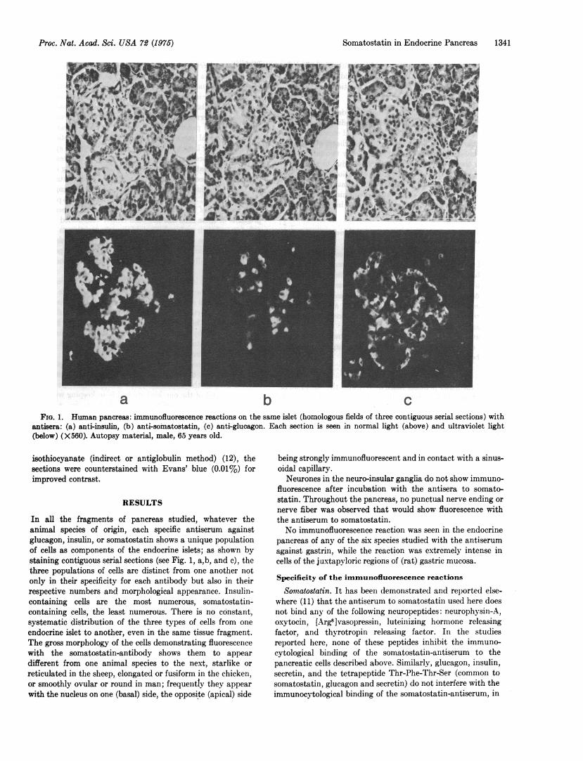
a b cFIG. 1. Human pancreas: immunofluorescence reactions on the same islet (homologous fields of three contiguous serial sections) with
antisera: (a) anti-insulin, (b) anti-somatostatin, (c) anti-glucagon. Each section is seen in normal light (above) and ultraviolet light(below) (X560). Autopsy material, male, 65 years old.
isothiocyanate (indirect or antiglobulin method) (12), thesections were counterstained with Evans' blue (0.01%) forimproved contrast.
RESULTS
In all the fragments of pancreas studied, whatever theanimal species of origin, each specific antiserum againstglucagon, insulin, or somatostatin shows a unique populationof cells as components of the endocrine islets; as shown bystaining contiguous serial sections (see Fig. 1, a,b, and c), thethree populations of cells are distinct from one another notonly in their specificity for each antibody but also in theirrespective numbers and morphological appearance. Insulin-containing cells are the most numerous, somatostatin-containing cells, the least numerous. There is no constant,systematic distribution of the three types of cells from one
endocrine islet to another, even in the same tissue fragment.The gross morphology of the cells demonstrating fluorescencewith the somatostatin-antibody shows them to appear
different from one animal species to the next, starlike or
reticulated in the sheep, elongated or fusiform in the chicken,or smoothly ovular or round in man; frequently they appear
with the nucleus on one (basal) side, the opposite (apical) side
being strongly immunofluorescent and in contact with a sinus-oidal capillary.Neurones in the neuro-insular ganglia do not show immuno-
fluorescence after incubation with the antisera to somato-statin. Throughout the pancreas, no punctual nerve ending ornerve fiber was observed that would show fluorescence withthe antiserum to somatostatin.No immunofluorescence reaction was seen in the endocrine
pancreas of any of the six species studied with the antiserumagainst gastrin, while the reaction was extremely intense incells of the j uxtapyloric regions of (rat) gastric mucosa.
Specificity of the immunofluorescence reactions
Somatostatin. It has been demonstrated and reported else-where (11) that the antiserum to somatostatin used here doesnot bind any of the following neuropeptides: neurophysin-A,oxytocin, [Arg8]vasopressin, luteinizing hormone releasingfactor, and thyrotropin releasing factor. In the studiesreported here, none of these peptides inhibit the immuno-cytological binding of the somatostatin-antiserum to thepancreatic cells described above. Similarly, glucagon, insulin,secretin, and the tetrapeptide Thr-Phe-Thr-Ser (common tosomatostatin, glucagon and secretin) do not interfere with theimmunocytological binding of the somatostatin-antiserum, in
Proc. Nat. Acad. Sci. USA 72 (1975)
Proc. Nat. Acad. Sci. USA 72 (1975)
concentrations up to 32 mg of peptide per ml of nondilutedantiserum. Only somatostatin in either the oxidized or reducedform inhibits the immunofluorescence reaction due to thesomatostatin-antiserum.
Insulin, Glucagon. None of the polypeptides listed aboveinhibit the immunofluorescence reaction due to insulin-antiserum, except insulin itself. Glucagon and secretin (whichshare a large degree of homology in terms of charge and polar-ity of amino-acid sequences) (13) are the only two substancesinhibiting the immunofluorescence reaction due to glucagon-antiserum. Actually, at very high concentrations, insulin andglucagon interfere reciprocally with the immunofluorescencereaction due to the other's antiserum, confirming the presenceof a minor contaminant of the other peptide in each prepara-tion.
Specificity of the organ or tissue localization ofimmuno-reactive somatostatin
Specific immunofluorescence due to the somatostatin-anti-serum (i.e., inhibited by competition with somatostatin only)is also observed in some discrete cells in the intestinal glands ofLieberkhuin, and of the juxtapyloric gastric mucosa; the latterare different from those cells showing immunofluorescencewith the antiserum to gastrin. A detailed study of theseextrapancreatic localizations of immunoreactive somatostatinwill be published elsewhere.Immunoreactive somatostatin is not observed by the
method described here in any of the other organs or tissueslisted above.
DISCUSSION
None of the many peptides listed above, with the exception ofsomatostatin itself, inhibits the antigen-antibody reactionobtained with somatostatin-antiserum. Thus, with the caveatthat must prevail when considering results obtained by im-munocompetence, we will assume that the antigen char-acterized by the immunocytological procedure is somato-statin. In favor of this assumption is the recent observation ofbioassayable somatostatin-activity in aqueous extracts of(rat) fetal pancreas (14).The presence of cells containing somatostatin in the islets
of the pancreas of all species studied is a totally unexpectedobservation, with possibly far-reaching significance in termsof the physiological mechanisms it evokes regarding secretionof glucagon and insulin. The statement relates to the nowwell-authenticated effect of exogenously administered somato-statin as an inhibitor of the secretion of both glucagon andinsulin by direct action at some receptor site on both the a2-and,3-cells of the endocrine pancreas. Due to the absence ofsensitive assay for measuring somatostatin in small volumes ofbody fluids, nothing is known at the moment of the dynamicsof the secretion of hypothalamic somatostatin; nor is it knownwhether somatostatin is present in peripheral blood and, if it isat all, if it is in variable amounts as a function of correspondingphysiological situations. In view of the very short biologicalhalf life of exogenously administered somatostatin, for soma-tostatin of hypothalamic origin to have a regulatory role at thelevel of the endocrine pancreas one can easily calculate thatunusually high levels of the hypothalamic peptide should besecreted to maintain peripheral concentrations high enough toexert a regulatory effect at the level of the endocrine pancreas.Thus, observing the presence of cells containing somatostatin
within the endocrine islets of the pancreas suggests that thesemay be part of a peripheral regulatory mechanism involving(pancreatic) somatostatin in the regulation of the secretion ofglucagon and/or insulin. One now asks about the mechanismsthat would regulate the secretion of pancreatic somatostatin;the obvious alternatives, are metabolic, endocrine or neural.Should a role of pancreatic somatostatin be demonstrated inthe physiological mechanisms involved in the secretion ofglucagon and/or insulin, alterations at any step of the systemmay be of significance in the physiopathology of diabetes.What is the nature of the pancreatic cells containing soma-
tostatin? They seem to be distinct from the a2-cells containingand secreting glucagon, and from the p3-cells containing andsecreting insulin. Are the somatostatin-containing cells part ofthe population of the a, or D cells? The nature of the secretionproducts of these cells has not been clearly established so far(15, 16) and their embryological origin is in question as possi-bly derived from neural crest anlage (17), a hypothesis thatwould explain how pancreatic cells would secrete a peptideobserved originally in cellular elements of the central nervous
system.In view of the intensity of the immunofluorescence reaction
observed in these cells and in view of the remarkable selec-tivity of the cells showing this reaction, it is unlikely that it bedue to some sort of nonspecific adsorption of (circulating)somatostatin. The immunocytological reaction seen here isbest explained by proposing that these cells synthesize andcontain somatostatin; and that they probably release it in stillundetermined circumstances.
Finally, the results reported here raise further the questionof the relative ubiquity of somatostatin. Its presence as
shown here in the endocrine pancreas of several species ofmammals and of the one bird studied is in keeping with thedemonstration of hypothalamic somatostatin in a largenumber of vertebrate species (11). In these various species,somatostatin now appears to be distributed in a series of tis-sues of neural crest origin, all with nearby effector. Itcertainly cannot be considered any longer as an exclusivelyhypothalamic hypophysiotropic peptide and may well have a
much broader physiological significance than originallysuspected.
I am indebted to each of the colleagues named in the text whograciously contributed aliquots of antisera or polypeptides usedin the studies referred to here and grateful to Prof. Roger Guille-min, The Salk Institute, La Jolla, Calif., whose interest and co-operation made these studies possible and who assumed the taskof putting this text in its final form. This work was supported byresearch Grant no. 74.4.445.35 Institut National de la Sante etRecherche Medicale (France).1. Burgus, R., Ling, N., Butcher, M. & Guillemin, R. (1973)
"Primary structure of somatostatin, a hypothalamic pep-tide that inhibits the secretion of pituitary growth hor-mone," Proc. Nat. Acad. Sci. USA 70, 684-688.
2. Brazeau, P., Vale, W., Burgus, R., Ling, N., Butcher, M.,Rivier, J. & Guillemin, R. (1973) "Hypothalamic polypep-tide that inhibits the secretion of immunoreactive pituitarygrowth hormone," Science 179, 77-79.
3. Vale, W., Brazeau, P., Grant, G., Nussey, A., Burgus, R.,Rivier, J., Ling, N. & Guillemin, R. (1972) "Premibres ob-servations sur le mode d'action de la somatostatine un fac-teur hypothalamique qui inhibe la secretion del'hormone decroissance," C. R. H. Acad. Sci. 275, 2913-2915.
4. Rivier, J. (1974) "Somatostatin: total solid phase synthe-sis," J. Amer. Chem. Soc. 96, 2986-2922.
5. Koerker, 1). J., Ruch, W., Chideckel, E., Palmer, J., Good-ner, C. J., Ensinck, J. & Gale, C. C. (1974) "Somatostatin:
1342 Physiology: Dubois
Proc. Nat. Acad. Sci. USA 72 (1975)
hypothalamic inhibitor of the endocrine pancreas," Science184, 482-484.
6. Gerich, J., Lovinger, R. & Grodsky, G. (1974) "Inhibitionof glucagon (IRG) and insulin (IRI) release from the invitro perfused rat pancreas by somatostatin (SRIF)," Endo-crinology, 94, A-190.
7. Fujimoto, W. Y., Ensinck, J. W. & Williams, R. W. (1974)"Somatostatin inhibits insulin and glucagon release bymonolayer cell cultures of rat endocrine pancreas," LifeSci. 15, 1999-2004.
8. Vale, W., Rivier, C., Palkovitis, M., Saavedna, M. M. &Brownstein, M. (1974) "Ubiquitous brain distribution ofinhibitors of adenohypophysial secretion," Endocrinology94, A-128.
9. Pelletier, G., Labrie, F., Arimura, A. & Schally, A. V. (1974)"Electron microscopic immunohistochemical localization ofgrowth hormone release inhibiting hormone (somatostatin)in the rat median eminence," Amer. J. Anat. 140, 445-450.
10. Hoekfelt, T., Efendic, J., Johansson, O., Luft, R. S. Arimura,A. (1974) "Immunohistochemical localization of somato-statin (growth hormone release inhibiting factor) in theguinea pig brain," Brain Res. 80, 165-169.
Somatostatin in Endocrine Pancreas 1343
11. Dubois, M. P., Barry, J. & Leonardelli, J. (1974) "Mise en6vidence par iminunofluorescence et r6partition de la soma-tostatine (SRIF) dans l'6minence m6diane de vert6br6s(Mammifbres, Oiseaux, Amphibiens, Poissons)," C.R.H.Acad. Sci. Ser. D 279, 1895-1903.
12. Goldman, M. (1968) in Fluorescent Antibody Methods(Academic Press, New York), pp. 157-160.
13. Rodbell, M. (1972) "Regulation of glucagon action at itsreceptors," in Glucagon, eds. LeFebvre, P. J. & Unger, R.H. (Pergamon Press, Oxford), pp. 61-77.
14. Vale, W. & Guillemin, R. (1975) "Physiology and biochem-istry of somatostatin and analogs," Rec. Progr. HormoneRes. 31, in press.
15. Caramia, F. (1963) "Electron microscopic description of athird cell type in the islets of the rat pancreas," Amer. J.Anat. 112, 53-64.
16. Hallenbeck, G. A. (1966) "Gastrin-like activity of tumors: areview," in Gastrin, ed. Grossman, M. I., (Butterworth &Co. Ltd., London, England), pp. 285-308.
17. Weichert, R. F. (1970) "The neural ectodermal origin ofthe peptide-secreting endocrine glands," Amer. J. Med. 49,232-241.