pan african university institute of basic sciences
TRANSCRIPT

ASSESSMENT OF NUTRITIONAL VARIATION AND
ESTABLISHMENT OF AN EFFICIENT TISSUE CULTURE
PROPAGATION PROTOCOL FOR Vitex doniana Sweet
(LAMIACEAE) FROM BENIN
(WESTERN AFRICA)
COLOMBE DADJO
MASTER OF SCIENCE
( MOLECULAR BIOLOGY AND BIOTECHNOLOGY)
PAN AFRICAN UNIVERSITY
INSTITUTE FOR BASIC SCIENCES TECHNOLOGY
AND INNOVATION
2014
Assessment of Nutritional Variation and Establishment of an Efficient
Tissue Culture Propagation Protocol for Vitex doniana Sweet
(Lamiaceae) from Benin (Western Africa)
Colombe Dadjo
MB 300-014/12
A Thesis submitted to Pan African University Institute for Basic
Sciences Technology and Innovation in partial fulfillment of the
requirements for the degree of
Master of Science in Molecular Biology and Biotechnology
2014
DECLARATION
This thesis is my original work and has not yet been presented to any other university for
examination.
Signature .............................................. Date.............................................
Colombe Dadjo
This thesis report has been submitted for examination with our approval as University
supervisors.
Signature……………………………………Date…………………..
Dr Daniel Sila
Food Sciences and Technology Department, Jomo Kenyatta University of Agriculture
and Technology
Signature Date…………………..
Dr Jane W. Kahia
Somatic Embryogenesis Scientist, World Agroforestry Centre (ICRAF)

DEDICATION
To my beloved mother,
My late father,
My brother and sisters

ACKNOWLEDGEMENTS
I would like to express my gratitude to the African Union through the Pan African
University for awarding me a scholarship and for funding my research.
My special thanks and gratitude to my supervisors Dr Daniel Sila and Dr Jane
Kahia who have guided this thesis and whose useful comments and fruitful suggestions
improved the manuscript considerably. I appreciate the efforts of Dr Jane Kahia who
dealt with all administrative matters related to my stay in Ivory Coast; I sincerely thank
her for her supportive kindness and excellent guidance.
My sincere gratitude goes to Dr. Kouassi Modeste, Scientist at Tissue Culture
laboratory of "Centre National de Recherche Agronomique" Cote d'Ivoire for various
suggestions which have helped to improve this work.
I am most grateful to the staff in the Somatic embryogenesis laboratory of the
World Agroforestry center (ICRAF), country Program Cote d'Ivoire and the technicians
of Food biochemistry laboratory of Jomo Kenyatta University of Agriculture and
Technology (JKUAT) for assisting me during my laboratory work and in collecting the
data.
I cannot forget the continuous support from all the teaching and administrative
staff of Pan African University Institute of Basic Sciences, Technology and Innovation
and Jomo Kenyatta University of Agriculture and Technology, especially Prof Naomi
Maina, program coordinator Molecular Biology and Biotechnology.
Last but not least, I am grateful to my parents who have supported me in various
ways throughout my studies.

TABLE OF CONTENTS
DECLARATION ............................................................................................................... i
DEDICATION ................................................................................................................. ii
ACKNOWLEDGEMENTS ........................................................................................... iii
LIST OF TABLES ........................................................................................................... x
LIST OF FIGURES ........................................................................................................ xi
LIST OF PLATES ........................................................................................................ xii
LIST OF APPENDICES ............................................................................................. xiii
LIST OF ABBREVIATIONS ...................................................................................... xiv
ABSTRACT ................................................................................................................... xvi
CHAPTER 1 INTRODUCTION .................................................................................... 1
1.1 Background .............................................................................................................. 1
1.2 Statement of Problem ............................................................................................... 3
1.3 Justification .............................................................................................................. 4
1.4 Objectives ................................................................................................................. 6
1.4.1 Main objective ................................................................................................... 6
1.4.2 Specific objectives ............................................................................................. 6

1.5 Hypotheses ............................................................................................................... 6
CHAPTER 2 LITERATURE REVIEW ........................................................................ 7
2.1 Botanical description of Vitex doniana .................................................................... 7
2.1.1 Taxonomy and structure .................................................................................... 7
2.1.2 Leaves ................................................................................................................ 7
2.1.3 Flowers............................................................................................................... 8
2.1.4 Fruits .................................................................................................................. 9
2.2 Ecology ................................................................................................................... 10
2 .3 Uses and nutritional value of V. doniana .............................................................. 10
2.3. 1 Food uses ........................................................................................................ 10
2.3.2 Medicinal uses ................................................................................................. 11
2.3.3 Nutritional composition of V. doniana leaves ................................................. 12
2.3.4 Nutritional value of V. doniana fruits .............................................................. 12
2.4 Phytochemical composition and properties ............................................................ 13
2.5 Propagation of V. doniana ...................................................................................... 13
2.6 Pests and diseases ................................................................................................... 14
2.7 Plant tissue culture .................................................................................................. 15
2.7.1 Micropropagation............................................................................................. 16
2.7.1.1 Stages of micropropagation .......................................................................... 16
2.7.1.2 Role of plant growth regulators................................................................. 17

2.7.2 Somatic embryogenesis ................................................................................... 18
2.7.2.1 Role of plant growth regulators................................................................. 19
2.7.2.2 Effects of carbohydrates sources and concentrations................................ 20
2.7.2.3 Effects of Activated charcoal and other additives..................................... 21
CHAPTER 3 MATERIALS AND METHODS ........................................................... 23
3.1 Introduction ............................................................................................................ 23
3.2 Nutritional characterization .................................................................................... 23
3.2.1 Sample collection Sites .................................................................................... 23
3.2.2 Sampling and sample collection ...................................................................... 24
3.2.3 Sample handling and preparation .................................................................... 24
3.2.4 Proximate analysis ........................................................................................... 26
3.2.4.1 Moisture Determination ............................................................................ 26
3.2.4.2 Crude Fat determination ............................................................................ 26
3.2.4.3 Crude Fibre ............................................................................................... 26
3.2.4.4 Crude Protein ............................................................................................ 27
3.2.4.5 Ash ............................................................................................................ 28
3.2.4.6 Carbohydrates ........................................................................................... 28
3.2.5 Minerals ........................................................................................................... 29
3.2.6 Vitamin A and C determination ....................................................................... 29
3.2.6.1 Vitamin C .................................................................................................. 29
3.2.6.2 Vitamin A (β-Carotene) ............................................................................ 30

3.2.7 Statistical analysis ............................................................................................ 30
3.3 Tissue culture ......................................................................................................... 30
3.3.1 Plant materials.................................................................................................. 30
3. 3.2 Media preparation ........................................................................................... 31
3.3.3 Plant growth regulators .................................................................................... 32
3.3.4 Aseptic techniques ........................................................................................... 32
3.3.5 Dissecting tools ................................................................................................ 33
3.3.6 Washing of glassware and vessels ................................................................... 33
3.3.7 Surface sterilization of explants ....................................................................... 33
3.3.8 Tissue culture experiments .............................................................................. 34
3.3.8.1 Somatic embryogenesis ............................................................................. 34
3.3.8.2 Micropropagation ...................................................................................... 37
3.3.9 Incubation conditions ....................................................................................... 38
3.3.10 Experimental design, data collection and analysis ........................................ 38
CHAPTER 4 RESULTS ................................................................................................ 40
4.1 Nutritional variation of Vitex doniana young leaves in Benin ............................... 40
4.1.1 Proximate composition of V. doniana young leaves ...................................... 40
4. 1.2 Mineral composition of Vitex doniana young leaves ..................................... 40
4.1.3 Vitamin A and C .............................................................................................. 41
4.2 Surface sterilization of V. doniana explants ........................................................... 42

4.2.1 Effects of super clean bleach and calcium hypochlorite on sterilization of V.
doniana leaf explants ................................................................................................ 42
4.2.2. Effect of calcium hypochlorite and mercuric chloride on V. doniana nodal
explants ..................................................................................................................... 43
4.3 Evaluation of some factors affecting induction and regeneration of somatic
embryogenesis .............................................................................................................. 44
4.3.1 Effects of growth regulators on the induction of somatic embryos ................. 44
4.3.1.1 Effects of cytokinins on the induction of somatic embryos ...................... 44
4.3.1.2 Effects of auxins on the induction of somatic embryos ............................ 45
4.3.2 Effect of carbon sources on induction of somatic embryogenesis................... 48
4.3.2.1 Effect of sucrose ........................................................................................ 48
4.3.2.2 Effect of different carbon sources on induction and regeneration of
somatic embryos.................................................................................................... 49
4.3.3 Effect of doubling potassium nitrate on the induction of somatic embryos .... 50
4.3.4 Effects of some amino acids on the induction of somatic embryos................. 51
4.3.5 Effects of silver nitrate on the induction of somatic embryos ......................... 52
4.3.6 Effects of activated charcoal and casein hydrolysate on the induction of
somatic embryos ....................................................................................................... 53
4.3.7 Effect of some organic extracts on induction of somatic embryogenesis ........ 54
4.3.8 Effect of liquid pulsing on the induction of somatic embryos ......................... 55
4.4 Effect of growth regulators on shoot and root regeneration from nodal explants .. 56
4.4.1 Effect of BAP on microshoot regeneration...................................................... 56

4.5.2 Effect of IBA on root regeneration .................................................................. 57
CHAPTER 5 DISCUSSION, CONCLUSION AND RECOMMENDATIONS ....... 59
5.1 Discussion .............................................................................................................. 59
5.1.1 Nutritional variation of V. doniana young leaves ............................................ 59
5.1.2 Surface sterilization of V. doniana explants .................................................... 60
5.1.3 Factors affecting somatic embryogenesis in V. doniana ................................. 61
5.1.4 Micropropagation in V. doniana ...................................................................... 72
5.2 Conclusion .............................................................................................................. 73
5.3 Recommendations .................................................................................................. 74
REFERENCES ............................................................................................................... 75
APPENDICES ................................................................................................................ 93

LIST OF TABLES
Table 3.1 Solvents used to dissolve plant growth substances .......................................... 32
Table 4.1 Proximate composition of V. doniana young leaves ....................................... 40
Table 4.2: Mineral concentration of V. doniana young leaves ........................................ 41
Table 4.3 Vitamin A and C concentration of V. doniana young leaves........................... 42
Table 4.4 Effects of calcium hypochlorite on elimination of surface contamination from
V. doniana leaf explants ................................................................................................... 43
Table 4.5 Effects of BAP, Kinetin and TDZ on callus induction .................................... 45
Table 4.6 Effects of Picloram on induction and regeneration of somatic embryos ......... 46
Table 4.7 Effects of 2, 4-D on induction and regeneration of somatic embryos ............. 47
Table 4.8 Effects of dicamba on induction and regeneration of somatic embryos .......... 47
Table 4.9 Effect of sucrose concentrations on induction of somatic embryos................. 49
Table 4.10 Effects of potassium nitrate on induction of somatic embryos ...................... 51
Table 4.11 Effects of silver nitrate on induction of somatic embryos ............................. 53
Table 4.12 Effects of casein hydrolysate on induction of somatic embryos .................... 53
Table 4.13 Effects of coconut water on induction and regeneration of embryos ............ 55
Table 4.14 Effect of BAP on microshoot regeneration .................................................... 57
Table 4. 15 Effect of IBA on root regeneration ............................................................... 58

LIST OF FIGURES
Figure 2.1 Flow chart summarizing tissue culture experiments (Hussain et al., 2012). .............. 15
Figure 3.1 Agro-climatic zones of Benin Republic ......................................................... 25
Figure 4.1 Effects of different sugars sources on percentage embryogenic cultures and
mean number of embryos ............................................................................................ 50
Figure 4.2 Effects of some amino acids on percentage embryogenic cultures and mean
number of embryos ..................................................................................................... 52
Figure 4.3 Effects of some organic extracts on percentage embryogenic cultures and
mean number of embryos ............................................................................................ 54
Figure 4.4 Effect of liquid pulsing on percentage embryogenic cultures and mean
number of embryos ..................................................................................................... 56

LIST OF PLATES
Plate 2.1 Vitex doniana leaves ...................................................................................... 8
Plate 2.2 Vitex doniana inflorescence ........................................................................... 9
Plate 2. 3 Vitex doniana fruit (a) unripe (b) ripe ........................................................... 9
Plate 3.1 V. doniana seedlings growing under shed in Cote d'Ivoire ............................... 31
Plate 4.1 (a) Callus (b) Globular embryos (c) Fully developed embryos ................... 48
Plate 4.2 Regenerated microshoot after two weeks on shooting media (b) Rooting of in
vitro shoots after four weeks of culture....................................................................... 58

LIST OF APPENDICES
Appendix 1 Composition of Murashige and Skoog's Medium ........................................ 93
Appendix 2 Standard curve used in determination of nutritional parameters .................. 94
Appendix 3 Statistical analysis outcomes ........................................................................ 96

LIST OF ABBREVIATIONS
%: Percent
°C : Degree Celsius
2, 4-D: Dichlorophenoxy – acetic acid
2iP: [ N6 - (2- isopentyl) adenine ]
3, 4-D: Trichlorophenoxy – acetic acid
AACC: Approved methods of American Association of Cereal Chemists
AC: Activated charcoal
AgNO3: Silver nitrate
ANOVA: Analysis of variance
AVGSAN: Analyse Globale de la Vulnérabilité, de la Sécurité Alimentaire et de la
Nutrition
AVRDC: Asian Vegetable Research and Development Center
BAP/BA: Benzylamino purine
CH: Casein hydrolysate
conc.: Concentration
CW: Coconut water
eq.: Equation
FAO: Food and Agriculture Organization
H2O: Water (dihydrogen monoxide)
HCl: Hydrochloride acid
HNO3: Nitric acid
HPLC: High Performance Liquid Chromatography
IBA: Indol-3- butyric acid
ICRAF: World Agroforestry Center
JKUAT: Jomo Kenyatta University of Agriculture and Technology
kg/ cm2: Kilogram per centimeter square
Kin: Kinetin (6-furfurylaminopurine)
KNO3: Potassium nitrate
MS: Murashige and Skoog medium
N: Normal
Na2SO4: Sodium sulphate
NAA: 1-naphtylacetic acid
NaOH: Sodium hydroxyde
NUS: Neglected and underutilized wild Species (NUS)
OECD: Organization for Economic Co-operation and Development
TDZ: Thidiazuron
V. doniana: Vitex doniana
v/ v: Volume by volume
WHO: World Health Organization

ABSTRACT
Black plum (Vitex doniana) tree is an indigenous wild species important for the
livelihoods of rural populations in Benin. It is highly nutritious and rich in
phytochemical compounds of health benefit. In Benin, the greatest economic potential of
Vitex doniana trees probably lies in the leaves. It has emerged as a potential species for
domestication. Despite some studies indicating that nutritional properties can vary
according to the sample provenances, no data on nutritional variation of V. doniana
leaves are available in Benin. On the other hand, the conventional methods of
propagating the plant produce inadequate number of planting materials as seeds of this
tree have a very weak germinating capacity and the macropropagation rate by stem
cuttings is slow. Tissue culture is a reliable alternative method. Therefore, the objective
of the current work was to evaluate the nutritional variation of leaves collected from
different agro climatic zones and to establish a feasible in vitro propagation method for
V. doniana using both somatic embryogenesis and micropropagation. During the current
study, proximate, minerals and vitamin A and C concentrations were analyzed. In the
Tissue Culture experiments, sterilization was evaluated using different concentrations of
a commercial bleach, mercuric chloride and calcium hypochlorite at varying time
intervals. For somatic embryogenesis, different growth regulators and additives were
evaluated. In the micropropagation experiments, microshoots were regenerated using
BAP and roots using IBA at concentrations 5, 10, 20, 40 µM. Results showed that V.
doniana young leaves are highly nutritious. Protein, Calcium, vitamin A and C
concentration vary significantly across agro climatic zones. The highest values of

Protein, Vitamin A and C were obtained in the Sudanian zone and the highest value of
calcium was obtained in the Sudano-Guinean zone. The highest number (91%) of clean
leaf explants were obtained when the leaf explants were subjected to sterilization using
2% calcium hypochlorite for 30 minutes followed by a second sterilization using 2%
hypochlorite for 15 minutes (double sterilization). The highest number (40%) of clean
nodal explants were obtained when the explants were subjected to sterilization using 2%
calcium hypochlorite for 45 minutes followed by a second sterilization using 2%
hypochlorite for 30 minutes. Inclusion of 4 µM Picloram, in the medium was observed
to increase the mean number of embryos per explant. Of the four amino acids tested,
only tryptophan evaluated at 150 µM had significant effect on the induction of somatic
embryogenesis with 6.5 embryos per explant. On the other hand, silver nitrate and casein
hydrolysate at 50 µM were found to enhance the number of embryos per explant. Liquid
pulsing treatment for 24 h produced the highest (9.19) mean number of embryos
compared to the control (1.2). The highest mean number of shoot/explant and the highest
mean length of shoot was obtained in media supplemented with 10 µM BAP. Rooting
was achieved using IBA. No optimum concentration of IBA was found for root
regeneration. The protocols developed during this study would be useful for the mass
propagation, and genetic transformation of selected elite lines. This work provides useful
information that can be used to increase domestication of V. doniana in Benin. However,
further investigations need to be done on the bioavailability of nutrients from the leaves
of V. doniana and the conversion and the performance in the field of both embryo-
derived plantlets and plantlets obtained from micropropagation.
~ 1 ~
CHAPTER 1 INTRODUCTION
1.1 Background
Plant genetic resources are essential for a sustainable agriculture and food
security. In the past, many indigenous wild species played a crucial role in the food
security, nutrition, health, income generation and food culture of the rural poor
(Magbagbeola et al., 2010). However, this is no longer the case. The lack of attention
has meant that their potential value is under-estimated and under-exploited. It also
places them in danger of continued genetic erosion and possible disappearance.
Agricultural biodiversity (agro biodiversity) has steadily declined with a
corresponding increase in dependence on a small number of food crops (Moore,
2010).
The Food and Agriculture Organization of the United Nations (FAO)
estimates that humans have used some 10,000 species for food throughout history.
However, only about 120 cultivated species provide around 90% of food
requirements and 4 species (Maize, Wheat, Rice and Potatoes) provide about 60% of
human dietary energy for the world's population (Muzzalupo et al., 2012). Many
neglected and underutilized wild species (NUS) are nutritionally rich (Ghane et al.,
2010; Johns and Eyzaguirre, 2006). Therefore, their erosion can have immediate
consequences on the nutritional status and food security of the poor and their
enhanced use can bring about better nutrition and fight hidden hunger. Even though
the link between agro biodiversity and diet diversity is not automatic (Burchi et al.,
2011), it is agreeable that the diminution of agro biodiversity, to some extent, places
considerable strain on the ease with which households are able to enjoy diversified,

~ 2 ~
balanced diets. Awareness of the importance and value of crop wild relatives and of
the need to conserve them has increased. Accordingly, a number of initiatives have
come forth in recognition of the importance of diversified diets notably the 2003/2004
joint Food and Agriculture Organization/World Health Organization (FAO/WHO)
consultations, all of which acknowledged, explicitly or implicitly, the indispensable
role of diet diversification for enhanced food security and nutrition outcomes. Food
system interventions that involve carefully formulated multi-sectorial activities along
the food chain- from food production to consumption and utilization (Burchi et al.,
2011) have been advanced as key to sustaining gains being made by short-term
micronutrient control measures because they simultaneously address multiple nutrient
and phytochemical needs for optimal health (Underwood, 2000). Crop diversity
contribute to the stability and sustainability of farming systems and are valued for
providing important attributes including inter alia agronomic characteristics, biotic
and abiotic stresses and other factors of cultural and socio-economic importance. In
addition, crop diversity contributes as direct or indirect source of several products,
such as medicines, life-saving drugs, vitamins, minerals, various industrial products.
Crop diversity also provide an insurance against unknown future needs/conditions as
these are likely to hold still undiscovered cures for known and emerging diseases and
is a fortune that can be tapped, as human needs change. FAO through the Second
Global Plan of Action for the conservation and sustainable Utilization of Plant
Genetic Resources for Food and agriculture supports activities improving in situ and
ex situ conservation of NUS (FAO, 2011). In situ conservation enables to preserve
evolutionary processes that generate new germplasm under conditions of natural

~ 3 ~
selection, maintain important field laboratories for crop biology and biogeography. It
also serves as a continuous source of germplasm for ex situ conservation. Ex- situ
conservation refers to the conservation of germplasm away from its natural habitat.
This complementary approach for conservation had begun on a wide scale about three
decades ago and is now practised, to some extent, in almost all countries as a means
to conserve crop species diversity for posterity. This strategy is particularly important
for crop gene pools, and can be achieved by propagating/ maintaining the plants in
genetic resource centre, botanical gardens, tissue culture repositories or in seed gene
banks (OECD, 1999).
About 187 leafy vegetables were reported in Benin by Dansi et al. (2008)
whilst the Darwin Initiative 15/003 surveys recorded 245 plants species which
include leafy, fruit and seed vegetables (Achigan-Dako et al., 2010).. These species,
mostly underutilized are potentially rich and important for rural populations. Vitex
doniana (Laminaceae) is one of the high-priority NUS for domestication in Benin. It
is the tallest and most frequent Pan African Vitex species, occurring from Senegal
east to Somalia and south to South Africa; Comoros and Seychelles (Louppe et al.,
2008). It can make a great contribution to the local, regional and national economy of
many West African countries (Codjia et al., 2003).
1.2 Statement of Problem
V. doniana is among the Neglected and Underutilized Species (NUS) known due to
their nutritional and medicinal properties (Orwa et al., 2009; Dadjo et al., 2012).
Despite the widely known nutritional, medicinal and economic uses of V. doniana
products, the species is still under- utilized, unimproved. Numerous studies have been
conducted on the nutritional concentration of V. doniana fruit (Olulosa, 1992; Glew et

~ 4 ~
al., 1997; Solebo and Aina, 2011; Vunchi et al., 2011; Osum et al., 2013). However,
very little information is available on the nutlritional quality of the leaves. Moreover,
despite some studies indicating that nutritional properties can vary according to the
sample provenances (Solebo and Aina, 2011; Assogbadjo et al., 2012) no data on
nutritional value of V. doniana leaves are available in Benin. The strong anthropic
pressure affecting this species has caused its numbers to fall increasingly in its natural
environment (Achigan-dako et al., 2010). The planting of seedlings is negligible and
the seeds of this tree have a very weak germinating capacity (Louppe et al., 2008).
Sanoussi et al. (2012) showed that macropropagation rate by stem cutting is slow. To
date, there are no reports on work carried out on tissue culture of the species.
1.3 Justification
The World Food Program comprehensive food security and vulnerability analysis
conducted in 2008 estimated that nearly one million people in Benin (10% of the
population) are food-insecure and nutrition insecure (AGVSAN, 2009). V. doniana is
distributed in all the climatic zones of Benin and has a great importance in the diet of
rural population. More recently, the species was chosen as a model species to be
domesticated in Benin (Codjia et al., 2003). The fruit of Vitex doniana is sweet and
tastes like prunes. It can be made into a jam and wine. Leaves are often used as
vegetables for cooking (Achigan-Dako et al., 2010; Dadjo et al., 2012). The different
parts of Vitex doniana are used in traditional medicine (Dadjo et al., 2012; Orwa et
al., 2009). In Benin, the greatest economic potential of Vitex doniana trees probably
lies in the leaves (Achigan-dako et al., 2010). It is the most consumed vegetables
during the dry season but also one of the most traded wild vegetables in Benin
(Codjia et al., 2003; Achigan-Dako et al., 2010). Considering the importance V.

~ 5 ~
doniana in people’s diet and for medicinal uses in Benin, it can be exploited at the
commercial level. The leaves, roots, bark and fruits can be used to get plant extracts
that may be helpful in overcoming nutritional related health problems among other
disorders/diseases that are predominant in the many rural areas of the country. It has
been demonstrated that the nutritional diversity of the plant is largely dependent on
the plant part and the geographic region from which the sample is obtained from
(Solebo and Aina, 2011). Therefore, elucidation of the nutritional characteristics of
the leaves collected from different phytoclimatic zones in Benin would be a critical
input for the selection of appropriate genotypes for cultivation and breeding purposes.
On the other hand, tissue culture is a reliable and advanced propagation
method that could be more promising than the conventional propagation methods
(Singh et al., 2013). In addition, tissue culture ensures mass production of elite clones
from hybrid or specific parental lines. Tissue culture ensures healthy seedlings with
desirable characters. The tissue culture of forest trees has shown promise in obtaining
regenerants and clonal multiplication for domestication of wild populations,
afforestation and economically important trees that have been cultivated for
generations. The application of in vitro culture for propagation of V. doniana tree will
assist in sustainable availability of propagules and plant products. In that perspective,
the characterization of nutritional variability for suitable material selection for tissue
and the establishment of an efficient tissue culture propagation protocol for V.
doniana presents a very important point in the biotechnology field, especially in the
domestication process of the species in Benin.

~ 6 ~
1.4 Objectives
1.4.1 Main objective
To enhance the domestication of Vitex doniana in Benin by characterizing the
nutritional variation of the species and establishing the optimum conditions for in
vitro propagation of the species.
1.4.2 Specific objectives
The specific objectives are to:
i. Determine the nutritional variation of Vitex doniana by assessing the
proximate composition, minerals, vitamin A and C content of its leaves
collected from different climatic zones.
ii. Determine the optimum sterilization procedure for V. doniana explants;
iii. Evaluate effect of growth regulators on direct and indirect somatic
embryogenesis in V. doniana;
iv. Compare effects of different cytokinin/auxin in regenerating microshoots and
roots from V. doniana nodal explants.
1.5 Hypotheses
i. There is no significant variation in nutritional composition of Vitex doniana
leaves according to the different climatic zones.
ii. There is no optimum sterilant concentration for sterilization of explants.
iii. There is no optimum cytokinin/auxin concentration for induction of somatic
embryos.
iv. There is no optimum cytokinin/auxin concentration for optimum shooting and
rooting of Vitex doniana.
v.

~ 7 ~
CHAPTER 2 LITERATURE REVIEW
2.1 Botanical description of Vitex doniana
2.1.1 Taxonomy and structure
Vitex doniana Sweet (Black plum) belongs to Vitex L. genus. The genus Vitex has
been considered in Verbenaceae family by different authors but in recent works it has
been transferred into Lamiaceae based on different evidences (Wagstaff et al., 1998).
Vitex doniana Sweet (syn. Vitex cuneata Thonn.) is the most widespread Vitex
species in Africa. V. doniana is a diploid species, with basic chromosome number of
(2n = 32). V. doniana is a medium-sized deciduous tree, 8-18 m high occasionally
25m, with a heavy rounded crown and a clear bole up to 5 m. Its bark is rough, pale
brown or greyish-white, rather smooth with narrow vertical fissures. The bases of old
trees have oblong scales.
2.1.2 Leaves
Leaves are opposite, glabrous and are 14-34 cm long. They are usually with 5 leaflets
on stalks of 6-14 cm long. Leaflets distinctly stalked, ovate, obovate-elliptic or
oblong, entire, are 8-22 cm long, and 2-9 cm wide.

~ 8 ~
Plate 2.1 Vitex doniana leaves
2.1.3 Flowers
V. doniana exhibits hermaphroditism, where both functional male and female organs
are in the same flower (Orwa et al., 2009). The flower petals are white except on
largest lobe, which are purple, in dense opposite and axillary cymes. Flowers are
numerous and small, blue or violet, 3-12 cm in diameter, only a few being open at a
time. Calyx and pedicels are densely hairy.

~ 9 ~
Plate 2.2 Vitex doniana inflorescence
2.1.4 Fruits
The fruits are oblong, about 3 cm long and they are green when young, turning
purplish-black on ripening and with a starchy black pulp. Each fruit contains 1 hard,
conical seed, of 1.5-2 cm long, and 1-1.2 cm wide (Orwa et al., 2009).
Plate 2. 3 Vitex doniana fruit (a) unripe (b) ripe
a b

~ 10 ~
2.2 Ecology
The wide geographical distribution of V. doniana reflects its wide ecological
tolerance. V. doniana occurs in a variety of habitats, from forest to Savannah, often in
wet localities and along rivers, and on termite mounds, up to 2000 m altitude. It is a
deciduous forest tree of coastal woodland, riverine and lowland forests and deciduous
woodland, extending as high as upland grassland (Orwa et al., 2009). It occurs in
regions with a mean annual rainfall of 750–2000 mm and requires a high water table.
Optimum growth is achieved when mean temperatures are between 10oC and 30
oC.
When it grows in open areas it develops a very thick bark which acts as protection
against fire. In swamps it has a much smoother, thinner bark. It occurs on a variety of
soils of varying origins but most commonly found on alluvial soils.
2 .3 Uses and nutritional value of V. doniana
2.3. 1 Food uses
The fruits and leaves are the edible part of the trees. They are either eaten raw or after
processing. The blackish pulp of the fruit is edible and sweet. It is eaten raw and
tastes like prunes; (Dadjo et al. 2012; Orwa et al. 2009; Louppe et al.2008). It is also
used for juice, syrup, wine, liquor and jam production (Orwa et al. 2009; Louppe et
al. 2008). Jam prepared from the fruits showed no significant difference in flavour,
colour, and overall acceptability from commercial plum jam. The black plum jam is
even preferred for consistency and spreadability. The syrup made from the pulp can
be used instead of other syrups as a nutritive sweetener. The seed inside the fruits
stone is edible (Louppe et al. 2008). Cooked young leaves are used as vegetable or in

~ 11 ~
sauces (Dadjo et al. 2012). The pounded leaves can be added to warm filtered grain
beer and then drunk (Louppe et al.2008).
2.3.2 Medicinal uses
Vitex doniana is widely used in traditional system of medicine. The leaves, fruits,
roots, barks and seed of the plant have been used as medication for liver disease,
anodyne, stiffness, leprosy, backache, hemiplegia, conjunctivity, rash, measles,
rachitis, febrifuge, as tonic galactagogue to aid milk production in lactating mothers,
sedative, digestive regulator and treatment of eye troubles, kidney troubles. It has also
been used for treatment of disease conditions such as infertility, anaemia, jaundice,
dysentery, gonorrhea, headaches, diabetes, chickenpox, rash and fever (Dadjo et al.
2012; Orwa et al. 2009, Louppe et al. 2008; Iwueke 2006). Leaf sap is used as an eye
drop to treat conjunctivitis and other eye complaints (Dadjo et al. 2012; Orwa et al.
2009; Louppe et al. 2008). The leaf is used against headache, stiffness, measles, rash
fever chickenpox, hemiplegia, febrifuge, and to treat respiratory diseases. Paste and
pounded leaves are applied to wound and burns. Leaves infusions are added to
alcoholic drink to make them stronger. Dried and fresh fruits are eaten against
diarrhea, dysentery, jaunice, anaemia, leprosy (Orwa et al. 2009). The bark is used
against leprosy, bleeding after child birth, liver diseases. The powdered bark is added
to water and taken to treat colic. The bark extract is used to treat kidney diseases and
stomachache. A root decoction is administered orally to treat ankylostomiasis,
rachitis, gastro-intestinal disorders, jaundice and as anodyne (Louppe et al. 2008).

~ 12 ~
2.3.3 Nutritional composition of V. doniana leaves
Olusola (1992) found that all the essential amino acids were present in the leave of V.
doniana and their levels compared favourably with the FAO reference protein except
for methionine and tryptophan where the level was lower. All the amino acids in the
leave were found to be biologically available and were of sufficient quality to sustain
normal growth (Olulosa, 1992). Adejumo et al., (2013) found that the moisture
content in V. doniana young leaves was high. Vitex doniana young leaf is very rich in
carbohydrates. Minerals such as sodium, calcium, iron, Magnesium, Zinc, Copper
were reported to be present in V. doniana young leaves (Adejumo et al., 2013; Osum
et al., 2013). The result on the analysis of mineral content of the V. doniana young
leaves revealed that Calcium (Ca) content is very high and Magnesium was moderate.
Sodium was low in V. doniana young leaf but rich in potassium (Adejumo et al.,
2013).
2.3.4 Nutritional value of V. doniana fruits
The chemical composition of proximate, amino acids, fatty acids, vitamins, and
minerals of V. doniana fruits has been reported (Osum et al., 2013; Vunchi et al.
2011; Glew et al. 1997). Reports on the mineral content of V. doniana have showed
that Iron, Magnesium, Manganese, Molybdenum, Phosphate, Zinc, Calcium and
Sodium are present (Vunchi et al., 2011; Osum et al., 2013). Glew et al. (1997)
reported that the most abundant mineral is Calcium. However, according to Vunchi et
al. (2011), V. doniana fruit pulp has moderate calcium value. The fruit has also high
value of vitamin C, carbohydrate and lipid. They suggested that the species could be
promoted as carbohydrate and lipid supplements for cereal-based diets in rural

~ 13 ~
communities while its moderate calcium value could be used for the management of
oesteomalacia. Agbede and Ibitoye (2007) studied the sugar content as well as the
anti- nutritional factor in the fruit. Qualitative studies on the crude extract of the
extract have detected presence of carbohydrates in moderate amount and protein in
small amount (Nwachukwu and Uzueto, 2010).
2.4 Phytochemical composition and properties
The medicinal value of a plant depends on its bioactive phytochemical constituents
that produce definite physiological action in the body. Several authors have evaluated
the phytochemical composition of V. doniana. They showed the presence of
flavonoids, tannins, saponins, anthraquinones, naphthoquinones, and resin in
methanol, ethanol, aqueous root, stem bark and leaf extract of V. doniana (Iwueke,
2006). Different extracts of the leaves, stem bark and roots have been used to
demonstrate the pharmacological activities of V. doniana. In general, the species
shows good anti-oxidant property, antimicrobial, antidiarrhoeal effect, local
anesthetic effect, antinociceptive activity (muscle relaxant), effect on pentobarbitone
sleeping time, antidiabetic and hepatoprotective effect, anticonvulsant and antipyretic
properties.
2.5 Propagation of V. doniana
The tree regenerates naturally by seed, coppice, wildings and root suckers. It is
thought forest fires help in inducing germination because they help break the hard
testa. Stones should be sown fresh after removal of the pulp and soaking in cold or
warm water for 24 hours. The seeds have a very weak germinating capacity even
when sown fresh. The growth rate of V. doniana is moderate. In Côte d’Ivoire, stones

~ 14 ~
dipped into sulphuric acid 95% for 60 minutes and subsequently in water for 72 hours
germinated after 26 days, but the germination rate was only 34% (Louppe et al.,
2008). Seedlings were on average 70–90 cm tall after 3 years, the tallest ones
reaching 170 cm. On good soils in southern Burkina Faso early growth is a bit faster
(www.prota.org). Untreated fruits may take very long to germinate; fire may
accelerate germination. Stones may contain several seeds and several seedlings may
germinate from one stone. Seeds can be stored for up to 1 year at 3–5°C.
Macropropagation by cuttings has been successful in Malawi (Louppe et al., 2008).
In Cameroon, Mapongmetsem et al., (2012) studied the effect of growth hormones
and substrate root growth using stem cuttings. They found that the rate of root growth
can reach 68.31% after eleven weeks. However in Benin, Sanoussi et al., (2012)
showed that propagation rate by stem cutting is slow while it is high (96.25%) by root
cutting after three months. Survival rates in plantations are normally good, about 80–
90% after 3 years (Louppe et al. 2008). To date there is no work reported on tissue
culture of the species.
2.6 Pests and diseases
Premature leaf shedding, heart rot and die back have been reported as some of the
diseases. V. doniana leaves were found bearing brown uredinia identified as rust the
rust fungus Olivea scitula. The pathogen has been reported from Sierra Leone,
Uganda, Nigeria and Zambia (Kapooria and Aime, 2005). According to Okia et al.
(2008), Caterpillars and worms are the major pests that defoliate the species.

~ 15 ~
2.7 Plant tissue culture
Plant Cell and tissue culture has already contributed significantly to crop
improvement and has great potential for the future (Kumar and Kumar, 1996).
Research efforts in plant cell and tissue culture have increased dramatically
worldwide in recent years including efforts in developing nations. The process starts
with the selection of plant tissues (explant) from a healthy, vigorous mother plant
(Murashige, 1974). Any part of the plant (leaf, apical meristem, bud and root) can be
used as explant. Hussain et al., (2012) divided the technique in 5 stages as
summarized in Figure 2.1.
Figure 2.1 Flow chart summarizing tissue culture experiments
Source: (Hussain et al., 2012).

~ 16 ~
2.7.1 Micropropagation
2.7.1.1 Stages of micropropagation
Stage 0: Preparation of donor plant: Any plant tissue can be introduced in vitro. To
enhance the probability of success, the mother plant should be ex vitro cultivated
under optimal conditions to minimize contamination in the in vitro culture.
Stage I: At the initiation stage, an explant is surface sterilized and transferred into
nutrient medium. Generally, the combined application of bactericide and fungicide
products is suggested. The selection of products depends on the type of explant to be
introduced. The surface sterilization of explant in chemical solutions is an important
step to remove contaminants with minimal damage to plant cells. The most
commonly used disinfectants are sodium hypochlorite, calcium hypochlorite, ethanol
and mercuric chloride (HgCl2). The cultures are incubated in growth chamber either
under light or dark conditions depending on the method of propagation.
Stage II: Multiplication stage aims at increasing the number of propagules. The
number of propagules is multiplied by repeated subcultures until the desired (or
planned) number of plants is attained.
Stage III: Rooting stage: The rooting stage may occur simultaneously in the same
culture media used for multiplication of the explants. However, in some cases it is
necessary to change media, including nutritional modification and growth regulator
composition to induce rooting and the development of strong root growth.
Stage IV: At acclimatization Stage, the in vitro plants are weaned and hardened.
Hardening is done gradually from high to low humidity and from low light intensity
to high light intensity. The plants are then transferred to an appropriate substrate
(sand, peat, compost etc.) and gradually hardened under greenhouse. In vitro growing

~ 17 ~
plantlets are under low light intensity (1,200–3,000 lux) and temperature (25 ± 2oC),
hence direct transfer to broad spectrum sunlight (4,000–12,000 lux) and temperature
(26–36oC) might cause charring of leaves and wilting of plantlets. It is therefore
necessary to accustom the plant in the natural conditions by a process of hardening or
acclimatization (Lavanya et al., 2009). The retardation in development of cuticle,
epicuticular waxes and functional stomatal apparatus during in vitro culture cause
high stomatal and cuticular transpiration rates of leaves in plantlets when taken out of
the culture vessels. In order to avoid this, slowly the plantlets should be transferred
from high humidity to low humidity conditions. Microshoots should be kept in the
shade with plugs loosened and after a week or two, they should be transferred to pots
containing sterile soil and sand mixture covered with polybags. Slowly stomatal and
cuticular transpiration rates gradually decreases because stomatal regulation of water
loss becomes more effective and cuticle and epicuticular waxes develop (Chandra et
al., 2010).
2.7.1.2 Role of plant growth regulators
Auxins, abscisic acid, cytokinins, ethylene, and gibberellins are commonly
recognized as the five main classes of naturally occurring plant hormones (Taiz and
Zeiger 2004). Auxins, cytokinins, and auxin-cytokinin interactions are usually
considered to be the most important for regulating growth and organized development
in plant tissue and organ cultures, as these two classes of hormones are generally
required.

~ 18 ~
- Cytokinins
The most commonly used cytokinins are the substituted purines: kinetin and BA.
Cytokinin is a phytohormone that participates in events in the course of whole plant
ontogeny, from fecundated ovule to senescence and death. It is present in processes
such as cell division, shoot initiation and growth, senescence delay and
photomorfogenic development, control of chloroplast division and growth,
modulation of metabolism and morphogenesis in response to environmental stimulus
(Pozo et al., 2005; Hirose et al. 2007). Although both auxin and cytokinin are usually
required for growth and morphogenesis, auxin can inhibit cytokinin accumulation,
whereas cytokinins can inhibit at least some of the actions of auxin.
- Auxins
Auxins exert a strong influence over processes such as cell growth expansion, cell
wall acidification, initiation of cell division, and organization of meristems giving rise
to either unorganized tissue (callus) or defined organs (generally roots) and promote
vascular differentiation. In organized tissue, auxins appear to be key players in
maintaining apical dominance, affecting abscission, promoting root formation, and
tropistic curvatures, delaying leaf senescence, and fruit ripening.
2.7.2 Somatic embryogenesis
In plants, embryo-like structures can be generated from cells other than gametes (i.e.
somatic cells) by circumventing the normal fertilization process, hence the term
somatic embryos (Parrott, 2000). As somatic embryos are formed without any
fertilization event they are genetically identical to the parent tissue and are therefore
clones. Somatic embryogenesis may be direct or indirect. Indirect somatic

~ 19 ~
embryogenesis involves dedifferentiation of organized tissue into callus prior to
embryo production whereas direct somatic embryogenesis involves production of
embryo from organized tissue without an intervening callus phase (Slater and Scott,
2003). A callus consists of an amorphous mass of loosely arranged thin walled
parenchyma cells arising from the proliferating cells of the cultured explants.
Frequently, as a result of wounding, a callus is formed at the cut end of a stem or root.
Using tissue culture techniques, callus formation can be induced in numerous plant
tissue and organs that do not usually develop callus in response to an injury. Many
parts of a whole plant may have an ultimate potential to proliferate in vitro, but it is
frequently found that callus cultures are more easily established from some organs
than others. Young meristematic tissues are most suitable, but meristematic areas in
older parts of a plant, such as the cambium, can give rise to callus. The callus formed
on an original explant is called primary callus. Secondary callus cultures are initiated
from pieces of tissue dissected from primary callus. Subculture can then often be
continued over many years, but the longer callus is maintained, the greater is the risk
that the cells thereof will suffer genetic change (George et al., 2008).
2.7.2.1 Role of plant growth regulators
Plant growth regulators play a key role in process potentials and dedifferentiation and
redifferention (Guo et al., 2011). Cytokinins are known to stimulate cells and, as
such, they are also suitable candidates for induction of somatic embryogenesis and
callogenesis. For example, in P. atlantica, callus induction was achieved by using
BAP 1mg/l (Aghaei et al., 2013). It has been suggested that TDZ is more effective
than other cytokinins used for somatic embryogenesis (Lin et al., 2004). The effects

~ 20 ~
of TDZ occur at lower concentrations than other cytokinins and it has been suggested
that it either directly promotes growth due to its own biological activity or through
inducing the synthesis and/or accumulation of endogenous cytokinins or auxins.
Specifically, TDZ was showed to be effective in Coffea arabica and Coffea
canephora somatic embryo formation (Giridhar et al., 2004). These regenerative
processes in cell and tissue cultures may be induced by cytokinins in collaboration
with other plant growth regulators (Guo et al., 2011). Many works have been reported
on induction of somatic embryogenesis using a combination of cytokinins and auxins.
Iin P. atlantica, the combination of BA 2 mg/l with NAA 1mg/1 was not effective
(Aghaei et al., 2013). Other growth regulators such as picloram and dicamba have
been used in somatic embryogenesis. In Phyla nodiflora, it was found that picloram
enhances formation of somatic embryos from leaf and stem explants in presence of
BA (Ahmed et al., 2011). Tournament of roses produced somatic embryos when
cultured on medium containing dicamba (2.3µM/l). Dicamba (0.02 mg/l) was also
found favourable for wheat regeneration.
2.7.2.2 Effects of carbohydrates sources and concentrations
Carbohydrates are generally considered as a carbon source needed for growth and
development of tissues in in vitro cultures. Sucrose is the most frequently used in the
culture medium compared with other sources of carbohydrates such as fructose,
glucose, sorbitol and mannitol. Several reports show that carbohydrate sources affect
the ability of the cells to induce somatic embryogenesis and its development as well
as their concentrations (Tokuhara and Mii, 2003). In several species, sucrose is the
preferred carbohydrate not only for induction, proliferation and embryos maturation

~ 21 ~
but playing a role as osmoticum (Li et al., 1998). In addition, Korbes and Droste
(2005) tested different maturation media by using 6% maltose, 3% sucrose or 6%
sucrose. Sucrose at 6% significantly enhanced the conversion percent of soybean
embryos. 0.15 M sucrose or sorbitol in induction medium while in proliferation stage
was effective on date palm (Phoenix dactylifera L.) callus induction (Mona and
Rania, 2012).
2.7.2.3 Effects of Activated charcoal and other additives
Activated charcoal has been shown to absorb 5-hydroxymethylfurfural, an
inhibitor formed by the degradation of sucrose during autoclaving, as well as
substantial amount of auxins and cytokinins. In sugarcane, 2% activated charcoal was
found to enhance somatic embryogenesis (Manchanda and Gosal, 2012).
Amino acids (glutamine, proline and tryptophan) have been identified as enhancers of
somatic embryogenesis in some species (Deo, 2010). In wheat culture, the efficiency
of the amino acids was genotype-based (Duran et al., 2013). Their efficacy in
embryogenesis has been attributed to their contribution to various cellular processes
such as improving cell signaling processes in various signal transduction pathway
(Lakshmanan and Taji, 2000), as precursor molecules for certain growth regulators.
The use of silver nitrate (AgNO3), an ethylene action inhibitor has been shown
to increase in vitro embryogenesis. AgNO3 played a minor role on in vitro embryo
induction frequency of Manchurian ash (Fraxinus mandshurica). However It has
been shown that AgNO3 enhanced synchronization and significantly inhibited
abnormal somatic embryo formation suggesting that AgNO3 might serve an important
function in controlling the development of somatic embryos in Manchurian ash
~ 22 ~
(Kong, et al., 2012). On the other hand, potassium nitrate (1.9 g/l) has been shown to
enhance embryo induction rate in Cotton (Gossypium hirsutum) (Haq and Zafar,
2004).
The positive effect of casein hydrolysate on somatic embryogenesis has been
found in species such as date palm (Phoenix dactylifera L.) (2g/l) (Khierallah and
Hussein, 2013), maize (100 mg/l) (Dhillon and Gosal, 2012). Some researchers have
reported that the inclusion of complex organic extracts, is essential for somatic
embryogenesis in some species (Deo et al., 2010). Coconut 10 and 20% coconut
water improved the production of white-opaque somatic embryos in avocado (Per´an-
Quesada et al., 2004). Potato extract alone or combined with components of
conventional culture media has been found to provide a useful medium for the anther
culture of wheat and some other cereals.
Liquid culture can be a positive condition for the induction of somatic
embryogenesis from various species, such as Alstroemeria (Akutsu and Sato 2002),
Lilium longiflorum (Nhut et al., 2006), V. vinifera L. (Jittayasothorn et al., 2007) and
Coffea arabica (Papanastasiou et al., 2008). Furthermore, liquid pulse treatments of
growth regulators could promote regeneration from plant cell culture though
organogenesis or somatic embryogenesis (Madhulatha et al., 2004). In addition, it has
previously been reported that initial pretreatment of leaf explants of squash
(Cucurbita pepo L.) with cytokinins for 6–48 h significantly promoted the formation
of somatic embryos which developed further to the torpedo-shape stage and
germinated (Kintzios et al., 2002).

~ 23 ~
CHAPTER 3 MATERIALS AND METHODS
3.1 Introduction
This work was conducted in three parts. First, the plant materials were collected in
Benin. Secondly, the nutritional analysis were done in the department of Food
Science and Technology at Jomo Kenyatta University of Agriculture and Technology
(Kenya). Lastly, the tissue culture studies were carried out in the Somatic
Embryogenesis laboratory of ICRAF (Cote d'Ivoire).
3.2 Nutritional characterization
3.2.1 Sample collection Sites
Samples were collected in the Republic of Benin, situated in West Africa between
latitudes 6o10’N and 12
o25’N and longitudes 0
o45’E and 3
o55’E (Figure 3.1). It is
bordered by the Republics of Togo in the west, Burkina Faso and Niger in the north,
and Nigeria in the east. The mean annual rainfall varies from 900 to 1300 mm. Its
lowest values are recorded in the southwest and in the far north (900-950 mm). The
highest precipitation (1200-1300 mm) is confined to Southeast Benin as well as the
tract Bassila-Djougou. The mean annual temperature ranges from 26 to 28°C and may
exceptionally reach 35-40oC in northern localities such as Kandi. The annual
temperature amplitude is low in the southern part (5-10oC) while it is high (11-13
oC)
in the northern part (at least from the latitude 8oN northwards). As in most West-
African countries, the climate is primarily determined by the annual cycle of the
“Inner Tropical Convergence Zone” (Adomou, 2005). Three climate zones can

~ 24 ~
broadly be distinguished (Akoègninou 2004). (1) The southern zone: From the coast
up to the latitude 7°N, the climate is Guinean or subequatorial with two rainy seasons
alternating with a longer dry season. The shorter dry season rarely exceeds two
months. (2) The transition zone: Between the latitudes 7° and 9°N, the climate
becomes subhumid or subsudanian with a tendency to a pattern of one rainy season
and one dry season. (3) The no rthern zone is characterised by a truly Sudanian
climate with a unimodal rainfall regime. The vegetation mainly consists of savannahs,
grasslands, farmlands, and fallows intermingled with small islands of closed forest
(semi-deciduous forest and swamp forest). Trees were randomly selected in different
localities within each agro climatic zones (Figure 3.1).
3.2.2 Sampling and sample collection
Sampling of leaves was done randomly to select three individuals of V. doniana in
each locality selected within each climatic zone of Benin. At least 1 km radius has
been considered between two sample trees in a climatic zone. Fresh and newly
emerging leaves (4 to 7 days) were hand harvested on each sampled tree from the top,
middle and lower part of the canopy.
3.2.3 Sample handling and preparation
Samples from the same agro climatic zones were pooled together to make a
composite sample, placed in polythene bag and clearly labeled. Samples were kept
fresh in a cool box containing ice during transportation and stored at -20oC in a deep
freezer before analysis was carried out.

~ 25 ~
Figure 3.1 Agro-climatic zones of Benin Republic
N
1
2
3
Collection sites
1
2
2
Guinean zone
Sudano Guinean
zone
Sudanian zone

~ 26 ~
3.2.4 Proximate analysis
3.2.4.1 Moisture Determination
Moisture content was estimated by drying 5 g of sample in an oven at 105±5°C till
constant weight (AOAC, 2000). The percentage of moisture was determined as
indicated in equation 1.
% Moisture = (weight of sample + dish before drying) - (weight of sample + dish
after drying) x 100 / weight of sample taken) (eq. 1)
3.2.4.2 Crude Fat determination
The ether extract of leaves represents the fat and oil in the leaves. The crude fat of
leaves was determined using hexane as a solvent in Soxhlet System according to the
procedure give in AOAC (2000). About 150ml of an anhydrous diethyl ether
(petroleum ether) of boiling point of 40-60ºC was placed in the flask. Five grams of
the sample was weighed into a thimble and the thimble was plugged with cotton
wool. The thimble with content was placed into the extractor; the ether in the flask
was then heated. As the ether vapour reaches the condenser through the side arm of
the extractor, it condenses to liquid form and drop back into the sample in the
thimble, the ether soluble substances are dissolved and are carried into solution
through the siphon tube back into the flask. The extraction continued for at least 4
hours. The thimble was removed and most of the solvent was distilled from the flask
into the extractor. The flask was then disconnected and placed in an oven at 65ºC for
4 hours, cool in desiccator and weighed. The percentage of ether extract was
determined using equation 2.
% Fat extract = weight of flask + extract- tare weight of flask x 100/ weight of sample
(eq. 2)
3.2.4.3 Crude Fibre
Crude fibre of the samples was determined following the procedure in AOAC (2000).
Crude fibre was estimated in fat free samples by treating with 1.25% H2SO4. Left
~ 27 ~
over material was subjected to further treatment with 1.25% NaOH solutions. The
solution was gently boiled for about 30mins, maintaining constant volume of acid by
the addition of hot water. The buckner flask funnel fitted with whatman filter was
pre-heated by pouring hot water into the funnel. The boiled acid sample mixture was
then vacuum filtered hot through a funnel. The residue was then washed several times
with boiling water (until the residue will be neutral to litmus paper) and transferred
back into the beaker. Then 200 ml of pre-heated 1.25% Na2SO4 was added and boiled
for another 30 min; filtered under suction and washed thoroughly with hot water and
twice with ethanol. The residue was dried at 65ºC for about 24hr and weighed. The
residue was transferred into a crucible and placed in muffle furnace (400-600ºC) and
ash for 4hours, then cooled in desiccator and weighed. The percentage of crude fibre
was calculated as indicated in equation 3.
% Crude fibre = dry weight of residue before ashing - weight of residue after ashing x
100/ weight of sample (eq. 3)
3.2.4.4 Crude Protein
Crude protein was determined by kjeldahl method as described in AOAC (2000). The
method involves: Digestion, Distillation and Titration.
Digestion: about 5g of the sample was weighed into kjeldahl flask and 25ml of
concentrated sulphuric acid, 0.5g of copper sulphate, 5g of sodium sulphate and a
speck of selenium tablet was added. Heat was applied in a fume cupboard slowly at
first to prevent undue frothing, digestion continued for 45mins until the digesta
became clear pale green. The digest was left until it cooled completely and 100mls of

~ 28 ~
distilled water was rapidly added. The digestion flask was rinsed 2-3 times and the
rinsing added to the bulk.
Distillation: Markham distillation apparatus was used for distillation. After steaming
up the distillation apparatus, about 10mls of the digest was added into the apparatus
via a funnel boiled. Ten milliliters of sodium hydroxide was added from the
measuring cylinder so that ammonia was not be lost. Then the distillation into 50mls
of 2% boric acid containing screened methyl red indicator was done.
Titration: the alkaline ammonium borate formed was titrated directly with 0.1N HCl.
The titre value which is the volume of acid used will be recorded. The volume of acid
used was fitted into the formula (equation 4) which becomes:
% Nitrogen = 0.014 x Volume of acid used x 0.1 x weight of sample x 100/1000
(eq.4)
The percentage of crude protein was determined as in equation 5:
% crude protein = % Nitrogen x 6.25 (eq.5)
3.2.4.5 Ash
Ash is the inorganic residue obtained by burning off the organic matter of the leaves
in muffle furnace (equation 6). Ash was estimated by direct incineration of 2 g of
sample; igniting it in a muffle furnace at 550°C till grayish white residue (AOAC,
2000). The crucible was then placed in the desiccator and weighed.
% Ash = weight of dish + ash - weight of dish x 100/ weight of sample (eq.6)
3.2.4.6 Carbohydrates
The Carbohydrate content was estimated by the difference method (equation 7).
% Carbohydrate = 100- (% Protein + % fat+ % Fibre+ %Ash) (eq. 7)

~ 29 ~
3.2.5 Minerals
The mineral elements comprising iron, calcium, magnesium, and zinc were
determined according to Adeyeye and Omotayo (2011) with slight modifications. The
ash obtained from section 3.2.3.5 was dissolved in 5 ml of HNO3/HCl/H2O (1:2:3)
and heated gently on a hot plate until brown fumes disappeared. To the remaining
material in each crucible, 5 ml of de – ionized water was added and heated until a
colorless solution was obtained. The mineral solution in each crucible was transferred
into a 100 ml volumetric flask by filtration through Whatman filter paper and the
volume was made to the mark with de – ionized water. This solution was used for
elemental analysis by flame atomic absorption spectrophotometer (Shimadzu, Kyoto,
Japan).
3.2.6 Vitamin A and C determination
3.2.6.1 Vitamin C
Vitamin C was determined by HPLC method according to Vikram et al. (2005). The
sample (5 g) was homogenised with 40 mls of 0.8% metaphosphoric acid (extracting
solution) and was centrifuged at 10000 rpm for 10 minutes. The supernatant was
filtered to obtain a clear extract and diluted with 10 mL of 0.8% metaphosphoric acid.
The mixture was then filtered through a Whatman filter paper. Various concentrations
of ascorbic acid standards (20 to 100 mg/l) were also made to make a calibration
curve. HPLC analysis was done using Shimadzu UV-VIS detector.

~ 30 ~
3.2.6.2 Vitamin A (β-Carotene)
β-Carotene was extracted using acetone and analyzed using column chromatography
and UV Spectrophotometer, (Rodriguez-Amaya and Kimura, 2004; AOAC, 2000).
The sample (5 g) was added with 2g of celite and homogenised using motar and
pestle. Fifty milliters of acetone was added. The filtrate was concentrated using a
rotary evaporator. The resulting extract was diluted to 10 ml with petroleum-ether.
Separation method involved reversed-phase liquid chromatography, with a mobile
phase of petroleum ether and the absorbance was read at 450 nm in a UV-Vis
spectrophotometer (Shimadzu model UV – 1601 PC, Kyoto, Japan). β-Carotene was
calculated using equation 8:
-carotene content ( ) = A x volume x 10000/Ap x sample weight (eq. 8)
Where A= absorbance; volume = total volume of extract (25 mL); Ap = absorption
coefficient of β−carotene in Petroleum Ether (2592).
3.2.7 Statistical analysis
Samples from each climatic zones were analyzed in triplicates. Results are expressed
as mean ± Standard Error (SE). The differences between the different nutritional
parameters was determined by performing an one way analysis of variance (ANOVA)
test on the mean values at each climatic zone and Tukey analysis was performed to
assess difference between means.
3.3 Tissue culture
3.3.1 Plant materials

~ 31 ~
V. doniana seedlings, originally wildings from the forests, were collected from field
in Benin and were maintained in a temporary shed in Abidjan, Cote d’ Ivoire where
they were watered daily. Leaves (for embryogenesis) and nodes (for microshoot and
root regeneration) were harvested from the seedlings for the experiments.
Plate 3.1 V. doniana seedlings growing under shed in Cote d'Ivoire
3. 3.2 Media preparation
The Murashige and Skoog (1962) media was used for all the experiments (Appendix
1). Media was prepared by dissolving the organic and inorganic components in
distilled water. The solution was stirred until dissolved and made up to final volume.
The media pH was adjusted between 5.7 and 5.8 by using either 1N HCl or 1N NaOH
before the gelling agent was added. Media was then heated on a hot plate with
continuous stirring using a magnetic stirrer until agar was dissolved and media
dispensed in the culture vessels. The culture vessels were capped with lids and placed
in trays and autoclaved. Autoclave was set at a temperature of 121oC and a pressure

~ 32 ~
of 1.1kg/cm2 for 20 minutes. All media was autoclaved within 12 hours of
preparation and when possible freshly autoclaved media was used. However, when it
was not possible to use the media immediately it was stored in a refrigerator at 4oC
for no longer than two weeks before use.
3.3.3 Plant growth regulators
Plant growth regulators were weighed and stocks prepared using appropriate solvents
(Table 3.1). The stocks were clearly labeled and stored in the refrigerator at 5oC.
Table 3.1 Solvents used to dissolve plant growth substances
Plant growth substance Solvent
BAP 0.1N NaOH and heat if required
Kinetin 0.1N NaOH
TDZ NaOH
3,4-D Absolute alcohol
2, 4-D Absolute alcohol
Picloram DMSO
Dicamba Distilled water
IBA Absolute alcohol
3.3.4 Aseptic techniques
The process of sterilization and dissection of plant materials was carried out under
sterile conditions in lamina flow cabinet. The cabinet was switched on and swabbed
down with 70% ethanol using cotton wool or sterile towel and kept running for about
15 minutes before the work in the cabinet starts. All the plant materials were
dissected on the sterile petridishes. The lamina flow cabinet was frequently swabbed
~ 33 ~
down with 70% alcohol. Hands were sprayed with 70% ethanol at suitable intervals
while working for protracted periods in front of the cabinets. Personal hygienic
precautions were observed by wearing a clean lab coat and gloves while working in
the lamina flow cabinet.
3.3.5 Dissecting tools
All tools were placed in an aluminum foil and sterilized in an autoclave. During their
use in the cabinet, tools were dipped in 70% ethanol followed by heat sterilization in
steribead sterilizer maintained at 250oC. In between operations, the tools were
frequently sterilized by dipping them in ethanol and in the steribead sterilizer for 30
seconds.
3.3.6 Washing of glassware and vessels
All glassware and vessels were washed in hot water to which few drops of liquid
detergent had been added. The glassware were then rinsed in cold water three times
followed by a final rinse in distilled water with a few drops of commercial bleach
(super clean®
). All this was carried out in a clean dust free washing room. The
glassware was then dried in the oven at 60oC in a clean dust free place.
3.3.7 Surface sterilization of explants
Leaf explants were harvested and transported from the greenhouse in a beaker
containing tap water to the laboratory. Once in the laboratory, they were cleaned with
liquid soap and cotton wool and kept under running tap water for 2 hours. They were
then dipped in 0.5% fungicide (Ridomil) for 1 hour. The explants were then
transferred to the lamina flow cabinet, immersed in 70% (v/v) ethanol for 30 seconds

~ 34 ~
and rinsed twice with sterile distilled water. The sterilization was carried out using
two sterilizing agents: 1%, 1.5% and 2% calcium hypochlorite and 15%, 20% and
25% bleach evaluated at different time interval. They were then rinsed four times in
sterile distilled water.
The Nodal explants were cleaned with cotton wool and liquid soap and soaked
in 0.5% fungicide solution for 1 hour. Thereafter, they were immersed in different
concentrations of mercuric chloride (0.1 and 0.5%) solution containing two drops of
Tween 20 for 5 minutes. They were then, rinsed two times with sterile distilled water
and dipped in 70% alcohol for 30s. Using calcium hypochlorite, the explants were
immersed in 2 and 4 % calcium hypochlorite for 45 min, rinsed with distilled water
and dipped in 70% alcohol for 30 s. They were sterilized again for 20 min and rinsed
four times.
3.3.8 Tissue culture experiments
3.3.8.1 Somatic embryogenesis
I. Effect of plant growth regulators
a. Effect of cytokinins
Cytokinins were evaluated for their abilities to induce somatic embryogenesis in V.
doniana. Leaf explants were cultured on half strength MS medium, 100mg/l inositol,
2% sucrose gelled with 0.3% phytagel supplemented with growth regulators. The
growth regulators evaluated were BAP, Kinetin (Kin) tested at either 5, 10, 20, 40
µM and TDZ tested at either 0.1, 0.5, 1 or 2 µM. Control culture consists on MS
medium without growth regulators.
b. Auxins

~ 35 ~
Four auxins were evaluated. There are 2, 4-D, 3,4-D, picloram and dicamba, at three
concentrations (either 2, 4 or 8 µM). Half strength MS medium supplemented with
0.5 µM TDZ, 100 mg/l myo inositol, 2% sucrose gelled with 0.3% phytagel were
supplemented with each of the auxins under evaluation in separate experiments. The
media without auxin is referred to as control.
II. Effect of carbon sources
c. Sucrose
Leaf explants were cultured on half strength MS media supplemented with 100 mg/l
myo inositol, 0.5 µM TDZ and different concentrations (0, 60, 90, 120, 150 and 180
mM). The media without sucrose is hereby referred to as control.
d. Other carbon sources
Mannitol, sorbitol, maltose, fructose and glucose were evaluated on their effect on
induction of somatic embryogenesis at concentrations of 90 mM, 120 mM and 150
mM. Explants were cultured on half strength MS media supplemented with 100mg/l
myo inositol, 0.5 µM TDZ and the different sugars under evaluation. The control
media had no sugar.
III. Effect of doubling potassium nitrate
The effects of doubling the amount of potassium nitrate in the MS medium on
induction of V. doniana somatic embryos were evaluated in two separate
experiments. In one of the experiments, explants were cultured in half strength MS
containing normal amount of potassium nitrate (950 mg/l) and supplemented with 0.5
µM TDZ while in the other, leaf explants were cultured on half strength MS media

~ 36 ~
containing double amount of potassium nitrate (1900 mg/l) and supplemented with
0.5 µM TDZ, 2% sucrose and gelled with 0.3% phytagel.
IV. Effect of amino acids
The effects of four amino acids (leucine, tryptophan, proline and lysine) on induction
and regeneration of somatic embryos were evaluated at concentration 50, 100 and 150
µM. Explants were cultured on half strength MS media supplemented with 100 mg/l
myo inositol, 0.5 µM TDZ, 2 % sucrose and the different amino acids under
evaluation. The media without the above four amino acids is referred to as the
control.
V. Effect of silver nitrate
Leaf explants were cultured in half strength MS containing silver nitrate at either 0,
50, 100 or 150 µM and supplemented with 100 mg/l myo inositol, 0.5 µM TDZ, 2%
sucrose and gelled with 0.3% phytagel.
VI. Effect of Activated Charcoal and Casein Hydrolysate
Activated charcoal (AC) and casein hydrolysate (CH) effects were evaluated on their
effects on induction of somatic embryos in V. doniana. AC was tested at 0.1, 0.2 and
0.3% (w/v) while casein hydrolysate was evaluated at 50, 100 and 150 mg/l. The
leaves were cultured on half strength MS medium supplemented with 0.5 µM TDZ
and gelled with 0.3% phytagel and the additive under evaluation.
VII. Effect of organic extract
~ 37 ~
The effects of four organic extracts: corn starch, papain, and potato starch and
coconut water on V. doniana somatic embryogenesis were evaluated. Corn starch,
papain, potato starch and coconut milk were evaluated at either 50, 100, 150 mg/l
while coconut water was evaluated at 10, 20 and 30% (v/v). Half strength MS
medium supplemented with 100 mg/l myo inositol, 0.5 µM TDZ, 2% sucrose, gelled
with 0.3% phytagel and the organic extract under evaluation was used. The media
without the organic extracts is referred to as control.
VIII. Effect of liquid pulsing
Liquid pulsing has been used to improve embryogenesis of non-embryogenic callus
in many plants. Leaf explants were cultured on half strength MS medium
supplemented, 2% sucrose, 100 mg/l myo inositol and gelled with 0.3% phytagel.
After 5 weeks on solid medium, callus tissues (non- embryogenic) were transferred to
the same media devoid of phytagel and put in a rotary shaker at three different time
intervals (6, 12 or 24 h). Calluses were transferred to petridishes containing half-
strength MS medium solidified with 0.3% phytagel and supplemented with
2%sucrose and 0.5µM TDZ. The control cultures consisted of callus tissues cultured
only on solid medium above, which were not exposed to the liquid pulses
pretreatment.
3.3.8.2 Micropropagation
I. Shoot regeneration
Nodal explants of V. doniana were cultured on full strength MS medium
supplemented with 30g/l sucrose, 100 mg/l myo-inositol, 6–Benzylaminopurine

~ 38 ~
(BAP) was evaluated at 5, 10, 20, 40 μM / l in separate experiments and gelled with
0.3% phytagel. The media without BAP was referred to as control.
II. Root regeneration
After one month on microshoot induction media. shoots were transferred to rooting
medium. The induction of roots was carried out on half strength Murashige and
Skoog medium supplemented with IBA at concentrations 5, 10, 20, 40 μM/l. 20g/l
sucrose, and 100mg/l myo-inositol and gelled with 0.3% phytagel. The media without
IBA was referred to as control.
3.3.9 Incubation conditions
For regeneration of somatic embryos, cultures were incubated in a dark room
maintained at 25oC. On the other hand, for regenerating microshoots and roots, the
cultures were incubated in growth rooms maintained at 25oC and 16 hours
photoperiod.
3.3.10 Experimental design, data collection and analysis
The experiments were laid out in completely randomized design (CRD) with each
treatment repeated two times. Ten (10) replicates per treatment were used at the
outset of experiments. The level of replication used per treatment combination varied
depending upon the availability of experimental materials. For the sterilization
experiment, percent clean explants were recorded (equation 9).
% clean explant= total number of contaminated explants x 100 / total number of
cultured explants (eq.9)

~ 39 ~
For the somatic embryogenesis experiment, data on the number of somatic embryos
per explants and the percentage embryogenic cultures were recorded (equation 10).
% embryogenic culture = total number of explants with embryo x 100 / total number
of cultured explants (eq. 10)
For the micropropagation experiment, the number of microshoots and roots per
explant were recorded. The length of microshoots was also recorded. All the data
were subjected to one-way analysis of variance (ANOVA) and the significant
differences between treatment means were assessed by using Minitab version 14
Software. The results are expressed as percentage (%) and mean ± Standard Error.

~ 40 ~
CHAPTER 4 RESULTS
4.1 Nutritional variation of Vitex doniana young leaves in Benin
4.1.1 Proximate composition of V. doniana young leaves
The proximate composition of the samples from the varying climatic zones is
indicated in Table 4.1. For the fat, carbohydrates, ash and fiber content, there were no
statistical significant difference (p = 0.05) between the Sudanian, Sudano-Guinean
zone and Guinean agro climatic zones. However, there were significant difference
between moisture and protein content of leaves from the different climatic zones. The
results show that the values of the moisture content were the highest (82.15%) in
leaves collected from Guinean zone while the lowest (79.04%) was observed from
those collected from the Sudanian zone. The percentage of protein in leaves collected
from Sudanian zone was the highest (7.60) while the lowest (4.95) was recorded from
samples collected from Guinean zone.
Table 4.1 Proximate composition of V. doniana young leaves
Climatic
zones
% Moisture % protein % Fat % Fibre % Ash %
Carbohydrate
Guinean 82.15 ± 0.38a* 4.95 ± 0.24a 1.86 ± 0.23a 1.21± 0.4a 1.31 ± 0.17a 8.53 ± 0.35a
Sudano
Guinean
80.49 ± 0.18b 5.82 ± 0.43a,b 2.69 ± 0.33a 1.95± 0.4a 1.91 ± 0.31 a 7.14 ± 0.6a
Sudanian 79.04 ± 0.28c 7.60 ± 0.64b 2.61± 0.15a 2.72 ±0 .06a 1.81 ± 0.18 a 7.54 ± 0.61a
P-value 0.001 0.018 0.102 0.063 0.216 0.067
* Values represent means ± SE. Means followed by the same letter are not
significantly different at P = 0.05. n=3.
4. 1.2 Mineral composition of Vitex doniana young leaves
V. doniana young leaves contain minerals such as calcium, magnesium, zinc, and iron
in variable proportions (Table 4.2). From the results of the analysis of variance

~ 41 ~
performed, it can be noted that there is no statistical significant difference between
the climatic zones for mineral concentration such as magnesium (Mg), zinc (Zn), iron
(Fe). However, statistical significant difference was noted between the climatic zones
for calcium (Ca) concentration. The highest value (242.95 mg/100g) of Ca was
observed in leaves collected in Sudano-Guinean zone.
Table 4.2: Mineral concentration of V. doniana young leaves
Climatic zones
Zn Fe Ca Mg
Mean (mg/100g) ± SE
Guinean 0.22 ± 0.1a* 2.45 ± 0.52a 121.98 ± 12.3a 57.39 ± 4.62a
Sudano Guinean 0.38 ± 0.24a 2.57 ± 0.54a 242.95 ± 10.4b 57.53 ± 3.49a
Sudanian 0.49 ± 0.19a 2.42 ± 0.08a 206.64 ± 6.23b,c 69.05 ± 6.54a
P-value 0.628 0.971 P<0.001 0.251
*Means followed by the same letter are not significantly different at P = 0.05. n=3.
4.1.3 Vitamin A and C
The results of Vitamin A and C analysis showed that there was a significant
difference between the vitamin A and C content of leaves collected from the different
climatic zones (Table 4.3). The Vitamin A and C content of the leaves collected in the
Sudanian zone was the highest at 5.5 mg /100g and 17.70 mg /100g respectively
while leaves collected from the Guinean zone exhibited the lowest Vitamin A (1.37)
and C (14.48 mg/100g).

~ 42 ~
Table 4.3 Vitamin A and C concentration of V. doniana young leaves
Climatic zones
Vitamin A Vitamin C
Mean (mg/100g) ±
SE
Guinean 1.37 ± 0.65b* 14.48 ± 0.09b
Sudano-Guinean 3.6 ± 0.9a,b 16.02 ± 0.26a,b
Sudanian 5.5 ± 0.88a 17.78 ± 0.87a
P-value 0.033 0.013
* Means followed by the same letter are not significantly different at P = 0.05. n=3.
4.2 Surface sterilization of V. doniana explants
4.2.1 Effects of super clean bleach and calcium hypochlorite on sterilization of V.
doniana leaf explants
The surface sterilization of leaves was performed using the household bleach
commonly called super clean®, which contains 3.85% active chlorine and calcium
hypochlorite. The highest percentage (70) of clean explants was obtained when 2 %
calcium hypochlorite was used for thirty (30) minutes. The results of the effect of
calcium hypochlorite concentrations on per cent clean explants fourteen days after
culturing are presented in Table 4.4. However, this percentage reduced further after
two week and only 50% clean explants were observed. All the treatments with bleach
were contaminated after fourteen days of culture. In order to achieve high rates of
clean explants a double sterilization experiment was conducted. This involved
sterilizing the explants using 2% calcium hypochlorite for 30 min rinsing two times
with sterile distilled water followed by quick dip in 70% ethanol. During the second
step, the explants were sterilized with 2% calcium hypochlorite for 15 min and finally

~ 43 ~
rinsed with sterile distilled water four times. This treatment raised the percentage of
clean explants to 91% after one month and it was used for all the subsequent
experiments.
Table 4.4 Effects of calcium hypochlorite on elimination of surface
contamination from V. doniana leaf explants
Concentrations of
Calcium hypochlorite
(in % w/v)
Exposure time (min) Percentage of
clean explants (%)
1 25 0
1 30 0
1 35 0
1 40 0
1.5 20 0
1.5 25 0
1.5 30 0
1.5 35 20
2 15 40
2 20 70
2 25 70
2 30 70
4.2.2. Effect of calcium hypochlorite and mercuric chloride on V. doniana nodal
explants
The surface sterilization of nodes was performed by using two sterilants: mercuric
chloride and calcium hypochlorite. All the treatments with mercuric chloride were
found toxic to the explants since the explants died. The highest (40%) percentage of
clean explant was achieved by using 2% calcium hypochlorite in double sterilization.
This consisted on sterilizing the explants using 2% calcium hypochlorite for 45 min

~ 44 ~
rinsing two times with sterile distilled water followed by quick dip in 70% ethanol.
During the second step, the explants were sterilized with 2% calcium hypochlorite for
20 min and finally rinsed with sterile distilled water four times.
4.3 Evaluation of some factors affecting induction and regeneration of somatic
embryogenesis
4.3.1 Effects of growth regulators on the induction of somatic embryos
4.3.1.1 Effects of cytokinins on the induction of somatic embryos
The initial aim was to regenerate direct somatic embryogenesis. However, after two
weeks callus was seen to be developing form the cut edges of the explant [Plate 4.1
(a)]. The percentage of callus formation was therefore recorded as the number of
explant with callus divided by total number of cultured explants.
The effects of cytokinins on callus induction are presented in Table 4.5. Callus
induction was first observed after ten days in medium supplemented with 0.5 and 1
µM TDZ. Callus form in these media were brown and friable while callus formed in
media containing BAP or Kin were loose and watery. The medium supplemented
with TDZ at 0.5 µM produced the highest (91%) frequency of callus while the
medium with 20 µM BAP produced the lowest (17%) frequency of callus. Fifty
(50%) of the explants cultured on media supplemented with Kin at 5 µM produced
callus. It was observed that the lower concentration of TDZ and higher concentrations
of Kin did not support embryogenesis Therefore, the half strength MS media
supplemented with 0.5 µM TDZ, 100mg/l inositol, 2% sucrose gelled with 0.3%
phytagel was used in all subsequent experiments and considered the control.

~ 45 ~
Table 4.5 Effects of BAP, Kinetin and TDZ on callus induction
Callus formation (%)
Cytokinin conc.
(µM)
TDZ BAP Kin
Control 0 0 0
0.1 0 - -
0.5 91 - -
1 50 - -
2 50 - -
5 - 0 50
10 - 25 0
20 - 17 0
40 - 0 0
4.3.1.2 Effects of auxins on the induction of somatic embryos
The effects of Picloram on induction and regeneration of somatic embryogenesis
[Plate 4.1 (b and c)] are presented in Table 4.6. Embryogenic cultures are white
brownish with nodular structures. An embryogenic culture can present one or more
embryos. Leaf explants cultured on medium without picloram produced the highest
(91%) percentage of embryogenic culture while the lowest (37%) percentage of
embryogenic culture was observed in medium supplemented with 2 µM picloram.
The highest (6.1) mean number of embryos was observed in medium supplemented
with 4 µM picloram and the lowest (1.2) was observed in the control.
The effects of 2,4-D on induction and regeneration of somatic embryogenesis
are presented in Table 4.7. The control produced the highest (91%) percentage of
embryogenic cultures. However, the lowest percentage of embryogenic cultures were
observed in the medium supplemented with 2 and 4 µM 2,4-D. There is no difference

~ 46 ~
in terms of mean number of somatic embryos per explant between the different
treatments.
The effects of dicamba on induction and regeneration of somatic
embryogenesis are presented in Table 4.8. Leaf explants cultured on medium without
dicamba produced the highest (91%) percentage of embryogenic culture. On the other
hand, the lowest (11%) percentage of embryogenic cultures were observed in medium
supplemented with 2 and 4 µM dicamba. There were no significant differences in
terms of mean number of somatic embryos per explant among the different
treatments. 3,4-D at all the concentration evaluated did not support somatic
embryogenesis.
Table 4.6 Effects of Picloram on induction and regeneration of somatic embryos
Picloram
conc (µM) Embryogenic cultures (%)
Mean number of
embryos/explant ± SE
0 91 1.2 ± 0.13c
2 37 3.39 ± 0.70b
4 61 6.1 ± 0.67a
8 39 2.71 ± 0.42b,c
P value
P< 0.001
*Means followed by the same letter are not significantly different at P= 0.05. n= 20

~ 47 ~
Table 4.7 Effects of 2, 4-D on induction and regeneration of somatic embryos
2,4 D conc
(µM) Embryogenic cultures ( %)
Mean number of embryos/explant
± SE
0 91 1.2 ± 0.13a*
2 8 2 ± 0.0a
4 8 2 ± 0.0a
8 13 1.5 ± 0.5a
P value
0.103
*Means followed by the same letter are not significantly different at P=0.05. n=20
Table 4.8 Effects of dicamba on induction and regeneration of somatic embryos
Dicamba conc. (µM) Embryogenic cultures (%) Mean number of
embryos/explant ± SE
0 91 1.2 ± 0.13a
2 11 2 ± 0.1a
4 11 1 ± 0a
8 0 0x
P value 0.193
xThe zero responses are not included in the analyses. *Means followed by the same
letter are not significantly different. n= 20
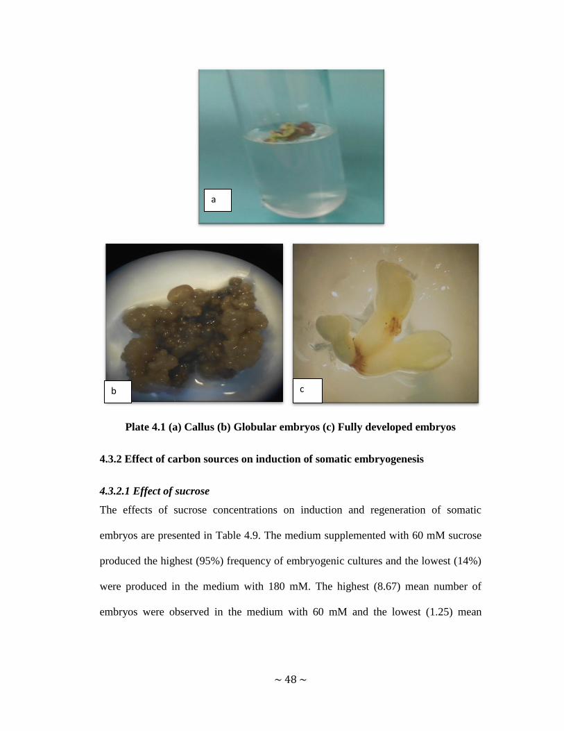
~ 48 ~
Plate 4.1 (a) Callus (b) Globular embryos (c) Fully developed embryos
4.3.2 Effect of carbon sources on induction of somatic embryogenesis
4.3.2.1 Effect of sucrose
The effects of sucrose concentrations on induction and regeneration of somatic
embryos are presented in Table 4.9. The medium supplemented with 60 mM sucrose
produced the highest (95%) frequency of embryogenic cultures and the lowest (14%)
were produced in the medium with 180 mM. The highest (8.67) mean number of
embryos were observed in the medium with 60 mM and the lowest (1.25) mean
a
b c

~ 49 ~
number of embryos were produced in the medium supplemented with 180 mM
sucrose. Media without sucrose did not support somatic embryogenesis.
Table 4.9 Effect of sucrose concentrations on induction of somatic embryos
Sucrose
conc (mM) Embryogenic cultures (%)
Mean number of
embryos/explant ± SE
0 0 0x
60 95 8.67 ± 0.55a*
90 29 1.67 ± 0.33b
120 25 1.57 ± 0.3b
150 45 3.11 ± 0.589b
180 14 1.25 ± 0.25b
P value
P<0.001
xThe zero responses are not included in the statistical analysis *Means followed by
the same letter are not significantly different at P=0.05
4.3.2.2 Effect of different carbon sources on induction and regeneration of somatic
embryos
The effects of different carbon sources on (a) percentage embryogenic cultures and
(b) mean number of embryos are presented in Figure 4.1. The highest (40%)
percentage of embryogenic cultures were observed in the medium supplemented with
90 mM maltose and the lowest (10%) percentage of embryogenic cultures were
produced in the medium supplemented with 150 mM glucose. The highest (3) mean
number of embryos was produced in medium supplemented with 90 mM fructose
while the lowest (1) was observed in the medium supplemented with 150 mM
glucose. However, no significant differences were observed in terms of mean number
of somatic embryos per explant between the different sugars.

~ 50 ~
Figure 4.1 Effects of different sugars sources on (a) percentage embryogenic
cultures and (b) mean number of embryos
4.3.3 Effect of doubling potassium nitrate on the induction of somatic embryos
Leaf explants cultured on medium supplemented with normal amount of potassium
nitrate produced the highest (91%) percentage of embryogenic cultures (Table 4.10).
Medium with double amount of potassium nitrate produced the lowest (33%)
frequency of embryogenic cultures. There were no differences in terms of mean
number of embryos per explant in the two concentration of potassium nitrate
evaluated.
0
5
10
15
20
25
30
35
40
45
Emb
ryo
gen
ic c
ult
ure
s (%
)
Sugar
90 µM
120 µM
150 µM
a
0
0.5
1
1.5
2
2.5
3
3.5
4
Me
an n
o o
f e
mb
ryo
s/e
xpla
nt
Sugar
90 µM
120 µM
150 µM
b

~ 51 ~
Table 4.10 Effects of potassium nitrate on induction of somatic embryos
KNO3
concentration
(mg/l)
Embryogenic
cultures ( %)
Mean number of
embryos/explant ± SE
normal (950) 91 1.20 ± 0.13a*
Double (1900) 33 1.25 ± 0.25a
P Value 0.852
*Means followed by the same letter are not significantly different at P=0.05. n=20
4.3.4 Effects of some amino acids on the induction of somatic embryos
The effects of the amino acids on (a) percentage embryogenic cultures and (b) mean
number of embryos are presented in Figure 4.2. The highest (91%) frequency of
embryogenic cultures were observed in the control and the lowest (6%) percentage of
embryogenic cultures were produced in medium supplemented with 150 µM proline.
Leaf explants cultured on medium supplemented with 150 µM tryptophan produced
the highest (6.5) mean number of somatic embryos and the lowest (1.2) mean number
of somatic embryos were produced in the control. The medium supplementing with
150 µM leucine did not support embryogenesis.

~ 52 ~
Figure 4.2 Effects of some amino acids on (a) percentage embryogenic cultures
and (b) mean number of embryos
4.3.5 Effects of silver nitrate on the induction of somatic embryos
The highest (91%) frequency of embryogenic cultures were observed in the control
and the lowest (25%) percentage of embryogenic cultures were produced in medium
with 150 µM silver nitrate (Table 4.11). Leaf explants cultured on medium
supplemented with 50 µM silver nitrate produced the highest (6.5) mean number of
somatic embryos. On the other hand, the lowest (1.2) mean number of somatic
embryos was produced in the control.
0
10
20
30
40
50
60
70
80
90
100
Emb
ryo
gen
ic c
ult
ure
s (%
)
Amino acids
control
90 µM
100 µM
150 µM
a
0
1
2
3
4
5
6
7
8
9
Me
an n
o o
f e
mb
ryo
s/ e
xpla
nt
Amino acids
control
90 µM
100 µM
150 µM
b

~ 53 ~
Table 4.11 Effects of silver nitrate on induction of somatic embryos
AgNO3 conc (µM) Embryogenic cultures (%) Mean number of embryos/explant ±
SE
0 91 1.2 ± 0.13b*
50 90 6.49 ± 0.38a
100 67 1.5 ± 0.27b
150 25 1.75 ± 0.25b
P-value P<0.001
*Means followed by the same letter are not significantly different at P=0.05. n= 20
4.3.6 Effects of activated charcoal and casein hydrolysate on the induction of
somatic embryos
The effects of CH are presented in Table 4.12. The highest (91%) percentage of
embryogenic cultures were observed in the control while the lowest (30%) were
observed in the medium supplemented with 100 mg/l CH. The highest mean number
of embryos were produced in the medium supplemented with 50 mg/l CH. On the
other hand, the lowest (1.2) mean number of embryos were produced in the control.
AC at all the concentrations evaluated did not support somatic embryogenesis.
Table 4.12 Effects of casein hydrolysate on induction of somatic embryos
CH conc.
(mg/l)
Embryogenic cultures (%) Mean number of embryos/explant
± SE
0 91 1.2 ± 0.13b*
50 58 4.15 ± 0.5a
100 50 3.38 ± 0.42a
150 30 2.67 ± 0.67a
P-value P˂0.01
*Means followed by the same letter are not significantly different at P= 0.05

~ 54 ~
4.3.7 Effect of some organic extracts on induction of somatic embryogenesis
The results of the effects of potato starch, corn starch and papain on percentage
embryogenic cultures and mean number of embryos are presented in Figure 4.3. The
highest (91%) percentage of embryogenic cultures were observed in the control and
the lowest (4%) were observed in the medium with 150 mg/l papain. The highest
(1.7) mean number of somatic embryos were produced in the medium supplemented
with corn starch and the lowest (1) in the medium with papain 100 and 150 mg/l.
Potato starch at concentrations 50 and 100 mg/l did not support embryogenesis.
However, there was an insignificant difference between the mean number of in
potato, corn starch and papain and the control (P≥0.05).
Figure 4.3 Effects of some organic extracts on (a) percentage embryogenic
cultures and (b) mean number of embryos
0
10
20
30
40
50
60
70
80
90
100
Emb
ryo
gen
ic c
ult
ure
s (%
)
Organic extracts conc (mg/l)
0
50
100
150
a
0
0.5
1
1.5
2
2.5
Me
an n
um
be
r o
fn e
mb
ryo
s
Organic extracts (mg/l)
0
50
100
150
b

~ 55 ~
The results of the effects of coconut milk on induction and regeneration of somatic
embryos are presented in Table 4.13. The highest (91%) percentage of embryogenic
cultures were observed in the control and the lowest (30%) were observed in the
medium with 30% coconut milk. The highest (3.18) mean number of somatic
embryos were produced in the medium supplemented with 20% coconut milk and the
lowest (1.2) in the control.
Table 4.13 Effects of coconut water on induction and regeneration of embryos
Coconut water conc.(%) Embryogenic cultures
(%)
Mean number of
embryos/explant ± SE
0 91 1.2 ±0.13 b*
10 50 1.6± 0.4 b
20 85 3.18± 0.3a
30 30 2.33±0.33a, b
P value P<0.001
*Means followed by the same letter are not significantly different at P= 0.05
4.3.8 Effect of liquid pulsing on the induction of somatic embryos
The effects of liquid pulsing on percentage embryogenic cultures and mean number
of embryos are presented in Figure 4.4. Callus pretreatment in liquid medium for
different periods had a very significant effect on somatic embryo induction and
proliferation. Callus tissues pretreated in liquid medium for 24 hours produced the
highest (94%) frequency of embryogenic cultures while the lowest frequency
embryogenic cultures was observed in calluses pretreated in liquid medium for 6

~ 56 ~
hours. The highest (9.19) mean number of somatic embryos were observed on callus
tissues pretreated in liquid medium for 24 hours and the lowest mean number (1.2) of
embryo somatic embryos was produced in the control.
Figure 4.4 Effect of liquid pulsing on (a) percentage embryogenic cultures and
(b) mean number of embryos
4.4 Effect of growth regulators on shoot and root regeneration from nodal
explants
4.4.1 Effect of BAP on microshoot regeneration
Shoots were first observed two weeks after culture. The effect of BAP on microshoot
regeneration is presented in Table 4.14 and Plate 4.2 (a). BAP at 10µM/l gave the
highest mean number (2.25) of shoots per explant and the highest mean length (0.9).
Increasing the concentration of BAP from 10 to 40 µM/l decreased the number of
microshoots/node and the mean shoot length.
0
10
20
30
40
50
60
70
80
90
100
0h 6h 12h 24h
Emb
ryo
gen
ic c
ult
ure
Pulsing duration (h)
0
1
2
3
4
5
6
7
8
9
10
0h 6h 12h 24h
Me
an n
o o
f gl
ob
ula
r e
mb
ryo
s
Pulsing duration (h)
a b

~ 57 ~
Table 4.14 Effect of BAP on microshoot regeneration
Concentration (M/l) Mean no. of microshoot
/explants (±SE)
Mean length of microshoot
(cm) (±SE)
0 0x 0
5 1.4 ± 0.25b* 0.32 ± 0.049b
10 2.25 ± 0.16a 0.9± 0.05a
20 2.0 ± 0b 0.38 ± 0.138b
40 1.67 ± 0.33b 0.33 ± 0.03b
P value 0.023 P<0.001
x The zero responses are not included in the statistical analysis. *Means followed by
the same letter are not significantly different. n= 20
4.5.2 Effect of IBA on root regeneration
After one month on microshoot induction media, shoots were transferred to rooting
medium. The effect of IBA on root regeneration is presented in Table 4.15 and plate
4.2 (b). IBA at 10 µM/l gave the highest mean number (2.2) of roots per explant.
However, there were no significant differences between the different treatments.
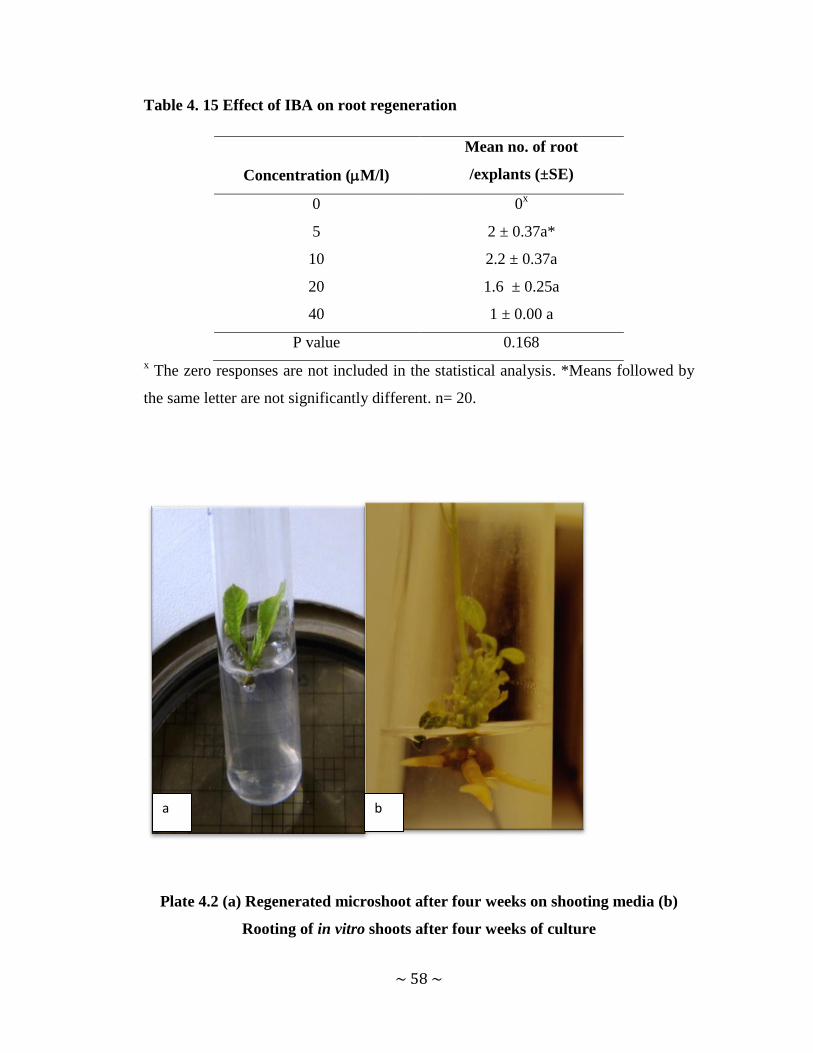
~ 58 ~
Table 4. 15 Effect of IBA on root regeneration
Concentration (M/l)
Mean no. of root
/explants (±SE)
0 0x
5 2 ± 0.37a*
10 2.2 ± 0.37a
20 1.6 ± 0.25a
40 1 ± 0.00 a
P value 0.168
x The zero responses are not included in the statistical analysis. *Means followed by
the same letter are not significantly different. n= 20.
Plate 4.2 (a) Regenerated microshoot after four weeks on shooting media (b)
Rooting of in vitro shoots after four weeks of culture
a b

~ 59 ~
CHAPTER 5 DISCUSSION, CONCLUSION AND
RECOMMENDATIONS
5.1 Discussion
5.1.1 Nutritional variation of V. doniana young leaves
Green leafy vegetables such as V. doniana young leaves form an indispensable
constituent of diet in Africa and in Benin in particular. In the present study, the
nutritional variation of V. doniana young leaves obtained from different climatic
zones in Benin were analyzed. Previous studies conducted on V. doniana young
leaves from different provenances in Nigeria have shown that the protein
concentration varies from 5.85% to 13.85 % in raw samples (Otitoju et al., 2014;
Osum et al. 2013; Adejumo et al., 2013 while the moisture content is in the range of
21.14 to 78.76% (Otitoju et al., 2014; Osum et al. 2013; Adejumo et al., 2013). Fat,
fiber, carbohydrate, ash content range from 1.10 and 2.92%, 1.85 and 2.75% , 7.57
and 12.92%, 1.63 and 6.21% respectively (Otitoju et al., 2014; Osum et al. 2013;
Adejumo et al., 2013). Most of the reported values are similar to the results obtained
in this study except for the moisture content. The Fe concentration found in the
present study is within range of the values reported by other authors (Otitoju et al.,
2014; Osum et al., 2013). However, the Ca concentration is twice to four times higher
than the maximum reported in the literature (Otitoju et al., 2014; Osum et al., 2013).
Vitamin C in V. doniana leaves is reported in the literature to vary from 20.80 to
91.28 mg/100g while Vitamin A varies from 0.53 to 2.15 mg/100g (Otitoju et al.,
2014; Osum et al., 2013; Adejumo et al., 2013). The reported values for Vitamin C
concentration in previous studies are higher than the results obtained in the current

~ 60 ~
study. On the other hand, the Vitamin A concentration in the Guinean zone was found
to be similar to the reported values but the concentration in the Sudanian zone was
found to be double the previously reported values. Among the nutritional parameters
evaluated, only the moisture, protein, calcium (Ca) and the vitamins A and C
concentrations showed significant differences due to the agro-climatic zones. Other
nutritive concentration of V. doniana leaves do not vary significantly (p > 0.05),
across agro-climatic zones. They could not therefore linked to the agro climatic zones
of provenance of the trees.
5.1.2 Surface sterilization of V. doniana explants
The study was conducted with an aim of optimizing the sterilization of Vitex doniana
explants. The use of field grown plants as a direct source of explant material for
obtaining ‘clean’ explant, presents a major challenge. Previous attempts to initiate
clean explants from field grown coffee, especially those from canopy close to the
ground, resulted in 100% contamination (Kahia, 1999). The disinfectant widely used
for surface sterilization is sodium hypochlorite (Miche and Balandreau, 2001). It is
usually purchased as household laundry bleach. A balance between concentration and
time must be determined empirically for each type of explant because of
phytotoxicity. Calcium hypochlorite being a mild sterilant, is rarely used for
decontaminating explants. It may be less injurious to plant tissues than sodium
hypochlorite (CSS 451, 2009). Bleach (1.4% NaOCl solution for 1 minute) has the
capability to be an effective sterilization agent for leaves as observed in A. crasna and
A. sinensis from greenhouse source (Okudera and Ito, 2009). Hassan et al. (2011)
achieved effective sterilization when using 50% bleach for 20 minutes on shoot tips

~ 61 ~
of A. hirta, from greenhouse while leaf explants of Allanblackia stuhlmannii were
best surface sterilized using 8% sodium hypochlorite (Neondo et al., 2011). During
the present study, sodium hypochlorite was found to be less effective than calcium
hypochlorite in sterilizing Vitex doniana leaf explants. Explants treated with sodium
hypochlorite turned brown and died. This is probably due to the fact that leaves used
were too young and the sterilant caused phytotoxicity. Mercuric chloride (0.1 and 0.5
%) was found toxic in sterilizing node explants. This result is contrary to those of
Vadawale et al. (2006) who achieved surface sterilization of Vitex negundo nodes
using 0.05% mercuric chloride. The toxicity observed in this study can explained by
the relatively high concentrations used. Many authors succeeded in surface
sterilization using calcium hypochlorite. Mihaljević et al., (2013) while working on
shoots of sour cherry recorded 80% clean explants when 3% Ca(OCl)2 for 15 minutes
was used. In order to increase the percent clean explant and reduce as well the
toxicity observed both with sodium hypochlorite and mercuric chlorite, a two- step
sterilization procedure was evaluated. This procedureallows the explants to be
sterilized twice and at the same reduces the number of dead explants due to toxicity.
Indeed, between the two steps explants are allowed to breathe while they are rinsed.
Similar results were reported by Nieves and Aspuria, (2011) who recorded 90 %
clean explants from cotyledon of Moringa oleifera using two steps comprising of 5%
Ca(OCl)2 for 15 minutes.
5.1.3 Factors affecting somatic embryogenesis in V. doniana
In vitro somatic embryogenesis may follow two routes: Direct embryogenesis in
which the embryos arise directly from the explants tissue in the absence of callus

~ 62 ~
proliferation. This occurs through pre-embryonic determined cells (PEDC). The other
route is indirect embryogenesis where there is a callus phase prior to embryo
formation. The cells from which embryos arise are referred to as induced
embryogenic determined cells (IEDC). In the majority of cases embryogenesis is
through the indirect method. In this type, specific growth regulator concentrations are
required for somatic embryos to arise. Media supplemented with cytokinins have
been known to induce direct somatic embryos (Kahia, 1999). Most of the reports have
indicated the requirement of an auxin alone or in combination with a cytokinin for
callus formation (Sheeba et al., 2013. Danya et al., 2012). In this study different types
and concentrations of cytokinins namely BAP, Kin and TDZ were tested for
induction of direct embryogenesis. Ten days after initiation callus induction was
observed. Similar report was made by Manisha and Rajesh (2013) who obtained
moderate to good callus growth in Tecomella undulata with different concentrations
of BAP and Kin alone. The formation of callus in media supplemented with cytokinin
alone may be explained by the presence of endogenous auxin and the sensitivity of
the species to the hormones. In woody trees, auxin is mainly produced in young
leaves (Aloni, 2007). During the current study very young leaves and this could be
the reason why wt callus was obtained instead of embryos. In the work being
reported, the best callus induction was obtained in the medium supplemented with
TDZ at concentration 0.5 µM compared to Kin and BAP. Similarly, Lin et al. (2004)
have suggested that TDZ is more effective than other cytokinins used for somatic
embryogenesis in sweet potato and bamboo respectively. It has been suggested that
TDZ either directly promotes growth due to its own biological activity or through

~ 63 ~
inducing the synthesis and/or accumulation of endogenous cytokinins or auxins
(Panaia et al., 2004). The latter could explain the effectiveness of TDZ as it may be
mediating levels of endogenous auxin levels within the cultured tissue.
The most commonly used protocol for induction of embryogenesis involves the
induction of callus in an auxin-supplemented medium (Deo et al., 2010). Contrary to
our findings, 2, 4-D has been found to be effective in many plant species. Wang et al.
(2006) reported that 2, 4-D (0.2 mg/L) induced the embryogenic callus in Chorispora
bungeana; similar results were observed in Phoenix dactylifera (Lin et al., 2004). It
was also observed that 3,4-D and dicamba were not effective. This probably is
because those growth regulators share similarities in structure and activity to 2, 4-D
(Balieldin et al., 2000). However, the results showed that picloram at concentration 4
µM significantly enhanced the mean number of embryos. Similarly, Surdarmonowati
and Henshaw (1996) observed that picloram (6 mg/l) improved embryogenesis in
cassava. It can therefore be concluded that the initiation of embryos cannot be
ascribed to any one plant growth regulator (Deo et al., 2010). It also appears that
different species have different levels of sensitivity towards various plant growth
regulators; hence, their response to embryogenesis is variable.
Carbohydrates such as sucrose, fructose and glucose, maltose, mannitol, sorbitol were
tested for their potential to induce and regenerate embryos. At low sucrose
concentration (60 mM), the highest percentage of embryogenic culture were observed
as well as the highest mean number of embryos. Increasing sucrose concentration did
not improve the mean number of embryos. These results contrary to those of
Gerdakaneh et al., (2009) who found that increasing sucrose concentration from 2 to

~ 64 ~
6 % in the medium improved the development of somatic embryos in strawberry.
However, they found that increasing the sucrose concentration from 6 to 12%
decrease the number of embryos per explant. Similarly, Pande and Khetmalas, (2012)
showed that culture medium supplied with 25 g/l sucrose did not influence somatic
embryogenesis in S. rebaudiana. Increased sucrose concentration to 45g/l in culture
medium resulted in less proliferation of callus and generation of somatic embryos.
This demonstrated that optimum concentration of sucrose is important for
embryogenesis. During the current study, mannitol, fructose, glucose, sorbitol did not
improve somatic embryogenesis. This showed that the best carbon source for embryo
culture in V. doniana is sucrose. Similar conclusion was made by Gerdakaneh et al.,
(2009) while comparing sucrose, fructose and glucose in strawberry. Our study
demonstrated that the type and concentration of sugar have important effects on the
embryo development in V. doniana.
The recalcitrance of some species can be overcome by manipulating media
components. During the current study the amount of KNO3 in MS media was
doubled. This resulted in no effect on the frequency of embryogenic cultures and the
mean number of embryos. Similar report was made by Haq and Zafar (2004) in
cotton where doubling the amount of KNO3 in half MS (1/2 MS + 1.9 g/l KNO3)
decreased the number of embryos from 100 i to 88.5.
Requirement of exogenous supply of amino acids for in vitro somatic embryogenesis
has been reported in a number of plant species (Gill et al., 2004; Coste et al., 2011).
For instance, glutamine has been found beneficial for embryo development in date
palm (El-Shiaty et al., 2004). MS medium supplemented with 100 mg /l glutamine

~ 65 ~
gave the highest number of embryos (3.33) followed by MS medium with 50 mg/l
glutamine which gave (3.00) with no significant difference between them. Sarker et
al., (2007) found that significant response was found using L-asparagine at 150 mg/l
concentration in four popular wheat Bangladeshi cultivars viz Kanchan, Shourav,
Gourav and Satabdi. In the work being reported, four amino acids namely proline,
lysine, leucine and tryptophan were evaluated. It was found that exogenous
application of a specific amino acid, especially tryptophan, promoted the process of
somatic embryogenesis and regeneration. A significant response was found using
tryptophan at 150 µM concentration giving the highest (6.5) mean number of
embryos. Similarly, when tryptophan (100 to 300 µM) was added to callus induction
medium of rice, it was observed a great enhancement of the frequency of
embryogenic cultures and regeneration (Shahsavari, 2011). Gill et al. (2004) reported
that proline at 560 mg/l was able to increase somatic embryogenesisin surgarcane. In
Peucedanum oreoselinum, embryo formation and maturation was enhanced by
addition of proline (5 mM) in MS medium (Coste et al., 2011). Although there was no
significant difference between the mean number of embryos in the media
supplemented with proline, it was observed that the lowest concentration of proline
increased almost 3 folds the mean number of embryos compared to the control. It was
also found that an increase in proline concentration has resulted in decrease in the
frequency of embryogenic cultures and the number of embryos. This results were
contrary to those found in rice by Chowdhri et al., (1993) who showed that proline
enhanced embryogenesis in rice. These authors reported that an increase in proline
concentration from 3 M to 12 mM resulted in a proportional increase of the

~ 66 ~
percentage of culture showing regeneration (the maximum being 63% as compared to
23% without proline). The results obtained in the work being reported with leucine
are contrary to those of Gill et al.(2004) and Coste et al. (2011) as. leucine at the
highest concentration (150 µM) was found to inhibit embryogenesis. On the other
hand, an increase in the frequency of embryogenic cultures and in the mean number
of embryos was observed when the concentration of lysine increased from 50 to 150
µM even if there were no significant differences between the different concentrations
and the control. Usefulness of tryptophan can be considered to provide organic
nitrogen supply associated with in vitro culture and somatic embryogenesis. On the
contrary, lysine, leucine and proline are not effective in providing nitrogen.
Differential responses of different amino acids indicate the requirement of specific
amino acids for somatic embryo regeneration.
The exact mechanism by which AgNO3 affects somatic embryogenesis is not
completely understood (Kong et al., 2012). In the present study, AgNO3 has been
found to enhance somatic embryogenesis in Vitex doniana. Media supplemented with
AgNO3 has also been shown to improve somatic embryogenesis in species such as
Coffea canephora (Rubiaceae) (Fuentes et al., 2000), Spinacia oleracea (Ishizaki et
al., 2000), Carthamus tinctorius (Asteraceae) (Mandal et al., 2001), Paspalum
scrobiculatum (Poaceae) (Vikrant and Rashid, 2002), Bactris gasipaes (Arecaceae)
(Steinmacher et al., 2007), Paspalum scrobiculatum (Poaceae) and Eleusine coracana
(Poaceae) (Kothari-Chajer et al., 2008), Hedychiurn bousigonianum (Gingeberacea)
(Sakhanokho et al., 2009), Gossypium nelsonii (Malvaceae) and G. australe (Yan et
al., 2010), and Pinus taeda (Pinaceae) (Pullman et al., 2003). Kong et al., (2012)

~ 67 ~
demonstrated that AgNO3 stimulates embryo formation in Manchurian ash. Ishizaki
et al., (2000) observed that in Spinacia oleracea addition of 10 mM AgNO3 to the
medium resulted in formation of about three times more embryos as compared with
controls. In the present study, 50 µM of AgNO3 was able to increase the number of
embryos These results are contrary to those of Hatanaka et al. (1995) who reported
that addition of AgNO3 (50 µM) inhibited somatic embryo formation in leaf explants
of Coffea canephora. Fuentes et al. (2000) found that the addition of AgNO3 caused
only small modifications in the ionic equilibrium of the medium and concluded the
effects of the compound on somatic embryogenesis were not attributable to any
substantial changes in available nutrients. In addition, the effects of AgNO3 on
somatic embryogenesis varied with the compound concentration. The lowest number
(one) of embryos per explant was induced in Manchurian ash at 2.5 mg L–1
AgNO3
while when 10 mg/L AgNO3 was included in the medium, the total number of
somatic embryos was the highest, with an average of 3.86 embryos per explant (Kong
et al., 2012). Kong et al., (2012) reported that the number of somatic embryos per
explant in Manchurian ash increased with increasing AgNO3 concentration. During
the current study, the higher concentrations of AgNO3 reduced the numbers of visible
embryos.
In the work being reported, addition of casein hydrolysate to the medium increased
the mean number of embryos. The highest mean number of embryos was observed in
media supplemented with 50 mg/l casein hydrolysate. Khierallah and Hussein, (2013)
observed the same result in date palm when 2 g/l casein hydrolysate was added to the
medium. Angeel and Elmeer (2011) found that the best result of somatic embryos per

~ 68 ~
treatment was achieved with 3 g/ l casein hydrolysate in date palm. The Increase in
the number of embryos by adding casein hydrolysate may be attributed to the fact that
casein as a simple protein decomposes into galactosamine and glucosamine or
mannosamine as well as simple sugars linked with protein with covalent bonds or
glycoside bond with hydroxyl group of the amino acids serine and threonine (Al-
Dalaly and Al-Rekaby, 1995). Activated charcoal has been found to have a positive
effect on many species such as Vitis vinifera (Lopez-Perez et al., 2005), pepper
(Ellialtıoğlu et al., 2001) and Aesculus hippocastanum (Calic-Dragosavac et al.,
2010). Lopez-Perez et al., (2005) found that addition of AC (0.25 %) to the medium
used for callus culture was an essential prerequisite to obtain somatic embryos in the
case of Vitis vinifera var. Crimson Seedless. In Italia and Don Mariano; it greatly
increased the frequency of embryogenic calli in var. Sugraone from 5.8 % in medium
without AC to 99.5 % with AC. Activated charcoal is commonly used in tissue
culture media to darken the immediate media surroundings and to absorb inhibitory or
toxic substances (Moshkov et al., 2008). Moreover, activated charcoal also has been
shown to absorb 5-hydroxymethylfurfural, an inhibitor formed by the degradation of
sucrose during autoclaving, as well as substantial amount of auxins and cytokinins.
Consequently, apart from removing inhibitors that would prevent growth, it may also
adsorb and reduce the levels of growth regulators. (Deo et al., 2010). In this study,
supplementation of the media with activated charcoal inhibited embryogenesis at all
the concentrations evaluated. This observation might be due to the fact that AC
degraded the growth regulator present in the medium and this affected
embryogenesis.

~ 69 ~
Some researchers have underscored the need to include complex organic extracts,
such as coconut water (CW), potato extract (PE), corn extract (CE) and papaya
extract (PAE) for inducing somatic embryogenesis in some species (Ichihashi and
Islam, 1999; Islam et al., 2003; Rahman et al., 2004). The current study investigated
the effects of coconut water, potato and corn starch, papain on somatic embryogenesis
in V. doniana and the results indicated that with potato starch, corn starch and papain
did not improve embryogenesis. On the contrary, the best result was obtained when
the modified VW medium supplemented with 20% potato extract produced the best
results on proliferation of Vanda Kasem’s Delight (VKD) orchid’s protocorm-like
bodies (Gnasekaran et al., 2012). Potato is a carbohydrate-rich food which contains
about 80% water and 20% dry matter, with 60 to 80% of the latter composed of starch
(Prokop and Albert 2008). There are antioxdants present in potatoes and beside them,
notable amounts of proteins and vitamin B6 of high biological value could also
promote somatic embryogenesis. Substantial amounts of alkaline salts in the form of
potassium may thwart acid overdose (Demigne et al., 2004) and prevent medium
fermentation, which in turn supports the production somatic embryos. In the current
study, Potato starch was used and the negative response observed might be due to the
concentrations evaluated which might be too low. Low concentrations of the potato
starch could not provide sufficient amounts of some of the biological compounds
present at trace levels in the potato starch. Similarly, concentrations of papain and
corn starch did not improve embryogenesis in V. doniana. Complex organic extracts
used in the present experiment contain carbohydrates, protein, fat, several vitamins,
phenolic compounds, a lower level of some amino acids, and organic acids. However,

~ 70 ~
there could be other unknown substances, alone or in combination which might
affect embryogenesis.
During the current study, supplementation of the media with coconut water improved
somatic embryogenesis in V. doniana, with 20 % coconut water giving the highest
response. Similarly, Khieralla and Hussein (2013) found that media supplemented
with 20 % coconut water gave the highest number of embryos (65 embryos) in date
palm. In contradictory to our results, Al-Khayri (2010) observed in date palm that 20
% coconut water decreased the number of embryos. The addition of coconut water to
the culture media resulted in the plants with a greater nutritional and carbohydrates
content as coconut itself contains 21.8 mg/L in total (George, 1993). The
enhancement of the number of mean number of embryos compared to the control
might be due to this high carbohydrate content. Mauney et al. (1952) purified a
growth factor from the aqueous extract of coconut meat which was found to be very
potent in promoting growth of tissue cultured plants. Coconut water was shown to
have cytokinin like activity by Kuraishi and Okumura (1961) and recognised natural
cytokinin substances have since been isolated [9-β-D-ribofuranosyl zeatin, and
several unidentified compounds but the levels of these compounds in various samples
of coconut water have not been reported. It has been observed that coconut contains
natural cytokinins, adding it to medium culture often has the same effect as adding
recognised cytokinin. Higher or lower concentrations (10 and 30%) of coconut water
did not significantly increase the number of embryos in the current study probably
due to reduction of the balance of nutrients or their availability.

~ 71 ~
It has been previously demonstrated that liquid pulse treatments with growth
regulators could promote regeneration from plant cell culture through organogenesis
or somatic embryogenesis (Madhulatha et al., 2004). In addition, it has previously
been reported that initial pretreatment of leaf explants of squash (Cucurbita pepo L.)
with cytokinins for 6–48 h significantly promoted the formation of somatic embryos
which developed further to the torpedo-shape stage and germinated (Kintzios et al.,
2002). Therefore, during the current study, it was found necessary to evaluate the
effect of the pretreatment of V. doniana callus in a liquid nutrient medium on the
induction and development of somatic embryos. In the present study, pretreatment in
liquid nutrient medium for regeneration of Vitex doniana plants have been established
for the first time. The induction and enhancement of somatic embryogenesis in leaf
derived callus tissues within the experimental period of the present study was
absolutely dependent on the physical state of the culture medium, since it only
occurred when cultures were liquid-pulsed for 6, 12 or 24 hrs. The stimulating effect
of liquid culture on Vitex doniana somatic embryogenesis could be associated with
increased uptake of TDZ by the cultured cells. All the treatments increased the mean
number of embryos compared to the control (leaf explants continuously cultured on
solid medium). The highest number of embryos was observed on callus tissues
pretreated in liquid medium for 24 h. This number was 8 fold higher than the mean
number recorded in the control. This results is similar to those of Papanastasiou et al.
(2008) who found that the highest number of embryos was observed on Coffea
arabica callus tissues pretreated in 50 µM BA for 24 h and subcultured on BA-
supplemented medium. However, they found that control and callus pretreated for 6 h
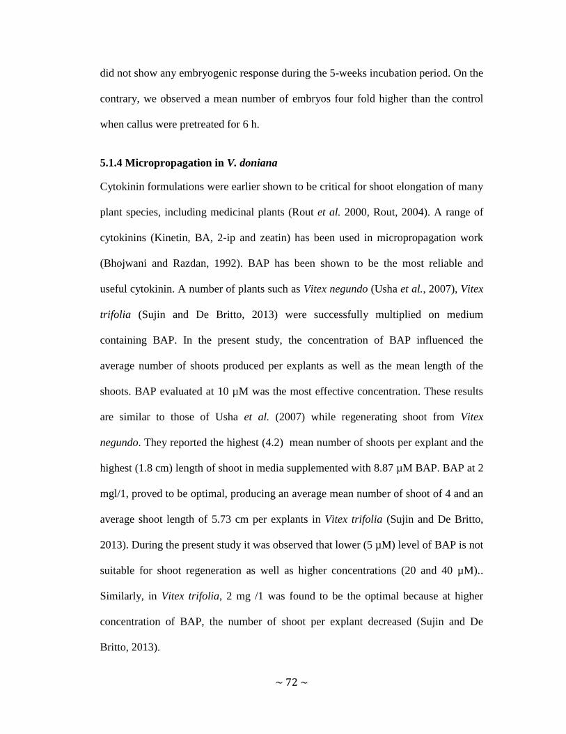
~ 72 ~
did not show any embryogenic response during the 5-weeks incubation period. On the
contrary, we observed a mean number of embryos four fold higher than the control
when callus were pretreated for 6 h.
5.1.4 Micropropagation in V. doniana
Cytokinin formulations were earlier shown to be critical for shoot elongation of many
plant species, including medicinal plants (Rout et al. 2000, Rout, 2004). A range of
cytokinins (Kinetin, BA, 2-ip and zeatin) has been used in micropropagation work
(Bhojwani and Razdan, 1992). BAP has been shown to be the most reliable and
useful cytokinin. A number of plants such as Vitex negundo (Usha et al., 2007), Vitex
trifolia (Sujin and De Britto, 2013) were successfully multiplied on medium
containing BAP. In the present study, the concentration of BAP influenced the
average number of shoots produced per explants as well as the mean length of the
shoots. BAP evaluated at 10 µM was the most effective concentration. These results
are similar to those of Usha et al. (2007) while regenerating shoot from Vitex
negundo. They reported the highest (4.2) mean number of shoots per explant and the
highest (1.8 cm) length of shoot in media supplemented with 8.87 µM BAP. BAP at 2
mgl/1, proved to be optimal, producing an average mean number of shoot of 4 and an
average shoot length of 5.73 cm per explants in Vitex trifolia (Sujin and De Britto,
2013). During the present study it was observed that lower (5 µM) level of BAP is not
suitable for shoot regeneration as well as higher concentrations (20 and 40 µM)..
Similarly, in Vitex trifolia, 2 mg /1 was found to be the optimal because at higher
concentration of BAP, the number of shoot per explant decreased (Sujin and De
Britto, 2013).

~ 73 ~
Shoots derived from nodal explants exhibited good response for root induction in a
medium supplemented with IBA. These results are similar to those of Rathore and
Shekhawat (2011) who reported in vitro rooting of micropropagated shoots of Vitex
negundo using IBA and maximum number of roots (6-7) of with a length of 4-5 cm
with 3 mg/l.
5.2 Conclusion
Results of this study showed that V. doniana young leaves are highly nutritious. The
protein, Calcium, vitamin A and C concentration vary significantly across agro
climatic zones. The highest content of protein (7.6 %), Vitamin A (0.55 mg/100g )
and C (17.78 mg/100g) were obtained in the Sudanian zone while the highest
concentration of calcium (242.95 mg/100g) was obtained in the Sudano-Guinean
zone.
It can be concluded that a double sterilization is more effective for sterilization of
field-grown derived leaf and nodal explants of Vitex doniana. V. doniana is
ameniable to tissue culture. Among all the treatments evaluated the liquid state of the
medium gave the best result on embryogenesis given a mean number of embryos
eight fold higher (9.19) than the control (1.2).
BAP was used for shoot regeneration. The best treatment was BAP 10µM which gave
the highest mean shoot number and mean shoot length. IBA at all evaluated
concentrations was able to regenerate roots but no optimum concentration was found.
This works provides useful information that can be used to catalyse increased
domestication of V. doniana in Benin based on the nutritional properties of the

~ 74 ~
species and the in vitro propagation protocols developed. However, more work needs
to be done to strengthen the current outputs as described in section 5.3.
5.3 Recommendations
V. doniana has a high content of crude protein, oils, minerals, and vitamins. We
recommend this species as a complex food supply. Further to the findings of our
study, we recommend:
more investigations on the bioavailability of nutrients from the leaves of V.
doniana as well as its anti-nutritional composition.
more investigations on the nutritional variation of V. doniana by analysis the
link between the soil types and the samples' provenances
Mature somatic embryos were obtained in this study after a long period. For more
efficient and complete tissue culture protocol, we recommend:
Optimization of maturation process in future studies which will provide
enough material for germination studies
Evaluation of other growth regulators on micropropagation for optimum
plantlets' regeneration
Studying the somaclonal variation
Conversion and the performance in the field of both embryo-derived plantlets
and plantlets obtained from micropropagation.
~ 75 ~
REFERENCES
Achigan-Dako EG, Pasquini MW, Assogba-Komlan F, N’danikou S, Yédomonhan H,
Dansi A Ambrose-Oji B (2010). Traditional vegetables in Benin: diversity,
distribution, ecology, agronomy, and utilisation. Institut National des
RecherchesAgricoles du Bénin, Benin. 252 pp.
Adejumo AA, Alaye SA, Ajagbe RO, Abi EA, Adedokun FT (2013). Nutritional And
Anti – Nutritional Composition Of Black – Plum (Vitex doniana) Journal of Natural
Sciences Research 3 (12): 144-148
Adeyeye IA, Omotayo FO (2011). Agriculture and Biology Journal of North
America, p 502
Adomou AC (2005): Vegetation patterns and environmental gradients in Benin:
Implications for biogeography and conservation. PhD Thesis Wageningen University,
Wageningen
Agbedje JO, Ibitoye AA (2007). Chemical composition of black plum (Vitex
doniana): an underutilised fruit. Journal of Food Agriculture and Environment 5: 95-
96.
Angeel S, Elmeer K (2011). Effects of Casein Hydrolysates and Glutamine on Callus
and Somatic Embryogenesis of Date Palm (Phoenix dactylifera L.). New York
Science Journal 4(7): 121-125
Aghaei P, Bahramnejad B, Mozafari AA (2013). Effect of different plant growth
regulators on callus induction of stem explants in Pistacia atlantica subsp. kurdica
Plant Knowledge Journal 2(3): 108-112

~ 76 ~
Ahmed ABA, Suryanarayana R A, Venkateswara MR, Taha
RM (2011). Effect of
picloram, additives and plant growth regulators on somatic embryogenesis of Phyla
nodiflora (L.) Greene. Brazilian archives of biology and technology 54 (1): 7-13
Akoègninou A (2004). Recherches botaniques et écologiques sur les forêts actuelles
du Bénin. Thèse d’Etat. Université de Cocody-Abidjan (Côte d’Ivoire), 326 pp.
Akutsu M, Sato H (2002). Induction of proembryos in liquid culture increases the
efficiency of plant regeneration from Alstroemeria calli. Plant Science 163:475–479
Al-Dalaly BK, Al-Rekaby K (1995). Food Chemistry, (in Arabic) Al-Kendy
Publisher, Baghdad, Iraq. pp. 234.
Al- Khayri JM (2010). Somatic embryogenesis of Date Palm (Phoenix dactylifera L.)
improved by coconut water. Biotechnology, 2010: 1-8
Aloni R (2007). Phytohormonal mechanisms may control wood quantity formation in
young and mature trees. In K Entwiistle, P Harris, J Walker (eds). The compromised
wood workshop. The Wood Technology Research Center, University of Canterbury,
Christchurch, New Zealand
Analyse Globale de la Vulnérabilité, de la Sécurité Alimentaire et de la Nutrition,
AGVSAN (2009). République du Bénin, Program Alimentaire Mondial (PAM),
UNICEF, FAO. PAM, Rome – Italy.
AOAC. (2000). Official Methods of Analysis 14th ed. Association of Official
Analytical Chemists Washington, D.C.
Assogbadjo AE, Chadare FJ, Glele Kakaï R, Fandohan B, Baidu-Forson JJ (2012).
Variation in biochemical composition of baobab (Adansonia digitata) pulp, leaves,

~ 77 ~
and seeds in relation to soil types and tree provenances. Agriculture, Ecosystems and
Environment 157: 94– 99
Bahieldin A, Dyer WE, QU R (2000). Concentration effects of dicamba on shoot
regeneration in wheat. Plant Breeding 119, 437-439
Barna KS, Wakhlu AK (1994) Whole plant regeneration of Cicer arietium from
callus culture via organogenesis. Plant Cell Reports 13:510-513.
Bhojwani SS, Razdan MK (1992). Plant tissue culture: Theory and practice, Elsevier,
Amsterdam, London, New York, Tokyo. Trifolium repens genotypes. Physiologia
Plantarum 52:187-190.
Burchi F, Fanzo J, Frison E (2011). The Role of Food and Nutrition System
Approaches in Tackling Hidden Hunger. International Journal of Environmental
Research and Public Health 8, 358-373.
Calic-Dragosavac D, Zdravkovi-Korac S, Bohanec B, Radojevic L, Vinterhalter B,
Stevovic S, Cingel A, Savic J (2010). Effect of activated charcoal, abscisic acid and
polyethylene glycol on maturation, germination and conversion of Aesculus
hippocastanum androgenic embryos. African Journal of Biotechnology 9 (25): 3786-
3793
Chandra S, Bandopadhyay Rajib, Kumar V, Chandra R (2010). Acclimatization of
tissue cultured plantlets: from laboratory to land. Biotechnology Letter 32, 1199–
1205
Chowdhry CN, Tyagi AK, Maheshwari N, Maheshwari SC (1993). Effect of L-
proline and L-tryptophan on somatic embryogenesis and plantlet regeneration of rice
(Oryza sativa L. cv. Pusa 169). Plant Cell Tissue and Organ. Culture. 32: 357-361.

~ 78 ~
Codjia JTC, Assogbadjo AE, Ekue MRM (2003). Diversite et valorization au niveau
local des resource vegetales forestieres alimentaires du Benin. Cahiers Agricultures
12: 321-331.
Coste A, Oltean B, Halmagyi A, Deliu C (2011). Direct somatic embryogenesis and
plant regeneration in Peucedanum oreoselinum (L.) Moench. Romanian
Biotechnological Letter 16(4):6451-6459
CSS 451 (2009) Biotechnology Applications for Plant Breeding and Genetic. January
13, Course and laboratory orientation; sterile techniques and tissue culture
https://www.msu.edu/course/css/451/ accesed on 24th May 2014
Dadjo C, Assogbadjo AE, Fandohan B, Glèlè Kakaï R, Chakeredza S, Houehanou
TD, Van Damme P, Sinsin B (2012). Uses and management of black plum (Vitex
doniana Sweet) in Southern Benin. Fruits 67(4) 239-248.
Dansi A, Adjatin A, Adoukonou-Sagbadja H, Faladé V, Yedomonhan H, Odou D,
Dossou B (2008). Traditional leafy vegetables and their use in the Benin Republic.
Genetic Resources and Crop Evolution 55, 1239-1256.
Danya U, MR Udhayasankar, K Arumugasamy, D Punitha (2012). An efficient
protocol devised for rapid callus induction from leaf explants of Stephania wightii
(Arn.) Dunn. - an endemic medicinal plant. Research in Plant Biology 2(5): 18-22
Demigne C, Sabboh H, Puel C, Remsey C, Coxam V (2004) Organic anions and
potassium salts in nutrition and metabolism, Nutrition Research Reviews, Cambridge
journals, Cambridge University Press 17: 249-258.

~ 79 ~
Deo PC, Tyagi AP, Taylor M, Harding RM, Becker DK (2010). Factors affecting
somatic embryogenesis and transformation in modern plant breeding. The South
Pacific Journal of Natural and Applied Sciences , 28(1): 27-40.
Dhillon NK, Gosal SS (2012). Effect of growth adjuvents on somatic embryogenesis
in maize (Zea mays L.) Journal of Cell & Tissue Research, 12(2), p3205
Duran RE, Coskun Y, Demırcı T (2013). Comparıson of amıno acıds for theır
effıcıency on regeneratıon ın wheat embryo culture. Asian Journal of Plant Science
and Research 3(1):115-119
Ellialtıoğlu Ş, Kaplan F, Abak K (2001). The effect of carrot extract and activated
charcoal on the androgenesis of pepper. XIth EUCARPIA Meeting on Genetics and
Breeding of Capsicum & Eggplant, 2001, Antalya-Turkey
El-Shiaty OH, El-Sharabasy SF, El‐ Kareim AHA (2004). Effect of some amino
acids and biotin on callus and proliferation of date palm (Phoenix dactylifera L.)
Sewy cultivar. Arabian Journal of Biotechnology 7: 265-272.
FAO (2011). Second Global Plan of Action for the conservation and sustainable
Utilization of Plant Genetic Ressources for Food and agriculture ISBN 978-92-5-
107163-2 Rome, Italy.(http://www.fao.org/docrep/015/i2624e/i2624e00.htm)
retrieved on 27, April, 2013.
FAO/WHO (2003). Diet, nutrition and the prevention of chronic diseases. Report of a
Joint FAO/WHO Expert Consultation. WHO Technical Report Series No. 916.
Geneva.

~ 80 ~
FAO/WHO (2004). Promoting fruit and vegetable consumption around the world.
Fruit and vegetables for health. Report of a Joint FAO/WHO Workshop, 1–3
September, 2004, Kobe, Japan.
Fuentes SRL, Calheiros MBP, Manetti-Filho J, Vieira LGE (2000). The effects of
silver nitrate and different carbohydrate sources on somatic embryogenesis in Coffea
canephora. Plant Cell Tissue and Organ Culture. 60: 5-13.
George EF, Hall MA, De Klerk G-J (2008). Plant Propagation by Tissue Culture:
Volume 1. The Background - Originally published by Exegetics, Basingstoke, UK
3rd ed.Springer. pp:81
Georges EF (1993). Plant propagation by tissue culture. The technology 2nd edition
London Exegetics
Gerdakaneh M, Mozafari AA, Khalighi A, Sioseh-mardah A (2009). The Effects of
Carbohydrate Source and Concentration on Somatic Embryogenesis of Strawberry
(Fragaria x ananassa Duch.). American-Eurasian Journal Agriculture. &
Environmental. Sciences. 6 (1): 76-80
Ghane SG, Lokhande V H., Ahire M L, Nikam TD (2010). Indigofera glandulosa
Wendl. (Barbada) a potential source of nutritious food: underutilized and neglected
legume in India. Genetic Resources and Crop Evolution 57(1): 147-153
Gill NK, Gill R, Gosal SS (2004). Factors enhancing somatic embryogenesis and
plant regeneration in surgarcane (Saccharum officinarum L.). Indian Journal of
Biotechnology, 3: 119-123

~ 81 ~
Giridhar P, Indu EP, Ravishankar GA, Chandrasekar A (2004). Influence of TRIA on
somatic embryogenesis in Coffea arabica L. and Coffea canephora P. EX FR. in vitro
cellular and Development Biology Plant 40:200-203.
Glew RH, Dorothy JV, Cassius L, Louis E, Garrett CS, Andrzej P, Mark M (1997).
Amino acid, fatty acid, and mineral composition of indigenous plants of Burkina
Faso. Journal of Food Composition and Analysis 10: 205-217.
Guo B, Abbasi BH, Zeb A, Xu LL, Wei YH (2011). Thidiazuron: A multi-
dimensional plant growth regulator. African Journal of Biotechnology, 10(45): 8984-
9000
Haq IU, Zafar Y, (2004). Effect of nitrates on embryo induction efficiency in cotton
(Gossypium hirsutum L.) African Journal of Biotechnology 3(6): 319-323
Hassan NH, Ali NAM, Zainudin F, Ismail H (2011). Effect of 6-benzylaminopurine
(BAP) in different basal media on shoot multiplication of Aquilaria hirta and
detection of essential oils in the in vitro shoots. African Journal of Biotechnology,
10(51):10500-10503
Hatanaka T, Sawabe E, Azuma T, Uchida N, Yasuda T (1995). The role of ethylene
in somatic embryogenesis from leaf discs of Coffea canephora. Plant Sciences.
107:199-204.
Hierallah HSM., Hussein NH (2013). The role of coconut water and casein
hydrolysate in somatic embryogenesis of date palm and genetic stability detection
using RAPD markers. Research in Biotechnology, 4(3): 20-28

~ 82 ~
Hirose N, Takei K, Kuroha T, Kamada-Nobusada T, Hayashi H, Sakakibara H
(2007). Regulation of cytokinin biosynthesis, compartmentalization, and
translocation. J Exp Bot 14: 1-9.
Hussain A, Qarshi IA, Nazir H, Ullah I (2012). available from
http://www.intechopen.com/download/pdf/40180. retrieved on 25th august 2013
Ichihashi S, Islam M O (1999). Effects of complex organic additives on callus growth
in three orchid genera, Phalaenopsis, Doritaenopsis and Neofinetia. Journal of
Japanese Society for Horticultural Science, 68: 269-274
Ishizaki T, Komai F, Msuda K (2000). Exogenous Ethylene Enhances Formation of
Embryogenic Callus and Inhibits Embryogenesis in Cultures of Explants of Spinach
Roots. ournal of the American Society for Horticultural Science. 125(1):21–24.
Islam M O, Rahman ARMM, Matsui S, Prodhan AKMA (2003). Effects of complex
organic extracts on callus growth and PLB regeneration through embryogenesis in the
Doritaenopsis orchid. Japan Agricultural Research Quarterly 37: 229-235.
Iwueke AV, Nwodo OFC, Okoli CO (2006). Evaluation of the anti-inflammatory and
analgesic activities of Vitex doniana leaves. African Journal of Biotechnology 5 (20):
1929-1935.
Jittayasothorn Y, LU J, Xu X, Thipyagong P, Boonkerd N (2007). A simple and
highly efficient protocol for somatic embryogenesis and plant regeneration from
proembryonic mass suspension culture in 'Autumn Royal Seedless' Vitis 46 (1): 45–
46

~ 83 ~
Johns T, Eyzaguirre PB (2006). Symposium on Wild-gathered plants: basic nutrition,
health and survival‟ Linking biodiversity, diet and health in policy and practice.
Proceedings of the Nutrition Society 65: 182–189
Jonsson U (2009). Paradigms in Applied Nutrition Paper .19th International Congress
of Nutrition (ICN) Bangkok, 4-9 October 2009
Kahia WJ (1999). In vitro propagation of the new Coffea arabica cultivar- Ruiru 11.
PhD thesis. University of London . United Kingdom
Kapooria RG, Aime MC (2005). First report of the rust fungus Olivea scitula on Vitex
doniana in Zambia. Plant Disease 89:431.
Kintzios S, Seretti E, Blouhos P, Drossopoulos JB, Kitsaki CK, Liopa-Tsakalidis A
(2002). Growth regulator pre-treatment improves somatic embryogenesis from leaves
of squash and melon. Plant Cell Reports 21:1–8
Kong D, Shen H, Li N (2012). Influence of AgNO3 on somatic embryo induction and
development in Manchurian ash (Fraxinus mandshurica Rupr.). African Journal of
Biotechnology 11(1): 120-125
Körbes AP, Droste A (2005). Carbon sources and polyethylene glycol on soybean
somatic embryo conversion. Pesquisa Agropecuária Brasileira, 40 (3): 211-216,
Kothari-Chajer A, Sharma M, Kachhwaha S, Kothari SL (2008). Micronutrient
optimization results into highly improved in vitro plant regeneration in kodo
(Paspalum scrobiculatum L.) and finger millets (Eleusine coracana (L.) Gaertn.).
Plant Cell Tissue Organ Culture 94: 105-112.
Kumar A, Kumar VA (1996). Plant Biotechnology and Tissue Culture Principles and
Perpectives, International Book Distributing Co, Lucknow.
~ 84 ~
Kuraishi S, Okumura FS (1961). A new green-leaf growth stimulating factor,
phyllococosine, from coconut water. Nature 189:148-149
Lakshmanan P, Taji A (2000). Somatic embryogenesis in leguminous plants. Plant
Biology 2: 136-148.
Lavanya M, Venkateshwarlu B, Devi BP (2009). Acclimatization of neem
microshoots adaptable to semi-sterile conditions. Indian Journal of Biotechnology
8:218–222
Li XY, Huang FH, Murphy JB,. Gbur Jr E.E (1998). Polyethylene glycol and maltose
enhance somatic embryo maturation in loblolly pine (Pinus taeda L.). In vitro
Cellular and Development Biology Plant 34:2–26.
Lin CS, Lin CC, Chang WC (2004). Effect of thidiazuron on vegetative tissue–
derived somatic embryogenesis and flowering of bamboo Bambusa edulis. Plant Cell,
Tissue and Organ Culture, 76, 75-82.
Lopez-Perez AJ, Carreno J, Martinez-Cutillas A, Dabauza M (2005) High
embryogenic ability and plant regeneration of table grapevine cultivars (Vitis vinifera
L.) induced by activated charcoal. Vitis 44 (2): 79–85
Louppe D, Oteng-Amoako AA, Brink M(2008). Timbers 1. Plant Resources of
Tropical Africa. Wageningen, the Netherlands. PROTA Foundation. Backhuys
Publishers. CTA. 7 (1) 704pp.
Madhulatha P, Anbalagan M, Jayachandran S, Sakthivel N (2004). Influence of liquid
pulse treatment with growth regulators on in vitro propagation of banana (Musa spp.
AAA). Plant Cell Tissue and Organ Culture 76:189–191

~ 85 ~
Magbagbeola JAO, Adetoso JA, Owolabi OA (2010). Neglected and underutilized
species (NUS): A panacea for community focused development to poverty
alleviation/poverty reduction in Nigeria. Journal of Economics and International
Finance 2(10): 208-211.
Manchanda P, Gosal S (2012). Effect of activated charcoal, carbon sources and
gelling agents on direct somatic embryogenesis and regeneration in sugarcane via leaf
roll segments. Sugar Technology.14:168-173.
Mandal AKA, Gupta DS, Chatterji AK (2001). Factors affecting somatic
embryogenesis from cotyledonary explants of safflower. Biologia Plantarum 44: 503-
507.
Manisha BP, Rajesh SP (2013). Effects of Plant Growth Regulators (PGRs) on callus
induction from leaf segment explant of Tecomella undulata (Sm.) Seem - An
Important Medicinal plant. International Journal of Scientific and Research
Publications 3(12): 1-4
Mapongmetsem PM, Djoumessi MC, Yemele MT, Fawa G, Doumara DG, Noubissie
JBT, Tientcheu A, Bellefontaine MLR (2012). Domestication de Vitex doniana
Sweet. (Verbenaceae): influence du type de substrat, de la stimulation hormonale, de
la surface foliaire et de la position du noeud sur l’enracinement des boutures
uninodales Journal of Agriculture and Environment for International Development
106 (1): 23 - 45
Mauney JK, Hillman WS, Miller CO, Skoog F, Clayton RA, Strong FM (1952).
Bioassay, purification and properties of Growth Factor from coconut. Physiologia
Plantarum 5(4):485-497

~ 86 ~
Miche L, Balandreau J (2001). Effects of rice seeds sterilization with Hypochlorite on
inoculated Burkholderia vietnamiensis. Applied and Environment Microbiology
67(7): 3046-3052
Mihaljević I, Dugalić K, Tomaš V, Viljevac M, Pranjić A, Čmelik Z, Puškar B,
Jurković Z (2013). In vitro sterilization procedures for micropropagation of
‘OBLAČINSKA’ sour cherry. Journal of Agricultural Sciences 58 (20): 117-126
Mona HM, Rania TA (2012). Callogenesis, Somatic Embryogenesis and
Regeneration of Date Palm Phoenix dactylifera L. Cultivars Affected by
Carbohydrate Sources. International Journal of Agricultural Research 7(5): p231
Moore G (2010). Multilateral and national regulatory regimes for agrobiodiversity. In
Lockie, S., and Carpenter, D. (Eds.). Agriculture, biodiversity and markets:
Livelihoods and agroecology in comparative perspective. Washington DC, Earthscan.
Moshkov IE, Novikova GV, Hall MA, George EF (2008). Plant growth regulators III:
gibberellins, ethylene, abscisic acid, their analogues and inhibitors; miscellaneous
compounds. In: George EF, Hall MA, De Klerk GJ (eds). Plant propagation by tissue
culture, 3rd edn, The background. Springer, Dordrecht, The Netherlands, 1: 227-282.
Murashige T and Skoog F (1962). A revised medium for rapid growth and bioassay
with tobacco tissue cultures. Plant Physiology, 15 : 473-497
Murashige T. (1974) Plant propagation through tissue culture. Ann. Rev. Plant
Physiol. 25: 135.
Neondo J, Machua J, Muigai A, Nyende AB, Munjuga M, Jamnadass R, Muchugi A
(2011). Micropropagation of Allanblackia stuhlmanii: Amenability to tissue culture

~ 87 ~
technique. International Journal for Biotechnology and Molecular Biology search
2(11):185-194
Nhut DT, Hanh NTM, Tuan PQ, Nquyet LTM, Tram NTH, Chinh NC, Nguyen NH,
Vinh DN (2006). Liquid culture as a positive condition to induce and enhance quality
and quantity of somatic embryogenesis of Lilium longiflorum. Scientia Horticulturae
Amsterdam 110:93–97
Nieves MC, Aspuria ET (2011). Callus Induction in Cotyledons of Moringa oleifera
Lam. Phillip Agric scientist 94 (3): 239-247
Nwachukwu E, Uzueto HO (2010). Antimicrobial activity of leaf of Vitex doniana
and Cajanus cajan on some bacteria. Researcher 2(3), 11p
OECD (1999). Handbook of Incentives Measures for biodiversity – Design and
Implementation.
Okia CA, Opolot VI, Sekatuba J, Katumba BM, Esegu JF (2008). Use and
Conservation of Indigenous Fruit Tree Diversity for Improved Livelihoods in Eastern
Africa Uganda Country Report National Forestry Resources Research Institute
(NaFORRI)
Olusola L (1992). An evaluation of the nutritive and medicinal values of Vitex
doniana available from http://dspace.unijos.edu.ng/handle/10485/1484 accessed on 4
june 2013.
Orwa C, Mutua A, Kindt R, Jamnadass R, Anthony S (2009). Agroforestree
Database: a tree reference and selection guide version 4.0
http://www.worldagroforestry.org/sites/treedbs/treedatabases.asp) retrieved on 23
April 2013.

~ 88 ~
Osum FI, Okonkwo TM, Okafor GI (2013). Effect of processing methods on the
chemical composition of Vitex doniana leaf and leaf products. Food Science and
Nutrition 3: 241-245.
Otitoju GTO, Nwamarah JU, Otitoju O, Iyeghe LU (2014). Nutrient composition of
some lesser known green leafy vegetables in Nsukka Lga of Enugu State. Journal of
Biodiversity and Environmental Sciences 4(4): 233-239
Pande S, Khetmalas M (2012). Effect of Concentration of Sucrose On Callus
Induction and Somatic Embryogenesis of Anti-Diabetic Plant: Stevia rebaudiana.
Asian Journal of Biochemical and Pharmaceutical Research 2 (1): 27-31
Papanastasiou I, Soukouli K, Moschopoulou G, Kahia J, Kintzios S. (2008). Effect of
liquid pulses with 6-benzyladenine on the induction of somatic embryogenesis from
coffee (Coffea arabica L.) callus cultures. Plant Cell Tissue and Organ Culture
92:215–225
Parrott (2000). Somatic embryogenesis of soybean [Electronic Version] from
http://www.cropsoil.uga.edu/homesoybean/somprot.htm.
Perán-Quesada R, Sánchez-Romero C, Barceló-Muñoz A, Pliego-Alfaro F (2004).
Factors affecting maturation of avocado somatic embryos. Scientia Horticulturae
102: 61–73
Pozo JC, Lopez-Matas MA, Ramirez-Parra E, Gutierrez C (2005) Hormonal control
of the plant cell cycle. Physiol Plant 123: 173-183.

~ 89 ~
Prokop S and Albert J, Potatoes, nutrition and diet, International year of the potato
(2008). http://www.potato2008.org/en/index.html [28th October 2014].
Protabase. Vitex doniana Sweet. www. prota.org retrieved on 14th April 2013
Pullman GS, Namjoshi K, Zhang Y (2003). Somatic embryogenesis in loblolly pine
(Pinus taeda L.): Improving culture initiation with abscisic acid, silver nitrate, and
cytokinin adjustments. Plant Cell Reports. 22: 85-95.
Rahman ARMM., Islam MO, Prodhan AKMA, Ichihashi S (2004). Effects of
complex organic extracts on plantlet regeneration from PLBs and plantlet growth in
the Doritaenopsis orchid. Japan Agricultural Research Quarterly, 37: 229-235.
Rathore MS, Shekhawat NS (2011). Micropropagation of Vitex negundo through in
vitro bud proliferation Agricultural Science Research Journals 1(8) : 191-198
Rodriguez-Amaya DB, and Kimura M (2004). HarvestPlus Handbook for Carotenoid
Analysis. Harvest Plus Technical Monograph 2.
Rout GR, Samantaray S, Dass P (2000). In vitro manipulation and propagation of
medicinal plants. Biotechnology Advances.18: 91–120.
Rout GR (2004). Effect of cytokinins and auxins on micropropagation of Clitoria
ternatea L. Biology letter, 41(1): 21-26
Sakhanokho HF, Rajasekaran K, Kelley RY (2009) Somatic embryogenesis in
Hedychium bousigonianum. Hort. Sci. 44: 1487-1490.
Sanoussi A, Ahoton LE. and Odjo, Th. (2012). Propagation of Black Plum (Sweet)
Using Vitex donania Stem and Root Cuttings in the Ecological Conditions of
South Benin. Tropicultura (2), 107-112.
~ 90 ~
Sarker KK, Kabir AH, Sharmin SA, Nasrin Z, Alam MF (2007). Improved somatic
embryogenesis using L-Asparagine in wheat (Triticum aestivum L.). Sjemenarstvo
24(3):187-196
Sebastin J, Barna KS (2003). Plant regeneration through callus culture of Iresine
lindenii. In vitro Cellular and Developmental Biology, Communicated.
Shahsavari E (2011). Contribution of sorbitol on regeneration of embryogenic calli in
upland rice. Int. J. Agric. Biol. 13: 838–840
Sharma SK, Bryan GJ, Millam S (2007). Auxin pulse treatment holds the potential to
enhance efficiency and practicability of somatic embryogenesis in potato. Plant Cell
Reports, 26: 945–950.
Sheeba E, Palanivel S, Parvathi S (2013) Effect of plant growth regulators on callus
induction in Physalis minima Linn. International Journal of Innovative Research in
Science,Engineering and Technology 2(9): 4847-4851
Singh SR, Singh R, Kalia S, Dalal S, Dhawan AK, Kalia RK (2013). Limitations,
progress and prospects of application of biotechnological tools in improvement of
bamboo—a plant with extraordinary qualities Physiology and Molecular Biology of
Plants, 19 (1):21–41
Slater A, Scott NW (2003). Plant Biotechnology, The genetic Manipulation of Plant.
Oxford Univeristy Press.
Solebo SO, Aina JO (2011). Physico-Chemical Properties And Sensory Evaluation Of
Jam Made From Black-Plum Fruit (Vitex doniana). African Journal of Food,
Agriculture, Nutrition and Development 11(3): 4772-4784

~ 91 ~
Spory S, Jacobsen E, Wendel G (1978). Production of monohaploids embryoids and
plantlets in cultured anthers of Solanum tuberosis. Plant Sciences12, 47-54
Steinmacher DA, Cangahuala-Inocente GC, Clement CR, Guerra MP (2007). Somatic
embryogenesis from peach palm zygotic embryos. In vitro Cellular Development
Biology- Plant 43: 124-132.
Taiz L, Zeiger E (2004). Fisiologia Vegetal. 3ª ed. Artmed, Porto Alegre. 719 p.
Tokuhara K, Mii M (2003). Highly efficient somatic embryogenesis from cell
suspension cultures of Phalaenopsis orchids by adjusting carbohydrate sources. In
vitro Cellular and Developmental Biology Plant, 39: 635-639..
Underwood BA (2000). Overcoming micronutrient deficiencies in developing
countries: Is there a role for agriculture? Food and Nutrition Bulletin 21(4). The
United Nations University.
Usha PK, Sailas B, Mohanan BV, Rhaghu AV (2007). An efficient micropropagation
system for Vitex negundo L., an important woody aromatic medicinal plant through
shoot tip culture. Research journal of botany 2(2): 102-107.
Vadawale AV, Barve DM, Dave AM (2006). In vitro flowering and rapid propagation
of Vitex negundo L.- A medicinal plant. Indian Journal of Biotechnology 5: 112-116
Vikram VB, Ramesh MN, Prapulla SG (2005). Thermal degradation kinetics of
nutrients in orange juice heated by electromagnetic and conventional methods.
Journal of Food Engeneering, 69: 31-40.
Vikrant RA (2002). Somatic embryogenesis from immature and mature embryos of a
minor millet Paspalum scrobiculatum L. Plant Cell Tissue and Organ Culture 69: 71-
77.
~ 92 ~
Vunchi MA, Umar AN, King M, Liman AA, Jeremiah G, Aigbe CO (2011).
Proximate, Vitamins and Mineral Composition of Vitex doniana (black plum) Fruit
Pulp. Nigerian Journal of Basic and Applied Sciences 19(1): 97-101.
Wagstaff SJ, Hickerson L, Spangelr R, Reeves PA, Olmstead G (1998). Phylogeny in
Labiatae S.L., inferred from cpDNA Sequences. Plant Systematic and Evolution, 265-
274.
Wang J, AN L, Wang R, Yang D, Si J, Fu X, Chang J, Xu S (2006). Plant
regeneration of Chorispora bungeana via somatic embryogenesis. In vitro Cellular
and Developmental Biology - Plant 42(2):148-151.
Yan SF, Zhang Q, Wang JE, Sun YQ, Daud MK, Zhu SJ (2010). Somatic
embryogenesis and plant regeneration in two wild cotton species belong to G
genome. In vitro Cellular and Developmental Biology - Plant 46: 298-305.

~ 93 ~
APPENDICES
Appendix 1 Composition of Murashige and Skoog's Medium
Stock
solution
Constituents Concentration in
stock solution g/l
Volume of
stock
solution in
final
medium
ml/l
Final
concentration
in medium
mg/l
A NH4NO3 82.5 20 1650
B KNO3 95.0 20 1900
C H3BO3 1.24 5 6.2
KH2PO4 34 170
KI 0.166 0.83
Na2MoO4.2H2O 0.05 0.25
COcl2.6H2O 0.005 0.025
D Cacl2.2H2O 88.0 5 440.0
E MgSO4.7 H2O 74.0 5 370.0
MnSO4.4 H2O 4.46 22.3
ZnSO4.7 H2O 1.72 8.6
CuSO4.5 H2O 0.005 0.025
F Na2.EDTA 7.45 5 37.35
FeSO4.7 H2O 5.57 27.85
G Thiamine HCL 0.02 5 0.1
Nicotinic Acid 0.1 0.5
Pyridoxine HCL 0.1 0.5
Glycine 0.4 2.0

~ 94 ~
Appendix 2 Standard curve used in determination of nutritional parameters
- Standard curve for Ca determination
- Standard curve for Mg determination
- Standard curve for Zn determination
y = 1.1429x + 4.5714 R² = 0.9938
0
5
10
15
20
0 5 10 15
abso
rban
ce
Conc ug/ml
y = 79.908x + 10.93 R² = 0.9943
0
50
100
150
200
250
300
0 1 2 3 4
Ab
sorb
ance
Conc ug/ml
y = 93.674x + 29.739 R² = 0.9977
0
50
100
150
200
250
300
350
0 1 2 3 4
abso
rban
ce
conc ug/ml

~ 95 ~
- Standard curve for Fe determination
- Standard curve for Vitamin C determination
y = 25x - 16 R² = 1
-10
0
10
20
30
40
50
60
70
0 1 2 3 4
abso
rban
ce
Conc ug/ml
y = 3739.7x - 78109 R² = 0.8757
-50000
0
50000
100000
150000
200000
250000
300000
0 20 40 60 80 100
abso
rban
ce
conc ug/ml
area

~ 96 ~
Appendix 3 Statistical analysis outcomes
One-way ANOVA: Protein versus Climatic Zones Source DF SS MS F P
Climatic Zones 2 10.977 5.488 8.38 0.018
Error 6 3.932 0.655
Total 8 14.909
S = 0.8095 R-Sq = 73.63% R-Sq(adj) = 64.84%
One-way ANOVA: Moisture versus Climatic Zones Source DF SS MS F P
Climatic Zones 2 14.496 7.248 27.95 0.001
Error 6 1.556 0.259
Total 8 16.052
One-way ANOVA: Vitamin C versus Climatic Zones Source DF SS MS F P
Climatic Zones 2 16.296 8.148 9.79 0.013
Error 6 4.996 0.833
Total 8 21.292
S = 0.9125 R-Sq = 76.54% R-Sq(adj) = 68.71%
One-way ANOVA: Vitamin A versus Climatic Zones Source DF SS MS F P
Climatic Zones 2 25.53 12.76 6.37 0.033
Error 6 12.02 2.00
Total 8 37.54
S = 1.415 R-Sq = 67.99% R-Sq(adj) = 57.33%
One-way ANOVA: Ca versus Climatic Zones Source DF SS MS F P
Climatic Zones 2 23121 11560 38.68 0.000
Error 6 1793 299
Total 8 24914
S = 17.29 R-Sq = 92.80% R-Sq(adj) = 90.40%