pals interim study guide - phs institute · pals study guide 220011666 bulletin: new resuscitation...
TRANSCRIPT

PALS Study Guide
222000111666
Bulletin: New resuscitation science and American Heart Association treatment guidelines were released
October 28, 2015!
The new AHA Handbook of Emergency Cardiac Care (ECC) contains these 2016 Guidelines and is required study for this course. The 2016 PALS Provider Manual is not yet available. This study guide will provide you with additional study information.
Website: www.heart.org/eccstudent Keyword: pals15 (Pretest) www.phsinstitute.com (study info. For class for rhythm review)
What is required to successfully complete PALS?
♥ C o m p l e t e d PALS Pre-test is required for admission to the course.
♥ S c o r e 84% on the multiple-choice post-test.
It is a timed test and you may be allowed to use your ECC Handbook.
♥ Y o u must be able to demonstrate:
• The PALS rapid cardiopulmonary assessment
• Effective infant and child CPR
• using an AED on a child
• Safe defibrillation with a manual defibrillator
• maintaining an open airway
• Confirmation of effective ventilation
• addressing vascular access
• stating rhythm appropriate drugs, route and dose
• Consideration of treatable causes
What happens if I do not do well in the course?
The Course Director or Instructor will first “remediate” (tutor) you and you may be allowed to continue in the course. If it is decided you need more time to study, you will be placed into the next course.
Where do I start?
• CPR/AED: You will be tested with no coaching. If you cann ot perform these skills well without coaching, you can/may be directed to take the course at another time. Know p. 7 of this study guide well.
• Arrhythmias: Before you come be sure you can identify: Sinus Rhythm (SR), Sinus Bradycardia (SB),
Sinus Tachycardia (ST), Supraventricular Tachycardia (SVT), Ventricular Tachycardia (VT), Ventricular Fibrillation (VF), Torsades de Pointes, Pulseless Electrical Activity (PEA) and Asystole.

< 1 month < 60 1 month – 1 year < 70 1 – 10 years < 70 + (2 x age in years) 10 + years < 90
5 Hs 5 Ts H ypoxia
H ypo-volemia
H ypo-thermia
Hypo /hyper kalemia
Hydro gen ion (acidosis)
Hydro- Glycemia
T amponade
T ension pneumothorax
T oxins – poisons, drugs
T hrombosis – coronary (AMI)
T hrombosis – pulmonary (PE)
T rauma
You will need to know:
* Respiratory Rate Heart Rate
Age Rate Age Sleeping - Awake
Infant 30 - 53 1- 12 months 90 - 205 Toddler 22 - 37 12 months - 2 years 90 - 180 Preschooler 20 - 28 2 – 5 years 80 - 140 School-age child Adolescent
18 - 25
12 - 20
5 - 10 years 10-15 years
58 - 118
50 - 100
ECC Handbook p. 77
* Hypotension by Systolic Blood Pressure (SBP)
Age SBP
Hypotension + signs of poor perfusion = Decompensated shock.
ECC Handbook p. 77
* Treat Possible Causes
.
Spacing separations may help as a memory aid.
Rapid Cardiopulmonary Assessment and Algorithms
This is a systematic head-to-toe assessment used to identify infants and children in respiratory distress and failure, shock and pulseless arrest. Algorithms are “menus” that guide you through recommended treatment interventions.
Know the following assessment because it begins all PALS case scenarios. The information you gather during
the assessment will determine which algorithm you choose for the patient’s treatment. After each intervention you will reassess the patient again using the head-to-toe assessment.

‹Start with child’s general appearance:
Is the level of consciousness: A= awake V= responds to verbal P= responds to pain U= unresponsive
Is the overall color: good or bad? Is the muscle tone: good or floppy?
‹Then assess CABs: (stop and give immediate support when needed, then continue with assessment)
Circulation: Is central pulse present or absent?
Is the rate normal or too slow or too fast? Is the rhythm regular or irregular? Is the QRS narrow or wide?
Airway: Open and hold with head tilt-chin lift
Breathing: Is it present or absent?
Is the rate normal or too slow or too fast? Is the pattern regular or irregular or gasping? Is the depth normal or shallow or deep? Is there nasal flaring or sternal retractions or accessory muscle use? Is there stridor or grunting or wheezing?
‹Next look at perfusion:
Is the central pulse versus peripheral pulse strength equal or unequal? Is skin color, pattern and temperature normal or abnormal? Is capillary refill normal or abnormal (greater than 2 seconds)? Is the liver edge palpated at the costal margin (normal or dry) or below the costal margin (fluid overload)?
‹And check:
Is systolic BP acceptable for age (normal or compensated) or hypotensive? Is urine output adequate for: infants and children (1– 2cc/kg/hr) or adolescents (30cc/hr)?
‹Now classify the physiologic status:
Stable: needs little support; reassess frequently
Unstable: needs immediate support and intervention
Respiratory distress: increased rate, effort and noise of breathing; requires much energy Respiratory failure: slow or absent rate, weak or no effort and is very quiet
Compensated shock: SBP is acceptable but perfusion is poor: central vs. peripheral pulse strength is unequal peripheral color is poor and skin is cool capillary refill is prolonged
Decompensated shock: Systolic hypotension with poor or absent pulses, poor color, weak compensatory effort.

‹Apply the appropriate treatment algorithm:
• Bradycardia with a Pulse
• Tachycardia with Adequate Perfusion
• Tachycardia with Poor Perfusion
• Pulseless Arrest: VF/VT and Asystole/PEA
Advanced Airway
A cuffed or uncuffed Endotracheal Tube (ET) may be used on Infants and children.
To estimate tube size: ECC Handbook p. 94
Uncuffed = (Age in years ÷ 4) + 4 Example: (4 years ÷ 4) = 1 + 4 = 5
Cuffed = (Age in years ÷ 4) + 3.5 Example: (4 years ÷ 4) = 1 + 3.5 = 4.5
Depth = (Age in years ÷ 2) + 12 Example: (4 years ÷ 2) = 2 + 12 = 14
Immediately confirm tube placement by clinical assessment and a device:
►Clinical assessment:
• Look for bilateral chest rise.
• Listen for breath sounds over stomach and the 4 lung fields (left and right anterior and midaxillary).
• Look for water vapor in the tube (if seen this is helpful but not definitive).
►Devices:
• End-Tidal CO2 Detector (ETD): if weight > 2 kg ƒ Attaches between the ET and Ambu bag; give 6 breaths with the Ambu bag:
- Litmus paper center should change color with each inhalation and each exhalation.
- Original color on inhalation = - Color change on exhalation =
Okay CO2!!
O2 is being inhaled: expected. Tube is in trachea.
- Original color on exhalation = Oh-OH!! Litmus paper is wet: replace ETD.
Tube is not in trachea: remove ET. Cardiac output is low during CPR.
• Esophageal Detector (EDD): if weight > 20 kg and in a perfusing rhythm * Resembles a turkey baster:
- Compress the bulb and attach to end of ET. - Bulb inflates quickly! Tube is in the trachea. - Bulb inflates poorly? Tube is in the esophagus.
* No recommendation for its use in cardiac arrest.
►When sudden deterioration of an intubated patient occurs, immediately check:
Displaced = tube is not in trachea or has moved into a bronchus (right main stem most common)
Obstruction = consider secretions or kinking of the tube
Pneumothorax = consider chest trauma or barotraumas or non-compliant lung disease
Equipment = check oxygen source and Ambu bag and ventilator

PALS Drugs
In Arrest:
Epinephrine: catecholamine ECC Handbook p. 92
Increases heart rate, peripheral vascular resistance and cardiac output; during CPR increases myocardial and cerebral blood flow.
IV/IO: 0.01 mg/kg of 1:10 000 solution (equals 0.1 mL/kg of the 1:10 000 solution); repeat q. 3–5 min
ET: 0.1 mg/kg of 1:1000 solution (equals 0.1 mL/kg of the 1:1000 solution); repeat q. 3–5 min
Anti-arrhythmic Drugs:
Amiodarone: atrial and ventricular antiarrhythmic ECC Handbook p. 89
Slows AV nodal and ventricular conduction, increases the QT interval and may cause vasodilation.
Refractory VF/PVT: IV/IO: 5 mg/kg bolus (may repeat up to 2 times) Perfusing VT: IV/IO: 5 mg/kg over 20-60 min
Perfusing SVT: IV/IO: 5 mg/kg over 20-60 min Max: 15 mg/kg per 24 hours – Max single dose 300mg
Caution: hypotension, Torsade; half-life is up to 40 days
Lidocaine: ventricular antiarrhythmic to consider when amiodarone is unavailable ECC Handbook p. 94
Decreases ventricular automaticity, conduction and repolarization. VF/PVT: IV/IO: 1 mg/kg bolus repeat >15 min
ÆET: 2 -3 mg/kg Perfusing VT: IV/IO: 1 mg/kg bolus repeat >15 min
Infusion: 20-50 mcg/kg/min
Caution: neuro toxicity → seizures
Magnesium: ventricular antiarrhythmic for Torsade and hypomagnesemia ECC Handbook p. 94
Shortens ventricular depolarization and repolarization (decreases the QT interval). IV/IO: 25-50 mg/kg over 10–20 min; give faster in Torsade Max: 2 gm
Caution: hypotension, bradycardia
Procainamide: atrial and ventricular antiarrhythmic to consider for perfusing rhythms ECC Handbook p. 96
Slows conduction speed and prolongs ventricular de- and repolarization (increases the QT interval). Perfusing recurrent VT: IV/IO: 15 mg/kg infused over 30–60 min Recurrent SVT: IV/IO: 15 mg/kg infused over 30–60 min
Caution: hypotension; use it with extreme caution with amiodarone as it can cause AV block or Torsade
Increase heart rate:
Epinephrine: drug of choice for pediatric bradycardia after oxygen and ventilation ECC Handbook p. 80
Increases heart rate, peripheral vascular resistance and cardiac output; during CPR increases myocardial and cerebral blood flow.
IV/IO: 0.01 mg/kg of 1:10 000 solution (equals 0.1 mL/kg of the 1:10 000 solution); repeat q. 3–5 min
ET: 0.1 mg/kg of 1:1000 solution (equals 0.1 mL/kg of the 1:1000 solution); repeat q. 3–5 min
Atropine: vagolytic to consider after oxygen, ventilation and epinephrine ECC Handbook p. 87
Blocks vagal input therefore increases SA node activity and improves AV conduction. IV/IO: 0.02 mg/kg; (max dose 0.5mg) Caution: do not give less than 0.1 mg or may worsen the bradycardia
2010 (New): Atropine is not recommended for routine use in
the management of PEA/asystole and has been removed from
the PALS Cardiac Arrest Algorithm. The treatment of PEA/asystole is now consistent in the PALS

Decrease heart rate:
Adenosine: drug of choice for symptomatic SVT & Wide Complex Monomorphic VT See ECC Handbook p. 88 for
injection technique
Blocks AV node conduction for a few seconds to interrupt AV node re-entry. IV/IO: first dose: 0.1 mg/kg max: 6 mg
second dose: 0.2 mg/kg max: 12 mg Caution: transient AV block or asystole; has very short half-life
Increase blood pressure:
Dobutamine: synthetic catecholamine ECC Handbook p. 92
Increases force of contraction and heart rate; causes mild peripheral dilation; may be used to treat shock. IV/IO infusion: 2- 20 mcg/kg/min infusion Caution: tachycardia
Dopamine: catecholamine ECC Handbook p. 92
May be used to treat shock; effects are dose dependent. Low dose: increases force of contraction and cardiac output. Moderate: increases peripheral vascular resistance, BP and cardiac output.
High dose: higher increase in peripheral vascular resistance, BP, cardiac work and oxygen demand. IV/IO infusion: 2–20 mcg/kg/min Caution: tachycardia
Miscellaneous:
Glucose: ECC Handbook p. 93
Increases blood glucose in hypoglycemia; prevents hypoglycemia when insulin is used to treat hyperkalemia. IV/IO: 0.5–1 g/kg; this equals: 2–4 mL/kg of D25 or 5–10 mL/kg of D10 or 10–20 mL/kg of D5
Caution: maximum recommended concentration should not exceed D25%; hyperglycemia may worsen neuro outcome
Naloxone: opiate antagonist ECC Handbook p. 95
Reverses respiratory depression effects of narcotics.
< 5 yr or 20 kg: IV/IO: 0.1 mg/kg > 5 yr or 20 kg: IV/IO: up to 2 mg
Caution: half-life is usually less than the half-life of narcotic, so repeat dosing is often required;
ÆET dose can be given but is not preferred; can also give IM or SQ.
Sodium bicarbonate: pH buffer for prolonged arrest, hyperkalemia, tricyclic overdose: ECC Handbook p. 97
Increases blood pH helping to correct metabolic acidosis. IV/IO: 1mEq/kg slow bolus; give only after effective ventilation is established
Caution: causes other drugs to precipitate so flush IV tubing before and after ET drug administration: distribution is unpredictable as is the resulting blood level of the drug; if there is no IV/IO access,
give the drug down the ET and flush with 5 mL NS then give 5 ventilations to disperse the drug.

2015 (Modification of Previous Recommendation):
For ease of placement and education, the anterior-lateral pad position is a reasonable default
electrode placement. Anyof 3 alternative pad positions (anterior-posterior, anterior-left infrascapular, and
anterior-right infrascapular) may beconsidered on the basis of individual patient characteristics.
Placement of AED electrode pads on the victim’ s bare chest inany of the 4 pad positions is reasonable
for defibrillation.
2015 : Continuous quantitative waveform capnography
is now recommended for intubated patients throughout the
periarrest period. When quantitative waveform capnography
is used for adults, applications now include recommendations
for confirming tracheal tube placement and for monitoring CPR
quality and detecting ROSC based on end-tidal carbon dioxide
Capnography to monitor effectiveness of resuscitation efforts. PETCO2 should read 35 to 40mm Hh in
individual of ROSC, High Quality CPR is confirmed by a Capnography read of >10mm Hg on the vertical axis
over time. This patient is intubated and receiving CPR. Note that the ventilation rate is approximately 8 to 10
breaths per minute. Chest compressions are given continuously at a rate of slightly faster than 100/min but are
not visible with this tracing.

Child and Infant CPR
Child CPR
1. Tap and ask: Are you OK?
o If inadequate:
• Send someone to call 911/call cod blue and bring an AED (AEDs are approved for children 0 – until puberty).
C. Check Brachial or femoral pulse for no more than 10 seconds.
• If pulse is felt, give 12-20 breaths per minute (one every 3-5 seconds).
• If pulse not definitely felt, give 30 compressions in center of chest on low half of the
Sternum.
• Compress 2” depth of chest wall with one or two hands. (at least 1/3 of the depth of the chest • One cycle of CPR is 30 compressions and 2 breaths.
• Give 5 cycles of CPR; minimize interruptions (about 2 minutes).
A. Open the airway with the head-tilt/chin lift.
• give 2 breaths over 1 second each.
• Each breath should make the chest rise.
4. When an AED arrives:
• After 5 cycles of CPR, turn it on and follow AED’s voice prompts.
• Use child pads or adult pads in victim’s age are 0 – until puberty.
• After the AED shocks or says “no shock advised”, resume CPR.
• After 5 cycles of CPR, check rhythm/pulse.
Child Two-rescuer CPR
1. When using a basic airway:
• One rescuer gives 15 compressions and pauses.
• Other rescuer gives 2 breaths during pause.
• One cycle of CPR is 15 compressions and 2 breaths (over 1 second each).
• Rescuers change “compressor” role after every 10 cycles of CPR.
2. When an advanced airway is in place:
• Give 100-120 continuous compressions per minute.
• give 12-20 breaths per minute (one every 3-5 seconds).
3. When an AED arrives:
• turn it on immediately and follow AED’s voice prompts.
• Use child pads or adult pads in victim’s age are 0 – until puberty.
• Continue CPR while attaching the AED until it says to not touch victim.
Infant CPR
• Same as Child CPR except compress sternum with two fingers and depth 1/3 of the chest
Depth or 1 ½ inches in depth or at least 1/3 of the depth of the chest
• AED is recommendation for use in infants under 1 year old.
Infant Two-rescuer CPR
• Same as Two-rescuer Child CPR except use the 2 thumb-encircling hands technique.

Bradycardia with a Pulse
ECC Handbook
CABs: rapid head-to-toe assessment (refer back to p. 3 of this guide)
Give oxygen: hypoxia is # 1 cause of bradycardia in infants/children
Attach monitor /defibrillator, IV/IO and EKG
Is bradycardia still causing symptoms?
NO Such as altered level of consciousness, respiratory distress,
poor perfusion, Hypotension, Signs of Shock
YES
Give oxygen
If HR < 60 with poor perfusion, start CPR
Consider expert consult
Give epinephrine:
IV/ IO: 0.01 mg/kg of 1:10 000 (0.1 mL/kg) ET: 0.1 mg/kg of 1:1000 (0.1 mL/kg)
Repeat every 3 to 5 minutes at same dose
Consider atropine: IV/IO: 0.02 mg/kg
may repeat minimum dose: 0.1mg max dose, child: 1mg
Consider cardiac pacing
Consider and treat possible causes: 5Hs and 5Ts
Refer back to p. 2 of this study guide.

Ventricular Tachycardia
Give oxygen if needed
Obtain IV access
Consider:
Mono Morphic VT Give Adenosine IV SLAM:
-first dose: 0.1mg/kg
-second dose 0.2mg/kg
-amiodarone 5 mg/kg IV over 30-60 min
-lidocaine 1 mg/kg IV bolus
-procainamide 15 mg/kg IV over 30-60 min
Tachycardia with Adequate Perfusion
ECC Handbook
CABs: rapid head-to-toe assessment
Give oxygen, IV, EKG, Check B/P & Oximetry
Attach monitor/defibrillator and identify rhythm
Narrow QRS Wide QRS
Sinus Tachycardia
Infants: HR < 220 bpm
Children: HR < 180 bpm
History makes sense for HR
HR varies
P waves present and normal
SVT Infants: HR > 220 bpm
Children: HR > 180 bpm
History is vague, nonspecific
HR does not vary
HR changes abruptly
P waves absent or abnormal
Give oxygen if needed
Treat the cause
Give oxygen if needed
Consider vagal maneuvers
Obtain IV access
Give adenosine IV SLAM!
- first dose: 0.1 mg/kg - repeat dose: 0.2 mg/kg
Consult pediatric cardiologist Consider synchronized cardioversion
- first dose: 0.5 – 1J/kg - next dose: 2J/kg
Sedate before cardioversion Obtain 12-lead ECG
Consider and treat possible causes: 5Hs and 5Ts
Ventricular Tachycardia
Synchronized cardioversion: - first dose: 0.5 – 1J/kg - next dose: 2J/kg
Sedate before cardioversion but do not delay
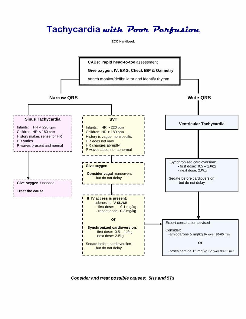
Tachycardia with Poor Perfusion
ECC Handbook
CABs: rapid head-to-toe assessment
Give oxygen, IV, EKG, Check B/P & Oximetry
Attach monitor/defibrillator and identify rhythm
Narrow QRS Wide QRS
Sinus Tachycardia
Infants: HR < 220 bpm
Children: HR < 180 bpm
History makes sense for HR
HR varies
P waves present and normal
SVT Infants: HR > 220 bpm
Children: HR > 180 bpm
History is vague, nonspecific
HR does not vary
HR changes abruptly
P waves absent or abnormal
Give oxygen if needed
Treat the cause
Give oxygen Consider vagal maneuvers
but do not delay
If IV access is present:
adenosine IV SLAM!
- first dose: 0.1 mg/kg - repeat dose: 0.2 mg/kg
or
Synchronized cardioversion:
- first dose: 0.5 – 1J/kg - next dose: 2J/kg
Sedate before cardioversion
but do not delay
Expert consultation advised
Consider: -amiodarone 5 mg/kg IV over 30-60 min
or
-procainamide 15 mg/kg IV over 30-60 min
Consider and treat possible causes: 5Hs and 5Ts

Pulseless Arrest – VF and Pulseless VT
ECC Handbook
CABs: Give CPR Give oxygen as soon as available
Attach monitor /defibrillator
Check rhythm: VF/ VT Check pulse: none
Resume CPR until defibrillator is charged
Give 1 shock at 2 J/kg Resume CPR immediately Give 5 cycles of CPR & IV/IO Access
Check rhythm: VF/ VT Check pulse in PVT
Resume CPR until defibrillator is charged
Give 1 shock at 4 J/kg Resume CPR immediately Give epinephrine:
IV/ IO: 0.01 mg/kg of 1:10 000 (0.1 mL/kg) ET: 0.1 mg/kg of 1:1000 (0.1 mL/kg)
Repeat : every 3-5 min
Give 5 cycles of CPR
Check rhythm: VF/ VT Check pulse in PVT
Resume CPR until defibrillator is charged
Give 1 shock at 4 J/kg Resume CPR immediately Consider:
-amiodarone 5 mg/kg IV
or - lidocaine 1 mg/kg IV
or -magnesium 25-50 mg/kg IV/IO if Torsade
Give 5 cycles of CPR
Consider and treat possible causes: 5Hs and 5Ts
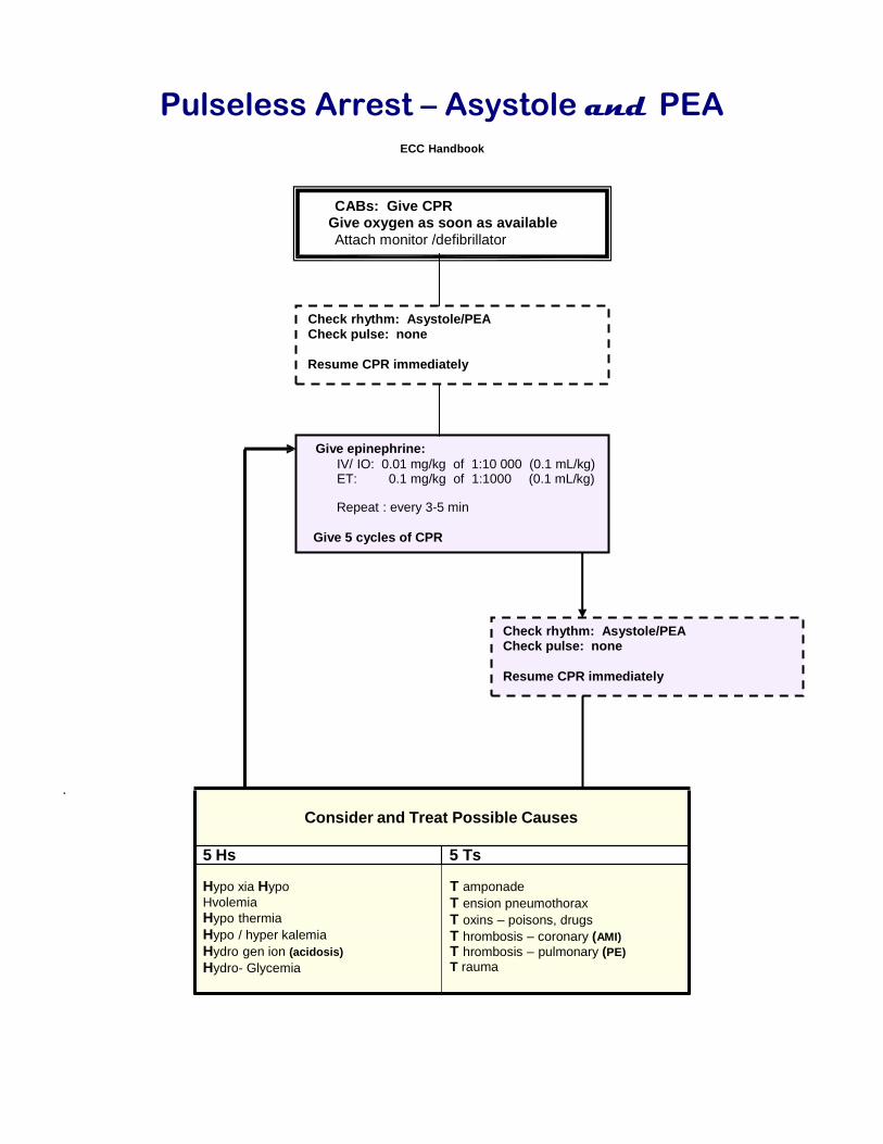
Pulseless Arrest – Asystole and PEA
ECC Handbook
CABs: Give CPR Give oxygen as soon as available
Attach monitor /defibrillator
Check rhythm: Asystole/PEA Check pulse: none
Resume CPR immediately
Give epinephrine:
IV/ IO: 0.01 mg/kg of 1:10 000 (0.1 mL/kg) ET: 0.1 mg/kg of 1:1000 (0.1 mL/kg)
Repeat : every 3-5 min
Give 5 cycles of CPR
Check rhythm: Asystole/PEA Check pulse: none
Resume CPR immediately
.
Consider and Treat Possible Causes
5 Hs 5 Ts
Hypo xia Hypo
Hvolemia
Hypo thermia
Hypo / hyper kalemia
Hydro gen ion (acidosis)
Hydro- Glycemia
T amponade
T ension pneumothorax
T oxins – poisons, drugs
T hrombosis – coronary (AMI)
T hrombosis – pulmonary (PE)
T rauma