pages 1-88 - us environmental protection agency
TRANSCRIPT
Acute Exposure Guideline Levelsfor Selected Airborne Chemicals
Volume 3
Subcommittee on Acute Exposure Guideline Levels
Committee on Toxicology
Board on Environmental Studies and Toxicology
Division on Earth and Life Studies
THE NATIONAL ACADEMIES PRESS 500 Fifth Street, N.W. Washington, D.C. 20001
NOTICE: The project that is the subject of this report was approved by the GoverningBoard of the National Research Council, whose members are drawn from the councils of theNational Academy of Sciences, the National Academy of Engineering, and the Institute ofMedicine. The members of the committee responsible for the report were chosen for theirspecial competences and with regard for appropriate balance.
This project was supported by Contract Nos. DAMD17-89-C-9086 and DAMD17-99-C-9049 between the National Academy of Sciences and the U.S. Army. Any opinions,findings, conclusions, or recommendations expressed in this publication are those of theauthor(s) and do not necessarily reflect the view of the organizations or agencies thatprovided support for this project.
International Standard Book Number 0-309-08883-6 (Book)International Standard Book Number 0-309-51590-4 (PDF)
Additional copies of this report are available from:
The National Academies Press500 Fifth Street, N.W.Box 285Washington, DC 20055
800-624-6242202-334-3313 (in the Washington metropolitan area)http://www.nap.edu
Copyright 2003 by the National Academy of Sciences. All rights reserved.
Printed in the United States of America
The National Academy of Sciences is a private, nonprofit, self-perpetuating society ofdistinguished scholars engaged in scientific and engineering research, dedicated to thefurtherance of science and technology and to their use for the general welfare. Upon theauthority of the charter granted to it by the Congress in 1863, the Academy has a mandatethat requires it to advise the federal government on scientific and technical matters. Dr.Bruce M. Alberts is president of the National Academy of Sciences.
The National Academy of Engineering was established in 1964, under the charter of theNational Academy of Sciences, as a parallel organization of outstanding engineers. It isautonomous in its administration and in the selection of its members, sharing with theNational Academy of Sciences the responsibility for advising the federal government. TheNational Academy of Engineering also sponsors engineering programs aimed at meetingnational needs, encourages education and research, and recognizes the superiorachievements of engineers. Dr. Wm. A. Wulf is president of the National Academy ofEngineering.
The Institute of Medicine was established in 1970 by the National Academy of Sciencesto secure the services of eminent members of appropriate professions in the examination ofpolicy matters pertaining to the health of the public. The Institute acts under theresponsibility given to the National Academy of Sciences by its congressional charter to bean adviser to the federal government and, upon its own initiative, to identify issues ofmedical care, research, and education. Dr. Harvey V. Fineberg is president of the Instituteof Medicine.
The National Research Council was organized by the National Academy of Sciences in1916 to associate the broad community of science and technology with the Academy’spurposes of furthering knowledge and advising the federal government. Functioning inaccordance with general policies determined by the Academy, the Council has become theprincipal operating agency of both the National Academy of Sciences and the NationalAcademy of Engineering in providing services to the government, the public, and thescientific and engineering communities. The Council is administered jointly by bothAcademies and the Institute of Medicine. Dr. Bruce M. Alberts and Dr. Wm. A. Wulf arechair and vice chair, respectively, of the National Research Council
www.national-academies.org
.

v
SUBCOMMITTEE ON ACUTE EXPOSURE GUIDELINE LEVELS
Members
DANIEL KREWSKI (Chair), University of Ottawa, Ottawa, Ontario, CanadaEDWARD C. BISHOP, Parsons Corporation, Fairfax, VAJAMES V. BRUCKNER, University of Georgia, AthensJOHN DOULL, University of Kansas Medical Center, Kansas CityKANNAN KRISHNAN, University of Montreal, CanadaSTEPHEN U. LESTER, Center for Health, Environment and Justice, Falls Church,
VAFRANZ OESCH, University of Mainz, Mainz, GermanyRICHARD B. SCHLESINGER, Pace University, Pleasantville, NYCALVIN C. WILLHITE, Department of Toxic Substances, State of California,
BerkeleyFREDERIK A. DE WOLFF, Leiden University, Leiden, Netherlands
Staff
KULBIR S. BAKSHI, Program Director KELLY CLARK, EditorAIDA C. NEEL, Senior Project Assistant
vi
COMMITTEE ON TOXICOLOGY
Members
BAILUS WALKER, JR. (Chair), Howard University Medical Center andAmerican Public Health Association, Washington, D.C.
MELVIN E. ANDERSEN, Chemical Industry Institute of Toxicology, Centers forHealth Research, Research Triangle Park, NC
EDWARD C. BISHOP, Parsons Corporation, Fairfax, VAGARY P. CARLSON, Purdue University, West Lafayette, INJANICE E. CHAMBERS, Mississippi State University, Mississippi StateLEONARD CHIAZZE, JR., Georgetown University, Washington, D.C.JUDITH A. GRAHAM, American Chemistry Council, Arlington, VASIDNEY GREEN, Howard University, Washington, D.C.MERYL KAROL, University of Pittsburgh, Pittsburgh, PASTEPHEN U. LESTER, Center for Health Environment and Justice, Falls Church,
VADAVID H. MOORE, Battelle Memorial Institute, Bel Air, MDCALVIN C. WILLHITE, Department of Toxic Substances, State of California,
Berkeley
Staff
KULBIR S. BAKSHI, Program Director SUSAN N.J. MARTEL, Senior Staff OfficerELLEN K. MANTUS, Senior Staff OfficerKELLY CLARK, Assistant EditorAIDA C. NEEL, Senior Project AssistantTAMARA DAWSON, Project Assistant

Acute Exposure Guideline Levelsfor Selected Airborne Chemicals
Volume 3

1
Introduction
This report is the third volume in the series Acute Exposure GuidelineLevels for Selected Airborne Chemicals.
In the Bhopal disaster of 1984, approximately 2,000 residents living neara chemical plant were killed and 20,000 more suffered irreversible damageto their eyes and lungs following accidental release of methyl isocyanate.The toll was particularly high because the community had little idea whatchemicals were being used at the plant, how dangerous they might be, andwhat steps to take in case of emergency. This tragedy served to focusinternational attention on the need for governments to identify hazardoussubstances and to assist local communities in planning how to deal withemergency exposures.
In the United States, the Superfund Amendments and ReauthorizationAct (SARA) of 1986 required that the U.S. Environmental ProtectionAgency (EPA) identify extremely hazardous substances (EHSs) and, incooperation with the Federal Emergency Management Agency and theDepartment of Transportation, assist Local Emergency Planning Commit-tees (LEPCs) by providing guidance for conducting health-hazard assess-ments for the development of emergency-response plans for sites whereEHSs are produced, stored, transported, or used. SARA also required thatthe Agency for Toxic Substances and Disease Registry (ATSDR) determinewhether chemical substances identified at hazardous waste sites or in theenvironment present a public-health concern.
2 ACUTE EXPOSU RE GUIDELINE LEVELS FOR SELECTED A IRBORN E CHEMICALS
1NAC is composed of members from EPA, DOD, many other federal and state
agencies, industry, academia, and other organizations. The roster of NAC is shown
on page 8.
As a first step in assisting the LEPCs, EPA identified approximately400 EHSs largely on the basis of their “immediately dangerous to life andhealth” (IDLH) values developed by the National Institute for OccupationalSafety and Health (NIOSH) in experimental animals. Although severalpublic and private groups, such as the Occupational Safety and HealthAdministration (OSHA) and the American Conference of GovernmentalIndustrial Hygienists (ACGIH), have established exposure limits for somesubstances and some exposures (e.g., workplace or ambient air quality),these limits are not easily or directly translated into emergency exposurelimits for exposures at high levels but of short duration, usually less than1 h, and only once in a lifetime for the general population, which includesinfants, children, the elderly, and persons with diseases, such as asthma,heart disease, or lung disease.
The National Research Council (NRC) Committee on Toxicology(COT) has published many reports on emergency exposure guidance levelsand spacecraft maximum allowable concentrations for chemicals used bythe Department of Defense (DOD) and the National Aeronautics and SpaceAdministration (NASA) (NRC 1968, 1972, 1984a,b,c,d, 1985a,b, 1986a,b,1987, 1988, 1994, 1996a,b, 2000). COT has also published guidelines fordeveloping emergency exposure guidance levels for military personnel andfor astronauts (NRC 1986b, 1992). Because of COT’s experience in rec-ommending emergency exposure levels for short-term exposures, in 1991EPA and ATSDR requested that COT develop criteria and methods fordeveloping emergency exposure levels for EHSs for the general population.In response to that request, the NRC assigned this project to the COT Sub-committee on Guidelines for Developing Community Emergency ExposureLevels for Hazardous Substances. The report of that subcommittee, Guide-lines for Developing Community Emergency Exposure Levels for Hazard-ous Substances (NRC 1993), provides step-by-step guidance for settingemergency exposure levels for EHSs. Guidance is given on what data areneeded, what data are available, how to evaluate the data, and how to pres-ent the results.
In November1995, the National Advisory Committee for Acute Expo-sure Guideline Levels for Hazardous Substances (NAC)1 was establishedto identify, review, and interpret relevant toxicologic and other scientificdata and to develop acute exposure guideline levels (AEGLs) for high-

INTRODUCTION 3
priority, acutely toxic chemicals. The NRC’s previous name for acute expo-sure levelsCcommunity emergency exposure levels (CEELs)Cwas replacedby the term AEGLs to reflect the broad application of these values to plan-ning, response, and prevention in the community, the workplace, transpor-tation, the military, and the remediation of Superfund sites.
Three levels—AEGL-1, AEGL-2, and AEGL-3—are developed for eachof five exposure periods (10 min, 30 min, 1 h, 4 h, and 8 h) and are distin-guished by varying degrees of severity of toxic effects.
The three AEGLs are defined as follows:
AEGL-1 is the airborne concentration (expressed as ppm [parts permillion] or mg/m3 [milligrams per cubic meter]) of a substance abovewhich it is predicted that the general population, including susceptibleindividuals, could experience notable discomfort, irritation, or certainasymptomatic nonsensory effects. However, the effects are not dis-abling and are transient and reversible upon cessation of exposure.
AEGL-2 is the airborne concentration (expressed as ppm or mg/m3) ofa substance above which it is predicted that the general population,including susceptible individuals, could experience irreversible or otherserious, long-lasting adverse health effects or an impaired ability toescape.
AEGL-3 is the airborne concentration (expressed as ppm or mg/m3) ofa substance above which it is predicted that the general population,including susceptible individuals, could experience life-threateninghealth effects or death.
Airborne concentrations below AEGL-1 represent exposure levels thatcan produce mild and progressively increasing but transient andnondisabling odor, taste, and sensory irritation or certain asymptomatic,nonsensory adverse effects. With increasing airborne concentrations aboveeach AEGL, there is a progressive increase in the likelihood of occurrenceand the severity of effects described for each corresponding AEGL. Al-though the AEGL values represent threshold levels for the general public,including susceptible subpopulations, such as infants, children, the elderly,persons with asthma, and those with other illnesses, it is recognized thatindividuals, subject to unique or idiosyncratic responses, could experiencethe effects described at concentrations below the corresponding AEGL.
4 ACUTE EXPOSU RE GUIDELINE LEVELS FOR SELECTED A IRBORN E CHEMICALS
SUMMARY OF REPORT ON GUIDELINES FOR DEVELOPING AEGLS
As described in the Guidelines for Developing Community EmergencyExposure Levels for Hazardous Substances (NRC 1993) and the NACguidelines report Standing Operating Procedures on Acute ExposureGuideline Levels for Hazardous Substances(NRC 2001), the first step inestablishing AEGLs for a chemical is to collect and review all relevantpublished and unpublished information available on a chemical. Varioustypes of evidence are assessed in establishing AEGL values for a chemical.These include information from (1) chemical-physical characterizations, (2)structure-activity relationships, (3) in vitro toxicity studies, (4) animaltoxicity studies, (5) controlled human studies, (6) observations of humansinvolved in chemical accidents, and (7) epidemiologic studies. Toxicitydata from human studies are most applicable and are used when availablein preference to data from animal studies and in vitro studies. Toxicity datafrom inhalation exposures are most useful for setting AEGLs for airbornechemicals because inhalation is the most likely route of exposure and be-cause extrapolation of data from other routes would lead to additionaluncertainty in the AEGL estimate.
For most chemicals, actual human toxicity data are not available orcritical information on exposure is lacking, so toxicity data from studiesconducted in laboratory animals are extrapolated to estimate the potentialtoxicity in humans. Such extrapolation requires experienced scientificjudgment. The toxicity data from animal species most representative ofhumans in terms of pharmacodynamic and pharmacokinetic properties areused for determining AEGLs. If data are not available on the species thatbest represents humans, the data from the most sensitive animal species areused to set AEGLs. Uncertainty factors are commonly used when animaldata are used to estimate risk levels for humans. The magnitude of uncer-tainty factors depends on the quality of the animal data used to determinethe no-observed-adverse-effect level (NOAEL) and the mode of action ofthe substance in question. When available, pharmacokinetic data on tissuedoses are considered for interspecies extrapolation.
For substances that affect several organ systems or have multiple ef-fects, all end points—including reproductive (in both sexes), develop-men-tal, neurotoxic, respiratory, and other organ-related effects—are evaluated,the most important or most sensitive effect receiving the greatest attention.For carcinogenic chemicals, excess carcinogenic risk is estimated, and theAEGLs corresponding to carcinogenic risks of 1 in 10,000 (1 × 10⁻⁴), 1 in

INTRODUCTION 5
100,000 (1 × 10⁻⁵), and 1 in 1,000,000 (1 × 10⁻⁶) exposed persons are esti-mated.
REVIEW OF AEGL REPORTS
As NAC began developing chemical-specific AEGL reports, EPA andDOD asked the NRC to review independently the NAC reports for theirscientific validity, completeness, and consistency with the NRC guidelinereports (NRC 1993; NRC in press). The NRC assigned this project to theCOT Subcommittee on Acute Exposure Guideline Levels. The subcommit-tee has expertise in toxicology, epidemiology, pharmacology, medicine,industrial hygiene, biostatistics, risk assessment, and risk communication.
The AEGL draft reports are initially prepared by ad hoc AEGL Devel-opment Teams consisting of a chemical manager, two chemical reviewers,and a staff scientist of the NAC contractor—Oak Ridge National Labora-tory. The draft documents are then reviewed by NAC and elevated from“draft” to “proposed” status. After the AEGL documents are approved byNAC, they are published in the Federal Register for public comment. Thereports are then revised by NAC in response to the public comments, ele-vated from “proposed” to “interim” status, and sent to the NRC Subcom-mittee on Acute Exposure Guideline Levels for final evaluation.
The NRC subcommittee’s review of the AEGL reports prepared byNAC and its contractors involves oral and written presentations to thesubcommittee by the authors of the reports. The NRC subcommittee pro-vides advice and recommendations for revisions to ensure scientific validityand consistency with the NRC guideline reports (NRC 1993, 2001). Therevised reports are presented at subsequent meetings until the subcommitteeis satisfied with the reviews.
Because of the enormous amount of data presented in the AEGL re-ports, the NRC subcommittee cannot verify all the data used by NAC. TheNRC subcommittee relies on NAC for the accuracy and completeness ofthe toxicity data cited in the AEGLs reports.
This report is the third volume in the series Acute Exposure GuidelineLevels for Selected Airborne Chemicals. AEGL documents for nerveagents (GA, GB, GD, GF, and VX), sulfur mustard, diborane, and methylisocyanate are published as an appendix to this report. The subcommitteeconcludes that the AEGLs developed in those documents are scientificallyvalid conclusions based on the data reviewed by NAC and are consistent
6 ACUTE EXPOSU RE GUIDELINE LEVELS FOR SELECTED A IRBORN E CHEMICALS
with the NRC guideline reports. AEGL reports for additional chemicalswill be presented in subsequent volumes.
REFERENCES
NRC (National Research Council). 1968. Atmospheric Contaminants in Space-
craft. Washington, DC: National Academy of Sciences.
NRC (National Research Council). 1972. Atmospheric Contaminants in Manned
Spacecraft. Washington, DC: National Academy of Sciences.
NRC (National Research Council). 1984a. Emergency and Continuous Exposure
Limits for Selected Airborne Contaminants, Vol. 1. Washington, DC: Na-
tional Academy Press.
NRC (National Research Council). 1984b. Emergency and Continuous Exposure
Limits for Selected Airborne Contaminants, Vol. 2. Washington, DC: Na-
tional Academy Press.
NRC (National Research Council). 1984c. Emergency and Continuous Exposure
Limits for Selected Airborne Contaminants, Vol. 3. Washington, DC: Na-
tional Academy Press.
NRC (National Research Council). 1984d. Toxicity Testing: Strategies to Deter-
mine Needs and Priorities. Washington, DC: National Academy Press.
NRC (National Research Council). 1985a. Emergency and Continuous Exposure
Guidance Levels for Selected Airborne Contaminants, Vol. 4. Washington,
DC: National Academy Press.
NRC (National Research Council). 1985b. Emergency and Continuous Exposure
Guidance Levels for Selected Airborne Contaminants, Vol. 5. Washington,
DC: National Academy Press.
NRC (National Research Council). 1986a. Emergency and Continuous Exposure
Guidance Levels for Selected Airborne Contaminants, Vol. 6. Washington,
DC: National Academy Press.
NRC (National Research Council). 1986b. Criteria and Methods for Preparing
Emergency Exposure Guidance Level (EEGL), Short-Term Public Emergency
Guidance Level (SPEGL), and Continuous Exposure Guidance level (CEGL)
Documents. W ashington, DC: National Academy Press.
NRC (National Research Council). 1987. Emergency and Continuous Exposure
Guidance Levels for Selected Airborne Contaminants, Vol. 7. Washington,
DC: National Academy Press.
NRC (National Research Council). 1988. Emergency and Continuous Exposure
Guidance Levels for Selected Airborne Contaminants, Vol. 8. Washington,
DC: National Academy Press.
NRC (National Research Council). 1992. Guidelines for Developing Spacecraft
Maximum Allowable Concentrations for Space Station Contaminants. Wash-
ington, DC: National Academy Press.
INTRODUCTION 7
NRC (National Research Council). 1993. Guidelines for Developing Community
Emergency Exposure Levels for Hazardous Substances. Washington, DC:
National Academy Press.
NRC (National Research Council). 1994. Spacecraft Maximum Allowable Con-
centrations for Selected Airborne Contaminants, Vol. 1. Washington, DC:
National Academy Press.
NRC (National Research Council). 1996a. Spacecraft Maximum Allowable
Concentrations for Selected Airborne Contaminants, Vol. 2. Washington, DC:
National Academy Press.
NRC (National Research Council). 1996b. Spacecraft M aximum Allowable
Concentrations for Selected Airborne Contaminants, Vol. 3. Washington, DC:
National Academy Press.
NRC (National Research Council). 2000. Spacecraft Maximum Allowable Con-
centrations for Selected Airborne Contaminants, Vol. 4. W ashington, DC:
National Academy Press.
NRC (National Research Coiuncil) 2001. Acute Exposure Guideline Levels for
Selected Airborne Chemicals. Washington, DC: National Academy Press.
NRC (National Research Council). 2001. Standing Operating Procedures for
Developing Acute Exposure Guideline Levels for Airborne Chemicals. Wash-
ington, DC: National Academy Press.
8
Roster of theNational Advisory Committee forAcute Exposure Guideline Levels
for Hazardous Substances
Committee Members
George RuschChair, NAC/AEGL CommitteeDepartment of Toxicology andRisk AssessmentHoneywell, Inc.Morristown, NJ
Steven BarbeeArch Chemicals, Inc.Norwalk, CT
Ernest FalkeChair, SOP WorkgroupU.S. Environmental ProtectionAgencyWashington, DC
Lynn BeasleyU.S. Environmental ProtectionAgencyWashington, DC
George AlexeeffOffice of Environmental HealthHazard AssessmentCalifornia EPAOakland, CA
David BelluckOffice of Environmental ServicesMinnesota Department ofTransportationOakdale, MN
ROSTER O F THE NATION AL ADV ISOR Y COMMITTEE FOR AEGLS 9
Robert BensonU.S. Environmental ProtectionAgencyRegion VIIIDenver, CO
Jonathan BorakYale UniversityNew Haven, CT
William BressVermont Department of HealthBurlington, VT
George CushmacOffice of Hazardous MaterialsSafetyU.S. Department of TransporationWashington, DC
Albert G. DietzU.S. Department of EnergyGermantown, MD
Larry GephartExxon Mobil Biomedical SciencesAnnandale, NJ
John P. HinzU.S. Air ForceBrooks Air Force Base, TX
James HollerAgency for Toxic Substances andDisease RegistryAtlanta, GA
Thomas C. HornshawOffice of Chemical SafetyIllinois Environmental ProtectionAgencySpringfield, IL
Nancy K. KimDivision of Environmental HealthAssessmentNew York State Department ofHealthTroy, NY
Loren KollerLoren Koller & AssociatesCorvallis, OR
Glenn LeachU.S. Army Center for HealthPromotion and PreventiveMedicineAberdeen Proving Grounds, MD
Mark A. McClanahanCenters for Disease Control andPrevention National Center forEnvironmental HealthDoraville, GA
John MorawetzInternational Chemical WorkersUnionCincinnati, OH
10 ACUTE EXPOSU RE GUIDELINE LEVELS FOR SELECTED A IRBORN E CHEMICALS
Richard W. NiemeierNational Institute for OccupationalSafety and HealthCincinnati, OH
Marinelle PaytonDepartment of Public HealthJackson State UniversityJackson, MS
Zarena PostTexas Natural ResourceConservation CommissionAustin, TX
George RodgersDepartment of PediatricsDivision of Critical CareUniversity of LouisvilleLouisville, KY
Robert SnyderEnvironmental and OccupationalHealth Sciences InstitutePiscataway, NJ
Thomas J. SobotkaU.S. Food and DrugAdministrationLaurel, MD
Kenneth StillNaval Health Research CenterWright-Patterson AFB, OH
Richard ThomasInternational Center forEnvironmental TechnologyMcLean, VA
Oak Ridge National Laboratory Staff
Po-Yung LuOak Ridge National LaboratoryOak Ridge, TN
Carol S. ForsythOak Ridge National LaboratoryOak Ridge, TN
Dennis OpreskoOak Ridge National LaboratoryOak Ridge, TN
Claudia TroxelOak Ridge National LaboratoryOak Ridge, TN
Robert YoungOak Ridge National LaboratoryOak Ridge, TN
Annetta WatsonOak Ridge National LaboratoryOak Ridge, TN
ROSTER O F THE NATION AL ADVISOR Y COMMITTEE FOR AEGLS 11
National Advisory Committee Staff
Roger L. GarrettDirector, AEGL ProgramU.S. Environmental ProtectionAgencyWashington, DC(Deceased, March 31, 2003)
Paul S. TobinDesignated Federal Officer, AEGLProgramU.S. Environmental ProtectionAgencyWashington, DC
Letty TahanSenior ScientistU.S. Environmental ProtectionAgencyWashington, DC


Appendixes
NERVE AGENTS GA, GB, GD, GF, AND VX 39
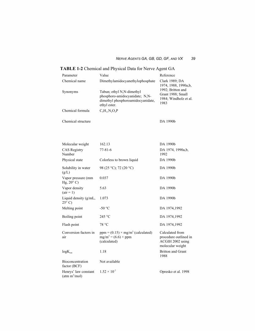
TABLE 1-2 Chemical and Physical Data for Nerve Agent GAParameter Value ReferenceChemical name Dimethylamidocyanethylophosphate Clark 1989; DA
1974, 1988, 1990a,b,1992; Britton andGrant 1988; Small1984; Windholz et al.1983
Synonyms Tabun; ethyl N,N-dimethylphosphoro-amidocyanidate; N,N-dimethyl phosphoroamidocyanidate,ethyl ester.
Chemical formula C5H11N2O2P
Chemical structure DA 1990b
Molecular weight 162.13 DA 1990bCAS RegistryNumber
77-81-6 DA 1974, 1990a,b,1992
Physical state Colorless to brown liquid DA 1990b
Solubility in water(g/L)
98 (25 °C); 72 (20 °C) DA 1990b
Vapor pressure (mmHg, 20° C)
0.037 DA 1990b
Vapor density (air = 1)
5.63 DA 1990b
Liquid density (g/mL,25° C)
1.073 DA 1990b
Melting point -50 °C DA 1974,1992
Boiling point 245 °C DA 1974,1992
Flash point 78 °C DA 1974,1992
Conversion factors inair
ppm = (0.15) × mg/m3 (calculated)mg/m3 = (6.6) × ppm (calculated)
Calculated fromprocedure outlined inACGIH 2002 usingmolecular weight
logKow 1.18 Britton and Grant1988
Bioconcentrationfactor (BCF)
Not available
Henrys’ law constant (atm m3/mol)
1.52 × 10⁻⁷ Opresko et al. 1998
40 ACUTE EXPOSURE GUIDELINE LEVELS FOR SELECTED AIRBORNE CHEMICALS
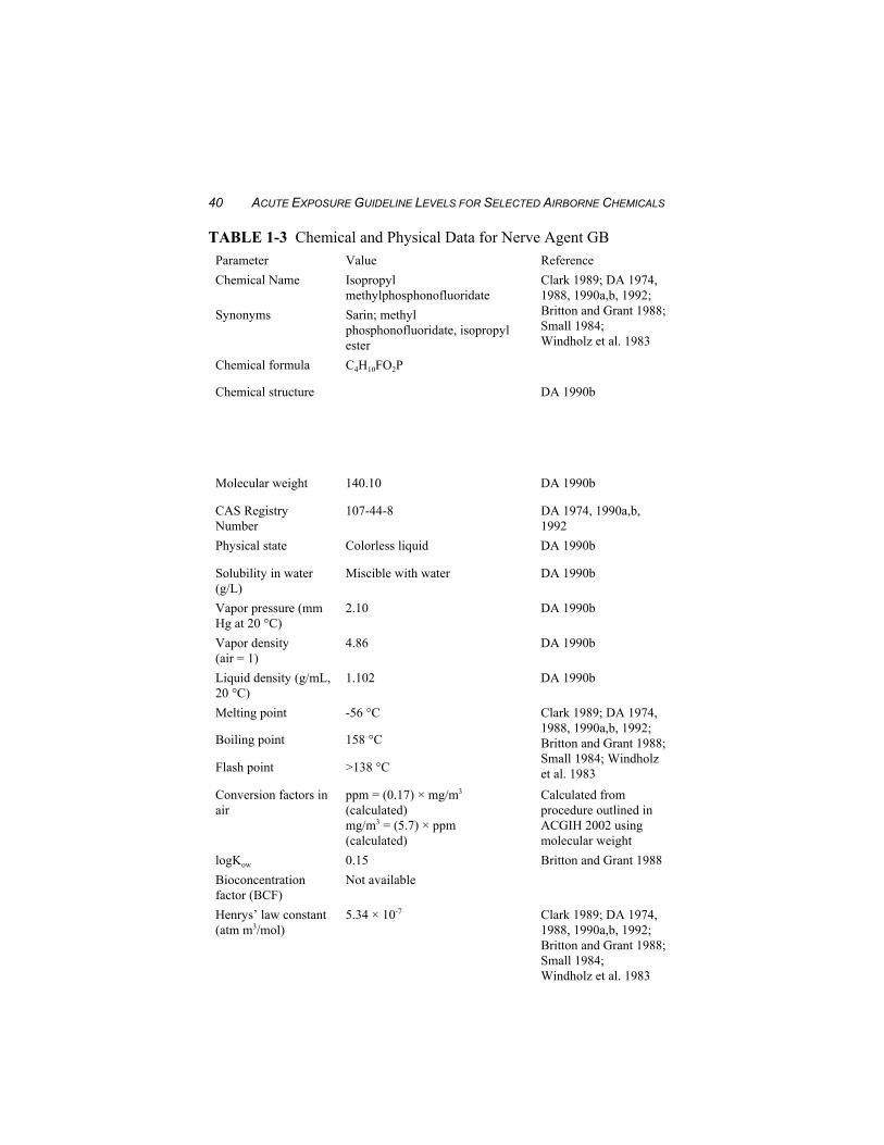
TABLE 1-3 Chemical and Physical Data for Nerve Agent GBParameter Value ReferenceChemical Name Isopropyl
methylphosphonofluoridateClark 1989; DA 1974,1988, 1990a,b, 1992;Britton and Grant 1988;Small 1984;Windholz et al. 1983
Synonyms Sarin; methylphosphonofluoridate, isopropylester
Chemical formula C4H10FO2P
Chemical structure DA 1990b
Molecular weight 140.10 DA 1990b
CAS RegistryNumber
107-44-8 DA 1974, 1990a,b,1992
Physical state Colorless liquid DA 1990b
Solubility in water(g/L)
Miscible with water DA 1990b
Vapor pressure (mmHg at 20 °C)
2.10 DA 1990b
Vapor density(air = 1)
4.86 DA 1990b
Liquid density (g/mL,20 °C)
1.102 DA 1990b
Melting point -56 °C Clark 1989; DA 1974,1988, 1990a,b, 1992;Britton and Grant 1988;Small 1984; Windholzet al. 1983
Boiling point 158 °C
Flash point >138 °C
Conversion factors inair
ppm = (0.17) × mg/m3
(calculated)mg/m3 = (5.7) × ppm (calculated)
Calculated fromprocedure outlined inACGIH 2002 usingmolecular weight
logKow 0.15 Britton and Grant 1988Bioconcentrationfactor (BCF)
Not available
Henrys’ law constant (atm m3/mol)
5.34 × 10⁻⁷ Clark 1989; DA 1974,1988, 1990a,b, 1992;Britton and Grant 1988;Small 1984;Windholz et al. 1983
NERVE AGENTS GA, GB, GD, GF, AND VX 41
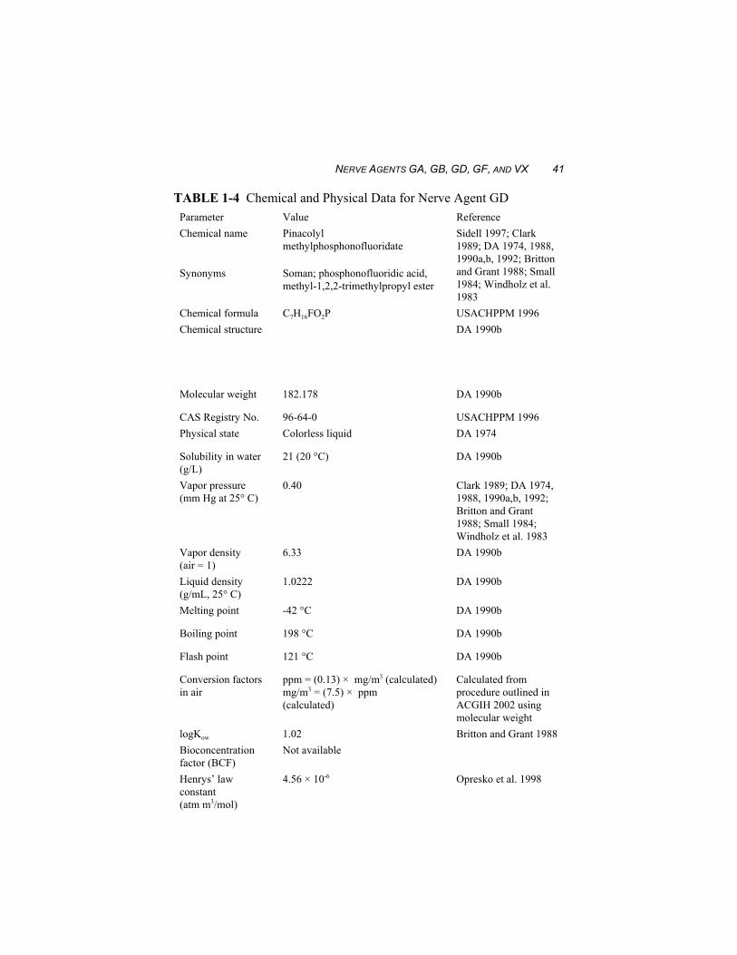
TABLE 1-4 Chemical and Physical Data for Nerve Agent GDParameter Value ReferenceChemical name Pinacolyl
methylphosphonofluoridateSidell 1997; Clark1989; DA 1974, 1988,1990a,b, 1992; Brittonand Grant 1988; Small1984; Windholz et al.1983
Synonyms Soman; phosphonofluoridic acid,methyl-1,2,2-trimethylpropyl ester
Chemical formula C7H16FO2P USACHPPM 1996Chemical structure DA 1990b
Molecular weight 182.178 DA 1990b
CAS Registry No. 96-64-0 USACHPPM 1996Physical state Colorless liquid DA 1974
Solubility in water(g/L)
21 (20 °C) DA 1990b
Vapor pressure (mm Hg at 25° C)
0.40 Clark 1989; DA 1974,1988, 1990a,b, 1992;Britton and Grant1988; Small 1984;Windholz et al. 1983
Vapor density (air = 1)
6.33 DA 1990b
Liquid density (g/mL, 25° C)
1.0222 DA 1990b
Melting point -42 °C DA 1990b
Boiling point 198 °C DA 1990b
Flash point 121 °C DA 1990b
Conversion factorsin air
ppm = (0.13) × mg/m3 (calculated)mg/m3 = (7.5) × ppm (calculated)
Calculated fromprocedure outlined inACGIH 2002 usingmolecular weight
logKow 1.02 Britton and Grant 1988Bioconcentrationfactor (BCF)
Not available
Henrys’ lawconstant (atm m3/mol)
4.56 × 10⁻⁶ Opresko et al. 1998
42 ACUTE EXPOSURE GUIDELINE LEVELS FOR SELECTED AIRBORNE CHEMICALS
TABLE 1-5 Chemical and Physical Data for Nerve Agent GFParameter Value ReferenceChemical name O-cyclohexyl-
methylfluorophosphonateDA 1990b
Synonyms Cyclohexylmethylphosphonofluoridate (CMPF)
Chemical formula C7H14FO2P DA 1990bChemical structure DA 1990b
Molecular weight 180.2 DA 1990bCAS RegistryNumber
329-99-7 DA 1990b
Physical state Liquid DA 1990b
Solubility in water 0.37% (20 °C); almost entirelyinsoluble in water
DA 1990b
Vapor pressure(mm Hg, 25° C)
0.044 DA 1990b
Vapor density (air= 1)
6.2 DA 1990b
Liquid Density(g/mL, 20° C)
1.1327 DA 1990b
Melting point -30 °C DA 1990b
Boiling point 239 °C DA 1990b
Flash point 94 °C DA 1990b
Conversion factorsin air
ppm = (0.14) × mg/m3 (calculated)mg/m3 = (7.4) × ppm (calculated)
Calculated fromprocedure outlined inACGIH 2002, usingmolecular weight
logKow Not availableBioconcentrationfactor (BCF)
Not available
Henrys’ lawconstant (atm m3/mol)
Not available
as a 2-10 min exposure estimate in the summary table. Thus, the LCt50 of35 mg"min/m3 assumes only short-term exposures of 2-10 min.

NERVE AGENTS GA, GB, GD, GF, AND VX 43
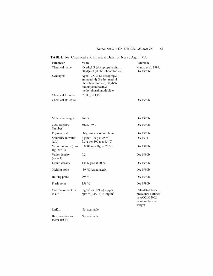
TABLE 1-6 Chemical and Physical Data for Nerve Agent VXParameter Value ReferenceChemical name O-ethyl-S-(diisopropylamino-
ethyl)methyl phosphonothiolateMunro et al. 1999;DA 1990b
Synonyms Agent VX; S-(2-diisopropyl-aminoethyl) O-ethyl methylphosphonothiolate; ethyl-S-dimethylaminoethylmethylphosphonothiolate
Chemical formula C11H 25 NO2PSChemical structure DA 1990b
Molecular weight 267.38 DA 1990b
CAS RegistryNumber
50782-69-9 DA 1990b
Physical state Oily, amber-colored liquid DA 1990bSolubility in water(g/L)
3 g per 100 g at 25 °C 7.5 g per 100 g at 15 °C
DA 1974
Vapor pressure (mmHg, 20° C)
0.0007 mm Hg at 20 °C DA 1990b
Vapor density (air = 1)
9.2 DA 1990b
Liquid density 1.006 g/cc at 20 °C DA 1990b
Melting point -39 °C (calculated) DA 1990b
Boiling point 298 °C DA 1990b
Flash point 159 °C DA 1990b
Conversion factorsin air
mg/m3 = (10.936) × ppm ppm = (0.0914) × mg/m3
Calculated fromprocedure outlinedin ACGIH 2002using molecularweight
logKow Not available
Bioconcentrationfactor (BCF)
Not available

44 ACUTE EXPOSURE GUIDELINE LEVELS FOR SELECTED AIRBORNE CHEMICALS
A subcommittee of the National Research Council’s Committee onToxicology (COT) has examined the Reutter and Wade (1994) analysis andrecommends that the proposed LCt50 estimates for agents GA, GB, GD, andGF for estimating vapor inhalation and percutaneous exposure effects inexposed military populations “should be lowered” in light of the need foradditional data characterizing vapor inhalation and percutaneous vaportoxicity. Furthermore, the subcommittee considered the estimates ofReutter and Wade (1994) inappropriate for civilian applications (NRC1997).
Agent VX
From animal data, Reutter and Wade (1994) estimated a LCt50 formilitary personnel of 15 mg"min/m3 for 2-10 min vapor exposures atmoderate temperatures (65-75 °F) for an individual with a respiratoryminute volume of 15 L. As in the case for agent GB, this LCt50 estimatewas calculated for 2-min exposure periods and then proposed as a 2-10 minexposure estimate. Thus, the LCt50 for VX at 15 mg"min/m3 assumes onlyshort-term exposures of 2-10 min.
The subcommittee recommends that the Reutter and Wade (1994)proposed LCt50 estimate of 15 mg"min/m3 for military personnel “should belowered” because of the low to moderate degree of confidence in theestimation, which considered effects from vapor inhalation andpercutaneous vapor exposures. Further, the subcommittee considered theestimates of Reutter and Wade (1994) inappropriate for civilian applications(NRC 1997).
Bide and Risk (2000) estimated the human 10-min LCt50 value for a VXaerosol based on lethality data for several animal species (see Section 3.1). The human LCt50 value was an estimated 7 mg"min/m3 for a 70 kg manbreathing 15 L/min for 10 min.
2.1.1. Case Reports
Agent GB
In 1994 and 1995, two incidents of chemical terrorism involving nerveagent GB (sarin) occurred in Japan; in both incidents, civilian populationswere deliberately exposed to lethal concentrations by followers of a cultoriginally local to Japan (Lillibridge 1995; Morita et al. 1995; Okumura et

NERVE AGENTS GA, GB, GD, GF, AND VX 45
al. 1996; Sidell 1996). Because of the state of emergency at the time ofrelease and the initial unknown nature of the source, exposures and dose-response could not be quantified.
The first incident occurred in June of 1994 in the central highland cityof Matsumoto, Japan, where seven people died shortly after exposure to anunknown vapor later determined to be agent GB (Morita et al. 1995)released into a residential area during the night. The Matsumoto incidentresulted in 56 hospital admissions as well as 253 cases in which the affectedindividuals sought medical consultation. Reports of “mild symptoms” werepresented by eight out of 53 rescue personnel and one attending physician(Morita et al. 1995). Prompt deaths (N = 3) and those who died beforearriving at the hospital (N = 4) appear to have been the result of respiratoryinsufficiency. At the time of the Morita et al. (1995) report, one patientremained “in a vegetative state because of anoxic encephalopathy”; a reporton the outcome of that case has not yet been found.
The second occurrence, widely known as the Tokyo Subway Incident,took place on March 20, 1995. The same terrorist group responsible for theMatsumoto incident employed sources of passive, evaporative release ofnerve agent GB in five individual subway cars serving three separatesubway lines during morning commuter rush hours (Lillibridge 1995;Okumura et al. 1996; Sidell 1996). Of the 5,510 persons known to havebeen given medical attention, there were eight prompt deaths; four moredied later (hours to days). The “later” group included individuals who hadinitially presented with “critical” respiratory effects requiring mechanicalventilation and intensive care (Lillibridge 1995). The 12 fatalities includedcommuters and subway transport employees, and death appeared to be theresult of respiratory insufficiency. On hospital day 28, an additional deathoccurred as a consequence of “severe hypoxic brain damage” sustainedduring the release incident (Okumura et al. 1996). This delayed fatality wasa previously healthy woman, 21 years of age, who presented withoutheartbeat or spontaneous respiration at the hospital but was revived withCPR and treated with agent antidotes. Plasma and RBC cholinesterasereturned to normal within a period of days, but the patient eventually suc-cumbed to hypoxic brain damage (Okumura et al. 1996).
Neuropathological examination of one individual who died 15 mo afterbeing severely exposed to agent GB during the Tokyo subway terroristattack indicated that the victim suffered marked nerve-fiber decrease in thesural nerve and moderate nerve-fiber loss in the sciatic nerve, with nochanges in the dorsal root ganglion, dorsal roots, or posterior column of thespinal cord (Himuro et al. 1998). The victim’s CNS showed severehypoxic-ischemic changes, which made it difficult to assess the specific
46 ACUTE EXPOSURE GUIDELINE LEVELS FOR SELECTED AIRBORNE CHEMICALS
effects of agent GB. Himuro et al. (1998) concluded that the observationswere consistent with the “dying back” of the peripheral nervous system andmight have been indicative of delayed neuropathy associated with inhibitionof neuropathy target esterase (NTE). Himuro et al. (1998) cite as additionalevidence of sarin-induced distal axonopathy an earlier study (Ishiyama1996) in which degeneration of intramuscular nerve fascicles withpreservation of the anterior horn cells was observed in a patient who died1 mo after the subway attack.
2.2. Nonlethal Toxicity
Exposure to acutely toxic concentrations of nerve agents can result inexcessive bronchial, salivary, ocular, and intestinal secretion, sweating,miosis, bronchospasm, intestinal hypermotility, bradycardia, musclefasciculations, twitching, weakness, paralysis, loss of consciousness,convulsions, and depression of the central respiratory drive (Dunn andSidell 1989). Minimal effects seen at very low exposure levels includemiosis and rhinorrhea. The effects of exposures to very low concentrationsof the nerve agents are evaluated in the literature, which includes clinicalcase reports as well as several studies using human volunteers. Key toacceptance of human subject data for use in the AEGL process is evidencethat subjects provided informed consent and that the studies were performedunder appropriate clinical supervision (NRC 2001). These criteria were metby the nonlethal studies summarized in Section 2.2.2.
A number of investigators consider both miosis and rhinorrhea to beearly signs of exposure to cholinesterase inhibitors (Sidell 1997;Mioduszewski et al. 2002b; H. van Helden, Pulmonary and CNSPharmacology Lab, TNO, the Netherlands, personal communtication; S.Tattersall, Biomedical Sciences Division, Porton Down, United Kingdom,personal communication). The presence of rhinorrhea can be indicative ofinhalation exposure and/or development of systemic effects, while miosisin the absence of other signs or symptoms is a local effect to the pupillarymuscles of the eye. In consequence, the presence of miosis is consideredan appropriately sensitive indicator of direct vapor exposure and has theadditional advantage of being readily recognized and quantifiable.
Recent nerve agent releases by terrorist groups have exposed civilianpopulations. Survivors of the incidents have been examined, and theresulting evaluations are summarized in Section 2.2.1.
NERVE AGENTS GA, GB, GD, GF, AND VX 47
2.2.1. Case Reports
Agent GB
Clinical case reports exist for the survivor population of the 1994 agentGB (sarin) release in Matsumoto, Japan, and the 1995 sarin release inTokyo; no estimates of exposure concentrations could be found in theliterature for either of these incidents. In the Matsumoto incident detailedabove (see Section 2.1.1), Morita and his colleagues (Morita et al. 1995)published the clinical and laboratory findings of 264 people who soughttreatment and the results of health examinations performed on 155Matsumoto residents at 3 weeks (wk) postexposure. During initialtreatment, severely poisoned individuals exhibited severe miosis,tachycardia followed by bradycardia, salivation, rhinorrhea, musclefasciculations, and abnormal epileptiform EEGs. Other reported acuteexposure signs and symptoms included headache, vision disturbances,fatigue, dizziness, nausea, dyspnea, ocular pain, and dysesthesia of theextremities. Clinical findings for the same group at the time of examinationincluded decreases in serum cholinesterase, erythrocyteacetylcholinesterase, and serum triglycerides as well as serum potassiumand chloride and increases in serum creatine kinase, leucocytes, and ketonesin urine. For a period of up to 30 d following the incident, some of theseverely exposed population exhibited slight continuous fever and someepileptiform EEG abnormalities (N = 2 out of nine “severely affectedpeople”). Nevertheless, follow-up examination revealed no persistent ab-normal physical findings in any individual; acetylcholinesterase activity inerythrocytes and serum cholinesterase returned to normal within 3 mo in theexamined population. Among some severe or moderately affected persons,subclinical miosis and some neuropathy were present 30 d after exposure.Morita et al. (1995) state that, in most people, “almost all symptoms of sarinexposure disappeared rapidly and left no sequelae.”
In the hours following the Tokyo subway release of agent GB, theemergency department of St. Luke’s International Hospital (located near theaffected subway stations) received 640 patients (Okumura et al. 1996).Additional details of the incident are provided above (see Section 2.1.1).Of the 640 admissions, 528 (82.5%) were diagnosed by Okumura and hiscolleagues (1996) as “mild” and exhibited “only eye signs or symptoms”such as miosis, eye pain, dim vision, and decreased visual acuity. Of theremaining 112 patients, one died in the emergency department, 107 were
48 ACUTE EXPOSURE GUIDELINE LEVELS FOR SELECTED AIRBORNE CHEMICALS
admitted as “moderate” cases (exhibiting “systemic signs and symptoms”such as weakness, fasciculations, convulsions, difficult breathing), and fourwere admitted as “severe” cases “requiring emergency respiratory support”such as intubation. Of the four severe cases, two patients experiencedcardiac arrest but were revived, treated with agent antidotes andanticonvulsants, and eventually recovered fully (discharged on hospital day3 and 5). Of the remaining two, both of whom required cardiopulmonaryresuscitation, one recovered after vigorous treatment and was discharged onday 6. The remaining severely affected patient originally presented with nopulse and died on hospital day 28. For the three severe cases discharged,RBC-cholinesterase remained below normal activity levels for 51-72 d.
In the early 1970s, three men (ages 27, 50, and 52 y) working atEdgewood Arsenal (now Aberdeen Proving Ground in Edgewood,Maryland) in a chemical agent area containing stored containers of agentGB (sarin) were brought to an emergency room after sudden onset ofrhinorrhea and respiratory discomfort approximately 20 min prior to arrivalat the emergency room (Sidell 1974). It was determined later that one ofthe agent GB (sarin) containers in the work area had developed a leak andthat the three individuals exhibiting signs had been working in the generalarea of the room where the leaking container was located. Examinationindicated the presence of “mild respiratory distress, marked miosis withslight eye pain, rhinorrhea, a moderate increase in salivation, and scatteredwheezes and rhonchi throughout all lung fields” (Sidell 1974). The menreceived no therapy but were observed for 6 h after emergency room arrivaland were asymptomatic upon discharge except for eye irritation and“decreased vision in dim light.” Blood cholinesterases were monitored andpupil diameter was recorded photographically for a period of 4 mofollowing exposure. Although 60-70% recovery of the ability to dark-adaptoccurred within 2 wk, complete recovery of the ability to dark-adaptrequired 2 mo. Sidell (1974) did not report any estimates of the GB agentconcentrations the men were exposed to.
Also in the early 1970s, a 52-y-old man in full protective gearemployed in cleaning an agent GB-contaminated area at Edgewood Arsenal(now Aberdeen Proving Ground in Edgewood, Maryland) experiencedbreathing difficulty and increased oral and nasal secretions (Sidell 1974).It was later determined that there was a crack in the man’s voicemitterdiaphragm through which exposure most likely had occurred. Upon arrivalat the emergency room 5-10 min after the first symptom, he was convulsingand cyanotic. Other evident signs included labored breathing, muscular

NERVE AGENTS GA, GB, GD, GF, AND VX 49
fasciculations, miosis, salivation, and rhinorrhea. He was treated aggres-sively with agent antidotes and provided assisted ventilation, and herecovered sufficiently to be able to walk through the ward by 9 hpostadmission. Red blood cell cholinesterase (RBC-ChE) was monitored,as were EKGs. “While ChE activity in his blood was undetectable,” theindividual was conscious and alert (Sidell 1997). By 18 h postadmission,miosis was still evident. On day 4 and thereafter, the patient wasasymptomatic; upon discharge 4 wk postexposure, he was “fullyambulatory and doing well.” A 4-mo-postexposure EKG “was entirelywithin normal limit” (Sidell 1974). Sidell (1974) did not report anyestimate of GB agent concentrations to which this individual was exposed.
In another incident of accidental exposure to GB vapors (0.09 mg/m3
for an undefined duration resulting from a faulty ventilation hood), two men(ages 46 and 53 y) exhibited significantly lowered RBC-ChE for 80-90 d(one showed depression to 19% of baseline activity, the other to 84% ofbaseline activity) and extreme miosis that persisted for 30-45 d (Rengstorff1985). These men exhibited no other signs or symptoms of nerve agentpoisoning and required no treatment with antidotes.
2.2.2. Acute Studies
Agent GB
Vapor Exposures
Fairley and Mumford (1948) exposed 16 male volunteers to GB at 0.3mg/m3 for 0.5 min. Nine of the test subjects reported that they could detectthe agent by smell; seven reported tightness of the chest, and 16 reportedrhinorrhea.
McKee and Woolcott (1949) evaluated the effects of low concentrationsof agent GB on 14 male volunteers. A single exposure to GB at 0.6 mg/m3
for 1 min or at 0.06 mg/m3 for 40 min resulted in miosis and slight tightnessof the chest (4/4 subjects exhibited those signs and symptoms in both the 1-min and 40-min tests; within 24 h, signs and symptoms resolved in subjectsreceiving 1-min exposures, although more than 48 h were required forresolution in subjects receiving 40-min exposures). Exposure of fiveindividuals to GB at 0.06 mg/m3 for 20 min/d resulted in miosis, but onlyafter the fourth day of exposure. When the subjects were exposed to GB at
50 ACUTE EXPOSURE GUIDELINE LEVELS FOR SELECTED AIRBORNE CHEMICALS
0.06 mg/m3 for 40 min/d, miosis occurred on the first or second day andadditional symptoms (headache, blurred vision, eye pain) appeared on thesecond, third, and fourth day of exposure.
In summarizing the toxicity studies conducted at Porton Down, UnitedKingdom, Mumford (1950) concluded that the threshold for ocular effectsis 1.5-5.0 mg"min/m3 (exposure times of 5-6 min) and that exposures to GBat 6-12 mg"min /m3 (exposure times of 5-8 min) would result in moderateto severe discomfort due to miosis and frontal headaches.
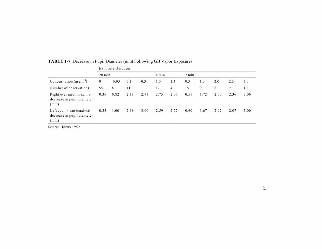
In a study reported by Johns (1952) and Harvey (1952), 128 adult malesvolunteered to be exposed to GB concentrations ranging from 0.05 mg/m3
to 3.0 mg/m3 for 2-20 min in a chamber. The corresponding Cts rangedfrom 1.0 mg"min/m3 to 6.0 mg"min/m3. The analytical methods used tomeasure the chamber concentrations of GB were not reported. Regressionanalysis of 150 observations, including 55 controls, indicated that the pointat which a 50% decrease in pupil diameter would be attained wasapproximately 4.1 mg"min/m3, with 90% confidence limits of about 2.7 and5.7 mg"min/m3 (Johns 1952). At the lowest test exposure level (0.05 mg/m3
for 20 min), there were mean maximum decreases in pupil diameter of 0.82mm (right eye) and 1.00 mm (left eye) (total of eight observations)compared with 0.36 mm (right eye) and 0.33 mm (left eye) in controls (55observations). Johns (1952) defines “mild miosis” as a “decrease of 1 to 2mm” in pupil diameter that usually disappears within 24 h. Although mildmiosis, as defined by the author, was observed in some subjects at thelowest Ct tested (Ct = 1.0 mg"min/m3), other subjects exhibited meanmaximal pupil decreases of <1 mm. This indicates that a likely responsethreshold was attained at this level of cumulative exposure. The results ofthe Johns (1952) study are presented in Table 1-7. It should be noted thatuntreated controls exhibited a pupil diameter decrease of $0.33 mm. Johns(1952) attributes this difference to observer bias and points out that there isstill a relative difference between the control group and the exposuregroups.
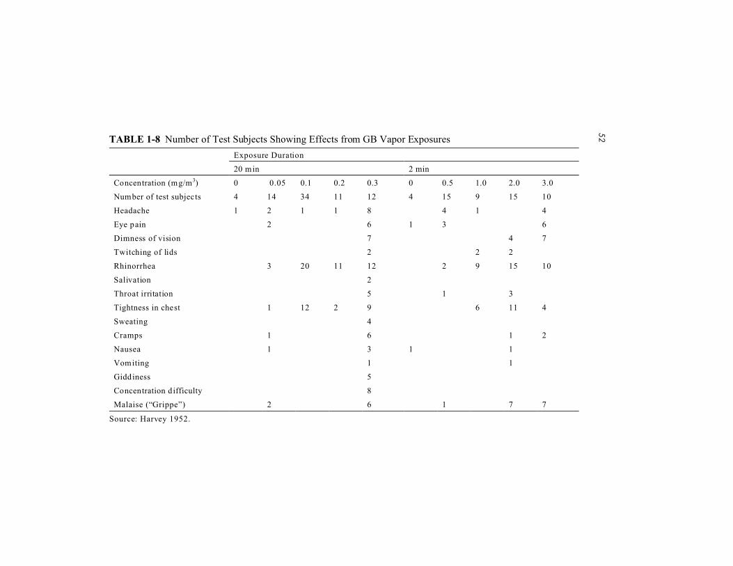
From the same overall study, Harvey (1952) reported signs andsymptoms resulting from the GB exposures; those results are presented inTable 1-8.
51
TABLE 1-7 Decrease in Pupil Diameter (mm) Following GB Vapor Exposures
Exposure Duration
20 min 4 min 2 min
Concentration (mg/m3) 0 0.05 0.2 0.3 1.0 1.3 0.5 1.0 2.0 2.3 3.0
Number of observations 55 8 11 11 12 4 15 9 8 7 10
Right eye; mean maximal
decrease in pupil diameter
(mm)
0.36 0.82 2.18 2.91 2.75 2.00 0.51 1.72 2.50 2.36 3.00
Left eye; mean maximal
decrease in pupil diameter
(mm)
0.33 1.00 2.18 3.00 2.59 2.22 0.60 1.67 2.92 2.07 3.00
Source: Johns 1952.

52TABLE 1-8 Number of Test Subjects Showing Effects from GB Vapor Exposures
Exposure Duration
20 min 2 min
Concentration (mg/m3) 0 0.05 0.1 0.2 0.3 0 0.5 1.0 2.0 3.0
Number of test subjects 4 14 34 11 12 4 15 9 15 10
Headache 1 2 1 1 8 4 1 4
Eye pain 2 6 1 3 6
Dimness of vision 7 4 7
Twitching of lids 2 2 2
Rhinorrhea 3 20 11 12 2 9 15 10
Salivation 2
Throat irritation 5 1 3
Tightness in chest 1 12 2 9 6 11 4
Sweating 4
Cramps 1 6 1 2
Nausea 1 3 1 1
Vomiting 1 1
Giddiness 5
Concentration d ifficulty 8
Malaise (“Grippe”) 2 6 1 7 7
Source: Harvey 1952.

NERVE AGENTS GA, GB, GD, GF, AND VX 53
In tests on human volunteers, Sim (1956) found that pupil constrictionoccurs more slowly and is less severe following exposure to GB at 5 mg/m3
than at 10 or 15 mg/m3 (1-3 min exposures). Some of the test subjects(number not given) reported restricted vision and eye pain.
Rubin et al. (1957) evaluated the effects of agent GB on the visualthreshold of three adult volunteers. The test subjects were exposed to GBat 2 mg/m3 for 2 min with the eyes protected or unprotected. With the eyesunprotected, the exposure resulted in moderate miosis and no other obvioussigns of cholinesterase inhibition, but there was a significant elevation ofthe absolute visual threshold in the dark-adapted eye.
Oberst et al. (1968) conducted inhalation studies in which 125volunteers were exposed to low concentrations of GB vapors in order tomeasure levels of GB retention and changes in RBC-ChE activity. In oneseries of tests in which resting subjects (N = 90; minute volumes 5.6-8.4 L)were exposed to GB (concentrations in the supply chamber were 16.2 to22.7 mg/m3, average 20.7 mg/m3) for 2 min, the calculated retained dosewas 3.4-3.8 µg/kg and the percent inhibition of RBC-ChE activity was 39-63% (average 49%). In a second series of tests, in which exercising men (N= 35; minute volumes 41.5-64.9 L) were exposed to GB (supply chamberconcentrations were 3.9 to 4.53 mg/m3, average 4.19 mg/m3) for 2 min, thecalculated retained dose was 3.2-4.0 µg/kg and the percent inhibition ofRBC-ChE activity was 29-58% (average 47%). The reported 2-min ChE50dose for all 125 subjects (grouped data) was 3.95 µg/kg. From these data,the 2-min EC50 for cholinesterase inhibition can be estimated asapproximately 21 mg/m3 for resting men breathing about 7 L/min and about4 mg/m3 for exercising men breathing about 50 L/min. In these studies thesubjects inhaled GB through a nosepiece or a mouthpiece; therefore, thepotential effects of the agent on the eyes (i.e., miosis) could not bedetermined.
McNamara and Leitnaker (1971) applied mathematical and conceptualmodels to human and animal data and estimated that the threshold forneuromuscular effects and the ECt50 of GB for miosis in humans would be4.0 mg"min/m3 (0.2 mg/m3 for 20 min). They further suggested that miosiswould not occur at a Ct of 0.5 mg"min/m3 (0.016 mg/m3 for 30 min).McNamara and Leitnaker (1971) also estimated the Ct at which 50%inhibition of blood cholinesterase would occur; it was reported to be 20mg"min/m3 (0.67 mg/m3 for 30 min). Blood cholinesterase activity was notexpected to be affected at a Ct of 0.5 mg"min/m3 (0.016 mg/m3 for 30 min).
Callaway and Dirnhuber (1971) evaluated the “miotogenic potency” ofGB vapor in humans exposed to GB “under goggles” (62 miosis responses
54 ACUTE EXPOSURE GUIDELINE LEVELS FOR SELECTED AIRBORNE CHEMICALS
in 26 human volunteers). The “goggle” experiments were designed todeliver GB vapor directly to the air volume around the eye and enclose thevapor as a means of controlling the exposure (no inhalation or percutaneousexposure) and delivering the vapor directly to the surface of the eye(thereby reducing variability). An airstream of GB vapor (flow rate 0.1L/min) was delivered to the space enclosed by each goggle. The unexposedpupil area of each eye was the baseline for pupil area decrementdeterminations for each eye. Exposure time periods ranged from 10 min to5 h. Callaway and Dirnhuber (1971) reported a 50% loss of pupil area inthe human dark-adapted eye at a Ct of 3.13 mg"min/m3 (95% confidenceinterval [CI] = 2.15-4.57 mg"min/m3). A 90% loss of pupil area occurredat a Ct of 13.85 mg"min/m3 (95% CI = 6.00-32.02 mg"min/m3).
Baker and Sedgwick (1996) exposed eight human volunteers to GB at0.5 mg/m3 for 30 min in an exposure chamber. During the exposure, testsubjects walked at a rate of 96 paces per minute and breathed normally. Itwas reported that the test Ct of 15 mg"min/m3 caused an inhibition of RBC-AChE activity to approximately 60% of individual baseline (reduction of40%) at both 3 h and 3 d postexposure. Subjects exhibited miosis and, insome cases, photophobia and mild dyspnea following exposure.Respiratory symptoms resolved within minutes, and ocular effects resolvedwithin 48 h. There were no clinical neuromuscular signs or symptoms;however, small changes in single fibre electromyography (SFEMG) of theforearm were measured at 3 h and 3 d postexposure and were still detectableat the first follow-up examination 4 to 15 mo postexposure. These changeswere not detectable at the second follow-up examination 15 to 30 mo afterexposure. Baker and Sedgwick (1996) suggested that theseelectrophysiological changes “may indicate subclinical onset of a non-depolarising type of neuromuscular block” that is fully reversible and hasno clinical significance.
Oral Exposures
In clinical studies conducted by Grob and Harvey (1958), GB wasadministered orally in aqueous solution to eight normal subjects. Doses of0.002 to 0.022 mg/kg resulted in 15-75% reduction in plasma and RBC-ChE activity. Grob and Harvey (1958) reported that the oral doseproducing 50% depression of RBC-ChE was 0.01 mg/kg.
NERVE AGENTS GA, GB, GD, GF, AND VX 55
Intra-arterial Exposures
In clinical studies conducted by Grob and Harvey (1958), GB wasadministered by intra-arterial injection to eight normal subjects. Grob andHarvey (1958) reported that the intra-arterial dose of GB producing 50%depression of RBC-ChE was 0.003 mg/kg.
Agent GD
Fairley and Mumford (1948) exposed 15 male volunteers to GD at 0.3mg/m3 for 0.5 min. Fourteen men reported that they could detect the agentby smell, seven reported tightness in the chest, and 11 reported rhinorrhea.
Agent GA
Uhde and Moore (1945, as cited in Mioduszewski et al. 1998) reportedthat four men exposed to T2104 (agent GA) at a concentration of 0.35mg/m3 for 2 min were able to detect the agent by smell, and all reportedslight, transient tightness of the chest, but none exhibited miosis. Ten menexposed to GA at 1.6 mg/m3 for 2 min were able to detect the agent bysmell, reported tightness of the chest, and exhibited miosis.
Agent VX
Local effects occurring at points of contact in the eyes and respiratorytract following exposure to low concentrations of VX vapor include miosis,rhinorrhea, and slight bronchoconstriction (Sidell 1992). These effects mayoccur without a significant decrease in activity of blood cholinesterases andwithout any signs of systemic toxicity (Sidell 1992). The ECt50 for mildeffects (ocular effects, accompanied perhaps by chest tightness andrhinorrhea) resulting from vapor exposures has been estimated at 0.09mg"min/m3 for 2-10 min exposures at moderate temperatures (65-75 °F) fora respiratory minute volume of 15 L (Reutter and Wade 1994). Exposuressufficiently high to result in systemic uptake can result in muscularweakness, tremors, difficulty breathing, convulsions, paralysis, and death.The ECt50 for severe effects resulting from vapor exposures has beenestimated at10 mg"min/m3 for 2-10 min exposures at moderate temperatures
56 ACUTE EXPOSURE GUIDELINE LEVELS FOR SELECTED AIRBORNE CHEMICALS
(65-75 °F) for a respiratory minute volume of 15 L (Reutter and Wade1994).
According to an unclassified NRC report (NRC 1997), the Reutter andWade (1994) estimated ECt50 of 0.09 mg"min/m3 for mild effects (oculareffects and rhinorrhea in humans) is based on the study by Bramwell et al.(1963) (percutaneous and direct ocular exposure to humans). The Bramwellet al. (1963) study is not considered credible for reasons that are discussedbelow under “Inhalation Exposures.” This conclusion also is supported bythe evaluation of a U.S. Surgeon General’s review panel in an August 2000public hearing (67 Fed. Reg. 894 [2002]; DHHS 2002).
Because agent VX is considered odorless, it possesses no olfactorywarning properties.
Vapor Exposures
Sixteen volunteers participated in an odor detection study of stabilizedand unstabilized VX (Koon et al. 1959). The agent was inhaled through anosmoscope attached to a chamber containing freshly generated agent vapor.The osmoscope permitted dilutions of the agent vapor with room air to yieldconcentrations down to one-sixty-fourth that in the chamber (0.05-3.34mg/m3). Each subject sniffed the agent in the morning and in the afternoonon two successive days (presumably only one sniff at each time point). Theestimated total doses for the four exposures ranged from 0.01 to 0.13 µg/kg.No significant changes in RBC- or plasma-ChE activity were demonstrated.Three subjects reported headaches the evening of the last test, and threeother subjects reported slight chest tightness, dryness of the mouth, andnasal irritation for 30 min following the test. There was no agreement as todescription of the odor. The median detectable concentration for VX vaporwas estimated to be 3.6 mg/m3 (95% CI = 0.8-16.4 mg/m3).
One of the few experimental attempts to evaluate human exposure toVX vapor for time durations greater than a few minutes is the historicallyimportant study of Bramwell et al. (1963) in which eight individuals wereexposed for time periods ranging from 2.25 seconds (s) to 24 min to VXvapor concentrations ranging from 0.23 mg/m3 to 5 mg/m3 (Cts = 0.7 to25.6 mg"min/m3). The Bramwell et al. (1963) study is not consideredcredible because of its seriously flawed exposure protocol but is presentedhere for completeness and context. The test subjects were exposed while
57
TABLE 1-9 ChE Inhibition in Humans Following Exposure to VX Vapors
Exposure Conditions Max ChE Inhibition
(% depression)Trial Subject Time (min) Concentration (mg/m3) Ct (mg"min/m3)
R1 SHE 3 0.2 0.6 20
R2 BIS 3 0.35 0.9 18
R3 LAD 3 0.31 0.9 22
R4 BUR 3 0.37 1.1 17
R5 BRA 3 0.4 1.2 14
R6 HOP 3 0.48 1.4 10
R7 CRO 3 0.57 1.7 12
R8 SHE 1.5 1.6 2.4 26
R9 BRA 1.5 1.73 2.6 25
R10 BUR 1.5 1.73 2.6 21
R11 BIS 1.5 1.93 2.8 28
R12 LAD 1.5 2.0 3.0 41
R13 HOP 1.5 2.07 3.1 18
R14 HOL 1.5 2.07 3.1 28
R15 CRO 1.5 2.4 3.6 20
R16 CRO 6 0.8 4.8 44
R17 LAD 7 0.79 5.5 70 (Continued)
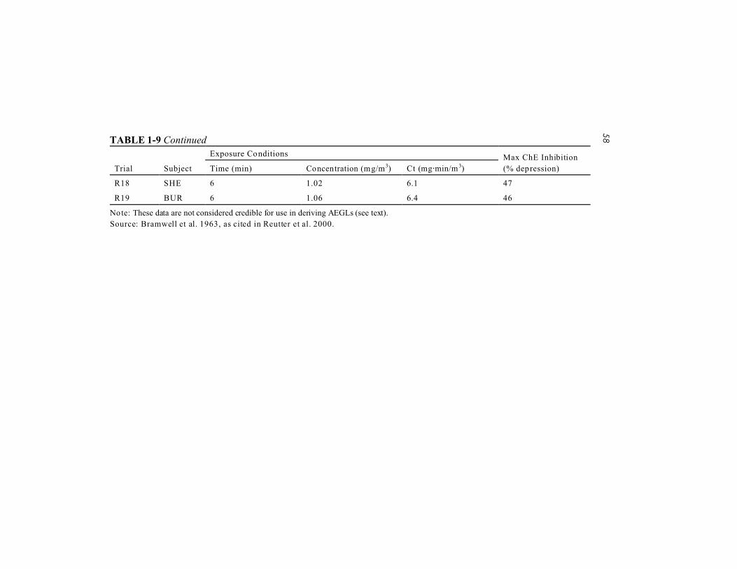
58TABLE 1-9 Continued
Exposure Conditions Max ChE Inhibition
(% depression)Trial Subject Time (min) Concentration (mg/m3) Ct (mg"min/m3)
R18 SHE 6 1.02 6.1 47
R19 BUR 6 1.06 6.4 46
Note: These data are not considered credible for use in deriving AEGLs (see text).
Source: Bramwell et al. 1963, as cited in Reutter et al. 2000.
NERVE AGENTS GA, GB, GD, GF, AND VX 59
standing or seated at the mouth of a tunnel from which VX vapor was flow-ing in an airstream at 1 m/min at a temperature of 32 °C. Only the head andneck of the test subjects were exposed. A total of 19 exposures wereconducted without respiratory protection (see Table 1-9). All but one of thetests were conducted with eyes closed without the use of eye protection inthe form of goggles or face mask. The only symptoms noted during theexposures were slight tightness in the throat and upper respiratory tract;these symptoms were not reported by all subjects. In the individual exposedwith eyes open (0.31 mg/m3 for 3 min), miosis developed suddenly 20 to 30min postexposure and was maximal at 1.5 h postexposure. In theindividuals exposed with eyes closed, some miosis usually developed 1 to3 h postexposure. The degree of miosis was quite variable among theindividuals and appeared to be concentration-dependent. The miosis wasoften accompanied by a fluttering or twitching of the eyelids. Although themuscle effects were clearly reported by the subjects, they were not alwaysobvious to the observers. Rhinorrhea occurred within 30 min of exposurein 14 of 19 trials. In four other trials, it developed more slowly; in one, itdid not develop at all. Excessive salivation, lasting for about an hour, wasreported in one subject after a 6-min exposure to a concentration of 1.06mg/m3. Two hours postexposure, one individual experienced some nauseaand sweating; RBC-ChE activity was 60% depressed at that time. Theseeffects abated somewhat and then recurred later when ChE inhibition hadreached 70%. Several individuals also experienced malaise and lethargy.Based on all 19 trials, the inhaled dose estimated to produce inhibition of50% of the RBC-ChE activity (ChE50) was 13 µg/kg. However, the authorsthought that apprehension had increased the subjects’ minute volume duringinitial exposures. That would have effectively increased the dose to whichthe individuals were exposed and was thought to account for a relativelyshallow probit slope. When those data were excluded, the estimated ChE50was about 8 µg/kg, which was thought to compare favorably withintravenous data.
The Bramwell et al. (1963) study is not considered credible for use inderiving AEGLs for agent VX. Reutter et al. (2000) examined theBramwell et al. study as a potential critical study for the estimation ofworker population levels (WPLs) and general population levels (GPLs) forchronic exposure to VX vapor (8-h time-weighted average for WPL; 24-hcontinuous exposure for GPL). Reutter et al. (2000) rejected the Bramwellet al. study because of multiple deficiencies; the concentration of VX towhich the subjects were exposed could not be determined (subjects wereseated in front of a “tunnel” down which generated VX vapor flowed in anairstream of known velocity), both C and t were varied (resulting in no
60 ACUTE EXPOSURE GUIDELINE LEVELS FOR SELECTED AIRBORNE CHEMICALS
replicate cumulative exposures), and the organic solvent benzene was usedto help disperse the agent in the airstream to which subjects were exposed(Bramwell et al. did not address the potential effect of the carrier solvent onagent absorption by the subject). The majority of a U.S. Surgeon General’sreview panel concurred with the appraisal of Bramwell et al. (1963) at apublic hearing convened by the CDC to examine the Reutter et al. (2000)report (67 Fed. Reg. 894 [2002]; DHHS 2002).
Oral Exposures
In clinical studies conducted by Sidell and Groff (1974), single oraldoses of VX at 2-4.5 µg/kg (stock solution in absolute ethanol diluted in asolution of saline and dextrose and swallowed by each subject undersupervision) produced gastrointestinal symptoms in 5 of 32 test subjects(more specific dose-response data not reported). Regression analysis of thedose-response data indicated that the RBC-ChE50 was 2.3 µg/kg. Sidell andGroff (1974) reported that the oral dose of VX needed to produce 70% ChEinhibition (4 µg/kg) was 3 times greater than that needed to produce thesame effect after intravenous administration.
Sim et al. (1964) reported no signs of toxicity in seven humanvolunteers receiving VX at 1.43 µg/kg/d for 7 d (in four daily doses of 500mL drinking water); however, average RBC-ChE activity was reduced 60%(to 40% of baseline values). The Sim et al. (1964) study resulted in a lowerRBC-ChE50 value than the Sidell and Groff (1974) oral study, probablybecause of the cumulative effects of VX given over the 7 d in the Sim et al.study. The total dose in the Sim et al. (1964) study was about twice thatused in the Sidell and Groff (1974) oral study.
Intravenous Exposures
Several studies have been conducted in which human volunteers wereinjected intravenously with VX. The experiment of Kimura et al. (1960)was performed with the informed consent of the participants, under fullclinical supervision and in a hospital setting considered suitable at the time(resuscitation team at bedside “to administer atropine, oximes, oxygen,artificial resuscitation, and tracheotomy if indicated”). Kimura et al. (1960)reported that a 30-s intravenous injection of 0.04 µg/kg in one adult testsubject caused frontal and retrobulbar headaches starting 45 min after theinjection. The subject reported being tired and appeared irritable to

NERVE AGENTS GA, GB, GD, GF, AND VX 61
observers, but no change in RBC or whole blood cholinesterase activity wasobserved. A subsequent 30-s intravenous injection of 0.08 µg/kg 3.5 h laterresulted in a 2-fold increase in airway resistance, a 25-30% decrease inrespiratory rate, and a 15% drop in pulse rate 15 min after the exposure, butno change in RBC-ChE. Headaches began 20 min postexposure, andminute volume increased from 15 L to 32 L 30-45 min postexposure. Peakeffects (increased sweating, lightheadedness, and abdominal cramping)appeared about 45 min after the dose was administered. A single 30-sintravenous dose of 0.225 µg/kg in one test subject resulted in a 27%decrease in baseline RBC-ChE activity within 15 min as well as retrobulbarheadaches. Many of these observed effects are for the single subjectparticipating in the dose-response range-finding study—Dr. Van Sim, MD,a principal investigator of the reported study. Six additional subjects(volunteers identified by subject code) received VX at 1 µg/kg byintravenous infusion over 1.75 to 4 h periods and exhibited 50-60%depression in cholinesterase activity but no signs of toxicity (except for one84-kg individual who reported headaches).
The Kimura et al. (1960) study meets the criteria for acceptance ofhuman subject data for use by the AEGL process (e.g., evidence thatsubjects provided informed consent and that the studies were performedunder appropriate clinical supervision).
In clinical studies conducted by Sidell and Groff (1974), 34 test subjectswere given VX by intravenous injection. The administered dose rangedfrom 1.2 to 1.7 µg/kg. An intrvenous dose of 1.5 µg/kg administered to18test subjects resulted in dizziness, nausea, and vomiting in 11, 4, and 6individuals, respectively; RBC-ChE was depressed 55-90% from baselinevalues (average about 75%). The test subjects exhibited a significantdecrement in performance on a number facility test within 1 h aftertreatment. Regression analysis of the dose response data indicated that theRBC-ChE50 was 1.1 µg/kg (three individuals tested at 1.2 µg/kg, 1.3 µg/kg,1.4 µg/kg, and 1.7 µg/kg; four at 1.6 µg/kg; and 18 at 1.5 µg/kg; estimatedfrom graphic presentation of the data) (Sidell and Groff 1974).
Percutaneous Exposures
Dermal vapor absorption is a low priority for this compound, althoughthere are certain release events that generate a dermal vapor threat. It isgenerally acknowledged that a specific toxicological end point for vaporexposure to nerve agent VX would be achieved at a lower concentrationexposure for the inhalation route than for other routes (e.g., the estimated
62 ACUTE EXPOSURE GUIDELINE LEVELS FOR SELECTED AIRBORNE CHEMICALS
human LCt50 for percutaneous vapor exposure to agent VX is 150mg"min/m3, while the estimated human LCt50 for inhalation vapor exposureto agent VX is <15 mg"min/m3) (NRC 1997). Thus, AEGL estimates basedon inhalation exposures are considered protective for both inhalation anddermal routes.
In studies conducted by Bramwell et al. (1963, as cited in Reutter et al.2000) eight individuals were exposed for time periods ranging from 2.25 sto 24 min to VX vapor concentrations ranging from 0.23 mg/m3 to 5 mg/m3
(Cts = 0.7 to 25.6 mg"min/m3). The test subjects were exposed whilestanding or seated at the mouth of a tunnel from which VX vapor wasflowing in an airstream at 1 m/min at a temperature of 32 °C. Only the headand neck of the test subjects were exposed. Thirty-five of the exposureswere performed with eyes closed (but without the use of eye protection inthe form of goggles or face mask) and with respiratory protection (a noseclip was used and the subjects were breathing through a spirometerconnected to a respirator canister). ChE inhibition was measurable withinan hour of exposure and was greatest at 8-12 h postexposure. No signs orsymptoms were noted during the exposure periods; however, 30 min ormore after the initial exposure, miosis appeared in nearly all subjects andbecame maximal several hours later. It was usually accompanied orfollowed by fluttering and twitching of the eyelids and was morepronounced at the higher concentrations. Flushing of the skin of the headand neck was observed in five of the eight subjects, and all eight individualsreported local sweating in one or more tests. Although some subjects hadthe perception that they were experiencing “tunnel vision” postexposure,visual perimetry studies following three of the exposures were notconfirmatory. Nor were there any changes in visual acuity or color vision.Five hours postexposure, one subject developed flatulence and abdominaldiscomfort. An hour later he did not feel well and was experiencing wavesof nausea. Eight hours postexposure, he deteriorated rapidly andexperienced severe nausea and vomiting. At that time, his RBC-ChEactivity was only 30% of baseline; no further inhibition occurred. Bouts ofvomiting and malaise continued, and he experienced cold sweating, pallor,and a feeling of motion sickness—minus the vertigo. At 12 h postexposure,he was able to sleep, but experienced a nightmare shortly after fallingasleep. By the next morning no signs or symptoms remained. TheBramwell et al. (1963) study is not considered credible for deriving AEGLvalues.
Lubash and Clark (1960) reported that percutaneous doses of undilutedVX (20 µg/kg or 35 µg/kg) applied to the volar forearm of male volunteers
NERVE AGENTS GA, GB, GD, GF, AND VX 63
resulted in significant decreases in blood ChE as well as signs andsymptoms of toxicity (lightheadedness, nausea, vomiting, diarrhea,hyperactive bowel sounds, epigastric discomfort, insomnia, and nightmares)in two of four subjects dosed with 20 µg/kg and in two of four subjectsdosed with 35 µg/kg (eight total subjects).
Sim (1962) reported that head and neck areas were the most sensitiveto percutaneously applied VX. A dose of VX at 5 µg/kg applied to theseareas resulted in signs and symptoms of systemic toxicity (nausea,vomiting, and weakness) in 54% (28 of 40) of the tested individuals. Wholeblood ChE was 50% of normal in 5.8 h and 33.5% of normal in 8.5 h. Itwas estimated that a VX dose of 5.1 µg/kg would be necessary to result inRBC-ChE30 (this end point was chosen because median ChE depression of30% was associated with the onset of gastrointestinal signs and symptomsof nausea and vomiting).
Cresthull et al. (1963) studied the effects of percutaneous absorption ofVX vapors on whole blood ChE activity in 29 male volunteers. Exposureswere to the arm or forearm. The VX concentrations ranged from 1.2 to 12.2mg/m3 and the exposure times were from 2 to 75 min (Cts ranged from 6 to765 mg"min/m3). Two men were exposed at 1.2-1.5 mg/m3 for 5 min (500cm2 surface area exposed); six to 2.5-4.9 mg/m3 for 5-10 min (500 cm2
surface area exposed); four to 4.8-7.3 mg/m3 for 12-20 min (500 cm2
surface area exposed); ten to 4.5-8.0 mg/m3 for 20-60 min (1,000 cm2
surface area exposed); and seven to 8.5-12.2 mg/m3 for 60-75 min (1,000cm2 surface area exposed). The median decrease in whole blood ChE inthose groups was 5%, 3%, 8%, 18%, and 43%, respectively. Althoughwhole blood ChE was inhibited as much as 76% at 20 h after exposure,none of the test subjects exhibited any toxic signs. Cresthull et al. (1963)estimated that the whole blood ChE50 vapor concentration for percutaneousexposures would be 141 mg"min/m3. The value was reported to be notstatistically meaningful because of the wide confidence limits (lower 95%CI = 35 mg/m3); however, by comparison with data for exposures to VXaerosols, Cresthull et al. concluded that the estimated ChE50 of 141 mg-min/m3 was acceptable. Cresthull et al. (1963) also estimated that 1 to 1.25h whole-body percutaneous exposure to a Ct of 38 mg"min/m3 (0.51 mg/m3
for 75 min) would not cause any signs of toxicity other than “partial”lowering of whole blood ChE (activity inhibition between 0% and 31%from baseline). Bowers et al. (1964) evaluated behavioral changes in93 volunteers who were exposed percutaneously to small amounts of liquidEA-1701 (agent VX). The actual amounts of VX applied were not reported.

64 ACUTE EXPOSURE GUIDELINE LEVELS FOR SELECTED AIRBORNE CHEMICALS
The test subjects were divided into three postexposure groups depending onthe level of reduction in their whole blood ChE (there was no controlgroup). Of 32 individuals whose whole blood ChE was 81-100% of controlvalues (not explicitly stated but presumed to be individual preexposurevalues) following exposure, 6% showed symptomatology of intellectualimpairment (impairment of ability to perform simple arithmetic tasks,inability to perform serial sevens, impairment of performance in reading orstandard games of concentration, and other subjective symptoms such as“impairment in orientation”), and 3% reported unusual dreams. Of the 24whose whole blood ChE was 40-80% of control values, 4% showedsymptomatology of intellectual impairment (by the measures reportedabove), 33% reported unusual dreams, 8% exhibited anxiety (determinedby the appearance of palpitations coupled with other, subjective symptomssuch as “restlessness”), and 4% exhibited psychomotor depression(determined by the appearance of reply latency, slowed speech, andevidence of fatigue in addition to other, subjective symptoms such asreported feelings of being “slowed down”). Of the 37 whose ChE was 10-40% of control values, 57% showed symptomatology of intellectual impair-ment (by the measures reported above), 38% reported unusual dreams, 30%exhibited anxiety, and 57% exhibited psychomotor depression. The moreseverely affected cases exhibited mood alterations as determined by Clydemood card sort before and after exposure, and some developed nausea andvomiting. Miosis, bronchoconstriction, hypermotility of the lower bowel,and muscle fasciculations were not observed in any of the test subjects.Bowers et al. (1964) concluded that, with the exception of excessivedreaming, psychological symptomatology did not develop in the exposedindividuals unless whole blood ChE fell to 40% or less of control values.Very few of the test subjects whose blood ChE was 80% or more of controlvalues exhibited any signs.
Data compiled by Sidell (1992) revealed that, for individuals exposedto VX percutaneously, gastrointestinal signs (vomiting) occurred in 0.6%(1/166) when RBC-ChE activity was at 50% of control values and in 8%(2/24) when RBC-ChE levels were 40-49% of controls. Thirty-threepercent exhibited such signs when RBC-ChE levels were 30-39% ofcontrols, and 45% (19/42) exhibited signs when RBC-ChE levels were 20-29% of controls. Sixty-seven percent (16/24) exhibited effects when RBC-ChE levels fell to less than 20% of control values.
NERVE AGENTS GA, GB, GD, GF, AND VX 65
2.2.3. Epidemiologic Studies
There are no human epidemiologic studies with dose-response datasuitable for deriving AEGL estimates for the G agents.
Occupational exposures to agent GB have been associated with alteredelectroencephalograms (EEGs) (Duffy et al. 1979; Burchfiel and Duffy1982). Burchfiel and Duffy (1982) evaluated the wake and sleep EEGs of77 industrial workers who had been exposed at least once to agent GB(sarin); however, no exposures had occurred in the year preceding the study.Spectral analysis of the EEGs indicated significant increases in brain betaactivity (12-30 Hz) in the exposed group compared with nonexposedcontrols, and sleep EEGs indicated significantly increased rapid eyemovement in the exposed workers. Combinations of EEG componentswere subjected to computer analysis in an attempt to identify an exposedindividual by EEG characteristics; however, the results were inconclusive.Burchfiel and Duffy (1982) concluded that there might be a threshold forthis type of effect. In evaluating the data of Burchfiel and Duffy (1982),DHHS (1988) considered the EEG changes to be “of questionablesignificance—given the difficulty of demonstrating such changes and theabsence of clinically significant effects even when EEG changes arepresent.”
A retrospective analysis of possible chronic or delayed adverse healtheffects among servicemen who participated in chemical agent effects andtherapy testing at Edgewood Arsenal during the years 1955-1975 wasconducted by the Committee on Toxicology of the National ResearchCouncil (NRC 1985). The primary source of information was provided byparticipant response to a questionnaire, but there were no exposure datafrom which to derive a dose-response relationship. The chronic healtheffects of concern were “excess cancer risk, and adverse mental, neurologic,hepatic and reproductive effects.”
Evaluation of questionnaire response indicated that data provided bysubjects historically tested with anticholinesterase compounds did notsignificantly differ from that of control subjects or those tested with othercompounds when self-evaluations of current health status were compared.The report candidly pointed out that the experimental design andcomparison groups available were such that “only large effects were likelyto be uncovered” because of the resulting large standard errors, self-reporting, and the potential for more than one exposure to eventually resultin development of the same biological end point (NRC 1985).
A number of studies have been conducted on individuals exposed toagent GB as a result of terrorist attacks in Japan. Morita et al. (1995)
66 ACUTE EXPOSURE GUIDELINE LEVELS FOR SELECTED AIRBORNE CHEMICALS
reported clinical findings for several hundred people who were exposed toagent GB in the city of Matsumoto in 1994. Subclinical miosis andneuropathy were still present in some individuals 30 d after exposure;however, most individuals exhibited no clinical signs of toxicity 6 mo afterthe exposure.
Several follow-up studies have examined the health of victims of theTokyo subway terrorist attack that occurred in March of 1995. No clinicalabnormalities were detected in 640 patients examined 3 mo after theincident (Okumura et al. 1996). Kato and Hamanaka (1996) examined 96victims for ocular effects. The primary ocular signs and symptoms includedmiosis, conjunctival injection, and ocular pain. Some individuals hadtemporary blurring of vision, 36 patients complained of subjectiveaccommodation impairment, and in 30 patients there were indications thatagent GB (sarin) had caused a reduction in intraocular pressure (intraocularpressure was 11.6 ± 1.9 mm Hg within 2 h of exposure but increased to 14.6± 1.8 mm Hg when the pupil diameter returned to normal). These signs andsymptoms spontaneously resolved within 3-21 d after exposure in mostcases. Kato and Hamanaka (1996) note that none of the victims developedcorneal injury, glaucoma attack, or retinal detachment, and although theocular condition of the patients returned to normal, they suggest thatexposure to agent GB may increase the risk of angle-closure glaucomacaused by anterior shift of the lens, retinal detachment, and vitreoushemorrhage caused by extensive contraction of the ciliary muscles. Murataet al. (1997) evaluated neurophysiological deficits in 18 victims of thesubway attack who had exhibited signs and symptoms of agent GBpoisoning (i.e., headache, miosis, increased lacrimation, dyspnea, nausea,diarrhea, para-esthesia, and decreased serum ChE activity). It was reportedthat 6 mo after the exposure, the exposed but no longer symptomaticindividuals exhibited significantly prolonged latencies in event-evokedpotentials and visual evoked potentials suggestive of persistent cognitiveand visual dysfunction.
In another study, Yokoyama et al. (1998a,b) evaluated chronic neuro-behavioral effects in nine male and nine female patients 6-8 mo after theincident. Although this study is for a very small number of those affected(only 18 out of approximately 5,500 people) and suffers from low recruit-ment, the results will be presented here for completeness. Theneurobehavioral tests included (1) digit symbol (psychomotor perfor-mance), (2) picture completion (visual perception), (3) digit span (attentionand memory), (4) finger tapping (psychomotor performance),(5) reactiontime (psychomotor performance), (6) continuous performance test
NERVE AGENTS GA, GB, GD, GF, AND VX 67
(sustained visual attention), (7) paired-associate learning (learning andmemory), (8) General Health Questionnaire (GHQ) (psychiatric symptoms),and (9) Profile of Mood States (POMS) (mood). Fifteen controls were usedin the tests. Analysis of covariance of the test results suggested to theinvestigators that “perhaps a chronic effect on psychomotor performance[digit symbol test only] was caused directly by acute agent GB (sarin)poisoning; on the other hand, the effects of psychiatric symptoms (GHQ)and fatigue (POMS) appeared to result from post-traumatic stress disorderinduced by exposure to sarin.” Yokoyama et al. (1998c) have also reportedves-tibulocerebellar effects (increased postural sway) in 18 patients tested6-8 mo after the incident. Postural sway was significantly greater thancontrols in exposed females but not in males. In both genders postural swaywas correlated with the plasma cholinesterase activity measuredimmediately after the exposure.
The U.S. Department of Defense reported in 1997 that militarypersonnel might have been exposed to nerve agents as a result of thedemolition of Iraqi munition storage sites. Retrospective studies haveevaluated the post-war health of soldiers who may have been exposed tonerve agents. Landrigan (1997) reviewed the principal epidemiologicstudies published before 1997. Kang and Bullman (1996) reported a 0.8%higher death rate among Gulf War veterans (10.4%) compared with otherveterans of the same time period (9.6%); this difference was largely due toaccidents, and no excess deaths from suicides or specific diseases wereobserved. Gray et al. (1996, 1999) reported no consistent pattern forincreased occurrence of any specific disease or hospitalization among GulfWar veterans. Further, Gray et al. (1999) indicate that “this data analysisdoes not support the hypothesis that Gulf War veterans are sufferingpostwar morbidity from subclinical nerve agent exposure.”
Other epidemiologic studies have reported an increase in neurologicdisorders among selected groups of Gulf War veterans but have not linkedany reported signs, symptoms, or clinical effects with potential nerve agentexposure (Goldstein et al. 1996; Kotler-Cope et al. 1996; Haley et al.1997a,b; Haley and Kurt 1997; Hom et al. 1997; Schwartz et al. 1997;Vasterling et al. 1998).
Epidemiologic studies regarding human exposure to agent VX were notfound in the available literature.
2.3. Neurotoxicity
The G agents (GA [tabun], GB [sarin], GD [soman], and GF) and agent
68 ACUTE EXPOSURE GUIDELINE LEVELS FOR SELECTED AIRBORNE CHEMICALS
VX are toxic organophosphate ester derivatives of phosphonic acid. Theyare commonly termed “nerve” agents as a consequence of their potentanticholinesterase properties and subsequent adverse effects on both smoothand skeletal muscle function as well as the central and peripheral nervoussystems. These neurotoxic properties were discussed in detail in Sections2.1 and 2.2.
Although the inhibition of cholinesterases within neuroeffectorjunctions or the effector itself is thought to be responsible for the majortoxic effects of nerve agents, these compounds can affect nerve impulsetransmission by more direct processes as well (e.g., direct effects onneurotransmitter receptors) (see Section 4.2).
2.4. Developmental and Reproductive Toxicity
The retrospective study of agent-exposed servicemen discussed inpreceding sections (NRC 1985) requested self-reported information onfertility. Two comparison groups of men were used. One was a “nochemical test” (NCT) group who met the requirements for military servicebut did not meet the more rigorous requirements (physical and mentalscreening exams for contraindications) necessary for chemical exposuretests. Those individuals were exposed to placebos, equipment only, or“FDA approved drugs” not otherwise identified. A second comparisongroup comprised men tested with compounds other than those beingevaluated in a particular test, the “other chemical test” (OCT) group. Theseindividuals also met the requirements for military service. They wereexposed to test chemicals other than the chemicals of interest. The OCTcompounds appear to include cannabinoids, “approved drugs,” and“innocuous chemicals and controls” not otherwise identified (NRC 1985).When the collected data were adjusted for volunteer age when the last testwas performed (to accommodate national trends toward smaller and delayedfamilies), there “was no difference between the observed fertility pattern ofmen exposed to anticholinergic chemicals and that expected on the basis ofmen who were exposed to other chemicals” (NRC 1985). Nevertheless,these data are not useful for application to the derivation of an AEGL giventhat no exposure data were collected.
Iranian soldiers and civilians were exposed to multiple chemicalwarfare agents during the Iran/Iraq conflict. Exposures may have been tothe nerve agents GA and/or GB as well as to the vesicant sulfur mustard.Follow-up studies have been conducted on some of the individuals. It hasbeen reported that the offspring of these chemical warfare victims born after
NERVE AGENTS GA, GB, GD, GF, AND VX 69
the Iran/Iraq conflict were more likely to have birth defects than those bornbefore the war (Pour-Jafari 1994a). It was also reported that the offspringhad an altered gender ratio (Pour-Jafari 1994b). Because of the possibilitythat exposures to multiple chemicals had occurred, it is impossible todetermine if, or to what extent, exposure to any of the G agents contributedto the reported effects.
There have been several reports of potential increased incidence of birthdefects among the offspring of military personnel who served in the PersianGulf War. Araneta et al. (1997) reported a slight increase (relative risk3.03, with 95% CI = 0.63-20.57) in Goldenhar syndrome among infantsborn in military hospitals to Gulf War veterans. Goldenhar syndrome is acraniofacial anomaly of unclear etiology. According to Araneta et al.(1997), suggested associations with its occurrence have includedchromosomal abnormalities, a genetic pattern of inheritance, maternaldiabetes, or prenatal exposure to several controlled or therapeutic drugs(e.g., cocaine, tamoxifen); the role of male-mediated effects is undefined.At least one mother of one case infant exhibited mild facial asymmetryupon examination, and the family of another case infant had a history ofbirth defects. In all five cases of confirmed Goldenhar syndrome among the34,069 infants born to veterans of the Gulf War and included in thisretrospective study, only the paternal parent served in the military. Amongnondeployed veterans (two cases of a total 41,345 births examined), onlythe paternal parent served in the military.
Araneta and colleagues (1997) point out that differences in prevalencerates of Goldenhar syndrome among the offspring of Gulf War veterans(14.7, 95% CI = 5.4-36.4) are not significantly different from those ofnondeployed veterans (4.8, 95% CI = 0.8-19.5) because of the small samplesizes and wide confidence intervals. Araneta et al. (1997) determined that,with the sample size maintained as a constant, “the risk would have had tobe at least 5.75 times higher among the Gulf War veterans’ infants in orderto be statistically significant.”
As a consequence of this finding of no significance, the Goldenharsyndrome issue was not further pursued in the AEGL analysis for chemicalwarfare agents. Its utility is further compromised by the fact that there isno confirmed report of veteran exposure to chemical warfare agents, and theGulf War fathers in the Araneta et al. (1997) study all served in differentunits and were deployed in theater at different times.
No data are available regarding the potential reproductive anddevelopmental toxicity of agent VX in humans.
70 ACUTE EXPOSURE GUIDELINE LEVELS FOR SELECTED AIRBORNE CHEMICALS
2.5. Genotoxicity
There is no information available to evaluate the genotoxicity of Gagents or agent VX in humans.
2.6. Carcinogenicity
There are no human data to suggest that G agents or agent VX arecarcinogenic.
2.7. Summary
G-series Agents
Available information on the acute inhalation toxicity of agent GB(sarin) to humans is summarized in Table 1-10. Minimal effects observedat low concentrations include miosis, tightness of the chest, rhinorrhea, anddyspnea. The threshold for minimal effects appears to fall in the range of0.05 to 0.5 mg/m3 for 10-30 min exposures. The results from differentstudies are not consistent in identifying the threshold, and that may be dueto differences in individual sensitivities, breathing rates of the test subjects,experimental protocols, or analytical methods.
A number of investigators consider both miosis and rhinorrhea to beearly signs of exposure to cholinesterase inhibitors (Sidell 1997;Mioduszewski et al. 2002b; H. van Helden, Pulmonary and CNSPharmacology Lab, TNO, the Netherlands, personal communication; S.Tattersall, Biomedical Sciences Division, Porton Down, United Kingdom,personal communication). The presence of rhinorrhea can be indicative ofinhalation exposure and/or development of systemic effects, while miosisalone in the absence of other signs or symptoms is a local effect to thepupillary muscles of the eye. As a consequence, the presence of miosis isconsidered an appropriately sensitive indicator of direct vapor exposure andhas the additional advantage of being readily recognized and quantifiable.
71
TABLE 1-10 Human Experimental Data For GB Vapor (Single Exposures)
Study
GB Concentration
(mg/m 3) Duration Ct (mg"min/m3) Signs and Symptoms
Harvey 1952 0.05 20 min 1 Headache, eye pain, rhinorrhea, tightness in
chest, cramps, nausea, malaise (N = 14)
Johns 1952 0.05 20 min 1 Mild miosis (mean maximum decrease in
pupil diameter 1-2 mm) in some of the test
subjects (150 observations)
McKee and W oolcott
1949
0.06 20 min 1.2 No reported effects (N = 5)
McKee and W oolcott
1949
0.06 40 min 2.0 “Threshold” for miosis; no other signs or
symptoms (N = 4)
Fairley and Mumford
1948
0.3 0.5 min 0.15 Rhinorrhea in 16 and tightness in chest in 7
(N = 16)
Baker and Sedgwick
1996
0.5 30 min 1.5 Miosis, dyspnea, photophobia, 40%
inhibition of RBC-ChE, changes in SFEMG
(N = 8)
McKee and W oolcott
1949
0.6 1 min 0.6 Miosis and slight tightness in chest (N = 4)
Rubin et al. 1957 2 2 min 4 Miosis; no other signs of ChE inhibition
(N = 3)
Callaway and
Dirnhuber 1971
10 min to 5 h 3.13a 50% pupil area decrement
(Continued)
72
TABLE 1-10 Continued
Study
GB Concentration
(mg/m 3) Duration Ct (mg"min/m3) Signs and Symptoms
Callaway and
Dirnhuber 1971
10 min to 5 h 13.85b 90% pupil area decrement
Oberst et al. 1968 4.19 2 min 8.38 47% inhibition of RBC-ChE; no other
effects; eyes not exposed (breathing rate
5.6-8.4 L/min)
Oberst et al. 1968 20.7 2 min 41.4 49% inhibition of RBC-ChE; no other
effects; eyes not exposed (breathing rate
47-65 L/min)
Note: Entries are from primary sources and known experimental dataa95% confidence limits 2.15-4.57 mg"min/m3.b95% confidence limits 6.00-32.02 mg"min/m3.

NERVE AGENTS GA, GB, GD, GF, AND VX 73
There is no evidence that exposure to any of the G agents results indevelopmental or reproductive toxicity, nor are there any data available toevaluate potential genotoxicity in humans. The G agents have not beenidentified as human carcinogens.
Agent VX
Experimental data on the effects of acute VX exposures to humans aresummarized in Tables 1-11 and 1-12; very few studies have been conductedusing exposures to VX vapor, and available data are not of sufficient qualityto be used directly in the development of AEGL estimates.
A comparison of the results of the intravenous studies, in terms of theestimated absorbed dose, allows for an evaluation of the dose-responserelationship (Table 1-11).
Studies indicate that an intravenous dose of about 1 µg/kg can result in50% ChE depression and some symptoms of toxicity (headaches); anintravenous dose of about 0.1 µg/kg is unlikely to affect RBC-ChE, but maycause mild effects (headache, chest tightness, dyspnea); and an intravenousdose of 0.01 µg/kg may be below the effects threshold. The estimatedequivalent air concentrations for these dose levels, using standard defaultvalues for body weight (70 kg) and breathing rate (0.0138 m3/min), are alsolisted in Table 1-11. They are highly derivative values and are onlypresented for comparative purposes.
Experimental data from the Bramwell et al. (1963) study aresummarized in Table 1-12. Although the Bramwell et al. (1963) data areconsidered suspect, they provide a means of comparison with the equivalentconcentrations estimated from the intravenous data. Both sets of datasuggest that a 10-min exposure to VX at 0.5 mg/m3 or higher may producesubstantial depression of RBC-ChE activity and some clinical signs oftoxicity.
Available data summarized suggest that a 10-min exposure to VX at0.5 mg/m3 or higher may produce depression of RBC-AChE activity andsome clinical signs of toxicity.
74TABLE 1-11 Human Experimental Data for VX
Dose (:g/kg) Exposure Route
Estimated Equivalent
Concentration (mg/m3)a End Point Reference
0.01-0.13
(estimated)
Inhalation
(sniff test)
0.05-3.34 No ChE change; headache,
chest tightness, dryness of the mouth
Koon et al.
1959
0.04 Intravenous
(30 s injection)
0.41 (0.02 for 10 min)b No ChE change; headache,
tiredness, irritability
Kimura et al.
1960
0.12 Intravenous
(2 doses over 3.5 h)
0.003 (0.06 for 10 min)b No ChE change; headache,
light headedness, abdominal cramps,
decrease in respiration and pulse rates,
increase in airway resistance and
minute volume
Kimura et al.
1960
1.0 Intravenous
(over 1.75-4 h)
0.021(0.5 for 10 min)b 50-60% depression in ChE activity;
headaches in 1/6 individuals
Kimura et al.
1960
1.0 Intravenous
(1 dose)
10 (0.51 for 10 min)b 50% inhibition of RBC-ChE Sidell and Groff
1974
1.5 Intravenous
(1 dose)
15(0.76 for 10 min)b 75% depression in ChE: dizziness
(11/18), nausea (4/18), vomiting
(6/18)
Sidell and Groff
1974
1.43 Oral
(1 dose/d for 7 d)
NA 60% inhibition of RBC-ChE; no signs
or symptoms of toxicity
Sim et al. 1964
2.3 Oral NA 50% inhibition of RBC-ChE Sidell and Groff
1974

75
2-4.5 Oral NA Gastrointestinal symptoms in 5/32 Sidell and Groff
1974
Entries are from primary sources and known experimental dataaEquivalent concentration estimated from intravenous dose, using as default values 70 kg body weight, and breathing rate of 0.0138
m3/min (13.8 L/min), and the maximum infusion time listed for the intravenous dose; for single intravenous doses, an estimated time
of 30 s was used. bValues in parentheses are for inhalation exposures, standardized to 10 min, using linear extrapolation. For a breathing rate of 0.055
m3/min (55 L/min) corresponding to heavy activity, the estimated 10-min equivalent concentrations would be approximately one-
fourth of the values listed.
76TABLE 1-12 Human Experimental Data For VX Vapor from Bramwell et al. (1963)a
VX Concentration
(mg/m 3) Duration
Estimated Ct
(mg"min/m3) Signs and Symptoms
0.2-0.57 3 min 0.6-1.7 10-22% ChE inhibition; slight tightness in throat and upper
respiratory tract, some miosis (N = 7); eyes closed
0.31 3 min 0.9 Sudden miosis in one individual with eyes open
1.6-2.4 1.5 min 2.4-3.6 18-41% ChE inhibition: slight tightness in throat and upper
respiratory tract; some miosis (N = 8); eyes closed
0.8-1.06 6-7 min 4.8-6.4 44-70% ChE inhibition; slight tightness in throat and upper
respiratory tract; some miosis (N = 4); eyes closed
aThe majority of a Surgeon General’s review panel convened by the CDC in Atlanta in August 2000 considered the Bramwell data to be “suspect” and recommended that they not be used in deriving exposure estimates (67 Fed. Reg. 894 [2002]; DHHS 2002). They are presented here for completeness.
NERVE AGENTS GA, GB, GD, GF, AND VX 77
3. ANIMAL TOXICITY DATA
3.1. Acute Lethality
Acute inhalation lethality data for agents GB, GA, GD, GF, and VX forseveral laboratory species are summarized in Tables 1-13 through 1-17.Additional lethality information is presented in the following subsections.
3.1.1. Nonhuman Primates
Agent GB
The National Defense Research Committee (NDRC) (1946) reportedan LCt50 value of 150 mg"min/m3 for 10-min exposure in five monkeys.Cresthull et al. (1959) reported that inhalation doses of GB at 19.2-48.4µg/kg (0.55-min exposures) resulted in 33-100% mortality in monkeys.
Johnson et al. (1988) conducted a series of lethality studies in whichnonhuman primates (baboons) were exposed to GB by the inhalationpathway. Baboons were considered to be a more appropriate test speciesthan rodents or dogs because of the similarities between baboon and humanlungs in both biochemical and functional characteristics. Young malebaboons were exposed to GB vapor at 1.25-1.3 LD50 (N = 6) or GD vaporat 2 LD50 (N = 5). As a result of these tests, Johnson et al. (1988) (see alsoAnzueto et al. [1990]) reported that inhalation exposures were in closeagreement with the reported intravenous LD50 values (approximately 13µg/kg). Woodard et al. (1994) reported an intravenous LD50 of 14.7 µg/kgfor GB in rhesus monkeys.
Vapor inhalation studies were conducted by Oberst (1961) on monkeys(species not identified) fitted with masks through which GB concentrationswere administered. The eyes were protected by eyepieces and were notexposed. The resulting 2-min LCt50 value of 42 mg"min/m3 is reported inTable 1-9.
Agent GA
DA (1974) (secondary source) reported LCt50 values of 187 and 135mg"min/m3 for monkeys.

78 ACUTE EXPOSURE GUIDELINE LEVELS FOR SELECTED AIRBORNE CHEMICALS
TABLE 1-13 Acute Inhalation Lethality Values for Agent GB in Animals(Toxicity Value, LCt50)Species Duration (min) Ct (mg"min/m3) ReferenceMonkey 10 74 DA 1974Monkey 2 42 DA 1974Monkey 0.167 27 DA 1974Monkey 10 150 NDRC 1946a
Monkey 2 42 Oberst 1961a
Dog 10 60 DA 1974Dog 2 56 Oberst 1961a
Rabbit 10 120 DA 1974Guinea pig 1 140 Oberst 1961a
Guinea pig 10 180 DA 1974Rat 5 164 (f) Mioduszewski et
al. 2000, 2001a
Rat 10 181 (f) Mioduszewski etal. 2000, 2001a
Rat 30 255 (f) Mioduszewski etal. 2000, 2001a
Rat 60 383 (f) Mioduszewski etal. 2000, 2001a
Rat 90 401 (f) Mioduszewski etal. 2000, 2001a
Rat 240 727 (f) Mioduszewski etal. 2000, 2001a
Rat 360 947 (f) Mioduszewski etal. 2000, 2001a
Rat 5 230 (m) Mioduszewski etal. 2000, 2001a
Rat 10 226 (m) Mioduszewski etal. 2000, 2001a
Rat 30 265 (m) Mioduszewski etal. 2000, 2001a
Rat 60 453 (m) Mioduszewski etal. 2000, 2001a
(continued)
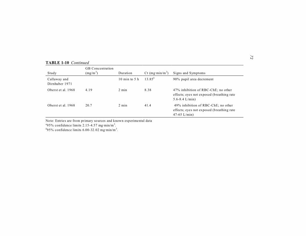
NERVE AGENTS GA, GB, GD, GF, AND VX 79
TABLE 1-13 ContinuedSpecies Duration (min) Ct (mg"min/m3) ReferenceRat 90 433 (m) Mioduszewski et
al. 2000, 2001a
Rat 240 982 (m) Mioduszewski etal. 2000, 2001a
Rat 360 1040 (m) Mioduszewski etal. 2000, 2001a
Rat 10 220 DA 1974Rat 10 300 Cohen et al. 1954a
Rat 1 220 (m), 118 (f) Callaway andBlackburn 1954a
Rat 5.0-6.7 191 (f) Schoene et al.1985a
Mouse 30 501 Bide et al. 1999Mouse 30 600 Husain et al. 1993a
Mouse (active) 10 240 DA 1974Mouse (resting) 10 310 DA 1974
aItalic entries are from primary sources and known experimental data.Abbreviations: f, female; m, male.
Agent GD
In tests conducted on baboons, Johnson et al. (1988) found that theeffects of inhalation exposures were in close agreement with the reportedintravenous LD50 values of 6.6 µg/kg. Adams et al. (1975) reported a 15-dintramuscular LD50 of 6.57 µg/kg.
3.1.2. Dogs
Agent GB
NDRC reported LCt50 values of 100-150 mg"min/m3 for 10-minexposures. Cresthull et al. (1959) reported that inhalation doses of GB at
80 ACUTE EXPOSURE GUIDELINE LEVELS FOR SELECTED AIRBORNE CHEMICALS
TABLE 1-14 Acute Inhalation Lethality Values for Agent GA in Animals(Toxicity Value, LCt50)Species Duration (min) Ct (mg"min/m3) ReferenceMonkey 10 187 DA 1974a
Monkey 10 135 DA 1974a
Dog 2 320 DA 1974a
Rabbit 10 960 DA 1974a
Rat 10 450 DA 1974a
aDA (1974) is a secondary source.
25.1 and 26.0 µg/kg (0.6- and 2.23-min exposures) were not lethal to dogs,but inhalation doses of GB at 32.5 µg/kg or higher caused 40% or moremortality. Bide et al. (1999) and Yee et al. (1999) developed a threedimensional probit model to calculate lethality values (LC05, LC50, LC95)from historic laboratory data and to estimate equivalent human values.Using the species-specific constants for inhalation rates, body mass, etcetera, provided by these authors, the 30-min LC50 value for dogs wascalculated to be 4.3 mg/m3.
Inhalation studies were conducted by Oberst (1961) on dogs (breed notidentified) fitted with masks through which GB vapor flowed. The eyeswere protected by eyepieces and were not exposed. The resulting 2-minLCt50 value of 56 mg"min/m3 is reported in Table 1-9.
Agent GA
DA (1974) (secondary source) reported an LCt50 value of 320mg"min/m3 for dogs.
Agent VX
Bide and Risk (2000) cite several earlier studies in which the LCt50values for VX aerosols were reported to be 15 mg"min/m3 (whole body)(Krackow 1956) and 15.1 mg"min/m3 (Punte and Atkinson 1960).
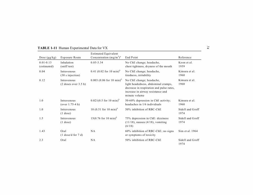
NERVE AGENTS GA, GB, GD, GF, AND VX 81
TABLE 1-15 Acute Inhalation Lethality Values for Agent GD in Animals(Toxicity Value, LCt50)Species Duration (min) Ct (mg"min/m3) ReferenceRabbit 10 160 DA 1974Rat 1 196 (m)
135 (f)Callaway andBlackburn 1954a
Rat 5.3-8.5 211 (f) Schoene et al.1985a
Rat 10 279 DA 1974
Rat 10 230 DA 1974Rat < 30
(threshold at 16)400(threshold at 335)
Aas et al. 1985a
Guinea pig 8 480 Langenberg et al.1998a
aEntries are from primary sources and known experimental data.Abbreviations: f, female; m, male.
3.1.3. Rats
Agent GB
NDRC (1946) reported LCt50 values of 150-300 mg"min/m3 for 10-minexposures. Bide et al. (1999) and Yee et al. (1999) developed a threedimensional probit model to calculate lethality values (LC05, LC50, LC95)from historic laboratory data and to estimate equivalent human values.Using the species-specific constants provided, the 30-min LC50 value forrats was calculated to be 8.2 mg/m3.
In studies conducted by Mioduszewski et al. (2000, 2001, 2002a), theacute lethal toxicity of GB to male and female Sprague-Dawley rats wasevaluated for time periods of 10, 30, 60, 90, 240 and 360 min in a whole-body dynamic chamber. The final report of this study (Mioduszewski et al.2001, 2002a) is further documentation of the findings presented below. Tenmales and 10 females were used for each concentration-time (Ct)combination, and 50 males and 50 females were used for each time point.GB concentrations ranged from about 2 mg/m3 to 56 mg/m3. Agentconcentrations were confirmed in the exposure chamber by three procedures(“Edgewood” bubblers, solid sorbent tubes, and a phosphorous monitor) to
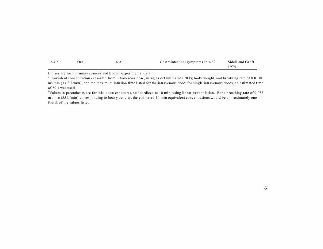
82 ACUTE EXPOSURE GUIDELINE LEVELS FOR SELECTED AIRBORNE CHEMICALS
TABLE 1-16 Acute Inhalation Lethality Values for Agent GF in Animals(Toxicity Value, LCt50)a
Species Duration (min) Ct (mg"min/m3) ReferenceRat 10 368 (m)
253 (f)Anthony et al. 2002
Rat 60 396 (m)334 (f)
Anthony et al. 2002
Rat 240 595 (m)533 (f)
Anthony et al. 2002
a24-h postexposure lethality.Abbreviations: f, female; m, male.
allow point and continuous determinations (Mioduszewski et al. 2000,2001, 2002a). Lethality was assessed at 24 h and at 14 d. Female rats werereported to be more sensitive to GB vapor toxicity than males over therange of exposure concentrations and durations studied (Mioduszewski etal. 2000, 2001, 2002a). Gender differences were reported to be significantat p < 0.01. Probit analysis (MINITAB, version 13) presented inMioduszewski et al. (2000) gave the following 14-d LC50 values for femalerats exposed to GB vapor: 18.1 mg/m3 for 10 min; 8.51 mg/m3 for 30 min;6.39 mg/m3 for 60 min; 3.03 mg/m3 for 4 h; and 2.63 mg/m3 for 6 h.
Probit analysis presented in Mioduszewski et al. (2000) gave thefollowing 14-d LC50 values for male rats:22.6 mg/m3 for 10 min; 8.84mg/m3 for 30 min; 7.55 mg/m3 for 60 min; 4.09 mg/m3 for 240 min; 2.89mg/m3 for 360 min.
Based on a probit analysis of the data (Mioduszewski et al. 2000), theestimated 14-d LC01 values for the females are as follows: 11.54 mg/m3 for10 min; 5.84 mg/m3 for 30 min; 4.01 mg/m3 for 60 min; 2.09 mg/m3 for 4h; 1.76 mg/m3 for 6 h.
This is the critical study and data set (females) for determination ofAEGL-3 values.
GB vapor exposure significantly inhibited rat blood cholinesteraseactivity in the Mioduszewski et al. (2000, 2001, 2002a) study. However,no correlation between severity of clinical signs and cholinesteraseinhibition was reported.
Cohen et al. (1954) reported that exposure of rats to GB at 30 mg/m3 for10 min (Ct = 300 mg"min/m3) resulted in mortality rates of close to 50%
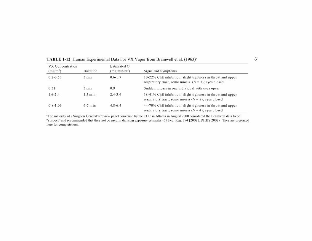
NERVE AGENTS GA, GB, GD, GF, AND VX 83
TABLE 1-17 Lethality in Laboratory Species Following Inhalation Exposure to Agent VX Vapor (End Point, LCt50)Species Duration (min) Ct (mg"min/m3) ReferenceMouse(head only)
10 min 13.6 Koon et al. 1960, ascited in NRC 1997
Mouse(whole body)
10 min 4.0 Koon et al. 1960, ascited in NRC 1997
Mouse(nose only)
_ 71 Carroll et al. 1957
Mouse(whole body)
_ 16.1 Carroll et al. 1957
Goat 10 min 9.2 Koon et al. 1960, ascited in NRC 1997
Mouse 6 h/d, 5 d/wk,2 wk
0.9 Crook et al. 1983(noncredible data)
Rat 6 h/d, 5 d/wk,2 wk
24.9 Crook et al. 1983(noncredible data)
Guinea pig 6 h/d, 5 d/wk,2 wk
238.6 Crook et al. 1983(noncredible data)
Rabbit 6 h/d, 5 d/wk,2 wk
No deaths Crook et al. 1983(noncredible data)
and a decrease in brain cholinesterase activity levels to less than 5% ofnormal.
Schoene et al. (1985) reported an LCt50 of 191 mg"min/m3 (95% CI =178-204 mg"min/m3) for exposure times of 5.0-6.7 min for female Wistarrats. The corresponding 5-min LC50 is 38 mg/m3. Callaway and Blackburn(1954) reported LCt50 values of 220 mg"min/m3 for male albino rats and 118mg"min/m3 for female albino rats (1-min exposures).
Agent GA
DA (1974) (secondary source) reported an LCt50 value of 450mg"min/m3 for the rat.
84 ACUTE EXPOSURE GUIDELINE LEVELS FOR SELECTED AIRBORNE CHEMICALS
Agent GD
In a study designed to secondarily examine agent GD toxicity, Aas etal. (1985) reported that the LCt50 for GD in rats (six animals tested at eachof three exposure levels for periods of time <30 min) was 400 mg"min/m3.Aas et al. (1985) graphically present their data as an LCt-versus-mortalitycurve. The lethality threshold estimated from the curve is about 335mg"min/m3. Because the reported GD concentration was fixed at 21 mg/m3
for the duration of the study, the exposure time corresponding to the lethalthreshold is 16 min (see Table 1-11).
The principal objective of the Aas et al. (1985) study was to test anexperimental dynamic flow system that would allow study of highly toxicvapors. In consequence, it was necessary to continually generate smallamounts of the toxic material in question. Agent GD (soman) was thecompound selected to best test the system. Secondary objectives of thestudy were to determine the (short-term) inhalation toxicity of agent GD(soman) and to study inhibition of acetylcholinesterase, cholinesterase, andcarboxylesterase activity in the respiratory tract (relative to other tissues).
Schoene et al. (1985) reported an LCt50 of 211 mg"min/m3 (95% CI =195-229 mg"min/m3) for exposure times of 5.3-8.5 min for female Wistarrats. Callaway and Blackburn (1954) reported LCt50 values of 196mg"min/m3 for male albino rats and 135 mg"min/m3 for female albino rats(1-min exposures) (Table 1-11).
Agent GF
Callaway and Blackburn (1954) reported LCt50 values of 181mg"min/m3 for male albino rats and 110 mg"min/m3 for female albino rats(1-min exposures). Kassa and Cabal (1999) reported an intramuscular LD50of 80 µg/kg for rats.
A recent study of GF vapor inhalation toxicity in male and female SDrats reported 24-h postexposure LC50 and LCt50 values for three exposureperiods (10, 60, and 240 min) (Anthony et al. 2002). Young adult rats wereexposed whole-body in a dynamic 750-L chamber under protocols similarto those previously published by Mioduszewski et al. (2001, 2002a) butwith additional accommodation for the lesser volatility of agent GF. Forfemale rats, Anthony et al. (2002) report 24-h LC50 values as follows: 10min, 25.3 mg/m3; 60 min, 5.56 mg/m3; 240 min, 2.22 mg/m3. For male rats,
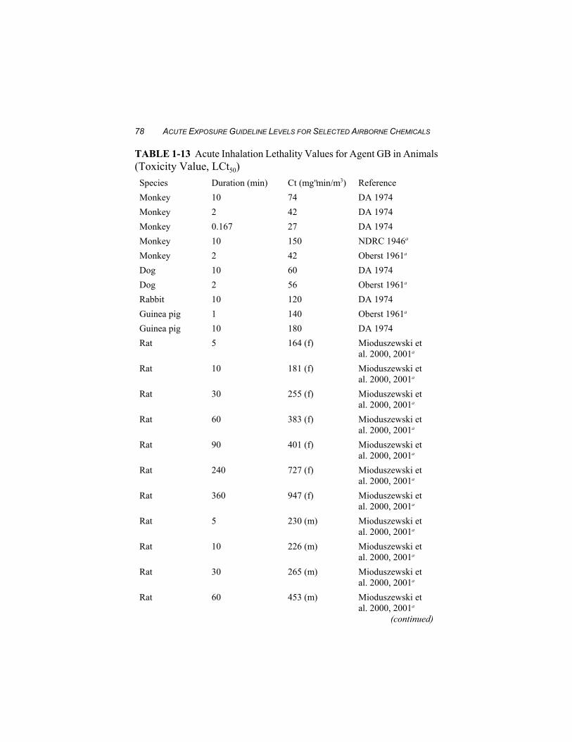
NERVE AGENTS GA, GB, GD, GF, AND VX 85
24-h LC50 values are as follows: 10 min, 36.8 mg/m3; 60 min, 6.60 mg/m3;240 min, 2.48 mg/m3. These results are summarized as LCt50 values inTable 1-16.
Agent VX
Crook et al. (1983) reported an LCt50 of 24.9 mg"min/m3 for animalsexposed 6 h/d, 5 d/wk, for 2 wk; however, the results of the study are notconsidered credible (see Section 3.2).
Bide and Risk (2000) exposed outbred male CD1(SD)BR rats to NaClaerosols containing entrained VX. The animals (five per test group) weretested with a nose-only inhalation system and for an exposure time of 12min. Test concentrations were not reported. The observed LCt50 was 67mg"min/m3.
In studies conducted by Maxwell (1992) on Sprague-Dawley rats, thesubcutaneous LD50 for VX was reported to be 0.027 µmol/kg.
DA (1974) (secondary source) reports intragastric LD50 values of 0.1mg/kg and 0.077-0.1280 mg/kg for rats.
3.1.4. Mice
Agent GB
NDRC (1946) reported LCt50 values of 150-300 mg"min/m3 for 10-minexposures, 360 mg"min/m3 for a 20-min exposure, and 420 mg"min/m3 fora 30-min exposure (14 mg/m3 for 30 min). Clement (1992) reported asubcutaneous LD50 value of 170 µg/kg for male CD-1 mice with bodyweight ranging from 30-40 g (five animals per test group). A subcutaneousLD50 of 0.212 mg/kg was reported for male and female Shanghai mice (18-22 g body weight, eight mice per group, five exposure groups) (Luo andLiang 1997). Bide et al. (1999) and Yee et al. (1999) developed a threedimensional probit model to calculate lethality values (LC05, LC50, LC95)from recently conducted laboratory experiments on mice and historictoxicity data for other laboratory species and to estimate equivalent humanvalues. Using the species-specific constants provided by Bide et al. (1999),the 30-min LC50 value for the mouse was calculated to be 16.7 mg/m3.
The LCt50 for agent GB in female Swiss albino mice has been reported
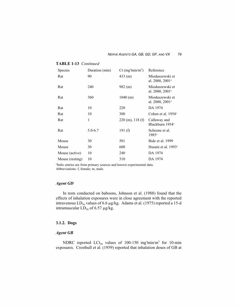
86 ACUTE EXPOSURE GUIDELINE LEVELS FOR SELECTED AIRBORNE CHEMICALS
to be 600 mg"min/m3 (Husain et al. 1993), equivalent to a 30-min exposureat 20 mg/m3. Lohs (1960) reported a 30-min inhalation lethality value ofvalue of 5 mg/m3.
Agent GD
Lohs (1960) reported a 30-min inhalation lethality value of 1 mg/m3.
Agent GF
Inhalation lethality data for agent GF were not found in the availableliterature. Luo and Liang (1997) reported an LD50 of 0.346 mg/kg in miceinjected with the agent subcutaneously. Clement (1992) reported asubcutaneous LD50 value of 243 µg/kg for male CD-1 mice with bodyweights ranging from 30-40 g (five animals per test group).
Agent VX
Ten-minute LCt50 values of 4.0 mg"min/m3 (whole body) and 13.6mg"min/m3 (head only) have been reported for mice exposed to VX vapors(Table 1-6) (Koon et al. 1960, as cited in NRC 1997). LCt50 values of 71mg"min/m3 for female mice for nose-only exposures and 16.1 mg"min/m3
for whole-body exposures were reported by Carroll et al. (1957) for femalemice; however, in this study it was reported that the concentration of theagent in the exposure chamber was not measured directly but was estimatedfrom the mortality level, which was correlated with the LD50 for anintravenous injection (estimated to be 17 µg/kg).
Crook et al. (1983) reported an LCt50 of 0.9 mg"min/m3 for animalsexposed 6 h/d, 5 d/wk, for 2 wk to VX vapors; however, the results of thisstudy are not considered credible.
Koplovitz et al. (1992) exposed Swiss ICR mice intramuscularly to GBand VX. The resulting acute (24-h) LD50 are as follows: LD50 for GB of204.81 µg/kg, LD50 of VX of 13.07 µg/kg.
Bide and Risk (2000) exposed outbred male CD1(ICR)BR mice toNaCl aerosols containing entrained VX. The animals (five per test group)were tested with a nose-only inhalation system and for a exposure time of

NERVE AGENTS GA, GB, GD, GF, AND VX 87
12 min. Test concentrations were not reported. The observed LCt50 was 72mg"min/m3. Bide and Risk (2000) also cite several earlier studies in whichthe LCt50 values for VX aerosols were reported to be 7 mg"min/m3
(Krackow 1956) and 6.1 mg"min/m3 (Punte and Atkinson 1960).
3.1.5. Guinea pigs
Agent GB
NDRC (1946) reported LCt50 values of 150-250 mg"min/m3 for 10-minexposures. Bide et al. (1999) and Yee et al. (1999) developed a threedimensional probit model to calculate lethality values (LC05, LC50, LC95)from historic laboratory data and to estimate equivalent human values.Using the species-specific constants provided, the 30-min LC50 value forguinea pigs was calculated to be 7.5 mg/m3.
Oberst (1961) conducted inhalation studies on guinea pigs administeredGB vapor by means of face masks “which filled the face without leakage.”The author does not mention eye protection for guinea pigs. The resulting1-min LCt50 of 140 mg"min/m3 is reported in Table 1-9.
Atchison et al. (2001) reported that subcutaneous injections of 0.6 LD50GB once per day, 5 d/wk, in young male Hartley guinea pigs (600 g)resulted in 50% mortality (two of four) after 2 wk of exposure and 100%mortality after 3 wk. The subcutaneous LD50 for guinea pigs was reportedto be 42 µg/kg.
Agent GD
Allon et al. (1998) reported an inhalation LD50 of 101 µg/kg for guineapigs, considerably higher than the reported intravenous LD50 values of 22µg/kg (Sterri et al. 1982) and 3.5 µg/kg (Due et al. 1993). For guinea pigsweighing 0.84 kg and breathing 0.4 m3/d (EPA defaults), a dose of 101µg/kg would be equivalent to an exposure to GD at 0.009 mg/m3 for 24 h,0.22 mg/m3 for 1 h, or 0.44 mg/m3 for 30 min. Langenberg et al. (1998)reported an 8-min LCt50 of 480 mg"min/m3 for guinea pigs. This value isequivalent to an LC50 of 60 mg/m3 for 8 min or 16 mg/m3 for 30 min(assuming linear scaling).
Atchison et al. (2001) reported that subcutaneous injections of 0.6 LD50
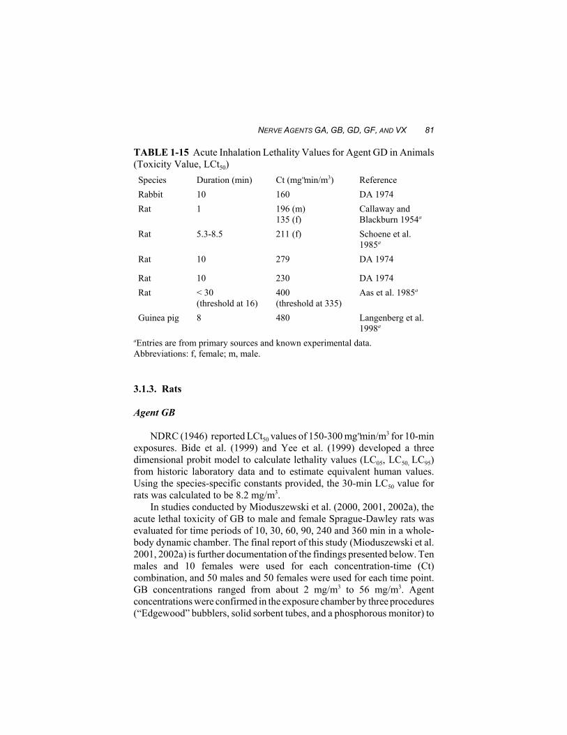
88 ACUTE EXPOSURE GUIDELINE LEVELS FOR SELECTED AIRBORNE CHEMICALS
GD once per day, 5 d/wk, in young male Hartley guinea pigs (600 g)resulted in 50% mortality (two of four) after 2 wk of exposure and 100%mortality after 9 wk. The subcutaneous LD50 for guinea pigs was reportedto be 28 µg/kg.
Agent VX
Crook et al. (1983) reported an LCt50 of 238 mg"min/m3 for animalsexposed 6 h/d, 5 d/wk, for 2 wk; however, the results of this study are notconsidered credible.
Koplovitz et al. (1992) exposed Hartley albino guinea pigs(subcutaneous) to GB and VX. The resulting acute (24-h) LD50 are asfollows: LD50 for GB of 41.26 µg/kg, LD50 for VX of 6.89 µg/kg.
Bide and Risk (2000) exposed outbred male (HA)BR guinea pigs toNaCl aerosols containing entrained VX. The animals (five per test group)were tested with a nose-only inhalation system and for an exposure time of12 min. Test concentrations were not reported. The observed LCt50 was 30mg"min/m3. Bide and Risk (2000) also cite several earlier studies in whichthe LCt50 values for VX aerosols were reported to be 30 mg"min/m3 (wholebody) (Krackow 1956) and 29.5 mg"min/m3 (Punte and Atkinson 1960).
Atchison et al. (2001) reported that subcutaneous injections of 0.6 LD50VX once per day, 5 d/wk, in young male Hartley guinea pigs (600 g)resulted in 33% mortality (two of six) after 10 wk of exposure and 83%mortality (five of six) after 9 wk. One animal survived the full 13 wk oftreatment. The subcutaneous LD50 for guinea pigs was reported to be 9µg/kg.
3.1. 6. Rabbits
Agent GB
NDRC (1946) reported LCt50 values of 120-250 mg"min/m3 for 10-minexposures. Bide et al. (1999) and Yee et al. (1999) developed a threedimensional probit model to calculate lethality values (LC05, LC50, LC95)from historic laboratory data and to estimate equivalent human values.Using the species-specific constants provided, the 30-min LC50 value forrabbits was calculated to be 5.6 mg/m3.