paediatrics notes 3 update pdf
DESCRIPTION
pediatric notesTRANSCRIPT

Umwangange. Paediatrics, BSNM 2013 Page 1
CHAPTER I. DISEASES AFFECTING VARIOUS SYSTEMS
I. RESPIRATORY SYSTEM
A. ACUTE BRONCHITIS
DEFINITION
Acute bronchitis is infection and inflammation of the bronchi. It is a common disease that can
occur at any time of the year, but most cases happen in the winter months. It is most common in
infants and young children.
CAUSES & RISK FACTORS
Acute bronchitis is the result of a viral( e.g. Respiratory syncitial virus) or bacterial infection
(e.g. Hemophilus influenza) which causes inflammation of the airways; bronchi. Mycaplasma
pneumoniae is a common cause in children older than 6 years.
Risk factors:
- Air pollution exposition
- Lung irritants
- exposition to cold
SIGNS & SYMPTOMS
Cold-like symptoms (headache, malaise, rhinorrhoea…)
Painful cough : Dry cough in early stages and productive cough (with purulent sputum)
usually in later stages (in 2 to 3 days).
Children younger than 5 years rarely expectorate, sputum is usually seen in vomitus (i.e.
posttussive emesis)
Wheezing
Fever (in case of bacterial bronchitis )
Generally sick
Trouble breathing: shortness of breath
Noisy breathing (rattling sound in the chest)

Umwangange. Paediatrics, BSNM 2013 Page 2
DIAGNOSIS
- Making a diagnosis of acute bronchitis begins with taking a thorough medical history,
including symptoms, and exposure to lung irritants.
- A physical examination is also performed and includes listening with a stethoscope to the
sounds that lungs make during respiration. Lung sounds that may point to a diagnosis of acute
bronchitis include wheezing, or crackling sound and decreased lung sounds.
- A chest X-ray and CT scan of the chest can help to exclude the diagnosis of pneumonia.
- Blood or sputum culture if antibiotic therapy is under consideration
DIFFERENTIAL DIAGNOSIS
Differential diagnosis include but are not limited to:
- Pneumonia
- Upper respiratory tract infections such as rhinitis
- influenza or cold flu
TREATMENT
- The goal of treatment of acute bronchitis is to control symptoms, such as fever, cough, and
shortness of breath, and to minimize the development of serious complications, such as
pneumonia.
The first step in treatment is prevention. The risk of developing acute bronchitis can be reduced
by avoiding air pollutants, cold…
- Ensure that the child has adequate oxygenation.
- General measures include:
Rest
Fluids for adequate hydration
Pain relief and Antipyretics: Paracetamol, Ibuprofen
Cough suppressant: such as Codeine, Bronchalene in case of dry cough,
Umwangange. Paediatrics, BSNM 2013 Page 3
Antibiotics : Such as Amoxycillin, in case of bacterial bronchitis. The combination of
Amoxycillin and clavulanic acid (Augmentin) should also be used. Erythromycin is used
in case of allergy or resistance to penicillin (amoxicillin)
Physiotherapy: respiratory exercise, postural drainage
Humidifier: to try to thin out sputum
Avoid very cold weather
COMPLICATIONS
- Bronchopneumonia
- Acute respiratory failure
B. PNEUMONIA
Pneumonia, inflammation of the pulmonary parenchyma, is common throughout childhood but
occurs more frequently in infancy and early childhood. Clinically, pneumonia may occur either
as a primary disease or as a complication of some illness.
Pneumonia is the single largest cause of death in children worldwide. Every year, it kills an
estimated 1.2 million children under the age of five years, accounting for 18% of all deaths of
children under five years old worldwide (WHO, 2012)
Morphologically, pneumonias are recognized as follows:
Lobar pneumonia: all or a large segment of one or more pulmonary lobes is involved.
When both lungs are affected, it is known as bilateral or “ double” pneumonia.
Bronchopneumonia: begins in the terminal bronchioles, which become clogged with
mucopurulent exudates to form consolidated patches in nearby lobules; also called
lobular pneumonia.
Intertitial pneumonia: The inflammatory process is more or less confined within the
alveolar walls (interstitium) and the peribronchial and interlobular tissues.

Umwangange. Paediatrics, BSNM 2013 Page 4
CAUSES
Viral: respiratory syncytial virus is the most common viral cause of pneumonia.
Atypical (mycoplasma)
Bacterial: pneumococcus/ Streptococcus pneumoniae is the most common cause of
bacterial pneumonia). Haemophilus influenza type b (Hib) is the second most common
cause of bacterial pneumonia.
Fungi (e.g. histomycosis, coccidiomycosis… )
In HIV- infected infants, Pneumocystis jiroveci is responsible for at least one quarter of
all pneumonia deaths.
Aspiration of foreign substances
RISK FACTORS
Compromised immune systems: in case of malnutrition, HIV infection, measles…
Environmental factors: indoor air pollution, living in crowded homes, parental
smoking…
PATHOLOGY/ EVOLUTION OF PNEUMOCOCCAL PNEUMONIA
Pneumococcal pneumonia evolves through a series of pathologic changes:
- The initial phase is one of edema: presence of a proteinaceous exudates and often of bacteria
in the alveoli.
- The 2nd
phase, red hepatization: The presence of erythrocytes in the cellular intraalveolar
exudate gives this second stage its name, but neutrophils are also present and are important from
the standpoint of host defense. Bacteria are occasionally seen in cultures of alveolar specimens
collected during this phase.
- The third phase, gray hepatization: no new erythrocytes are extravasating, and those already
present have been lysed and degraded. The neutrophil is the predominant cell, fibrin deposition is
abundant, and bacteria have disappeared. This phase corresponds with successful containment of
the infection and improvement in gas exchange.

Umwangange. Paediatrics, BSNM 2013 Page 5
-The final phase, resolution, the macrophage is the dominant cell type in the alveolar space,
and the debris of neutrophils, bacteria, and fibrin has been cleared, as has the inflammatory
response.
This pattern has been described best for pneumococcal pneumonia and may not apply to
pneumonias of all etiologies, especially viral or Pneumocystis pneumoni
CLINICAL MANIFESTATIONS
Cough. Newborns (neonates) with pneumonia rarely cough.
Fever, chills
Tachypnea: the absence of tachypnea is a strong indication that the child does not have
pneumonia
Wheezes and/or crackles on auscultation
In severe cases there is the use of accessory muscles, retractions including sub-costal and
inter-costal, sub-xyphoid, nasal flaring
Hypoxemia may be present, but is often a late sign.
Prostration. Very severely ill infants may be unable to feed or drink and may also
experience unconsciousness, hypothermia and convulsions.
These symptoms may be preceded by minor Upper Respiratory Infections (URIs)
symptomatology including low- grade fever and rhinorrhea.
DIAGNOSTIC
The clinical manifestations and the physical examination can make a diagnosis of
Pneumonia.
Leucocytosis (WBC > 15,000-20,000/mm3) in association with fever of 39
0C often
suggests a bacterial etiology.
Blood culture especially in severe pneumonia and in infants under 3 months of age.
Gram stain and culture on respiration secretions (sputum, bronchoalveolar lavage, pleural
fluids) can be helpful
Chest Radiography to confirm the presence and location of pneumonia; however, a
normal chest X-ray does not rule out pneumonia.

Umwangange. Paediatrics, BSNM 2013 Page 6
MANAGEMENT
- Hospitalization: its indication includes Oxygen requirement (sat. < 95%), infants < 2 months,
presence of moderate to large pleural effusions and in other very severe cases.
- Antibiotics in inpatient:
. Ampicilline + Gentamycine for neonates 0-30 days
. Cefotaxin is antibiotic of choice in children between 1 month and 5 years
. If Chlamydia trachomatis or Brodetera pertussis are suspected, give a Macrolide e.g.
Erythromycine
. For Hospitalized children over 5 years: Cefotaxin+ Erythromycin
The duration of therapy depends on the patient’s response but in general is 10-14 days for
children up to 3 months of age and 7-10 days for children older than 3 months.
- For outpatient:
. Children under 5 years: Amoxycillin in High dose (80-90mg/kg/day)
. If the child is vomiting, an initial parental dose of Ceftriaxone is given
. For children over 5 years, give a macrolide (Erythromycin)
- In case of viral pneumonia, there is no need of antibiotic.
PREVENTION
- Preventing pneumonia in children is an essential component of a strategy to reduce child
mortality. Immunization against Hib, pneumococcus, measles and whooping cough (pertussis) is
the most effective way to prevent pneumonia.
- Adequate nutrition: starting with exclusive breastfeeding for the first six months of life.
- Prevent environmental factors
- In children infected with HIV, the antibiotic cotrimoxazole is given daily to decrease the risk
of contracting pneumonia.

Umwangange. Paediatrics, BSNM 2013 Page 7
C. BRONCHIAL ASTHMA
Asthma is the most common chronic disorder in children and adolescents. About 5 million of
children under 18 years have asthma. This includes an estimated 1.3 million children under the
age of 5 years.
PATHOPHYSIOLOGY
Asthma is a chronic inflammatory disorder of airway in which many cells play a role, including
mast cells and eosinophils. The symptoms are associated with airway obstruction
(bronchoconstriction) due to inflammation with mucosal edema leading to bronchospasm. The
airway narrowing is often reversible either spontaneously or with treatment, and causes an
associated increase in airway responsiveness to variety of stimuli as well as bronchial
hypersecresion.
Risk factors
exposure to tobacco smoke
Previous allergic reactions: food allergies, allergic rhinitis, allergic sinusitis, eczema
Family history of asthma, family history of allergy
Exposure to air pollution
Recurrent respiratory viral infections
Triggers
The asthma triggers vary from child to child and can include:
Viral infections such as the common cold
Exposure to air pollutants, such as tobacco smoke
Allergies to dust mites, pet dander, pollen or mold
Physical activity
Weather changes or cold air
Emotions and stress
Sometimes, asthma symptoms occur with no apparent triggers.

Umwangange. Paediatrics, BSNM 2013 Page 8
CLINICAL FEATURES
Cough
Cough at night or with exercise
Allergic symptoms such as rhinitis, sinusitis, eczema
Wheezing
Episodic wheeze, ronchi
Shortness of breath
chest tightness
Symptoms increase in case of aeroallergens or exercise, they worsen at night, awakening
the patient.
THERAPY
- Assess the severity: intermittent, mild persistent, moderate persistent, severe persistent.
- Pharmacological management: the use of agents to control and agents for quick relief of
symptoms.
Control agents: inhaled corticosteroids (e.g. Fluticasone, Beclomethasone…),
long acting Beta- agonists/bronchodilators (e.g. Salmeterol…),
Theophylline/Aminophylline, Leucotrienes modifiers (e.g.Montelukast/
Singulair), anti-immunoglobulin E (e.g. Omalizumab)
Relief medications: short- acting bronchodilators (e.g. Albuterol/Salbutamol), oral
and parenteral corticosteroids (e.g. Prednisolone)
- Acute asthma management includes the use of B-Agonist for a quick ;When B-agonists
are ineffective or required more than four times a day give bronchodilatator e.g.
salbutamol add oral corticoids (1-2mg/kg/day for 3-5days eg dexamethazone,
prednisolone.
- In case of acute severe asthmatic episodes (Status asthmaticus): Correct the significant
hypoxemia with supplemental oxygen. Mechanically assisted ventilation may be required
in alveolar hypoventilation.
Umwangange. Paediatrics, BSNM 2013 Page 9
PROGNOSIS
There is no cure for asthma. Symptoms sometimes decrease over time. With repetitive attacks,
school absenteeism is one of the outcomes.
II. GASTRO- INTESTINAL SYSTEM
A. DIARRHEA
Diarrhea is loss of watery stools (or bloody) for more than 3 times a day (more than 5 times a
day for the neonates and small infants). Acute diarrhea is a common problem that usually lasts 1
or 2 days, it can be prolonged and persist for more than 2 days and poses the risk of dehydration.
Chronic diarrhea may be a feature of a chronic disease.
Diarrhea is a common gastrointestinal problem during infancy and early childhood. In
developing countries, diarrhea causes around two million child deaths annually.
CAUSES OF DIARRHEA
Acute diarrhea
Bacterial infections. Several types of bacteria consumed through contaminated food or
water can cause diarrhea. Common causes include Campylobacter, Salmonella, Shigella,
and Escherichia coli (E. coli).
Viral infections. Many viruses cause diarrhea, including rotavirus, Norwalk virus,
cytomegalovirus, herpes simplex virus, and viral hepatitis.
Food intolerances. Some people are unable to digest food components such as lactose;
the sugar found in milk, infants can also have milk-protein allergies.
Parasites. Parasites can enter the body through food or water and settle in the digestive
system. Parasites that cause diarrhea include Giardia lamblia, Entamoeba histolytica
Reaction to medicines. Antibiotics, cancer drugs (chemotherapy), and antacids
containing magnesium can all cause diarrhea.
Intestinal diseases. Inflammatory bowel disease, colitis, Crohn’s disease, Congenital
aganglionic megacolon often lead to diarrhea.

Umwangange. Paediatrics, BSNM 2013 Page 10
Functional bowel disorders. Diarrhea can be a symptom of irritable bowel syndrome.
Chronic diarrhea
Diarrhea that persists longer than 4 weeks is considered to be chronic.
SIGNS & SYMPTOMS
-Frequent elimination: liquid stool (watery or bloody) accompanied by cramping, abdominal
pain, nausea. Depending to the cause, the child may have fever, vomiting.
DIAGNOSTIC
Stool culture
Stool examination
Blood tests
Sigmoidoscopy/ colonoscopy
COMPLICATIONS
Dehydration is the principal complication of diarrhoea,
Malnutrition in case of chronic diarrhoea
Rectal prolapse in case of repetitive diarrhoea or chronic diarrhoea.
MANAGEMENT OF DIARRHEA
- Prevent the dehydration by giving ORS (Oral Rehydrating Solution)
-Teach the mother on hygiene and sanitation
-To treat aetiology: e.g anti-infectious medication…
- Zinc supplementation in children over 6 months: in developing countries, many children have
zinc deficiency. Zinc sulfate reduces the duration of diarrhoea and accelerates appetite
- Treat Dehydration
Table 1: Clinical assessment of degree of dehydration (Goldman et al., 2008)

Umwangange. Paediatrics, BSNM 2013 Page 11
Degree of
dehydration
Mild
(5-7% body weight)
Moderate
(7-9% body weight)
Severe
(>10% body weight)
Fontanelle Slightly sunken Very sunken Very sunken
Mucous membranes Slightly humid Dry Very dry
Skin turgor Normal Slightly
decreased(<2sec)
Markedly
decreased(≥2sec)
Capillary refill time Normal (<3 seconds) Normal (<3 seconds) Delayed (≥3 seconds)
Urine output Normal Slightly decreased Decreased or absent
Mental status Normal Slightly fussy Irritable or lethargic
Based on the degree of dehydration, the following approach to management has been suggested:
Table 2: Management of Dehydration
Degree of
dehydration Management
Mild Home-based treatment
Moderate
Oral rehydration as the child needs; and give 50-100 ml for every watery stool
(<12months), 100-200ml for every watery stool (≥ 12months)
oral rehydration therapy (ORS 75ml/kg in 4 hours) if the child vomits use
nasogastric tube
Reassess, if the child is now in mild dehydration, adjust the treatment.
Severe
Admit patient, do bloodwork - give 30 mL/kg bolus IV fluids over 1 hour
(<12months) or over 30min (≥12months), check pH, bicarbonate, nitrogen.
Continue IV as required: 70ml/kg in 5hours (<12months) and 70ml/kg in 2h 30
(≥ 12months). Assess the patient every one hour in less than 12 months children
and every 30 minutes in 12months and more.(Use surveillance sheet)
Oral rehydration therapy include ORS with electrolytes, breastmilk and not tea, sugar drinks
such as apple juice and other juices, homemade remedies.
In a child, the normal fluid requirement (ORS) per 24 hours (maintenance therapy) is
approximately:
100 ml/kg for the first 1–10 kilograms of body weight
50 ml/kg for the next 11–20 kilograms of body weight
20 ml/kg for every kilogram of body weight exceeding 20 kg.
Example: fluid requirement in a child weighing 26 kg is 10 × 100 ml + 10 × 50 ml + 6 ×
20 ml = 1 620 ml. ( From EBM Guidelines)
Umwangange. Paediatrics, BSNM 2013 Page 12
Indications for referral to hospital (From Evidence-based medicine Guidelines )
The child is referred if even one of the following criteria is met:
→ Age below 6 months
→ Profuse diarrhoea or vomiting, poor general condition
→ Dehydration of 8% or more (at least moderately severe dehydration)
→ Diarrhea lasting for more than 5 days (the general condition and loss of weight are
decisive factors)
→ Colicky abdominal pain (and sudden ceasing of the diarrhea): intussusception?
→ Bloody diarrhoea
→ Inability to treat the child at home
Correction of the estimated dehydration is often possible by administration of a corrective
solution through a nasogastric tube at the outpatient department of a hospital. Afterwards,
the child is examined and weighed and can usually be discharged for follow-up care at
home.
If the child is in shock when referred, infuse Ringer solution 20 ml/kg in 15 minutes.
In a hospitalized patient, fluid balance parameters, CRP, basic blood picture and blood
gas analysis are examined.
B. PARASITOSIS
ASCARIASIS
This is caused by ascaris lumbricoides. It is seen more commonly in the children between the
age of one and five years with lower socioeconomic status (poor hygiene). It is transmitted
through contaminated food, water, hands, and utensils.
Manifestations
Slight fever due to larvae;
Ascaris pneumonia due to larvae in the lungs with dry cough (Lo ffler syndrome);
Diarrhoea, abdominal pain and irritability
Loss of appetite, loss of weight
Diagnosis
Stool examination: eggs of ascaris in the stool

Umwangange. Paediatrics, BSNM 2013 Page 13
Complications
In heavy infections, a large bolus of entangled worms can cause pain and small-bowel
obstruction, sometimes complicated by perforation or volvulus.
A large worm can enter and occlude the biliary tree, causing biliary colic, cholecystitis,
cholangitis, pancreatitis, or (rarely) intrahepatic abscesses.
Treatment: Mebendazole.
Prevention: - Hygiene (use of proper water to drink, to clean fruits and vegetables...)
- Encourage routine hand washing (e.g. after the toilet, before eating) in children
also should minimize infestation
- use of sanitary latrines minimize infestation
-For children under 5 years, give mebendazole 500mg every 3 months.
OXYUROSE
It is caused by Enterobius vermicularis and is transmitted because of unsanitary living
conditions in the school, hostel and families.
It is transmitted through contaminated soil, fingers, flies.
manifestations
General symptoms found:
loss of weight
lack of appetite
abdominal pain
Itching at anal region.
diagnosis
Stool examination.
treatment

Umwangange. Paediatrics, BSNM 2013 Page 14
- Mebendazole 100mg is prescribed and administered twice a day.
-It is advisable to treat whole family at the same time.
-Hygiene, cut finger nails to avoid sores due to itching
ANCYLOSTOMIASIS
Is commonly found in children of rural area or in the urban area where children live in the
neighbourhood of field or open grounds. it is caused by Ancylostoma duodenale or Necator
americanus.
The infective larvae enter the human skin through hair follicles.
manifestations
- Epigastric pain;
- Fatigue and weakness;
- Anemia should be present
Diagnosis
- Stool examination
- Eosinophilia may be found.
Treatment
- mebendazole (500 mg once), and pyrantel pamoate (11 mg/kg for 3 days).
- Oral Iron in case of mild iron-deficiency anemia.
- Severe hookworm disease with protein loss and malabsorption necessitates nutritional support
and oral iron replacement.
prevention
- Use of the sanitary latrines helps to prevent the spread of the infestation.
- Habit of using a foot wear helps to prevent the contact with the soil contaminated with worms,
in the open fields.
Umwangange. Paediatrics, BSNM 2013 Page 15
AMOEBIASIS
It is caused by Entamoeba hystolitica and transmitted through ingestion of contaminated water,
food, hands...
Manifestations
Diffuse lower abdominal pain
Dysentery: diarrhoea with mucus stool accompanied by back pain after defecation
Fever is less common
Diagnosis
Stool examination
Treatment
Metronidazole
C. CONSTIPATION
Functional constipation in childhood is currently defined by Rome criteria as 12 weeks (which
need not to be consecutive) over 12 months of hard stool, sensation of incomplete evacuation or
fewer than three defecations per week.
Physiology
Although breast-fed infants are less likely to develop constipation than those fed cow milk-
based formulas their normal stool frequency can vary widely from seven per day to one every
7days.
Normal stool frequency ranges from an average of four per day during the neonatal period to two
per day by one year of age.
Diagnosis: Functional constipation is clinical diagnosis that can generally be made on the basis
of typical history and an essentially normal physical examination including one of the rectal
examination is a key part of the initial evaluation.
Umwangange. Paediatrics, BSNM 2013 Page 16
Treatment
Education involves a clear explanation, of the disorder to the parents.
Oral polyethylene glycol is better tolerated if administrated with metoclopramide to enhance
gastric empting.
III. URINARY SYSTEM
A.WILMS TUMOUR
Wilms tumour is the tumour of the kidneys, that affect 1/10000 children younger than 15 years
of age.
It is associated with multiple congenital abnormalities and in some cases with identified
syndromes:
Aniridia (absence of iris) 1%
WAGR ( Wilms tumour, aniridia, genitourinary malformation, and mental retardation)
Deny Drash Syndrome (Wilms tumour, pseudohermaphroditism, and glomerulopathy)
Beckwith-Wiedemann Syndrome (BWS) (macroglosia, gigantism, umbilical hernia)
Trisomy 18
Genitourinary anomalies (5%)
The exact cause is not known. It may be unilateral or bilateral. In most cases, the tumour is
vascular, soft in character.
Manifestation
A firm abdominal mass (common)
Abdominal pain (common)
Hypertension (less common)
Fever (less common)
Haematuria (less common)
Anaemia (less common)
The neoplasm metastasises early, in the perirenal tissue, lymph nodes, the liver, the diaphragm,
abdominal muscles and the lungs.
Staging
Stage I: Tumour limited to the kidney and completely excised
Stage II: Tumour beyond the kidney but completely excised

Umwangange. Paediatrics, BSNM 2013 Page 17
Stage III: Residual non-hematogenous tumour confined to the abdomen
Stage IV: Hematogenous metastases ( lungs, liver, bone, brain)
Stage V: Bilateral kidney involvement at diagnosis.
Investigation
Abdominal CT and Scanner, MRI
Intravenous pyelography to determine the tumour and assess the kidneys
Urinalysis
Blood urea Nitrogen, Uric acid and alkaline phosphates
Biopsy
Management
- If unilateral tumour: chemotherapy: Adriamycin, vincristine or doxorubicine for 52
weeks followed by nephrectomy
- If bilateral tumours : • Partial nephrectomy and chemotherapy
• Radiotherapy
ACUTE GLOMERULONEPHRITIS
Acute gromerulonephritis is a glomerular disease that can affect children. It is more common in
the age group of 2 to 10 years. It is immune complex disease due to antigen- antibody reaction
following haemolytic streptococcal infection, the antibodies affect the glomerulus causing
proliferation and swelling of endothelial cells, which increase the glomerular capillaries
permeability and lead to hematuria and proteinuria. The amount of the glomerulus is reduced and
it allows the passage of the blood cells and proteins into filtrate.
Manifestations
The symptoms develop about 1 to 3 weeks after the streptococcal infection.
Oedema start with the per orbital oedema in the morning, edema of the ankles, feet, and
occasionally ascites or pleural effusions
Urine out put is decreased with the high colour urine resembling black tea or coca-cola
due to lysis of red blood cells

Umwangange. Paediatrics, BSNM 2013 Page 18
Hypertension may be present, and may cause headache, vomiting, somnolence, and other
central nervous system manifestations, including seizures.
The fever may or not be present
Children look pale, lethargic, and irritable
Children may complain of a headache
Gastrointestinal disturbances may occur( abdominal pain)
The most severely affected children develop acute renal failure with oliguria.
investigations
1. Urine examination for:
-hematuria
-albumin
-white blood cells and epithelial cells
2. Blood examination for:
-urea nitrogen and creatinin
-Serum albumin
3 .Anti sreptolysin o titer (ASLO)
management
Umwangange. Paediatrics, BSNM 2013 Page 19
The treatment is symptom-specific
steroids to minimize immune response
Antibiotics, such as long acting penicillin may be given to treat infection
Antihypertensive drugs may be prescribed to control the hypertension
magnesium sulphate may be prescribed in the encephalopathy to reduce cerebral oedema
sedatives may be required in restless patients
Rest may be required for two to four weeks
The vital signs should be observed to detect early signs of complications
Observations of the intake and output is important
salty food items should be avoided
Salty restricted regular food may be allowed,
Fluid should be supplied according to the prescription. The parents should be explained
about the accurate fluid intake.
Recreational facilities and play in the bed can help to divert the children’s mind.
NEPHROTIC SYNDROME
Nephrotic syndrome is a clinical state in which a group of symptoms can be developed in many
renal diseases, where there is increased glomerular permeability to pass plasma proteins.
Proteinuria increases and plasma proteins decrease.
Main characteristics of nephrotic syndrome are oedema, proteinuria, hypoalbunaemia, and
hypercholesterolemia.
Manifestations
Progressive gain in weight, oedema around the eyes, puffiness of the face which may become
generalized. If ascites increase, the child may become breathless
Vomiting, anorexia, and diarrhea may occur due to poor absorption because of the oedema of
the gastrointestinal mucosa.
Children become irritable and get easily fatigued
Foamy urine
Urine output decreases.

Umwangange. Paediatrics, BSNM 2013 Page 20
Recurrent infections may occur.
Investigations
Urine analysis for proteins
Blood for total serum protein and albumin and globulin levels
Erythro sedimantation rate
Serum cholesterol
Management
Immunosupressive drugs like cyclophosphamide, Levamisole are prescribed in case of
relapses
Diuretics are used to relieve oedema (e.g furosemide)
Antibiotics may be used to treat the infection
Bed rest is required during the stage of oedema
Diet should be low in sodium and high in proteins
The fluid intake requirement is calculated according to the output and weight of the child
Observation of early signs of infection is necessary, because these patients have low
resistance.
Daily urine examination for albumin is required.
Nutritional needs should be met with the diet containing high protein, for example eggs,
milk, soy bean, and ground nuts
IV. TEGUMENTARY SYSTEM (oral candidosis, scabies, psoriasis).
PSORIASIS
Psoriasis is a chronic inflammatory skin disorder with a prevalence of 1-3% in the general
population with one –third of individuals presenting before 20 years of age.
Genetic, immunologic, environmental and infectious factors all play a role in the pathogenesis of
psoriasis.
clinical features and differential diagnosis
Umwangange. Paediatrics, BSNM 2013 Page 21
Plaque type in the most common form of psoriasis seen in children and adult.
Lesions are erythematous, well-demarcated asymptomatic plaques and can be localized
or generalized on any body site. Scalp is a common site of involvement. Nails can be
involved with pitting, yellowing, subungual hyperkeratosis.
Arthritis, typically of the distal interphalangeal joints occurs in a small percentage of
children with psoriasis.
Streptococcal infection may be a trigger for appearance of a disease
The differential diagnosis includes seborrheic dermatitis, atopic dermatitis and bacterial
folliculitis (pustular variant)
Treatment and Prognosis
topical corticosteroids are used twice daily One is applied in the morning and the other is
applied in the evening
.Topic refined preparations such as liquor carbonic detergents can be combined with
corticosteroids and used once or twice daily.
SCABIES OR ITCH
Definition
Scabies are very tiny parasites (0.33mm in length) almost invisible to the naked eye, that live in
the folds to the skin, digging short, thin burrows in which they deposit their eggs, causing in this
way, irritation and itching. They do not suck blood but are nourished by cell wastes and the
exudates resulting from the irritation provoked. They may live in human skin for years,
periodically reproducing.
Mode of transmission
Direct transmission of the mites from a diseased to a healthy individual or , more infrequently ,
indirectly through clothing containing the mites or by sleeping in the same bed used by an
individual with scabies.

Umwangange. Paediatrics, BSNM 2013 Page 22
There is no actual penetration, but external parasitism, since the mites like on the surface, inside
the epidermis.
Main risk of infection
Poor communities, with little personal hygiene, in which soap is not used and people
share crowded sleeping accommodation.
Signs and symptoms
Incubation period: 3-30 days
Intense itching, especially at night, in different parts of the body: between the fingers of the
hands, at the wrists, bends of the elbow, breasts, chest, buttocks and waist, in breast feed children
also on the soles of the feet, ankles and palms of the hands.
Formation of small, pearly, white vesicles and short, thin, linear burrows/ visible under a lens,
lesion caused by scratching and the resulting skin infection.
Nodules and scalp involvement are seen mainly in infants.
Laboratory diagnosis
The localization of itching and the appearance of the lesions are in themselves characteristic.
Microscopic examination of skin scrapings can reveal the mites.
Treatment
* Hot bath followed by applications of a soapy emulsion of benzyl – benzoate 25% or
anti scabies ointment, to be repeated for 2 consecutive days
* At the same time, sterilize the clothing, towels and sheets by boiling and dried on heat
* Treat all the member of the family who have scabies.
*treat itch and secondary infection if necessary

Umwangange. Paediatrics, BSNM 2013 Page 23
PROPHYLAXIS
Individual
* Hygiene of the hand and skin
* Avoid contact with individual suspected of having scabies: Don’t sleep in their bed, clothing or
towels
Collective
Health education which primarily aims to teach the rules of personnel hygiene. The habitual use
of soap in cleaning the body and knowledge of the causative agent of scabies (mites) the life
cycle and modes of transmission.
ORAL CANDIDOSIS
Definition:
Oral candidiasis is fungus disease causing thrush (fungus diseases especially of children
affecting mouth and throat.
Candida infection include superficial mucosal infection, such as oropharyngeal (thrush,) glositis,
and otitis external
There is many species of Candida that can cause this disease but the primary agent is Candida
albicans
Signs: white plaque covering the mucosa when scraped, these plaques usually reveal an easily
hemorrhagic .It can cover all or part of oral mucosa; it can be a discrete lesion or cover all of the
oro pharyngeal mucosa.
Diagnosis
-Diagnosis of Candida infection is supported by potassium hydroxide preparation or gram stain
of lesions fluids.
Biopsy specimens or by culture on standard.

Umwangange. Paediatrics, BSNM 2013 Page 24
*The chronic oral Candida infection can occur in:
- Acquired immunodeficiency syndrome
- Nutritional deficiencies
- Head and neck radiation.
Complications
septicemia
Meningitis
Oesophageal candidosis
Digestive disorders
Treatment
-Nystatin
-In older children clotrimazole is recommended to keep the medication in contact with the lesion
longer.
-Daktarin gele of the mouth.
- The management concerns the: Complete diet, hygiene of the mouth, nasogastric tube.
V. NERVOUS SYSTEM (Meningitis, Convulsions, Encephalitis.)
A. CONVULSIONS
Definition:
Convulsions are violent motion of the limbs or body caused by involuntary contraction of
muscles.
Causes
Convulsions can be conditioned by
Fever: Febrile convulsion

Umwangange. Paediatrics, BSNM 2013 Page 25
Epilepsy
Breathing difficulty
Whooping cough
Brain inflammation
Meningitis
Encephalitis
Head injury
Brain tumor
Hypoglycemia
Drug over dose
Stroke
Certain types of poisoning
Alcohol withdrawal
Febrile convulsions: occur in young children when there is a rapid increase in their body
temperature. It affects up to 1 in 20 children between the ages of one and four but can affect
children between six months and about five years old.
Children who are at risk may naturally have a lower resistance to febrile convulsion than others.
Symptoms of Convulsions
The signs and symptoms mentioned in various sources for Convulsions includes the following:
Body twitching
Body spasms
Jerking limbs
Head spasms
Facial spasms
Bladder incontinence
Bowel incontinence
Loss of consciousness
Sleeping after convulsion

Umwangange. Paediatrics, BSNM 2013 Page 26
Complications
Complications and sequelae of Convulsions include:
Acute confusional state
Incontinence
Gastric content aspiration
Involuntary muscular movements
Falls, injuries
Reduced level of consciousness
Treatment
Treatments depend on the type of seizure
First, give Anticonvulsant medications
Then, Treatment of the underlying cause
*Drugs and Medications used to treat Convulsions:
Phenytoin
Phenobarbital
Diazepan
MENINGITIS
Meningitis is an inflammation of the meninges, the membranes that cover the brain and spinal
cord. It is usually caused by bacteria, viruses, or fungi but it can also be caused by certain
medications or illnesses.
Bacterial meningitis is rare, but is usually serious and can be life-threatening if it's not treated
right away. Viral meningitis (also called aseptic meningitis) is relatively common and far less
serious. It often remains undiagnosed because its symptoms can be similar to those of the
common flu.

Umwangange. Paediatrics, BSNM 2013 Page 27
Kids of any age can get meningitis, but because it can be easily spread between people, living in
close quarters, teens, college students, and boarding-school students are at higher risk for
infection.
If dealt promptly, meningitis can be treated successfully. So it is important to get routine
vaccinations and know the signs of meningitis
Transmission
Most cases of meningitis (both viral and bacterial) result from infections that are contagious,
spread via tiny drops of fluid from the throat and nose of someone who is infected. The drops
may become airborne when the person coughs, laughs, talks, or sneezes. They then can infect
others when people breathe them in or touch the drops and then touch their own noses or mouths.
Sharing food, drinking glasses, eating utensils, tissues, or towels all can transmit infection as
well. Some infectious organisms can spread through a person's stool, and someone who comes in
contact with the stool such as a child in day care may contract the infection.
The infections most often spread between people who are in close contact, such as those who
live together or people who are exposed by kissing or sharing eating utensils.
Causes of Meningitis
Many of the bacteria and viruses that cause meningitis are fairly common and are typically
associated with other routine illnesses. Bacteria and viruses that infect the skin, urinary system,
gastrointestinal or respiratory tract can spread by the bloodstream to the meninges through
cerebrospinal fluid, the fluid that circulates in and around the spinal cord.
In some cases of bacterial meningitis, the bacteria spread to the meninges from a severe head
trauma or a severe local infection, such as a serious ear infection (otitis media) or nasal sinus
infection (sinusitis).

Umwangange. Paediatrics, BSNM 2013 Page 28
Many different types of bacteria can cause bacterial meningitis.
In infants, the most common causes are: Group B streptococcus, Escherichia coli, and Listeria
monocytogenes.
In older kids: Streptococcus pneumoniae (pneumococcus) and Neisseria meningitidis
(meningococcus) are more often the causes.
Another bacterium, Haemophilus influenza type b (Hib), can also cause the illness but because of
widespread childhood immunization, these cases are now rare.
Similarly, many different viruses can lead to viral meningitis, including enteroviruses (such as
coxsackievirus, poliovirus, and hepatitis A and the herpesvirus.
Cryptococcus neoformans can cause meningitis in immunocompromized individual i.e People
living with HIV
Symptoms of Meningitis
The symptoms of meningitis vary and depend both on the age of the child and on the cause of the
infection. Because the flu-like symptoms can be similar in both types of meningitis, particularly
in the early stages, and bacterial meningitis can be very serious, it's important to quickly
diagnose an infection.
The first symptoms of bacterial or viral meningitis can come on quickly or surface several days
after a child has had a cold and runny nose, diarrhea and vomiting, or other signs of an infection.
Common symptoms include:
fever
lethargy
irritability
headache

Umwangange. Paediatrics, BSNM 2013 Page 29
photophobia (eye sensitivity to light)
stiff neck
skin rashes
seizures
Infants with meningitis may not have those symptoms, and might simply be extremely irritable,
lethargic, or have a fever. They may be difficult to be comforted, even when they are picked up
and rocked.
Other symptoms of meningitis in infants can include:
jaundice (a yellowish tint to the skin)
stiffness of the body and neck (neck rigidity)
fever or lower-than-normal temperature
poor feeding
a weak suck
a high-pitched cry
bulging fontanel
Viral meningitis tends to cause flu-like symptoms, such as fever and runny nose, and may be so
mild that the illness goes undiagnosed. Most cases of viral meningitis resolve completely within
7 to 10 days, without any complications or need for treatment.
Diagnosis
Because bacterial meningitis can be so serious, if you suspect that your child has any form of
meningitis, it's important to do investigation to confirm or to exclude the illness.
The tests will likely include a lumbar puncture (spinal tap) to collect a sample of spinal fluid.
This test will show any signs of inflammation, and whether a virus or bacteria is causing the
infection.
Umwangange. Paediatrics, BSNM 2013 Page 30
Treatment
*A child who has viral meningitis may be hospitalized, although some kids are allowed to
recover at home if they are not too ill. Treatment, including rest, fluids, and over-the-counter
pain medication, is given to relieve symptoms (e.g. paracetamol)
*If bacterial meningitis is diagnosed, or even suspected:
- start intravenous (IV) antibiotics as soon as possible ( e.g. cefotaxim, ceftriaxon, Ampicilline+
gentamycine)
- Fluids may be given to replace those lost to fever, sweating, and vomiting,
- corticosteroids may help reduce inflammation of the meninges, depending on the cause of the
disease. (e.g. head trauma)
- anticonvulsants might be given for seizures.
- Some kids may need supplemental oxygen or mechanical ventilation if they have difficulty
breathing.
Complications & prognosis
hearing loss,
visual impairment,
seizures, and
learning disabilities (mental retardation)
The heart, kidneys, and adrenal glands also may be affected.
Although some kids develop long-lasting neurological problems, most who receive
prompt diagnosis and treatment recover fully.
Prevention
* Routine immunization: the vaccines against Hib, measles, mumps, polio, meningococcus, and
pneumococcus can protect against meningitis caused by these microorganisms.
Umwangange. Paediatrics, BSNM 2013 Page 31
* In certain countries, the 11 years old get vaccinated for meningococcal disease, a serious
bacterial infection that can lead to meningitis. The Children who have not had the vaccine and
are over 11 years old should also be immunized, particularly if they're going to college, boarding
school, camp, or other settings where they are going to be living in close quarters with others.
This vaccine may also be recommended for people who are travelling to countries where
meningitis is more common.
* Good hygiene is an important way to prevent any infection: encourage kids to wash their hands
thoroughly and often, particularly before eating and after using the toilet.
* Avoiding close contact with someone who is obviously ill and not sharing food, drinks, or
eating utensils can help stop the spread of germs as well.
* In certain cases, may decide to give antibiotics to anyone who has been in close contact with
the person who is ill to help prevent additional cases of illness.
ENCEPHALITIS
Encephalitis literally means an inflammation of the brain, but it usually refers to brain
inflammation caused by a virus. It's a rare disease that occurs in approximately 0.5 per 100,000
individuals most commonly in children, the elderly, and people with weakened immune systems
(i.e., those with HIV/AIDS or cancer).
Although several thousand cases of encephalitis (also called acute viral encephalitis or aseptic
encephalitis) are reported to the Centers for Disease Control and Prevention (CDC) every year,
experts suspect that many more may go unreported because the symptoms are so mild.
Signs and Symptoms
* Symptoms in milder cases of encephalitis usually include:
fever
headache
Umwangange. Paediatrics, BSNM 2013 Page 32
poor appetite
loss of energy
a general sick feeling
* In more severe cases of encephalitis, the symptoms include:
High fever
severe headache
nausea and vomiting
stiff neck
confusion
disorientation
personality changes
convulsions (seizures)
problems with speech or hearing
hallucinations
memory loss
Difficulty in walking
drowsiness (lethargy)
coma
It's harder to detect some of these symptoms in infants, but important signs to look for include:
Vomiting
a full or bulging fontanel
crying that doesn't stop or that seems worse when an infant is picked up or handled in some
way
body stiffness
Causes
Because encephalitis can be caused by many types of germs, the infection can be spread in
several different ways.

Umwangange. Paediatrics, BSNM 2013 Page 33
- One of the most dangerous and most common causes of encephalitis is the herpes simplex
virus (HSV). HSV is the same virus that causes cold sores around the mouth, but when it attacks
the brain it may occasionally be fatal. Fortunately, HSV encephalitis is very rare.
- Encephalitis can be a very rare complication of Lyme disease transmitted by ticks, or of rabies
spread by rabid animals.
- Milder forms of encephalitis can follow or accompany common childhood illnesses, including
measles, mumps, chickenpox, rubella, and mononucleosis. Viruses like chickenpox spread
mostly via the fluids of the nose and throat, usually during a cough or sneeze.
- Less commonly, encephalitis can result from a bacterial infection, such as bacterial meningitis,
or it may be a complication of other infectious diseases like syphilis.
- Certain parasites, like toxoplasmosis, can also cause encephalitis in people with weakened
immune systems.
Prevention
- Encephalitis cannot be prevented except to try to prevent the illnesses that may lead to it.
- Encephalitis that may be seen with common childhood illnesses can be largely prevented
through proper immunization.
- Kids should also avoid contact with anyone who already has encephalitis.
Diagnosis
- Imaging tests, such as computed tomography (CT) scans or magnetic resonance imaging
(MRI), to check the brain for swelling, bleeding, or other abnormalities
- Electroencephalogram (EEG), which records the electrical signals in the brain, to check for
abnormal brain waves
Umwangange. Paediatrics, BSNM 2013 Page 34
- Blood tests to confirm the presence of bacteria or viruses in the blood, and whether a person is
producing antibodies (specific proteins that fight infection) in response to a germ
- Lumbar puncture, in which cerebrospinal fluid is checked for signs of infection
Treatment
Hospitalization
Monitor the blood pressure, heart rate, and breathing,
Monitor body fluids, to prevent further swelling of the brain.
Antiviral drugs can be used to treat some forms of encephalitis, especially the type
caused by the herpes simplex virus.
Corticosteroids may also be used in some cases to reduce brain swelling.
If a child is having seizures, anticonvulsants may also be given.
Over-the-counter (OTC) medications, like acetaminophen (Paracetamol), can be used to
treat fever and headaches.
Evolution & complications: For most forms of encephalitis, the acute phase of the illness (when
symptoms are the most severe) usually lasts up to a week. Full recovery can take much longer,
often several weeks or months. Most people with encephalitis make a full recovery.
In a small percentage of cases, some complications may occur, including:
- learning disabilities,
- speech problems,
- memory loss, or
- lack of muscle control.
- Rarely, if the brain damage is severe, encephalitis can lead to death. Infants younger than 1 year
are at greatest risk of death from encephalitis.
VI. ENDOCRIN SYSTEM

Umwangange. Paediatrics, BSNM 2013 Page 35
DIABETES MELLITUS
Diabetes mellitus (DM) or simply diabetes is a chronic health condition in which the body either
fails to produce sufficient amount of insulin (reduced insulin secretion) or responds abnormally
to insulin (decreased glucose utilization). The ultimate outcome for all the types of diabetes is
high blood glucose level or hyperglycemia.
TYPES OF DIABETES MELLITUS
1. Type 1 diabetes: insulin deficiency
2. Type 2 diabetes: insulin resistance
3. Gestational Diabetes Mellitus (GDM)
Type 1 diabetes
Etiology
- Genetic factor
-Immune-mediated = cell destruction, usually leading to absolute insulin deficiency
-Idiopathic
- Pancreatitis with destruction of islet cells
- Some viruses such as rubella, mumps, coxsackie, cytomegalovirus are related to the destruction
of beta cells.
Pathophysiology
Type 1 diabete result from a severe, absolute lack of insulin caused by loss of beta cells.
Destruction of islet cells is related to genetic susceptibility and autoimmunity. There are islet cell
autoantibodies that participate in damage of islet cells. When 80% to 90% of the insulin-
secreting beta cells of the islet of Langerhans are destroyed, then the hyperglycemia and other
symptoms occur.
The pathophysiology of diabetes mellitus (all types) is related to the hormone insulin, which is
secreted by the beta cells of the pancreas. This hormone is responsible for maintaining glucose
level in the blood. It allows the body cells to use glucose as a main energy source. However, in a

Umwangange. Paediatrics, BSNM 2013 Page 36
diabetic person, due to abnormal insulin metabolism, the body cells and tissues do not make use
of glucose from the blood, resulting in an elevated level of blood glucose or hyperglycemia.
Over a period of time, high glucose level in the bloodstream can lead to severe complications,
such as eye disorders, cardiovascular diseases, kidney damage and nerve problems.
Manifestations
Polydipsia
Polyuria
Polyphagia
Weight loss: the glucose is not used by cells because there is lack of insulin, so, fats and
proteins are used as source of energy.
Fatigue
Type 2 Diabetes mellitus
Etiology & risk factors
- Obesity is the most powerful risk factor
- Familiar
- MODY (Maturity Onset of Diabetes of Youth), a subset of type 2 diabetes is
thought to be autosomal because it affects 50% of first-degree relatives
Pathophysiology
Cellular resistance is a factor for 60 - 80% of individuals with type 2 diabetes. Insulin resistance
is increased with obesity. The release of free fatty acids from adipocytes blocks insulin receptors.
A decreased number of insulin receptors is responsible for insulin resistance followed by
hyperinsulinemia which occurs often in early stages of type2 diabetes. This hyperinsulinemia is a
compensatory adaptation to insulin resistance in tissues induced by obesity until pancreas can not
continue to overproduce insulin. Since the body cells and tissues are non responsive to insulin,
glucose remains in the bloodstream resulting in hyperglycemia.
Manifestations
- Classic symptoms: polydipsia, polyphagia (unexplained weight loss), polyuria
Umwangange. Paediatrics, BSNM 2013 Page 37
- monilial infection with pruritis especially in girls
Diagnosis
2 hour stimulated Glycemia > 200mg/dl
Fasting plasma glucose > 126mg/dl
Urine analysis: Glucosuria, ketonuria
Treatment
- Insuline in case of type 1 diabetes, and in late stage of type 2 diabetes
- Oral hypoglycemiant agents in type 2 diabetes in early stages
- Dietetic measures
- Monitor glycemia before meal and before bedtime
Complications
Acute complications
- Diabetic ketoacidosis: with hyperglycemia and ketonuria manifested by acetone smelling
(fruity odor)
- Hyperosmolar coma: with hyperglycemia(> 600mg/dl) and dehydration
- Hypoglycemia
Chronic complications
- Hypertension
- Prolonged wound healing
- Infection
- Neuropathy
- Nephropathy
EDUCATION
- Education about signs and symptoms of hypoglycemia
- During any journey, the patient should carry the drugs on him/her. The diabetic card is also
necessary, and the patient should not forget to carry some foods or fluid containing sugar to take
in case of hypoglycemia
- Education on self injection: number of injections per day, way of administration (S/C route)
and site of injection
Umwangange. Paediatrics, BSNM 2013 Page 38
- General hygiene specifically feet hygiene
- Educate the patient and the family the importance of insulin therapy and some complications
of diabetes if insulin is not taken carefully
- Don’t forget the importance of diabetic regimen
- Advise the patient/parents to be in diabetic association (it will help her or him to have
medication on low cost, and he/she will learn more about the diabetic mellitus)
CHAPTER II. NUTRITIONAL DEFICIENCIES
2.1 PROTEIN- ENERGY MALNUTRITION
Protein deficiency: KWASHIORKOR
Caloric deficiency: MARASMUS
Mixed form: PROTEIN-ENERGY MALNUTRITION
Malnutrition is directly responsible for 300,000 deaths per year in children younger than 5 years
in developing countries and contributes indirectly to over half the deaths in childhood
worldwide.
KWASHIORKOR

Umwangange. Paediatrics, BSNM 2013 Page 39
It is a severe deficiency of protein. This type of malnutrition in children often occurs after the
children weaned between age of 18 and 48 months. It is caused by deficiency in protein, and is
characterized by edema.
Causes & Risk factors
Inadequate protein intake/ protein deficiency
Poverty: lack of food
Ignorance: about food preparation i.e. balanced food
Weaning: early weaning associated with poverty, late weaning also predispose to
kwashiorkor
Short interval between birth: the first child will be neglected and may get kwashiorkor
Low birth weight, Poor growth in the first few months.
Twins
Diarrhoea in case of mal absorption
Culture: taboos
MARASMUS
Marasmus is a state of starvation due to calories deficiency, in which almost all muscle fat is
used, characterized by reduced muscle skinfold, the appearance of necked bone and the child that
is look like a worried old man.
Causes & risk factors
Inadequate food intake / Deficiency in nutritional composition: with low calories
Caloric deficiency secondary to acute diseases especially diarrhoea.
Malabsorption: e.g.: Lactose intolerance
Child abuse: child neglect with lack of food: hunger
Low socio-economic factors: poverty, ignorance, culture (taboos)
Comparison of Marasmus and Kwashiorkor
Elements of comparison Marasmus Kwashiorkor

Umwangange. Paediatrics, BSNM 2013 Page 40
P.E.M
Protein-energy malnutrition also called proteino-caloric malnutrition is a mixed form of
marasmus and kwashiorkor. The predominant signs and symptoms indicate which form to
manage.
Causes & risk factors
- inadequate intake of calories and proteins
- bottle feeding: milk that is very diluted (much water)
- inadequate knowledge of proper child care: child neglect
- generally lack of the food: poverty, famine
cause Energy intake deficiency Protein intake deficiency
Clinical features Starved appearance (thin) Well-nourished appearance
Weigh loss > 10% Easy hair plackability
Subcutaneous fat is minimal:
triceps skinfold < 3mm
Edema: forearm, legs
mid upper-arm
circumference(MUAC) <
125mm (infants ≥ 6months)
Weak and looks like hungry:
appetite, crying is weak
No appetite, apathetic child,
crying for along time
Laboratory findings Serum Albumin level < 2.8
g/dl
Diagnostic criteria Triceps skinfold < 3mm
Mid- upper-arm circumference
< 125mm
-Serum albumin level < 2.8
g/dl
At least one of the following:
-Edema of 2 legs
-Easy hair plackability
-Poor wound healing, skin
breakdown, decubitus ulcers

Umwangange. Paediatrics, BSNM 2013 Page 41
- improper absorption
- socio-cultural factors: ignorance, taboos
Anthropometric measures
weight
height
mid- upper-arm circumference (MUAC)
Skinfold thickness
Using Z- score, check weight for age, weight for height, and height for age Z- score according to
sex. a Z-score with a standard deviation ≤ -2 is considered abnormal. Thus an abnormal Z- score
or abnormal MUAC explains one or another form of malnutrition (severe or moderate, acute or
chronic).
Other tests
- Blood tests: serum albumin level, CBC
- Stool examination
MANAGEMENT
Prevention
Immunization
Adequate breast feeding: exclusive breastfeeding up to 6 months, then introduce other
fluids, food, and continue to breastfeed up to 2 years.
Weaning process should be started on time (6 months)
Prompt treatment of any illness is important
Parental education on prevention: behaviour and/ belief change
Encourage the family and the community on home gardens and the use of the available
food to prevent malnutrition
Advice on both environmental and personal hygiene to prevent repetitive diarrhea that
can be a factor of malnutrition
Help the community to know the source of carbohydrates such as rice, vegetable,
cassava, wheat, Irish potatoes, cereals, grains, sweet potatoes, sugar, honey,… and the
source of proteins such as eggs, meat, fish, beans, soy beans, peas, milk…

Umwangange. Paediatrics, BSNM 2013 Page 42
Surveillance of the child’s weight, height, MUAC through monthly follow-up of the
children under 5 years, in order to detect early the symptoms of malnutrition, and act
accordingly.
Treatment
dietary intervention in collaboration with a dietitian or other nutritional professionals
- Providing diet with high quality of protein, carbohydrate, vitamins and minerals
- Increase the number of meals per day
- Give therapeutic milk (see course of dietetics and nutrition)
Treat the underlying cause
Treat the symptoms
Nasogastric tube in case of vomiting
Regular weighing of children to assess the evolution (Children with edema must be
assessed carefully for actual nutritional status because edema may mask the severity of
malnutrition).
In case of kwashiorkor, check serum albumin level
CHAPTER III. PREVENTION OF MOTHER TO CHILD TRANSMISSION/ CARE FOR
HIV- INFECTED CHIDREN
An estimated 3.4 million children were living with HIV at the end of 2011, 91% of them in sub-
Saharan Africa. Most of these children acquire HIV from their HIV-infected mothers during
pregnancy, birth or breastfeeding (WHO, 2012)
The mother to child transmission of HIV may be :
In utero: 5-8 %
Perinatal: 10-20%

Umwangange. Paediatrics, BSNM 2013 Page 43
Postnatal: 14-20%
Total: 30-40% without PMTCT
Risk factors of mother to child transmission
Advanced state of the infection for the mother (increased viral load)
primo infection
CD4 less than 500/µl
Obstetrical factors
Prolonged labour with membranes ruptured
premature rupture of membranes
episiotomy
Use of forceps and vaccum extractor (ventouse)
Prematurity of the baby
Twin pregnancy
Dystocic birth
*Breast Feeding mixed with artificial feeding is of high percentage
So either exclusive artificial feeding should be adopted, if not possible especially in developing
country, breastfeeding up to 18 months to minimize mortality due to malnutrition in infants
(exposed children are on ARVs for 6 weeks and Cotrimoxazole when the child is still negative).
Breastfeeding is also protected by mother’s ARVs.
Strategies of prevention
primary prevention by IEC on the spread of sexual transmitted infections and voluntary
HIV testing
prevention of unwanted pregnancies by using family planning methods for those who are
infected
prevention of in uterus transmission by giving Antiretrovirals : Tritherapy
(TDF+3TC+EFV) for pregnant mothers at the beginning of the 2nd
trimester of pregnancy
(14week of pregnancy)

Umwangange. Paediatrics, BSNM 2013 Page 44
safe methods of birth and breastfeeding: e.g Tritherapy for the mother when starting
labour, and Niverapine for the infant within 6-12 hours and continue up to 6 weeks (see
the new PMTCT protocol 2011,). Minimize the risk for infection by promoting C- section
especially for those with STIs and vaginal ulcerations, minimizing vaginal examination,
episiotomy and avoiding amniocentesis.
Breastfeeding avoidance may be helpful for the mothers who have other source of
nutrients. If breastfeeding is adopted, exclusive breastfeeding in 6 months, then introduce
appropriate food and continue breastfeeding for 12 months.
ANTIRETROVIRAL DRUGS FOR TREATING PREGNANT WOMEN
AND PREVENTING HIV INFECTION IN INFANTS (2010 WHO recommendations)
Antiretroviral treatment options recommended for HIV-infected pregnant
women who are eligible for treatment
Eligibility criteria
- CD4 ≤ 350/mm3 regardless on clinical stages
- clinical stages 3 or 4 regardless on CD4 cell count
Maternal ART + infant ARV prophylaxisher
Maternal antepartum daily ART, starting as soon as possible irrespective of gestational age, and
continued during pregnancy, delivery and thereafter. Recommended regimens include:
AZT + 3TC + NVP or
AZT + 3TC + EFV* or
TDF + 3TC + NVP or
TDF + 3TC + EFV*
Infant
Infant: Daily NVP or twice-daily AZT from birth until 4 to 6 weeks of age (irrespective of the
mode of infant feeding).
Two options are recommended for HIV-infected pregnant women who are not eligible for
ART: option A is maternal AZT + infant ARV prophylaxis; option B is maternal triple ARV
prophylaxis.
Option A: maternal AZT + infant ARV prophylaxis
- antepartum twice-daily AZT, plus sd-NVP at the onset of labour , plus twice daily
AZT + 3TC during labour and delivery and continued for 7 days postpartum.
- In breastfeeding infants, daily administration of NVP to the infant from birth until 1
week after all exposure to breast milk has ended, or for 4 to 6 weeks if breastfeeding

Umwangange. Paediatrics, BSNM 2013 Page 45
stops before 6 weeks (but at least 1 week after the early cessation of breastfeeding), is
recommended.
- In infants receiving only replacement feeding, daily administration of NVP from birth
or sd-NVP at birth plus twice-daily AZT from birth until 4 to 6 weeks of age is
recommended.
Option B: maternal triple ARV prophylaxis
- antepartum daily triple ARV prophylaxis until delivery, or,
- if breastfeeding, until 1 week after all exposure to breast milk has ended.
Recommended regimens include
AZT + 3TC+ LPV/r,
AZT + 3TC + ABC,
AZT + 3TC + EFV, or
TDF + 3TC + EFV.
In infants, regardless of infant feeding practices (breastfeeding or replacement feeding),
the maternal triple ARV prophylaxis should be combined with the daily administration of
NVP or twice-daily AZT to the infant from birth until 4 to 6 weeks
Palliative care
Palliative care are all care that improve the quality of life for patients and families facing the
problems associated with life-threatening illness by preventing and relieving sufferings through
the early identification, assessment and treatment of pain and other physical, psychosocial and
spiritual problems ( WHO, 2003)
Goal of palliative care
The goal of palliative care is to provide support and care that makes life comfortable for patients
throughout all phases of the disease, so they can leave as fully and comfortably as possible.
Types of palliative care
The following 4 categories of essential palliative care are: clinical care, psychological care,
social care, and spiritual care.
a) Clinical care:
providing ART drugs to the patient,

Umwangange. Paediatrics, BSNM 2013 Page 46
prophylaxis for opportunistic infections ( co-trimoxazole),
treatment and care for opportunistic infections,
nutritional support for ART adherence.
WHO Classification of HIV infection: Staging
STAGES CHARACTERISTICS
Stage I
Primo-infection
Stage II
Asymptomatic (latent phase)
Stage III
Symptomatic (Prolonged diarrhea, fever >1
months, lymph nodes swelling, herpes
zoster…)
Stage IV
Opportunistic diseases
Paediatric classification of HIV infection stages according to CDC (Centre for diseases
control): immunologic evaluation
Stages 0-11 months 1-5 years 6-12 years
No immunity
deficiency
CD4 ≥1500 / µl CD4 ≥1000 / µl CD4 ≥500 / µl
Moderate deficiency CD4 750-1499 / µl CD4 500-999 / µl CD4 200-499 / µl
Severe deficiency CD4 <750 / µl CD4 <500 / µl CD4 <200 / µl

Umwangange. Paediatrics, BSNM 2013 Page 47
Proposed CD4 thresholds for initiation of ART in infants and children; WHO stages
>18 months with WHO stage 3 and 4: Treat all
Criteria
< 18 months
18-35
months
36-60 months
> 5years
CD4/µl
Treat all
<1000
<750
<350
Note: The new national protocol (2011) proposes that all children under 5 years of age who are
HIV positive are treated with ARVs regardless of their CD4 cell count or clinical stage. (for
details, see new PMTCT protocol 2011)
ART regimen in children
1st and 2
nd line regimen in non-exposed to sd NVP (PMTCT exposure)
Recommended 1st line regimen Recommended 2
nd line regimen
ABC+3TC+EFV/NVP
AZT+3TC+LPV/RTV
Alternative 1st line regimen Alternative 2
nd regimen

Umwangange. Paediatrics, BSNM 2013 Page 48
AZT+3TC+EFV/NVP
ABC+3TC+LPV/RTV
1st and 2nd
line regimen in exposed to sd NVP
Recommended 1st line Recommended 2
nd line
ABC+ 3TC+ LPV/RTV AZT+ 3TC+ LPV/RTV
Alternative Alternative
AZT+ 3TC+ LPV/RTV ABC+ 3TC+ LPV/RTV
Protease inhibitors in 1st line treatment of children
*Use LPV/RTV in 1st line therapy if:
Mother ever failed NVP, or
Mother or infant got NVP for PMTCT, or
Mother took NVP while breastfeeding, or
Child ever failed NVP,or
Intolerance or other contraindication to NNRTI
b) Psychological care:
assess the child for psychological problems and give support through counseling (
counseling of the family, counseling of the child, group support)
Treatment of HIV related psychiatric illness, such as depression. Some tools to
screen children’s depression are available such as Maria Kovacs’ children’s
depression inventory.
c) Social care:
stigma prevention

Umwangange. Paediatrics, BSNM 2013 Page 49
Advocate for school adherence: some NGOs help HIV- infected children in
school affairs, by paying school fees, providing school materials…
d) Spiritual care: counseling related to hopes and forgiveness
Umwangange. Paediatrics, BSNM 2013 Page 50
CHAPTER IV. INFANTILE SURGERY
1. PHIMOSIS & PARAPHIMOSIS
Phimosis and paraphimosis are both disorders in which the foreskin (prepuce) is “too tight” to
move easily over the glans penis.
Phimosis is a condition in which the foreskin cannot be retracted back over the glans, whereas
paraphimosis is the opposite: the foreskin is retracted and cannot be moved forward to cover the
glans.
a) Phimosis
The inability to retract the foreskin is normal in infancy and is caused by congenital adhesions.
During the 1st three years of life, congenital adhesions (between the foreskin and the glans)
separate naturally with penile erections and are not an indication for circumcision.
Phimosis can occur at any age and is most commonly caused by poor hygiene and chronic
infection (e.g balanitis).
Reasons for seeking treatment include: edema, redness, tenderness of the prepuce and purulent
discharge; inability to retract the foreskin is a less common complain.
Circumcision, if needed, is performed after infection has been eradicated by antibiotics.
Complications:- Inflammation of the glans (balanitis)
- Inflammation of the prepuce (posthitis)
- Paraphimosis
b) Paraphimosis
The retracted foreskin can constrict the penis, causing edema of the glans.
Causes: it may happen after rigorous cleaning, catheter insertion
Treatment: If the foreskin cannot be reduced manually, surgery (circumcision) must be
performed to prevent necrosis of the glans caused by constricted blood vessels.

Umwangange. Paediatrics, BSNM 2013 Page 51
Complication: Necrosis of the glans.
2. CRYPTORCHIDISM
The cryptorchidism is a condition whereby one or both testes fail to descend into the scrotum. It
is the most common congenital condition involving the testes.
About 3% to 6% of all full-term males and 20% to 30% of all premature males have
undescended testes at birth.
The testes may remain in the abdomen, or descent may be arrested in the inguinal canal
or the puboscrotal junction.
In approximately 75% to 90% of infants with cryptorchidism, the testes descend into the
scrotum by 1 year of age.
Causes
- Developmental delay
- Defect of the testis
- Deficient maternal gonadotropin stimulation
- Some mechanical factor that prevents descent through the inguinal canal such as*short
spermatic cord, *fibrous bands or adhesions in the normal path of the testes, * or
narrowed inguinal canal.
Cryptorchidism does not prevent puberty or maintenance of secondary sex characteristics if the
testis is otherwise normal.
Complications
- Infertility if untreated
- Neoplastic processes: the undescended testes are susceptible to cancer with the risk of 35
to 50 times greater for men with cryptorchidism or a history of cryptorchidism than for
the general male population.
Physical examination
Palpation of the scrotum: Absence of one or both testes in the scrotum

Umwangange. Paediatrics, BSNM 2013 Page 52
Diagnosis
Ultrasonography or a CT scan can help clinicians locate a non palpable testis that has
migrated intra-abdominally.
Treatment
- The treatment often begins with hormonal therapy.
- If hormonal therapy is not successful, to preserve fertility, surgical correction
(ORCHIOPEXY) of cryptorchidism is attempted when the child is about 2 years of
age. Orchiopexy is recommended no later than age 5 or 6 years.
- Placement of the cryptorchid testes into the scrotal sac does not decrease the potential
for malignancy, but it does facilitate the examination and tumor detection.
3. TORSION OF THE TESTIS
The torsion of the testis is the rotation of the spermatic cord on its vascular pedicle, interrupting
its blood supply.
Torsion of the testis is the one of several conditions that causes an acute scrotum (testicular pain,
and swelling).
It is responsible for 16% to 42% of cases of boys with acute scrotum.
The torsion can occur at any age, but is most common among children and adolescents,
particularly at puberty.
Clinic
The onset of torsion may be spontaneous or follow physical exertion or trauma.
Torsion twists (make a helix) the arteries and veins in the spermatic cord, reducing or
stopping circulation to the testis.
Vascular engorgement and ischemia develop, causing acute scrotal swelling and severe
pain not relieved by rest or scrotal support
Treatment
Torsion of the testis is a surgical emergency

Umwangange. Paediatrics, BSNM 2013 Page 53
If it cannot be reduced manually (scrotal elevation), surgery must be performed within 6
hours after the onset of symptoms to preserve normal testicular function.
ORCHIOPEXY: spermatic cord is untwisted and testicle is sutured to scrotum.
4. HYPOSPADIAS Definition: A condition where the urethral orifice opens in abnormal position on the ventral
surface of the penis or scrotum.
Causes /risk factors
- Use of maternal estrogen or progesterone during pregnancy
- Hereditary
Signs and symptoms
- Difficulty directing the urinary stream and stream spraying
- Chordee
- Males with this condition often have a downward curve (ventral curvature or chordee) of the
penis during an erection
- Abnormal spraying of urine
- Having to sit down to urinate
- Malformed foreskin that makes the penis look “hooded”
Investigations
- A physical examination can diagnose this condition
- A buccal smear and karyotyping
- Urethroscopy
- cystoscopy
- Excretory urography
Complications
- Difficulty with toilet training
- Problems with sexual intercourse in adulthood
- Urethral strictures and fistulas may form throughout the boy’s life
Management
- Infants with hypospadias should not be circumcised
- For a Minor degree of hypospadias (e.g. glandular hypospadias) require no treatment
Surgical management: During the surgery, the penis is straightened and the hypospadias is
corrected using tissue grafts from the foreskin. The repair may require multiple surgeries
Umwangange. Paediatrics, BSNM 2013 Page 54
• Relief of the chordee
• Urethral reconstruction
• In some cases, more surgery is needed to correct fistulas or a return of the abnormal penis
curve
- Surgery is usually done before the child starts school
- Surgery can be done as young as 4 months old, better before the child is 18 months old
5. HERNIAS
a) INGUINAL HERNIA
An inguinal hernia is a condition in which intra-abdominal fat or part of the small
intestine, also called the small bowel, lumps or bulges through a weak area in the lower
abdominal muscles called inguinal canal.
An inguinal hernia can occur any time from infancy to adulthood and is much more
common in males than females. It is the 2nd
hernia in childhood, after umbilical hernia.
TYPES
Indirect inguinal hernias
Indirect inguinal hernias are congenital hernias and are much more common in males than
females because of the way males develop in the womb.
In a male fetus, the spermatic cord and both testicles starting from an intra-abdominal location
normally descend through the inguinal canal into the scrotum. Sometimes the entrance of the
inguinal canal at the inguinal ring does not close as it should just after birth, leaving a fault in the
abdominal wall. Fat or part of the small intestine slides through the weakness into the inguinal
canal, causing a hernia. In females, an indirect inguinal hernia is caused by the female organs or
the small intestine sliding into the groin through a weakness in the abdominal wall.
Premature infants are especially at risk for indirect inguinal hernias because there is less time for
the inguinal canal to close.
Direct inguinal hernias
Direct inguinal hernia are acquired hernia, and are less common in infants and children.

Umwangange. Paediatrics, BSNM 2013 Page 55
A direct hernia develops gradually because of continuous pressure on the muscles. One or more
of the following factors can cause pressure on the abdominal muscles and may worsen the
hernia:
lifting heavy objects
straining on the toilet because of constipation
weight gain
chronic coughing
Indirect and direct inguinal hernias usually slide back and forth spontaneously through the
inguinal canal and can often be moved back into the abdomen with gentle massage.
Symptoms
a bulge (swelling) in the groin,
discomfort or sharp pain,
a feeling of pressure in the groin,
a burning, or aching feeling at the bulge.
Complications
Incarceration: Incarcerated inguinal hernia is a hernia that becomes stuck in the groin
or scrotum and cannot be massaged back into the abdomen.
Strangulation: a strangulated hernia, in which the blood supply to the incarcerated small
intestine is limited, is a serious condition and requires immediate medical attention.
Symptoms of strangulation include:- extreme tenderness and redness in the area of the bulge,
- sudden pain that worsens quickly,
- fever and rapid heart rate,
- nausea, and vomiting.
Diagnosis
Inguinal hernia is diagnosed through physical examination
Treatment

Umwangange. Paediatrics, BSNM 2013 Page 56
Surgery: herniorraphy
b) UMBILICAL HERNIA
Definition: It is a bulge in the navel due to intestine, fat, or fluid that pushes through a hole in
the baby’s abdomen muscles (umbilical ring that is not closed just before birth).
Cause
The ring of muscle and other tissue that forms where blood vessels in the umbilical cord enter a
fetus's body is known as the umbilical ring. This ring usually closes before the baby is born. If it
does not close, tissue may bulge through the opening, creating an umbilical hernia.
Clinical manifestations
An umbilical hernia is usually seen after the umbilical cord stump falls off, within a few weeks
after birth. But some children don’t get a hernia until they are infants or toddlers.
Symptoms may include: - A soft bulge under the skin of your child’s belly button (navel)
- The part of the bulge can be pushed back in
- The bulge may be easier to see when your child sits or stands upright
or when a child is crying, coughing, or having a bowel movement.
- Most children don't feel pain from the hernia.
- The child may experience vomiting and signs of infection such as
redness and swelling on the belly button associated with fever.
Diagnosis
Umbilical hernia is diagnosed through physical examination.
Treatment

Umwangange. Paediatrics, BSNM 2013 Page 57
Umbilical hernias usually close on their own before a baby is 1 year old. If a hernia has not
closed by the time your child is 5 years old, your child probably will need surgery to close it.
The child may have surgery before he or she is age 5 if:
The hernia is large and has not closed by age 2.
There is another problem, such as an infection.
The way the hernia looks worries you or your child.

Umwangange. Paediatrics, BSNM 2013 Page 58
CHAPTER V. PEDIATRIC EMERGENCIES: HOME ACCIDENTS
I. BITES & STING
1. SNAKEBITES
Epidemiology
Venomous snakes bite rates are highest in temperate and tropical regions where the population
subsists by manual agriculture. Estimates indicate >5 million bites annually by venomous snakes
worldwide, with >125,000 deaths.
Clinical Manifestations
- Local swelling
- Pain (myalgia)
- Ecchymosis
- local tissue necrosis caused by proteolytic enzymes of the venom
- Myocardial depressant factors of the venom reduce cardiac output, and
- Neurotoxins of the venom inhibit peripheral nerve impulses and can cause ptoses, altered
mental status
- Severe poisoning may result in paralysis, including the muscles of respiration, and lead to
death due to respiratory failure and aspiration.
- Hemorragins molecular of the venom promote vascular leakage and cause both local and
systemic bleeding including hemorrhagic bullae and serum-filled vesicles.
Treatment
Field Management
- Reassure the patient
- Immobilize the extremity
- Get to the hospital
- Inform the physician to telltale the signs and symptoms

Umwangange. Paediatrics, BSNM 2013 Page 59
Hospital Management
- Any patient with signs of venom poisoning should be observed in the hospital for at least
24 h.
- Unstable patients should be admitted to a monitored setting.
- The victim should be closely monitored (vital signs, cardiac rhythm, oxygen saturation,
urine output) while a history is quickly obtained and a rapid, systematic physical
examination is performed.
- Victims should be watched carefully for evidence of difficulty swallowing or respiratory
insufficiency, which should prompt definitive securing of the airway by endotracheal
intubation.
- The level of swelling in a bitten extremity should be marked and limb circumferences
measured in several locations every 15 min until swelling has stabilized.
- Large-bore IV access in unaffected extremities should be established.
- Fluid resuscitation with Normal saline should be initiated for clinical shock.
- The key to management of venomous snakebite is the administration of specific
antivenom. Circulating venom components bind quickly with heterologous antibodies
produced in animals immunized with the venom in question (or a very closely related
venom). Antivenoms may be monospecific (for a particular snake species) or polyspecific
(covering several medically important species in the region)
. Example of antivenom: crotalidae polyvalent immune fab
- Indications for antivenom administration in victims of viperid bites include any evidence
of systemic envenomation (systemic symptoms or signs; laboratory abnormalities) and
(possibly) significant, progressive local findings (e.g., soft tissue swelling crossing a joint
or involving more than half the bitten limb in the absence of a tourniquet).
- For viperid bites, antivenom administration should generally be continued as needed until
the victim shows definite improvement (e.g., stabilized vital signs, reduced pain, restored
coagulation).
- Neurotoxicity from elapid bites may be harder to reverse with antivenom. Once
neurotoxicity is established and endotracheal intubation is required, further doses of

Umwangange. Paediatrics, BSNM 2013 Page 60
antivenom are unlikely to be beneficial. In such cases, the victim must be maintained on
mechanical ventilation until recovery occurs, which may take days to weeks.
- In many developing countries, antivenom resources are inadequate, contributing to high
morbidity and mortality rates in these regions. The rates of acute anaphylactoid reactions
to some of these products exceed 50%. If the risk of allergic reaction is significant,
pretreatment with appropriate loading doses of IV antihistamines (e.g., diphenhydramine,
1 mg/kg to a maximum of 100 mg; and cimetidine, 5–10 mg/kg to a maximum of 300
mg) may be considered. In some regions, a prophylactic SC or IM dose of epinephrine is
given in an effort to reduce the risk of reaction.
- Care of the bite wound includes application of a dry sterile dressing and
splinting/immobilization of the extremity with padding between the digits.
- Once the administration of an indicated antivenom has been initiated, the extremity
should be elevated above heart level to relieve edema.
- Tetanus immunization should be updated as appropriate.
- Antibiotics can be considered, however, if misguided first-aid efforts have included
incisions or mouth suction. Give cefalosporine: e.g. Ceftriaxone.
- A dose of IV mannitol (1 g/kg) can be given in an effort to reduce muscle edema if the
patient's hemodynamic status is stable.
- Wound care in the days after the bite may require careful aseptic debridement of clearly
necrotic tissue once coagulation has been restored. Intact serum-filled vesicles or
hemorrhagic blebs should be left undisturbed. If ruptured, they should be debrided with
sterile technique.
- Outpatient analgesic treatment should be continued.
- In the event of serum sickness (fever, chills, urticaria, myalgias, arthralgias, and possibly
renal or neurologic dysfunction developing 1–2 weeks after antivenom administration),
the victim should be treated with systemic glucocorticoids (e.g., oral prednisone, 1–2
mg/kg daily) until all findings resolve, at which point the dose is tapered over 1–2 weeks.
Oral antihistamines (e.g., diphenhydramine in standard doses) provide additional relief of
symptoms.
2. BEE STING
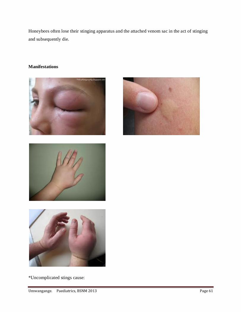
Umwangange. Paediatrics, BSNM 2013 Page 61
Honeybees often lose their stinging apparatus and the attached venom sac in the act of stinging
and subsequently die.
Manifestations
*Uncomplicated stings cause:

Umwangange. Paediatrics, BSNM 2013 Page 62
- immediate pain,
- a wheal-and-flare reaction,
- local edema and swelling that subside in a few hours.
*Stings from accidentally swallowed insects may induce life-threatening edema of the upper
airways.
*Multiple stings can lead to:
- vomiting,
- diarrhea,
- generalized edema,
- dyspnea,
- hypotension, and collapse.
- intravascular hemolysis may cause renal failure.
- Death from the direct effects of venom has followed 300–500 honeybee stings.
Treatment
- Stingers from honeybees embedded in the skin should be removed as promptly as
possible, using any method available, to limit the quantity of venom delivered.
- The site should be cleansed and disinfected and ice packs applied to slow the spread of
venom.
- Elevate the affected site
- Administration of analgesics and oral antihistamines relieve symptoms.
- Give the steroids ( e.g. Oral prednisolone) in case of large local reactions.
- Patients with numerous stings should be monitored for 24 h for evidence of renal failure
or coagulopathy.
- In case of anaphylaxis:
. Give Epinephrine
. A tourniquet may slow the spread of venom.
. Give parenteral antihistamines,
. fluid resuscitation,
Umwangange. Paediatrics, BSNM 2013 Page 63
. bronchodilators,
. oxygen and intubation.
.Patients should be observed for 24 h for recurrent anaphylaxis.
II. INGESTION OF CHEMALS
1. INGESTION OF HYDROCARBONS
Hydrocarbon is the one of the most toxic exposures, usually occurring in children younger than 5
years old. Hydrocarbons are commonly used as fuel, polishes,…
Ingested hydrocarbons generally produce little systemic toxicity, but during ingestion they pose a
serious risk of pulmonary aspiration. Hydrocarbons destroy pulmonary surfactant, leading to
ventilation- perfusion mismatch, hypoxia.
Manifestations
- Prolonged cough, gasping, or choking following ingestion often indicates aspiration
- Respiratory distress within 2-6 hours in case of aspiration
- Fever due to direct tissue toxicity
Treatment
- Gastric lavage
- Asymptomatic patient after 4-6 hours will be discharge
2. CAUSTIC AGENTS ( Acids or Alcalins)
Caustic agents which cause direct tissue injury, are present in many household. Ingested caustic
agents include dishwasher detergents, swimming pool and toilet bowel cleaners, battery acids.
Acids cause a caogulative necrosis which tends to limit tissue damage, in contrast, alcalines
cause liquefactive necrosis with penetration into the deeper tissues, resulting in extensive tissue
damage.

Umwangange. Paediatrics, BSNM 2013 Page 64
Manifestations
- Severe pain
- Spontaneous vomiting
- Injury to the esophagus from alkalines has been graded.
*Grade I: injury, mucosal hyperhemia without ulceration.
*Grade II: burns, with submucosal lesions and ulcerations.
*Grade III: burns, with deep ulcers and tissue necrosis. There is risk for perforation.
- The chest and abdominal X- ray may show esophageal or gastric perforation.
Treatment
- Dilution with milk or water may be helpful if given within the 1st few minutes after the
exposure in those who have no airway complains, vomiting, no abdominal pain, and are
able to speak.
III. INGESTION OF DRUG OVERDOSAGE
Drug overdose is common among children less than 6 years of age. Poisoning due to drug
overdose may be local (e.g., skin, eyes, or lungs) or systemic depending on the chemical and
physical properties of the poison, its mechanism of action, and the route of exposure.
Management
Treatment goals include:
Support of vital signs,
Prevention of further poison absorption,
Enhancement of poison elimination,
Administration of specific antidotes,
and prevention of reexposure.

Umwangange. Paediatrics, BSNM 2013 Page 65
a) Supportive care
Airway protection
Oxygenation/ventilation
Treatment of arrhythmias
Hemodynamic support
Treatment of seizures
Correction of temperature abnormalities
Correction of metabolic derangements
Prevention of secondary complications
a) Prevention of Further Poison Absorption
Gastrointestinal decontamination
Syrup of ipecac–induced emesis
Gastric lavage
Activated charcoal
Whole-bowel irrigation
Dilution
Endoscopic/surgical removal
Decontamination of other sites
Eye decontamination
Skin decontamination
Body cavity evacuation
Enhancement of Poison Elimination
Multiple-dose activated charcoal
Diuresis
Alteration of urinary pH
Extracorporeal removal
Peritoneal dialysis

Umwangange. Paediatrics, BSNM 2013 Page 66
Hemodialysis
Hemoperfusion
Hemofiltration
Plasmapheresis
Exchange transfusion
Hyperbaric oxygenation
Administration of Antidotes
Neutralization by antibodies
Neutralization by chemical binding
Metabolic antagonism
Physiologic antagonism
Prevention of Reexposure
Education of older children
Child-proofing
Psychiatric referral in case of suicide attempt
ACETAMINOPHEN OVERDOSAGE
Acetaminophen ( Paracetamol ) is the most common pharmaceutical agent involved in overdose.
Clinical features
An acute ingestion 150-200mg/kg of acetaminophen in a child is potentially hepatotoxic.
Early after the acute overdose, there are minor symptoms like: Nausea, vomiting and
anorexia.
Approximately 36 hours after ingestion: * transaminase levels rise, *Encephalopathy,
*metabolic acidosis, *increasing prothrombin time.

Umwangange. Paediatrics, BSNM 2013 Page 67
Differential diagnosis
Viral hepatitis
Hepatobiliary disease
Management
Activated charcoal within1-2 hours
Give N-acetylcystein within8 hours of ingestion to prevent hepatotoxicity. Oral or via
nasogastric tube 140mg/kg loading dose, then 70mg/kg every 4 hours for 17 doses.
Antiemetics may be necessary
Clinical Features and Associated Poisons (Toxidromes)
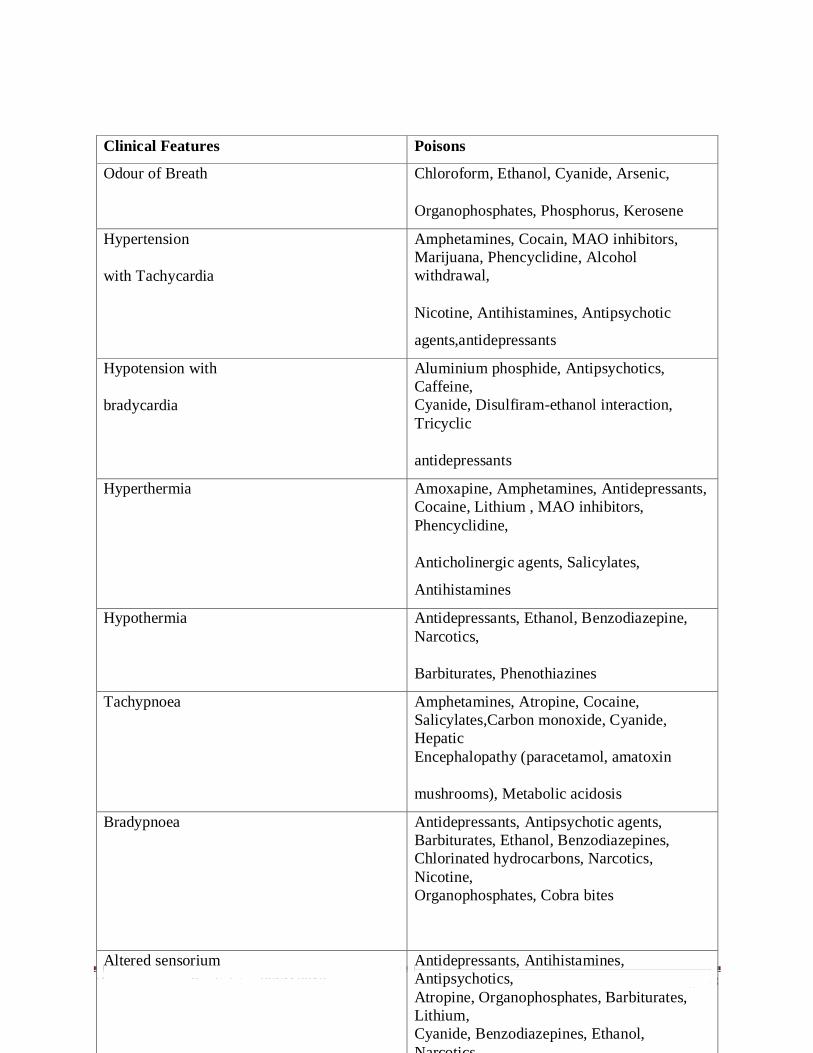
Umwangange. Paediatrics, BSNM 2013 Page 68
Clinical Features Poisons
Odour of Breath Chloroform, Ethanol, Cyanide, Arsenic,
Organophosphates, Phosphorus, Kerosene
Hypertension
with Tachycardia
Amphetamines, Cocain, MAO inhibitors,
Marijuana, Phencyclidine, Alcohol
withdrawal,
Nicotine, Antihistamines, Antipsychotic
agents,antidepressants
Hypotension with
bradycardia
Aluminium phosphide, Antipsychotics,
Caffeine,
Cyanide, Disulfiram-ethanol interaction,
Tricyclic
antidepressants
Hyperthermia Amoxapine, Amphetamines, Antidepressants,
Cocaine, Lithium , MAO inhibitors,
Phencyclidine,
Anticholinergic agents, Salicylates,
Antihistamines
Hypothermia Antidepressants, Ethanol, Benzodiazepine,
Narcotics,
Barbiturates, Phenothiazines
Tachypnoea Amphetamines, Atropine, Cocaine,
Salicylates,Carbon monoxide, Cyanide,
Hepatic
Encephalopathy (paracetamol, amatoxin
mushrooms), Metabolic acidosis
Bradypnoea Antidepressants, Antipsychotic agents,
Barbiturates, Ethanol, Benzodiazepines,
Chlorinated hydrocarbons, Narcotics,
Nicotine,
Organophosphates, Cobra bites
Altered sensorium Antidepressants, Antihistamines,
Antipsychotics,
Atropine, Organophosphates, Barbiturates,
Lithium,
Cyanide, Benzodiazepines, Ethanol,
Narcotics,

Umwangange. Paediatrics, BSNM 2013 Page 69