overview of the structure–function relationships of
TRANSCRIPT

HAL Id: hal-02388775https://hal-amu.archives-ouvertes.fr/hal-02388775
Submitted on 21 Jan 2020
HAL is a multi-disciplinary open accessarchive for the deposit and dissemination of sci-entific research documents, whether they are pub-lished or not. The documents may come fromteaching and research institutions in France orabroad, or from public or private research centers.
L’archive ouverte pluridisciplinaire HAL, estdestinée au dépôt et à la diffusion de documentsscientifiques de niveau recherche, publiés ou non,émanant des établissements d’enseignement et derecherche français ou étrangers, des laboratoirespublics ou privés.
Distributed under a Creative Commons Attribution| 4.0 International License
Overview of the Structure–Function Relationships ofMannose-Specific Lectins from Plants, Algae and Fungi
Annick Barre, Yves Bourne, Els van Damme, Pierre Rougé
To cite this version:Annick Barre, Yves Bourne, Els van Damme, Pierre Rougé. Overview of the Structure–FunctionRelationships of Mannose-Specific Lectins from Plants, Algae and Fungi. International Journal ofMolecular Sciences, MDPI, 2019, Protein–Carbohydrate Interactions: Structure–Function Relation-ships, 20 (2), pp.254. �10.3390/ijms20020254�. �hal-02388775�

International Journal of
Molecular Sciences
Review
Overview of the Structure–Function Relationships ofMannose-Specific Lectins from Plants, Algaeand Fungi
Annick Barre 1, Yves Bourne 2, Els J. M. Van Damme 3 and Pierre Rougé 1,*1 UMR 152 PharmaDev, Institut de Recherche et Développement, Faculté de Pharmacie, Université Paul
Sabatier, 35 Chemin des Maraîchers, 31062 Toulouse, France; [email protected] Centre National de la Recherche Scientifique, Aix-Marseille Univ, Architecture et Fonction des
Macromolécules Biologiques, 163 Avenue de Luminy, 13288 Marseille, France;[email protected]
3 Department of Biotechnology, Faculty of Bioscience Engineering, Ghent University, Coupure links 653,B-9000 Ghent, Belgium; [email protected]
* Correspondence: [email protected]; Tel.: +33-069-552-0851
Received: 11 December 2018; Accepted: 31 December 2018; Published: 10 January 2019�����������������
Abstract: To date, a number of mannose-binding lectins have been isolated and characterizedfrom plants and fungi. These proteins are composed of different structural scaffold structureswhich harbor a single or multiple carbohydrate-binding sites involved in the specific recognitionof mannose-containing glycans. Generally, the mannose-binding site consists of a small, central,carbohydrate-binding pocket responsible for the “broad sugar-binding specificity” toward a singlemannose molecule, surrounded by a more extended binding area responsible for the specificrecognition of larger mannose-containing N-glycan chains. Accordingly, the mannose-bindingspecificity of the so-called mannose-binding lectins towards complex mannose-containing N-glycansdepends largely on the topography of their mannose-binding site(s). This structure–functionrelationship introduces a high degree of specificity in the apparently homogeneous group ofmannose-binding lectins, with respect to the specific recognition of high-mannose and complexN-glycans. Because of the high specificity towards mannose these lectins are valuable tools fordeciphering and characterizing the complex mannose-containing glycans that decorate both normaland transformed cells, e.g., the altered high-mannose N-glycans that often occur at the surface ofvarious cancer cells.
Keywords: lectin; plant; fungi; mannose-binding specificity; structure; function; use as tools
1. Introduction
Protein-carbohydrate interactions are part of the most efficient signaling pathways occurringinside living organisms or between living organisms and their environment. Lectins orCarbohydrate-Binding Agents (CBAs) are proteins that have specialized in the specific recognition ofcarbohydrates during the evolution of all living organisms. The large family of carbohydrate-bindingproteins contains a large variety of carbohydrate-binding domains (CBDs), each with one or morecarbohydrate-binding sites (CBSs) which specifically recognize simple or more complex sugars.Depending on the lectin, the carbohydrate-binding domains belong to distinct structural scaffoldsusually organized in homo- or hetero-dimeric or tetrameric structures [1]. According to the nature andthe organization of their domains, plant and fungal lectins have been classified in two groups of lectins,(1) lectins exclusively composed of carbohydrate-binding domains and (2) chimerolectins composedof a carbohydrate-binding domain linked to another domain(s) devoid of any carbohydrate-binding
Int. J. Mol. Sci. 2019, 20, 254; doi:10.3390/ijms20020254 www.mdpi.com/journal/ijms

Int. J. Mol. Sci. 2019, 20, 254 2 of 49
properties [1]. With respect to their binding properties, plant and fungal lectins can be subdividedin different groups, such as for example Man-specific lectins, Gal/GalNAc-specific lectins, andFuc-specific lectins [2]. However, the binding of lectins towards simple sugars is probably not reallyrelevant. It is more realistic to assume that lectins will interact with the more complex N-glycan chainsthat decorate the cell surface of all living organisms [3]. In addition, the idea has progressively emergedthat, besides lectins which are abundantly distributed in storage tissues like seeds and bulbs andplay a defensive/protective role, other less abundant lectins participate in more discrete carbohydraterecognition processes equally necessary for the proper functioning of the living organisms [4]. In thisrespect, the discovery of Nictaba, a lectin localized in the nucleus of tobacco (Nicotiana tabacum) cells,represents a milestone in our vision of the function devoted to plant and fungal lectins in vivo [5].
Owing to the huge amount of structural and functional data that have been accumulated forseveral decades these carbohydrate-binding proteins from plants and fungi have become a toolto decipher the structure–function relationships inherently associated to protein macromolecules.In this respect, lectins involved in the specific recognition of mannosyl residues, the so-calledmannose-binding lectins, represent an important group of functional proteins taking into accountthe widespread distribution of mannose-containing N-glycans of the N-acetyllactosamine type andhigh-mannose type. The present review aims to present an exhaustive overview that summarizesall published informations related to the structure–function relationships of mannose-specific lectinsfrom plants and fungi, and their possible applications as analytical and therapeutic tools forbiomedical research.
2. Diversity of Mannose-Binding Lectins in Higher Plants
To date, lectins with a mannosyl-binding specificity have been identified in many different plantfamilies, including monocotyledonous as well as dicotyledonous species (Table 1). Among the monocotfamilies, research has focused on the Liliaceae and Amaryllidaceae [6], whereas the Fabaceae familyoccupies a predominant position in the dicot group [6]. Following to the pioneering work of Agrawal& Goldstein [7], who reported that concanavalin A (Con A), the lectin from Jack bean (Canavaliaensiformis) seeds, was easily retained by simple filtration through a column containing cross-linkeddextran gel (Sephadex, Pharmacia) and subsequent desorbtion by the addition of glucose or mannoseto the eluting buffer, both Con A and many other mannose-specific lectins (Table 1) were easily purifiedusing a single affinity chromatography step. Mannose-specific lectins were also successfully isolatedfrom different algae, mushrooms and lower plant species [8]. Moreover, some mannose-specific lectinsfrom red algae specifically recognize the core (α1-6)-fucosylated N-glycans of cancer cells and can beused as biomarkers for the detection of cancer glycoforms [9]. In this respect, they resemble LcA fromLens culinaris, PsA from Pisum sativum and LoL-I from Lathyrus ochrus, which show strong binding tocore-fucosylated mono- and bi-antennary N-glycans [10,11].

Int. J. Mol. Sci. 2019, 20, 254 3 of 49
Table 1. Overview of plant, algae and fungi lectins with a mannosyl-binding specificity (β-sandwich: βs, β-prism: βp, n.d.: not determined).
Plant, Alga, Mushroom Family Plant, Alga, Mushroom Species Lectin Structural Scaffold Oligomer Ref.
Pteridophyta Phlebodium aureum PAL β barrel 2 [12]
GymnospermsAraucaria brasiliensis
Lectin I n.d. 10[13]Lectin 2 n.d. 6
Gingko biloba Gnk2 α β 1 [14]Cycas revoluta CRLL β-prism 2 [15,16]
Fabaceae
Bowringia mildbraedii BMA β-sandwich 2/4 [17]Cajanus cajan CcL βs 2 [18]
Camptosema pedicellatum CPL βs 4 [19]Canavalia boliviana ConBo βs 4 [20]
Canavalia bonariensis CaBo βs 4 [21]Canavalia brasiliensis ConBr βs 4 [22]Canavalia ensiformis ConA βs 4 [23]Canavalia gladiata CGL βs 4 [24]
Canavalia grandiflora ConGF βs 4 [25]Canavalia maritima ConM βs 4 [26]
Canavalia virosa ConV βs 4 [27]Centrolobium microchaete CML βs 4 [28]Centrolobium tomentosum CTL βs 4 [29]
Cladrastis lutea CLAI,II βs 4 [30]Cratylia floribunda CFL βs 2/4 [31]
Cratylia mollis CRAMOLL βs 2/4 [32]Cymbosema roseum CRLI βs 4 [33]Dioclea grandiflora DGL βs 4 [34,35]Dioclea guianensis Dguia βs 4 [36]Dioclea lasiocarpa DLL βs 4 [37]Dioclea lasiophylla DlyL βs 4 [38]
Dioclea reflexa DrfL βs 4 [39]Dioclea rostrata DRL βs 4 [40]
Dioclea sclerocarpa DSL βs 4 [41]Dioclea violacea DVL βs 4 [42]Dioclea virgata DvirL βs 4 [43]Dioclea wilsonii DwL βs 4 [44]Lathyrus aphaca LaphL βs 2 [45]
Lathyrus articulatus LarL βs 2 [45]

Int. J. Mol. Sci. 2019, 20, 254 4 of 49
Table 1. Cont.
Plant, Alga, Mushroom Family Plant, Alga, Mushroom Species Lectin Structural Scaffold Oligomer Ref.
Lathyrus cicera LcL βs 2 [45]Lathyrus hirsutus LhL βs 2 [46]Lathyrus nissolia LnL βs 1 [47]Lathyrus ochrus LoL βs 2 [48]
Lathyrus odoratus LodL βs 2 [49]Lathyrus sativus LsL βs 2 [50]
Lathyrus sphaericus LsphL βs 1 [51]Lathyus sylvestris LsiL βs 2 [52]
Lathyrus tingitanus LtL βs 2 [46]Lens culinaris LcA βs 2 [53]
Millettia dielsiana MDL βs 2 [54]Onobrychis viciifolia βs n.d. [55]
Pisum arvense PAL βs 2 [56]Pisum sativum PsA βs 2 [57]
Pterocarpus angolensis PAL βs 2 [58]Sophora flavescens SFL βs 2 [59]
Trigonella foenumgraecum βs n.d. [60]Vicia cracca βs 2 [61]Vicia ervilia βs 4 [62]Vicia faba VfA βs 2 [63]
Vicia sativa βs 2 [64]
MimosaceaeParkia biglobosa PBL βs 2 [65]
Parkia platycephala PPL βs 2 [66]
Dalbergieae Platypodium elegans nPELa βs 2 [67]Platymiscium floribundum PFL βs 2 [68]
Fagaceae Castanea crenata CCA βs 6/8 [69]
Moraceae
Artocarpus heterophyllus ArtinM β-prism 4 [70,71]Artocarpus incisa Frutapin βp 4 [72]Artocarpus integer CMB βp 4 [73,74]
Artocarpus integrifolia artocarpin βp 4 [75,76]jacalin βp 4 [77,78]
Artocarpus lakoocha artocarpin βp 4 [79]Morus nigra Moniga-M βp 4 [80]

Int. J. Mol. Sci. 2019, 20, 254 5 of 49
Table 1. Cont.
Plant, Alga, Mushroom Family Plant, Alga, Mushroom Species Lectin Structural Scaffold Oligomer Ref.
Asteraceae Helianthus tuberosus Heltuba βp 8 [81]
Brassicaceae Arabidopsis thaliana PP2-A1 βp n.d. [82]
Ranonculaceae Clematis montana CML βp 2 [83]
Aloeae Aloe arborescens ALOE βp 4 [84]
Araceae
Arisaema lobatum ALA n.d. 2+2 [85]Arisaema heterophyllum AHA βp n.d [86]
Arum maculatum AMA βp 2+2 [87]Colocasia esculenta CEA, tarin βp 2+2 [88]
Dieffenbachia sequina βp 2+2 [87]Lysichiton camtschatcensis βp 2+2 [89]
Pinellia ternata PTA βp 2+2 [90]Remusatia vivipara RVL βp 2+2 [91]
Typhonium divaricatum TDL βp 2+2 [92]Xanthosoma sagittifolium XSL βp 2+2 [93]Zantedeschia aethiopica ZAA βp n.d. [94]
AsparagaceaeOphiopogon japonicus OJL βp n.d. [95]
Polygonatum cyrtonema PCL βp 4 [96]Polygonatum multiflorum PMA βp 4 [97]
ConvolvulaceaePolygonatum odoratum POL βp 4 [98]
Calystegia sepium Calsepa βp 2 [99]Ipomoea batatas ipomoelin βp 4 [100]
Alliaceae
Allium altaicum AALTA βp 2 [101]Allium ascalonicum AAA βp 2 [102]
Allium cepa ACA βp 2 [103]Allium porrum APA βp 2 [103]Allium sativum ASA-I/II βp 2 [104]
Allium tuberosum ATA βp 2 [105]Allium ursinum AUA-I/II βp 2 [106]

Int. J. Mol. Sci. 2019, 20, 254 6 of 49
Table 1. Cont.
Plant, Alga, Mushroom Family Plant, Alga, Mushroom Species Lectin Structural Scaffold Oligomer Ref.
Amaryllidaceae
Amaryllis vittata AVA βp n.d. [107]Clivia miniata CMA βs 2 [108]
Crinum asiaticum CAA βp n.d. [109]Galanthus nivalis GNA βp 4 [110]
Hippeastrum hybrid HHA βp 2 [111]Leucojum vernum LVL βp n.d. [112]
Zephyranthes candida ZCA βp 4 [113]Zephyranthes grandiflora ZGA βp 4 [114]
Lycoris aurea LAA βp 2 [115]Lycoris radiata LRA βp 2 [116]
DioscoreaceaeDioscorea batatas DB1 βp 1 [117]
Dioscorea bulbifera DBL βp 1 [118]
IridaceaeCrocus sativus CSL βp n.d. [119,120]Crocus vernus CVA βp 4 [121]
Liliaceae
Aspidistra elatior AEL n.d. 2 [122]Narcissus pseudonarcissus NPA βp 2,4 [111]
Narcissus tazetta NTL βp 2 [123]Narcissus tortifolius NTA βp n.d. [124]
Tulipa hybrid TxLCI βp 4[125]TL-MII βp 2
Smilacaceae Smilax glabra SGM2 βp 3 [126]
Hyacintheae Scilla campanulata SCAman βp 2 [127]
MusaceaeMusa acuminata BanLec βp 2 [128]Musa paradisiaca βp 2 [129]
Pandanaceae Pandanus amaryllifolius pandanin βp n.d. [130]
Orchidaceae
Cymbidium hybridum CHA βp 2 [131]Dendrobium officinale DOA2 βp n.d. [132]Epipactis helleborine EHMBP βp 2 [131]
Gastrodia elata gastrodianine βp 2 [133]Liparis noversa LNL βp 2 [95]
Listera ovata LNL βp 2 [131]

Int. J. Mol. Sci. 2019, 20, 254 7 of 49
Table 1. Cont.
Plant, Alga, Mushroom Family Plant, Alga, Mushroom Species Lectin Structural Scaffold Oligomer Ref.
Poaceae Oryza sativa Orysata βp 2 [134]
Red algae
Bryothamnion seaforthii BSL n.d. 1 [135]Bryothamnion triquetrum BTL n.d. 1,2 [136]
Euchema denticulatum EDA n.d. 1 [137]Eucheuma serra ESA n.d. 1 [138]Griffithsia sp. griffithsin n.d. 2 [139,140]
Hypnea cervicornis HCA n.d. 1 [141]Hypnea japonica HJA n.d. 1 [9]
Hypnea musciformis HMA n.d. 1 [142]Kappaphycus alvarezii KAA-2 n.d. 1 [143]Kappaphycus striatum KSA n.d. 1 [144]
Green algae Boodlea coacta BCA β-prism 1 [145]Halimeda renschii HRL40-1/2 n.d. 4 [146]
Hydnangiaceae Laccaria bicolor tectonin 2 β-propeller n.d. [147,148]
Trichocomaceae Penicillium chrysogenum PeCL n.d. n.d. [149]
Saccharomycetaceae Saccharomyces cerevisiae Flo5A β-sandwich 2 [150]Saccharomyces pasteurianus Flo1p βs 4 [151]
Schizosaccharo-mycetaceae Schizosaccharomyces pombe glucosidase βs 2 [152]
Hygrophoraceae Hygrophorus russula HRL n.d. 4 [153]
Marasmiaceae Marasmus oreades MOA β-prism 2 [154]
Pteridaceae Ceratopteris richardii cyanovirin CVN-fold 1 [155]
Sordariaceae Neurospora crassa cyanovirin CVN-fold 1 [155]
Tuberaceae Tuber borchii cyanovirin CVN-fold 1 [155]

Int. J. Mol. Sci. 2019, 20, 254 8 of 49
3. Structural Organization of the Plant, Algal and Fungal Mannose-Binding Lectins
3.1. Structure of Mannose-Specific Plant Lectins
Mannose-specific lectins from plants essentially belong to three distinct structural scaffolds thatassemble in different ways to generate more complex oligomeric structures:
3.1.1. The β-Sandwich Fold
The jelly roll scaffold occurring in legume lectins (Fabaceae) consists of either a single or twopolypeptide chains. In two-chain lectins, the light (α) and heavy (β) chains made of six and sevenstrands of antiparallel β-sheet, respectively, non-covalently associate in a β-sandwich protomer(Figure 1A). Protomers associate by non covalent bonds to give the homodimeric lectins of theVicieae tribe, e.g., pea lectin (Pisum sativum agglutinin PsA) [57], lentil lectin (Lens culinaris agglutininLcA) [156], yellow vetch lectin (Lathyrus ochrus lectin Lol) [48] (Figure 1B), and the faba bean lectin(Vicia faba agglutinin VfA or favin) [63] (Figure 1B). In contrast, the Man-specific lectin from Lathyrussphaericus consists of an uncleaved single chain protomer [51]. The single-chain protomers associateinto homotetramers. Examples are the mannose-binding lectins characterized in the tribes Baphieae(Bowringia mildbraedii agglutinin BMA) [17], Dalbergieae (Centrolobium tomentosum lectin CTL [29],Pterocarpus angolensis lectin PAL [58]), Diocleae (Con A [23,157] (Figure 1C), Cymbosema roseum CRL [33],Dioclea grandiflora lectin Con GF [25], and other Dioclea sp. lectins). Dimeric lectins such as PsA,possess two identical mannose-binding sites whereas tetrameric lectins like Con A, exhibit fourmannose-binding sites. Gal/GalNAc-specific lectins from other legume tribes such as the soybeanagglutinin SBA (Glycine max) from the Glycinae tribe (PDB code 1SBF) [158], the peanut agglutininPNA (Arachis hypogaea) from the Aeschynomeneae (PDB code 2PEL) [159], the coral tree lectin EcorL(Erythrina corallodendron) from the Erythrinae tribe (PDB code 1AXY) [160], and the kidney beanleucoagglutinin PHA-L (PDB code 1FAT) [161] and erythroagglutinin PHA-E (PDB code 3WCR) [162],(Phaseolus vulgaris) belonging to the Phaseolae tribe, all strikingly resemble Con A and other Diocleaelectins but differ in the topological organization for the single-chain protomers that constitute the lectin.
3.1.2. The β-Prism I Fold
The β-prism I scaffold serves as a building block for the mannose-binding lectins in seeds of theMoraceae such as artocarpin, the lectin from the Jackfruit (Artocarpus integrifolia) seeds which servesas a prototype for this group [163]. The β-prism I scaffold consists of three bundles of four antiparallelβ-strands forming three Greek keys 1, 2 and 3, arranged into a β-prism structure along a longitudinalaxis (Figure 1D). Depending on the lectins, a posttranslational proteolytic cleavage between theβ-strands β1 and β2 of Greek key 1 occurs during seed ripening, to liberate the light α-chain with aterminal Gly1 residue exhibiting a free H2N- group, and the heavy β-chain comprising the rest of theβ-prism structure. This proteolytic cleavage occurs in the Gal/GalNAc-specific homotetrameric lectinsof Moraceae, such as jacalin (Figure 1E) (PDB code 1JAC) [164], the MPA lectin from Osage orange(Maclura pomifera) seeds (PDB code 1JOT) [165], and the Gal/GalNAc-specific lectin Morniga-G fromthe bark of blackberry (Morus nigra) [80]. However, the Man-specific lectins from the Moraceae family,e.g., artocarpin from Jackfruit [163] and Morniga-M from blackberry [166], consist of an uncleavedsingle-chain β-prism polypeptide chain. Similarly, Heltuba, the lectin from the Jerusalem artichocke(Helianthus tuberosus), also consists of a single-chain β-prism polypeptide chain made of 8 β-prismsnon-covalentlty associated around a central axis to form a flattened star-shaped architecture comprising8 identical carbohydrate-binding sites (Figure 1F) [81].
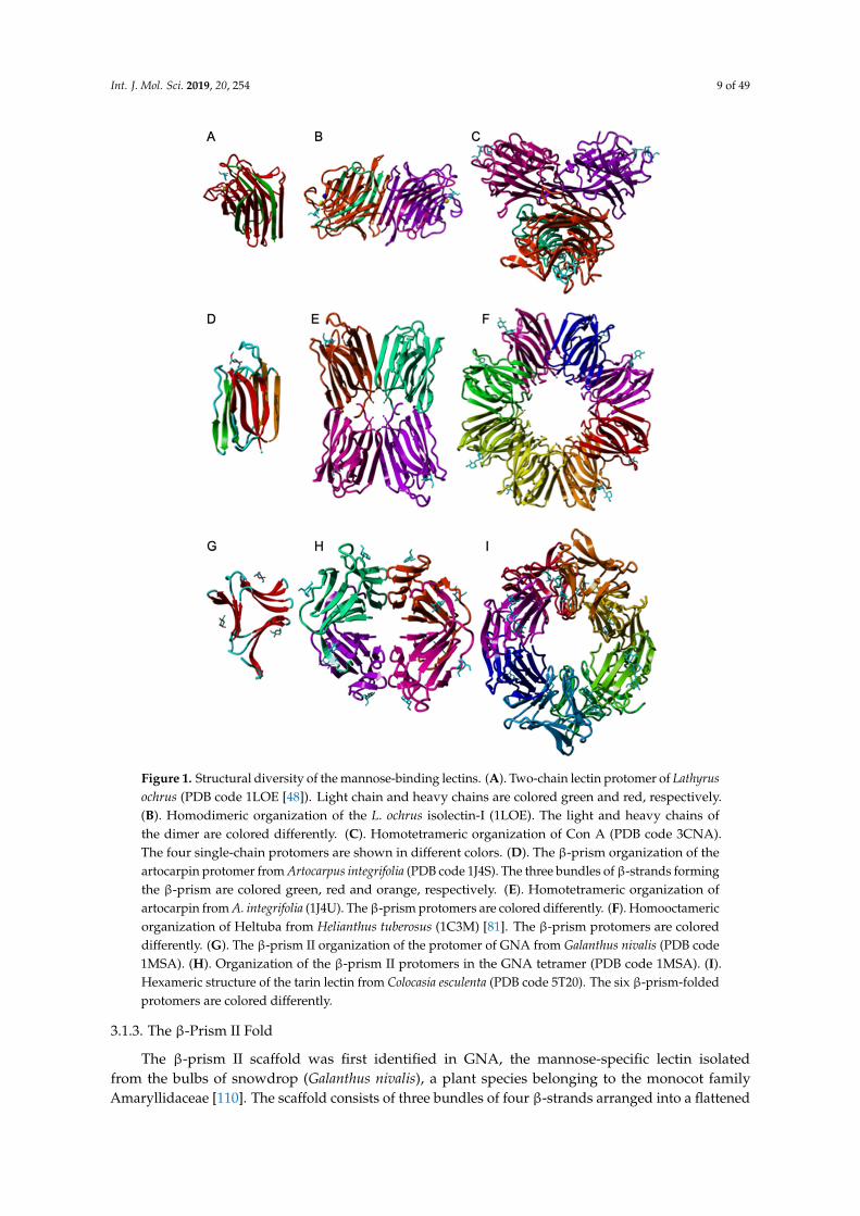
Int. J. Mol. Sci. 2019, 20, 254 9 of 4911
Int. J. Mol. Sci. 2019, 20
Figure 1. Structural diversity of the mannose-binding lectins. (A). Two-chain lectin protomer of
Lathyrus ochrus (PDB code 1LOE [48]). Light chain and heavy chains are colored green and red,
respectively. (B). Homodimeric organization of the L. ochrus isolectin-I (1LOE). The light and heavy
chains of the dimer are colored differently. (C). Homotetrameric organization of Con A (PDB code
3CNA). The four single-chain protomers are shown in different colors. (D). The β-prism organization
of the artocarpin protomer from Artocarpus integrifolia (PDB code 1J4S). The three bundles of β-strands
forming the β-prism are colored green, red and orange, respectively. (E). Homotetrameric
organization of artocarpin from A. integrifolia (1J4U). The β-prism protomers are colored differently.
(F). Homooctameric organization of Heltuba from Helianthus tuberosus (1C3M) [81]. The β-prism
protomers are colored differently. (G). The β-prism II organization of the protomer of GNA from
Galanthus nivalis (PDB code 1MSA). (H). Organization of the β-prism II protomers in the GNA
tetramer (PDB code 1MSA). I. Hexameric structure of the tarin lectin from Colocasia esculenta (PDB
code 5T20). The six β-prism-folded protomers are colored differently.
3.1.3. The β-prism II Fold
The β-prism II scaffold was first identified in GNA, the mannose-specific lectin isolated from the
bulbs of snowdrop (Galanthus nivalis), a plant species belonging to the monocot family
Amaryllidaceae [110]. The scaffold consists of three bundles of four β-strands arranged into a
flattened β-prism structure around a central pseudoaxis (Figure 1G). A carbohydrate-binding site
Figure 1. Structural diversity of the mannose-binding lectins. (A). Two-chain lectin protomer of Lathyrusochrus (PDB code 1LOE [48]). Light chain and heavy chains are colored green and red, respectively.(B). Homodimeric organization of the L. ochrus isolectin-I (1LOE). The light and heavy chains ofthe dimer are colored differently. (C). Homotetrameric organization of Con A (PDB code 3CNA).The four single-chain protomers are shown in different colors. (D). The β-prism organization of theartocarpin protomer from Artocarpus integrifolia (PDB code 1J4S). The three bundles of β-strands formingthe β-prism are colored green, red and orange, respectively. (E). Homotetrameric organization ofartocarpin from A. integrifolia (1J4U). The β-prism protomers are colored differently. (F). Homooctamericorganization of Heltuba from Helianthus tuberosus (1C3M) [81]. The β-prism protomers are coloreddifferently. (G). The β-prism II organization of the protomer of GNA from Galanthus nivalis (PDB code1MSA). (H). Organization of the β-prism II protomers in the GNA tetramer (PDB code 1MSA). (I).Hexameric structure of the tarin lectin from Colocasia esculenta (PDB code 5T20). The six β-prism-foldedprotomers are colored differently.
3.1.3. The β-Prism II Fold
The β-prism II scaffold was first identified in GNA, the mannose-specific lectin isolatedfrom the bulbs of snowdrop (Galanthus nivalis), a plant species belonging to the monocot familyAmaryllidaceae [110]. The scaffold consists of three bundles of four β-strands arranged into a flattened

Int. J. Mol. Sci. 2019, 20, 254 10 of 49
β-prism structure around a central pseudoaxis (Figure 1G). A carbohydrate-binding site occurs in agroove located at the center of the bundle of β-strands forming each β-sheet. The monocot-specificlectins result from the non-covalent association of four β-prism II scaffolds. Depending on the lectin,four identical β-prism II of 12 kDa form a homotetramer, e.g., in GNA (Figure 1H) [167], whereasother lectins consist of heterotretramers built up from the symmetrical association of two 12 kDa andtwo 14 kDa β-prism subunits, e.g., the Araceae lectins [6]. Usually, all three carbohydrate-bindingsites occurring in each β-prism scaffold are readily functional but in a few lectins, one or twocarbohydrate-binding sites are apparently inactive due to point mutation(s) in key residues involvedin the H-bonding of mannose. Tarin from Colocasia esculenta assembles into homohexameric structuresmade of 6 β-prism scaffolds [168] (Figure 1I).
The β-trefoil scaffold, another β-prism II scaffold, has been primarily identified in type IIRibosome-Inactivating Proteins (RIP-II), in amaranthin, a T antigen-specific lectin from amaranth(Amaranthus caudatus) [169], and it also occurs in the stress inducible lectins composed of EUL (Euonymuslectin) domains, such as the lectins from rice (Oryza sativa) and Arabidopsis [170]. The β-trefoil scaffoldconsists of six β-hairpins arranged around an approximate three-fold symmetry axis, linked to extendedloops that simulate the three lobes of a trefoil leaf (Figure 2). The Man-binding sites are located in theshallow depressions of the β-strands but, usually not all binding sites are functional.
12 Int. J. Mol. Sci. 2019, 20 occurs in a groove located at the center of the bundle of β-strands forming each β-sheet. The monocot-
specific lectins result from the non-covalent association of four β-prism II scaffolds. Depending on
the lectin, four identical β-prism II of 12 kDa form a homotetramer, e.g., in GNA (Figure 1H) [167],
whereas other lectins consist of heterotretramers built up from the symmetrical association of two 12
kDa and two 14 kDa β-prism subunits, e.g., the Araceae lectins [6]. Usually, all three carbohydrate-
binding sites occurring in each β-prism scaffold are readily functional but in a few lectins, one or two
carbohydrate-binding sites are apparently inactive due to point mutation(s) in key residues involved
in the H-bonding of mannose. Tarin from Colocasia esculenta assembles into homohexameric
structures made of 6 β-prism scaffolds [168] (Figure 1I).
The β-trefoil scaffold, another β-prism II scaffold, has been primarily identified in type II
Ribosome-Inactivating Proteins (RIP-II), in amaranthin, a T antigen-specific lectin from amaranth
(Amaranthus caudatus) [169], and it also occurs in the stress inducible lectins composed of EUL
(Euonymus lectin) domains, such as the lectins from rice (Oryza sativa) and Arabidopsis [170]. The β-
trefoil scaffold consists of six β-hairpins arranged around an approximate three-fold symmetry axis,
linked to extended loops that simulate the three lobes of a trefoil leaf (Figure 2). The Man-binding
sites are located in the shallow depressions of the β-strands but, usually not all binding sites are
functional.
Figure 2. Three-dimensional models for the EUL domain of EUL-domains of rice lectin Orysata,
showing the β-trefoill organization made of three bundles of antiparallel β-sheets (I, II, III).
An unexpected four-bladed β-propeller structure was found to occur in a PA2 albumin from
chickpea (Cicer arietinum), which displays a well documented hemagglutinating activity most
probably related to a lectin with an unusual hemopexin fold [171].
3.2. Structure of Mannose-Specific Algal Lectins
The mannose-specific lectin griffthsin from the red alga Griffthsia sp., consists of a domain-
swapped dimer made of two protomer exhibiting the β-prism I fold, that closely resembles to the
jacalin-related lectin organization (PDB code 2GTY) [140]. Swapping results from the participation of
two β-strands of one molecule in the completion of the three four-stranded sheets forming the β-
prism of the other molecule, and vice versa. As a result of this swapping, both molecules in the dimer
consist of a complete β-prism organization (Figure 3).
Figure 2. Three-dimensional models for the EUL domain of EUL-domains of rice lectin Orysata,showing the β-trefoill organization made of three bundles of antiparallel β-sheets (I, II, III).
An unexpected four-bladed β-propeller structure was found to occur in a PA2 albumin fromchickpea (Cicer arietinum), which displays a well documented hemagglutinating activity most probablyrelated to a lectin with an unusual hemopexin fold [171].
3.2. Structure of Mannose-Specific Algal Lectins
The mannose-specific lectin griffthsin from the red alga Griffthsia sp., consists of a domain-swapped dimer made of two protomer exhibiting the β-prism I fold, that closely resembles to thejacalin-related lectin organization (PDB code 2GTY) [140]. Swapping results from the participation oftwo β-strands of one molecule in the completion of the three four-stranded sheets forming the β-prismof the other molecule, and vice versa. As a result of this swapping, both molecules in the dimer consistof a complete β-prism organization (Figure 3).
In spite of a high number of cloned and sequenced lectins from different species of red and greenalgae, their three-dimensional organization(s) were poorly investigated and still remain unknown.Their amino acid sequences readily differ from that of griffithsin and, most probably, they also differfrom griffithsin by their three-dimensional structure and monomer organization.

Int. J. Mol. Sci. 2019, 20, 254 11 of 49
12 Int. J. Mol. Sci. 2019, 20 occurs in a groove located at the center of the bundle of β-strands forming each β-sheet. The monocot-
specific lectins result from the non-covalent association of four β-prism II scaffolds. Depending on
the lectin, four identical β-prism II of 12 kDa form a homotetramer, e.g., in GNA (Figure 1H) [167],
whereas other lectins consist of heterotretramers built up from the symmetrical association of two 12
kDa and two 14 kDa β-prism subunits, e.g., the Araceae lectins [6]. Usually, all three carbohydrate-
binding sites occurring in each β-prism scaffold are readily functional but in a few lectins, one or two
carbohydrate-binding sites are apparently inactive due to point mutation(s) in key residues involved
in the H-bonding of mannose. Tarin from Colocasia esculenta assembles into homohexameric
structures made of 6 β-prism scaffolds [168] (Figure 1I).
The β-trefoil scaffold, another β-prism II scaffold, has been primarily identified in type II
Ribosome-Inactivating Proteins (RIP-II), in amaranthin, a T antigen-specific lectin from amaranth
(Amaranthus caudatus) [169], and it also occurs in the stress inducible lectins composed of EUL
(Euonymus lectin) domains, such as the lectins from rice (Oryza sativa) and Arabidopsis [170]. The β-
trefoil scaffold consists of six β-hairpins arranged around an approximate three-fold symmetry axis,
linked to extended loops that simulate the three lobes of a trefoil leaf (Figure 2). The Man-binding
sites are located in the shallow depressions of the β-strands but, usually not all binding sites are
functional.
Figure 2. Three-dimensional models for the EUL domain of EUL-domains of rice lectin Orysata,
showing the β-trefoill organization made of three bundles of antiparallel β-sheets (I, II, III).
An unexpected four-bladed β-propeller structure was found to occur in a PA2 albumin from
chickpea (Cicer arietinum), which displays a well documented hemagglutinating activity most
probably related to a lectin with an unusual hemopexin fold [171].
3.2. Structure of Mannose-Specific Algal Lectins
The mannose-specific lectin griffthsin from the red alga Griffthsia sp., consists of a domain-
swapped dimer made of two protomer exhibiting the β-prism I fold, that closely resembles to the
jacalin-related lectin organization (PDB code 2GTY) [140]. Swapping results from the participation of
two β-strands of one molecule in the completion of the three four-stranded sheets forming the β-
prism of the other molecule, and vice versa. As a result of this swapping, both molecules in the dimer
consist of a complete β-prism organization (Figure 3).
Figure 3. Three-dimensional model of griffithsin (PDB code 2GTY), showing the β-prism organizationmade of three four-stranded β-sheets in each monomer. The four stranded β-sheets are colored red,pink and magenta in monomer (A), and blue, light blue and purple in monomer (B), respectively. Thestars indicate the localization of the carbohydrate-binding sites in each monomer.
3.3. Structure of Mannose-Specific Fungal Lectins
Mannose-specific lectins isolated from fungi result from the non-covalent association of differentstructural scaffolds resulting in more complex oligomeric structures:
An unusual six-bladed β-propeller organization built up from 4-stranded anti-parallel β-sheetswas identified in tectonin 2, a lectin from the mushroom Laccaria bicolor AAL (PDB code 5FSC), thatspecifically recognizes O-methylated glycans [148] (Figure 4).
13 Int. J. Mol. Sci. 2019, 20
Figure 3. Three-dimensional model of griffithsin (PDB code 2GTY), showing the β-prism organization
made of three four-stranded β-sheets in each monomer. The four stranded β-sheets are colored red,
pink and magenta in monomer (A), and blue, light blue and purple in monomer (B), respectively. The
stars indicate the localization of the carbohydrate-binding sites in each monomer.
In spite of a high number of cloned and sequenced lectins from different species of red and green
algae, their three-dimensional organization(s) were poorly investigated and still remain unknown.
Their amino acid sequences readily differ from that of griffithsin and, most probably, they also differ
from griffithsin by their three-dimensional structure and monomer organization.
3.3. Structure of Mannose-Specific Fungal Lectins
Mannose-specific lectins isolated from fungi result from the non-covalent association of different
structural scaffolds resulting in more complex oligomeric structures:
An unusual six-bladed β-propeller organization built up from 4-stranded anti-parallel β-sheets
was identified in tectonin 2, a lectin from the mushroom Laccaria bicolor AAL (PDB code 5FSC), that
specifically recognizes O-methylated glycans [148] (Figure 4).
Figure 4. (A). Beta-propeller organization of tectonin 2 from the mushroom Laccaria bicolor in complex
with allyl-α4-methyl-mannoside. The lectin consists of 6 antiparallel strands of β-sheet (colored
differently) organized in 6 blades around the axis of the β-propeller. The allyl-mannoside residues
(M) anchored to the carbohydrate-binding sites of the lectin are colored purple (PDB code 5FSC) (B).
Sixth mannose-binding site of tectonin 2 in complex with allyl-α4-methyl-mannoside. Hydrogen
bonds connecting the monosaccharides to the amino acid residues Ser200, Asn216 and Tyr222,
forming the monosaccharide-binding site are represented by black dashed lines. Aromatic residues
Trp3 and Tyr222, paticipating in stacking interactions with the sugar ring are colored orange. The
molecular surface of the lectins is colored dark grey and their extended oligosaccharide-binding areas
are delineated by white dashed lines. (C). The shallow depression corresponding to the
monosaccharide-binding site that accommodates the allyl-mannoside residue (colored purple) at the
molecular surface (colored according to the oulombic charges) of tectonin 2, is delineated by a green
dashed line.
A similar 6-bladed β-propeller structure was observed in the fucose-binding lectins from the
bacteria Ralstonia solanacearum [172], Photorhabdus luminescens [173], Photorhabdus asymbiotica [174], as
well as in the tachylectin from the Japanese horseshoe crab Tachypleus tridentatus [175]. However, the
β-propeller scaffold is not specific for the fucose-binding property since a β-propeller structure was
shown to occur in other lectins with quite different sugar-binding specificities, e.g., the Neu5Ac- and
GlcNAc-specific lectins from the mushrooms Psathyrella velutina [176] and Psathyrella asperospora
[177], and the lectin Bambl from the bacterium Burkholderia ambifaria, which specifically interacts with
the lewis x antigen, the blood H type 1 and H type 2 tetrasaccharides and the blood group B epitope
[178].
The β-sandwich scaffold is another structural scaffold found in the mannose-binding N-terminal
domain of flocculins Flo1 and Flo5 from Saccharomyces cerevisiae, and Flo1 from S. pasteurianus
M M
Figure 4. (A). Beta-propeller organization of tectonin 2 from the mushroom Laccaria bicolor in complexwith allyl-α4-methyl-mannoside. The lectin consists of 6 antiparallel strands of β-sheet (coloreddifferently) organized in 6 blades around the axis of the β-propeller. The allyl-mannoside residues(M) anchored to the carbohydrate-binding sites of the lectin are colored purple (PDB code 5FSC) (B).Sixth mannose-binding site of tectonin 2 in complex with allyl-α4-methyl-mannoside. Hydrogen bondsconnecting the monosaccharides to the amino acid residues Ser200, Asn216 and Tyr222, forming themonosaccharide-binding site are represented by black dashed lines. Aromatic residues Trp3 and Tyr222,paticipating in stacking interactions with the sugar ring are colored orange. The molecular surface ofthe lectins is colored dark grey and their extended oligosaccharide-binding areas are delineated bywhite dashed lines. (C). The shallow depression corresponding to the monosaccharide-binding sitethat accommodates the allyl-mannoside residue (colored purple) at the molecular surface (coloredaccording to the oulombic charges) of tectonin 2, is delineated by a green dashed line.
A similar 6-bladed β-propeller structure was observed in the fucose-binding lectins from thebacteria Ralstonia solanacearum [172], Photorhabdus luminescens [173], Photorhabdus asymbiotica [174], aswell as in the tachylectin from the Japanese horseshoe crab Tachypleus tridentatus [175]. However, theβ-propeller scaffold is not specific for the fucose-binding property since a β-propeller structure wasshown to occur in other lectins with quite different sugar-binding specificities, e.g., the Neu5Ac- andGlcNAc-specific lectins from the mushrooms Psathyrella velutina [176] and Psathyrella asperospora [177],

Int. J. Mol. Sci. 2019, 20, 254 12 of 49
and the lectin Bambl from the bacterium Burkholderia ambifaria, which specifically interacts with thelewis x antigen, the blood H type 1 and H type 2 tetrasaccharides and the blood group B epitope [178].
The β-sandwich scaffold is another structural scaffold found in the mannose-bindingN-terminal domain of flocculins Flo1 and Flo5 from Saccharomyces cerevisiae, and Flo1 fromS. pasteurianus [150,151]. These surface-adhesins possess a N-terminal domain that readily accomodatesMan and α1,2-mannobiose via a network of hydrogen bonds and stacking interactions with aromaticresidues, very similar to those occurring in Man-specific lectins of higher plants (Figure 5). Most of theaminoacid residues involved in the binding of mannose also serve as ligands for a Ca2+ ion located atthe bottom of the mannose-binding site. The mannose-binding activity of Flo1 and Flo5 proteins playsa key role in the self-recognition processes occurring during the growth of the yeasts.
14 Int. J. Mol. Sci. 2019, 20 [150,151]. These surface-adhesins possess a N-terminal domain that readily accomodates Man and
α1,2-mannobiose via a network of hydrogen bonds and stacking interactions with aromatic residues,
very similar to those occurring in Man-specific lectins of higher plants (Figure 5). Most of the
aminoacid residues involved in the binding of mannose also serve as ligands for a Ca2+ ion located at
the bottom of the mannose-binding site. The mannose-binding activity of Flo1 and Flo5 proteins plays
a key role in the self-recognition processes occurring during the growth of the yeasts.
Figure 5. (A,C). Beta-sandwich organization of Flo5 from the yeast Saccharomyces cerevisiae in complex
with mannose (A) (PDB code 2XJP) and α1,2-mannobiose (C) (PDB code 2XJS). The mannose-binding
N-terminal domain of Flo5 consists of two strands of β-sheet forming a β-sandwich structure. (B).
Network of hydrogen bonds anchoring mannose (colored purple) to the amino acid residues forming
the carbohydrate-binding site located at the top of the β-sandwich. Two stacking interactions of the
pyranose ring of mannose with aromatic residues Tyr54 and Trp228 (colored orange), complete the
interaction. (D). Network of hydrogen bonds anchoring α1,2-mannobiose (colored purple) Flo5,
showing additional hydrogen bonds anchoring α1,2-mannobiose to Gln117 and Ser 227 residues.
Residues Asp160, Asp161, Val226 and Trp228, also serve as ligands for a Ca2+ ion (colored red in A
and C) located at the bottom of the mannose-binding pocket.
The cyanovirin-fold (CVN-fold) also occurs as a structural scaffold identified in the cyanovirin-
N family of mannose-binding fungal lectins, including the ascomycetous fungi Ceratopteris richardii
(CrCVNH), Neurospora crassa (NcCVNH) and Tuber borchii (TbCVNH) [155]. The NcCVNH lectin
consists of a two swapped domains polypeptide chain of 111 amino acids, built up from a domain A
of 56 residues (residues 1–42 and residues 100–111), and a domain B of 57 residues (residues 43–99).
According to the swapping occurring between both domains, domain A comprises the triple-
stranded β-sheet (β1,β2,β3) associated to the β-hairpin (β9,β10), whereas domain B consists of the
triple-stranded sheet (β6,β7,β8) associated to the β-hairpin (β4,β5) (Figure 6A). Other CrVNH and
TbCVNH exhibit a very similar organization.
Gln117
Figure 5. (A,C). Beta-sandwich organization of Flo5 from the yeast Saccharomyces cerevisiae in complexwith mannose (A) (PDB code 2XJP) and α1,2-mannobiose (C) (PDB code 2XJS). The mannose-bindingN-terminal domain of Flo5 consists of two strands of β-sheet forming a β-sandwich structure. (B).Network of hydrogen bonds anchoring mannose (colored purple) to the amino acid residues formingthe carbohydrate-binding site located at the top of the β-sandwich. Two stacking interactions ofthe pyranose ring of mannose with aromatic residues Tyr54 and Trp228 (colored orange), completethe interaction. (D). Network of hydrogen bonds anchoring α1,2-mannobiose (colored purple) Flo5,showing additional hydrogen bonds anchoring α1,2-mannobiose to Gln117 and Ser 227 residues.Residues Asp160, Asp161, Val226 and Trp228, also serve as ligands for a Ca2+ ion (colored red in A andC) located at the bottom of the mannose-binding pocket.
The cyanovirin-fold (CVN-fold) also occurs as a structural scaffold identified in the cyanovirin-Nfamily of mannose-binding fungal lectins, including the ascomycetous fungi Ceratopteris richardii(CrCVNH), Neurospora crassa (NcCVNH) and Tuber borchii (TbCVNH) [155]. The NcCVNH lectinconsists of a two swapped domains polypeptide chain of 111 amino acids, built up from a domainA of 56 residues (residues 1–42 and residues 100–111), and a domain B of 57 residues (residues43–99). According to the swapping occurring between both domains, domain A comprises thetriple-stranded β-sheet (β1, β2, β3) associated to the β-hairpin (β9, β10), whereas domain B consistsof the triple-stranded sheet (β6, β7, β8) associated to the β-hairpin (β4, β5) (Figure 6A). Other CrVNHand TbCVNH exhibit a very similar organization.
It is notheworthy that most of the Man-specific lectins identified in bacteria consist of the so-calledCVN family fold (Table 2), which comprises cyanovirin, actinohivin, and microvirin occurring incyanobacteria (ex blue-green algae) as a two swapped domains polypeptide chain, each domain builtup from a β-sheet of three anti-parallel β-strands linked to a β-hairpin by a short α-helical turn [179](Figure 6B).
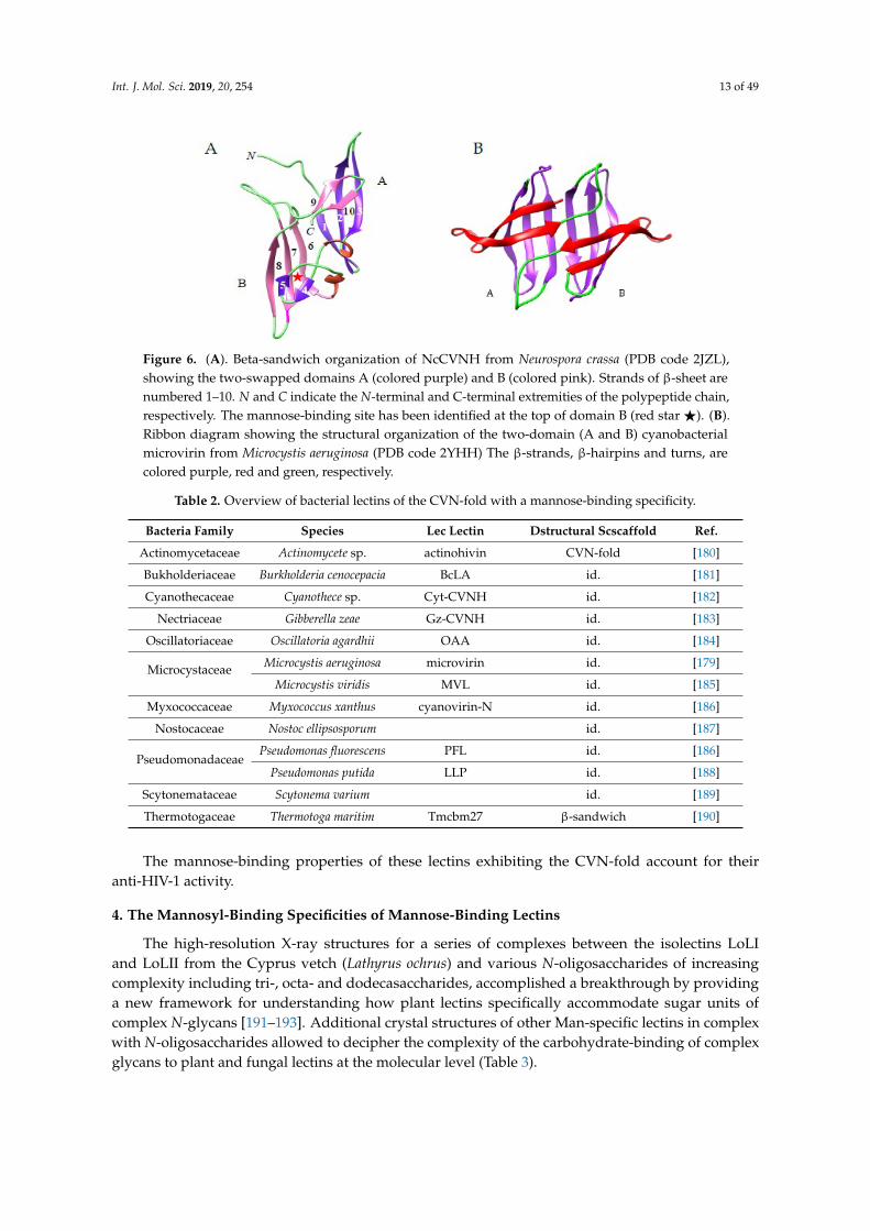
Int. J. Mol. Sci. 2019, 20, 254 13 of 4915
Int. J. Mol. Sci. 2019, 20
Figure 6. (A). Beta-sandwich organization of NcCVNH from Neurospora crassa (PDB code 2JZL),
showing the two-swapped domains A (colored purple) and B (colored pink). Strands of β-sheet are
numbered 1–10. N and C indicate the N-terminal and C-terminal extremities of the polypeptide chain,
respectively. The mannose-binding site has been identified at the top of domain B (red star ★). (B).
Ribbon diagram showing the structural organization of the two-domain (A and B) cyanobacterial
microvirin from Microcystis aeruginosa (PDB code 2YHH) The β-strands, β-hairpins and turns, are
colored purple, red and green, respectively.
It is notheworthy that most of the Man-specific lectins identified in bacteria consist of the so-
called CVN family fold (Table 2), which comprises cyanovirin, actinohivin, and microvirin occurring
in cyanobacteria (ex blue-green algae) as a two swapped domains polypeptide chain, each domain
built up from a β-sheet of three anti-parallel β-strands linked to a β-hairpin by a short α-helical turn
[179] (Figure 6B).
Table 2. Overview of bacterial lectins of the CVN-fold with a mannose-binding specificity.
Bacteria Family Species Lec Lectin Dstructural Scscaffold Ref.
Actinomycetaceae Actinomycete sp. actinohivin CVN-fold [180]
Bukholderiaceae Burkholderia cenocepacia BcLA id. [181]
Cyanothecaceae Cyanothece sp. Cyt-CVNH id. [182]
Nectriaceae Gibberella zeae Gz-CVNH id. [183]
Oscillatoriaceae Oscillatoria agardhii OAA id. [184]
Microcystaceae Microcystis aeruginosa microvirin id. [179]
Microcystis viridis MVL id. [185]
Myxococcaceae Myxococcus xanthus cyanovirin-N id. [186]
Nostocaceae Nostoc ellipsosporum id. [187]
Pseudomonadaceae Pseudomonas fluorescens PFL id. [186]
Pseudomonas putida LLP id. [188]
Scytonemataceae Scytonema varium id. [189]
Thermotogaceae Thermotoga maritim Tmcbm27 β-sandwich [190]
The mannose-binding properties of these lectins exhibiting the CVN-fold account for their anti-
HIV-1 activity.
Figure 6. (A). Beta-sandwich organization of NcCVNH from Neurospora crassa (PDB code 2JZL),showing the two-swapped domains A (colored purple) and B (colored pink). Strands of β-sheet arenumbered 1–10. N and C indicate the N-terminal and C-terminal extremities of the polypeptide chain,respectively. The mannose-binding site has been identified at the top of domain B (red star F). (B).Ribbon diagram showing the structural organization of the two-domain (A and B) cyanobacterialmicrovirin from Microcystis aeruginosa (PDB code 2YHH) The β-strands, β-hairpins and turns, arecolored purple, red and green, respectively.
Table 2. Overview of bacterial lectins of the CVN-fold with a mannose-binding specificity.
Bacteria Family Species Lec Lectin Dstructural Scscaffold Ref.
Actinomycetaceae Actinomycete sp. actinohivin CVN-fold [180]
Bukholderiaceae Burkholderia cenocepacia BcLA id. [181]
Cyanothecaceae Cyanothece sp. Cyt-CVNH id. [182]
Nectriaceae Gibberella zeae Gz-CVNH id. [183]
Oscillatoriaceae Oscillatoria agardhii OAA id. [184]
Microcystaceae Microcystis aeruginosa microvirin id. [179]
Microcystis viridis MVL id. [185]
Myxococcaceae Myxococcus xanthus cyanovirin-N id. [186]
Nostocaceae Nostoc ellipsosporum id. [187]
PseudomonadaceaePseudomonas fluorescens PFL id. [186]
Pseudomonas putida LLP id. [188]
Scytonemataceae Scytonema varium id. [189]
Thermotogaceae Thermotoga maritim Tmcbm27 β-sandwich [190]
The mannose-binding properties of these lectins exhibiting the CVN-fold account for theiranti-HIV-1 activity.
4. The Mannosyl-Binding Specificities of Mannose-Binding Lectins
The high-resolution X-ray structures for a series of complexes between the isolectins LoLIand LoLII from the Cyprus vetch (Lathyrus ochrus) and various N-oligosaccharides of increasingcomplexity including tri-, octa- and dodecasaccharides, accomplished a breakthrough by providinga new framework for understanding how plant lectins specifically accommodate sugar units ofcomplex N-glycans [191–193]. Additional crystal structures of other Man-specific lectins in complexwith N-oligosaccharides allowed to decipher the complexity of the carbohydrate-binding of complexglycans to plant and fungal lectins at the molecular level (Table 3).

Int. J. Mol. Sci. 2019, 20, 254 14 of 49
Table 3. PDB codes of lectins from plants and fungi, complexed with simple sugars (m),oligomannosides (o), and complex (c) mannose-containing glycans.
Plant Species: Lectin: PDB Code: Ref.
Bowringia mildbraedii BMA 2FMD(o) [194]
Canavalia ensiformis ConA 1BXH(o), 1CVN(o), 1I3H(o), 1ONA(o), 1QDC(o),1QDO(o), 1TEI(o), 1VAM(m), 5CNA(m), 5WEY(o) [195–203]
Canavalia gladiata CGL 2D7F(m), 2EF6(o), 2OVU(o) [204,205]
Canavalia maritima ConM 2OW4(o), 2P37(o) [205]
Canavalia virosa ConV 5F5Q(m) [27]
Centrolobium tomentosum CTL 5EYX(o), 5EYY(o) [29]
Cymbosema roseum CRLI 4MYE(m)
Dioclea grandiflora DGL 1DGL(o) [35]
Dioclea lasiocarpa DLL 5UUY(m) [37]
Dioclea lasiophylla DlyL 6CJ9(m) [38]
Dioclea reflexa DrfL 5TG3(m) [39]
Dioclea rostrata DRL 2ZBJ [40]
Dioclea sclerocarpa DSL 4NOT(m) [41]
Dioclea virgata DvirL 3RS6(m) [43]
Lathyrus ochrus LoLI 1LOA(m), 1LOB(m), 1LOF(o), 1LOG(o) [191,192,206]
LoLII 1LGB(c), 1LGC(c) [193]
Pisum arvense PAL 5T7P(m) [207]
Pisum sativum PsA 1BQP(m), 1RIN(o) [208,209]
Pterocarpus angolensis PAL
1Q8O(o), 1Q8P(o), 1Q8Q(o), 1Q8S(o), 1Q8V(o),1UKG(m), 2AR6(o), 2ARB(o), 2ARE(m), 2ARX(o),
2AUY(o), 2GN3(m), 2GN7(o), 2GMM(o), 2GMP(o),2PHF(o), 2PHR(o), 2PHT(o), 2PHU(o), 2PHW(o),
2PHX(o)
[210–213]
Parkia biglobosa PBL 4MQ0(m)
Artocarpus incisa frutapin 5M6O(m) [72]
Artocarpus integrifolia artocarpin 1J4U(m), 1VBO(o), 1VBP(o) [163,214]
jacalin 1KUJ(m), 1WS4(m), 1WS5(m) [77,78]
Morus nigra Morniga-M 1XXR(m) [168]
Helianthus tuberosus Heltuba 1C3M(o), 1C3N(o) [81]
Colocasia esculenta tarin 5D9Z(m), 5T20(o) [165]
Ipomoea batatas ipomoelin 3R51(m), [99]Calystegia sepium Calsepa 1OUW(m), 5AV7(o), 5XF1(o) [98]
Allium sativum ASA 1BWU(m), 1KJ1(m) [215,216]
Galanthus nivalis GNA 1JPC(o), 1MSA(m), 1NIV(o) [217,218]
Narcissus pseudonarcissus NPA 1NPL(o), 3DZW(o) [219]
Musa acuminata 3MIT(m), 3MIU(o), 4PIK(o), 4PIT(o) [128,220]
Musa paradisiaca 1X1V(m) [127]
Oryza sativa Orysata 5XFH(c), 5XFI(c) [221]
Fungal/Algal Species: Lectin: PDB Code: Ref.
Griffthsia sp. griffthsin 2GUC(m), 2GUD(m), 2HYQ(o), 3LL2(c) [140,222,223]
Saccharomyces cerevisiae adhesin Flo1 4LHK(o), 4LHN(m) [151]
Saccharomyces pastorianus flocculin Flo5 2XJP(m), 2XJR(o), 2XJS(o), 2XJT(o), 2XJU(o) [150]
Schizosaccharomyces pombe glucosidase II 4XQM(m) [152]
Marasmus oreades cyanovirin-N 4TKC(m)
Actinomyces sp. actinohivin 4P6A(o) [224]

Int. J. Mol. Sci. 2019, 20, 254 15 of 49
Depending on the molecular complexity of the recognized carbohydrates, two types of closelyinterlinked carbohydrate-binding specificities can occur at the carbohydrate-binding site of plant andfungal lectins:
1. A monosaccharide-binding specificity, allowing the lectin to specifically recognize a simplesugar, e.g., mannose Man, and its derivatives, e.g., α-methylmannoside. This type of monosacchariderecognition by lectins corresponds to the so-called “broad sugar-binding specificity” of lectins, whichrelies on the occurrence of a monosaccharide-binding pocket within the carbohydrate-binding site.
2. An oligosaccharide-binding specificity, which consists of the simultaneous accommodationof several sugar units of a complex N-glycan, e.g., high-mannose glycans, also known as the “finesugar-binding specificity” of the lectins. This type of oligosaccharide recognition involves most of thesurface of the carbohydrate-binding site, including the monosaccharide-binding site.
The monosaccharide-binding site is part of a more extended oligosaccharide-binding site.In physiological conditions, however, plant and fungal lectins are almost always involved in therecognition of complex glycans, rather than simple sugars, simply because the amount of freemonosaccharides in cells and tissues is very low. The binding of plant and fungal lectins to mannosewas first observed in hapten inhibition experiments, by introducing free mannose or mannosederivatives to prevent or reverse the in vitro interaction between lectins and red blood cells or complexglycans. Obviously, the affinity of mannose-specific lectins for simple sugars, e.g., for Man or Manderivatives, is far weaker compared to the affinity measured for more complex glycans, e.g., forcomplex N-glycans or high-mannose type glycans (Table 4) [10,225].
Table 4. Minimum concentrations (mM) of various oligosaccharidic structures and glycopeptidesnecessary to completely inhibit red blood cells agglutination by Con A, LcA from lentil and favin (fromref. [225]).
Oligosaccharidic Structures Con A LcA Favin
Man 1.25 2.5 0.625
αMan(1,3)βMαν(1,4)GlcNAc 0.104 0.83 0.104
αMαν(1,2)αMαν(1,3)βMan(1,4)GlcNAc 0.026 0.21 0.105
αMαν(1,2)αMan(1,2)αMαν(1,3)βMαν(1,4)GlcNAc 0.026 0.206 0.105
18 Int. J. Mol. Sci. 2019, 20
Table 4 Minimum concentrations (mM) of various oligosaccharidic structures and glycopeptides
necessary to completely inhibit red blood cells agglutination by Con A, LcA from lentil and favin
(from ref. [225]).
Oligosaccharidic Structures Con A LcA Favin
Man 1.25 2.5 0.625
αMan(1,3)β(1,4)GlcNAc 0.104 0.83 0.104
α(1,2)α(1,3)βMan(1,4)GlcNAc 0.026 0.21 0.105
α(1,2)αMan(1,2)(1,3)β(1,4)GlcNAc 0.026 0.206 0.105
0.0003 0.157 0.31
0.026 0.003 0.013
0.026 0.0004 0.0008
4.1. The Mannose-Binding Specificity
The recognition and binding of simple sugars by lectins occurs through non covalent
interactions occurring between some hydroxyls of the sugar ring and a few, essentially polar, amino
acid residues forming a shallow depression at the lectin surface, the so-called monosaccharide-
binding site. Usually, most of these interactions consist of hydrogen bonds (H-bond) often associated
to a hydrophobic stacking of the pyranose ring of the sugar to the phenolic ring of an aromatic residue
such as Phe (F), Tyr (Y), or Trp (W), located in the close vicinity of the monosaccharide-binding cavity.
Acidic residues like Asp (D) and Glu (E), often participate in the interaction with simple sugars, thus
attributing a more or less pronounced electronegative character to the monosaccharide-binding site.
Both acidic residues Asp and Glu, play a key role in the binding of simple sugars due to their capacity
to create multiple H-bonds with the hydroxyls emerging from the sugar ring.
Detailed structural information is available for the binding of α-D-mannose (Man) to the
monosaccharide-binding site of Man-specific legume lectins including Con A [202], LoLI isolectin
from Lathyrus ochrus [206], favin from the broad bean Vicia faba [63], pea lectin PsA [209] and PAL
from Pterocarpus angolensis [210]. A very similar binding scheme occurs for both the two-chain (LolLI,
favin, PsA) and single-chain (Con A) lectins: a few amino acid residues located on three distinct loops
exposed at the top of the dome-shaped lectin protomer, form a shallow depression which
accommodates the Man ligand via a network of hydrogen bonds connected to the O3, O4, O5 and O6
atoms of the sugar. An acidic residue (Asp208 of Con A, Asp81 of LoLI and PsA), which also
participates in the binding of a Ca2+ ion located in the close vicinity of the binding site, plays a key
role in ligand binding. An additional stacking interaction between the pyranose ring of Man and one
(Phe123 of LoLI) or two (Tyr12 and Tyr100 of Con A) aromatic residues located in the vicinity of the
monosaccharide-binding site, reinforces anchorage of the sugar to the binding site (Figure 7A–D). A
few water molecules also participate in the binding of Man to the monosaccharide-binding site of the
lectins. Very similar binding observations were reported for the binding of Man or α-methyl-D-
0.0003 0.157 0.31
18 Int. J. Mol. Sci. 2019, 20
Table 4 Minimum concentrations (mM) of various oligosaccharidic structures and glycopeptides
necessary to completely inhibit red blood cells agglutination by Con A, LcA from lentil and favin
(from ref. [225]).
Oligosaccharidic Structures Con A LcA Favin
Man 1.25 2.5 0.625
αMan(1,3)β(1,4)GlcNAc 0.104 0.83 0.104
α(1,2)α(1,3)βMan(1,4)GlcNAc 0.026 0.21 0.105
α(1,2)αMan(1,2)(1,3)β(1,4)GlcNAc 0.026 0.206 0.105
0.0003 0.157 0.31
0.026 0.003 0.013
0.026 0.0004 0.0008
4.1. The Mannose-Binding Specificity
The recognition and binding of simple sugars by lectins occurs through non covalent
interactions occurring between some hydroxyls of the sugar ring and a few, essentially polar, amino
acid residues forming a shallow depression at the lectin surface, the so-called monosaccharide-
binding site. Usually, most of these interactions consist of hydrogen bonds (H-bond) often associated
to a hydrophobic stacking of the pyranose ring of the sugar to the phenolic ring of an aromatic residue
such as Phe (F), Tyr (Y), or Trp (W), located in the close vicinity of the monosaccharide-binding cavity.
Acidic residues like Asp (D) and Glu (E), often participate in the interaction with simple sugars, thus
attributing a more or less pronounced electronegative character to the monosaccharide-binding site.
Both acidic residues Asp and Glu, play a key role in the binding of simple sugars due to their capacity
to create multiple H-bonds with the hydroxyls emerging from the sugar ring.
Detailed structural information is available for the binding of α-D-mannose (Man) to the
monosaccharide-binding site of Man-specific legume lectins including Con A [202], LoLI isolectin
from Lathyrus ochrus [206], favin from the broad bean Vicia faba [63], pea lectin PsA [209] and PAL
from Pterocarpus angolensis [210]. A very similar binding scheme occurs for both the two-chain (LolLI,
favin, PsA) and single-chain (Con A) lectins: a few amino acid residues located on three distinct loops
exposed at the top of the dome-shaped lectin protomer, form a shallow depression which
accommodates the Man ligand via a network of hydrogen bonds connected to the O3, O4, O5 and O6
atoms of the sugar. An acidic residue (Asp208 of Con A, Asp81 of LoLI and PsA), which also
participates in the binding of a Ca2+ ion located in the close vicinity of the binding site, plays a key
role in ligand binding. An additional stacking interaction between the pyranose ring of Man and one
(Phe123 of LoLI) or two (Tyr12 and Tyr100 of Con A) aromatic residues located in the vicinity of the
monosaccharide-binding site, reinforces anchorage of the sugar to the binding site (Figure 7A–D). A
few water molecules also participate in the binding of Man to the monosaccharide-binding site of the
lectins. Very similar binding observations were reported for the binding of Man or α-methyl-D-
0.026 0.003 0.013
18 Int. J. Mol. Sci. 2019, 20
Table 4 Minimum concentrations (mM) of various oligosaccharidic structures and glycopeptides
necessary to completely inhibit red blood cells agglutination by Con A, LcA from lentil and favin
(from ref. [225]).
Oligosaccharidic Structures Con A LcA Favin
Man 1.25 2.5 0.625
αMan(1,3)β(1,4)GlcNAc 0.104 0.83 0.104
α(1,2)α(1,3)βMan(1,4)GlcNAc 0.026 0.21 0.105
α(1,2)αMan(1,2)(1,3)β(1,4)GlcNAc 0.026 0.206 0.105
0.0003 0.157 0.31
0.026 0.003 0.013
0.026 0.0004 0.0008
4.1. The Mannose-Binding Specificity
The recognition and binding of simple sugars by lectins occurs through non covalent
interactions occurring between some hydroxyls of the sugar ring and a few, essentially polar, amino
acid residues forming a shallow depression at the lectin surface, the so-called monosaccharide-
binding site. Usually, most of these interactions consist of hydrogen bonds (H-bond) often associated
to a hydrophobic stacking of the pyranose ring of the sugar to the phenolic ring of an aromatic residue
such as Phe (F), Tyr (Y), or Trp (W), located in the close vicinity of the monosaccharide-binding cavity.
Acidic residues like Asp (D) and Glu (E), often participate in the interaction with simple sugars, thus
attributing a more or less pronounced electronegative character to the monosaccharide-binding site.
Both acidic residues Asp and Glu, play a key role in the binding of simple sugars due to their capacity
to create multiple H-bonds with the hydroxyls emerging from the sugar ring.
Detailed structural information is available for the binding of α-D-mannose (Man) to the
monosaccharide-binding site of Man-specific legume lectins including Con A [202], LoLI isolectin
from Lathyrus ochrus [206], favin from the broad bean Vicia faba [63], pea lectin PsA [209] and PAL
from Pterocarpus angolensis [210]. A very similar binding scheme occurs for both the two-chain (LolLI,
favin, PsA) and single-chain (Con A) lectins: a few amino acid residues located on three distinct loops
exposed at the top of the dome-shaped lectin protomer, form a shallow depression which
accommodates the Man ligand via a network of hydrogen bonds connected to the O3, O4, O5 and O6
atoms of the sugar. An acidic residue (Asp208 of Con A, Asp81 of LoLI and PsA), which also
participates in the binding of a Ca2+ ion located in the close vicinity of the binding site, plays a key
role in ligand binding. An additional stacking interaction between the pyranose ring of Man and one
(Phe123 of LoLI) or two (Tyr12 and Tyr100 of Con A) aromatic residues located in the vicinity of the
monosaccharide-binding site, reinforces anchorage of the sugar to the binding site (Figure 7A–D). A
few water molecules also participate in the binding of Man to the monosaccharide-binding site of the
lectins. Very similar binding observations were reported for the binding of Man or α-methyl-D-
0.026 0.0004 0.0008
4.1. The Mannose-Binding Specificity
The recognition and binding of simple sugars by lectins occurs through non covalent interactionsoccurring between some hydroxyls of the sugar ring and a few, essentially polar, amino acid residuesforming a shallow depression at the lectin surface, the so-called monosaccharide-binding site. Usually,most of these interactions consist of hydrogen bonds (H-bond) often associated to a hydrophobicstacking of the pyranose ring of the sugar to the phenolic ring of an aromatic residue such as Phe (F),Tyr (Y), or Trp (W), located in the close vicinity of the monosaccharide-binding cavity. Acidic residueslike Asp (D) and Glu (E), often participate in the interaction with simple sugars, thus attributing amore or less pronounced electronegative character to the monosaccharide-binding site. Both acidic
Int. J. Mol. Sci. 2019, 20, 254 16 of 49
residues Asp and Glu, play a key role in the binding of simple sugars due to their capacity to createmultiple H-bonds with the hydroxyls emerging from the sugar ring.
Detailed structural information is available for the binding of α-D-mannose (Man) to themonosaccharide-binding site of Man-specific legume lectins including Con A [202], LoLI isolectinfrom Lathyrus ochrus [206], favin from the broad bean Vicia faba [63], pea lectin PsA [209] and PALfrom Pterocarpus angolensis [210]. A very similar binding scheme occurs for both the two-chain (LolLI,favin, PsA) and single-chain (Con A) lectins: a few amino acid residues located on three distinctloops exposed at the top of the dome-shaped lectin protomer, form a shallow depression whichaccommodates the Man ligand via a network of hydrogen bonds connected to the O3, O4, O5 andO6 atoms of the sugar. An acidic residue (Asp208 of Con A, Asp81 of LoLI and PsA), which alsoparticipates in the binding of a Ca2+ ion located in the close vicinity of the binding site, plays a keyrole in ligand binding. An additional stacking interaction between the pyranose ring of Man and one(Phe123 of LoLI) or two (Tyr12 and Tyr100 of Con A) aromatic residues located in the vicinity of themonosaccharide-binding site, reinforces anchorage of the sugar to the binding site (Figure 7A–D). A fewwater molecules also participate in the binding of Man to the monosaccharide-binding site of the lectins.Very similar binding observations were reported for the binding of Man or α-methyl-D-mannoside(MeMan) to other Canavalia [20,21,25,27] and Dioclea lectins [34–44], Parkia biglobosa (PDB code 4MQ0)and Cymosema roseum (PDB code 4MYE) Man-specific lectins from the Brasilian flora.
The accommodation of Man by artocarpin, a Man-specific jacalin-related lectin, shows avery similar network of 9 H-bonds between four amino acid residues (Gly15, Asp138, Leu139,Asp141) located at the top of the β-prism protomer, and the O1, O3, O4, O5, and O6 atomsof the sugar (Figure 7E,F). No stacking interactions occur between the aromatic residues of themonosaccharide-binding site and the sugar. In addition, jacalin, another member of the jacalin-relatedlectins, offers an interesting example of sugar-binding promiscuity because this Gal-specific lectinsalso interacts, albeit with lower affinity, with other simple sugars like Man, Glc and GalNAc via a verysimilar H-bond network [77]. Another Man-specific lectin with a β-prism architecture, Heltuba ofHelianthus tuberosus, also accommodates Man through a very similar network of H-bonds betweenfour amino acid residues (Gly18, Asp136, Val137, Asp139), which form the monosaccharide-bindingsite also located at the top of the β-prism protomer, and the O3, O4, O5 and O6 atoms of the sugar(Figure 7G,H).
The recognition of Man by GNA, the Man-specific snowdrop (Galanthus nivalis) lectin, and othermonocot Man-binding lectins harboring a similar β-prism architecture (a β-prism in which the strandscomposing the β-sheet are arranged perpendicularly to the axis of the prism), exhibits a differentmode of binding due to the fact that three out of eight H-bonds connecting the Gln89, Asp91, Asn93and Tyr97 residues from the 3rd mannose-binding site to the O2, O3, O4, and O6 atoms of Man,are connected to the axial O2 atom (Figure 5I,J). Residue Tyr97 also provides a stacking interactionwith one face of the Man pyranose ring. An additional hydrophobic interaction with Val95, anotherresidue of the consensus sequence stretch QXDXNXVXY of the monosaccharide-binding binding site,reinforces the anchorage of Man to the binding site.
Molecular modeling and in silico docking suggest that other nucleocytoplasmic EUL domain-containing lectins from rice (Oryza sativa) and Arabidopsis with a β-prism architecture, also interactwith mannose via a very similar network of H-bonds and stacking interactions with aromatic aminoacid residues located in the close vicinity of the monosaccharide-binding site (Figure 8) [170]. However,some promiscuity was shown to occur at the monosaccharide-binding site of the EUL-lectins, which inaddition to high mannose N-glycans also recognize blood group B related structures and galactosylatedepitopes [226].

Int. J. Mol. Sci. 2019, 20, 254 17 of 4920
Int. J. Mol. Sci. 2019, 20
Figure 7. (A,B). ConA from Canavalia ensiformis in complex with α-methylmannoside (PDB code
5CNA). (C,D). Isolectin LoLI from Lathyrus ochrus in complex with Man (PDB code 1LOB). (E,F).
Artocarpin from Artocarpus integrifolia in complex with α-mthylmannoside (PDB code 1J4U). (G,H).
Heltuba from Helianthus tuberosus in complex with Manα1,3Man (PDB code 1C3M). (I,J). Third Man-
binding site of GNA from Galanthus nivalis in complex with α-methylmannoside (PDB code 1MSA).
Hydrogen bonds connecting the monosaccharides to the amino acid residues forming the
monosaccharide-binding site are represented by black dashed lines. Aromatic residues participating
in stacking interactions with the sugar rings are colored orange. The molecular surface of the lectins
is colored dark grey and their extended oligosaccharide-binding areas are delineated by white dashed
lines. The shallow depression corresponding to the monosaccharide-binding site that accommodates
simple sugars is delineated by a green dashed line. The green and violet spheres correspond to the
Ca2+ and Mn2+ ions, that have a stabilizing effect on the carbohydrate-binding site.
Molecular modeling and in silico docking suggest that other nucleocytoplasmic EUL domain-
containing lectins from rice (Oryza sativa) and Arabidopsis with a β-prism architecture, also interact
with mannose via a very similar network of H-bonds and stacking interactions with aromatic amino
acid residues located in the close vicinity of the monosaccharide-binding site (Figure 8) [170].
However, some promiscuity was shown to occur at the monosaccharide-binding site of the EUL-
lectins, which in addition to high mannose N-glycans also recognize blood group B related structures
and galactosylated epitopes [226].
Figure 7. (A,B). ConA from Canavalia ensiformis in complex with α-methylmannoside (PDB code 5CNA).(C,D). Isolectin LoLI from Lathyrus ochrus in complex with Man (PDB code 1LOB). (E,F). Artocarpinfrom Artocarpus integrifolia in complex with α-mthylmannoside (PDB code 1J4U). (G,H). Heltuba fromHelianthus tuberosus in complex with Manα1,3Man (PDB code 1C3M). (I,J). Third Man-binding site ofGNA from Galanthus nivalis in complex with α-methylmannoside (PDB code 1MSA). Hydrogen bondsconnecting the monosaccharides to the amino acid residues forming the monosaccharide-binding siteare represented by black dashed lines. Aromatic residues participating in stacking interactions withthe sugar rings are colored orange. The molecular surface of the lectins is colored dark grey and theirextended oligosaccharide-binding areas are delineated by white dashed lines. The shallow depressioncorresponding to the monosaccharide-binding site that accommodates simple sugars is delineated by agreen dashed line. The green and violet spheres correspond to the Ca2+ and Mn2+ ions, that have astabilizing effect on the carbohydrate-binding site.

Int. J. Mol. Sci. 2019, 20, 254 18 of 4921
Int. J. Mol. Sci. 2019, 20
Figure 8. Docking of αMeMan to the monosaccharide-binding site of the active sub-domain III of
OsEULS3. Hydrogen bonds connecting Man to the amino acid residues forming the monosaccharide-
binding site are shown by black dashed lines and distances are indicated in Å. The aromatic Trp136
residue participating in stacking interactions with the sugar ring is colored orange.
4.2. The Oligosaccharide-Binding Specificity
Although the monosaccharide-binding capacity of Man-specific lectins has been widely
investigated, it is obvious that simple sugar residues like Man probably cannot be considered as the
natural ligands for plant and fungal lectins, due to the extreme scarcity of simple sugars as free
ligands occurring in living organisms, compared to other complex carbohydrates. Along this line, the
affinity of Man-specific lectins for complex high-mannose N-glycans is much higher than that
measured for free Man [10,225]. In fact, once the first crystallographic structures of complexes of Man-
specific lectins with oligomannosides were solved at atomic resolution [191–193], it became evident
that the so-called monosaccharide-binding site is in fact part of a more surface-extended
oligosaccharide-binding site, comprising other amino acid residues susceptible to chemical
interaction with other sugar units distinct from that recognized by the monosaccharide-binding site.
Such a multiplicity of interactions readily accounts for the higher affinity of Man-specific lectins for
high-mannose N-glycans (inhibitory activity in the mM range), compared to free Man (inhibitory
activity in the µM range) [225]. In addition, depending on the degree of freedom of the different O-
glycosidic linkage types, e.g., α1-2, α1-3, α1-4 or α1-6, occurring along the glycan chain, complex
glycans can more or less fit the shape of the lectin oligosaccharide-binding site.
Structural analysis of different lectin-oligosaccharide complexes (Table 5), including Con A in
complex with a pentasaccharide (Figure 9A,B), isolectin LoLII from Lathyrus ochrus in complex with
a biantennary octasaccharide of the N-acetyllactosamine type from lactotransferrin (Figure 9C,D),
GNA in complex with a mannopentaose (Figure 9E,F), and PAL from Pterocarpus angolensis in
complex with a mannotetraose (Figure 9G,H), show that a complex network of H-bonds, stacking
and hydrophobic interactions, links several sugar units of the glycan chain to the oligosaccharide-
binding site of the lectin. However, depending on the lectin, important discrepancies occur in the
accommodation of sugar units. In this respect, isolectins of Lathyrus ochrus and other two-chain
Vicieae lectins such as pea PsA and lentil LcA lectins, which differ from Con A by a higher affinity
for fucosylated glycans of the N-acetyllactosaminic type [10,225], strongly interact with the α1,6-Fuc
residue linked to the Asn-bound GlcNAc of the glycan whereas Con A does not interfere at all with
the Fuc residue. Similarly, the accommodation of structurally closely-related oligomannosides by
GNA (Figure 9F) and PAL (Figure 7H), illustrates how discrepancies observed in the topographical
features (shape and size) of the oligosaccharide-binding site can affect the binding of complex glycans
to different Man-specific lectins belonging to distinct scaffold architectures.
Figure 8. Docking of αMeMan to the monosaccharide-binding site of the active sub-domain III ofOsEULS3. Hydrogen bonds connecting Man to the amino acid residues forming the monosaccharide-binding site are shown by black dashed lines and distances are indicated in Å. The aromatic Trp136residue participating in stacking interactions with the sugar ring is colored orange.
4.2. The Oligosaccharide-Binding Specificity
Although the monosaccharide-binding capacity of Man-specific lectins has been widelyinvestigated, it is obvious that simple sugar residues like Man probably cannot be considered as thenatural ligands for plant and fungal lectins, due to the extreme scarcity of simple sugars as free ligandsoccurring in living organisms, compared to other complex carbohydrates. Along this line, the affinityof Man-specific lectins for complex high-mannose N-glycans is much higher than that measured forfree Man [10,225]. In fact, once the first crystallographic structures of complexes of Man-specific lectinswith oligomannosides were solved at atomic resolution [191–193], it became evident that the so-calledmonosaccharide-binding site is in fact part of a more surface-extended oligosaccharide-binding site,comprising other amino acid residues susceptible to chemical interaction with other sugar units distinctfrom that recognized by the monosaccharide-binding site. Such a multiplicity of interactions readilyaccounts for the higher affinity of Man-specific lectins for high-mannose N-glycans (inhibitory activityin the mM range), compared to free Man (inhibitory activity in the µM range) [225]. In addition,depending on the degree of freedom of the different O-glycosidic linkage types, e.g., α1-2, α1-3, α1-4or α1-6, occurring along the glycan chain, complex glycans can more or less fit the shape of the lectinoligosaccharide-binding site.
Structural analysis of different lectin-oligosaccharide complexes (Table 5), including Con A incomplex with a pentasaccharide (Figure 9A,B), isolectin LoLII from Lathyrus ochrus in complex with abiantennary octasaccharide of the N-acetyllactosamine type from lactotransferrin (Figure 9C,D), GNAin complex with a mannopentaose (Figure 9E,F), and PAL from Pterocarpus angolensis in complex witha mannotetraose (Figure 9G,H), show that a complex network of H-bonds, stacking and hydrophobicinteractions, links several sugar units of the glycan chain to the oligosaccharide-binding site of thelectin. However, depending on the lectin, important discrepancies occur in the accommodation ofsugar units. In this respect, isolectins of Lathyrus ochrus and other two-chain Vicieae lectins such aspea PsA and lentil LcA lectins, which differ from Con A by a higher affinity for fucosylated glycansof the N-acetyllactosaminic type [10,225], strongly interact with the α1,6-Fuc residue linked to theAsn-bound GlcNAc of the glycan whereas Con A does not interfere at all with the Fuc residue. Similarly,the accommodation of structurally closely-related oligomannosides by GNA (Figure 9F) and PAL(Figure 7H), illustrates how discrepancies observed in the topographical features (shape and size) ofthe oligosaccharide-binding site can affect the binding of complex glycans to different Man-specificlectins belonging to distinct scaffold architectures.

Int. J. Mol. Sci. 2019, 20, 254 19 of 4922
Int. J. Mol. Sci. 2019, 20
Figure 9. (A,B). ConA from Canavalia ensiformis in complex with β-D-GlcNAc-(1,2)-α-D-Man-(1,6)-[β-
D-GlcNAc-(1,2)-α-D-Man-(1,6]-αD-Man (PDB code 1TEI) [196]. (C,D). Isolectin LoLII from Lathyrus
ochrus in complex with a biantennary octasaccharide of the N-acetyllactosamine type from
lactotransferrin (PDB code 1LOF). (E,F). GNA from Galanthus nivalis in complex with three mannosyl
residues from a mannopentaose (PDB code 1JPC). (G,H). PAL from Pterocarpus angolensis in complex
with a mannotetraose (PDB code 2PHF). Hydrogen bonds connecting the oligosaccharides to the
amino acid residues forming the extended carbohydrate-binding site are represented by black dashed
lines. Aromatic residues participating in stacking interactions with the sugar rings are colored orange.
The electrostatic potentials were calculated and mapped on the molecular surface of the lectins, using
YASARA. The extended oligosaccharide-binding areas are delineated by white dashed lines. The
shallow depression corresponding to the monosaccharide-binding site that accommodates simple
sugars, is delineated by a green dashed line.
Table 5. Structure of the branched oligosaccharides complexed to Con A (PDB code 1TEI), LoLII (PDB
code 1LOF), GNA (PDB code 1JPC) and PAL (PDB code 2PHF).
Oligosaccharides/Glycopeptide Complexed to:
Figure 9. (A,B). ConA from Canavalia ensiformis in complex with β-D-GlcNAc-(1,2)-α-D-Man-(1,6)-[β-D-GlcNAc-(1,2)-α-D-Man-(1,6]-αD-Man (PDB code 1TEI) [196]. (C,D). Isolectin LoLII from Lathyrus ochrusin complex with a biantennary octasaccharide of the N-acetyllactosamine type from lactotransferrin(PDB code 1LOF). (E,F). GNA from Galanthus nivalis in complex with three mannosyl residuesfrom a mannopentaose (PDB code 1JPC). (G,H). PAL from Pterocarpus angolensis in complex witha mannotetraose (PDB code 2PHF). Hydrogen bonds connecting the oligosaccharides to the aminoacid residues forming the extended carbohydrate-binding site are represented by black dashed lines.Aromatic residues participating in stacking interactions with the sugar rings are colored orange.The electrostatic potentials were calculated and mapped on the molecular surface of the lectins,using YASARA. The extended oligosaccharide-binding areas are delineated by white dashed lines.The shallow depression corresponding to the monosaccharide-binding site that accommodates simplesugars, is delineated by a green dashed line.

Int. J. Mol. Sci. 2019, 20, 254 20 of 49
Table 5. Structure of the branched oligosaccharides complexed to Con A (PDB code 1TEI), LoLII (PDBcode 1LOF), GNA (PDB code 1JPC) and PAL (PDB code 2PHF).
Oligosaccharides/Glycopeptide Complexed to:
23 Int. J. Mol. Sci. 2019, 20
Con A
LoLII
GNA
PAL
Investigations on the oligosaccharide-binding specificity of Man-specific bacterial lectins,
showed a highly similar binding scheme associated to the recognition of oligomannosides and
complex high-mannose N-glycans. However, depending both on the extent of the glycan chain and
the shape and size of the oligosaccharide-binding site in the lectin monomer, which possesses a β-
prism- (griffthsin) or a β-barrel-architecture (actinohivin), rather distinct accommodation schemes
were observed for these lectins (Figure 10) [227]. The oligosaccharide-binding sites of griffthsin and
actinohivin readily differ by the shape, the size and the discrete distribution of charged residues that
account for the differences observed in the accommodation of oligomannosides and high-mannose
branched glycans by the lectins. Similar to plant lectins, the monosaccharide-binding pocket occupies
a pivotal position at the centre of the binding site and fully participates in the binding of the complex
glycans.
Obviously, the binding of complex glycan chains to lectins is a highly complex interaction
process due to the extreme variability observed in the topographical features of the oligosaccharide-
binding site of lectins, associated to the extreme diversity of the recognized glycan structures.
Hopefully, the recent developments in glycan array technology [228], and the improvement of frontal
affinity chromatography [229], offer new important tools for deciphering the biomolecular
interactions between plant lectins and the large panel of complex glycans.
Con A
23 Int. J. Mol. Sci. 2019, 20
Con A
LoLII
GNA
PAL
Investigations on the oligosaccharide-binding specificity of Man-specific bacterial lectins,
showed a highly similar binding scheme associated to the recognition of oligomannosides and
complex high-mannose N-glycans. However, depending both on the extent of the glycan chain and
the shape and size of the oligosaccharide-binding site in the lectin monomer, which possesses a β-
prism- (griffthsin) or a β-barrel-architecture (actinohivin), rather distinct accommodation schemes
were observed for these lectins (Figure 10) [227]. The oligosaccharide-binding sites of griffthsin and
actinohivin readily differ by the shape, the size and the discrete distribution of charged residues that
account for the differences observed in the accommodation of oligomannosides and high-mannose
branched glycans by the lectins. Similar to plant lectins, the monosaccharide-binding pocket occupies
a pivotal position at the centre of the binding site and fully participates in the binding of the complex
glycans.
Obviously, the binding of complex glycan chains to lectins is a highly complex interaction
process due to the extreme variability observed in the topographical features of the oligosaccharide-
binding site of lectins, associated to the extreme diversity of the recognized glycan structures.
Hopefully, the recent developments in glycan array technology [228], and the improvement of frontal
affinity chromatography [229], offer new important tools for deciphering the biomolecular
interactions between plant lectins and the large panel of complex glycans.
LoLII
23 Int. J. Mol. Sci. 2019, 20
Con A
LoLII
GNA
PAL
Investigations on the oligosaccharide-binding specificity of Man-specific bacterial lectins,
showed a highly similar binding scheme associated to the recognition of oligomannosides and
complex high-mannose N-glycans. However, depending both on the extent of the glycan chain and
the shape and size of the oligosaccharide-binding site in the lectin monomer, which possesses a β-
prism- (griffthsin) or a β-barrel-architecture (actinohivin), rather distinct accommodation schemes
were observed for these lectins (Figure 10) [227]. The oligosaccharide-binding sites of griffthsin and
actinohivin readily differ by the shape, the size and the discrete distribution of charged residues that
account for the differences observed in the accommodation of oligomannosides and high-mannose
branched glycans by the lectins. Similar to plant lectins, the monosaccharide-binding pocket occupies
a pivotal position at the centre of the binding site and fully participates in the binding of the complex
glycans.
Obviously, the binding of complex glycan chains to lectins is a highly complex interaction
process due to the extreme variability observed in the topographical features of the oligosaccharide-
binding site of lectins, associated to the extreme diversity of the recognized glycan structures.
Hopefully, the recent developments in glycan array technology [228], and the improvement of frontal
affinity chromatography [229], offer new important tools for deciphering the biomolecular
interactions between plant lectins and the large panel of complex glycans.
GNA
23 Int. J. Mol. Sci. 2019, 20
Con A
LoLII
GNA
PAL
Investigations on the oligosaccharide-binding specificity of Man-specific bacterial lectins,
showed a highly similar binding scheme associated to the recognition of oligomannosides and
complex high-mannose N-glycans. However, depending both on the extent of the glycan chain and
the shape and size of the oligosaccharide-binding site in the lectin monomer, which possesses a β-
prism- (griffthsin) or a β-barrel-architecture (actinohivin), rather distinct accommodation schemes
were observed for these lectins (Figure 10) [227]. The oligosaccharide-binding sites of griffthsin and
actinohivin readily differ by the shape, the size and the discrete distribution of charged residues that
account for the differences observed in the accommodation of oligomannosides and high-mannose
branched glycans by the lectins. Similar to plant lectins, the monosaccharide-binding pocket occupies
a pivotal position at the centre of the binding site and fully participates in the binding of the complex
glycans.
Obviously, the binding of complex glycan chains to lectins is a highly complex interaction
process due to the extreme variability observed in the topographical features of the oligosaccharide-
binding site of lectins, associated to the extreme diversity of the recognized glycan structures.
Hopefully, the recent developments in glycan array technology [228], and the improvement of frontal
affinity chromatography [229], offer new important tools for deciphering the biomolecular
interactions between plant lectins and the large panel of complex glycans.
PAL
Investigations on the oligosaccharide-binding specificity of Man-specific bacterial lectins, showeda highly similar binding scheme associated to the recognition of oligomannosides and complexhigh-mannose N-glycans. However, depending both on the extent of the glycan chain and theshape and size of the oligosaccharide-binding site in the lectin monomer, which possesses a β-prism-(griffthsin) or a β-barrel-architecture (actinohivin), rather distinct accommodation schemes wereobserved for these lectins (Figure 10) [227]. The oligosaccharide-binding sites of griffthsin andactinohivin readily differ by the shape, the size and the discrete distribution of charged residuesthat account for the differences observed in the accommodation of oligomannosides and high-mannosebranched glycans by the lectins. Similar to plant lectins, the monosaccharide-binding pocket occupiesa pivotal position at the centre of the binding site and fully participates in the binding of thecomplex glycans.
Obviously, the binding of complex glycan chains to lectins is a highly complex interaction processdue to the extreme variability observed in the topographical features of the oligosaccharide-bindingsite of lectins, associated to the extreme diversity of the recognized glycan structures. Hopefully,the recent developments in glycan array technology [228], and the improvement of frontal affinitychromatography [229], offer new important tools for deciphering the biomolecular interactions betweenplant lectins and the large panel of complex glycans.

Int. J. Mol. Sci. 2019, 20, 254 21 of 4924 Int. J. Mol. Sci. 2019, 20
Figure 10. Structural diversity of the mannose-binding lectins. (A,B,C). Ribbon diagrams (A lateral
view, B upper view) and surface electrostatic potentials (C) of griffthsin in complex with a high-
mannose branched glycan (colored cyan) (PDB code 3LL2), showing the β-prism organization of the
lectin. Note the electronegatively charged character (colored red) of the Man-binding pockets at the
upper face of the β-prism. (D,E,F). Ribbon diagrams (D lateral view, E upper view) and surface
electrostatic potentials (F) of actinohivin in complex with a high-mannose branched glycan (colored
cyan) (PDB code 3S5X), showing the β-trefoil (β-prism II) organization of the lectin. Note the
electronegatively (colored red) and electropositively (colored blue) charged character of the Man-
binding pockets at the edges of the β-trefoil. (G,H,I). Ribbon diagrams (G lateral view, H upper view)
and surface electrostatic potentials (I) of actinohivin in complex with α-1,2-mannotriose (colored
cyan) (PDB code 4P6A), showing the organization of the lectin. Note the electronegatively charged
character (colored red) of the Man-binding pockets.
5. Functions of Mannose-Specific Lectins
Figure 10. Structural diversity of the mannose-binding lectins. (A–C). Ribbon diagrams (A lateral view,B upper view) and surface electrostatic potentials (C) of griffthsin in complex with a high-mannosebranched glycan (colored cyan) (PDB code 3LL2), showing the β-prism organization of the lectin.Note the electronegatively charged character (colored red) of the Man-binding pockets at the upperface of the β-prism. (D–F). Ribbon diagrams (D lateral view, E upper view) and surface electrostaticpotentials (F) of actinohivin in complex with a high-mannose branched glycan (colored cyan) (PDBcode 3S5X), showing the β-trefoil (β-prism II) organization of the lectin. Note the electronegatively(colored red) and electropositively (colored blue) charged character of the Man-binding pockets at theedges of the β-trefoil. (G–I). Ribbon diagrams (G lateral view, H upper view) and surface electrostaticpotentials (I) of actinohivin in complex with α-1,2-mannotriose (colored cyan) (PDB code 4P6A),showing the organization of the lectin. Note the electronegatively charged character (colored red) ofthe Man-binding pockets.

Int. J. Mol. Sci. 2019, 20, 254 22 of 49
5. Functions of Mannose-Specific Lectins
The Man-specific lectins present in seeds or storage organs (bulbs, rhizomes) of plants, areabundant proteins with a dual role as storage proteins and defense proteins [230,231]. In contrast,Man-specific lectins occurring in the nucleus or in the cytoplasmic compartment are usuallysynthesized at low levels. Since lectin concentrations are higher after exposure of the plant to e.g., saltor drought stress, or pathogen infections these stress inducible lectins are involved in plant immunityand can help the plant to cope with environmental stresses [1,231].
5.1. Insecticidal Activity
A large group of GNA-related lectins have been investigated with respect to their insecticidalproperties. The interest in the monocot Man-specific lectins was triggered because these lectins showedtoxicity towards aphid pests responsible for serious crop damage (Table 6). The expression of GNAand other monocot lectins in various transgenic plants conferred enhanced resistance to sap-suckingaphid predators. In addition, these lectins often caused a higher larval mortality and retardation inlarval development. Similarly legume lectins such as Con A were investigated for their deleteriouseffects on aphid growth and development. At present, the mechanism of entomotoxicity still remainpoorly understood and most probably depends on diverse, complementary mechanisms [232].
The detrimental effects of Man-specific lectins on aphids relies on their ability to recognizeand bind high-mannose glycan receptors present in the peritrophic membrane and the underlyingmidgut epithelium. Receptors proteins for the monocot Man-specific lectins (ACA of Allium cepa,Diefenbachia sequina lectin, CEA of Colocacia esculenta, AMA of Arum maculatum) have been identifiedin brush border membrane vesicles of the midgut [233], and two major receptors for AMA of40 kDa and 35 kDa, respectively, were detected in the brush border membrane vesicles of the aphidsLipaphis erysimi and Aphis craccivora [234]. A major binding protein for Con A was identified as amembrane-bound aminopeptidase of 130 kDa, in the pea aphid Acyrthosiphon pisum [235]. Two otherabundant membrane-associated proteins, an alanyl aminopeptidase N and a sucrase, have also beenpostulated as possible receptors in Acyrthosiphon pisum, for both garlic lectins ASAI and ASAII [236].Interestingly, a putative glycosylated receptor of 37 kDa identified in the mushroom Rhizoctonia solanicross-reacted with the homodimeric Allium sativum leaf lectin and the interaction, which dependson the oligomeric assembly of the lectin, was specifically inhibited by addition of mannose [237].Binding partners of CEA, the Colocasia esculenta Man-specific lectin, were identified as ATPase and ATPsynthase in Bemisia tabaci, and ATP synthase, HSP70 and clathrin heavy chain in Lipaphis erysimi [238].The dietary ingestion of Con A resulted in a marked decrease of the α-glucosidase and alkalinephosphatase activity in the bird cherry-oat aphid Rhopalosiphum padi [232]. Taken together, these resultsargue for multiple so-called lectin “receptors” occurring in aphid pests.
Table 6. List of Man-specific lectins investigated for their toxicity towards aphids (aphid predatorspecies are indicated with an asterisk *).
Lectin class Lectin Aphid Ref.
Monocot lectins
GNA (Galanthus nivalis) Aulacorthum solani [239]Myzus persicae [240,241]
Caratovacuna lanigera [242]Myzus persicae [243]
Rhopalosiphum maidis [244]Chrysoperla carnea* Adalia punctata*, Coccinella
septempunctata* [245]
Sitobium avenae, Schizaphis graminum, Rhopalosiphum padi [246]
PTA (Pinellia ternata) Myzus persicae [247,248]
PPA (Pinellia pedatisecta) Sitobium avenae [249]
AAA (Allium altaicum) Aphis gossypii [100]
ACA (Allium cepa)) Myzus persicae [250]
Int. J. Mol. Sci. 2019, 20, 254 23 of 49
Table 6. Cont.
Lectin class Lectin Aphid Ref.
ASA (Allium sativum)
Myzus persicae [251]Aphis craccivora [252]Myzus nicotianae [253]
Acyrthosiphon pisum [236]
AHA (Arisaema heterophyllum) Myzus persicae [254]
MDA (Monstera deliciosa) Myzus persicae [255]
Orysata (Oryza sativa) Acyrthosiphon pisum, Myzus persicae [256]
Dieffenbachia sequina Aphis craccivora, Lipaphis erysimi [233]
CEA (Colocacia esculenta) Lipaphis erysimi, Aphis craccivora [233]
AMA (Arum maculatum) Aphis craccivora, Lipaphis erysimi [234]
ZGA (Zephyranthes grandiflora) Myzus nicotianae [257]
Legume lectins Con A (Canavalia ensiformis) Acyrthosiphon pisum [235]Rhopalosiphum padi [232]
β-prism lectins HTA (Helianthus tuberosus) Myzus persicae [258]
Fungal lectins PeCl (Penicillium chrysogenum) Myzus persicae [149]
Beyond the alterations resulting from the binding of Con A to the midgut epithelial cells inthe pea aphid Acyrthosiphon pisum, e.g., the cellular swelling of epithelial cells associated withhypersecretion [259], other systemic effects of Con A like DNA damage accompanied with anincrease in caspase 3 activity in the gut tissues, were observed in the aphid Rhopalosiphum padifed with a Con A-containing diet [232]. A similar entomotoxic effect accompagnied by DNAfragmentation and caspase-3-dependent apoptosis, was observed in Acyrthosiphon pisum fed witha diet containing the lectin SNA-I from Sambucus nigra, a chimerolectin corresponding to a type IIRIP with a carbohydrate-binding B chain displaying sialic acid-binding specificity [260]. Finally, inaddition to the direct effect of aphicidal lectins on death of gut epithelial cells, an effect on the feedingbehavior has been invoked to account for the entomotoxicity of plant lectins towards aphid pests [232].
5.2. Resistance to Abiotic (and Biotic) Stresses
Although most lectins studied at present are constitutively expressed in plant tissues, some lectinsare considered as stress inducible proteins. The discovery of Nictaba, a tobacco (Nicotiana tabacum)lectin which is synthesized in response to a jasmonic acid (JA) treatment, and insect herbivory, andaccumulates in the cytosol and the nucleus of leaf cells [5], shed a new light on plant lectins andallowed the development of very new concepts on the role(s) of plant lectins [4,231,261,262].
Besides Nictaba, which appears as the prototype of a group of closely related lectins [263,264],other groups of stress inducible lectins in the nucleocytoplasmic compartment have been identified,such as the Euonymus europaeus EUL-related lectins [170,265] and the group of mannose-bindingjacalin-related lectins [221,266]. Among the stress inducible cytoplasmic/nuclear lectins identifiedso far, Nictaba belonging to the Nictaba-related lectins [267], Orysata belonging to the jacalin-relatedlectins [268], and OrysaEULD1A belonging to the EUL-related lectins [269], all readily interact withhigh-mannose glycan structures (Table 7). Accordingly, they participate as signaling molecules in theplant response to stress conditions [270]. In this respect, a member of the EUL-related lectin family,AthEULS3 is involved in abscissic acid (ABA)-induced stomatal closure [271]. Overexpression of theNictaba-like lectin genes GmLLL1 and GmNLL2 from soybean in Arabidopsis thaliana, was reported toconfer tolerance to Pseudomonas syringae infection, aphid (Aphis glycines) infestation and salt stress [272].Similarly, the involvement of Nictaba homologs from Arabidopsis thaliana in the plant stress responsewas recently demonstrated [273].

Int. J. Mol. Sci. 2019, 20, 254 24 of 49
Table 7. List of the stress inducible, nucleocytoplasmic lectin families identified in plants.
Lectin Families Carbohydrate-Binding Specificity
Jacalin-related lectin family High-mannose N-glycans
EUL-related lectin family Galactosides, high-mannose N-glycans
Nictaba-related lectin family Chitooligosaccharides, recognition of the (GlcNAc)2-Man3 core ofhigh-mannose N-glycans and complex glycans
Recent studies for lectin sequences in several complete plant genomes reported the occurrenceof chimeric proteins composed of a lectin domain with known Man-binding specificity, such as e.g.,the GNA-like domain or legume lectin domain, linked to an intracellular kinase domain through atransmembrane linker domain. These lectin-receptor-like kinases (LecRLK), play a role in the signalingcascades triggered in response to biotic and abiotic stress [270].
6. Medical Applications for the Mannose-Specific Lectins
So far, medical applications of Man-specific lectins, have been developed in two domains: (1)as inhibitors of the entry of HIV-1 into CD4+ T-lymphocytes and, (2) as anticancer drugs for thechemotherapeutic treatment of cancers.
6.1. Mannose-Specific Lectins as Immunomodulators
Soon after the identification of high-mannose N-glycans decorating the gp120 protein of HIV-1(Figures 11 and 12) [274], many studies focused on the use of mannose-specific lectins from bacteria,mushrooms and plants as tools to decipher the importance of the high-mannose moiety of gp120 for therecognition by the CD4+ T-lymphocytes as well as for preventing the virion infectivity of HIV towardthe host cells in vitro [275–280]. Two classes of mannose-specific lectins from the Vicieae tribe and theGNA-related lectins, were particularly investigated with respect to their blocking capacity (Table 8).
Mannose-binding lectins of bacterial origin were identified as potent HIV-1-inactivating proteinsthrough their specific binding to the envelope glycoprotein gp120. Studies have been performed foractinohivin [180], cyanovirin-N [281,282], MVL from Microcystis viridis [283], OAA from Oscillatoriaagardhii [284] and the lectin from Scytonema varium [285].
27 Int. J. Mol. Sci. 2019, 20
Nictaba-related lectin family Chitooligosaccharides, recognition of the (GlcNAc)2-Man3
core of high-mannose N-glycans and complex glycans
Recent studies for lectin sequences in several complete plant genomes reported the occurrence
of chimeric proteins composed of a lectin domain with known Man-binding specificity, such as e.g.
the GNA-like domain or legume lectin domain, linked to an intracellular kinase domain through a
transmembrane linker domain. These lectin-receptor-like kinases (LecRLK), play a role in the
signaling cascades triggered in response to biotic and abiotic stress [270].
6. Medical Applications for the Mannose-Specific Lectins
So far, medical applications of Man-specific lectins, have been developed in two domains: (1) as
inhibitors of the entry of HIV-1 into CD4+ T-lymphocytes and, (2) as anticancer drugs for the
chemotherapeutic treatment of cancers.
6.1. Mannose-Specific Lectins as Immunomodulators
Soon after the identification of high-mannose N-glycans decorating the gp120 protein of HIV-1
(Figures 11 and 12) [274], many studies focused on the use of mannose-specific lectins from bacteria,
mushrooms and plants as tools to decipher the importance of the high-mannose moiety of gp120 for
the recognition by the CD4+ T-lymphocytes as well as for preventing the virion infectivity of HIV
toward the host cells in vitro [275–280]. Two classes of mannose-specific lectins from the Vicieae tribe
and the GNA-related lectins, were particularly investigated with respect to their blocking capacity
(Table 8).
Mannose-binding lectins of bacterial origin were identified as potent HIV-1-inactivating
proteins through their specific binding to the envelope glycoprotein gp120. Studies have been
performed for actinohivin [180], cyanovirin-N [281,282], MVL from Microcystis viridis [283], OAA
from Oscillatoria agardhii [284] and the lectin from Scytonema varium [285].
Figure 11. Three-dimensional structure of the highly-mannosylated gp120 molecule associated to the
O-glycosylated gp41 molecule (PDB code 5FYK). Surface-exposed Man residues of high-mannose N-
glycoproteins decorating gp120 are colored green. O-glycans of gp41 are colored blue.
Table 8. List of the mannose-specific lectins inhibiting HIV infection by binding to the viral gp120
envelope protein.
Lectin Class Lectin Ref.
Figure 11. Three-dimensional structure of the highly-mannosylated gp120 molecule associated tothe O-glycosylated gp41 molecule (PDB code 5FYK). Surface-exposed Man residues of high-mannoseN-glycoproteins decorating gp120 are colored green. O-glycans of gp41 are colored blue.

Int. J. Mol. Sci. 2019, 20, 254 25 of 49
Table 8. List of the mannose-specific lectins inhibiting HIV infection by binding to the viral gp120 envelope protein.
Lectin Class Lectin Ref.
Vicieae lectins
Con A (Canavalia ensiformis) [276,286–294]
LcA (Lens culinaris) [280]
LoLI (Lathyrus ochrus) [280]
PsA (Pisum sativum) [280,295]
Monocot lectins
CHA (Cymbidium hybrid) [280,296]
EHA (Epipactis helleborine) [280,296]
GNA (Galanthus nivalis) [280,297–302]
HHA (Hippeastrum hybrid) [280,297,302]
LOA (Listera ovata) [280,297]
NPA (Narcissus pseudonarcissus) [280,297,303]
NTA (Narcissus tazetta) [304]
Narcissus confusus, N. leonensis and N. tortifolius [124]
PCL (Polygonatum cytonema) [96,305]
Convolvulaceae Calsepa (Calystegia sepium) [280]
Urticaceae UDA (Urtica dioica) [296]
Araceae RVL (Remusatia vivipara) [91]
Musaceae BanLec (Musa acuminata) [306]
Poaceae GNAmaize (Zea mays) [307]
Asteraceae Heltuba (Helianthus tuberosus) [280]
Red algae Griffithsin (Griffithsia sp.) [139,308]
Green algae KAA (Kappaphycus alvarezii) [309]
BCA (Boodlea coacta) [143] 29 Int. J. Mol. Sci. 2019, 20
Figure 12. Three-dimensional structure of the gp120-gp41 tandem complexed to a CD4 molecule (PDB
code 47VP). Gp120, gp41 and CD4 are colored pink, purple, and orange/yellow, respectively. The
high-mannose N-glycan chains decorating gp120 are represented in cyan colored sticks. The
carbohydrate binding agents (red arrow) specifically recognize some of the high-mannose N-glycans
exposed at the surface of gp120, thus preventing the recognition of gp120 by the CD4 molecule of the
CD4+ T lymphocytes. In fact, the association of three gp120-gp41 tandems forms the HIV-1-envelope
spike, which facilitates the HIV-1 entry. The Env spike consists of a transmembrane trimer of gp41
associated to an extracellular trimer of gp120 offering exposed high-mannose glycans to the CD4
recognition process.
Long term exposure of HIV to cyanovirin or monocot mannose-binding lectins like GNA, HHA
and NPA, was shown to progressively result in the deletions of some N-glycan chains decorating the
envelope gp120, in an attempt of the retrovirus to diminish the drug pressure and acquire resistance
against the carbohydrate-binding agents [314–316]. In addition, the associated treatment of mutant
virus strains by 1-deoxymannojirimycin, a potent inhibitor of the α(1,2)-mannosidase, strongly
enhanced the suppressive effect of carbohydrate-binding agents on VIH-1 replication [317]. A similar
synergistic effect was also observed when combining two carbohydrate-binding agents that
recognize distinct N-glycan structures decorating the gp120 [318].
Additionaly, a 13 kDa monomeric mannose-binding lectin from edible chive (Allium tuberosum),
exhibited pronounced inhibitory activity against the HIV-1 reverse transcriptase, a key enzyme in the
replication of the HIV-1 genome [319]. However, other lectins with different carbohydrate-binding
specificities like PHA from Phaseolus vulgaris, RCA from Ricinus communis and ABA from the
mushroom Agaricus bisporus, also exhibited a similar inhibitory activity against the HIV-1 reverse
transcriptase [320].
Moreover, the bacterial carbohydrate-binding agents cyanovirin-N, griffithsin and scytovirin,
also inhibit the syncytium formation in different HIV-1 infected and uninfected cell lines by
preventing the DC-SIGN receptor-directed HIV-1 capture by monocyte-derived dendritic cells (DCs),
and subsequent transmission to CD4+ T-lymphocytes [321–324].
Other lectins with very different carbohydrate-binding specificities like the Gal/GalNAc-specific
jacalin from Artocarpus integrifolia and the GlcNAc-specific Nictaba from Nicotiana tabacum, are also
potent inhibitors for the HIV-1 infection of CD4+ T lymphocytes [325,326]. In fact, both lectins exhibit
some mannose-binding promiscuity as shown from X-ray crystallographic experiments for jacalin
[77,78], and glycan array experiments for Nictaba [267], respectively.
6.2. Mannose-Specific Lectins as Cancer Biomarkers and Anti-Cancer Drugs
The ability of mannose-specific lectins to distinguish between normal and diseased cancer cells
through the selective recognition of the altered hypermannosylation N-glycans associated to various
tumor cell transformations, has led to the application of mannose-binding lectins as potential
biomarkers for the detection and the follow up of different tumor cells. In this respect, a variety of
legume Man-specific legume lectins and GNA-like lectins were deeply investigated (Table 9).
Figure 12. Three-dimensional structure of the gp120-gp41 tandem complexed to a CD4 molecule(PDB code 47VP). Gp120, gp41 and CD4 are colored pink, purple, and orange/yellow, respectively.The high-mannose N-glycan chains decorating gp120 are represented in cyan colored sticks.The carbohydrate binding agents (red arrow) specifically recognize some of the high-mannoseN-glycans exposed at the surface of gp120, thus preventing the recognition of gp120 by the CD4molecule of the CD4+ T lymphocytes. In fact, the association of three gp120-gp41 tandems forms theHIV-1-envelope spike, which facilitates the HIV-1 entry. The Env spike consists of a transmembranetrimer of gp41 associated to an extracellular trimer of gp120 offering exposed high-mannose glycans tothe CD4 recognition process.

Int. J. Mol. Sci. 2019, 20, 254 26 of 49
Investigating the surface carbohydrates of gp120 showed that resistance of cyanovirin N- andCon A-resistant HIV-1 strains highly depends on mutations that have eliminated N-linked glycanson gp120 [310]. In general, the number of N-glycan deletions in gp120 correlated with the level ofphenotypic resistance to cyanovirin of the mutated VIH-1A strains [311]. A similar observation waspreviously reported in a series of mutant HIV-1 isolates resistant to GNA (Galanthus nivalis) and HHA(Hippeastrum sp. hybrid) lectins, showing that the major amino acid mutations occur at several putativeN-glycosylation sites NXT and NXS, and especially, at the ultimate T or S residues [312]. Removal oftwo high-mannose N-glycans in gp120 resulted in an enhanced resistance of HIV-1 to griffithsin [308].In fact, the association of three gp120-gp41 forming the HIV1-envelope spike, will be necessary for therecognition by CD4+ T-lymphocytes [313].
Long term exposure of HIV to cyanovirin or monocot mannose-binding lectins like GNA, HHAand NPA, was shown to progressively result in the deletions of some N-glycan chains decoratingthe envelope gp120, in an attempt of the retrovirus to diminish the drug pressure and acquireresistance against the carbohydrate-binding agents [314–316]. In addition, the associated treatment ofmutant virus strains by 1-deoxymannojirimycin, a potent inhibitor of the α(1,2)-mannosidase, stronglyenhanced the suppressive effect of carbohydrate-binding agents on VIH-1 replication [317]. A similarsynergistic effect was also observed when combining two carbohydrate-binding agents that recognizedistinct N-glycan structures decorating the gp120 [318].
Additionaly, a 13 kDa monomeric mannose-binding lectin from edible chive (Allium tuberosum),exhibited pronounced inhibitory activity against the HIV-1 reverse transcriptase, a key enzyme in thereplication of the HIV-1 genome [319]. However, other lectins with different carbohydrate-bindingspecificities like PHA from Phaseolus vulgaris, RCA from Ricinus communis and ABA from themushroom Agaricus bisporus, also exhibited a similar inhibitory activity against the HIV-1 reversetranscriptase [320].
Moreover, the bacterial carbohydrate-binding agents cyanovirin-N, griffithsin and scytovirin, alsoinhibit the syncytium formation in different HIV-1 infected and uninfected cell lines by preventing theDC-SIGN receptor-directed HIV-1 capture by monocyte-derived dendritic cells (DCs), and subsequenttransmission to CD4+ T-lymphocytes [321–324].
Other lectins with very different carbohydrate-binding specificities like the Gal/GalNAc-specificjacalin from Artocarpus integrifolia and the GlcNAc-specific Nictaba from Nicotiana tabacum, are alsopotent inhibitors for the HIV-1 infection of CD4+ T lymphocytes [325,326]. In fact, both lectinsexhibit some mannose-binding promiscuity as shown from X-ray crystallographic experiments forjacalin [77,78], and glycan array experiments for Nictaba [267], respectively.
6.2. Mannose-Specific Lectins as Cancer Biomarkers and Anti-Cancer Drugs
The ability of mannose-specific lectins to distinguish between normal and diseased cancercells through the selective recognition of the altered hypermannosylation N-glycans associated tovarious tumor cell transformations, has led to the application of mannose-binding lectins as potentialbiomarkers for the detection and the follow up of different tumor cells. In this respect, a variety oflegume Man-specific legume lectins and GNA-like lectins were deeply investigated (Table 9).
The recognition of altered N-glycans covering the cancer cells by lectins resulted in programmedcell death through targeting of different apoptotic and autophagic pathways. However, the effects ofplant lectins on programmed cell death of cancer cells is not limited to Man-specific lectins since, otherplant lectins with distinct carbohydrate-binding specificities, e.g., T/Tn-specific lectins, also interferewith other altered O-glycans covering tumor cells to exert their cytotoxic effects [327].
Plant and fungal lectins affect both apoptosis and autophagy in cancer cells by modulatingdiverse signaling pathways associated to various pro-apoptotic gene families including, but notexclusively, the Bcl-2 family, caspase family, ROS-p38-p53, P73-Foxo1a-Bim apoptosis, PI3K/Akt,ERK, BNIP3-mediated mitochondrial autophagy, Ras-Raf family and ATG family [328–330] (Table 9).

Int. J. Mol. Sci. 2019, 20, 254 27 of 49
However, depending on both the lectins and the type of targeted cancer cells, some discrepancies occurwith respect to the apoptotic and autophagic pathways leading to the programmed cell death.
Table 9. Cytotoxic effects of Man-specific lectins on cancer cells (reported during the last decade).
Lectin Cancer Cell Apoptosis Autophagy Ref.
Cabo (Canavalia bonariensis) glioma + [21]
PsA (Pisum sativum)colorectal cancer + [331]
Erlich acites carcinoma + [332]
MOSL (Moringa oleifera)Erlich acites carcinoma, + [333]
murine malanoma + [334]
DLasiL (Dioclea lasiocarpa)glioma + [37]
ovarian, lung, brestbreast,prostate carcinoma + [335]
POL (Polygonatum odoratum)
melanoma + + [336]
lung adenocarcinoma + + [337]
breast cancer + + [338]
lung cancer (non-small cells) + [339]
melanoma + [98]
murine fibrosarcoma + [340]
Hyacinthus sp. Caco-2, Hela + [341]
RVL (Remusatia vivipara) breast cancer + + [342]
ArtinM (Artocarpus heterophyllus) Jurkat T cells + + [70]
PCL (Polygonatum cyrtonema)
lung adenocarcinoma + [343]
murine fibrosarcoma + + [344]
melanoma + + [345]
melanoma + + [346]
AHA (Arisema heterophyllum) lung cancer + [347]
LcA (Lens culinaris) nasopharyngeal carcinoma + [348]
ASA (Allium sativum) oral carcinoma + [349]
Con A (Canavalia ensiformis)
breast carcinoma + [350]
leukemia + + [351]
glioblastoma + + [352]
ovarian cancer + + [353]
melanoma + + [354]
SFL (Sophora flavescens) HeLa cells + [59]
breast carcinoma + [350]
CML (Clematis montana)HeLa, breast cancer, + [83]
hepatocellular carcinoma
ConBr (Canavalia brasiliensis)murine melanoma + [355]
leukemia [351]
PTA (Pinellia ternata) hepatoma + [356]

Int. J. Mol. Sci. 2019, 20, 254 28 of 49
Table 9. Cont.
Lectin Cancer Cell Apoptosis Autophagy Ref.
ESA (Eucheuma serra)
osteosarcoma + [357]
mice colon adenocarcinoma + [358]
colon cancer, HeLa + [138]
OJL (Ophiopogon japonicus) murine fibrosarcoma + [96]
LNL (Liparis noversa) murine fribrosarcoma + [96]
Following to these cytotoxic effects on cancer cells, some therapeutic applications have beenconsidered, essentially for the Man-specific legume lectins (Con A) and the GNA-related lectins(Polygonatum cyrtonema) [359–361]. To date, however, the use of plant lectins as targeting tools fortherapeutic applications has rarely been used [362].
7. Biomedical Perspectives for Mannose-Specific Lectins
Obviously, Man-specific lectins from plants, algae and fungi are interesting probes to target thealtered hypermannosylated N-glycan expressed at the surface of malignant cells. Our knowledge onthe fine carbohydrate-binding specificity of plant and fungal lectins revealed the extreme versatility ofthe Man-specific lectins to specifically recognize discrete/subtle differences in the expression of alteredglycans by tumor cells. Depending on the discrepancies observed in both the shape and size of theirextended carbohydrate-binding site, the affinity towards high-mannose N-glycans and their chemicalsubstitutions such as sialylation or sulfation varies widely from one lectin to another. The ability ofMan-specific lectins to accommodate large mannosylated chains to the extended carbohydrate-bindingsite via a complex network of hydrogen bonds and hydrophobic interactions, readily accounts forsuch versatility. Compared to monoclonal antibodies used as standard probes for the detection ofthe glycan aberrations occurring at the cancer cell surface, plant and fungal Man-specific lectins area complementary and equally powerful tool for the recognition of high-mannose N-glycans [362].Besides the high-mannose N-glycan recognition, Man-specific lectins can exert cytotoxic effects on thetargeted cancer cells. They induce apoptotic and autophagic death through modulation of differentsignaling pathways in cancer cells. These encouraging results suggest the potential use of carefullyselected Man-specific lectins for the treatment of cancers [361–363].
Besides their cytotoxic effects detrimental for cancer cells, Man-specific lectins have been provento act as valuable anti-HIV drugs in vitro and in vivo. The Man-specific lectins from plant, fungaland bacterial origin constitute an important class of HIV entry inhibitors by virtue of their capacity tospecifically recognize and bind the oligomannoside chains decorating the evelope gp120 of HIV [363].Moreover, a long-term exposure of HIV to plant and fungal lectins results in deletions in some of theN-glycan chains of gp120, as an attempt of the virus to escape drug pressure, that improves the antiviralactivity of these carbohydrate-binding agents [318]. However, the therapeutic use of Man-specificlectins still suffers from several limitations dealing with their high manufacturing costs, formulationand potential mitogenicity, as stated in [364]. In spite of these limitations, encouraging results havebeen reported using lectins via topical mucosa administration [324].
Author Contributions: A.B. and Y.B. provided the bibliographic informations and analyses. P.R. provided themolecular docking pictures. E.J.M.V.D. and P.R. participated in the writing of the review. All authors approvedthe final manuscript.
Funding: This research received no external funding.
Conflicts of Interest: The authors declare no conflict of interest.

Int. J. Mol. Sci. 2019, 20, 254 29 of 49
Abbreviations
ABA Agaricus bisporus agglutininACA Allium cepa agglutininAMA Arum maculatum agglutininASA Allium sativum agglutininAsp, D Aspartic acidAth Arabidopsis thalianaATP Adenosine triphosphateBMA Bowringia mildbraedii agglutininCBA Carbohydrate-binding agentCBM Carbohydrate-binding moduleCBS Carbohydrate-binding siteCEA Colocasia esculenta agglutininCon A Concanavalin ACTL Centrolobium tomentosum lectinCVN Cyanovirin NEUL Euonymus europaeus lectinGlu, E Glutamic acidGly, G GlycineGNA Galanthus nivalis agglutininHeltuba Helianthus tuberosus agglutininHHA Hippeastrum hybdrid agglutininHIV Human Immunodeficiency virusHSP Heat shock proteinLeu, L LeucineLcA Lens culinaris agglutininLoL Lathyrus ochrus lectinMPA Maclura pomifera agglutininMVL Microcystis viridis lectinOAA Oscillatoria agardhii agglutininPAL Pterocarpus angolensis lectinPDB Protein data bankPHA PhytohemagglutininPhe, F PhenylalaninePNA Peanut agglutininPsA Pisum sativum agglutininROS Reactive oxygen speciesRIP Ribosome inactivating proteinSBA Soybean agglutininSNA Sambucus nigra agglutininTrp, W TryptophaneTyr, Y TyrosineVal, V ValineVfA Vicia faba agglutinin
References
1. Van Damme, E.J.M.; Rougé, P.; Peumans, W.J. Plant lectins. In Carbohydrate-Protein Interactions: Plant Lectins;Kamerling, J.P., Boons, G.J., Lee, Y.C., Suzuki, A., Taniguchi, N., Voragen, A.G.I., Eds.; Elsevier: New York,NY, USA, 2007; pp. 564–599.
2. Wu, A.M.; Song, S.C.; Tsai, M.S.; Herp, A. A guide to the carbohydrate specificities of applied lectins-2.Adv. Exp. Med. Biol. 2001, 491, 551–585.

Int. J. Mol. Sci. 2019, 20, 254 30 of 49
3. De Schutter, K.; Van Damme, E.J.M. Protein-carbohydrate interactions as part of plant defense and animalimmunity. Molecules 2015, 20, 9029–9053. [CrossRef] [PubMed]
4. Van Damme, E.J.M.; Lannoo, N.; Fouquaert, E.; Peumans, W.J. The identification of inducible cytoplasmic/nuclear carbohydrate-binding proteins urges to develop novel concepts about the role of plant lectins.Glycoconj. J. 2004, 20, 449–460. [CrossRef] [PubMed]
5. Chen, Y.; Peumans, W.J.; Hause, B.; Bras, J.; Kumar, M.; Proost, P.; Barre, A.; Rougé, P.; Van Damme, E.J.M.Jasmonic acid methyl ester induces the synthesis of a cytoplasmic/nuclear chito-oligosaccharide bindinglectin in tobacco leaves. FASEB J. 2002, 16, 905–907. [CrossRef] [PubMed]
6. Van Damme, E.J.M.; Peumans, W.J.; Barre, A.; Rougé, P. Plant lectins: A composite of several distinct familiesof structurally and evolutionary related proteins with diverse biological roles. Crit. Rev. Plant Sci. 1998, 17,575–692. [CrossRef]
7. Agrawal, B.B.; Goldstein, I.J. Specific binding of concanavalin A to cross-linked dextran gels. Biochem. J.1965, 96, 23C–25C. [CrossRef]
8. Singh, R.S.; Bhari, R.; Kaur, H.P. Mushroom lectins: Current status and future perspectives.Crit. Rev. Biotechnol. 2010, 30, 99–126. [CrossRef]
9. Okuyama, S.; Nakamura-Tsuruta, S.; Tateno, H.; Hirabayashi, J.; Matsubara, K.; Hori, K. Strict bindingspecificity of small-sized lectins from the red alga Hypnea japonica for core (α1-6) fucosylated N-glycans.Biosci. Biotechnol. Biochem. 2009, 73, 912–920. [CrossRef]
10. Debray, H.; Rougé, P. The fine sugar specificity of the Lathyrus ochrus seed lectin and isolectins. FEBS Lett.1984, 176, 120–124. [CrossRef]
11. Tateno, H.; Nakamura-Tsuruta, S.; Hirabayashi, J. Comparative analysis of core-fucose-binding lectinsfrom Lens culinaris and Pisum sativum using frontal affinity chromatography. Glycobiology 2009, 19, 527–536.[CrossRef]
12. Tateno, H.; Winter, H.C.; Petryniak, J.; Goldstein, I.J. Purification, characterization, molecular cloning, andexpression of novel members of jacalin-related lectins from rhizomes of the true fern Phlebodium aureum (L.)J. Smith (Polypodiaceae). J. Biol. Chem. 2003, 278, 10891–10899. [CrossRef] [PubMed]
13. Datta, P.K.; Figueroa, M.O.R.; Lajolo, F.M. Purification and characterization of two major lectins fromAraucaria brasiliensis syn Araucaria angustifolia seeds (pinhao). Plant Physiol. 1991, 97, 856–862. [CrossRef]
14. Miyakawa, T.; Hatano, K.; Miyauchi, Y.; Suwa, Y.; Sawano, Y.; Tanokura, M. A secreted protein withplant-specific cysteine-rich motif functions as a mannose-binding lectin that exhibits antifungal activity.Plant Physiol. 2014, 166, 766–778. [CrossRef] [PubMed]
15. Yagi, F.; Iwaya, T.; Haraguchi, T.; Goldstein, I.J. The lectin from leaves of Japanese cycad, Cycas revolutaThunb. (Gymnosperm) is a member of the jacalin-related family. Eur. J. Biochem. 2002, 269, 4335–4341.[CrossRef] [PubMed]
16. Shimokawa, M.; Haraguchi, T.; Minami, Y.; Yagi, F.; Hiemori, K.; Tateno, H.; Hirabayashi, J. Two carbohydraterecognizing domains from Cycas revoluta leaf lectin show the distinct sugar-binding specificity-A uniquemannooligosaccharide recognition by N-terminal domain. J. Biochem. 2016, 160, 27–35. [CrossRef]
17. Animashaun, T.; Hughes, R.C. Bowringia milbraedii agglutinin. Specificity of binding to early processingintermediates of asparagine-linked oligosaccharide and use as a marker of endoplasmic reticulumglycoproteins. J. Biol. Chem. 1989, 264, 4657–4663. [PubMed]
18. Siddiqui, S.; Hasan, S.; Salahuddin, A. Isolation and characterization of Cajanus cajan lectin.Arch. Biochem. Biophys. 1995, 319, 426–431. [CrossRef]
19. Souza Teixeira, C.; da Silva, H.C.; de Moura, T.R.; Pereira-Júnior, F.N.; do Nascimento, K.S.; Nagano, C.S.;Sampaio, A.H.; Delatorre, P.; Rocha, B.A.; Cavada, B.S. Crystal structure of the lectin of Camptosemapedicellatum: Implication of a conservative substitution at the hydrophobic subsite. J. Biochem. 2012, 152,87–98. [CrossRef]
20. Bezerra, G.A.; Viertlmayr, R.; Moura, T.R.; Delatorre, P.; Rocha, B.A.; do Nascimento, K.S.; Figueiredo, J.G.;Bezerra, I.G.; Teixeira, C.S.; Simões, R.C.; et al. Structural studies of an anti-inflammatory lectin fromCanavalia boliviana seeds in complex with dimannosides. PLoS ONE 2014, 9, e97015. [CrossRef]
21. Cavada, B.S.; Silva, M.T.L.; Osterne, V.J.S.; Pinto-Junior, V.R.; Nascimento, A.P.M.; Wolin, I.A.V.; Heinrich, I.A.;Nobre, C.A.S.; Moreira, C.G.; Lossio, C.F.; et al. Canavalia bonariensis lectin: Molecular bases ofglycoconjugates interaction and antiglioma potential. Int. J. Biol. Macromol. 2018, 106, 369–378. [CrossRef]

Int. J. Mol. Sci. 2019, 20, 254 31 of 49
22. Sanz-Aparicio, J.; Hermoso, J.; Grangeiro, T.B.; Calvete, J.J.; Cavada, B.S. The crystal structure of Canavaliabrasiliensis lectin suggests a correlation between its quaternary conformation and its distinct biologicalproperties from Concanavalin A. FEBS Lett. 1997, 405, 114–118. [CrossRef]
23. Agrawal, B.B.; Goldstein, I.J. Physical and chemical characterization of concanavalin A, the hemagglutininfrom jack bean (Canavalia ensiformis). Bhiochim. Biophys. Acta 1967, 133, 376–379. [CrossRef]
24. Kojima, K.; Ogawa, H.; Seno, N.; Matsumoto, I. Purification and characterization of Canavalia gladiataagglutinin. Carbohydr. Res. 1991, 213, 275–282. [CrossRef]
25. Barroso-Neto, I.L.; Simões, R.C.; Rocha, B.A.; Bezerra, M.J.; Pereira-Junior, F.N.; Silva Osterne, V.J.;Nacscimento, K.S.; Nagano, C.S.; Delatorre, P.; Pereira, M.G.; et al. Vasorelaxant activity of Canavaliagrandiflora seed lectin: A structural analysis. Arch. Biochem. Biophys. 2014, 543, 31–39. [CrossRef]
26. Perez, G.; Perez, C.; Sousa-Cavada, B.; Moreira, R.; Richardson, M. Comparison of the amino acid sequenceof the lectins from seeds of Dioclea lehmanni and Canavalia maritima. Phytochemistry 1991, 30, 2619–2621.[CrossRef]
27. Osterne, V.J.S.; Silva-Filho, J.C.; Santiago, M.Q.; Pinto-Junior, V.R.; Almeida, A.C.; Barreto, A.A.G.C.;Wolin, I.A.V.; Nascimento, A.P.M.; Amorim, R.M.F.; Rocha, B.A.M.; et al. Structural characterization of alectin from Canavalia virosa seeds with inflammatory and cytotoxic activities. Int. J. Biol. Macromol. 2017, 94,271–282. [CrossRef]
28. Vasconcelos, M.A.; Alves, A.C.; Carneiro, R.F.; Dias, A.H.; Martins, F.W.; Cajazeiras, J.B.; Nagano, C.S.;Teixeira, E.H.; Nascimento, K.S.; Cavada, B.S. Purification and primary structure of a novel mannose-specificlectin from Centrolobium microchaete Mart seeds. Int. J. Biol. Macromol. 2015, 81, 600–607. [CrossRef]
29. Almeida, A.C.; Osterne, V.J.; Santiago, M.Q.; Pinto-Junior, V.R.; Silva-Filho, J.C.; Lossio, C.F.; Nascimento, F.L.;Almeida, R.P.; Teixeira, C.S.; Leal, R.B.; et al. Structural analysis of Centrolobium tomentosum seed lectin withinflammatory activity. Arch. Biochem. Biophys. 2016, 596, 73–83. [CrossRef]
30. Van Damme, E.J.M.; Barre, A.; Bemer, V.; Rougé, P.; Van Leuven, F.; Peumans, W.J. A lectin and a lectin-relaredprotein are the two most prominent proteins in the bark of yellow wood (Cladrastis lutea). Plant Mol. Biol.1995, 29, 579–598. [CrossRef]
31. Del Sol, F.G.; Cavada, B.S.; Calvete, J.J. Crystal structures of floribunda seed lectin at acidic and basic pHs.Insights into the structural basis of the pH-dependent dimer-tetramer transition. J. Struct. Biol. 2007, 158,1–9. [CrossRef]
32. Varejão, N.; Correia, M.T.; Foguel, D. Chqaracterization of the unfolding process of the tetrameric anddimeric forms of Cratylia mollis seed lectin (CRAMOLL1): Effects of natural fragmentation on proteinstability. Biochemistry 2011, 50, 7330–7340. [CrossRef] [PubMed]
33. Rocha, B.A.; Delatorre, P.; Oliveira, T.M.; Benevides, R.G.; Pires, A.F.; Sousa, A.A.; Souza, L.A.; Assreuy, A.M.;Debray, H.; de Azevedo, X.W.F.; et al. Structural basis for noth pro- and anti-inflammatory response inducedby mannose-specific legume lectin from Cymbosema roseum. Biochimie 2011, 93, 806–816. [CrossRef] [PubMed]
34. Moreira, R.A.; Barros, A.C.; Stewart, J.C.; Pusztai, A. Isolation and characterization of a lectin from the seedsof Dioclea grandiflora (Mart.). Planta 1983, 158, 63–69. [CrossRef]
35. Rozwarski, D.A.; Swami, B.M.; Brewer, C.F.; Sacchetini, J.C. Crystal structure of the lectin from Diocleagrandiflora complexed with core trimannoside of asparagine-linked carbohydrates. J. Biol. Chem. 1998, 273,32818–32825. [CrossRef] [PubMed]
36. Wah, D.A.; Romero, A.; Gallego del Sol, F.; Cavada, B.S.; Ramos, M.V.; Grangeiro, T.B.; Sampaio, A.H.;Calvete, J.J. Crystal structure of native and Cd/Cd-substituted Dioclea guianensis seed lectin. A novelmanganese-binding site and structural basis of dimer-tetramer association. J. Mol. Biol. 2001, 310, 885–894.[CrossRef] [PubMed]
37. Nascimento, K.S.; Santiago, M.Q.; Pinto-Junior, V.R.; Osterne, V.J.S.; Martins, F.W.V.; Nascimento, A.P.M.;Wolin, I.A.V.; Heinrich, I.A.; Martins, M.G.Q.; Silva, M.T.L.; et al. Structural analysis of Dioclea lasiocarpalectin: A C6 cells apoptosis-inducing protein. Int. J. Biochem. Cell Biol. 2017, 92, 79–89. [CrossRef] [PubMed]
38. Leal, R.B.; Pinto-Junior, V.R.; Osterne, V.J.S.; Wolin, I.A.V.; Nascimento, A.P.M.; Neco, A.H.B.; Araripe, D.A.;Welter, P.G.; Neto, C.C.; Correia, J.L.A.; et al. Crystal structure of DlyL, a mannose-specific lectin from Dioclealasiophylla Mart. Ex Benth seeds that display cytotoxic effects against C6 glioma cells. Int. J. Biol. Macromol.2018, 114, 64–76. [CrossRef] [PubMed]

Int. J. Mol. Sci. 2019, 20, 254 32 of 49
39. Pinto-Junior, V.R.; Osterne, V.J.; Santiago, M.Q.; Correia, J.L.; Pereira-Junior, F.N.; Leal, R.B.; Pereira, M.G.;Chicas, L.S.; Nagano, C.S.; Rocha, B.A.; et al. Structural studies of a vasorelaxant lectin from Dioclea reflexaHook seeds: Crystal structure, molecular docking and dynamics. Int. J. Biol. Macromol. 2017, 98, 12–23.[CrossRef] [PubMed]
40. De Oliveira, T.M.; Delatorre, P.; da Rocha, B.A.; de Souza, E.P.; Nascimento, K.S.; Bezerra, G.A.; Moura, T.R.;Benevides, R.G.; Bezerra, E.H.; Moreno, F.B.; et al. Crystal structure of Dioclea rostrata lectin: Insights intounderstanding the pH-dependent dimer-tetramer equilibrium and the structural basis for carbohydraterecognition in Diocleinae lectins. J. Struct. Biol. 2008, 164, 177–182. [CrossRef]
41. Barroso-Neto, I.L.; Delatorre, P.; Teixeira, C.S.; Correia, J.L.; Cajazeiras, J.B.; Pereira, R.I.; Nascimento, K.S.;Laranjeira, E.P.; Pires, A.F.; Assreuy, A.M.; et al. Structural analysis of a Dioclea sclerocarpa lectin: Study onthe vasorelaxant properties of Dioclea lectins. Int. J. Biol. Macromol. 2016, 82, 464–470. [CrossRef]
42. Bezerra, M.J.; Rodrigues, N.V.; Pires Ade, F.; Bezerra, G.A.; Nobre, C.B.; Alencar, K.L.; Soares, P.M.;do Nascimento, K.S.; Nagano, C.S.; Martins, J.L.; et al. Crystal structure of Dioclea violacea lectin and acomparative study of vasorelaxant properties with Dioclea rostrata lectin. Int. J. Biochem. Cell Biol. 2013, 45,807–815. [CrossRef] [PubMed]
43. Batista da Nóbrega, R.; Rocha, B.A.; Gadelha, C.A.; Santi-Gadelha, T.; Pires, A.F.; Assreuy, A.M.;Nascimento, K.S.; Nagano, C.S.; Sampaio, A.H.; Cavada, B.S.; et al. Structure of Dioclea virgata lectin:Relations between carbohydrate binding site and nitric oxide production. Biochimie 2012, 94, 900–906.[CrossRef] [PubMed]
44. Rangel, T.B.; Rocha, B.A.; Bezerra, G.A.; Assreuy, A.M.; Pires Ade, F.; do Nascimento, A.S.; Bezerra, M.J.; doNascimento, K.S.; Nagano, C.S.; Sampaio, A.H.; et al. Crystal structure of a pro-inflammatory lectin from theseeds of Dioclea wilsonii Standl. Biochimie 2012, 94, 525–532. [CrossRef] [PubMed]
45. Cavada, B.S.; Richardson, M.; Yarwood, A.; Père, D.; Rougé, P. The amino acid sequences of the α subunitsof the lectins from Lathyrus cicera, L. aphaca and L. articulatus. Phytochemistry 1986, 25, 115–118. [CrossRef]
46. Yarwood, A.; Richardson, M.; Cavada, B.S.; Père, D.; Rougé, P. The amino acid sequences of the α subunitsof the lectins from the seeds of Lathyrus hirsutus and Lathyrus tingitanus. Phytochemistry 1986, 25, 2109–2112.[CrossRef]
47. Yarwood, A.; Richardson, M.; Morphet, B.; Westby, M.; Père, D.; Rougé, P. The amino acid sequences of twoatypical single-chain Vicieae isolectins from seeds of Lathyrus nissolia L. Phytochemistry 1988, 27, 1719–1721.[CrossRef]
48. Bourne, Y.; Abergel, C.; Cambillau, C.; Frey, M.; Rougé, P.; Fontecilla-Camps, J.C. X-ray crystal structuredetermination and refinement at 1.9 Å resolution of isolectin I from the seeds of Lathyrus ochrus. J. Mol. Biol.1990, 214, 571–584. [CrossRef]
49. Kolberg, J. Isolation and partial characterization of a mitogenic lectin from Lathyrus odoratus seeds. ActaPathol. Microbiol. Scand. C 1978, 86C, 99–104. [CrossRef]
50. Sletten, K.; Kolberg, J. The primary structure of the α chain of a mitogenic lectin from the seeds of Lathyrussativus. Hoppe Seylers Z. Physiol. Chem. 1983, 364, 1047–1051. [CrossRef]
51. Richardson, M.; Yarwood, A.; Rougé, P. The amino acid sequence of an atypical single-chain lectin fromseeds of Lathyrus sphaericus. FEBS Lett. 1987, 216, 145–150. [CrossRef]
52. Tichá, M.; Zeineddine, I.; Kocourek, J. Studies on lectins. XLVIII. Isolation and characterization of lectins fromthe seeds of Lathyrus odoratus L. and Lathyrus silvestris L. Acta Biol. Med. Ger. 1980, 39, 649–655. [PubMed]
53. Foriers, A.; Van Driessche, E.; De Neve, R.; Kanarek, L.; Strosberg, A.D. The subunit structure and N-terminalsequences of the α- and β-subunits of the lentil lectin (Lens culinaris). FEBS Lett. 1977, 75, 237–240. [CrossRef]
54. Gao, S.; An, J.; Wu, C.F.; Gu, Y.; Chen, F.; Yu, Y.; Wu, Q.Q.; Bao, J.K. Effect of amino acid residue andoligosaccharide chain chemical modifications on spectral and hemagglutinating activity of Millettia dielsianaHarms. ex Diels. lectin. Acta Biochim. Biophys. Sin. (Shangai) 2005, 37, 47–54. [CrossRef]
55. Young, N.M.; Williams, R.E.; Roy, C.; Yaguchi, M. Structural comparison of the lectin from sainfoin(Onobrychis viciifolia) with concanavalin A and other D-mannose specific lectins. Can. J. Biochem. 1982,60, 933–941. [CrossRef]
56. Cavada, B.S.; da Silva, L.I.; Ramos, M.V.; Galvani, F.R.; Grangeiro, T.B.; Leite, K.B.; Assreuy, A.M.;Cajazeiras, J.B.; Calvete, J.J. Seed lectin from Pisum arvense: Isolation, biochemical characterization and aminoacid sequence. Protein Pept. Lett. 2003, 10, 607–617. [CrossRef]

Int. J. Mol. Sci. 2019, 20, 254 33 of 49
57. Einspahr, H.; Pareks, E.H.; Suguna, K.; Subramanian, E.; Suddath, F.L. The crystal structure of pea lectin at3.0-Å resolution. J. Biol. Chem. 1986, 261, 16518–16527.
58. Loris, R.; Imberty, A.; Beeckmans, S.; Van Driessche, E.; Read, J.S.; Bouckaert, J.; De Greve, H.; Buts, L.;Wyns, L. Crystal structure of Pterocarpus angolensis lectin in complex with glucose, sucrose, and turanose.J. Biol. Chem. 2003, 278, 16297–16303. [CrossRef]
59. Liu, Z.; Liu, B.; Zhang, Z.T.; Zhou, T.T.; Bian, H.J.; Min, M.W.; Liu, Y.H.; Chen, J.; Bao, J.K. A mannose-bindinglectin from Sophora flavescens induces apoptosis in HeLa cells. Phytomedicine 2008, 15, 867–875. [CrossRef]
60. Naeem, A.; Ahmad, E.; Ashraf, M.T.; Khan, R.H. Purification and characterization of mannose/glucose-specific lectin from seeds of Trigonella foenumgraecum. Biochemistry (Mosc) 2007, 72, 44–48. [CrossRef]
61. Baumann, C.M.; Strosberg, A.D.; Rüdiger, H. Purification and characterization of a mannose/glucose-specificlectin from Vicia cracca. Eur. J. Biochem. 1982, 122, 105–110. [CrossRef]
62. Fornstedt, N.; Porath, J. Characterization studies on a new lectin found in seeds of Vicia ervilia. FEBS Lett.1975, 57, 187–191. [CrossRef]
63. Reeke, G.N., Jr.; Becker, J.W. Three-dimensional structure of favin: Saccharide binding-cyclic permutation inleguminous lectins. Science 1986, 234, 1108–1111. [CrossRef] [PubMed]
64. Gebauer, G.; Schiltz, E.; Rüdiger, H. The amino-acid sequence of the alpha subunit of the mitogenic lectinfrom Vicia sativa. Eur. J. Biochem. 1981, 113, 319–325. [CrossRef] [PubMed]
65. Silva, H.C.; Bari, A.U.; Rocha, B.A.; Nascimento, K.S.; Ponte, E.L.; Pires, A.F.; Delatorre, P.; Teixeira, E.H.;Debray, H.; Assreuy, A.M.; et al. Purification and primary structure of a mannose/glucose-binding lectinfrom Parkia biglobosa Jacq. seeds with antinociceptive and anti-inflammatory properties. J. Mol. Recognit.2013, 26, 470–478. [CrossRef] [PubMed]
66. Mann, K.; Farias, C.M.; Del Sol, F.G.; Santos, C.F.; Grangeiro, T.B.; Nagano, C.S.; Cavada, B.S.; Calvete, J.J. Theamino acid sequence of the glucose/mannose-specific lectin isolated from Parkia platycephala seeds revealsthree tandemly arranged jacalin-related domains. Eur. J. Biochem. 2001, 268, 4414–4422. [CrossRef] [PubMed]
67. Cavada, B.S.; Araripe, D.A.; Silva, I.B.; Pinto-Junior, V.R.; Osterne, V.J.S.; Neco, A.H.B.; Laranjeira, E.P.P.;Lossio, C.F.; Correia, J.L.A.; Pires, A.F.; et al. Structural studies and nociceptive activity of a native lectinfrom Platypodium elegans seeds (nPELa). Int. J. Biol. Macromol. 2018, 107, 236–246. [CrossRef] [PubMed]
68. Pereira-Junior, F.N.; Silva, H.C.; Freitas, B.T.; Rocha, B.A.; Nascimento, K.S.; Nagano, C.S.; Leal, R.B.;Sampaio, A.H.; Cavada, B.S. Purification and characterization of a mannose/N-acetyl-D-glucosamine-specificlectin from the seeds of Platymiscium floribundum Vogel. J. Mol. Recognit. 2012, 25, 443–449. [CrossRef][PubMed]
69. Nomura, K.; Ashida, H.; Uemura, N.; Kushibe, S.; Ozaki, T.; Yoshida, M. Purification and characterization ofa mannose/glucose-specific lectin from Castanea crenata. Phytochemistry 1998, 49, 667–673. [CrossRef]
70. Da Silva, T.A.; Oliveira-Brito, P.K.M.; Gonçalves, T.E.; Vendruscolo, P.E.; Roque-Barreira, M.C. ArtinMmediates murine T cell activation and induces cell death in Jurkat human leukemic T cells. Int. J. Mol. Sci.2017, 18, 1400. [CrossRef]
71. Pereira-da-Silva, G.; Roque-Barreira, M.C.; Van Damme, E.J. ArtiM: A rational substitution for the namesartocarpin and KM+. Immunol. Lett. 2008, 119, 114–115. [CrossRef]
72. de Sousa, F.D.; da Silva, B.B.; Furtado, G.P.; Carneiro, I.S.; Lobo, M.D.P.; Guan, Y.; Guo, J.; Coker, A.R.;Lourenzoni, M.R.; Guedes, M.I.F.; et al. Frutapin, a lectin from Artocarpus incisa (breadfruit): Cloning,expression and molecular insights. Biosci. Rep. 2017, 37. [CrossRef] [PubMed]
73. Gabrielsen, M.; Abdul-Rahman, P.S.; Isaacs, N.W.; Hashim, O.H.; Copgdell, R.J. Crystallization and initialX-ray diffraction analysis of a mannose-binding lectin from champedak. Acta Crystallogr. Sect. F Struct. Biol.Cryst. Commun. 2010, 66, 592–594. [CrossRef] [PubMed]
74. Gabrielsen, M.; Abdul-Rahman, P.S.; Othman, S.; Hashim, O.H.; Cogdell, R.J. Structures and bindingspecificity of galactose- and mannose-binding lectins from champedak: Differences from jacfruit lectins. ActaCrystallogr. F Struct. Biol. Commun. 2014, 70, 709–716. [CrossRef]
75. Rosa, J.C.; De Oliveira, P.S.; Garratt, R.; Beltramini, L.; Resing, K.; Roque-Barreira, M.C.; Greene, L.J. KM+,a mannose-binding lectin from Artocarpus integrifolia: Amino acid sequence, predicted tertiary structure,carbohydrate recognition, and analysis of the β-prism fold. Protein Sci. 1999, 8, 13–24. [CrossRef] [PubMed]
76. Misquith, S.; Rani, P.G.; Surolia, A. Carbohydrate binding specificity of the B-cell maturation mitogen fromArtocarpus integrifolia seeds. J. Biol. Chem. 1994, 269, 30393–30401. [PubMed]

Int. J. Mol. Sci. 2019, 20, 254 34 of 49
77. Bourne, Y.; Houlès-Astoul, C.; Zamboni, V.; Peumans, W.J.; Menu-Bouaouiche, L.; Van Damme, E.J.M.;Barre, A.; Rougé, P. Structural basios for the unusual carbohydrate-binding specificity of jacalin towardsgalactose and mannose. Biochem. J. 2002, 364, 173–180. [CrossRef] [PubMed]
78. Jeyaprakash, A.A.; Jayashree, G.; Mahanta, S.K.; Swaminathan, C.P.; Sekar, K.; Surolia, A.; Vijayan, M.Structural basis for the energetics of jacalin-sugar interactions: Promiscuity versus specificity. J. Mol. Biol.2005, 347, 181–188. [CrossRef] [PubMed]
79. Chjowdhury, S.; Ahmed, H.; Chatterjee, B.P. Chemical modificatrion studies of Artocarpus lakoocha lectinartocarpin. Biochimie 1991, 73, 563–571. [CrossRef]
80. Van Damme, E.J.; Hause, B.; Hu, J.; Barre, A.; Rougé, P.; Proost, P.; Peumans, W.J. Two distinct jacalin-relatedlectins with a different specificity and subcellular location are major vegetative storage proteins in the barkof the black mulberry tree. Plant Physiol. 2002, 130, 757–769. [CrossRef]
81. Bourne, Y.; Zamboni, V.; Barre, A.; Peumans, W.J.; Van Damme, E.J.; Rougé, P. Helianthus tuberosus lectinreveals a widespread scaffold for mannose-binding lectins. Structure 1999, 7, 1473–1482. [CrossRef]
82. Beneteau, J.; Renard, D.; Marché, L.; Douville, E.; Lavenant, L.; Rahbé, Y.; Dupont, D.; Vilaine, F.; Dinant, S.Binding properties of the N-acetylglucosamine and high-mannose N-glycan PP2-A1 phloem lectin inArabidopsis. Plant Physiol. 2010, 153, 1345–1361. [CrossRef]
83. Peng, H.; Lv, H.; Wang, Y.; Liu, Y.H.; Li, C.Y.; Meng, L.; Chen, F.; Bao, J.K. Clematis montana lectin, a novelmannose-binding lectin from traditional Chinese medicine with antiviral and apoptosis-inducing activities.Peptides 2009, 30, 1805–1815. [CrossRef] [PubMed]
84. Koike, T.; Titani, K.; Suzuki, M.; Beppu, H.; Kuzuya, H.; Maruta, K.; Shimpo, K.; Fujita, K. The completeamino acid sequence of a mannose-binding lectin from "Kidachi Aloe" (Aloe arborescens Miller var. natalensisBerger). Biochem. Biophys. Res. Commun. 1995, 214, 163–170. [CrossRef] [PubMed]
85. Lin, J.; Zhou, X.; Pang, Y.; Gao, H.; Fei, J.; Shen, G.A.; Wang, J.; Li, X.; Sun, X.; Tang, K. Cloning andcharacterization of an agglutinin gene from Arisaema lobatum. Biosci. Rep. 2005, 25, 345–362. [CrossRef][PubMed]
86. Zhao, X.; Yao, J.; Liao, Z.; Zhang, H.; Chen, F.; Wang, L.; Lu, Y.; Sun, X.; Yu, S.; Tang, K. Molecular cloning ofa novel mannose-binding lectin gene from Arisaema heterophyllum. Plant Sci. 2003, 165, 55–60. [CrossRef]
87. Van Damme, E.J.; Goossens, K.; Smeets, K.; Van Leuven, F.; Verhaert, P.; Peumans, W.J. The major tuberstorage protein of araceae species is a lectin. Characterization and molecular cloning of the lectin from Arummaculatum L. Plant Physiol. 1995, 107, 1147–1158. [CrossRef]
88. Pereira, P.R.; Winter, H.C.; Vericimo, M.A.; Meagher, J.L.; Stckey, J.A.; Goldstein, I.J.; Paschoalin, V.M.;Silva, J.T. Structural analysis and binding properties of isoforms of tarin, the GNA-related lectin fromColocasia esculenta. Biochim. Biophys. Acta 2015, 1854, 20–30. [CrossRef]
89. Nakagawa, Y.; Sakamoto, H.; Tateno, H.; Hirabayashi, J.; Oguri, S. Purification, characterization, andmolecular cloning of lectin from winter buds of Lysichiton camtschatcensis (L.) Schott. Biosci. Biotechnol.Biochem. 2012, 76, 25–33. [CrossRef]
90. Yao, J.H.; Zhao, X.Y.; Liao, Z.H.; Lin, J.; Chen, Z.H.; Chen, F.; Song, J.; Sun, X.F.; Tang, K.X. Cloning andmolecular characterization of a novel lectin gene from Pinellia ternata. Cell Res. 2003, 13, 301–308. [CrossRef]
91. Shetty, K.N.; Bhat, G.G.; Inamdar, S.R.; Swamy, B.M.; Suguna, K. Crystal structure of a β-prism II lectin fromRemusatia vivipara. Glycobiology 2012, 22, 56–69. [CrossRef]
92. Luo, Y.; Xu, X.; Liu, J.; Li, J.; Sun, Y.; Liu, Z.; Liu, J.; Van Damme, E.; Balzarini, J.; Bao, J. A novelmannose-binding tuber lectin from Typhonium divaricatum (L.) Decne (family Araceae) with antiviral activityagainst HSV-II and anti-proliferative effect on human cancer cell lines. J. Biochem. Mol. Biol. 2007, 40, 358–367.[CrossRef] [PubMed]
93. Mo, H.; Rice, K.G.; Evers, D.L.; Winter, H.C.; Peumans, W.J.; Van Damme, E.J.; Goldstein, I.J. Xanthosomasagittifolium tubers contain a lectin with two different types of carbohydrate-binding sites. J. Biol. Chem. 1999,274, 33300–33305. [CrossRef]
94. Chen, Z.; Pang, Y.; Liu, X.; Wang, X.; Deng, Z.; Sun, X.; Tang, K. Molecular cloning and characterization of anovel mannose-binding lectin cDNA from Zantedeschia aethiopica. Biocell 2005, 29, 187–193.
95. Liu, B.; Peng, H.; Yao, Q.; Li, J.; Van Damme, E.; Balzarini, J.; Bao, J.K. Bioinformatics analyses ofthe mannose-binding lectins from Polygonatum cyrtonema, Ophiopogon japonicus and Liparis noversa withantiproliferative and apoptosis-inducing activities. Phytomedicine 2009, 16, 601–608. [CrossRef] [PubMed]

Int. J. Mol. Sci. 2019, 20, 254 35 of 49
96. Ding, J.; Bao, J.; Zhu, D.; Zhang, Y.; Wang, D.C. Crystal structure of a novel anti-HIV mannose-binding lectinfrom Polygonatum cyrtonema Hua with unique ligand-binding property and super-structure. J. Struct. Biol.2010, 171, 309–317. [CrossRef] [PubMed]
97. Van Damme, E.J.; Barre, A.; Rougé, P.; Van Leuven, F.; Balzarini, J.; Peumans, W.J. Molecular cloning of thelectin and a lectin-related protein from common Solomon’s seal (Polygonatum multiflorum). Plant Mol. Biol.1996, 31, 657–672. [CrossRef] [PubMed]
98. Yang, Y.; Xu, H.L.; Zhang, Z.T.; Liu, J.J.; Li, W.W.; Ming, H.; Bao, J.K. Characterization, molecular cloning,and in silico analysis of a novel mannose-binding lectin from Polygonatum odoratum (Mill.) with anti-HSV-IIand apoptosis-inducing activities. Phytomedicine 2011, 18, 748–755. [CrossRef] [PubMed]
99. Bourne, Y.; Roig-Zamboni, V.; Barre, A.; Peumans, W.J.; Astoul, C.H.; Van Damme, E.J.; Rougé, P. The crystalstructure of the Calystegia sepium agglutinin reveals a novel quaternary arrangement of lectin subunits with aβ-prism fold. J. Biol. Chem. 2004, 279, 527–533. [CrossRef]
100. Chang, W.C.; Liu, K.L.; Hsu, F.C.; Jeng, S.T.; Cheng, Y.S. Ipomoelin, a jacalin-related lectin with a compacttetrameric association and versatile carbohydrate binding properties regulated by its N terminus. PLoS ONE2012, 7, e40618. [CrossRef]
101. Upadhyay, S.K.; Saurabh, S.; Singh, R.; Rai, P.; Dubey, N.K.; Chandrashekar, K.; Negi, K.S.; Tuli, R.; Singh, P.K.Purification and characterization of a lectin with high hemagglutination property isolated from Alliumaltaicum. Protein J. 2011, 30, 374–383. [CrossRef]
102. Mo, H.; Van Damme, E.J.; Peumans, W.J.; Goldstein, I.J. Purificartion and characterization of amannose-specific lectin from shallot (Allium ascalonicum) bulbs. Arch. Biochem. Biophys. 1993, 306, 431–438.[CrossRef] [PubMed]
103. Van Damme, E.J.; Smeets, K.; Engelborghs, I.; Aelbers, H.; Balzarini, J.; Pusztai, A.; van Leuven, F.;Goldstein, I.J.; Peumans, W.J. Cloning and characterization of the lectin cDNA clones from onion, shallotand leek. Plant. Mol. Biol. 1993, 23, 365–376. [CrossRef] [PubMed]
104. Van Damme, E.J.; Smmets, K.; Torrekens, S.; van Leuven, F.; Goldstain, I.J.; Peumans, W.J. The closelyrelataed homomeric and heterodimeric mannose-binding lectins from garlic are encoded by one-domainand two-domain lectin genes, respectively. Eur. J. Biochem. 1992, 206, 413–420. [CrossRef] [PubMed]
105. Ooi, L.S.; Yu, H.; Chen, C.M.; Sun, S.S.; Ooi, V.E. Isolation and characterization of a bioactive mannose-bindingprotein from the Chinese chive Allium tuberosum. J. Agric. Food Chem. 2002, 50, 696–700. [CrossRef] [PubMed]
106. Van Damme, E.J.; Smeets, K.; Torrekens, S.; Van Leuven, F.; Peumans, W.J. The mannose-specific lectinsfrom ramsons (Allium ursinum L.) are encoded by three sets of genes. Eur. J. Biochem. 1993, 217, 123–129.[CrossRef] [PubMed]
107. Wu, C.-F.; An, J.; He, X.-J.; Deng, J.; Hong, Z.-X.; Liu, C.; Hong-Zhou, L.; Li, Y.-J.; Wang, C.-J.; Chen, F.; Bao, J.Molecular cloning of a novel mannose-binding lectin gene from bulbs of Amaryllis vittata (Amaryllidaceae).Sheng Wu Yi Xue Gong Cheng Xue Za Zhi 2004, 46, 1301–1306. (In Chinese)
108. Van Damme, E.J.M.; Smeets, K.; Van Leuven, F.; Peumans, W.J. Molecular cloning of mannose-binding lrctinsfrom Clivia miniata. Plant Mol. Biol. 1994, 24, 825–830. [CrossRef]
109. Chai, Y.; Pang, Y.; Liao, Z.; Zhang, L.; Sun, X.; Lu, Y.; Wang, S.; Tang, K. Molecular cloning and characterizationof a mannose-binding lectin gene from Crinum asiaticum. J. Plant Physiol. 2003, 160, 913–920. [CrossRef]
110. Van Damme, E.J.; Kaku, H.; Perini, F.; Goldstein, I.J.; Peeters, B.; Yagi, F.; Decock, B.; Peumans, W.J.Biosynthesis, primary structure and molecular cloning of snowdrop (Galanthus nivalis L.) lectin. Eur. J.Biochem. 1991, 202, 23–30. [CrossRef] [PubMed]
111. Kaku, H.; Van Damme, E.J.; Peumans, W.J.; Goldstein, I.J. Carbohydrate-binding specificity of the daffodil(Narcissus pseudonarcissus) and amaryllis (Hippeastrum hybr.) bulb lectins. Arch. Biochem. Biophys. 1990, 279,298–304. [CrossRef]
112. Antoniuk, L.L.A.; Antoniuk, V.O. Interaction of immobilized lectin from Leucojum vernum L. withpolysaccharides and glycoproteins. Ukrainskii Biokhimicheskii Zhurnal 1978, 65, 69–77. (In Ukrainian)
113. Wu, C.-F.; Li, J.; An, J.; Chang, L.-Q.; Che, F.; Bao, J.-K. Purification, biological activities, and molecularcloning of a novel mannose-binding lectin from bulbs of Zephyranthes candida herb (Amaryllidaceae). J. Integr.Plant Biol. 2006, 48, 223–231. [CrossRef]
114. Bao, J.; Wu, C.; An, J.; Gao, S.; Zhao, X.; Chang, L.; Rong, Y.; Wang, C.; Chen, F. Molecular cloning andanalysis of a monocot mannose-binding agglutinin from Zephyranthes grandiflora (family Amaryllidaceae).Sheng Wu Yi Xue Gong Cheng Xue Za Zhi 2004, 21, 812–818. (In Chinese) [PubMed]

Int. J. Mol. Sci. 2019, 20, 254 36 of 49
115. Liu, J.; Xu, X.; Liu, J.; Balzarini, J.; Luo, Y.; Kong, Y.; Li, J.; Chen, F.; Van Damme, E.J.; Bao, J. A novel tetramericlectin from Lycoris aurea with four mannose binding sites per monomer. Acta Biochim. Pol. 2007, 54, 159–166.[PubMed]
116. Zhao, X.; Yao, J.; Sun, X.; Tang, K. Molecular cloning and characterization of a novel lectin gene from Lycorisradiata. DNA Seq. 2003, 14, 223–226. [CrossRef] [PubMed]
117. Ohizumi, Y.; Gaidamashvili, M.; Ohwada, S.; Matsuda, K.; Kominami, J.; Nakamura-Tsuruta, S.;Hirabayashi, J.; Naganuma, T.; Ogawa, T.; Muramoto, K. Mannose-binding lectin from yam (Dioscoreabatatas) tubers with insecticidal properties against Helicoverpa armigera (Lepidoptera:Noctuidae). J. Agric.Food Chem. 2009, 57, 2896–2902. [CrossRef]
118. Sharma, M.; Vishwanathreddy, H.; Sindhura, B.R.; Kamalanathan, A.S.; Swamy, B.M.; Inamdar, S.R.Purification, characterization and biological significance of mannose binding lectin from Dioscorea bulbiferabulbils. Int. J. Biol. Macromol. 2017, 101, 1146–1155. [CrossRef] [PubMed]
119. Oda, Y.; Nakayama, K.; Abdul-Rahman, B.; Kinoshita, M.; Hashimoto, O.; Kawasaki, N.; Hayakawa, T.;Kakehi, K.; Tomiya, N.; Lee, Y.C. Crocus sativus lectin recognizes Man3GlncNAc in the N-glycan core structure.J. Biol. Chem. 2000, 275, 26772–27779. [PubMed]
120. Escribano, J.; Rubio, A.; Alvarez-Ortí, M.; Molina, A.; Fernández, J.A. Purification and characterization of amannan-binding lectin specifically expressed in corms of saffrom plant (Crocus sativus L.). J. Agric. Food Chem.2000, 48, 457–463. [CrossRef]
121. Van Damme, E.J.; Astoul, C.H.; Barre, A.; Rougé, P.; Peumans, W.J. Cloning and characterization of amonocot mannose-binding lectin from Crocus vernus (family Iridaceae). Eur. J. Biochem. 2000, 267, 5067–5077.[CrossRef]
122. Xu, X.; Wu, C.; Liu, C.; Luo, C.; Luo, Y.; Li, J.; Zhao, X.; Van Damme, E.; Bao, J. Purificationand characterization of a mannose-binding lectin from the rhizomes of Aspidistra elatior Blume withantiproliferative activity. Acta Biochimi. Biophys. Sin. (Shanghai) 2007, 39, 507–519. [CrossRef]
123. Ooi, L.S.; Ho, W.S.; Ngai, K.L.; Tian, L.; Chan, P.K.; Sun, S.S.; Ooi, V.E. Narcissus tazetta lectin shows stronginhibitory effects against respiratory syncytial virus, influenza A (H1N1, H3N2, H5N1) and B viruses.J. Biosci. 2010, 35, 95–103. [CrossRef] [PubMed]
124. López, S.; Armand-Ugon, M.; Bastida, J.; Viladomat, F.; Esté, J.A.; Stewart, D.; Codina, C. Anti-humanimmunodeficiency virus type 1 (HIV-1) activity of lectins from Narcissus species. Planta Med. 2003, 69,109–112. [CrossRef] [PubMed]
125. Van Damme, E.J.; Briké, F.; Winter, H.C.; Van Leuven, F.; Goldstein, I.J.; Peumans, W.J. Molecular cloningof two different mannose-binding lectins from tulip bulbs. Eur. J. Biochem. 1996, 236, 419–427. [CrossRef][PubMed]
126. Ooi, L.S.; Sun, S.S.; Wang, H.; Ooi, V.E. New mannose-binding lectin isolated from the rhizome of SarsaparillaSmilax glabra Roxb. (Liliaceae). J. Agric. Food Chem. 2004, 52, 6091–6095. [CrossRef] [PubMed]
127. Wright, L.M.; Wood, S.D.; Reynolds, C.D.; Rizkallah, P.J.; Peumans, W.J.; Van Damme, E.J.; Allen, A.K.Purification, crystalolization and preliminary X-ray analysis of a mannose-binding lectin from bluebell (Scillacampanulata) bulbs. Acta Crystallogr. D Biol. Crystallogr. 1996, 52, 1021–1023. [CrossRef] [PubMed]
128. Sharma, A.; Vijayan, M. Influence of glycosidic linkage on the nature of carbohydrate binding in β-prismI fold lectins: An X-ray and molecular dynamics investigation on banana lectin-carbohydrate complexes.Glycobiology 2011, 21, 23–33. [CrossRef]
129. Singh, D.D.; Saikrishnan, K.; Kumar, P.; Surolia, A.; Sekar, K.; Vijayan, M. Unusual sugar specificity of bananalectin from Musa paradisiaca and its probable evolutionary origin. Crystallographic and modelling studies.Glycobiology 2005, 15, 1025–1032. [CrossRef]
130. Ooi, L.S.; Sun, S.S.; Ooi, V.E. Purification and characterization of a new antiviral protein from the leaves ofPandanus amaryllifolius (Pandanaceae). Int. J. Biochem. Cell Biol. 2004, 36, 1440–1446. [CrossRef]
131. Van Damme, E.J.; Smeets, K.; Torrekens, S.; Van Leuven, F.; Peumans, W.J. Characterization and molecularcloning of mannose-binding lectins from the Orchidaceae species Listera ovata, Epipactis helleborine andCymbidium hybrid. Eur. J. Biochem. 1994, 221, 769–777. [CrossRef]
132. Chen, Z.; Sun, X.; Tang, K. Cloning and expression of a novel cDNA encoding a mannose-binding lectinfrom Dendrobium officinale. Toxicon 2005, 45, 535–540. [CrossRef]

Int. J. Mol. Sci. 2019, 20, 254 37 of 49
133. Liu, W.; Yang, N.; Ding, J.; Huang, R.H.; Hu, Z.; Wang, D.C. Structural mechanism governing the quaternaryorganization of monocot mannose-binding lectin revealed by the novel monomeric structure of an orchidlectin. J. Biol. Chem. 2005, 280, 14865–14976. [CrossRef] [PubMed]
134. Zhang, W.; Peumans, W.J.; Barre, A.; Astoul, C.H.; Rovira, P.; Rougé, P.; Proost, P.; Truffa-Bachi, P.; Jalali, A.A.;Van Damme, E.J. Isolation and characterization of a jacalin-related mannose-binding lectin from salt-stressedrice (Oryza sativa) plants. Planta 2000, 210, 970–978. [PubMed]
135. Do Nascimento-Neto, L.G.; Carneiro, R.F.; da Silva, S.R.; Rocha da Silva, B.; Arruda, F.V.S.; Carneiro, V.A.;do Nascimento, K.S.; Saker-Sampaio, S.; da Silva, V.A., Jr.; Figueiredo Porto, A.L.; et al. Characterization ofisoforms of the lectin isolated from the red algae Bryothamnion seaforthii and its pro-healing effect. Mar. Drugs2012, 10, 1936–1954. [CrossRef] [PubMed]
136. Calvete, J.J.; Costa, F.H.; Saker-Sampaio, S.; Murciano, M.P.; Nagano, C.S.; Cavada, B.S.; Grangeiro, T.B.;Ramos, M.V.; Bloch, C., Jr.; Silveira, S.B.; et al. The amino acid sequence of the agglutinin isolated from thered marine alga Bryothamnion triquetrum defines a novel lectin structure. Cell Mol. Life Sci. 2000, 57, 343–350.[CrossRef]
137. Hung, L.D.; Hirayama, M.; Ly, B.M.; Hori, K. Purification, primary structure, and biological activity of thehigh-mannose N-glycan-specific lectin from cultivated Eucheuma denticulatum. J. Appl. Phycol. 2015, 27,1657–1669. [CrossRef]
138. Sugahara, T.; Ohama, Y.; Fukuda, A.; Hayashi, M.; Kawakubo, A.; Kato, K. The cytotoxic effect of Eucheumaserrata agglutinin (ESA) on cancer cells and its application to molecular probe for drug delivery system usinglipid vesicles. Cytotechnology 2001, 36, 93–99. [CrossRef]
139. Mori, T.; O’Keefe, B.R.; Sowder, R.C., 2nd; Bringans, S.; Gardella, R.; Berg, S.; Cochran, P.; Turpin, J.A.;Buckheit, R.W., Jr.; McMahon, J.B.; et al. Isolation and characterizatiopn of griffithsin, a novelHIV-inactivating protein, from the red alga Griffithsia sp. J. Biol. Chem. 2005, 280, 9345–9353. [CrossRef]
140. Zlółkowska, N.E.; O’Keefe, B.R.; Mori, T.; Zhu, C.; Giomarelli, B.; Vojdani, F.; Palmer, K.E.; McMahon, J.B.;Wlodawer, A. Domain-swapped structure of the potent antiviral protein griffithsin and its mode ofcarbohydrate binding. Structure 2006, 14, 1127–1138. [CrossRef]
141. Nascimento, K.S.; Nagano, C.S.; Nunes, E.V.; Rodrigues, R.F.; Goersch, G.V.; Cavada, B.S.; Calvete, J.J.;Saker-Sampaio, S.; Farias, W.R.; Sampaio, A.H. Isolation and characterization of a new agglutinin from thered marine alga Hypnea cervicornis J. Agardh. Biochem. Cell Biol. 2006, 84, 49–54. [CrossRef]
142. Nagano, C.S.; Moreno, F.B.; Bloch, C., Jr.; Prates, M.V.; Calvete, J.J.; Saker-Sampaio, S.; Farias, W.R.;Tavares, T.D.; Nascimento, K.S.; Grangeiro, T.B.; et al. Purification and characterization of a new lectin fromthe red marine alga Hypnea musciformis. Protein Pept. Lett. 2002, 9, 159–166. [CrossRef]
143. Sato, Y.; Morimoto, K.; Hirayama, M.; Hori, K. High mannose-specific lectin (KAA-2) from the red algaKappaphycus alvarezii potently inhibits influenza virus infection in a strain-independent manner. Biochem.Biophys. Res. Commun. 2011, 405, 291–296. [CrossRef] [PubMed]
144. Hung, L.D.; Sato, Y.; Hori, K. High-mannose N-glycan-specific lectin from the red alga Kappaphycus striatum(Carrageenophyte). Phytochemistry 2011, 72, 855–861. [CrossRef] [PubMed]
145. Sato, Y.; Hirayama, M.; Morimoto, K.; Yamamoto, N.; Okuyama, S.; Hori, K. High-mannose-binding lectinwith preference for the cluster of α1-2-mannose from the green alga Boodlea coacta is a potent entry inhibitorof HIV-1 and influenza viruses. J. Biol. Chem. 2011, 286, 19446–19458. [CrossRef]
146. Mu, J.; Hirayama, M.; Sato, Y.; Morimoto, K.; Hori, K. A novel high-mannose specific lectin from the greenalga Halimeda renschii exhibits a potent anti-influenza virus activity through high-affinity binding to the viralhemagglutinin. Mar. Drugs 2017, 15, 255. [CrossRef] [PubMed]
147. Wohlschlager, T.; Butschi, A.; Grassi, P.; Sutov, G.; Gauss, R.; Hauck, D.; Schmieder, S.S.; Knobel, M.; Titz, A.;Dell, A.; et al. Methylated glycans as conserved targets of animal and fungal innate defense. Proc. Natl. Acad.Sci. USA 2014, 111, E2787–E2796. [CrossRef]
148. Sommer, R.; Makshakova, O.N.; Wohlschlager, T.; Hutin, S.; Marsh, M.; Titz, A.; Künzler, M.; Varrot, A.Crystal structure of fungal tectonin in complex with O-methylated glycans suggest key role in innate immunedefense. Structure 2018, 26, 391–402. [CrossRef] [PubMed]
149. Francis, F.; Jaber, K.; Colinet, F.; Portetelle, D.; Haubruge, E. Purification of a new fungal mannose-specificlectin from Penicillium chrysogenum and its aphicidal properties. Fungal Biol. 2011, 115, 1093–1099. [CrossRef][PubMed]

Int. J. Mol. Sci. 2019, 20, 254 38 of 49
150. Veelders, M.; Brückner, S.; Ott, D.; Unverzagt, C.; Mösch, H.U.; Essen, L.O. Structural basis offlocculin-mediated social behavior in yeast. Proc. Natl. Acad. Sci. USA 2010, 107, 22511–22516. [CrossRef]
151. Goossens, K.V.; Ielasi, F.S.; Nookaew, I.; Stals, I.; Alonso-Sarduy, L.; Daenen, L.; Van Mulders, S.E.; Stassen, C.;van Eisjden, R.G.; Siewers, V.; et al. Molecular mechanism of flocculation self-recognition in yeast and itsrole in mating and survival. MBio 2015, 6, e00427-15. [CrossRef]
152. Olson, L.J.; Orsi, R.; Peterson, F.C.; Parodi, A.J.; Kim, J.J.; D’Alessio, C.; Dahms, N.M. Crystal structureand functional analyses of the lectin domain of glucosidase II: Insights into oligomannose recognition.Biochemistry 2015, 54, 4097–4111. [CrossRef]
153. Suzuki, T.; Sugiyama, K.; Hirai, H.; Ito, H.; Morita, T.; Dohra, H.; Murata, T.; Usui, T.; Tateno, H.;Hirabayashi, J.; et al. Mannose-specific lectin from the mushroom Hygrophorus russula. Glycobiology 2012, 22,616–629. [CrossRef]
154. Shimokawa, M.; Fukudome, A.; Yamashita, R.; Minami, Y.; Yagi, F.; Tateno, H.; Hirabayashi, J.Characterization and cloning of GNA-like lectin from the mushroom Marasmius oreades. Glycoconj. J.2012, 29, 457–465. [CrossRef] [PubMed]
155. Koharudin, L.M.; Viscomi, A.R.; Jee, J.G.; Ottonello, S.; Gronenborn, A.M. The evolutionary conserved familyof cyanovirin-N homologs: Structures and carbohydrate specificity. Structure 2008, 16, 570–584. [CrossRef][PubMed]
156. Loris, R.; Van Overberge, D.; Dao-Thi, M.H.; Poortmans, F.; Maene, N.; Wyns, L. Structural analysis of twocrystal forms of lentil lectin at 1.8 Å resolution. Proteins 1994, 20, 330–346. [CrossRef] [PubMed]
157. Hardman, K.D.; Ainsworth, C.F. Structure of concanavalin A at 2.4-Å resolution. Biochemistry 1972, 11,4910–4919. [CrossRef] [PubMed]
158. Olsen, L.R.; Dessen, A.; Gupta, D.; Sabesan, S.; Sacchettini, J.C.; Brewer, C.F. X-ray crystallographic studies ofunique cross-linked lattices between four isomeric biantennary oligosaccharides and soybean agglutinin.Biochemistry 1997, 36, 15073–15080. [CrossRef] [PubMed]
159. Banerjee, R.; Das, K.; Ravishankar, R.; Suguna, K.; Surolia, A.; Vijayan, M. Conformation,protein-carbohydrate interactions and a novel subunit association in the refined structure of peanutlectin-lactose complex. J. Mol. Biol. 1996, 259, 281–296. [CrossRef]
160. Elgavish, S.; Shaanan, B. Structures of the Erythrina corallodendron lectin and of its complexes with mono-and disaccharides. J. Mol. Biol. 1998, 277, 917–932. [CrossRef]
161. Hamelryck, T.W.; Dao-Thi, M.H.; Poortmans, F.; Chrispeels, M.J.; Wyns, L.; Loris, R. The crystallographicstructure of phytohemagglutinin-L. J. Biol. Chem. 1996, 271, 20479–20485. [CrossRef]
162. Nagae, M.; Soga, K.; Morita-Matsumoto, K.; Hanashima, S.; Ikeda, A.; Yamamoto, K.; Yamaguchi, Y.Phytohemagglutinin from Phaseolus vulgaris (PHA-E) displays a novel glycan recognition mode using acommon legume lectin fold. Glycobiology 2014, 24, 368–378. [CrossRef] [PubMed]
163. Pratap, J.V.; Jeyaprakash, A.A.; Rani, P.G.; Sekar, K.; Surolia, A.; Vijayan, M. Crystal structure of artocarpin, aMoraceae lectin with mannose specificity, and its complex with methyl-α-D-mannose: Implications to thegeneration of carbohydrate specificity. J. Mol. Biol. 2002, 317, 237–247. [CrossRef] [PubMed]
164. Sankaranarayanan, R.; Sekar, K.; Banerjee, R.; Sharma, V.; Surolia, A.; Vijayan, M. A novel mode ofcarbohydrate recognition in jacalin, a Moraceae plant lectin with a β-prism fold. Nat. Struct. Biol. 1996, 3,596–603. [CrossRef]
165. Lee, X.; Thompson, A.; Zhang, Z.; Ton-that, H.; Biesterfeldt, J.; Ogata, C.; Xu, L.; Johnston, R.A.; Young, N.M.Structure of the complex of Maclura pomifera agglutinin and the T-antigen disaccharide, Galβ1,3GalNAc.J. Biol. Chem. 1998, 273, 6312–6318. [CrossRef]
166. Rabijns, A.; Barre, A.; Van Damme, E.J.M.; Peumans, W.J.; De Ranter, C.J.; Rougé, P. Structural analysis of thejacalin-related lectin MornigaM from the black mulberry (Morus nigra) in complex with mannose. FEBS J.2005, 272, 3725–3732. [CrossRef] [PubMed]
167. Hester, G.; Kaku, H.; Goldstein, I.J.; Wright, C.S. Structure of mannose-specific snowdrop (Galmanthus nivalis)lectin is representative of a new plant lectin family. Nat. Struct. Biol. 1995, 2, 472–479. [CrossRef] [PubMed]
168. Pereira, P.R.; Meagher, J.L.; Winter, H.C.; Goldstein, I.J.; Paschoalin, V.M.; Silva, J.T.; Stuckey, J.A.High-resolution crystal structures of Colocasia esculenta tarin lectin. Glycobiology 2017, 27, 50–56. [CrossRef]
169. Transue, T.R.; Smith, A.K.; Mo, H.; Goldstein, I.J.; Saper, M.A. Structure of benzyl T-antigen disaccharidebound to Amaranthus caudatus agglutinin. Nat. Struct. Biol. 1997, 4, 779–783. [CrossRef] [PubMed]

Int. J. Mol. Sci. 2019, 20, 254 39 of 49
170. De Schutter, K.; Tsaneva, M.; Kulkarni, S.R.; Rougé, P.; Vandepoele, K.; Van Damme, E.J.M. Evolutionaryrelationships and expression analysis of EUL domain proteins in rice (Oryza sativa). Rice 2017, 10, 26.[CrossRef]
171. Sharma, U.; Katre, U.V.; Suresh, C.G. Crystal structure of a plant albumin from Cicer arietinum (chickpea)possessing hemopexin fold and hemagglutination activity. Planta 2015, 241, 1061–1073. [CrossRef]
172. Kostlánová, N.; Mitchell, E.P.; Lortat-Jacob, H.; Oscarson, S.; Lahmann, M.; Gilboa-Garber, N.; Chambat, G.;Wimmerová, M.; Imberty, A. The fucose-binding lectin from Ralstonia solanacearum. A new type of β-propellerarchitecture formed by oligomerization and interacting with fucoside, fucosyllactose, and plant xyloglucan.J. Biol. Chem. 2005, 280, 27839–27849. [CrossRef] [PubMed]
173. Kumar, A.; Sýkorová, P.; Demo, G.; Dobeš, P.; Hyršl, P.; Wimmerová, M. A novel fucose-binding lectin fromPhotorhabdus luminescens (PPL) with an unusual heptabladed β-propeller tetrameric structure. J. Biol. Chem.2016, 291, 25032–25049. [CrossRef] [PubMed]
174. Jancariková, G.; Houser, J.; Dobeš, P.; Demo, G.; Hyršl, P.; Wimmerová, M. Characterization of novel banglelectin from Photorhabdus asymbiotica with dual sugar-binding specificity and its effect on host immunity.PLoS Pathog. 2017, 13, e1006564. [CrossRef] [PubMed]
175. Yadid, I.; Kirshenbaum, N.; Sharon, M.; Dym, O.; Tawfik, D.S. Metamorphic proteins mediate evolutionarytransitions of structure. Proc. Natl Acad. Sci. USA 2010, 107, 7287–7292. [CrossRef] [PubMed]
176. Cioci, G.; Mitchell, E.P.; Chazalet, V.; Debray, H.; Oscarson, S.; Lahmann, M.; Gautier, C.; Breton, C.; Perez, S.;Imberty, A. Beta-propeller crystal structure of Psathyrella velutina lectin: An integrin-like fungal proteininteracting with monosaccharides and calcium. J. Mol. Biol. 2006, 357, 1575–1591. [CrossRef] [PubMed]
177. Ribeiro, J.P.; Ali Abol Hassan, M.; Rouf, R.; Tiralongo, E.; May, T.W.; Day, C.J.; Imberty, A.; Tiralongo, J.;Varrot, A. Biophysical characterization and structural determination of the potent cytotoxic Psathyrellaasperospora lectin. Proteins 2017, 85, 969–975. [CrossRef]
178. Audfray, A.; Claudinon, J.; Abounit, S.; Ruvoën-Clouet, N.; Larson, G.; Smith, D.F.; Wimmerová, M.;Le Pendu, J.; Römer, W.; Varrot, A.; et al. Fucose-binding lectin from opportunistic pathogen Burkholderiaambifaria binds to both plant and human oligosaccharidic epitopes. J. Biol. Chem. 2012, 287, 4335–4347.[CrossRef]
179. Shahzad-ul-Huassan, S.; Gustchina, E.; Ghirlando, R.; Clore, G.M.; Bewley, C.A. Solution structure of themonovalent lectin microvirin in complex with Manα(1-2)Man provides a basis for anti-HIV activity withlow toxicity. J. Biol. Chem. 2011, 286, 20788–20796. [CrossRef]
180. Chiba, H.; Inokoshi, J.; Okamoto, M.; Matsuzaki, K.; Iwama, M.; Mizumoto, K.; Tanaka, H.; Oheda, M.;Fujita, K.; Nakashima, H.; et al. Actinohivin, a novel anti-HIV protein from an actinomycete that inhibitssyncytium formation: Isolation, characterization, and biological activites. Biochem. Biophys. Res. Commun.2001, 282, 595–601. [CrossRef] [PubMed]
181. Lameignere, E.; Malinovská, L.; Sláviková, M.; Duchaud, E.; Mitchell, E.P.; Varrot, A.; Sedo, O.; Imberty, A.;Wimmerová, M. Structural basis for mannose recognition by a lectin from opportunistic bacteria Burkholderiacenocapacia. Biochem. J. 2008, 411, 307–318. [CrossRef] [PubMed]
182. Matei, E.; Basu, R.; Furey, W.; Shi, J.; Calnan, C.; Aiken, C.; Gronenborn, A.M. Structure and glycan bindingof a new cyanovirin-N homolog. J. Biol. Chem. 2016, 291, 18967–18976. [CrossRef] [PubMed]
183. Matei, E.; Louis, J.M.; Jee, J.; Gronenborn, A.M. NMR solution structure of a cyanovirin homolog from wheathead blight fungus. Proteins 2011, 79, 1538–1549. [CrossRef] [PubMed]
184. Koharudin, L.M.I.; Furey, W.; Gronenborn, A.M. Novel fold and carbohydrate specificity of the potentanti-HIV cyanobacterial lectin from Oscillatoria agardhii. J. Biol. Chem. 2011, 286, 1588–1597. [CrossRef][PubMed]
185. Williams, D.C., Jr.; Lee, J.Y.; Cai, M.; Bewley, C.A.; Clore, G.M. Crystal structures of the HIV-1 inhibitoryprotein MVL free and bound to Man3GlcNAc2: Structural basis for specificity and high-affinity binding tothe core pentasaccharide from N-linked oligomannoside. J. Biol. Chem. 2005, 280, 29269–29276. [CrossRef][PubMed]
186. Koharudin, L.M.; Kollipara, S.; Aiken, C.; Gronenborn, A.M. Structural insights into the anti-HIV activity ofthe Oscillatoria agardhii agglutinin homolog lectin family. J. Biol. Chem. 2012, 287, 33796–33811. [CrossRef][PubMed]
Int. J. Mol. Sci. 2019, 20, 254 40 of 49
187. Clore, G.M.; Bewley, C.A. Using conjoined rigid body.torsion angle simulated annealing to determine therelative orientation of covalently linked protein domains from dipolar couplings. J. Magn. Reson. 2002, 154,329–335. [CrossRef] [PubMed]
188. Ghequire, M.G.; Garcia-Pino, A.; Lebbe, E.K.; Spaepen, S.; Loris, R.; De Mot, R. Structural determinants foractivity and specificity of the bacterial toxin LlpA. PLoS Pathog. 2013, 9, e1003199. [CrossRef] [PubMed]
189. McFeeters, R.L.; Xiong, C.; O’Keefe, B.R.; Bokesh, H.R.; McMahon, J.B.; Ratner, D.M.; Castelli, R.;Seeberger, P.H.; Byrd, R.A. The novel fold of scytovirin reveals a new twist for antiviral entry inhibitors.J. Mol. Biol. 2007, 369, 451–461. [CrossRef]
190. Boraston, A.B.; Revett, T.J.; Boraston, C.M.; Nurizzo, D.; Davies, G.J. Structural and thermodynamicdissection of specific mannan recognition by a carbohydrate binding module, TmCBM27. Structure 2003,665–675. [CrossRef]
191. Bourne, Y.; Rougé, P.; Cambillau, C. X-ray structure of a (α-Man(1-3)β-Man(1-4)GlcNAc)-lectin complexat 2.1-Å resolution. The role of water in sugar-lectin interaction. J. Biol. Chem. 1990, 265, 18161–18165.[PubMed]
192. Bourne, Y.; Rougé, P.; Cambillau, C. X-ray structure of a biantennary octasaccharide-lectin complex refinedat 2.3-Å resolution. J. Biol. Chem. 1992, 267, 197–203. [PubMed]
193. Bourne, Y.; Mazurier, J.; Legrans, D.; Rougé, P.; Montreuil, J.; Spik, G.; Cambillau, C. Structure of alegume lectin complexed with the human lactotransferrin N2 fragment, and with an isolated biantennaryglycopeptide: Role of the fucose moiety. Structure 1994, 2, 209–219. [CrossRef]
194. Buts, L.; Garcia-Pino, A.; Wyns, L.; Loris, R. Structural basis of carbohydrate recognition by a Man(α1-2)Man-specific lectin from Bowringia milbraedii. Glycobiology 2006, 16, 635–640. [CrossRef] [PubMed]
195. Naismith, J.H.; Field, R.A. Structural basis of trimannoside recognition by concanavalin A. J. Biol. Chem.1996, 271, 972–976. [CrossRef] [PubMed]
196. Moothoo, D.N.; Naismith, J.H. Concanavalin A distorts the β-GlcNAc-(1,2)-Man linkage ofβ-GlcNAc-(1,2)-α-Man-(1,3)-[β-GlcNAc-(1,2)-α-Man)(1,6)]-Man upon binding. Glycobiology 1998, 8, 173–181.[CrossRef] [PubMed]
197. Moothoo, D.N.; Canan, B.; Field, R.A.; Naismith, J.H. Manα1-2Manα-OMe-concanavalin A complex revealsa balance of forces involved in carbohydrate recognition. Glycobiology 1999, 9, 539–545. [CrossRef] [PubMed]
198. Sanders, D.A.; Moothoo, D.N.; Reftery, J.; Howard, A.J.; Helliwell, J.R.; Naismith, J.H. The 1.2 Å resolutionstructure of the Con A-dimannose complex. J. Mol. Biol. 2001, 310, 875–884. [CrossRef]
199. Loris, R.; Maes, D.; Poortmans, F.; Wyns, L.; Bouckaert, J. A structure of the complex between concanavalinA and methyl-3,6-di-O-(α-D-mannopyranosyl)-α-D-mannopyranoside reveals two binding modes. J. Biol.Chem. 1996, 271, 30614–30618. [CrossRef]
200. Bouckaert, J.; Hamelryck, T.W.; Wyns, L.; Loris, R. The crystal structure of Man(α1-3)Man(α1-O)Me andMan(α1-6)Man(α1-O)Me in complex with concanavalin A. J. Biol. Chem. 1999, 274, 29188–29195. [CrossRef]
201. Kanellopoulos, P.N.; Pavlou, K.; Perrakis, A.; Agianian, B.; Vorgias, C.E.; Mavrommatis, C.; Soufi, M.;Tucker, P.A.; Hamodrakas, S.J. The crystal structure of the complexes of concanavalin A with 4′-nitrophenyl-α-D-mannopyranoside and 4′-nitrophenyl-α-D-glycopyranoside. J. Struct. Biol. 1996, 116, 345–355. [CrossRef]
202. Naismith, J.H.; Emmerich, C.; Habash, J.; Harrop, S.J.; Helliwell, J.R.; Hunter, W.N.; Raftery, J.; Kalb, A.J.;Yariv, J. Refined structure on concanavalin A complexed with methyl α-D-mannopyranoside at 2.0 Åresolution and comparison with the saccharide-free structure. Acta Crystallogr. D Biol. Crystallogr. 1994, 50,847–858. [CrossRef]
203. Gerlits, O.O.; Coates, L.; Woods, R.J.; Kovalevsky, A. Mannobiose binding induces changes in hydrogenbonding and protonation states of acidic residues in concanavalin A as revealed by neutron crystallograohy.Biochemistry 2017, 56, 4747–4750. [CrossRef]
204. Delatorre, P.; Rocha, B.A.; Souza, E.P.; Oliveira, T.M.; Benzerra, G.A.; Moreno, F.B.; Freitas, B.T.;Santi-Gadelha, T.; Sampaio, A.H.; Azevedo, W.F., Jr.; et al. Structure of a lectin from Canavalia gladiataseeds: New structureinsightys for old molecules. BMC Struct. Biol. 2007, 7, 52. [CrossRef] [PubMed]
205. Bezerra, G.A.; Oliveira, T.M.; Moreno, F.B.; de Souza, E.P.; da Rocha, B.A.; Benevides, R.G.; Delatorre, P.;de Azevedo, W.F., Jr.; Cavada, B.S. Structural analysis of Canavalia maritima and Canavalia gladiata lectinscomplexed with different dimannosides: New insights into the understaznding of the structure-biologicalactivity relationship in legume lectins. J. Struct. Biol. 2007, 160, 168–176. [CrossRef] [PubMed]

Int. J. Mol. Sci. 2019, 20, 254 41 of 49
206. Bourne, Y.; Roussel, A.; Frey, M.; Rougé, P.; Fontecilla-Camps, J.C.; Cambillau, C. Three-dimensionalstructures of Lathyrus ochrus isolectin I with glucose and mannose: Fine specificity of the monosaccharide-binding site. Proteins 1990, 8, 365–376. [CrossRef] [PubMed]
207. Pinto-Junior, V.R.; Santiago, M.Q.; Nobre, C.B.; Osterne, V.J.S.; Leal, R.B.; Cajazeiras, J.B.; Lossio, C.F.;Rocha, B.A.M.; Martins, M.G.Q.; Nobre, C.A.S.; et al. Crystal structure of Pisum arvense seed lectin (PAL) andcharacterization of its interaction with carbohydrates by molecular docking and dynamics. Arch. Biochem.Biophys. 2017, 630, 27–37. [CrossRef] [PubMed]
208. Ruzheinikov, S.N.; Mikhailova, I.Y.; Tsygannik, I.N.; Pangborn, W.; Duax, W.; Pletnev, V.Z. The structure ofthe pea lectin-D-mannopyanose complex at 2.1 Å resolution. Russ. J. Bioorg. Chem. 1998, 24, 277–279.
209. Rini, J.M.; Hardman, K.D.; Einspahr, H.; Suddath, F.L.; Carver, J.P. X-ray crystal structure of a pealectin-trimannoside complex at 2.6 Å resolution. J. Biol. Chem. 1993, 268, 10126–10132.
210. Loris, R.; Van Walle, I.; De Greve, H.; Beeckmans, S.; Deboeck, F.; Wyns, L.; Bouckaert, J. Structural basisof oligomannose recognition by the Pterocarpus angolensis seed lectin. J. Mol. Biol. 2004, 335, 1227–1240.[CrossRef]
211. Buts, L.; Garcia-Pino, A.; Imberty, A.; Amiot, N.; Boon, G.J.; Beeckmans, S.; Versées, W.; Wyns, L.; Loris, R.Structural basis for the recognition of complex-type biantennary oligosaccharides by Pterocarpus angolensislectin. FEBS J. 2006, 273, 2407–2420. [CrossRef]
212. Garcia-Pino, A.; Buts, L.; Wyns, L.; Loris, R. Interplay between metal binding and cis/trans isomerization inlegume lectins: Structural and thermodynamic study of P. angolensis lectin. J. Mol. Biol. 2006, 361, 153–167.[CrossRef] [PubMed]
213. Garcia-Pino, A.; Buts, L.; Wyns, L.; Imberty, A.; Loris, R. How a plant lectin recognizes high mannoseoligosaccharides. Plant Physiol. 2007, 144, 1733–1741. [CrossRef] [PubMed]
214. Jeyaprakash, A.A.; Srivastav, A.; Surolia, A.; Vijayan, M. Structural basis for the carbohydrate specificities ofartocarpin: Variation in the length of a loop as a strategy for generating ligand specificity. J. Mol. Biol. 2004,338, 757–770. [CrossRef] [PubMed]
215. Chandra, N.R.; Ramachandraiah, G.; Bachhawat, K.; Dam, T.K.; Surolia, A.; Vijayan, M. Crystal structureof a dimeric mannose-specific agglutinin from garlic: Quaternary association and carbohydrate specificity.J. Mol. Biol. 1999, 285, 1157–1168. [CrossRef]
216. Ramachandraiah, G.; Chandra, N.R.; Surolia, A.; Vijayan, M. Re-refinement using processed data to improvethe quality of the structure: A case study involving garlic lerctin. Acta Crystallogr. D Biol. Crystallogr. 2002,58, 414–420. [CrossRef] [PubMed]
217. Wright, C.S.; Hester, G. The 2.0 Å structure of a cross-linked complex between snowdrop lectin and abranched mannopentaose: Evidence for two unique binding modes. Structure 1996, 4, 1339–1352. [CrossRef]
218. Hester, G.; Wright, C.S. The mannose-specific bulb lectin from Galanthus nivalis (snowdrop) binds mono-and dimannosides at distinct sites. Structure analysis of refined complexes at 2.3 Å and 3.0 Å resolution.J. Mol. Biol. 1996, 262, 516–531. [CrossRef] [PubMed]
219. Sauerborn, M.K.; Wright, L.M.; Reynolds, C.D.; Grossmann, J.G.; Rizkallah, P.J. Insights into carbohydraterecognition by Narcissus pseudonarcissus lectin: The crystal structure at 2 Å resolution in complex with α1-3mannobiose. J. Mol. Biol. 1999, 290, 185–199. [CrossRef] [PubMed]
220. Swanson, M.D.; Boudreaux, D.M.; Salmon, L.; Chugh, J.; Winter, H.C.; Meagher, J.L.; Andre, S.; Murphy, P.V.;Oscarson, S.; Roy, R.; et al. Engineering a therapeutic lectin by uncoupling mitogenicity from antiviralactivity. Cell 2015, 163, 746–758. [CrossRef]
221. Nagae, M.; Mishra, S.K.; Hanashima, S.; Tateno, H.; Yamaguchi, Y. Distinct roles for each N-glycan branchinteracting with mannose-binding type jacalin-related lectins Orysata and Calsepa. Glycobiology 2017, 27,1120–1133. [CrossRef]
222. Zlólkowska, N.E.; Shenoy, S.R.; O’Keefe, B.R.; McMahon, J.B.; Palmer, K.E.; Dwek, R.A.; Wormald, M.R.;Wlodawer, A. Crystallographic, thermodynamic, and molecular modeling studies of the mode of binding ofoligosaccharides to the potent antiviral protein griffthsion. Proteins 2007, 67, 661–670. [CrossRef] [PubMed]
223. Moulaei, T.; Shenoy, S.R.; Giomarelli, B.; Thomas, C.; McMahon, J.B.; Dauter, Z.; O’Keefe, B.R.; Wlodawer, A.Monomerization of viral entry inhibitor griffithsin elucidates the relationship between multivalent bindingto carbohydrates and anti-HIV activity. Structure 2010, 18, 1104–1115. [CrossRef] [PubMed]
Int. J. Mol. Sci. 2019, 20, 254 42 of 49
224. Zhang, F.; Hoque, M.M.; Jiang, J.; Suzuki, K.; Tsunoda, M.; Takeda, Y.; Ito, Y.; Kawai, G.; Tanaka, H.;Takenaka, A. The characteristic structure of anti-HIV actinohivin in complex with three HMTG D1 chains ofHIV-gp120. Chembiochem. 2014, 15, 2766–2773. [CrossRef] [PubMed]
225. Debray, H.; Decout, D.; Strecker, G.; Spik, G.; Montreuil, J. Specificity of twelve lectins towardsoligosaccharides and glycopeptides related to N-glycosylproteins. Eur. J. Biochem. 1981, 117, 41–55.[CrossRef] [PubMed]
226. Fouquaert, E.; Van Damme, E.J.M. Promiscuity of the Euonymus carbohydrate-binding domain. Biomecules2012, 2, 415–434. [CrossRef] [PubMed]
227. Koharudin, L.M.; Gronenborn, A.M. Structural basis of the anti-HIV activity of the cyanobacterial Oscillatoriaagardhii agglutinin. Structure 2011, 19, 1170–1181. [CrossRef]
228. Van Damme, E.J.M.; Smith, D.; Cummings, R.; Peumans, W.J. Glycan arrays to decipher the specificity ofplant lectins. Adv. Exp. Med. Biol. 2011, 705, 757–767.
229. Hirabayashi, J.; Arata, Y.; Kasai, K. Reinforcement of frontal affinity chromatography for effective analysis oflectin-oligosaccharide interactions. J. Chromatogr. A 2000, 890, 261–271. [CrossRef]
230. Peumans, W.J.; Van Damme, E.J.M. Lectins as plant defense proteins. Plant Physiol. 1995, 109, 347–352.[CrossRef]
231. Van Damme, E.J.M.; Fouquaert, E.; Lannoo, N.; Vanderborre, G.; Schouppe, D.; Peumans, W.J. Novel conceptsabout the role of lectins in the plant cell. Adv. Exp. Med. Biol. 2011, 705, 271–294.
232. Sprawka, I.; Golawska, S.; Parzych, T.; Golawski, A.; Czerniewicz, P.; Sytykiewicz, H. Mechanism ofentomotoxicity of the concanavalin A in Rhopalosiphum padi (Hemiptera: Aphididae). J. Insect Sci. 2014, 14,232. [CrossRef] [PubMed]
233. Majumder, P.; Banerjee, S.; Das, S. Identification of receptors responsible for binding of the mannose specificlectin to the gut epithelial membrane of the target insects. Glycoconj. J. 2004, 20, 525–530. [CrossRef][PubMed]
234. Majumder, P.; Mondal, H.A.; Das, S. Insecticidal activity of Arum maculatum tuber lectin and its binding tothe glycosylated insect gut receptors. J. Agric. Food Chem. 2005, 53, 6725–6729. [CrossRef] [PubMed]
235. Cristofoletti, P.T.; de Sousa, F.A.; Rahbé, Y.; Terra, W.R. Characterization of a membrane-boundaminopeptidase purified from Acyrthosiphon pisum midgut cells. A major binding site for toxic mannoselectins. FEBS Lett. 2006, 273, 5574–5588. [CrossRef] [PubMed]
236. Fitches, E.; Wiles, D.; Douglas, A.E.; Hinchliffe, G.; Audsley, N.; Gatehouse, J.A. The insecticidal activity ofrecombinant garlic lectins towards aphids. Insect Biochem. Mol. Biol. 2008, 38, 905–915. [CrossRef] [PubMed]
237. Banerjee, N.; Sengupta, S.; Roy, A.; Ghosh, P.; Das, K.; Das, S. Functional anteration of a dimeric insecticvidallectin to a monomeric antifungal protein correlated to its oligomeric status. PLoS ONE 2011, 6, e18593.[CrossRef]
238. Roy, A.; Gupta, S.; Hess, D.; Das, K.P.; Das, S. Binding of insecticidal lectin Colocasia esculenta tuber agglutinin(CEA) to midgut receptors of Bemisia tabaci and Lipaphis erysimi provides clues to its insecticidal potential.Proteomics 2014, 14, 1646–1659. [CrossRef]
239. Down, R.E.; Gatehouse, A.M.R.; Hamilton, W.D.O.; Gatehouse, J.A. Snowdrop lectin inhibits developmentand decreases fecundity of the glasshouse potato aphid (Aulacorthum solani) when administered in vitroand via transgenic plants both in laboratory and glasshouse trials. J. Insect Physiol. 1996, 42, 1035–1045.[CrossRef]
240. Zhou, Y.; Tian, Y.; Wu, B.; Mang, K. Inhibition effect of transgenic tobacco plants expressing snowdrop lectinon the population development of Myzus persicae. Chin. J. Biotechnol. 1998, 14, 9–16.
241. Down, R.E.; Ford, L.; Woodhouse, S.D.; Davison, G.M.; Majerus, M.E.; Gatehouse, J.A.; Gatehouse, A.M.Tritrophic interactions between transgenic potato expressing snowdrop lectin (GNA), an aphid pest(peach-potato aphid; Myzus persicae (Sulz.) and a beneficial predator (2-spot ladybird; Adalia bipunctata L.).Transgenic Res. 2003, 12, 229–241. [CrossRef]
242. Luo, S.; Zhangsun, D.; Tang, K. Functional GNA expressed in Escherichia coli with high efficiency and itseffect on Ceratovacuna lanigera Zehntner. Appl. Microbiol. Biotechnol. 2005, 69, 184–191. [CrossRef] [PubMed]
243. Down, R.E.; Fitches, E.C.; Wiles, D.P.; Corti, P.; Bell, H.A.; Gatehouse, J.A.; Edwards, J.P. Insecticidal spidervenom toxin fused to snowdrop lectin is toxic to the peach-potato aphid, Myzus persicae (Hemiptera: Aphidae)and the rice brown planthopper, Nilaparvata lugens (Hemiptera: Delphacidae). Pest Manag. Sci. 2006, 62,77–85. [CrossRef] [PubMed]

Int. J. Mol. Sci. 2019, 20, 254 43 of 49
244. Wang, Z.; Zhang, K.; Sun, X.; Tang, K.; Zhang, J. Enhancement of resistance to aphids by introducing thesnowdrop lectin gene GNA into maize plants. J. Biosci. 2005, 30, 627–638. [CrossRef] [PubMed]
245. Hogervorst, P.A.; Ferry, N.; Gatehouse, A.M.; Wäckers, F.L.; Romeis, J. Direct effects of snowdrop lectin(GNA) on larvae of three aphid predators and fate of GNA after ingestion. J. Insect Physiol. 2006, 52, 614–624.[CrossRef] [PubMed]
246. Miao, J.; Wu, Y.; Xu, W.; Hu, L.; Yu, Z.; Xu, Q. The impact of transgenic wheat expressing GNA (snowdroplectin) on the aphids Sitobion avenae, Schizaphis graminum, and Rhopalosiphum padi. Environ. Entomol. 2011, 40,743–748. [CrossRef]
247. Qi, G.; Lan, N.; Ma, X.; Yu, Z.; Zhao, X. Controlling Myzus persicae with recombinant endophytic fungiChaetomium globosum expressing Pinellia ternata agglutinin: Using recombinant endophytic fungi to controlaphids. J. Appl. Microbiol. 2011, 110, 1314–1322. [CrossRef]
248. Xiao, Y.; Wang, K.; Ding, R.; Zhang, H.; Di, P.; Chen, J.; Zhang, L.; Chen, W. Transgenic tetraploid Isatisindigotica expressing Bt Cry1Ac and Pinellia ternata agglutinin showed enhanced resistance to moths andaphids. Mol. Biol. Rep. 2012, 39, 485–491. [CrossRef]
249. Duan, X.; Hou, Q.; Liu, G.; Pang, X.; Niu, Z.; Wang, X.; Zhang, Y.; Li, B.; Liang, R. Expression of Pinelliapedatisecta lectin gene in transgenic wheat enhances resistance to wheat aphids. Molecules 2018, 23, 748.[CrossRef]
250. Javaid, S.; Amin, I.; Jander, G.; Mukhtar, Z.; Saeed, N.A.; Mansoor, S. A transgenic approach to controlhemipteran insects by expressing insecticidal genes under phloem-specific promoters. Sci. Rep. 2016, 6,34706. [CrossRef]
251. Chakraborti, D.; Sarkar, A.; Mondal, H.A.; Schuermann, D.; Hohn, B.; Sarmah, B.K.; Das, S. Cre/loxsystem to develop selectable marker free transgenic tobacco plants conferring resistance against sap suckinghomopteran insect. Plant Cell Rep. 2008, 27, 1623–1633. [CrossRef]
252. Chakraborti, D.; Sarkar, A.; Mondal, H.A.; Das, S. Tissue expression of potent insecticidal, Allium sativumleaf agglutinin (ASAL) in important pulse crop, chickpea (Cicer arietinum L.) to resist the phloem feedingAphis craccivora. Transgenic Res. 2009, 18, 529–544. [CrossRef] [PubMed]
253. Sadeghi, A.; Broeders, S.; De Greve, H.; Hernalsteens, J.P.; Peumans, W.J.; Van Damme, E.J.M.; Smagghe, G.Expression of garlic leaf lectin under the control of the phloem-specific Asus1 from Arabidopsis thalianaprotects tobacco plants against the tobacco aphid (Myzus nicotianae). Pest Manag. Sci. 2007, 63, 1215–1223.[CrossRef]
254. Yao, J.; Zhao, X.; Qi, H.; Wan, B.; Chen, F.; Sun, X.; Yu, S.; Tang, K.X. Transgenic tobacco expressing anArisaema heterophyllum agglutinin gene displays enhanced resistance to aphids. Can. J. Plant Sci. 2004, 84,785–790.
255. Kai, G.; Ji, Q.; Lu, Y.; Qian, Z.; Cui, L. Expression of Monstera deliciosa agglutinin (MDA) in tobacco confersresistance to peach-potato aphids. Integr. Biol. 2012, 4, 937–944. [CrossRef]
256. Al Atalah, B.; Smagghe, G.; Van Damme, E.J.M. Orysata, a jacalin-related lectin from rice, could protectplants against biting-chewing and piercing-sucking insects. Plant Sci. 2014, 221–222, 21–28. [CrossRef][PubMed]
257. Ye, S.H.; Chen, S.; Zhang, F.; Wang, W.; Tian, Q.; Liu, J.Z.; Chen, F.; Bao, J.K. Transgenic tobacco expressingZephyranthes grandiflora agglutinin confers enhanced resistance to aphids. Appl. Biochem. Biotechnol. 2009,158, 615–630. [CrossRef] [PubMed]
258. Chang, T.; Chen, L.; Chen, S.; Cai, H.; Liu, X.; Xiao, G.; Zhu, Z. Transformation of tobacco with genesencoding Helianthus tuberosus agglutinin (HTA) confers resistance to peach-potato aphid (Myzus persicae).Transgenic Res. 2003, 12, 607–614. [CrossRef]
259. Sauvion, N.; Nardon, C.; Febvay, G.; Gatehouse, A.M.; Rahbé, Y. Binding of the insecticidal lectinconcanavalin A in pea aphid, Acyrthosiphon pisum (Harris) and induced effects on the structure of midgutepithelial cells. J. Insect Physiol. 2004, 50, 1137–1150. [CrossRef]
260. Shahidi-Noghabi, S.; Van Damme, E.J.; Mahdian, K.; Smagghe, G. Entomotoxic action of Sambucus nigraagglutinin I in Acyrthosiphon pisum aphids and Spodoptera exigua caterpillars through caspase-3-like-dependentapoptosis. Arch. Insect. Biochem. Physiol. 2010, 75, 207–220. [CrossRef]
261. Van Damme, E.J.M.; Barre, A.; Rougé, P.; Peumans, W.J. Cytoplasmic/nuclear plant lectins: A new story.Trends Plant Sci. 2004, 9, 484–489. [CrossRef]

Int. J. Mol. Sci. 2019, 20, 254 44 of 49
262. Lannoo, N.; Van Damme, E.J.M. Nucleocytoplasmic plant lectins. Biochim. Biophys. Acta 2010, 1800, 190–201.[CrossRef] [PubMed]
263. Delporte, A.; Van Holle, S.; Lannoo, N.; Van Damme, E.J.M. The tobacco lectin, prototype of the family ofNictaba-related proteins. Curr. Protein Pept. Sci. 2015, 16, 5–16. [CrossRef] [PubMed]
264. Van Holle, S.; Rougé, P.; Van Damme, E.J.M. Evolution and structural diversification of Nictaba-like genes infood crops with focus on soybean (Glycine max). Ann. Bot. 2017, 119, 901–914. [PubMed]
265. Fouquaert, E.; Peumans, W.J.; Vandekerckhove, T.T.M.; Ongenaert, M.; Van Damme, E.J.M. Proteins with anEuonymus-like domain are ubiquitous in embryophyta. BMC Plant Biol. 2009, 9, 136. [CrossRef]
266. Esch, L.; Schaffrath, U. An update on jacalin-like lectins and their role in plant defense. Int. J. Mol. Sci. 2017,18, 1592. [CrossRef] [PubMed]
267. Lannoo, N.; Peumans, W.J.; Pamel, E.V.; Alvarez, R.; Xiong, T.C.; Hause, G.; Mazars, C.; Van Damme, E.J.M.Localization and in vitro binding studies suggest that the cytoplasmic/nuclear tobacco lectin can interact insitu with high mannose and complex N-glycans. FEBS Lett. 2006, 580, 6329–6337. [CrossRef] [PubMed]
268. Al Atalah, B.; Fouquaert, E.; Vanderschaeghe, D.; Proost, P.; Balzarini, J.; Smith, D.F.; Rougé, P.; Lasanajak, Y.;Callewaert, N.; Van Damme, E.J.M. Expression analysis of the nucleocytoplasmic lectin ‘Orysata’ from ricein Pichia pastoris. FEBS J. 2011, 278, 2064–2079. [CrossRef] [PubMed]
269. Al Atalah, B.; Vanderschaeghe, D.; Bloch, Y.; Proost, P.; Plas, K.; Callewaert, N.; Savvides, S.N.;Van Damme, E.J.M. Characterization of a type D1A EUL-related lectin from rice expressed in Pichia pastoris.Biol. Chem. 2014, 395, 413–424. [CrossRef] [PubMed]
270. Van Holle, S.; Van Damme, E.J.M. Signaling through plant lectins: Modulation of plant immunity andbeyond. Biochem. Soc. Trans. 2018, 46, 217–233. [CrossRef] [PubMed]
271. Van Hove, J.; De Jaeger, G.; De Winne, N.; Guisez, Y.; Van Damme, E.J.M. The Arabidopsis lectin EULS3 isinvolved in stomatal closure. Plant Sci. 2015, 238, 312–322. [CrossRef] [PubMed]
272. Van Holle, S.; Smagghe, G.; Van Damme, E.J.M. Overexpression of Nictaba-like lectin genes from Glycinemax confers tolerance toward Pseudomonas syringae infection, aphid infestation and salt stress in transgenicArabidopsis plants. Front. Plant Sci. 2016, 7, 1590. [CrossRef] [PubMed]
273. Eggermont, L.; Stefanowicz, K.; Van Damme, E.J.M. Nictaba homologs from Arabidopsis thaliana are involvedin plant stress responses. Front. Plant Sci. 2018, 8, 2218. [CrossRef] [PubMed]
274. Stewart-Jones, G.B.; Soto, C.; Lemmin, T.; Chuang, G.Y.; Druz, A.; Kong, R.; Thomas, P.V.; Wagh, K.; Zhou, T.;Behrens, A.J.; et al. Trimeric HIV-en structures define glycan shields from clades A, B, and G. Cell 2016, 165,813–826. [CrossRef] [PubMed]
275. Lifson, J.; Coutré, S.; Huang, E.; Engleman, E. Role of evelope glycoprotein carbohydrate in humanimmunodeficiency virus (HIV) infectivity and virus-induced cell fusion. J. Exp. Med. 1986, 164, 2101–2106.[CrossRef] [PubMed]
276. Robinson, W.E., Jr.; Montefiori, D.C.; Mitchell, W.M. Evidence that mannosyl residues are involved in humanimmunodeficiency virus type 1 (HIV-1) pathogenesis. AIDS Res. Hum. Retrovir. 1987, 3, 165–182. [CrossRef][PubMed]
277. Müller, W.E.; Renneisen, K.; Kreuter, M.H.; Schröder, H.C.; Winkler, I. The D-mannose-specific lectin fromGerardia savaglia blocks binding to human immunodeficiency virus type I to H9 cells and human lymphocytesin vitro. J. Acquir. Immune Defic. Syndr. 1988, 1, 453–458. [PubMed]
278. Hansen, J.E.; Nielsen, C.M.; Nielsen, C.; Heegaard, P.; Mathiesen, L.R.; Nielsen, J.O. Correlation betweencarbohydrates structures on the envelope glycoprotein gp120 of HIV-1 and HIV-2 and syncytium inhibitionwith lectins. AIDS 1989, 3, 635–641. [CrossRef]
279. Hammar, L.; Eriksson, S.; Morein, B. Human immunodeficiency virus glycoproteins: Lectin bindingproperties. AIDS Res. Hum. Retrovir. 1989, 5, 495–506. [CrossRef]
280. Houlès Astoul, C.; Peumans, W.J.; Van Damme, E.J.M.; Rougé, P. Accessibility of the high-mannose glycansof glycoprotein gp120 from human immunodeficiency virus type 1 probed by in vitro interaction withmannose-binding lectins. Biochem. Biophys. Res. Commun. 2000, 274, 455–460. [CrossRef]
281. Boyd, M.R.; Gustafson, K.R.; McMahon, J.B.; Shoemaker, R.H.; O’Keefe, B.R.; Mori, T.; Gulakowski, R.J.;Wu, L.; Rivera, M.I.; Laurencot, C.M.; et al. Discovery of cyanovirin-N, a novel human immunodeficiencyvirus-inactivating protein that binds viral surface envelope glycoprotein gp120: Potential applications tomicrobicide development. Antimicrob. Agents Chemother. 1997, 41, 1521–1530. [CrossRef]

Int. J. Mol. Sci. 2019, 20, 254 45 of 49
282. Dey, B.; Lerner, D.L.; Lusso, P.; Boyd, M.R.; Elder, J.H.; Berger, E.A. Multiple antiviral activities ofcyanovirin-N: Blocking of human immunodeficiency virus type 1 gp120 interaction with CD4 and coreceptorand inhibition of diverse enveloped viruses. J. Virol. 2000, 74, 4562–4569. [CrossRef] [PubMed]
283. Bewley, C.A.; Cai, M.; Ray, S.; Ghirlando, R.; Yamaguchi, M.; Muramoto, K. New carbohydrate specificity andHIV-1 fusion bloking activity of the cyanobacterial protein MVL: NMR, ITC and sedimentation equilibriumstudies. J. Mol. Biol. 2004, 339, 901–914. [CrossRef] [PubMed]
284. Férirn, G.; Huskens, D.; Noppen, S.; Koharudin, L.M.; Gronenborn, A.M.; Schols, D. Broad anti-HIV activityof the Oscillatoria agardhii agglutinin homologue lectin family. J. Antimicrob. Chemother. 2014, 69, 2746–2758.[CrossRef] [PubMed]
285. Bokesch, H.R.; O’Keefe, B.R.; McKee, T.C.; Pannell, L.K.; Patterson, G.M.; Gardella, R.S.; Sowder, R.C., 2nd;Turpin, J.; Watson, K.; Buckheit, R.W., Jr.; et al. A potent novel anti-HIV protein from the culturedcyanobacterium Scytonema varium. Biochemistry 2003, 42, 2578–2584. [CrossRef]
286. Robinson, J.E.; Holton, D.; Liu, J.; McMurdo, H.; Murciano, A.; Gohd, R. A novel enzyme-linkedimmunosorbent assay (ELISA) for the detection of antibodies to HIV-1 envelope glycoproteins basedon immobilization of viral glycoproteins in microtiter wells coated with concanavalin A. J. Immunol. Methods1990, 132, 63–71. [CrossRef]
287. Gattegno, L.; Ramdani, A.; Jouault, T.; Saffar, L.; Gluckman, J.C. Lectin-carbohydrate interactions andinfectivity of human immunodeficiency virus type 1 (HIV-1). AIDS Res. Hum. Retrovir. 1992, 8, 27–37.[CrossRef] [PubMed]
288. Hart, T.K.; Klinkner, A.M.; Ventre, J.; Bugelski, P.J. Morphometric analysis of envelope glycoprotein gp120distribution on HIV-1 virions. J. Histochem. Cytochem. 1993, 41, 265–271. [CrossRef]
289. Pal, R.; DeVico, A.; Rittenhouse, S.; Sarngadharan, M.G. Conformational perturbation of the enveloprglycoprotein gp120 of human immubodeficiency virus type 1 by soluble CD4 and the lectin succinyl Con A.Virology 1993, 194, 833–837. [CrossRef]
290. Yeh, J.C.; Seals, J.R.; Murphy, C.I.; van Halbeek, H.; Cummings, R.D. Site-specific N-glycosylation andoligosaccharide structures of recombinant HIV-1 gp120 derived from a baculovirus expression system.Biochemistry 1993, 32, 11087–11099. [CrossRef]
291. Gram, G.J.; Hemming, A.; Bolmstedt, A.; Jansson, B.; Olofsson, S.; Akerblom, L.; Nielsen, J.O.; Hansen, J.E.Identification of an N-linked glycan in the V1-loop of HIV-1 gp120 influencing neutralization by anti-V3antibodies and soluble CD4. Arch. Virol. 1994, 139, 253–261. [CrossRef]
292. Gu, J.J.; Harriss, J.V.; Ozato, K.; Gottlieb, P.D. Induction by concanavalin A of specific mRNAs and cytolyticfunction in a CD8-positive T cell hybridoma. J. Immunol. 1994, 153, 4408–4417. [PubMed]
293. Akashi, M.; Niikawa, T.; Serizawa, T.; Hayakawa, T.; Baba, M. Capture of HIV-1 gp120 and virions bylectin-immobilized polystyrene nanospheres. Bioconj. Chem. 1998, 9, 50–53. [CrossRef] [PubMed]
294. Hayakawa, T.; Kawamura, M.; Okamoto, M.; Baba, M.; Niikawa, T.; Takehara, S.; Serizawa, T.; Akashi, M.Concanavalin A-immobilized polystyrene nanospheres capture HIV-1 vririons and gp120: Potential approachtowards prevention of viral transmission. J. Med. Virol. 1998, 56, 327–331. [CrossRef]
295. No, T.B.; Chan, Y.S.; Ng, C.C.; Wong, J.H. Purification and characterization of a lectin from green split peas(Pisum sativum). Appl. Biochem. Biotechnol. 2015, 177, 1374–1385.
296. Balzarini, J.; Neyts, J.; Schols, D.; Hosoya, M.; Van Damme, E.; Peumans, W.; De Clercq, E. Themannose-specific lectins from Cymbidium hybrid and Epipactis helleborine and the (N-acetylglucosamine)n-specific plant lectin from Urtica dioica are potent and selective inhibitors of human immunodeficiency virusand cytomegalovirus replication in vitro. Antivir. Res. 1992, 18, 191–207. [PubMed]
297. Balzarini, J.; Schols, D.; Neyts, J.; Van Damme, E.; Peumans, W.J.; De Clercq, E. α-(1-3)- andα-(1-6)-D-mannose-specific plant lectins are markedly inhibitory to human immunodeficiency virus andcytomegalovirus infections in vitro. Antimicrob. Agents Chemother. 1991, 35, 410–416. [CrossRef] [PubMed]
298. Mahmood, N.; Hay, A.J. An ELISA utilizing immobilized snowdrop lectin GNA for the detection of envelopeglycoproteins of HIV and SIV. J. Immunol. Methods 1992, 151, 9–13. [CrossRef]
299. Gilljam, G. Envelope glycoproteins of HIV-1, HIV-2, and SIV purified with Galanthus nivalis agglutinininduce strong immune responses. AIDS Res. Hum. Retrovir. 1993, 9, 431–438. [CrossRef]
300. Hammar, L.; Hirsch, I.; Machado, A.; De Mareuil, J.; Baillon, J.; Chermann, J.C. Lectin effects on HIV-1infectivity. Ann. N. Y. Acad. Sci. 1994, 724, 166–169. [CrossRef]

Int. J. Mol. Sci. 2019, 20, 254 46 of 49
301. Hammar, L.; Hirsch, I.; Machado, A.A.; De Mareuil, J.; Baillon, J.G.; Bolmont, C.; Chermann, J.C.Lectin-mediated effects on HIV type 1 infection in vitro. AIDS Res. Hum. Retroviruses 1995, 11, 87–95.[CrossRef]
302. Balzarini, J.; Hatse, S.; Vermeire, K.; Princen, K.; Aquaro, S.; Perno, C.-F.; De Clercq, E.; Egberink, H.; VandenMooter, G.; Peumans, W.; et al. Mannose-specific plant lectins from the amaryllidaceae family qualifyas efficient microbicides for prevention of human immunodeficiency virus infection. Antimicrob. AgentsChemother. 2004, 48, 3858–3870. [CrossRef] [PubMed]
303. Weiler, B.E.; Schröder, H.C.; Stefanovich, V.; Stewart, D.; Forrest, J.M.; Allen, L.B.; Bowden, B.J.; Kreuter, M.H.;Voth, R.; Müller, W.E. Sulphoevernan, a polyanionic polysaccharide, and narcissus lectin potently inhibithuman immunodeficiency virus infection by binding to viral envelope protein. J. Gen. Virol. 1990, 71,1957–1963. [CrossRef] [PubMed]
304. Gao, Z.; Zheng, B.; Wang, W.; Li, Q.; Yuan, Q. Cloning and functional characterization of a GNA-like lectinfrom Chinese narcissus (Narcissus tazetta var. Chinensis Roem). Physiol. Plant. 2011, 142, 193–204. [CrossRef][PubMed]
305. An, J.; Liu, J.Z.; Wu, C.F.; Li, J.; Dai, L.; Van Damme, E.; Balzarini, J.; De Clercq, E.; Chen, F.; Bao, J.K.Anti-HIV I/II activity and molecular cloning of a novel mannose/sialic acid-binding lectin from rhizome ofPolygonatum cyrtonema Hua. Acta Biochim. Biophys. Sin. (Shanghai) 2006, 38, 70–78. [CrossRef] [PubMed]
306. Swanson, M.D.; Winter, H.C.; Goldstein, I.J.; Markovitz, D.M. A lection isolated from bananas is a potentinhibitor of HIV replication. J. Biol. Chem. 2010, 285, 8646–8655. [CrossRef] [PubMed]
307. Hoorelbeke, B.; Van Damme, E.J.; Rougé, P.; Schols, D.; Van Laethem, K.; Fouquaert, E.; Balzarini, J.Differences in the mannose oligomer specificities of the closely related lectins from Galanthus nivalis and Zeamays strongly determine their eventual anti-HIV activity. Retrovirology 2011, 8, 10. [CrossRef] [PubMed]
308. Huang, X.; Jin, W.; Griffin, G.E.; Shattock, R.J.; Hu, Q. Removal of two high-mannose N-linked glycanson gp120 renders human immunodeficiency virus 1 largely resistant to the carbohydrate-binding agentgriffithsin. J. Gen. Virol. 2011, 92, 2367–2373. [CrossRef]
309. Hirahyama, M.; Shibata, H.; Imamura, K.; Sakaguchi, T.; Hori, K. High-mannose specific lectin and itsrecombinants from a carrageenophyta Kappaphycus alvarezii represent a potent anti-HIV activity throughhigh-affinity binding to the viral envelope glycoprotein gp120. Mar. Biotechnol. 2016, 18, 215–231. [CrossRef]
310. Witvrouw, M.; Fikkert, V.; Hantson, A.; Pannecouque, C.; O’Keefe, B.R.; McMahon, J.; Stamatatos, L.; deClercq, E.; Bolmstedt, A. Resistance of human immunodeficiency virus type 1 to the high-mannose bindingagents cyanovirin N and concanavalin A. J. Virol. 2005, 79, 7777–7784. [CrossRef]
311. Balzarini, J.; Van Laethem, K.; Peumans, W.J.; Van Damme, E.J.; Bolmstedt, A.; Gago, F.; Schols, D. Mutationalpathways, resistance profile, and side effects of cyanovirin relative to human immunodefiociency virus type1 strains with N-glycan deletions in their gp120 envelopes. J. Virol. 2006, 80, 8411–8421. [CrossRef]
312. Balzarini, J.; Van Laethem, K.; Hatse, S.; Vermeire, K.; De Clercq, E.; Peumans, W.; Van Damme, E.;Vandamme, A.-M.; Böhlmstedt, A.; Schols, D. Profile of resistance of human ommunodeficiency virusto mannose-specific plant lectins. J. Virol. 2004, 78, 10617–10627. [CrossRef] [PubMed]
313. Pancera, M.; Zhou, T.; Druz, A.; Georgiev, I.S.; Soto, C.; Gorman, J.; Huang, J.; Acharya, P.; Chuang, G.-Y.;Ofek, G.; et al. Structure and immune recognition of trimeric prefusion HIV1 Env. Nature 2014, 514, 455–461.[CrossRef] [PubMed]
314. Balzarini, J.; Van Laethem, K.; Hatse, S.; Froeyen, M.; Peumans, W.; Van Damme, E.; Schols, D.Carbohydrate-binding agents cause deletions of highly conserved glycosylation sites in HIV gp120: Anew therapeutic concept to hit the Achilles heel of HIV. J. Biol. Chem. 2005, 280, 41005–41014. [CrossRef][PubMed]
315. Balzarini, J.; Van Laethem, K.; Hatse, S.; Froeyen, M.; Van Damme, E.; Bolmstedt, A.; Peumans, W.; DeClercq, E.; Scols, D. Marked depletion of glycosylation sites of HIV-1 gp120 under selective pressure bythe mannose-specific plant lectins of Hippeastrum hybrid. and Galanthus nivalis. Mol. Pharemacol. 2005, 67,1556–1565. [CrossRef] [PubMed]
316. Balzarini, J. Carbohydrate-binding agents: A potential future cornerstone for the chemotherapy of envelopedviruses? Antivir. Chem. Chemother. 2007, 18, 1–11. [CrossRef] [PubMed]
317. Balzarini, J. The α(1,2)-mannosidase I inhibitor 1-deoxymannojirimycin potentiates the antiviral activity ofcarbohydrate-binding agents against wild-type and mutant HIV-1 strains containing glycan deletions ingp120. FEBS Lett. 2007, 581, 2060–2064. [CrossRef] [PubMed]

Int. J. Mol. Sci. 2019, 20, 254 47 of 49
318. Férir, G.; Huskens, D.; Palmer, K.E.; Boudreaux, D.M.; Swanson, M.D.; Markovitz, D.M.; Balzarini, J.;Schols, D. Combinations of griffithsin with other carbohydrate-binding agents demonstrate superior activityagainst HIV type 1, HIV type 2, and selected carbohydrate-ding agent-resistant HIV type 1 strains. AIDS Res.Hum. Retrovir. 2012, 28, 1513–1523. [CrossRef] [PubMed]
319. Lam, Y.W.; Ng, T.B. A monomeric mannose-binding lectin from inner shhots of the edible chive (Alliumtuberosum). J. Protein Chem. 2001, 20, 361–366. [CrossRef]
320. Wang, H.X.; Ng, T.B. Examination of lectins, polysaccharopeptide, polysaccharide, alkaloid, coumarin andtrypsin inhibitors for inhibitory activity against human immunodeficiency virus reverse transcriptase andglycohydrolases. Planta Med. 2001, 67, 669–672. [CrossRef]
321. Pollicita, M.; Schols, D.; Aquaro, S.; Peumans, W.J.; Van Damme, E.J.; Perno, C.F.; Balzarini, J.Carbohydrate-binding agents (CBAs) inhibit HIV-1 infection in human primary monocyte-derivedmacrophages (MDMs) and efficienctly prevent MDM-directed viral capture and subsequent trabsmission toCD4+ T lymphocytes. Virology 2008, 370, 382–391. [CrossRef]
322. Alexandre, K.B.; Gray, E.S.; Mufhandu, H.; McMahon, J.B.; Chakauya, E.; O’Keefe, B.R.; Chikwamba, R.;Morris, L. The lectins griffithsin, cyanovirin-N and scytovirin inhibit HIV-1 binding to the DC-SIGN receptorand transfer to CD4(+) cells. Virology 2012, 423, 175–186. [CrossRef] [PubMed]
323. Hoorelbeke, B.; Xue, J.; LiWang, P.J.; Balzarini, J. Role of the carbohydrate-binding sites of griffithsin in theprevention of DC-SIGN-mediated capture and transmission of HIV-1. PLoS ONE 2013, 8, e64132. [CrossRef][PubMed]
324. Mitchell, C.A.; Ramessar, K.; O’Keefe, B.R. Antiviral lectins: Selective inhibitors of viral entry. Antivir. Res.2017, 142, 37–54. [CrossRef] [PubMed]
325. Lafont, V.; Dornand, J.; Covassin, L.; Liautard, J.P.; Favero, J. The lectin jacalin triggers CD4-mediatedlymphocyte signaling by binding CD4 through a protein-protein interaction. J. Leukoc. Biol. 1996, 59, 691–696.[CrossRef] [PubMed]
326. Gordts, S.C.; Renders, M.; Férir, G.; Huskens, D.; Van Damme, E.J.; Peumans, W.; Balzarini, J.;Scols, D. NICTABA and UDA, two GlcNAc-binding lectins with unique antiviral activity profiles.J. Antimicrob. Chemother. 2015, 70, 1674–1685. [CrossRef]
327. Poiroux, G.; Barre, A.; Van Damme, E.J.M.; Benoist, H.; Rougé, P. Plant lectins targeting O-glycans at thecell surface as tools for cancer diagnosis, prognosis and therapy. Int. J. Mol. Sci. 2017, 18, 1232. [CrossRef][PubMed]
328. Li, W.W.; Yu, J.Y.; Xu, H.L.; Bao, J.K. Concanavalin A: A potential anti-neoplastic agent targeting apoptosis,autophagy and anti-angiogenesis for cancer therapeutics. Biochem. Biophys. Res. Commun. 2011, 414, 282–286.[CrossRef] [PubMed]
329. Wu, L.; Bao, J.K. Anti-tumor and anti-viral activities of Galanthus nivalis agglutinin (GNA)-related lectins.Glycoconj. J. 2013, 30, 269–279. [CrossRef]
330. Jiang, Q.L.; Zhang, S.; Tian, M.; Zhang, S.Y.; Xie, T.; Chen, D.Y.; Chen, Y.J.; He, J.; Liu, J.; Ouyang, L.; et al.Plant lectins, from ancient sugar-binding proteins to emerging anti-cancer drugs in apoptosis and autophagy.Cell Prolif. 2015, 48, 17–28. [CrossRef]
331. Islam, F.; Gopalan, V.; Lam, A.K.; Kabir, S.R. Pea lectin inhibits cell growth by inducing apoptosis in SW480and SW48 cell lines. Int. J. Biol. Macromol. 2018, 117, 1050–1057. [CrossRef]
332. Kabir, S.R.; Nabi, M.M.; Hague, A.; Uz Zaman, R.; Mahmud, Z.H.; Reaza, M.A. Pea lectin inhibits growth ofEhrlich ascites carcinoma cells by inducing apoptosis and G2/M cell cycle arrest in vivo in mice. Phytomedicine2013, 20, 1288–1296. [CrossRef] [PubMed]
333. Asaduzzaman, A.K.M.; Hasan, I.; Chakrabortty, A.; Zaman, S.; Islam, S.S.; Ahmed, F.R.S.; Kabir, K.M.A.;Nurujjaman, M.; Uddin, M.B.; Alam, M.T.; et al. Moringa oleifera seed lectin inhibits Ehrlich ascites carcinomacell growth by inducing apoptosis through the regulation of Bak and NF-kB gene expression. Int. J.Biol. Macromol. 2018, 107, 1936–1944. [CrossRef] [PubMed]
334. De Andrade Luz, L.; Rossato, F.A.; Costa, R.A.P.E.; Napoleão, T.H.; Paiva, P.M.G.; Coelho, L.C.B.B.Cytotoxicity of the coagulant Moringa oleifera lectin (cMOL) to B16-F10 melanoma cells. Toxicol In Vitro 2017,44, 94–99. [CrossRef] [PubMed]
335. Gondim, A.C.S.; Romero-Canelón, I.; Sousa, E.H.S.; Blindauer, C.A.; Butler, J.S.; Romero, M.J.;Sanchez-Cano, C.; Sousa, B.L.; Chaves, R.P.; Nagano, C.S.; et al. The potent anti-cancer activity of Dioclealasiocarpa lectin. J. Inorg. Biochem. 2017, 175, 179–189. [CrossRef] [PubMed]

Int. J. Mol. Sci. 2019, 20, 254 48 of 49
336. Luan, V.V.; Qian, Y.; Ni, X.; Chandra, T.K.; Xia, Y.; Wang, J.; Yan, Y.; Xu, B. Polygonatum odoratum lectinpromotes BECN1 expression and induces autophagy in malignant melanoma by regulation of miR1290.OncoTargets Ther. 2017, 10, 4569–4577. [CrossRef] [PubMed]
337. Wu, L.; Liu, T.; Xiao, Y.; Li, X.; Zhu, Y.; Zhao, Y.; Bao, J.; Wu, C. Polygonatum odoratum lectin induces apoptosisand autophagy by regulation of microRNA-1290 and microRNA-15a-3p in human lung adenocarcinomaA549 cells. Int. J. Biol. Macromol. 2016, 85, 217–226. [CrossRef] [PubMed]
338. Ouyang, L.; Chen, Y.; Wang, X.Y.; Lu, R.F.; Zhang, S.Y.; Tian, M.; Xie, T.; Liu, B.; He, G. Polygonatum odoratumlectin induces apoptosis and autophagy via targeting EGFR-mediated Ras-Raf-MEK-ERK pathway in humanMCF-7 breast cancer cells. Phytomedicine 2014, 21, 1658–1665. [CrossRef] [PubMed]
339. Li, C.; Chen, J.; Lu, B.; Shi, Z.; Wang, H.; Zhang, B.; Zhao, K.; Qi, W.; Bao, J.; Wang, Y. Molecular switch ofAkt in Polygonatum odoratum lectin-induced apoptosis and autophagy in human non-small cell lung cancerA549 cells. PLoS ONE 2014, 9, e101526. [CrossRef]
340. Liu, B.; Zhang, B.; Min, M.W.; Bian, H.J.; Chen, L.F.; Liu, Q.; Bao, J.K. Induction of apoptosis by Polygonatumodoratum lectin and its molecular mechanisms in murine fibrosarcoma L929 cells. Biochim. Biophys. Acta 2009,1790, 840–844. [CrossRef]
341. Naik, S.; Rawat, R.S.; Khandai, S.; Kumar, M.; Jena, S.S.; Vijayalakshmi, M.A.; Kumar, S. Biochemicalcharacterisation of lectin from Indian hyacinth plant bulbs with potential inhibitory action against humancancer cells. Int. J. Biol. Macromol. 2017, 105, 1349–1356. [CrossRef]
342. Sindhura, B.R.; Hegde, P.; Chachadi, V.B.; Inamdar, S.R.; Swamy, B.M. High mannose N-glycan bindinglectin from Remusatia vivipara (RVL) limits cell growth, motility and invasiveness of human breast cancercells. Biomed. Pharmacother. 2017, 93, 654–665. [CrossRef] [PubMed]
343. Liu, T.; Wu, L.; Wang, D.; Wang, H.; Chen, J.; Yang, C.; Bao, J.; Wu, C. Role of reactive oxygen species-mediatedMAPK and NF-kB activation in Polygonatum cyrtonema lectin-induced apoptosis and autophagy in humanlung carcinoma A549 cells. J. Biochem. 2016, 160, 315–324. [CrossRef] [PubMed]
344. Liu, B.; Wu, J.M.; Li, J.; Liu, J.J.; Li, W.W.; Li, C.Y.; Xu, H.L.; Bao, J.K. Polygonatum cyrtonema lectin inducesmurine fibrosarcoma L929 cell apoptosis and autophagy via blocking Ras-Raf and PI3K-Akt signalingpathways. Biochimie 2010, 92, 1934–1938. [CrossRef] [PubMed]
345. Liu, B.; Cheng, Y.; Bian, H.J.; Bao, J.K. Molecular mechanisms of Polygonatum cyrtonema lectin-inducedapoptosis and autophagy in cancer cells. Autophagy 2009, 5, 253–255. [CrossRef] [PubMed]
346. Liu, B.; Cheng, Y.; Zhang, B.; Bian, H.J.; Bao, J.K. Polygonatum cyrtonema lectin induces apoptosis andautophagy in human melanoma A375 cells through a mitochondria-mediated ROS-p38-p53 pathway.Cancer Lett. 2009, 275, 54–60. [CrossRef]
347. Feng, L.X.; Sun, P.; Mi, T.; Liu, M.; Liu, W.; Yao, S.; Cao, Y.M.; Yu, X.L.; Wu, W.Y.; Jiang, B.H.; et al. Agglutininisolated from Arisema heterophyllum Blume induces apoptosis and autophagy in A549 cells through inhibitingPI3K:Akt pathway and inducing ER stress. Clin. J. Nat. Med. 2016, 14, 856–864.
348. Chan, Y.S.; Yu, H.; Xia, L.; Ng, T.B. Lectin from green speckled lentil seeds (Lens culinaris) triggered apoptosisin nasopharyngeal carcinoma cell lines. Chin. Med. 2015, 10, 25. [CrossRef]
349. Kumar, S.; Jitendra, K.; Singh, K.; Kappor, V.; Sinha, M.; Xess, I.; Das, S.N.; Sharma, S.; Singh, T.P.;Dey, S. Biological properties and characterization of ASL50 protein from aged Allium sativum bulbs.Appl. Biochem. Biotechnol. 2015, 176, 1914–1927. [CrossRef]
350. Shi, Z.; Chen, J.; Li, C.Y.; An, N.; Wang, Z.J.; Yang, S.L.; Huang, K.E.; Bao, J.K. Antitumor effects ofconcanavalin A and Sophora flavescens lectin in vitro and in vivo. Acta Pharmacol. Sin. 2014, 35, 248–256.[CrossRef]
351. Faheina-Martins, G.V.; da Silveira, A.L.; Cavalcanti, B.C.; Ramos, M.V.; Moraes, M.O.; Pessoa, C.; Araújo, D.A.Antiproliferative effects of lectins from Canavalia ensiformis and Canavalia brasiliensis in human leukemia celllines. Toxicol. In Vitro 2012, 26, 1161–1169. [CrossRef]
352. Pratt, J.; Roy, R.; Annabi, B. Concanavalin-A-induced autophagy biomarkers requires membrane type-1metalloproteinase intracellular signaling in glioblastoma cells. Glycobiology 2012, 22, 1245–1255. [CrossRef][PubMed]
353. Amin, A.R.; Thakur, V.S.; Gupta, K.; Jackson, M.W.; Harada, H.; Agarwal, M.K.; Shin, D.M.; Wald, D.N.;Agarwal, M.L. Restoration of p53 functions protects cells from concanavalin A-induced apoptosis.Mol. Cancer Ther. 2010, 9, 471–479. [CrossRef]

Int. J. Mol. Sci. 2019, 20, 254 49 of 49
354. Liu, B.; Min, M.W.; Bao, J.K. Induction of apoptosis by concanavalin A ans its molecular mechanisms incancer cells. Autophagy 2009, 5, 432–433. [CrossRef] [PubMed]
355. Silva, F.O.; Santos, P.N.; Figueirôa, E.O.; de Melo, C.M.; de Andradre Lemoine Neves, J.K.; Arruda, F.V.;Cajazeiras, J.B.; do Nascimento, K.S.; Teixeira, E.H.; Cavada, B.S.; et al. Antiproliferative effect of Canavaliabrasiliensis lectin on B16F10 cells. Res. Vet. Sci. 2014, 96, 276–282. [CrossRef] [PubMed]
356. Zhou, W.; Gao, Y.; Xu, S.; Yang, Z.; Xu, T. Purification of a mannose-binding lectin Pinellia ternata agglutininand its induction of apoptosis in Bel-7404 cells. Protein Expr. Purif. 2014, 93, 11–17. [CrossRef] [PubMed]
357. Hayashi, K.; Walde, P.; Miyazaki, T.; Sakayama, K.; Nakamura, A.; Kameda, K.; Masuda, S.; Umakoshi, H.;Kato, K. Active targeting to osteosarcoma cells and apoptotic cell death induction by the novel lectinEucheuma serra agglutinin isolated from a marine red alga. J. Drug Deliv. 2012, 2012, 842785. [CrossRef][PubMed]
358. Fukuda, Y.; Sugahara, T.; Ueno, M.; Fukuta, Y.; Ochi, Y.; Akiyama, K.; Miyazaki, T.; Masuda, S.; Kawakubo, A.;Kato, K. The anti-tumor effect of Eucheuma serra agglutinin on colon cancer cells in vitro and in vivo.Anticancer Drugs 2006, 17, 943–947. [CrossRef] [PubMed]
359. Yau, T.; Dan, X.; Ng, C.C.W.; Ng, T.B. Lectins with potential for anti-cancer therapy. Molecules 2015, 20,3791–3810. [CrossRef] [PubMed]
360. Estrada-Martínez, L.E.; Moreno-Celis, U.; Cervantes-Jiménez, R.; Ferriz-Martínez, R.A.; Blanco-Labra, A.;García-Gasca, T. Plant lectins as medical tools against digestive system cancers. Int. J. Mol. Sci. 2017, 18, 1403.[CrossRef] [PubMed]
361. Lagarda-Dias, I.; Guzman-Partida, A.M.; Vazquez-Moreno, L. Legume lectins: Proteins with diverseapplications. Int. J. Mol. Sci. 2017, 18, 1242. [CrossRef] [PubMed]
362. Poiroux, G.; Barre, A.; Rougé, P.; Benoist, H. Targeting glycosylation aberrations to improve the efficiency ofcancer phototherapy. Curr. Cancer Drug Targets 2018. [CrossRef] [PubMed]
363. François, K.O.; Balzarini, J. Potential of carbohydrate-binding agents as therapeutics against envelopedviruses. Mol. Res. Rev. 2012, 32, 349–387. [CrossRef] [PubMed]
364. Mahalingam, A.; Geonnotti, A.R.; Balzarini, J.; Kiser, P.F. Activity and safety of synthetic lectins basedon benzoboroxole-functionalized polymers for inhibition of HIV entry. Mol. Pharm. 2011, 8, 2465–2475.[CrossRef] [PubMed]
© 2019 by the authors. Licensee MDPI, Basel, Switzerland. This article is an open accessarticle distributed under the terms and conditions of the Creative Commons Attribution(CC BY) license (http://creativecommons.org/licenses/by/4.0/).