other names - mjf veterinary college
TRANSCRIPT
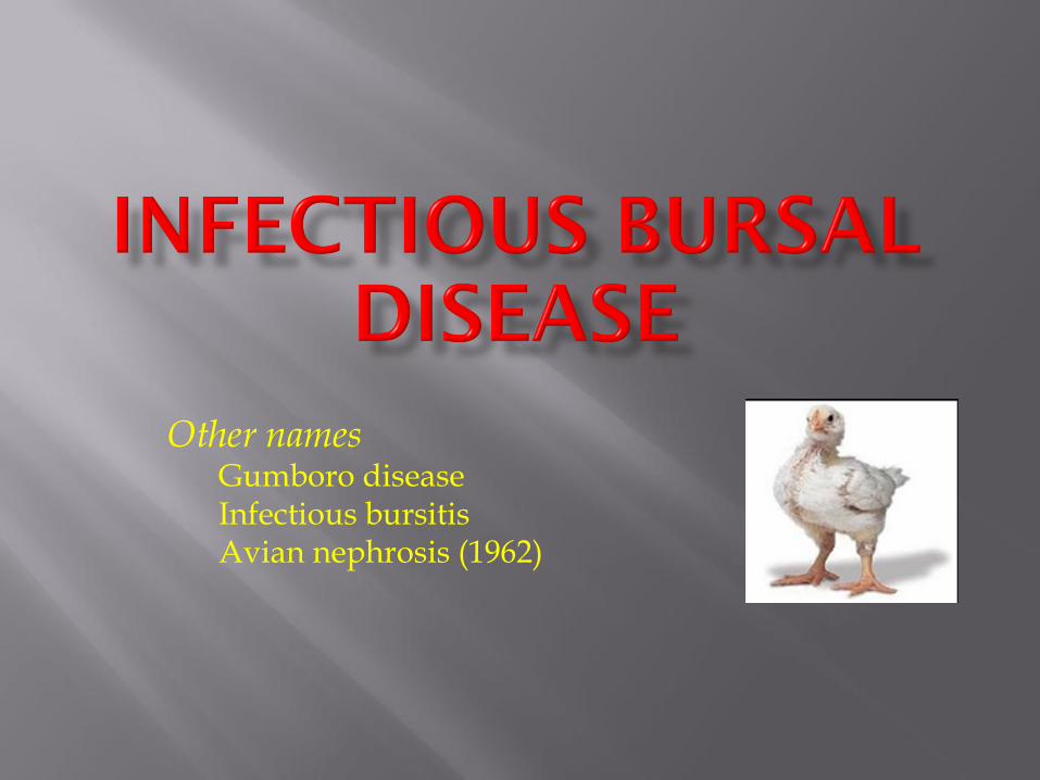
Other namesGumboro diseaseInfectious bursitisAvian nephrosis (1962)
B- Lymphocytes are the primary target cells
Bursa, lymphoid organ, is severely affected
First report in Gumboro (Delware District of USA)
Birna virus
( ds RNA)

IBD is highly contagious Affected birds excrete the virus in faeces for 10-14 days Virus is very stable
Remains highly infectious for many months (up to 122 days) in the poultry environment
Remains infectious even after 52 days in water, feed anddroppings. Hardy nature of this virus survives heat, cleaning and disinfectant
procedures Survival in the environment between outbreaks
Role of mechanical vectors (Human, wild birds, insects) Meal worms and litter mites remains infective for up to 8
weeks No vertical transmission Older birds (due to bursal regression) are more resistant to
infection
B - cells and their precursors are the main target cells
T- Lymphocytes are relatively unaffected
Renal pathology (swollen with urate deposits and cell debris) are due to severely swollen bursa
Mechanism for muscular haemorrhage is may be due to interference of virus with the normal blood clotting mechanism
Bursal infection in early life can result in impaired immune responses
Severity depends upon age, breed, and MDA level of the chick as well as the virulence of virus
Acute form Incubation period: 2- 3 days 3 -6 wks old chicks are affected
Signs Depression White watery diarrhoea Soiled vent Anorexia Ruffled feathers Reluctance to move Closed eyes and death
Morbidity - 10 – 100% Mortality 0 - 20% (Normally) and 90 – 100% (VVIBDV)
Milder form - Little or No signs Suboptimal (growth) / response to vaccination

Dehydration of carcass
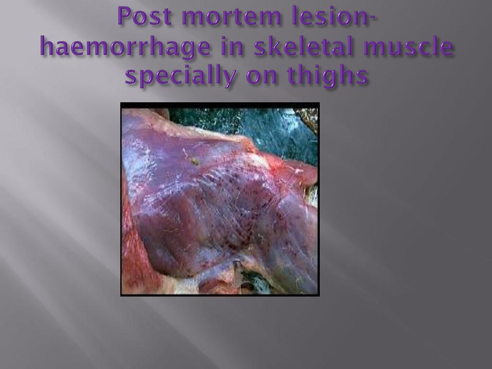
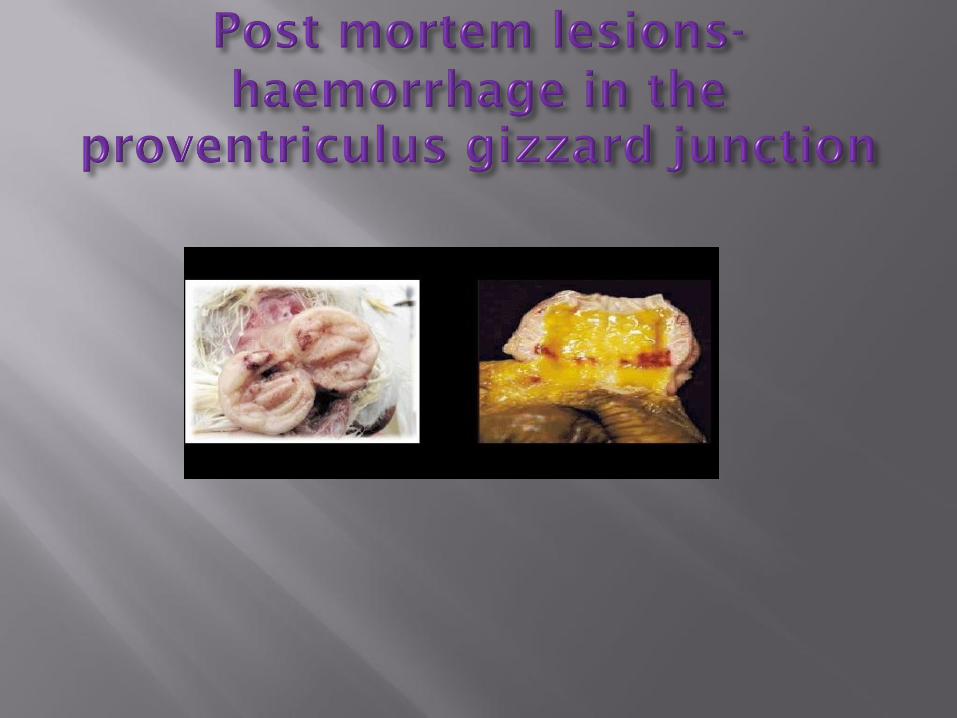
Muscular haemorrhage (thigh and pectoral) and some times at the junction of proventriculus and gizzard.
Haemorrhages of leg muscles are typical of IBD
Intestine with excess mucus
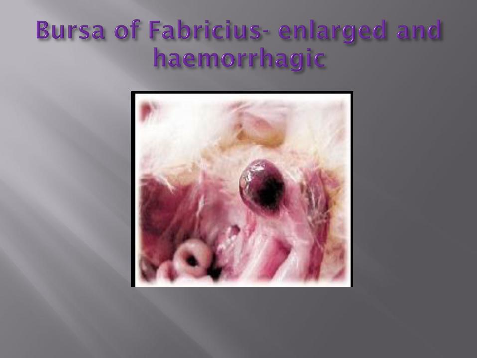
Bursa is enlarged, inflamed, edematous and cream coloured (early) and then Atrophy (after 3 – 8 days)
Haemorrhage on the internal and serosal surfaces
Other organs
Liver- Hepatomegaly and peripheral infarcts
Spleen- Splenomegaly
Kidneys- Swelling and white appearance, dilatation of tubules with urates( cell debris, occasionally).

Microscopic changes are mainly seen in lymphoid organs.
Bursa- inflammatory response with hyperaemia, oedema, infilteration of neutrophils, B lymphoid cell necrosis.
Spleen - Moderate lymphoid cell necrosis
Thymus and caecal tonsil - Lymphoid cellular reaction (early stage), but less extensive damage
Harderian gland - Depletion of plasma cells
Kidneys - Non - specific
Liver - Mild perivascular infiltration of monocytes.


Based on history clinical signs gross lesions ( for acute disease) Serological test AGPT (using macerated bursa) ELISA (against a known positive antiserum) Immunoperoxidase staining Immunofluorescence (in frozen bursal sections or smears) Virus isolation (rarely) – Inoculation of suspected bursa into 10 – 11 days old
embryonated eggs Nucleic acid probe, Ag-capture ELISA (using MCAbs.,), RT-
PCR