orthodontics i course review enoch ng, dds 2014 intro …enoch/resources/ortho_i.pdf ·...
TRANSCRIPT

Orthodontics I Course Review Enoch Ng, DDS 2014
Intro to Ortho - Tooth size and number decreasing, but slower than jaw size
o From softer foods, refined sugars, genetics
- Population Stats
o 45% people have ideal Mx occlusion = 55% people have Mx crowding
o 35% people have ideal Mn occlusion = 65% people have Mn crowding
o 45-55% people have ideal OJ, 15% have class II, <5% have class III
o 50% people have ideal OB, 30% have deep bite, <5% have open bites
Problems happen from deep bites, not open bites
- Angle’s Occlusion
US population, classification of anterior teeth only (did not look at molars)
o 30% normal
o 55% class I malocclusion
o 15% class II
o <1% class III
Bone Biology - Flat bones – intramembranous – direct ossification without cartilage template – cranial vault, Mn body, Mx
- Long bones – endochondral – indirect ossification, requires cartilage – femur, cranial base, condyle
o Complex multistep, sequential formation/degradation of cartilage, postnatal growth and repair
- Ages
o 0-20y/o = BF>BR
o 20-50y/o = BF=BR
o >50y/o = BF<BR
- Osteoclasts needed for bone formation – osteoclastic number (not activity) control bone formation
- Drugs
o Bisphosphonates – osteoporosis
Nitrogen containing = affects ruffled membrane
Non-nitrogen containing = causes cell death
o Glucocorticosteroids – arthritis

Orthodontics I Course Review Enoch Ng, DDS 2014
Craniofacial Growth/Development 1 - Cephalocaudal gradient (head to tail bone)
o 2 months = 50% head
o Birth = 30% head
Head bigger than face (small Mn) = easier to get through birth canal
o 25y/o = 12.5% head
Cranium closest to adult size at birth, stops growing first
Mn last bone to finish growing
- Scammon’s Curve
o 7y/o – cranial sutures close, neural development finished, ideal time to screen for ortho
o 10y/o – lymphatics done, start to shrink
o 10-12y/o – puberty starts, genital and general growth spurts begin
- Growth Patterns
o Boys start developing 2 years later, develop for longer, and grow larger than girls
o Growth spurt starts 2 years before sexual maturation
- Apposition – periosteum experiences hyperplasia, hypertrophy, and ECM secretion at surfaces (not internally)
Craniofacial Growth/Development 2 - Cranial vault – intramembranous formation/ossification, growth at sutures and apposition along fontanelles,
resorption along internal surface
- Cranial base – endochondral from spheno-occipital, intersphenoid, and spheno-ethmoidal synchondroses
o Growth stops at age 7
- Mx is displaced anterior inferiorly, with resorption along anterior surface and apposition on posterior surfaces
o Best age to pull Mx forward is age 7
o Palatal sutures close around 13-15 y/o
o Lengthening of Mx arch from apposition along Mx tuberosity
- Mn intramembranous formation
Mn ramus = intramembranous ossification, condyle = endochondral ossification
o Apposition along posterior surface, resorption along anterior surface of ramus (space for 3rd molars)
- For young kids, growth of the alveolar process is most important to accommodate the developing dentition
- Soft tissue – loses collagen with age
o Sags – decreased exposure of upper incisors and increased exposure of lower ones
o Thinner, less vermillion displace, less protruded
- Cartilage growth
o Nasal bone growth stops at age 10, cartilage finishes after adolescent growth spurt
o Females = stops age 17-19/o
o Males = stops age 19-21y/o
- Mn crowding – late Mn growth = crowding earlier, but may resolve later on
o Bones stop growing in width first, then in length. Growth in height is the last to stop
- Adolescence o Treat females around 2y earlier than boys o Lots of individual variation o Mid-palatal suture close 13-15y/o o Mn last bone to grow o Space for 3rd molars
- Adults o Facial tissue grows more than hard tissue o Lower incisal crowding o Lip line to upper incisors o Chin accentuation

Orthodontics I Course Review Enoch Ng, DDS 2014
Development of Dentition 1 - Stages of Development
o Primary dentition
o Early mixed – presence of permanent incisors and molars
o Late mixed – loss of deciduous molars and canines
o Permanent dentition
- Primary dentition
o Centrals, laterals, 1st molars, canines, 2nd molars
4 month rule
Variations of up to 6m for eruption normal
Dentition is stable from 3-6 y/o – development of permanent dentition
o Primate space = M to canine in Mx, D to canine in Mn
o Shallow overbite/excess overjet
o Increased horizontal overlap of anterior teeth
Mx grows AP faster than vertically
- 71% Flush terminal plane – class I or II
- 19% Mesial step – class I or III
- 10% Distal step – class II
- Primary dentition less proclined than permanent dentition
o Permanent arches are more tapered, primary arches are more ovoid
- Leeway space – space from difference in size between primary and permanent teeth
C (canine) = 0
D (premolar 1) – Mx = 0.0mm, Mn = 0.5mm
E (premolar 2) – Mx = 1.5mm, Mn = 2.0mm
o Mn arch = 5mm leeway space
o Mx arch = 3mm leeway space
- Mesial shifting
o Early mesial shift (63% of population) – mesial migration of Mn 1st molar
Uses up primate space, occurs around age 6
o Late mesial shift (100% of population) – mesial migration of Mn 1st molar AFTER primary 2nd molar loss
Uses up leeway space, occurs around age 11
- Teeth move occusally, mesially, buccally in adulthood

Orthodontics I Course Review Enoch Ng, DDS 2014
Development of Dentition 2 - 1st molars, Mn centrals, Mx centrals, Mn laterals, Mx laterals, Mn canines, Mx premolars, Mn premolars, Mx
canines, 2nd molars, 3rd molars
- 3 principles of treatment planning impacted teeth
o Prognosis related to extent of displacement and surgical trauma
o Eruption should happen through keratinized mucosa
o Adequate space created prior to surgery
- Transitional midline diastema – closes with eruption of Mx canines
o >2mm = begin pondering treatment
- Dental arch length decreases with transition from primary to permanent dentition
- Incisor liability – canine eruption, primate spacing, incisor proclination
- Potential problems with eruption
o Premature loss of deciduous teeth – if primary 2nd molar is lost, ALWAYS maintain the space
o Interproximal decay, over-retained primary teeth
o Impaction – contralateral teeth should erupt within 6months of each other
o Ankylosis – grey in color, dull to percussion
o Positional anomalies – ectopic eruption (wrong location) of lower incisors
Transposition – most commonly upper lateral and canine
Palatal eruption – may be genetic
Canines erupted in line, but if crowded likely to erupt labially
o Crossbites (posterior and/or anterior)

Orthodontics I Course Review Enoch Ng, DDS 2014
Biology of Tooth Movement - PDL required, acts as a shock absorber
- Physiologic function – fast (<5s) and heavy loading, intermittent
o Fluids and ligaments stabilize against gross displacement, alveolar bone bends, no pain
- Undermining resorption
o Heavy forces, rapid pain, compressed PDL decreases bloodflow = necrosis hyalinization of tissue
o Takes longer to move tooth – must heal first
- Frontal resorption
o Light forces, relatively painless, reduced blood flow causing signaling, not cell death, remodeling occurs
o Tension and compression sides for remodeling
Tension = apposition – osteoblasts and fibroblasts, laying down osteoid
Compression = resorption – osteoclasts
o Minimum 4-6h to get orthodontic tooth movement, want around 20-350grams of force
- Tissue changes
o Enamel = no effect
o Cementum = localized perforations, repaired from cellular cementum zone
o Dentin = resorption possible in areas of perforated cementum
o Pulp = transitory inflammation – loss of tooth vitality in teeth with history of trauma
- Types of movement
o Tipping
o Translation
o Rotation
o Extrusion
o Intrusion
- Force types
o Continuous force – never declines to zero. Think of a NiTi coil spring
o Interrupted force – declines to zero, then replaced. Think of a power chain
o Intermittent force – declines to zero, but appliance is removable. Think of headgear or elastics
- Drugs
Prostaglandins and IL1β increases quickly in PDL during orthodontic tooth movement
Prefer to use Tylenol instead of NSAIDs, as NSAIDs act centrally and block prostaglandins
o Depress OTM
Bisphosphonates, prostaglandin inhibitors (NSAIDs), tricyclic antidepressants, antyarrhythmics,
glucocorticosteroids, antimalarials, anticonvulsants, tetracyclines
o Increase OTM
Vitamin D, prostaglandins

Orthodontics I Course Review Enoch Ng, DDS 2014
Patient Exam and Diagnosis - Psychosocial
o Develop rapport with patient
o Write down the CC verbatim, ADDRESS THE CC
Why are you here? Why now? What do your parents say?
Why do you think you need braces?
o MHx/DHx
- 3 major reasons for ortho treatment
o Impaired dentofacial esthetics
o Impaired function
o Enhancement of dentofacial esthetics
- Be problem oriented so as not to fixate on only 1 portion – idea is to create a database for planning
o Prioritize the problem list – should address primary CC, ensures all issues are addressed, includes
pathologic, functional, and developmental problems
- Patient interview
o Physical growth evaluation – growth charts, sexual maturation, growth prediction
o Social/behavior evaluation – internal motivation/expectation, documentation of patient compliance, etc
- Clinical Exam
o Oral health – perio charting, caries, pulpal disease
o Jaw and occlusion – mastication/speech, habits, breathing, TMD/other dysfunctions
o Facial/dental appearance
Macroesthetics – frontal exam (symmetry, proportions of width/height), developmental age,
facial proportions, profile analysis
Brachyfacial, mesofacial, dolichofacial
Rule of 3rd (forehead, Mx, Mn areas)
Rule of 5th (bisecting nose (1/5), eyes (2/5), to edge of ears (2/5))
Convex = class II, flat = class I, concave = class III
Miniesthetics – teeth, smile framework (gingival display, midlines, etc), crossbites, malocclusion
Microesthetics – details on individual tooth
- Not necessary to mount child casts – TMJ is not done developing, hard to find reproducible CR

Orthodontics I Course Review Enoch Ng, DDS 2014
Classification of Malocclusion - Andrew’s 6 keys
o Molar relationship
o Mesial crown angulation
o Crown inclination – incisor proclination, canine and posterior lingual inclination
o No rotations present
o No spaces present
o Flat (or slightly curved) occlusal plane
- Other components of normal occlusion
o Normal apical base relationship
o Good interdigitation
o Minimum overjet and overbite
o Smooth/coordinated arch shapes
o Symmetrical dental arches, matching midlines
o Normal axial root inclination
o No crossbites
o No crowding or spacing
o No supernumary or missing teeth
o No oversized or undersized teeth
o CR/CO shift <2mm
o Normal curves of Wilson and Spee
- Malocclusion – deviation from accepted norm that presents a hazard to person’s wellbeing, associated with
dentofacial abnormalities
o Angle’s classification – Mx MB cusp in Mn B groove, first molar analysis only!
o Ackerman and Proffit classification – transverse plane relationship, AP plane relationship, vertical plane
relationship, soft tissue relationship, intra arch dental relationship
- Class II – end to end, or full step (Mx DB cusp in Mn B groove)
o Division 1 – accentuated OJ – usually end to end
o Division 2 – acceptable OJ (Mx incisors usually retroclined) – usually full step
o Subdivision – patient’s left or right side
- Vertical dysplasias
o Supraversion – teeth out of alignment, excessive eruption occlusally – deep bite
o Infraversion – teeth out of alignment, insufficient eruption (doesn’t reach occlusal plane) – open bite
- Etiology
o Pathological, developmental, accidental, genetic, acquired
CLP (embryonic disturbance)
Congenital missing/supernumary teeth
Ectopic eruption, impactions
Early loss of primaries, caries
Trauma, habits
- Example Classification
o Sagittal – class II, Division 1, Subdivision left
o Transverse – unilateral left posterior crossbite
o Vertical – anterior open bite

Orthodontics I Course Review Enoch Ng, DDS 2014
Cephalometrics 1 - Goals – evaluate relationships (horizontally and vertically) of 5 major functional components
o Cranial base
o Maxilla
o Maxillodentoalveolar process
o Mandibulodentoalveolar process
o Mandible
- Standard cephalometric arrangement to standardize study of:
o Skeletal relationships
o Underlying malocclusion etiology
o Pattern of craniofacial growth
o Prediction on timing of maximum growth
- Completion of growth – Nasion-Menton should not change between 6 month cephalometric radiographs
- Cephalometric analysis
o Soft tissue – facial contour, proportions, lip positions
o Skeletal – Mx and Mn basal arches, AP and vertical relationships, Mn plane, facial plane
o Dental – incisor/molar angulation, AP/vertical angulation, occlusal plane angulation, OJ and OB
o Lateral – AP dysplasia, vertical dysplasia, incisor position/inclination, balance of soft tissue/facial contour
o Frontal – transverse dysplasia, asymmetries
Cephalometrics 2 - Refer to diagrams in notes

Orthodontics I Course Review Enoch Ng, DDS 2014
Study Model Analysis
What to look for - Symmetry – superimpose a plastic grid (or have computer do it), pay attention to subdivisions, look for midline
deviations
- Occlusion – count teeth, check for patterns of surface wear
o Sagittal, vertical, transverse – check for excessive, level, or reversed Curve of Spee
- Intra-arch
o Space – crowding, spacing
o Irregularities – size, rotations, translations
o Tooth size
Space Analysis - TSALD – tooth size arch length discrepancy
o Space available – space required = amount of crowding or spacing
- Arch length available – measured with a brass wire, or sum of 4 segments (bilateral – 2 posterior, 2 anterior)
- Arch length required
o Permanent dentition
Merrifield analysis
Arch length required = total dental width, measured M-D of each tooth summed
o Mild = 1-3mm crowding/spacing
o Moderate = 4-6mm crowding/spacing
o Severe > 7mm crowding/spacing
Irregularity index
Measure each overlap from M to M of the 2 bilateral first molars and sum the
measurements – most commonly used for Mn anterior teeth in relapse studies
Reported as a length (mm) – higher the value, more severe the crowding
o Mixed dentition
Must discover size of permanent teeth, and account for changes in arch size by growth
Moyer’s analysis – based on Caucasians (tendency to overestimate)
Sum width of 4Mn incisors/2 – prediction of Mx and Mn Cs, PM1, PM2
o Widths of C, PM1, PM2 provided in charts – no radiographs required
o CI = 75% - 75% will have < value, STD of +1mm
Tanaka Johnson’s analysis – greatest variability of error (usually overestimates 2o teeth size)
Sum width of 4Mn incisors/2 + 10.5mm = Mn C, PM1, PM2
Sum width of 4Mn incisors/2 + 11.0mm = Mx C, PM1, PM2
Radiographic method – need high quality X-rays, no effect of ethnicity
Radiographically measure MD width of unerupted permanent teeth
Use a scaling coefficient to account for distortion
(1o Clinical width / 1o rad width) x (2o rad width) = estimated 2o clinical width
o Films must be // to MD crown axis (BW or PA, cannot use PAN)
o Teeth must lie in same Bu/Li plane
o Has poor estimation of canine M/D widths due to curvature

Orthodontics I Course Review Enoch Ng, DDS 2014
- Bolton Analysis – estimates tooth size discrepancy between Mx and Mn, related to perfect class I dentition
~5% of population has size discrepancy, discrepancy <1.5mm rarely clinically significant
If there is a discrepancy, check for peg laterals
o Mn excess (deficient OJ) – consider interproximal reduction of Mn incisors, or buildup of Mx incisors
o Mx excess (increased OJ) – consider interproximal reduction of Mx incisors, or leave extra OJ
Class I canine with anterior crossbite or edge to edge bite – probably excess Mn anteriors
Class I canine with excess OJ – probably Mn anterior deficiency
Class I canine with crossbite – probably excess Mn anteriors – want to open space and restore it
o Anterior ratio – sum of 6 Mn anteriors / sum of 6 Mx anteriors = 77.2%
>77.2% = excess Mn tooth size
<77.2% = excess Mx tooth size
o Overall ratio – sum of 12 Mn teeth / sum of 12 Mx teeth = 91.3%
>91.3% = excess Mn tooth size
<91.3% = excess Mx tooth size

Orthodontics I Course Review Enoch Ng, DDS 2014
Removable Appliances - Can be removed from the mouth by the patient
General Points - Development in Europe
o Little influence of Angle’s classifications
o Social welfare system – limited ortho treatment for the masses
o Scarce precious metals available for fixed ortho work
- Advantages
o Lab made – reduces dentist chair time
o Oral hygiene easier – can be removed for cleaning
o Can be removed for aesthetic occasions
- Disadvantages
o Success is patient compliance dependent
o Move only a few teeth at a time
o Almost impossible to produce complex tooth movements
Take Home Messages - Removable ortho appliances used for
o Growth modification
o Simple tooth movement in kids
o Retention
- They consist of a
o Framework
o Retentive elements
o Active elements
- Typically cheaper and easier to clean than fixed appliances, but can only create tipping forces
- Can be used as retainers to keep teeth in position after active ortho movement
Common Appliances - Functional appliances – passive tooth-borne appliance for guiding growth
o Similar to headgear (treats class II) – headgear moves Mx back, functional appliance moves Mn forwards
Each functional appliance, no matter what name, is simply a melding of wire and plastic parts
o Passive – no intrinsic force generated from springs or screws
o Changes posture of the Mn, pressures created by soft tissue stretch transmitted to bone – moves teeth
and modifies growth (commonly used to correct class II retruded Mn)
Capping incisors blocks Mn incisor proclination
1900s – Monobloc
1920s – Activator
Block of plastic covers palate and teeth of both arches
Advances Mn for class II correction, opens bite by 3-4mm
Shelves between teeth provide vertical control, angled flutes in acrylic guide posterior
tooth eruption

Orthodontics I Course Review Enoch Ng, DDS 2014
1960s – Bionator
Cut down activator with incisor capping and no palatal coverage
o Omega wire covers the palate
Lingual flanges stimulate forward posturing of Mn
1960s – Functional regulator
Tissue borne, buccal shields and lip pads reduce pressure on teeth
Lingual pad dictates Mn position, buccal stretching of cheeks causing alveolar apposition
via periosteal stretching
1970s – Twin block (popular in Britain)
Designed to be worn all day and to be used in function
Individual plates with ramps which guide Mn forward when patient closes down
Allows nearly full range of movement and reasonable speech
Mx plate usually includes expansion screw (expand as patient grows)
Also, grind down posteriors to allow supraeruption, add braces, fine tune
Components approach – combine appropriate components that deal with specific aspects of
patient’s problems for custom designed appliance for individual
- Active plates – tooth borne appliances for tipping motion
Retention
Clasps – fits into undercuts for good retention, needed for plate retention
o Adam’s clasp – most useful and versatile for removable appliances
Molars and premolars, 0.7mm stainless steel wire
o Arrow clasp – simple retention for removable appliances
Used in continuous row of teeth, 0.7mm stainless steel wire
Labial bow – retention of plate when in undercuts, limits tooth movement from springs,
can be used for tooth movement when activated
o Horizontally follows curvature of incisors
o Vertically positioned in middle third of clinical crown
o U-loops in canine region allow for activation – close loop to tighten the bow
Baseplate – complete or segmented
Made of acrylic – palatal coverage = main source of appliance anchorage
Can be configured to serve as a bite plane
As thin as possible for patient comfort
Active elements – screws and springs
Screws – opening with a key separates sections of the plate
o Heavy rapidly decaying forces – not ideal for tooth movement (uncontrolled)
o If force level is too high, the appliance gets displaced
Springs – contacts tooth at single point, creating tipping forces
o Light continuous forces, must be guided to only exert action in desired direction
o Early/interceptive treatment
Arch expansion – corrects posterior crossbite
Incisor tipping – corrects anterior crossbite
Space maintenance
o Regular treatment
Simple cases needed only tooth tipping movement

Orthodontics I Course Review Enoch Ng, DDS 2014
- Retainers – usually passive and used after active treatment
Allows for small movements only because the rest of the retainer should be passing
o Tooth borne appliance to prevent intra-arch instability after ortho treatment
Passive or active
Removable or fixed
o Hawley Retainer – molar clasps and outer bow with adjustment loops from canine to canine
Preferred retainer in Mx, especially after palatal expansion
Minimal wirework crossing occlusion – allows for vertical settling
Horseshoe shaped baseplate – improved speech
Must consider pre-treatment situation before designing retainer – prevent relapse
o Active removable retainer – realignment of incisors with spring retainer fabricated on a lab model where
teeth were reset into alignment
o Vacuum-formed retainers – cheap, less lab time
Occlusal coverage – blocks vertical settling (this is NOT good)
Should block 2nd molars – prevent anterior open bite from over eruption
Thickness of material may prove uncomfortable
Appliance bulk distal to canine prone to fracture
- Clear aligners/invisalign
Vacuum-formed sheets on casts with slightly reset teeth to fix mild irregularities
Sequence of casts can be made to incrementally correct irregularities with new vacuum-formed
retainers made for each resetting
o Invisalign – Align Technologies in 1990s, heavy marketing to public
Only treats mature dentition – growth changes cannot be predicted (not designed for kids)
Success requires patient to wear aligners 20-22h/day
If not worn, next set of aligners will not work – new set will be required
Tooth-colored composite attachments allow correction of severe malpositioned individual teeth
Good for - Mild crowding with IPR or expansion - Posterior dental expansion - Correction of mild spacing - Absolute intrusion of individual teeth - Lower incisor extraction for severe crowding
o Simple movements only, not complex
Bad for - Dental expansion of blocked out teeth - Extrusion of incisors - Severe rotations - Relative intrusion - Molar uprighting - Molar translation - Closure of premolar spaces after extraction
o Production Process
CT scan of impressions – create accurate 3D digital model
Digital sectioning of teeth
Movement of teeth following clinician’s instructions
Preliminary plan placed online for clinician review as a ClinCheck
Transfer of digital models to a cast production facility where stereolithographic model for each
step fabricated
Clear plastic retainers formed over each model
Aligners shipped to clinician
Orthodontics I Course Review Enoch Ng, DDS 2014
Fixed Appliances I
Configurations - Standard edgewise – brackets at 90o to the wire (no tipping, torqueing, or in/out variations), all teeth get
standard brackets
o Most contemporary braces use standard edgewise
o Wire bends to adapt to tooth/teeth malposition
- PreAdjusted (straightwire) edgewise – built for averages, may require small adjustments
Better than large adjustments starting from a scratch wire
o Tip (mesial/distal) – angulation
Rhomboid shape, with the slot’s angulation positioned to match roots/long axis of tooth
o Torque (buccal/lingual) – inclination
Values are more negative as you move posteriorly in the Mn – want natural lingual inclination
o In-out (distance between surface of tooth and bracket (thickness)) – offset
- Slot sizes – two types
o 0.018” x 0.025” – stainless steel, but people used to gold dimensions preferred to keep 0.022”
o 0.022” x 0.028” – originally for when gold wires were used
Types of Tooth Movement - Occlusal/gingival
- Mesial/distal rotation
- Buccal/lingual (in/out)
- Mesial/distal angulation (tipping)
- Buccal/lingual inclination (torque)
Type of Archwire Bends - First Order – visible from occlusal view
o Adjusts in/out (buccal/lingual position)
o Adjusts mesial/distal rotation
- Second Order – visible from lateral/frontal view
o Adjusts occlusal/gingival (up/down)
o Adjusts mesial/distal angulation (tipping)
- Third Order – twist in the wire
o Adjust buccal/lingual inclination (torque)
o Only possible with rectangular wires (not possible with circular ones)

Orthodontics I Course Review Enoch Ng, DDS 2014
Components - Bracket (bonded or banded) – important to keep organized, so proper bracket goes on proper tooth
o Archwire slot
Closed – self ligating Open – requires ligatures
o Tie wings – undercuts for elastic ligatures
o Bracket base – bonded to tooth or band
o Indicator dot – ALWAYS oriented to the distal gingival
o Buccal tubes (for molars)
Mx – triple tube – auxiliary tube, main archwire tube, headgear tube
Mx – double tube – auxiliary tube, main archwire tube
- Archwire – energy is stored in the archwire for tooth movement
Wire gets distorted to fit malocclusion slowly releases energy to move teeth as it restores
itself to normal form/straightens out
Metal Alloys - Stainless steel – stiffest - TMA (β-titanium) – half as stiff as stainless steal - NiTi – half as stiff as TMA
Sizes – many different sizes - Rounds – 0.014”, 0.018”, 0.020” - Rectangular – 0.016x0.016”, 0.016”x0.022”,
0.019”x0.025”
- Ligatures
o Elastic chain form
o Wire ties
Fixed Appliances II
Auxiliaries - Transpalatal Arch – removable or fixed (soldered) – spans between banded Mx first molars
Fisrt molars require banding with lingual auxillary tubes
o Anchorage support
o Molar rotation
o Arch width adjustment
- Nance appliance
o Acrylic button used to anchor Mx molar position
o Typically used in mixed dentition to hold space
- Lower Lingual Arch – similar to TPA in concept
o Arch width control
o Anchorage control
o Arch length management
o May (or may not) contact incisors
If it doesn’t contact incisors, allows for incisor movement to increase arch width
- Lip Bumper
o Uses muscles to hold molars in place or tip them (usually to upright them)
o Difficult to create molar translation, but tipping forces are possible

Orthodontics I Course Review Enoch Ng, DDS 2014
- Coil springs (stainless steel or NiTi) – placed over archwire
o Used to open or close spaces
o Can be combined with temporary alveolar implants for closing spaces
o Metal wire ligatures on either side of the coil spring to prevent tooth rotation (remember, bracket is on
facial of tooth, so forces applied through facial surface)
- Power thread (zing string) – elastic monofilament coil spring
o Very technique sensitive – requires perfect square knot for attachment to coil spring
o Must be active AFTER tie is completed; if tied incorrectly it is often passive after tie completed
o Tooth attached to a [gold] chain and tied to the coil spring via a thread to activate the tooth
- Expanders
o RME/RPE – rapid maxillary expander/rapid palatal expander (they are the same thing)
As rigid connection as possible to minimize tooth movement when active
Increase arch by separating mid-palatal suture
o Quad helix (4 circles) – activated in the office, not at home (spring appliance, does not have a screw)
o Pendulum appliance – moves molars backwards, can move other teeth distally afterwards
Always bonded to premolar occlusal surfaces for anchorage
- Herbst Appliance
o Bars attached from posterior Mx to anterior Mn – forces Mn to occlude anteriorly
Elastics - Class I – intra-arch – closes spaces inside an arch
- Class II – class II malocclusion – pulls Mx back and Mn forward
o Control the vertical force component, or may cause supraeruption and give patient a gummy smile
- Class III – class III malocclusion – pulls Mx forward and Mn back
- Vertical – used to correct open bites
- Anterior cross elastics – used to correct crossbites
o Do not use for a long time (3+ months) or patient’s occlusal plane may kant
- Posterior cross elastics
- Posterior box – used to close larger vertical open areas
Separators - Metal springs/elastics placed into proximal contacts
- Coil springs attached to temporary alveolar bone implants
o Stretched – closes spaces as it comes together
o Compressed – opens spaces as it pushes apart

Orthodontics I Course Review Enoch Ng, DDS 2014
Construction, Debonding, Debanding
Separators - Brightly colored radiopaque separators used to open proximal contacts to allow for band seating
- Should not be used for longer than 2 weeks (usually 2-7 days, longer may cause attachment loss)
- Elastic separators work 95% of time – if elastics separators don’t work, use elastic springs
o Place between beaks of separating pliers, stretch, snap one side through contact
o Use 2 loops of floss, stretch separator, snap floss through contact and pull separator underneath
contact, pull separator up to snap one side through contact, remove floss
Banding vs Bonding o Until 1980s, only way to secure a bracket was to band the tooth
o Now, only routinely banded teeth are Mx molars
- Banding Indications
o Teeth receiving heavy intermittent forces against attachments – headgear
o Require attachment of intraoral or extraoral auxiliaries – transpalatal arch
o Need both labial and lingual attachments
- Ideal band position
o Parallel to cusps and marginal ridges – all cusps are equally visible
o Band margins just below marginal ridges and above contact points
o Tube straddles buccal groove mesial-distally, perpendicular to long axis of the tooth
- Adhesives for Bands
- Ideal bracket position (use of a PANO helps locate roots and gives better idea for ideal placement)
o Center of clinical crown
o Bracket slot parallel to incisal edges/marginal ridges
o Tie wings parallel to long axis of tooth
o Inaccuracies
Horizontal error – unwanted tooth rotation
Axial error – unwanted tooth tipping
Thickness error – improper torque/rotation
Vertical error – extrusion/intrusion, torque error, in/out error
- Adhesives for bands – light cure adhesive systems used in ortho
o RMGIC for othro use – light cured, greatly reduces problems with leakage beneath bands and allows for
control of working time
o 2part powder/liquid GIC for ortho use – dual cure from visible light and chemical causes rapid set, better
bond strength than light cured cements
o Sandblasting – increases bond strength and mean survival time, reduces clinical failure rate
o Bonding to non-enamel – roughen surface with micro-etch, diamond, or green stone
Bond metals and ceramic brackets to enamel
Resin based primer penetrates into enamel rods
Possible to add pontics to fill spaces temporarily – for patient esthetics

Orthodontics I Course Review Enoch Ng, DDS 2014
- Bonding – >90% of orthodontists in USA use direct bonding with light cured resins with a median fail rate of 5%
o Clean the tooth – pumice to remove plaque and organic pellicle
o Pre-select appropriate band size using patient plaster casts as a guide
o Remove separators, trial seat and adapt the bands to the teeth
o Remove bands and thoroughly clean and dry teeth and band before bonding
o Cover occlusal openings with tape, fill attachments and tubes to prevent obturation with cement
o Label bands, mix cement, apply to inner surfaces of bands
o Seat bands in ideal position, clean off excess cement with cotton rolls, pellets, and scalers
o Keep cement layer between tooth and band as thin as possible
- Enamel surface preparation
o Use cheek retractors and saliva ejector to keep area dry, better environment for adhesives
o Etch 37% phosphoric acid (blue to contrast with tooth, gel so it stays on tooth) for 15s anterior, 30s
posterior, rinse thoroughly, dry thoroughly – etched areas should appear frosty white (if not, etch again)
o Apply thin coat of primer
o Have adhesives on bracket before placement – adhesive pre-coated brackets are available
o Place slight excess adhesive on bracket base, place bracket on tooth in correct position
Bracket at midpoint of clinical crown, tie wings parallel to long axis
Push bracket firmly toward tooth surface – extrudes excess cement
Remove excess adhesive with explorer or scaler
Check vertical position using a bracket gauge
o Light cure from ALL sides – time depends on bracket type, adhesive, and light source
o Recheck bracket position
o Tie in archwires
Debanding/Debonding - Debanding – band remover pliers
- Debonding metal brackets
o Debonding pliers – used to deform metal base to collapse bracket
o Lift-off debonding pliers – high risk of enamel damage
o Weingart pliers – used to deform metal base to collapse bracket
o Ligature wire cutter
- Debonding ceramic brackets – increased risk of shattering (danger to eyes)
o Position tips against mesial and distal sides of brackets
o Gently squeeze bracket until it collapses
o Gently rock mesial/distally to completely separate from enamel
o Ideally, adhesive stays on the tooth (bond between bracket/adhesive should be weaker than between
tooth/adhesive)
- Post-debonding polishing
o Spiral fluted carbide finishing bur in low speed with light pressure and painting motion
o Prophy paste or pumice slurry with rubber cup
o Brown and green polishing cups for highly polished final clinical appearance
o Prophy polisher for enamel polish – optimum at 6,000rpm, no air/water spray necessary for cooling

Orthodontics I Course Review Enoch Ng, DDS 2014
Clinical Example - Remove bracket by creating fracture between adhesive and bracket base
o Keep archwire in place – pop off brackets THEN remove the bands (holds everything in 1 piece)
- Break cement attachment, life band off tooth by elevating buccal and lingual surfaces with band-removing pliers
- Remove excess adhesive, polish tooth
Polishing Effects Surface cleaning Acid etch Low speed High speed Polish Enamel thickness
5um 7-20um 10um 20-25um 10um 1500-2000um
Take Home - Separators create space between teeth for band fitting and cementation
- Bands routinely used only for teeth that require attachment of intraoral or extraoral appliances
- Brackets used on all other teeth, can be bonded to enamel and non-enamel surfaces
- Ortho fixed appliances causes minimal enamel wear/loss

Orthodontics I Course Review Enoch Ng, DDS 2014
Treatment Timing - 2 phase treatment for problems that are appropriate for early referral/treatment
- 1 phase treatment for problems solvable in early permanent dentition
Progressive Discrepancy o Increase in problem severity/treatment difficulty over time
Recommend 2 phase treatment
- Habits (thumb/finger sucking) – hard to break habit after age 9, especially in females
- Anterior crossbite – fix before incisal facial wearing affects esthetic appearance
- Mild/moderate class III – palatal arch expansion/headgear
o Severe class III requires surgery
- Moderate/severe space deficiency – arch expansion
o Extraction of primary teeth for temporary solution
- Unilateral posterior crossbite with shift – Intervene before TMJ remodels
o If the TMJ remodels, will make shift permanent
Stable Discrepancy o No increase in problem severity or treatment difficulty over time
Recommend 1 phase treatment
- Mild/moderate crowding
- Most class II problems (with mixed dentition and no significant spacing concerns)
- Deep bite – only if you see a soft tissue effect
- Mild open bite

Orthodontics I Course Review Enoch Ng, DDS 2014
Biomechanics
Important Concepts - Force = mass x acceleration = load applied to an object that tends to move said object
o Major components
Magnitude (N) = in ortho, measured in grams [negligible in tooth movement]
Varies with type of tooth movement
Light, continuous forces considered most effective in inducing tooth movement,
undermining resorption occurs if too great a force is applied too fast
o Tipping = 35-60gm
o Translation = 70-120gm
o Root uprighting = 50-100gm
o Rotation = 35-60gm
o Extrusion = 36-50gm
o Intrustion = 10-20gm
Direction – X, Y, Z components
Force is a vector, can be broken down into its directional components
Point of Application – one point along line of action (ortho bracket)
Moment = tendency of a body to rotate when force is applied
o Units = Nmm (gmm), direction is either clockwise or counterclockwise
- Center of Resistance
o Ideally 1/3 distance from alveolar crest to apex
o For multirooted teeth, center of resistance is just apical to the furcation
o For individual tooth, center of resistance doesn’t change unless apex or crestal bone changes
o Consider
Number of roots
Degree of crestal bone loss
Root/apical resorption
- Center of Rotation
o Point at which rotation occurs
o Most often does not match center of resistance, but is possible
Results of Force Application - Pure translation
o Center of rotation at infinity (far away from the tooth) – essentially no rotation
o All parts (root and crown) moving in the same direction
- Pure rotation
o Center of rotation = center of resistance
o Root and crown move in opposite directions
- Controlled tipping
o Center of rotation just apical or matching root apex
o Tipping, both crown and root moving in same direction
- Uncontrolled tipping
o Center of rotation between center of resistance and root apex
o Tipping, crown and root moving in opposite directions

Orthodontics I Course Review Enoch Ng, DDS 2014
Fixed Appliances - Type of movement depends on moment to force ratio
- Most forces applied to crown = causes tipping motion
o To get desired motion:
Increase magnitude
ID center of rotation, place in desired location (control moment/force ratio)
- Moment/force ratio
o Translation – create a moment and apply it so it cancels out the moment caused by primary force
Using a thicker wire fully engages slot – allows placement of center of rotation more
towards infinity (further from the root)
o Final result ultimately based on biological response of teeth and tissues (perio, bone, etc)
o Magnitude of force is vital
Removable appliances - Only tipping motions, usually uncontrolled tipping
Biomechanical Examples - Intrusion arch – used when there is a deep bite promoted by all teeth (very rare)
o Fx – proclines incisors, distally tips molars (not seen in type 1)
o Fy – anterior intrusion, molar extrusion
o Fz – not significant
- Power chain space closure
o Fx – canine moves distally, molar moves mesially
o Fy – not significant
o Fz – canine rotates distolingually, molar rotates mesiolingually
Always use 2 parameters when describing rotation – surface/cusp and direction it is moving
Prevent rotation with stainless steel ties
- Class II elastics
o Fx – Mx canine moves distally, Mn molar moves mesially
o Fy – extrusion of both canine and molar
o Fz – Mx canine rotates distolingually, Mn molar rotates mesiolingually
Place elastic on lateral incisor and 2nd molar – greater X component, smaller Y component
- Class III elastics
o Fx – Mn canine moves distally, Mx molar moves mesially
o Fy – extrusion of both canine and molar
o Fz – Mn canine rotates distolingually, Mx molar rotates mesiolingually
- Molar uprighting from orthodontics – takes 6-12 months
Improves direction and distribution of occlusal loads (increases restoration durability)
Decreases tooth reduction needed for parallel abutments
Decreases probability for endo, perio, or advanced prosth procedures
Removes plaque-retentive areas – increases perio health
Improves alveolar contour and crown/root ratio
o Fx – not significant
o Fy – anterior intrusion, molar extrusion
o Fz – uprights molar, proclines anterior segment

Orthodontics I Course Review Enoch Ng, DDS 2014
Anchorage
Principles - Differential force theory – rate of tooth movement related to force/unit area of root surface
o Tooth with more root surface area have higher resistance, therefore higher anchorage
- Relationship between surface area and tooth movement is NOT linear
- Tooth movement increases with increased applied force, but has a maximum
- Optimal level exists after which there is no increase in movement, only in strain on anchor units
Assessing Requirements - Anchorage available in an arch is related to space in that arch, usually an extraction space
o How much space is needed to complete the correction?
o How much might the anchor teeth move?
- Group A – anterior retraction
o High anchorage needed in posterior – maximum posterior anchorage
o Retraction of anterior teeth without mesial posterior tooth movement
- Group B – equivalent retraction/protraction – reciprocal anchorage
o Moderate anchorage needed in posterior
o Equal movements of anterior and posterior movements distal and mesial, respectively
- Group C – posterior protraction – maximum anterior anchorage
o No anchorage in posterior
o Protraction of posteriors without distal movement of anterior teeth
Anchorage Control - Differential response to pressure allows for moving some teeth more than others, though unwanted tooth
movements will still occur
1. Reinforcement
- Addition of teeth to anchorage unit – increases root surface area, dissipating force over more teeth
- Addition of teeth from opposite arch via elastics (intermaxillary traction)
2. Subdivision of desired movement
- Pit resistance of a group of teeth against movement of single toth
o Move canine back individually, add it to anchorage group, then move back incisors
3. Friction control strategies
Supplemental Anchorage - Extraction decision
- Non-dental sources of anchorage – if structures other than teeth are used for anchorage, possible to
produce wanted movement without creating any unwanted movement
o Intraoral sources
Mucosa and underlying bone
Pendulum/pendex appliance – moves posterior teeth back without moving other
teeth too far forwards
Nance appliance
Soft tissue and perioral musculature
Lip bumper

Orthodontics I Course Review Enoch Ng, DDS 2014
o Extraoral sources
Forehead, basal bones
Protraction facemask – reverse pull headgear, anteriorly pulls Mn forwards
Cranial vault, occipital bone, neck
Headgear – posterior forces Mx back
o Has an inner and outer bow
o Inserts into headgear tube on molar band
o Exerts distal force on molars
o Skeletal (absolute) anchorage
Temporary anchorage devices
Osseointegrated implants
o Palatal implants need 3-6 months to osseointegrate
Miniscrews – titanium screws that penetrate through gingiva into alveolar bone, but
no osseointegration needed (can be loaded immediately)
o Distalize anterior teeth w/o moving molars forward (good for severe class II)
o Absolute intrusion – must be placed on both B and L to avoid tipping tooth
Miniplates/bone anchors – placed beneath soft tissue, usually in zygomatic buttress
area of Mx
Uses of Headgear - Orthodontic anchorage
o 100g/side
o Hold position of Mx posterior teeth in the arch
- Tooth movement
o 150-250g/side
o Distalize Mx molars
- Growth modification
o 500g/side
o Create differential growth between Mx and Mn
- Vertical effects of headgear can negate forward Mn growth
- High pull – occipital
o Level of force through the center of resistance = backward upward translation of molar
If combined with short facebow and a Mx splint, causes Mx rotation
o Level of force below or above center of resistance = crown or root tipping
- Low pull – cervical
o Level of force through center of resistance = backward downward translation of molar
o Level of force below or above center of resistance = crown or root tipping
o Negative effect = clockwise movement of Mn
- Straight pull – both occipital and cervical
o Level of force through center of resistance = backward translation of molar

Orthodontics I Course Review Enoch Ng, DDS 2014
Class I Treatment
US Population o 30% normal occlusion
o 50-55% class I malocclusion
o 15-20% class II malocclusion
o 1% class III malocclusion
- Etiology – genetics, tooth size discrepancy (Bolton analysis), big/small jaws, # of teeth, shape of teeth,
inappropriate function, rotations, vertical problems, condylar fractures, congenital anomalies, etc
Treatment Planning Factors - Chief complaint
- MHx, medications
- Internal motivation, realistic expectations
- Perio, prosth, restorative, other dental needs
- Crowding, incisal position/inclination, Bolton discrepancy, OJ, OB, transverse and vertical relationships
- Facial proportions, soft tissue
- Growth potential
o Class I malocclusion patients don’t need any growth modification
Treatment Timing for Class I - No need to modify growth
- Usually best started in late mixed/permanent dentition stage
o Minor problems can be fixed later (Adult stages – avoid uncompliant teen stage)
- Transverse problems (orthognathic problems) should be treated earlier
o Orthopedic palatal expansion before palatal suture closes
o Lateral and anterior shifts treat immediately before TMJ remodeling to avoid permanent assymetry
o Lateral shift – unilateral crossbite where there is actually bilateral crossbite, bit hidden by shift
- Vertical growth problems (habits) may need to be addressed earlier
o Open bites are more problematic than deep bites, unless there is perio damage or palatal
impingement
- Severe crowding (>10mm) may benefit from serial extraction in mixed dentition stage
Potential Problems and Treatments - Arch-space discrepancy
o Crowding – IPR, extractions, dental and/or skeletal expansion
o Spacing – close spaces (retention, Bolton analysis, anchorage requirements, restorative plan)
- Antero-posterior discrepancy – no posterior discrepancies in Class I malocclusion (molar relationship), so
problems are in anterior region
Skeletal class I – normal ANB and facial convexity
Dental class I – class I molars
o Bimaxillary dentoalveolar prognathism (Jaws are in Class I, but are prognathic to cranial base –
common in African Americans) – extraction
o Anterior crossbite – extraction, incisor advancement, Bolton
o Excessive OJ – incisor advancement, space closure, Bolton

Orthodontics I Course Review Enoch Ng, DDS 2014
- Transverse discrepancy
o Posterior crossbite (buccal and lingual crossbites) – lingual crossbites more common
Skeletal posterior crossbite – Mx expansion via opening of midpalatal suture
Dental posterior crossbite – removable appliances, expanding archwires, cross elastics
o Bilateral buccal crossbite (scissors bite) – Mn completely tucked under Mx – very rare
Scissors bite – dental expansion of Mn arch, constriction of Mx arch, surgery
- Vertical discrepancy
o Deep bite
Incisor intrusion, leveling curve of Spee, extrusion of posteriors to open the bite
o Anterior open bite
Extraction mechanics usually deepens bite (wedge theory)
Surgical impaction of Mx for gummy smile, reduce lip strain
Incisor extrusion, level curve of Spee, intrusion of posteriors to deepen the bite
- Tooth anomalies (form, #, position)
o Open/consolidate space of missing teeth
o Create space for impacted teeth, then expose/bond and bring into the arch
Open the space first before asking the surgeon to expose the canine
- Soft tissue problems
o Bimaxillary dentoalveolar prognathism
o Lip/mentalis strain (strain when lips are sealed together) – 1st premolar extraction. retract anteriors
o Gummy smile
Short upper lip – nothing can be done to treat, everything looks good but when they smile
there’s lots of gingiva displayed
Upper lip hypermobility – treat with botox
Gingival hyperplasia – perio surgery
Extraction vs Non-Extraction - 0-4mm crowding = IPR, expansion, incisor advancement and proclination
o Only extract if there is severe incisor protrusion or severe vertical discrepancy
Also for patients with bimaxillary prognathism who want their “full lips” corrected
- 5-9mm = both non-extraction and extraction techniques
o Extraction dependent on patient’s hard and soft tissue characteristics, incisor position/angulation
o Nonextraction requires transverse expansion across molars and premolars
- >10mm = premolar extraction needed
Extraction Options - Option 1 – extract 4s (1st premolars)
o Maximum posterior anchorage, maximum retraction of anterior teeth
o Good for bimaxillary prognathism
- Option 2 – extract 5s (2nd premolars)
o Less anchorage, less anterior teeth retraction
o More difficult to correct anterior crowding
o Can help treat open bites – “wedge theory” = 5s are closer to hinge; let molars move mesial to close
anterior openbite
o Good for open bites and hyperdivergent Mn (steep Mn plane)
Orthodontics I Course Review Enoch Ng, DDS 2014
- Serial Extraction – patients who fit all criteria are very rare
o First extract certain deciduous teeth, then later on some permanent teeth
Does NOT avoid braces – avoids CROWDING
o Common for people with congenitally missing 2nd premolars with crowding
o For skeletal class I, normal OB (or open bite tendency), severe crowding (>10mm), all teeth present
radiographically with good eruption positioning
o Patients between 8-9y/o, incisors are crowded
o Subsequent orthodontic treatment REQUIRED
- Dewel’s Method
o Very simple, but has lots of risks – rarely done because of the risks
o Extract deciduous canines to create space for incisors (tongue pressure will usually align incisors)
o Extract deciduous first molars to aid in quick first molar eruption
o Extract first premolars to create space for canines
o Modified Dewel’s
First premolars are enucleated during extraction of first deciduous molars – expedite
eruption of canines (more often used in Mn)
o Risks
Deepening the bite
Possible loss of space via mesial migration if there is an eruption delay
Poorly executed serial extraction may worsen the case
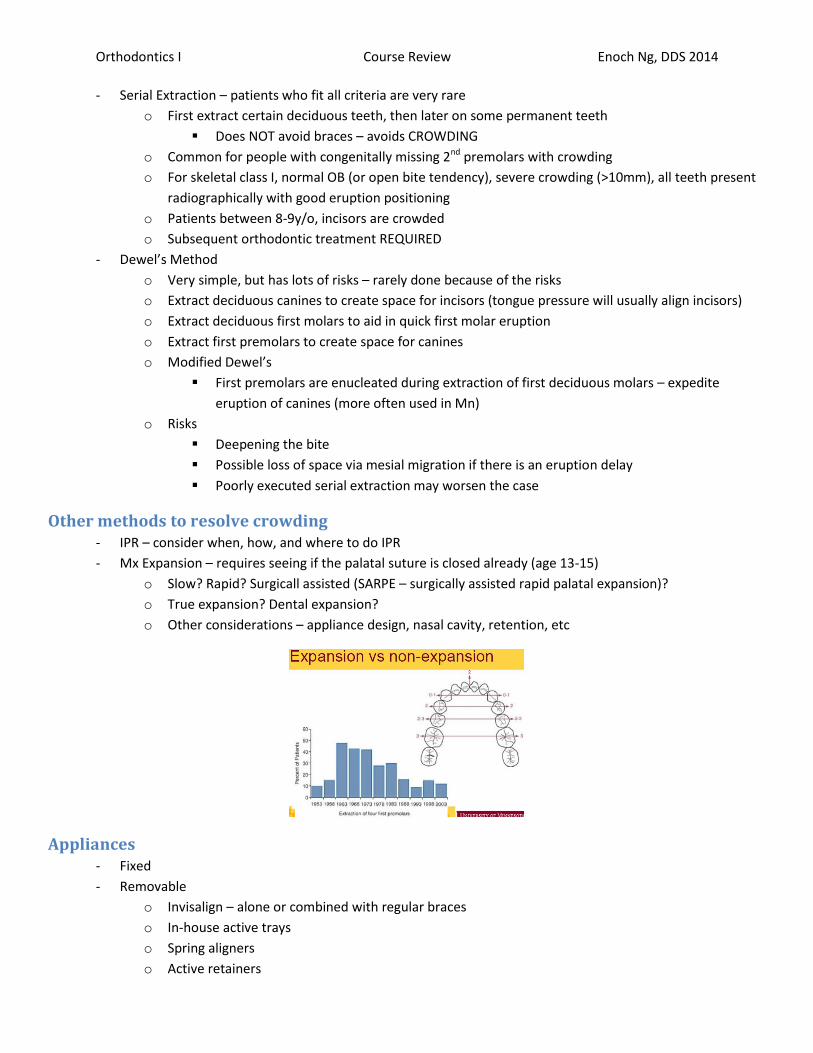
Other methods to resolve crowding - IPR – consider when, how, and where to do IPR
- Mx Expansion – requires seeing if the palatal suture is closed already (age 13-15)
o Slow? Rapid? Surgicall assisted (SARPE – surgically assisted rapid palatal expansion)?
o True expansion? Dental expansion?
o Other considerations – appliance design, nasal cavity, retention, etc
Appliances - Fixed
- Removable
o Invisalign – alone or combined with regular braces
o In-house active trays
o Spring aligners
o Active retainers

Orthodontics I Course Review Enoch Ng, DDS 2014
Class II Treatment
Treatment Planning Factors - Same etiology as Class I, except there is an A-P posterior dysplasia
o Requirement to ID which jaw is at fault
o Need to understand growth potential and patient compliance
- For othognathic patients, need to ensure growth completed before surgery
o Take cephalometrics every 6 months and superimpose to check for completed growth
Potential Problems - Antero-posterior discrepancy
o Increased ANB and facial plane
- Class II Division I
o Dental problems
Class II molars and canines
Excessive overjet, often flared incisors
Can be deep, normal, or open bite
Subdivisions (left or right)
o Skeletal Problems
SNA and SNB
Greater SNA – Mx prognathism
Greater SNB – Mn retrognathism
Can also have combination
ANB > 4o
Facial convexity greater than 15o
o Habits
AAPD – thumb sucking for > 3 years to cause malocclusion
- Class II Division II
o Dental problems
Class II molars and canines
Incisors retroclined, limited overjet
Lateral incisors/canines flared labially
Subdivisions (left or right)
o Skeletal problems
SNA and SNB
Greater SNA – Mx prognathism
Greater SNB – Mn retrognathism
Can also have combination
ANB > 4o
Facial convexity greater than 15o
Often with hypodivergent growth pattern and DEEP bite

Orthodontics I Course Review Enoch Ng, DDS 2014
Class II Treatment Strategies o Growth stimulation – larger than what would normally be, causes MORE growth in a period than
would have been expected without treatment
Does NOT occur in orthodontics
o Growth modification – differential acceleration of growth (functional appliances) or restraint of
growth (headgear) – headgear effect = functional appliances tightens lips, restrains Mx growth
THIS is used in orthodontics
- Differential growth
o Facilitation of growth – functional appliances worn for 16-20h/day
o Restraint of growth – headgear worn for 12-14h/day (better/easier compliance)
150g force to move molars
500g force to cause skeletal effect
High pull – good for open bite, moves teeth posteriorly and intrudes them
Cervical pull – good for deep bites (hypodivergent, flat Mn plane, horizontal growth), moves
teeth posteriorly and extrudes them
Combination – for people with a good bite, moves teeth backwards only
- Dental movement
o Dentoalveolar compensation
Used when extractions are not indicated
For mild problems (50% for class II molars)
Better for growing instead of non-growing patients
Class II mechanics – tends to bring forward Mn and procline Mn incisors
Is UNSTABLE if excessive tooth movement occurs
o Molar distalization
Headgear, fixed molar distalizers, trans-arch distalization
Mild skeletal problems, designed to finish with class I molars and canines
Only used if facial height permits

Orthodontics I Course Review Enoch Ng, DDS 2014
- Extraction therapy
o Mild/moderate compromised skeletal relationship
o Usually involves upper first premolars
o Designed to finish in Class I OR class II molars, only class I canines
Decisions options – facial appearance, degree of crowding/protrusion and proclination of
Mx incisors, open bite tendency
o Extraction of Mx 4’s – correct malocclusion while making the soft-tissue discrepancy less apparent
Resolve Mx crowding, less compliance required
Finish in Class I canines, class II molars
o Extraction of Mx 4’s and Mn 5’s
Lower anteriors with moderate crowding or proclined
Lower 2nd premolar space used for correcting crowding/retraction of anteriors as well as to
mesialize into Class I
Finish in Class I canines and class I molars
o Extraction of Mx 4’s and lower incisors
Adults with moderate/severe lower anterior crowding
Resolves crowding to get class I canines
Bolton discrepancy created – may finish with excessive overjet
- Orthognathic therapy
o For patients who have stopped growing and/or to camouflage extraction treatment is not indicated
o Pre-surgical orthodontics (decompensation) for 12-18 months, then post-surgical treatment to
finalize occlusion (6 months)
o Importance of clear treatment plan – direction of tooth movement and extraction pattern, definitive
and cannot be reversed
o Typical procedures include BSSO and/or LeFort I Maxillary impaction
o Mandibular advancements up to 12mm are possible
o For severe problems, mandibular advancement can be done at age 14-15 (psychosocial aspect)
Summary - Extraction treatment can be effective if used when indicated
- Non-extraction treatment with excessive movement of teeth within their bony bases is unstable
- Orthognathic surgery when problems are severe or not suitable for growth modification (adults) or
extractions

Orthodontics I Course Review Enoch Ng, DDS 2014
Class III Treatment
Treatment Planning Factors - <1% of the population, same etiology as others, except there is a strong genetic/hereditary component
o Check for mom and dad’s history of ortho treatment, compliance, etc
- Mesial relationship of Mn teeth to Mx arch
Know the cusp/fossa relationships of class I, II, and III
o ANB <2o – overjet is end-to-end or negative
o Straight to concave profile
- Potential dental compensations – because of function (eating, speech). May be associated with GERD, may
have speech pathology
o Proclined Mx incisors
o Retroclined Mn incisors
- Very common conditions in craniofacial patients
Potential Discrepancies (Not just these 3, best to intervene early) - Class III dental – anterior crossbite, retroclined Mn incisors, pseudo class III
o Treat early
- Class III skeletal (growing patient) – Mx retro, Mn prognathism
o Treat early, growth modification, Mx protraction/chin cup
- Class III skeletal (non-growing patient) – Mx retro, Mn prognathism
o Treat later – potential surgery
Treatment Planning Factors - Chief complaint
- Medical history/medications
- Internal motivation, compliance, realistic expectations
- Perio, prosth, restorative, other restorative needs
- Crowding, incisal position/inclination, Bolton discrepancy, OJ/OB, transverse and vertical relationships
- Facial proportions and soft tissue
- Growth potential
- Growth modification should be done early, treat before age 10
o Need to differentiate true Class III from pseudo class III (class I with anterior shift when closing,
causing appearance of class III)
- More significant skeletal discrepancies may need to be monitored until growth is completed
o Skeletal assessment – 6 month serial cephalometrics, no skeletal changes is gold standard
- Continued Mn growth during treatment or after treatment makes prediction of class III treatment difficult
o Class III treatment is potentially the most complicated and difficult treatment
o Post-pubertal Mn growth can mis-align jaws/teeth, even after ortho therapy. Some orthos leave
excessive overjet (similar to class II) to compensate for future post-pubertal Mn growth
- Pseudo Class III – CR/CO discrepancy, anterior shifting of the Mn forward, commonly due to anterior
interference even though patient is class I
Orthodontics I Course Review Enoch Ng, DDS 2014
Class III Treatment Strategies - Consider questions
o Skeletal discrepancy, dental, or both?
o Is a shift present?
o Which jaw is at fault? Or both?
o Mild, moderate, or severe?
For severe cases, intervene quickly with growth modification, or else expect an orthognathic
case – mild/moderate is case dependent
o Growth and growth potential?
- Differential growth
o Differential promotion of growth – protraction facemask (reverse pull headgear)
250-450g, 12-14h/day, downward and forwards force
Promotes forward movement of Mx teeth relative to Mx, downwards ad backward
rotation of Mn
Create hyperdivergent Mn plane, hard to treat Class III open bites because of this
Indicated for normal positioned teeth, retrusive upper incisors, brachyfacial (short
face form)
o Differential growth restraint/redirection – chin cup, functional appliance
Chin cup – 350-450g, 14-16h/day, redirected Mn growth downwards
Increases facial height for decreased chin prominence, works best for brachyfacial
patients (best for Class III with deep bite)
Functional appliance
Similar to chin cups – rotates Mn downward and backwards
o Only redirects growth, doesn’t stop/promote growth
Requires 20-24h/day, is a compliance problem
- Dental movement
o Dentoalveolar compensation – elastics, only for moderate tooth movement within the socket
Mild problems
Finish up treatment
Can be used on growing or adult patients (works better on growing patients)
Results are unstable if used to create excessive tooth movements
o Mx dental protrusion/Mn dental retrusion
- Extraction therapy
o Mild to moderate compromised skeletal relationship
o Usually involves lower first premolars
Finish in class I molars and canines OR class I canines and class III molars
o Extraction of Mx 5’s and Mn 4’s
Lower anteriors with moderate/severe crowding/proclination
Upper 2nd premolars used to mesialize molars to class I
Finish with canine and molar class I
o Lower incisor extraction
Often in adults with moderate/severe lower anterior crowding
May or may not mask skeletal apperanace
Creates a Bolton discrepancy – only corrects anteriors, ends with class III molars

Orthodontics I Course Review Enoch Ng, DDS 2014
- Orthognathic surgery
o For when patient has stopped growing, or camouflage extraction treatment is not indicated
o Pre-surgical orthodontics (decompensation) for 12-18months
Patient will look worse before they look better because of decompensation
Ortho to correct teeth in relation to jaw, then orthognathic surgery will relate jaws
correctly
Post-surgical, keep braces on for minor modifications, will use lots of elastics
o Post-surgical finalization for 6 months
o Importance of clear treatment plan – direction of tooth movement and extraction pattern