original article critical role of mitochondrial ros is dependent on
TRANSCRIPT

Am J Cardiovasc Dis 20166(3)93-108wwwAJCDus ISSN2160-200XAJCD0034177
Original ArticleCritical role of mitochondrial ROS is dependent on their site of production on the electron transport chain in ischemic heart
Ngonidzashe B Madungwe12 Netanel F Zilberstein3 Yansheng Feng1 Jean C Bopassa1
1Department of Physiology School of Medicine University of Texas Health Science Center at San Antonio TX USA 2Department of Biomedical Engineering University of Texas at San Antonio TX USA 3Sackler School of Medicine Tel Aviv University Israel Equal contributors
Received June 21 2016 Accepted August 21 2016 Epub September 15 2016 Published September 30 2016
Abstract Reactive oxygen species (ROS) generation has been implicated in many pathologies including ischemiareperfusion (IR) injury This led to multiple studies on antioxidant therapies to treat cardiovascular diseases but paradoxically results have so far been mixed as ROS production can be beneficial as a signaling mechanism and in cardiac protection via preconditioning interventions We investigated whether the differential impact of increased ROS in injury as well as in protection could be explained by their site of production on the mitochondrial elec-tron transport chain Using amplex red to measure ROS production we found that mitochondria isolated from hearts after IR produced more ROS than non-ischemic when complex I substrate (glutamatemalate) was used Interestingly the substrates of complex II (succinate) and ubiquinone (sn-glycerol 3-phosphate G3P) produced less ROS in mitochondria from IR hearts compared to normal healthy hearts The inhibitors of complex I (rotenone) and complex III (antimycin A) increased ROS production when glutamatemalate and G3P were used in contrast they reduced ROS production when the complex II substrate was used Mitochondrial calcium retention capacity required to induce mitochondrial permeability transition pore (mPTP) opening was measured using calcium green fluores-cence and was found to be higher when mitochondria were treated with G3P and succinate compared to glutamatemalate Furthermore Langendorff hearts treated with glutamatemalate exhibited reduced cardiac functional re-covery and increased myocardial infarct size compared to hearts treated with G3P Thus ROS production by the stimulated respiratory chain complexes I and III has opposite roles cardio-deleterious when produced in complex I and cardio-protective when produced in complex III The mechanism of these ROS involves the inhibition of the mPTP opening a key event in cell death following ischemiareperfusion injury
Keywords Reactive oxygen species electron transport chain ischemia reperfusion mitochondrial permeability transition pore
Introduction
Oxidative stress generated by dysfunctional mitochondria in ischemiareperfusion (IR) in- jury contributes to heart failure and other ma- jor cardiovascular diseases [1-3] The genera-tion of reactive oxygen species (ROS) and their by-products by the defective mitochondrial electron transport chain (ETC) activates cyto-toxic mechanisms responsible for cell death via apoptosis and necrosis [1] Cardiovascular research has for a long time been focused on antioxidant therapies like vitamin E and CoQ10 based on promising experiments but clinical trials were inconclusive at best show- ed no improvement versus placebo and were even harmful in some trials [2-6] Speculation
about why these trials failed includes question-ing the doses used disease progression at the time of administration length of the trials as well as the antioxidants tested but a more plausible argument has been the recognition that such therapies need to be more target- ed towards mitochondria and the respiratory chain which are the main sources of ROS in cardiac cells [2 7] From this focus new treat-ments have been proposed such as MitoQ which has shown positive results in experi-ments but unfortunately again the clinical data is not definitive [8 9]
Adding to the confusing data ROS are also known to be second-messenger molecules involved in growth factor and cytokine signal-
ROS production during ischemiareperfusion
94 Am J Cardiovasc Dis 20166(3)93-108
ing initiation of mitogen-activated protein ki- nase (MAPK) pathways and communication vital for cell adhesion [10] In particular ROS are known to be upregulated in ischemic and pharmacological preconditioning studies to protect the myocardium against IR injury by activating pathways involving p38 MAPK MA- PKAP kinase 2 and NFκB [11-14] Paradoxical- ly ROS inhibition has been shown to induce cardioprotection against IR injury in post-con-ditioning studies [15 16] but ROS upregula- tion has also been observed as being neces-sary to activate mechanisms for limiting re- perfusion injury [17] In all there still has yet to be proposed a unified theory that satisfa- ctorily explains both the cardio-beneficial and cardio-deleterious effects of ROS in IR
As many studies have shown the opposing ROS impacts on cardiac tissue it has become common to attribute the differences in out-comes of protection or damage to the quan- tity of ROS produced by mitochondria In this theory low quantities of ROS are responsible for the cell survival mechanisms while excess amounts lead to cell and tissue death Appe- aling as this may be it fails to account for the fact that an increase in ROS is observed in both protection and damage thus despite lowphysiological quantities of ROS being gen-erated naturally for signaling purposes abnor-mal quantities are needed in both diseased
cardio-protective effects of delaying the open-ing of the mitochondrial permeability transition pore (mPTP) in isolated mitochondria as well as improving functional recovery and reducing infarct size in isolated hearts following IR stress Oppositely our data suggests that ROS from complex I is damaging to mitochondria and whole-heart tissue as judged by the same measures of transition pore opening functional recovery and infarct size after IR
Materials and methods
Animals
Male adult mice (C57BL6J) 8-16 weeks old were used Protocols received approval from the UT Health Science Center at San Antonio Institutional Animal Care and Use Committee The protocols conformed to the Guide for the Care and Use of Laboratory Animals Eighth Edition published by the National Research Council The investigation conformed to the Guide for the Care and Use of Laboratory Ani- mals published by the US National Institute
of Health (NIH Publication No 85-23 revised 1996)
Langendorff preparation and heart perfusion
All materials used were purchased from Sigma-Aldrich (St Louis MO) unless otherwise stat-
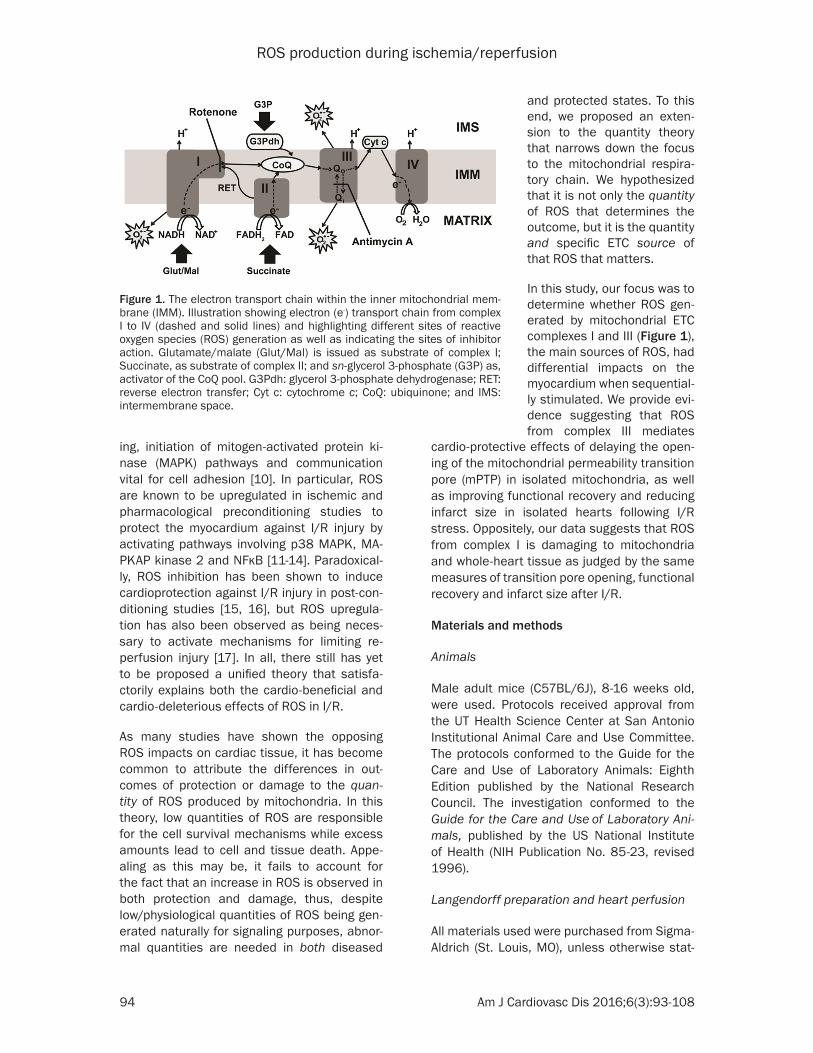
Figure 1 The electron transport chain within the inner mitochondrial mem-brane (IMM) Illustration showing electron (e-) transport chain from complex I to IV (dashed and solid lines) and highlighting different sites of reactive oxygen species (ROS) generation as well as indicating the sites of inhibitor action Glutamatemalate (GlutMal) is issued as substrate of complex I Succinate as substrate of complex II and sn-glycerol 3-phosphate (G3P) as activator of the CoQ pool G3Pdh glycerol 3-phosphate dehydrogenase RET reverse electron transfer Cyt c cytochrome c CoQ ubiquinone and IMS intermembrane space
and protected states To this end we proposed an exten-sion to the quantity theory that narrows down the focus to the mitochondrial respira-tory chain We hypothesized that it is not only the quantity of ROS that determines the outcome but it is the quantity and specific ETC source of that ROS that matters
In this study our focus was to determine whether ROS gen-erated by mitochondrial ETC complexes I and III (Figure 1) the main sources of ROS had differential impacts on the myocardium when sequential-ly stimulated We provide evi-dence suggesting that ROS from complex III mediates
ROS production during ischemiareperfusion
95 Am J Cardiovasc Dis 20166(3)93-108
ed Mice were anesthetized by intraperitoneal injection of pentobarbital (60 mgkg) and hep-arin (200 UIkg) was used to prevent blood coagulation Hearts were surgically removed and immediately arrested in cold (4degC) Krebs Henseleit bicarbonate buffer (KH) solution (mM) glucose 11 NaCl 118 KCl 47 MgSO4 12 KH2PO4 12 NaHCO3 25 and CaCl2 3 pH 74 The aorta was rapidly cannulated and the heart was retrograde-perfused at a constant rate (3 mlmin) in the Langendorff mode using KH buffer bubbled with 95 O25 CO2 at 37degC After 30 minutes of equilibration global normothermic ischemia was induced by stop-ping KH buffer supply to the aorta for 30 min-utes followed by 20 minutes of reperfusion for mitochondrial isolation For myocardial infarct size imaging studies hearts were reperfused for 120 minutes to allow for the development of IR damage We preferentially considered good hearts the ones that reached a minimum left ventricular developed pressure (LVDP) of 80 mmHg at the end of the basal perfusion (before ischemia) In the mouse model this IR proto-col typically results in ~50 of infarct size Sham hearts were not subjected to IR but only were perfused for the same duration as the IR protocol To determine the role of the stimulation of different ETC complexes in myocardial infarction and cardiac functional recovery hearts were perfused with the KH buffer supplemented with 3 mM of each of the substrates glutamatemalate succinate or sn-glycerol 3-phosphate
Functional measurements
The heart function was recorded throughout the experiments using a catheter (14F SPR-671 Millar Instruments Colorado Springs CO) connected to a MAC LAB (Powerlab) acquisi- tion system from ADInstruments (Sydney Aus- tralia) as previously described in our articles [18 19] The catheter was directly inserted into the left ventricle (LV) after a left atrial incision was made to expose the mitral annu-lus the LV end-systolic pressure (LVSP) the LV end-diastolic pressure (LVEDP) and heart rate (HR) were directly obtained from Lab- Chart55 (ADInstruments) software as describ- ed in our article [20] The LV developed pres-sure (LVDP = LVSP - LVEDP) and Rate-Pressure Product (RPP = LVDP times HR) were calculated from the recordings
Myocardial infarct size
The heart was removed from the Langen- dorff apparatus at the end of the reperfusion period and cut into four transverse slices paral-lel to the atrio-ventricular groove as previous- ly described [18] Slices were incubated for 7 minutes in 2 triphenyltetrazolium chloride (TTC) at 37degC followed by fixation with 4 paraformaldehyde This staining differentiated the infarcted (pale) from viable (red) myocar- dial tissue The slices were photographed us- ing digital microscopic imaging (Moticam 5 MP camera) The area of necrosis was quantified by computerized planimetry with Adobe Photo- shop CS6 and the total area of necrosis was calculated and expressed as a percentage of the total heart area
Preparation of isolated mitochondria
Mitochondria were isolated from fresh isch-emic (20-minute reperfusion) and non-ischemic (sham) hearts at 4degC as previously described in [20] Myocardial sections (approximately 015-022 g) were placed in isolation buffer A (mM) sucrose 70 mannitol 210 EDTA 1 and Tris-HCl 50 pH 74 The tissue was finely minced and homogenized in the same Buffer A (01 g of tissueml of buffer) The homoge- nate was centrifuged at 3000 rpm for 3 min-utes in a Galaxy 20R centrifuge (VWR Radnor PA) the supernatant was centrifuged at 13000 rpm for 10 minutes The mitochondrial pellet was resuspended in isolation Buffer B (mM) sucrose 150 KCl 50 KH2PO4 2 succinic acid 5 and TrisHCl 20 pH 74) Additional isola- tion buffers C and D were prepared with 5 mM glutamatemalate and 5 mM sn-glyecerol 3-phosphate respectively instead of 5 mM succinate as used in Buffer B Protein concen-tration was estimated using the Bradford me- thod assay kit (Bio-Rad Hercules CA)
Mitochondrial H2O2 measurement
Mitochondrial ROS generation was measured spectrofluorometrically (560 nm excitation and 590 nm emission) in 100 microg mitochondrial pro-tein incubated in a solution containing 20 mM Tris 250 mM sucrose 1 mM EGTA 1 mM EDTA and 015 bovine serum albumin adjusted to pH 74 at 30degC with continuous stirring Superoxide generated in mitochondria has a short half-life and does not diffuse easily across
ROS production during ischemiareperfusion
96 Am J Cardiovasc Dis 20166(3)93-108
the membranes but the H2O2 derived from dismutation easily diffuses through the mem-branes [21 22] As a result generation of ROS can be monitored as a function of H2O2 le- vels Hydrogen peroxide was measured with the H2O2-sensitive dye amplex red reagent (10 microM) (Thermo Fisher Waltham MA) in the pre- sence of horseradish peroxidase (0345 U mL) and H2O2 levels were calculated from a calibration curve obtained from fluorescence emission intensity as a function of H2O2 con-centration The sodium salts of glutamatema- late (3 mM) succinate (3 mM) and sn-glycerol 3-phosphate (G3P) (3 mM) were used to acti-
levels as Ca2+ enters the mitochondrial matrix via uptake by the Ca2+ uniporter With increas-ing calcium loading the extra-mitochondrial Ca2+ concentration starts accumulating there-by reflecting a lower capacity for mitochondrial Ca2+ uptake This is followed by a sustained Ca2+ increase indicating a massive release of the mitochondria Ca2+ by the mPTP opening The Ca2+ retention capacity (CRC) was defined as the amount of Ca2+ required to trigger this massive Ca2+ release which is used here as an indicator of the mPTP sensitivity to Ca2+ CRC is expressed as nmol of CaCl2 per mg of mito-chondrial protein
Figure 2 ROS production from activated ETC complexes measured as H2O2 release in normal mitochondria and mitochondria following ischemiareper-fusion (IR) injury Typical measurements of H2O2 release from normal (black trace) and IR (red trace) mitochondria when complex I (3 mM glutamatemalate GlutMal) (A) complex II (3 mM succinate) (B) and CoQ (3 mM sn-glycerol 3-phosphate G3P) (C) were stimulated in the presence of amplex red and horseradish peroxidase (Left) Graphs showing how the rate of H2O2 production measured for a 60 second period after addition of substrate in-creased in IR mitochondria after GlutMal was added but decreased after succinate and G3P were added (Right) Values are mean plusmn SEM p lt 005 normal versus IR mitochondria n = 5-6group
vate complexes I and II as well as the coenzyme Q pool respectively Complex I inhibi-tor rotenone (2 microM) and com-plex III inhibitor antimycin A from Streptomyces sp (20 microM) were also used
Ca2+-induced mitochondrial permeability transition
The installation of mitochon-drial permeability transition pores (mPTP) was assessed following in vitro Ca2+ over- load as previously described [23 24] Free Ca2+ concentra-tion outside the mitochon- dria was recorded using 01 microM calcium green-5N (Ther- mo Fisher) which binds re- versibly to Ca2+ using exci- tation and emission wave-lengths set at 500 and 530 nm respectively Isolated mi- tochondria (500 microg of pro- tein) were suspended in 2 ml isolation Buffer B C or D and pre-incubated for 90 seconds in a spectrofluo- rometer (Hitachi F-2710) set at 30degC CaCl2 pulses (10 micromoles or 10 microL of 1 mM stock solution) were applied every 60 seconds to the cuvette leading to 20 nmol Ca2+ (per mg of mitochondrial protein) The Ca2+ pulses in- duce a peak of extra-mito-chondrial Ca2+ concentration that returns to near-baseline
ROS production during ischemiareperfusion
97 Am J Cardiovasc Dis 20166(3)93-108
Statistical analysis
The data shown in bar graphs are expressed as means with error bars that are the stand- ard errors of the mean (plusmn SEM) for a mini- mum of five independent hearts (n ge 5) The Studentrsquos t-test and one-way ANOVA with post-
opposite ROS production when complex I is activated compared to when complex II and CoQ are activated in mitochondria isolated from ischemic hearts and in normal mitochon-dria We also noted the marked difference in rates when each complex is stimulated with succinatecomplex II stimulation resulting in
Figure 3 ROS production from activated ETC complexes measured as H2O2 release in normal mitochondria after insult with pro-apoptotic drugs Typical measurements of H2O2 release in healthy mitochondria from control (black trace) with complex I inhibitor rotenone (Rot orange trace) and with com-plex III inhibitor antimycin A (AA purple trace) when complex I (3 mM glu-tamatemalate GlutMal) (A) complex II (3 mM succinate) (B) and CoQ (3 mM sn-glycerol 3-phosphate G3P) (C) were stimulated in the presence of amplex red and horseradish peroxidase (Left) Bar graphs showing changes in the rate of H2O2 production measured for a 60 second period after ad-dition of each inhibitor (Right) Values are mean plusmn SEM p lt 005 control versus+inhibitor n = 5-6group
hoc Dunnettrsquos Tukeyrsquos and Sidakrsquos corrections for multi-ple comparisons were appro-priately used to assess the differences observed Analy- sis was performed using Pri- sm 6 (Graphpad Software La Jolla CA) A difference of p lt 005 was considered to be statistically significant
Results
Following IR stimulation of complex I increases ROS production but stimulation of complex II and CoQ de-creases it
We first sought to ascertain the production of ROS by sti- mulated complexes in isolat-ed mitochondria from both sham and ischemic myocardi-um using commonly used [25-27] de novo ETC substrates glutamatemalate (complex I) and succinate (complex II) as well as CoQ substrate G3P In line with past studies follow-ing ischemic injury ROS pro-duction increases in complex I [28] Activating complex I (glutamatemalate) resulted in an increase in ROS produc-tion in mitochondria isolated from IR hearts as compared to mitochondria isolated from non-ischemic hearts (Figure 2A) Surprisingly treatment of IR mitochondria with the complex II substrate succi-nate or the Q-pool substrate G3P resulted in reduced ROS production rate in IR mito-chondria when compared to non-ischemic tissue (Figure 2B 2C) These results show
ROS production during ischemiareperfusion
98 Am J Cardiovasc Dis 20166(3)93-108
tremendously more ROS production than com-plex I and CoQ
duction to those observed following IR injury (Figure 2) and therefore can be used to
Figure 4 Mitochondrial Ca2+ induced opening of the mPTP for buffers of each substrate A Left Example of recordings showing the calcium over-load required to induce the mPTP opening (colored arrows) of isolated mi-tochondria re-suspended in a buffer containing 5 mM glutamatemalate (GlutMal) as substrate (blue trace) and after supplementation with 3 mM succinate (Succ red trace) and 3 mM sn-glycerol 3-phosphate (G3P green trace) Right Bar graph showing improved calcium retention capacity (CRC) when GlutMal is supplemented with other substrates Values are mean plusmn SEM p lt 005 supplements versus control (GlutMal) n = 6group B Left Example of recordings showing the calcium overload required to induce the mPTP opening (colored arrows) of isolated mitochondria re-suspended in a buffer containing 5 mM succinate (Succ) as substrate (red trace) and after supplementation with 3 mM glutamatemalate (GlutMal blue trace) and 3 mM sn-glycerol 3-phosphate (G3P green trace) Right Bar graph showing calcium retention capacity (CRC) when succinate is supplemented with other substrates Values are mean plusmn SEM p lt 005 supplements versus con-trol (Succ) n = 6group C Left Example of recordings showing the calcium overload required to induce the mPTP opening (colored arrows) of isolated mitochondria re-suspended in a buffer containing 5 mM sn-glycerol 3-phos-phate (G3P) as substrate (green trace) and after supplementation with 3 mM succinate (Succ red trace) and 3 mM glutamatemalate (GlutMal blue trace) Right Bar graph showing improved calcium retention capacity (CRC) when G3P is supplemented with other substrates Values are mean plusmn SEM p lt 005 supplements versus control (G3P) n = 6group
Pro-apoptotic drugs increase ROS production when com-plex I and the Q pool are stimulated but reduce ROS production when complex II is stimulated in healthy mito-chondria
Next we studied if pro-apop-totic drugs resulted in similar responses to complex stimu-lation As shown in Figure 3 addition of complex I blocker rotenone showed an increase in ROS generation by activat-ed complex I and a modest increase on the activated Q cycle The addition of antimy-cin A which blocks the Q0 site of complex III led to dramatic increase in ROS generation when both complex I and CoQ were activated (Figure 3A 3C) However when complex II was stimulated the addi- tion of both inhibitors result- ed in reduction of ROS gener-ation In fact in the pre- sence of rotenone a complex I blocker and hence a blocker of reverse electron transfer (RET) ROS rate slowed down the most (succinate+rote- none 1010 plusmn 139 versus suc-cinate 2274 plusmn 225 pmolesminmg n = 5 p = 0001) when compared to antimycin A (succinate+antimycin A 1538 plusmn 88 pmolesminmg) suggesting that the electron transfer from the stimula- ted complex II to complex I (RET) is a larger source of ROS production than the for-ward transfer to complex III Together these findings in- dicate that pro-apoptosis ETC inhibitors induce oppo-site ROS production when complex I or II is stimulated These results also show that these pro-apoptotic drugs that affect the ETC [29] have similar effects on ROS pro-
ROS production during ischemiareperfusion
99 Am J Cardiovasc Dis 20166(3)93-108
approximate IR effects when the different complexes are being studied
Activation of complex II and the Q-pool result in increased mitochondrial calcium retention capacity while activation of complex I shows a decrease in capacity
We then asked what impact the increases in ROS from each complex would have on the opening of the mPTP a pivotal event known to cause cell death in IR injury [30] and whorsquos inhibition was recently shown by our group to be necessary in cardioprotection against IR injury via the G-protein coupled estrogen recep-tor 1 (GPER1) [31] As the mPTP opening may be induced by calcium overload [32] we com-
of complex I to mitochondria increases the sensitivity of the mPTP opening to calcium overload when compared to succinate the substrate of complex II We also found that when glutamatemalate was used to prepare the isolation buffer addition of either succi- nate (3 mM) or the ETC intermediate G3P (3 mM) rescued the mitochondrial CRC with suc-cinate showing a greater recovery (glutamatemalate 73 plusmn 11 versus glutamatemalate+suc- cinate 183 plusmn 24 nmolmg of mitochondrial pro- tein n = 6 p = 0002) This recovery was grea- ter than that for G3P (glutamatemalate+succi- nate 183 plusmn 24 versus glutamatemalate+G3P 150 plusmn 20 nmolmg of mitochondrial protein n = 6) (Figure 4A) On the other hand when suc-cinate the substrate of complex II was used to
Figure 5 Mitochondrial Ca2+ induced opening of the mPTP for buffers of each substrate with pro-apoptotic inhibitors A Typical recording showing the calcium overload required to induce the mPTP opening (colored arrows) of isolated mitochondria re-suspended in a buffer containing 5 mM gluta-matemalate (GlutMal blue trace) 5 mM succinate (Succ red trace) and 5 mM sn-glycerol 3-phosphate (G3P green trace) as substrate (control) B Typical recording showing the calcium overload required to induce the mPTP opening (colored arrows) of isolated mitochondria re-suspended in a buffer containing 5 mM glutamatemalate (GlutMal blue trace) 5 mM succinate (Succ red trace) and 5 mM sn-glycerol 3-phosphate (G3P green trace) and treated with rotenone (Rot 2 μM) C Typical recording showing the calcium overload required to induce the mPTP opening (colored arrows) of isolated mitochondria re-suspended in a buffer containing 5 mM glutamatemalate (GlutMal blue trace) 5 mM succinate (Succ red trace) and 5 mM sn-glycer-ol 3-phosphate (G3P green trace) and treated with antimycin A (AA 20 μM) D Bar graphs showing mitochondrial calcium retention capacity (CRC) mea-sured of mitochondria re-suspended in a buffer containing each substrate and supplemented with either rotenone or antimycin A Values are mean plusmn SEM p lt 005 inhibitor treated versus respective control (same substrate) p lt 005 Succ+Rot versus other substrates+Rot n = 6group
pared the sensitivity of mPTP opening as a function of calcium retention capacity in isolated fresh mitochondria treated with each complexrsquos substrate to stimulate ROS production Figure 4 shows the time course of addition of free Ca2+ pulses in the solu-tion surrounding mitochon-dria The initial Ca2+ concen-tration in the buffer was set to Ca2+ contaminant that declined after adding mito-chondria due to a global reduction of the solutionrsquos optical transmittance as a result of the increase in tur-bidity after loading mitochon-dria The following slower phase prior to the addition of the Ca2+ pulses reflects the rate of initial Ca2+ uptake by the mitochondria
In isolated mitochondria us- ing buffers of de novo ETC substrates glutamatemalate and succinate we found that the control mitochondrial cal-cium retention capacity (CRC) measurements were 73 plusmn 11 and 240 plusmn 5 nmolmg of mi- tochondrial protein (n = 6) re- spectively These results in- dicate that application of glu-tamatemalate the substrate
ROS production during ischemiareperfusion
100 Am J Cardiovasc Dis 20166(3)93-108
prepare the isolation buffer addition of gluta-matemalate (3 mM) slightly decreased the CRC (succinate 240 plusmn 5 versus succinate+glu- tamatemalate 217 plusmn 11 nmolmg of mito-chondrial protein n = 6 not significant) while the addition of G3P (3 mM) led to a statistically significant increase in CRC (succinate 240 plusmn 5 nmolmg of mitochondrial protein versus suc-
cinate+G3P 293 plusmn 8 nmolmg of mitochondrial protein n = 6 p = 0001) (Figure 4B) Use of the less traditional substrate G3P as buffer (5 mM) resulted in poor CRC (G3P 33 plusmn 4 nmolmg of mitochondrial protein) which was only aided by addition of 3 mM of the more com-monly used ETC substrates glutamatemalate (G3P+glutamatemalate 120 plusmn 20 nmolmg of mitochondrial protein n = 6 p = 0002 ver-sus G3P control) and succinate (G3P+succinate 193 plusmn 16 nmolmg of mitochondrial protein n = 6 p lt 00001 versus G3P control) (Figure 4C)
Furthermore we compared effects of ETC in- hibitors rotenone and antimycin A when gluta-matemalate succinate or G3P were used as ROS substrate for mitochondrial CRC As shown in Figure 5 addition of antimycin A resulted in mitochondrial depolarization when all three substrates were used In fact we found that addition of antimycin A to the cuvette contain-ing pre-incubated mitochondria immediately led to the opening of the mPTP Interestingly when complex I (glutamatemalate buffer) was activated addition of rotenone led to similar mitochondrial depolarization with immediate mPTP opening even before addition of Ca2+ pulses commenced (n = 6 p lt 0001) When CoQ (G3P buffer) was activated addition of rotenone did not show any change in CRC (G3P+rotenone 27 plusmn 4 versus G3P 33 plusmn 4 nmolmg of mitochondrial protein n = 5-6 not significant) Interestingly when complex II (suc-cinate buffer) was stimulated addition of rote-none remarkably enhanced CRC (succinate+ rotenone 267 plusmn 7 versus succinate 240 plusmn 5 nmolmg of mitochondrial protein n = 6 p = 002) suggesting a delay of the mPTP open- ing Together these results indicate that the ROS production in complex I promotes the mPTP opening while ROS produced in com- plex III induces delay of the mPTP opening and presumably may lead to cardioprotection
Activation of CoQ in contrast to complexes I and II improved cardiac functional recovery and reduced myocardial infarct size following IR
Finally we asked whether resultant differences in ROS produced by each complex could be observed in an ex vivo animal model and pos-sibly highlight clinically relevant effects Using Langendorff retrograde perfusion we studied myocardial infarct size and cardiac functional
Figure 6 Measure of cardiac functional recovery in control and after addition of substrates after IR Cardiac function was recorded throughout the ex-periments in isolated perfused hearts treated with KH buffer for control and after addition of different substrates (3 mM) A Bar graph showing an improve-ment of the cardiac functional recovery (Rate-Pres-sure Product RPP) at 120 min reperfusion (end of reperfusion) in sn-glycerol 3-phosphate (G3P) group compared to control (Ctrl) and glutamatemalate (GlutMal) groups B Bar graph indicating that the left ventricular developed pressure (LVDP) at 120 min reperfusion did not change in control or the supplements of each substrate C Bar graph show-ing an improvement of the heart rate (HR) at 120 min reperfusion (end of reperfusion) in sn-glycerol 3-phosphate (G3P) group compared to control and glutamatemalate (GlutMal) groups Values are mean plusmn SEM p lt 005 substrate versus control p lt 005 G3P versus GlutMal n = 5-6group
ROS production during ischemiareperfusion
101 Am J Cardiovasc Dis 20166(3)93-108
recovery following IR stress with reduced infarct size and higher rate-pressure product (RPP) indicating cardioprotection We com-pared these parameters in isolated hearts per-fused with KH buffer that was enriched with each of the three substrates to stimulate ROS production With regards to the heart regaining cardiac function following IR the RPP values obtained at 120 minutes of reperfusion as a percent of RPP before ischemia were in the order G3P (72 plusmn 9) succinate (43 plusmn 7) and glutamatemalate (36 plusmn 1) compared to con-trol (36 plusmn 4) (n = 5-7) (Figure 6A) As shown in Figure 6B 6C the resultant RPP at the end of reperfusion was mostly driven by changes in heart rate as the LVDP readings were fairly similar across all conditions Incitement of ROS
production at complex III by activating the Q-pool with G3P resulted in smaller infarct sizes (G3P 42 plusmn 2) as compared to the con- trol (56 plusmn 4 n = 5-7 p = 001) However acti-vating complex I with glutamatemalate incre- ased the infarct size from that exhibited by con-trols (glutamatemalate 59 plusmn 4 versus con-trol 56 plusmn 4 n = 5-7 not significant) (Figure 7A 7B) Comparing G3P (complex III ROS) to glutamatemalate (complex I ROS) showed statistically significant benefits of G3P (Figures 6 7 Infarct size p = 0001 and RPP p = 004 n = 5-7) These results suggest that complex III ROS induce cardioprotective effects against ischemia reperfusion injury while complex I ROS appear to promote myocardial injury
Discussion
In this study we demonstrated that not only is the quantity of ROS important but the quan-tity and mitochondrial ETC source of that ROS in mitochondria is pertinent to its impact as either cardio-protective or cardio-deleterious in a heart IR model Specifically we showed that ROS from complex I results in a reduction of mitochondriarsquos ability to sequester Ca2+ ions in healthy mitochondria greater myocardial infarct size and less functional recovery in the whole heart following IR injury Conversely we revealed how ROS from complex III leads to an improvement in mitochondrial Ca2+ buffering as highlighted by the delay in the mPTP open-ing and reduces the overall heart infarct size while improving recovery after IR Our results show that stimulating complex III either from complex II or Q-pool substrates leads to ROS production that is cardio-beneficial as oppos- ed to that from stimulating complex I which is detrimental to both mitochondria and the stressed heart as a whole
ROS production at complexes I and III
The increase in production of mitochondrial ROS has been demonstrated using cell labeling methods as being a relevant process in many pathological conditions like heart attack dia-betes and cancer [33-35] and it also partici-pates in necrosis and apoptosis associated with cardiac IR injury (reviewed in detail in [36 37]) Complex I is a major source of electron leakage which results in ROS production in the ETC Previous studies have debated the exact site of ROS generation on complex I [36 38]
Figure 7 Measure of myocardial infarct size in con-trol and after supplement of substrates after IR Myocardial infarct size was measured at the end of 120 min reperfusion in isolated perfused hearts treated with KH buffer for control and after addition of different substrates A Representative images from 4 slices of the same heart in each condition White areas correspond to the infarcted zone B Bar graphs showing reduction of myocardial infarct size in sn-glycerol 3-phosphate (G3P) group compared to control (Ctrl) and glutamatemalate (GlutMal) groups Values are mean plusmn SEM of infarct size p lt 005 substrate treated versus control p lt 005 G3P versus GlutMal n = 6-7group
ROS production during ischemiareperfusion
102 Am J Cardiovasc Dis 20166(3)93-108
but the ubiquinone-binding domain appears to be main site during succinate-driven reverse electron transfer (RET) from complex II [39] and this also happens to be the same site at which rotenone blocks transfer of electrons to CoQ [40] Following ischemic damage the iron sul-fur clusters proximal to this ubiquinone binding site are thought to be the locations for ROS pro-duction which result in the increase in ROS that we also observed [38] (Figure 2A) ROS produc-tion from complex I is more dependent on ΔpH than ΔΨ (membrane potential) [39 41] and thus less reliant on mitochondrial energization Thus IR injury and rotenone increase ROS generation from the same region on complex I when this complex is activated by blocking electron escape as we observed (Figures 2A and 3A)
Complex II influences ROS production at com-plex I (via RET) [28 29] and complex III [36] and although some evidence has suggest- ed complex II to be a source of ROS under cer-tain conditions [42] it is only complexes I and III that are globally accepted as main genera-tors Following IR insult or application of rote-none to complex I we observed a decrease in succinate-driven ROS (Figures 2B and 3B) which are in line with a loss of RET to complex I For complex II our findings further highlight its dependence on its neighboring complexes for ROS production From this it may be inferr- ed that following IR injury activated complex II no longer influences ROS production at com-plexes I or III hence the observed decrease (Figure 2) while rotenone and antimycin A also lead to diminished ROS formation as complex IIrsquos access to the adjacent enzymatic units is diminished (Figure 3)
Complex III has two known sites of ROS pro- duction with one releasing ROS into the inter-membrane space and the other into the matrix [43] Complex III works by transferring electrons from ubiquinol to ferricytochrome c by utilizing hemes bL and bH and the Rieske iron-sulfur protein [36] The contribution of complex III to ROS production in normal physiology remains uncertain with some groups suggesting it to be smaller [44 45] while others postulate it to be larger [38 46 47] than complex I invo- lvement However there is consensus on com-plex III being a major site of ROS production after IR [36 38] Ischemic damage reduced ROS production from G3P stimulation (Figure
2C) contrary to the expectation of an increase IR disrupts complex III function [48] and G3PCoQ stimulation probably would have been impaired from affecting ROS from this complex Accordingly downstream application of anti- mycin A which blocks electron transfer from the Q0 site of complex III increased ROS pro- duction when complex I and complex III were stimulated (Figure 3) And despite complex III ROS relying more on ΔΨ than ΔpH [49] appli- cation of the transfer blocker antimycin A re- sults in ROS production As succinate influenc-es ROS at both complexes I and III we opted to include G3P in order to gain further insight into ROS production from complex III G3P also known as L-α-glycerophosphate is thought to be an inadequate substrate by itself for study-ing H2O2 production in heart mitochondria [50] but when coupled with glutamate or succinate can augment ROS production at complex III Mracek et al have shown that glycerophos-phate dehydrogenase (G3Pdh in Figure 1) is a source of ROS particularly in brown adipose tissue but that in heart mitochondria stimulat-ing the dehydrogenase with G3P results in the bulk of ROS being made at complex III [51] hence we opted to use it in our studies to in- fluence ROS generation at complex III
Opposing effects of ROS from complex I versus complex III
Kalogeris et al summed up current opinion that the ldquodouble-edged swordrdquo impact of ROS prompted by IR may be traced back to i) the type of ROS produced ii) the amount and time when the ROS is produced and iii) the cellular and subcellular loci of creation [52] First reac-tive species known to cause damage to cells and tissue include the superoxide anion radi- cal (O2
bull-) hydroxyl radical (bullOH) hydrogen perox-ide (H2O2) and peroxynitrite (ONOO-) [53] In cardiovascular tissue the most important and abundant form of ROS is O2
bull- [54] and within this tissue it is known that mitochondria and specifically the respiratory chain are the domi-nant sources of O2
bull- [2 7 36] With regards to the heart IR damage results in increased O2
bull- production by both subsarcolemmal and interfibrillary mitochondria [55 56] which were the focus of our study
Second the amount of ROS and when it is pro-duced are critical to the effect it has The amount of ROS following IR overwhelms the
ROS production during ischemiareperfusion
103 Am J Cardiovasc Dis 20166(3)93-108
hitherto tightly-regulating antioxidant machin-ery of the cell and the ROS can thus attack DNA proteins and subcellular structures [52] In our experiments we used the ETC substrates glutamatemalate succinate and G3P at non-physiological concentrations of 3 mM to insti-gate this increase in ROS As evidenced by the increase in ROS in pre- and postconditioning the time when ROS increase commences is also vital [11-14 17] During ischemia cells are starved of both nutrients and oxygen and when reperfusion begins it is the accumulation of Ca2+ in the matrix depletion of adenine nucleo-tide increased inorganic phosphate concentra-tion and oxidative stress that leads to the opening of mPTP [32] whose effects include depolarizing mitochondria uncoupling the ETC allowing for efflux of Ca2+ and intermembrane proteins such as the pro-apoptotic cytochrome c and causing swelling of the mitochondrial matrix [57]
Thirdly the cellularsubcellular location of ROS generation is pertinent to the outcomes of IR This idea of compartmentalization for the con-trol of ROS is both logical and supported by a large body of evidence (reviewed in [58-60]) In normal physiology the cell highly regulates the amount of ROS particularly through superoxide dismutase (SOD) which degrades ROS to H2O2 which can be further degraded to water and molecular oxygen The mitochondrial matrix contains manganese-SOD (Mn-SOD or SOD-2) while the cytoplasm and the intermembrane space in mitochondria contain copperzinc-SOD (CuZn-SOD or SOD-1) [52 61] Insults like IR ignite overwhelming quantities of ROS that overpower these safeguards and leads to senescence autophagy and apoptotic path-ways associated with cardiovascular diseases [1]
In the present study we observed that not only is it the subcellular compartment key to ROS impact but the mitochondrial ETC complex generating that ROS is crucial as well By study-ing isolated mitochondriarsquos ability to sequester calcium before opening the mPTP we found that the impact of ROS produced by activated complexes I and III on the opening of the mPTP is contradictory (Figure 4) The mPTP opening is known to be associated with an increase in ROS and its opening is a trademark event of mitophagy following IR injury [20] Our data indicate that the treatment of fresh and healthy
mitochondria with succinate buffer (5 mM) (for complex II) delays the opening of the mPTP as the calcium retention capacity (CRC) increases while the stimulation of complex I (glutamatemalate buffer 5 mM) actually reduces mito-chondrial CRC (Figure 4A 4B) For each of these substrates addition of the other sub-strate either aids or impedes the CRC effect As shown in Figure 4 when glutamatemalate buf-fer (5 mM) is supplemented with either succi-nate or G3P (3 mM) this serves to improve the mitochondriarsquos CRC on the contrary addition of glutamatemalate (3 mM) to succinate buf-fer (5 mM) solution slightly reduces CRC and as can be deduced from our hypothesis addi-tion of 3 mM G3P (for complex III) to succinate buffer (5 mM) increases CRC As discussed ear-lier G3P by itself is a poor cardiac ROS sub-strate and this was also shown by its low CRC values when it was used as substrate buffer (5 mM) (Figure 4C) however it was improved sig-nificantly in the presence of the natural sub-strates glutamatemalate (3 mM) and succi-nate (3 mM)
Following our hypothesis that the ETC source of ROS is critical in cardioprotection we studied the CRC effects of increased ROS production when complexes I and III were sequentially blocked by use of rotenone and antimycin A as blockers Rotenone inhibits oxidation of the iron-sulfur clusters in complex I while also blocking RET from complex II [36 62] (Figure 1) Antimycin A blocks the Qi site of complex III resulting in increased ROS production at the complexrsquos Q0 site with this ROS being expelled into the intermembrane space from the Q0 site [43 51 63 64] Application of antimycin A a drug with no biological equivalent to mitochon-dria results in the collapse of membrane poten-tial opening of the mPTP [62 65] and hence the inability to absorb Ca2+ as we also found (Figure 5) Furthermore addition of rotenone to activated complex I which increases ROS at this site also depleted mitochondrial CRC when using glutamatemalate but rotenonersquos block-ing of RET improved CRC when complex II was activated probably due to more ldquogoodrdquo ROS coming from complex III We found that limiting ROS from resting state complex I by applying rotenone and stimulating complex II using suc-cinate served to enhance the cardio-positive effects of the ROS from complex III by increas-ing CRC (Figure 5D) The fact that the stimula-
ROS production during ischemiareperfusion
104 Am J Cardiovasc Dis 20166(3)93-108
tion of the complexes II and III (while inhibiting influence of complex I) increases the mitochon-drial CRC indicate that ROS produced under these conditions are cardioprotective since it is well established that the inhibition of mPTP opening protects the heart during pre- and post-conditioning and controlled reperfusion [37]
The differential effects of ROS from complexes I and III in isolated mitochondria suggested opposite roles and we sought to determine if these roles extended to intact myocardium For this we compared cardiac functional recovery as well as myocardial infarct size in isolated hearts perfused with KH buffer versus KH with supplementation of 3 mM of each substrate Although our previous experiments had been in isolated mitochondria with substrates easily accessing the inner membrane of mitochon-dria all three substrates are found in the cyto-sol and can be transported across cell mem-branes from exogenous sources [66-69]
The rate-pressure product (RPP) or cardiovas-cular product used in hemodynamic stress studies in cardiac physiology is used to predict myocardial oxygen consumption or quite sim-ply myocardial workload [70] As the product of heart rate and systolic blood pressure RPP can be used to monitor the functional impact of IR injury on the heart [18 71 72] and in our stud-ies the RPP was used to determine the level of functional recovery following IR In this study we found that G3P showed the greatest recov-ery with glutamatemalate showing the least further evidence for the opposing complexesrsquo ROS The left ventricular developed (LVDP) was similar in all groups and the improvement of the RPP with G3P infusion was mostly due to the increase in the heart rate (HR) (Figure 6A-C) suggesting that G3P may also directly modu-late pace maker activity
Myocardial infarct size was measured using histological staining and planimetry and the results showed significantly different outcomes following IR (Figure 7) Following the trends we observed in mitochondrial CRC applying the substrates to whole tissue further cemented our proposition that complex III ROS are cardio-protective and complex I ROS are cardio-dele- terious G3P resulted in reduced infarct size compared to glutamatemalate As KH buffer already contains a mitochondrial stimulant and
Krebs cycle driver in the form of glucose we did not expect dramatically pronounced differenc-es between the samples This was because glu-cose was stimulating complexes I and II as well as supporting an active tricarboxylic acid cycle which is necessary for the heart to survive ex vivo Nevertheless we did observe significant differences between glutamatemalate and G3P treatment enough to support the notion of opposed impacts of ROS With regards to succinate we also did observe smaller infarct sizes compared to glutamatemalate but not to the same extent as those for G3P (Figure 7) As succinatecomplex II feeds electrons to both complexes I and III this intermediate result was not surprising Furthermore Chouchani and colleagues have shown that in vivo the accu-mulation of succinate during ischemia is the main driver for ROS generation by complex I via RET during reperfusion and that a reduction of this RET is adequate enough to mitigate IR injury in murine models [73]
Impact on cardioprotection and antioxidant therapy
Based on our results a fundamental question that arises is how exactly cardiomyocytes or specifically mitochondria recognize or delin-eate between ldquogoodrdquo versus ldquobadrdquo ROS in times of stress As ROS in normal conditions and controlled quantities features as a signal-ing conduit [10] it may be possible that under such circumstances neither complex I nor com-plex III ROS are good or bad In pathophysio- logical conditions resulting from IR or exoge-nous inhibitors we have shown that ROS from complexes I and III are not the same but the mechanism or even the sites of action of these ROS leading to cardiac protection or damage are the subject of future studies Drose et al have suggested a mechanistic model to explain lsquosignaling ROSrsquo by studying diazoxide effects on mitochondrial ROS generation [63] They proposed that diazoxide a complex II blocker could stimulate the production of lsquosig-nalling ROSrsquo at the Q0 site of complex III by in- hibition of the succinate-ubiquinone oxidore-ductase (complex II) thereby partially oxidizing the Q-pool They hypothesized that following stress lsquosignaling ROSrsquo activates mitochondrial signaling pathways that during reoxygenation eventually reduce the production of lsquodeleteri-ousrsquo ROS by NADH-ubiquinone oxidoreductase (complex I) via RET and that in addition diazox-
ROS production during ischemiareperfusion
105 Am J Cardiovasc Dis 20166(3)93-108
ide can directly reduce oxidative damage evoked by RET at complex I by inhibition of com-plex II However studies in H9c2 myoblasts by other investigators have suggested complex I being a source of signaling ROS for muscle dif-ferentiation [74] In our study our data agree with the Drose group in suggesting that the source of this lsquosignalingrsquo ROS in heart mito-chondria to be complex III while also highlight-ing the deleterious effects of ROS from com-plex I We extend this premise into the impact that both ETC sources have on cardiac tissue recovery following ischemic insult Despite this model offering some insights into ROS classifi-cation much is still needed to fully elucidate how cells respond to ROS from different ETC sites Multiple factors need consideration when defining such pathways including the varying needs and responses of mitochondria in differ-ent organs [25 50] and how for example in the brain glutamate is both an excitatory neu-rotransmitter and a cause of neurotoxicity [75] Understanding these mechanisms will be vital to designing targeted therapeutic agents for cardioprotection as current models of coen-zyme Q10 treatment [6] or applying indiscrimi-nant antioxidants as one-size fits all panaceas have proven to be somewhat fruitless as both good and bad ROS are destroyed [3]
In conclusion we demonstrated the opposite impact of ROS generated by stimulation of com-plexes I II and III after ischemia-reperfusion and also application of pro-apoptotic drugs in fresh mitochondria We propose an extension to the concept that the quantity of ROS is important by showing that the mitochondrial ETC source complex of that ROS is also pivotal We went on to demonstrate the positive impact of the ROS from mitochondrial complex III in cardioprotection via the inhibition of the mPTP opening and following IR reduction of myo-cardial infarct size and improvements in cardi-ac functional recovery while showing the car-dio-deleterious effects of ROS from complex I
Acknowledgements
Supported by Bopassarsquos AHA fellowship 09- POST2190008 Voelcker fund (Bopassa)
Disclosure of conflict of interest
None
Address correspondence to Jean C Bopassa De- partment of Physiology School of Medicine Uni- versity of Texas Health Science Center at San Antonio 7703 Floyd Curl Dr San Antonio TX 78- 229 USA Tel 210-567-0429 Fax 210-567-4410 E-mail bopassauthscsaedu
References
[1] Brown DI and Griendling KK Regulation of signal transduction by reactive oxygen species in the cardiovascular system Circ Res 2015 116 531-549
[2] Steinhubl SR Why have antioxidants failed in clinical trials Am J Cardiol 2008 101 14D-19D
[3] Madamanchi NR Vendrov A and Runge MS Oxidative stress and vascular disease Ar- terioscler Thromb Vasc Biol 2005 25 29-38
[4] Bjelakovic G Nikolova D Gluud LL Simonetti RG and Gluud C Mortality in randomized trials of antioxidant supplements for primary and secondary prevention systematic review and meta-analysis JAMA 2007 297 842-857
[5] Miller ER 3rd Pastor-Barriuso R Dalal D Riemersma RA Appel LJ and Guallar E Meta-analysis high-dosage vitamin E supplementa-tion may increase all-cause mortality Ann Intern Med 2005 142 37-46
[6] Pepe S Marasco SF Haas SJ Sheeran FL Krum H and Rosenfeldt FL Coenzyme Q10 in cardiovascular disease Mitochondrion 2007 7 Suppl S154-167
[7] Chandel NS and Tuveson DA The promise and perils of antioxidants for cancer patients N Engl J Med 2014 371 177-178
[8] Snow BJ Rolfe FL Lockhart MM Frampton CM OrsquoSullivan JD Fung V Smith RA Murphy MP Taylor KM and Protect Study G A double-blind placebo-controlled study to assess the mitochondria-targeted antioxidant MitoQ as a disease-modifying therapy in Parkinsonrsquos dis-ease Mov Disord 2010 25 1670-1674
[9] Smith RA and Murphy MP Animal and hu- man studies with the mitochondria-targeted antioxidant MitoQ Ann N Y Acad Sci 2010 1201 96-103
[10] Bartosz G Reactive oxygen species destroyers or messengers Biochem Pharmacol 2009 77 1303-1315
[11] Das DK Maulik N Sato M and Ray PS Reactive oxygen species function as second messenger during ischemic preconditioning of heart Mol Cell Biochem 1999 196 59-67
[12] Maslov LN Naryzhnaya NV Podoksenov YK Sementsov AS and Gorbunov AS [Signaling Mechanism of Cardioprotective Effect of Re- active Oxygen Species] Ross Fiziol Zh Im I M Sechenova 2015 101 377-385
ROS production during ischemiareperfusion
106 Am J Cardiovasc Dis 20166(3)93-108
[13] Halestrap AP Clarke SJ and Khaliulin I The role of mitochondria in protection of the heart by preconditioning Biochim Biophys Acta 2007 1767 1007-1031
[14] Zhou T Chuang CC and Zuo L Molecular Characterization of Reactive Oxygen Species in Myocardial Ischemia-Reperfusion Injury Biomed Res Int 2015 2015 864946
[15] Sun HY Wang NP Kerendi F Halkos M Kin H Guyton RA Vinten-Johansen J and Zhao ZQ Hypoxic postconditioning reduces cardiomyo-cyte loss by inhibiting ROS generation and in-tracellular Ca2+ overload Am J Physiol Heart Circ Physiol 2005 288 H1900-1908
[16] Kin H Zhao ZQ Sun HY Wang NP Corvera JS Halkos ME Kerendi F Guyton RA and Vinten-Johansen J Postconditioning attenuates myo-cardial ischemia-reperfusion injury by inhibit-ing events in the early minutes of reperfusion Cardiovasc Res 2004 62 74-85
[17] Penna C Rastaldo R Mancardi D Raimondo S Cappello S Gattullo D Losano G and Pag- liaro P Post-conditioning induced cardioprotec-tion requires signaling through a redox-sensi-tive mechanism mitochondrial ATP-sensitive K+ channel and protein kinase C activation Basic Res Cardiol 2006 101 180-189
[18] Feng Y and Bopassa JC Oxygen surrounding the heart during ischemic conservation deter-mines the myocardial injury during reperfu-sion Am J Cardiovasc Dis 2015 5 127-139
[19] Rahman S Li J Bopassa JC Umar S Iorga A Partownavid P and Eghbali M Phosphorylation of GSK-3beta mediates intralipid-induced car-dioprotection against ischemiareperfusion injury Anesthesiology 2011 115 242-253
[20] Bopassa JC Eghbali M Toro L and Stefani E A novel estrogen receptor GPER inhibits mito-chondria permeability transition pore opening and protects the heart against ischemia-reper-fusion injury Am J Physiol Heart Circ Physiol 2010 298 H16-23
[21] Bienert GP and Chaumont F Aquaporin-facilitated transmembrane diffusion of hydro-gen peroxide Biochim Biophys Acta 2014 1840 1596-1604
[22] Loschen G Azzi A Richter C and Flohe L Superoxide radicals as precursors of mito-chondrial hydrogen peroxide FEBS Lett 1974 42 68-72
[23] Bopassa JC Ferrera R Gateau-Roesch O Couture-Lepetit E and Ovize M PI 3-kinase regulates the mitochondrial transition pore in controlled reperfusion and postconditioning Cardiovasc Res 2006 69 178-185
[24] Ferrera R Bopassa JC Angoulvant D and Ovize M Post-conditioning protects from cardiople-gia and cold ischemia via inhibition of mito-
chondrial permeability transition pore J Heart Lung Transplant 2007 26 604-609
[25] Vial G Dubouchaud H Couturier K Cottet-Rousselle C Taleux N Athias A Galinier A Casteilla L and Leverve XM Effects of a high-fat diet on energy metabolism and ROS pro-duction in rat liver J Hepatol 2011 54 348-356
[26] Muller FL Song W Jang YC Liu Y Sabia M Richardson A and Van Remmen H Denervation-induced skeletal muscle atrophy is associated with increased mitochondrial ROS production Am J Physiol Regul Integr Comp Physiol 2007 293 R1159-1168
[27] Liu Y Fiskum G and Schubert D Generation of reactive oxygen species by the mitochondrial electron transport chain J Neurochem 2002 80 780-787
[28] Tahara EB Navarete FD and Kowaltowski AJ Tissue- substrate- and site-specific character-istics of mitochondrial reactive oxygen species generation Free Radic Biol Med 2009 46 1283-1297
[29] Wolvetang EJ Johnson KL Krauer K Ralph SJ and Linnane AW Mitochondrial respiratory chain inhibitors induce apoptosis FEBS Lett 1994 339 40-44
[30] Argaud L Gateau-Roesch O Muntean D Chalabreysse L Loufouat J Robert D and Ovize M Specific inhibition of the mitochon-drial permeability transition prevents lethal re-perfusion injury J Mol Cell Cardiol 2005 38 367-374
[31] Kabir ME Singh H Lu R Olde B Leeb-Lundberg LM and Bopassa JC G Protein-Coupled Estrogen Receptor 1 Mediates Acute Estrogen-Induced Cardioprotection via MEKERKGSK-3beta Pathway after IschemiaReperfusion PLoS One 2015 10 e0135988
[32] Lemasters JJ The mitochondrial permeability transition and the calcium oxygen and pH par-adoxes one paradox after another Cardiovasc Res 1999 44 470-473
[33] Tsutsui H Kinugawa S and Matsushima S Mitochondrial oxidative stress and dysfunction in myocardial remodelling Cardiovasc Res 2009 81 449-456
[34] Rolo AP and Palmeira CM Diabetes and mito-chondrial function role of hyperglycemia and oxidative stress Toxicol Appl Pharmacol 2006 212 167-178
[35] Weinberg F Hamanaka R Wheaton WW Weinberg S Joseph J Lopez M Kalyanaraman B Mutlu GM Budinger GR and Chandel NS Mitochondrial metabolism and ROS genera-tion are essential for Kras-mediated tumorige-nicity Proc Natl Acad Sci U S A 2010 107 8788-8793
ROS production during ischemiareperfusion
107 Am J Cardiovasc Dis 20166(3)93-108
[36] Chen YR and Zweier JL Cardiac mitochondria and reactive oxygen species generation Circ Res 2014 114 524-537
[37] Bopassa JC Protection of the ischemic myo-cardium during the reperfusion between hope and reality Am J Cardiovasc Dis 2012 2 223-236
[38] Chen Q Moghaddas S Hoppel CL and Les- nefsky EJ Ischemic defects in the electron transport chain increase the production of re-active oxygen species from isolated rat heart mitochondria Am J Physiol Cell Physiol 2008 294 C460-466
[39] Lambert AJ and Brand MD Superoxide produc-tion by NADHubiquinone oxidoreductase (complex I) depends on the pH gradient across the mitochondrial inner membrane Biochem J 2004 382 511-517
[40] Chance B Williams GR and Hollunger G Inhibition of electron and energy transfer in mi-tochondria I Effects of Amytal thiopental ro-tenone progesterone and methylene glycol J Biol Chem 1963 238 418-431
[41] Lambert AJ and Brand MD Inhibitors of the quinone-binding site allow rapid superoxide production from mitochondrial NADH ubiqui-none oxidoreductase (complex I) J Biol Chem 2004 279 39414-39420
[42] Quinlan CL Orr AL Perevoshchikova IV Treberg JR Ackrell BA and Brand MD Mitochondrial complex II can generate reactive oxygen spe-cies at high rates in both the forward and re-verse reactions J Biol Chem 2012 287 27255-27264
[43] Muller FL Liu Y and Van Remmen H Complex III releases superoxide to both sides of the in-ner mitochondrial membrane J Biol Chem 2004 279 49064-49073
[44] Jastroch M Divakaruni AS Mookerjee S Treberg JR and Brand MD Mitochondrial pro-ton and electron leaks Essays Biochem 2010 47 53-67
[45] Zhang DX and Gutterman DD Mitochondrial reactive oxygen species-mediated signaling in endothelial cells Am J Physiol Heart Circ Physiol 2007 292 H2023-2031
[46] Han D Antunes F Canali R Rettori D and Cadenas E Voltage-dependent anion channels control the release of the superoxide anion from mitochondria to cytosol J Biol Chem 2003 278 5557-5563
[47] St-Pierre J Buckingham JA Roebuck SJ and Brand MD Topology of superoxide production from different sites in the mitochondrial elec-tron transport chain J Biol Chem 2002 277 44784-44790
[48] Lee HL Chen CL Yeh ST Zweier JL and Chen YR Biphasic modulation of the mitochondrial electron transport chain in myocardial isch-
emia and reperfusion Am J Physiol Heart Circ Physiol 2012 302 H1410-1422
[49] Rottenberg H Covian R and Trumpower BL Membrane potential greatly enhances super-oxide generation by the cytochrome bc1 com-plex reconstituted into phospholipid vesicles J Biol Chem 2009 284 19203-19210
[50] Kwong LK and Sohal RS Substrate and site specificity of hydrogen peroxide generation in mouse mitochondria Arch Biochem Biophys 1998 350 118-126
[51] Mracek T Pecinova A Vrbacky M Drahota Z and Houstek J High efficiency of ROS pro-duction by glycerophosphate dehydrogenase in mammalian mitochondria Arch Biochem Biophys 2009 481 30-36
[52] Kalogeris T Bao Y and Korthuis RJ Mitochon- drial reactive oxygen species a double edged sword in ischemiareperfusion vs precondi-tioning Redox Biol 2014 2 702-714
[53] Victor VM and Rocha M Targeting antioxidants to mitochondria a potential new therapeutic strategy for cardiovascular diseases Curr Pharm Des 2007 13 845-863
[54] Taniyama Y and Griendling KK Reactive oxy-gen species in the vasculature molecular and cellular mechanisms Hypertension 2003 42 1075-1081
[55] Chen Q Moghaddas S Hoppel CL and Lesnefsky EJ Reversible blockade of electron transport during ischemia protects mitochon-dria and decreases myocardial injury following reperfusion J Pharmacol Exp Ther 2006 319 1405-1412
[56] Chen CH Budas GR Churchill EN Disatnik MH Hurley TD and Mochly-Rosen D Activation of aldehyde dehydrogenase-2 reduces isch-emic damage to the heart Science 2008 321 1493-1495
[57] Bernardi P and Petronilli V The permeability transition pore as a mitochondrial calcium re-lease channel a critical appraisal J Bioenerg Biomembr 1996 28 131-138
[58] Jones DP and Go YM Redox compartmental-ization and cellular stress Diabetes Obes Metab 2010 12 Suppl 2 116-125
[59] Cardoso AR Chausse B da Cunha FM Lue- vano-Martinez LA Marazzi TB Pessoa PS Queliconi BB and Kowaltowski AJ Mitochon- drial compartmentalization of redox process-es Free Radic Biol Med 2012 52 2201-2208
[60] Hansen JM Go YM and Jones DP Nuclear and mitochondrial compartmentation of oxi- dative stress and redox signaling Annu Rev Pharmacol Toxicol 2006 46 215-234
[61] Okado-Matsumoto A and Fridovich I Sub- cellular distribution of superoxide dismutases (SOD) in rat liver CuZn-SOD in mitochondria J Biol Chem 2001 276 38388-38393
ROS production during ischemiareperfusion
108 Am J Cardiovasc Dis 20166(3)93-108
[62] Drose S Differential effects of complex II on mitochondrial ROS production and their rela-tion to cardioprotective pre- and postcondition-ing Biochim Biophys Acta 2013 1827 578-587
[63] Drose S Hanley PJ and Brandt U Ambivalent effects of diazoxide on mitochondrial ROS pro-duction at respiratory chain complexes I and III Biochim Biophys Acta 2009 1790 558-565
[64] Drose S and Brandt U The mechanism of mito-chondrial superoxide production by the cyto-chrome bc1 complex J Biol Chem 2008 283 21649-21654
[65] Bernardi P Broekemeier KM and Pfeiffer DR Recent progress on regulation of the mito-chondrial permeability transition pore a cyclo-sporin-sensitive pore in the inner mitochondri-al membrane J Bioenerg Biomembr 1994 26 509-517
[66] Inoue K Zhuang L and Ganapathy V Human Na+ -coupled citrate transporter primary structure genomic organization and trans- port function Biochem Biophys Res Commun 2002 299 465-471
[67] Nakayama T Kawakami H Tanaka K and Nakamura S Expression of three glutamate transporter subtype mRNAs in human brain regions and peripheral tissues Brain Res Mol Brain Res 1996 36 189-192
[68] Hara-Chikuma M and Verkman AS Physio- logical roles of glycerol-transporting aquapo-rins the aquaglyceroporins Cell Mol Life Sci 2006 63 1386-1392
[69] Bartoloni L Wattenhofer M Kudoh J Berry A Shibuya K Kawasaki K Wang J Asakawa S Talior I Bonne-Tamir B Rossier C Michaud J McCabe ER Minoshima S Shimizu N Scott HS and Antonarakis SE Cloning and character-ization of a putative human glycerol 3-phos-phate permease gene (SLC37A1 or G3PP) on 21q223 mutation analysis in two candidate phenotypes DFNB10 and a glycerol kinase de-ficiency Genomics 2000 70 190-200
[70] Fletcher GF Ades PA Kligfield P Arena R Balady GJ Bittner VA Coke LA Fleg JL Forman DE Gerber TC Gulati M Madan K Rhodes J Thompson PD Williams MA American Heart Association Exercise CR Prevention Com- mittee of the Council on Clinical Cardiology CoNPA Metabolism CoC Stroke N Council on E and Prevention Exercise standards for test-ing and training a scientific statement from the American Heart Association Circulation 2013 128 873-934
[71] Pasdois P Parker JE Griffiths EJ and Halestrap AP Hexokinase II and reperfusion injury TAT-HK2 peptide impairs vascular function in Langendorff-perfused rat hearts Circ Res 2013 112 e3-7
[72] Cadete VJ Arcand SA Chaharyn BM Doroszko A Sawicka J Mousseau DD and Sawicki G Matrix metalloproteinase-2 is activated during ischemiareperfusion in a model of myocardial infarction Can J Cardiol 2013 29 1495-1503
[73] Chouchani ET Pell VR Gaude E Aksentijevic D Sundier SY Robb EL Logan A Nadtochiy SM Ord EN Smith AC Eyassu F Shirley R Hu CH Dare AJ James AM Rogatti S Hartley RC Eaton S Costa AS Brookes PS Davidson SM Duchen MR Saeb-Parsy K Shattock MJ Robinson AJ Work LM Frezza C Krieg T and Murphy MP Ischaemic accumulation of succi-nate controls reperfusion injury through mito-chondrial ROS Nature 2014 515 431-435
[74] Lee S Tak E Lee J Rashid MA Murphy MP Ha J and Kim SS Mitochondrial H2O2 generated from electron transport chain complex I stimu-lates muscle differentiation Cell Res 2011 21 817-834
[75] Atlante A Calissano P Bobba A Giannattasio S Marra E and Passarella S Glutamate neuro-toxicity oxidative stress and mitochondria FEBS Lett 2001 497 1-5
ROS production during ischemiareperfusion
94 Am J Cardiovasc Dis 20166(3)93-108
ing initiation of mitogen-activated protein ki- nase (MAPK) pathways and communication vital for cell adhesion [10] In particular ROS are known to be upregulated in ischemic and pharmacological preconditioning studies to protect the myocardium against IR injury by activating pathways involving p38 MAPK MA- PKAP kinase 2 and NFκB [11-14] Paradoxical- ly ROS inhibition has been shown to induce cardioprotection against IR injury in post-con-ditioning studies [15 16] but ROS upregula- tion has also been observed as being neces-sary to activate mechanisms for limiting re- perfusion injury [17] In all there still has yet to be proposed a unified theory that satisfa- ctorily explains both the cardio-beneficial and cardio-deleterious effects of ROS in IR
As many studies have shown the opposing ROS impacts on cardiac tissue it has become common to attribute the differences in out-comes of protection or damage to the quan- tity of ROS produced by mitochondria In this theory low quantities of ROS are responsible for the cell survival mechanisms while excess amounts lead to cell and tissue death Appe- aling as this may be it fails to account for the fact that an increase in ROS is observed in both protection and damage thus despite lowphysiological quantities of ROS being gen-erated naturally for signaling purposes abnor-mal quantities are needed in both diseased
cardio-protective effects of delaying the open-ing of the mitochondrial permeability transition pore (mPTP) in isolated mitochondria as well as improving functional recovery and reducing infarct size in isolated hearts following IR stress Oppositely our data suggests that ROS from complex I is damaging to mitochondria and whole-heart tissue as judged by the same measures of transition pore opening functional recovery and infarct size after IR
Materials and methods
Animals
Male adult mice (C57BL6J) 8-16 weeks old were used Protocols received approval from the UT Health Science Center at San Antonio Institutional Animal Care and Use Committee The protocols conformed to the Guide for the Care and Use of Laboratory Animals Eighth Edition published by the National Research Council The investigation conformed to the Guide for the Care and Use of Laboratory Ani- mals published by the US National Institute
of Health (NIH Publication No 85-23 revised 1996)
Langendorff preparation and heart perfusion
All materials used were purchased from Sigma-Aldrich (St Louis MO) unless otherwise stat-
Figure 1 The electron transport chain within the inner mitochondrial mem-brane (IMM) Illustration showing electron (e-) transport chain from complex I to IV (dashed and solid lines) and highlighting different sites of reactive oxygen species (ROS) generation as well as indicating the sites of inhibitor action Glutamatemalate (GlutMal) is issued as substrate of complex I Succinate as substrate of complex II and sn-glycerol 3-phosphate (G3P) as activator of the CoQ pool G3Pdh glycerol 3-phosphate dehydrogenase RET reverse electron transfer Cyt c cytochrome c CoQ ubiquinone and IMS intermembrane space
and protected states To this end we proposed an exten-sion to the quantity theory that narrows down the focus to the mitochondrial respira-tory chain We hypothesized that it is not only the quantity of ROS that determines the outcome but it is the quantity and specific ETC source of that ROS that matters
In this study our focus was to determine whether ROS gen-erated by mitochondrial ETC complexes I and III (Figure 1) the main sources of ROS had differential impacts on the myocardium when sequential-ly stimulated We provide evi-dence suggesting that ROS from complex III mediates
ROS production during ischemiareperfusion
95 Am J Cardiovasc Dis 20166(3)93-108
ed Mice were anesthetized by intraperitoneal injection of pentobarbital (60 mgkg) and hep-arin (200 UIkg) was used to prevent blood coagulation Hearts were surgically removed and immediately arrested in cold (4degC) Krebs Henseleit bicarbonate buffer (KH) solution (mM) glucose 11 NaCl 118 KCl 47 MgSO4 12 KH2PO4 12 NaHCO3 25 and CaCl2 3 pH 74 The aorta was rapidly cannulated and the heart was retrograde-perfused at a constant rate (3 mlmin) in the Langendorff mode using KH buffer bubbled with 95 O25 CO2 at 37degC After 30 minutes of equilibration global normothermic ischemia was induced by stop-ping KH buffer supply to the aorta for 30 min-utes followed by 20 minutes of reperfusion for mitochondrial isolation For myocardial infarct size imaging studies hearts were reperfused for 120 minutes to allow for the development of IR damage We preferentially considered good hearts the ones that reached a minimum left ventricular developed pressure (LVDP) of 80 mmHg at the end of the basal perfusion (before ischemia) In the mouse model this IR proto-col typically results in ~50 of infarct size Sham hearts were not subjected to IR but only were perfused for the same duration as the IR protocol To determine the role of the stimulation of different ETC complexes in myocardial infarction and cardiac functional recovery hearts were perfused with the KH buffer supplemented with 3 mM of each of the substrates glutamatemalate succinate or sn-glycerol 3-phosphate
Functional measurements
The heart function was recorded throughout the experiments using a catheter (14F SPR-671 Millar Instruments Colorado Springs CO) connected to a MAC LAB (Powerlab) acquisi- tion system from ADInstruments (Sydney Aus- tralia) as previously described in our articles [18 19] The catheter was directly inserted into the left ventricle (LV) after a left atrial incision was made to expose the mitral annu-lus the LV end-systolic pressure (LVSP) the LV end-diastolic pressure (LVEDP) and heart rate (HR) were directly obtained from Lab- Chart55 (ADInstruments) software as describ- ed in our article [20] The LV developed pres-sure (LVDP = LVSP - LVEDP) and Rate-Pressure Product (RPP = LVDP times HR) were calculated from the recordings
Myocardial infarct size
The heart was removed from the Langen- dorff apparatus at the end of the reperfusion period and cut into four transverse slices paral-lel to the atrio-ventricular groove as previous- ly described [18] Slices were incubated for 7 minutes in 2 triphenyltetrazolium chloride (TTC) at 37degC followed by fixation with 4 paraformaldehyde This staining differentiated the infarcted (pale) from viable (red) myocar- dial tissue The slices were photographed us- ing digital microscopic imaging (Moticam 5 MP camera) The area of necrosis was quantified by computerized planimetry with Adobe Photo- shop CS6 and the total area of necrosis was calculated and expressed as a percentage of the total heart area
Preparation of isolated mitochondria
Mitochondria were isolated from fresh isch-emic (20-minute reperfusion) and non-ischemic (sham) hearts at 4degC as previously described in [20] Myocardial sections (approximately 015-022 g) were placed in isolation buffer A (mM) sucrose 70 mannitol 210 EDTA 1 and Tris-HCl 50 pH 74 The tissue was finely minced and homogenized in the same Buffer A (01 g of tissueml of buffer) The homoge- nate was centrifuged at 3000 rpm for 3 min-utes in a Galaxy 20R centrifuge (VWR Radnor PA) the supernatant was centrifuged at 13000 rpm for 10 minutes The mitochondrial pellet was resuspended in isolation Buffer B (mM) sucrose 150 KCl 50 KH2PO4 2 succinic acid 5 and TrisHCl 20 pH 74) Additional isola- tion buffers C and D were prepared with 5 mM glutamatemalate and 5 mM sn-glyecerol 3-phosphate respectively instead of 5 mM succinate as used in Buffer B Protein concen-tration was estimated using the Bradford me- thod assay kit (Bio-Rad Hercules CA)
Mitochondrial H2O2 measurement
Mitochondrial ROS generation was measured spectrofluorometrically (560 nm excitation and 590 nm emission) in 100 microg mitochondrial pro-tein incubated in a solution containing 20 mM Tris 250 mM sucrose 1 mM EGTA 1 mM EDTA and 015 bovine serum albumin adjusted to pH 74 at 30degC with continuous stirring Superoxide generated in mitochondria has a short half-life and does not diffuse easily across
ROS production during ischemiareperfusion
96 Am J Cardiovasc Dis 20166(3)93-108
the membranes but the H2O2 derived from dismutation easily diffuses through the mem-branes [21 22] As a result generation of ROS can be monitored as a function of H2O2 le- vels Hydrogen peroxide was measured with the H2O2-sensitive dye amplex red reagent (10 microM) (Thermo Fisher Waltham MA) in the pre- sence of horseradish peroxidase (0345 U mL) and H2O2 levels were calculated from a calibration curve obtained from fluorescence emission intensity as a function of H2O2 con-centration The sodium salts of glutamatema- late (3 mM) succinate (3 mM) and sn-glycerol 3-phosphate (G3P) (3 mM) were used to acti-
levels as Ca2+ enters the mitochondrial matrix via uptake by the Ca2+ uniporter With increas-ing calcium loading the extra-mitochondrial Ca2+ concentration starts accumulating there-by reflecting a lower capacity for mitochondrial Ca2+ uptake This is followed by a sustained Ca2+ increase indicating a massive release of the mitochondria Ca2+ by the mPTP opening The Ca2+ retention capacity (CRC) was defined as the amount of Ca2+ required to trigger this massive Ca2+ release which is used here as an indicator of the mPTP sensitivity to Ca2+ CRC is expressed as nmol of CaCl2 per mg of mito-chondrial protein
Figure 2 ROS production from activated ETC complexes measured as H2O2 release in normal mitochondria and mitochondria following ischemiareper-fusion (IR) injury Typical measurements of H2O2 release from normal (black trace) and IR (red trace) mitochondria when complex I (3 mM glutamatemalate GlutMal) (A) complex II (3 mM succinate) (B) and CoQ (3 mM sn-glycerol 3-phosphate G3P) (C) were stimulated in the presence of amplex red and horseradish peroxidase (Left) Graphs showing how the rate of H2O2 production measured for a 60 second period after addition of substrate in-creased in IR mitochondria after GlutMal was added but decreased after succinate and G3P were added (Right) Values are mean plusmn SEM p lt 005 normal versus IR mitochondria n = 5-6group
vate complexes I and II as well as the coenzyme Q pool respectively Complex I inhibi-tor rotenone (2 microM) and com-plex III inhibitor antimycin A from Streptomyces sp (20 microM) were also used
Ca2+-induced mitochondrial permeability transition
The installation of mitochon-drial permeability transition pores (mPTP) was assessed following in vitro Ca2+ over- load as previously described [23 24] Free Ca2+ concentra-tion outside the mitochon- dria was recorded using 01 microM calcium green-5N (Ther- mo Fisher) which binds re- versibly to Ca2+ using exci- tation and emission wave-lengths set at 500 and 530 nm respectively Isolated mi- tochondria (500 microg of pro- tein) were suspended in 2 ml isolation Buffer B C or D and pre-incubated for 90 seconds in a spectrofluo- rometer (Hitachi F-2710) set at 30degC CaCl2 pulses (10 micromoles or 10 microL of 1 mM stock solution) were applied every 60 seconds to the cuvette leading to 20 nmol Ca2+ (per mg of mitochondrial protein) The Ca2+ pulses in- duce a peak of extra-mito-chondrial Ca2+ concentration that returns to near-baseline
ROS production during ischemiareperfusion
97 Am J Cardiovasc Dis 20166(3)93-108
Statistical analysis
The data shown in bar graphs are expressed as means with error bars that are the stand- ard errors of the mean (plusmn SEM) for a mini- mum of five independent hearts (n ge 5) The Studentrsquos t-test and one-way ANOVA with post-
opposite ROS production when complex I is activated compared to when complex II and CoQ are activated in mitochondria isolated from ischemic hearts and in normal mitochon-dria We also noted the marked difference in rates when each complex is stimulated with succinatecomplex II stimulation resulting in
Figure 3 ROS production from activated ETC complexes measured as H2O2 release in normal mitochondria after insult with pro-apoptotic drugs Typical measurements of H2O2 release in healthy mitochondria from control (black trace) with complex I inhibitor rotenone (Rot orange trace) and with com-plex III inhibitor antimycin A (AA purple trace) when complex I (3 mM glu-tamatemalate GlutMal) (A) complex II (3 mM succinate) (B) and CoQ (3 mM sn-glycerol 3-phosphate G3P) (C) were stimulated in the presence of amplex red and horseradish peroxidase (Left) Bar graphs showing changes in the rate of H2O2 production measured for a 60 second period after ad-dition of each inhibitor (Right) Values are mean plusmn SEM p lt 005 control versus+inhibitor n = 5-6group
hoc Dunnettrsquos Tukeyrsquos and Sidakrsquos corrections for multi-ple comparisons were appro-priately used to assess the differences observed Analy- sis was performed using Pri- sm 6 (Graphpad Software La Jolla CA) A difference of p lt 005 was considered to be statistically significant
Results
Following IR stimulation of complex I increases ROS production but stimulation of complex II and CoQ de-creases it
We first sought to ascertain the production of ROS by sti- mulated complexes in isolat-ed mitochondria from both sham and ischemic myocardi-um using commonly used [25-27] de novo ETC substrates glutamatemalate (complex I) and succinate (complex II) as well as CoQ substrate G3P In line with past studies follow-ing ischemic injury ROS pro-duction increases in complex I [28] Activating complex I (glutamatemalate) resulted in an increase in ROS produc-tion in mitochondria isolated from IR hearts as compared to mitochondria isolated from non-ischemic hearts (Figure 2A) Surprisingly treatment of IR mitochondria with the complex II substrate succi-nate or the Q-pool substrate G3P resulted in reduced ROS production rate in IR mito-chondria when compared to non-ischemic tissue (Figure 2B 2C) These results show
ROS production during ischemiareperfusion
98 Am J Cardiovasc Dis 20166(3)93-108
tremendously more ROS production than com-plex I and CoQ
duction to those observed following IR injury (Figure 2) and therefore can be used to
Figure 4 Mitochondrial Ca2+ induced opening of the mPTP for buffers of each substrate A Left Example of recordings showing the calcium over-load required to induce the mPTP opening (colored arrows) of isolated mi-tochondria re-suspended in a buffer containing 5 mM glutamatemalate (GlutMal) as substrate (blue trace) and after supplementation with 3 mM succinate (Succ red trace) and 3 mM sn-glycerol 3-phosphate (G3P green trace) Right Bar graph showing improved calcium retention capacity (CRC) when GlutMal is supplemented with other substrates Values are mean plusmn SEM p lt 005 supplements versus control (GlutMal) n = 6group B Left Example of recordings showing the calcium overload required to induce the mPTP opening (colored arrows) of isolated mitochondria re-suspended in a buffer containing 5 mM succinate (Succ) as substrate (red trace) and after supplementation with 3 mM glutamatemalate (GlutMal blue trace) and 3 mM sn-glycerol 3-phosphate (G3P green trace) Right Bar graph showing calcium retention capacity (CRC) when succinate is supplemented with other substrates Values are mean plusmn SEM p lt 005 supplements versus con-trol (Succ) n = 6group C Left Example of recordings showing the calcium overload required to induce the mPTP opening (colored arrows) of isolated mitochondria re-suspended in a buffer containing 5 mM sn-glycerol 3-phos-phate (G3P) as substrate (green trace) and after supplementation with 3 mM succinate (Succ red trace) and 3 mM glutamatemalate (GlutMal blue trace) Right Bar graph showing improved calcium retention capacity (CRC) when G3P is supplemented with other substrates Values are mean plusmn SEM p lt 005 supplements versus control (G3P) n = 6group
Pro-apoptotic drugs increase ROS production when com-plex I and the Q pool are stimulated but reduce ROS production when complex II is stimulated in healthy mito-chondria
Next we studied if pro-apop-totic drugs resulted in similar responses to complex stimu-lation As shown in Figure 3 addition of complex I blocker rotenone showed an increase in ROS generation by activat-ed complex I and a modest increase on the activated Q cycle The addition of antimy-cin A which blocks the Q0 site of complex III led to dramatic increase in ROS generation when both complex I and CoQ were activated (Figure 3A 3C) However when complex II was stimulated the addi- tion of both inhibitors result- ed in reduction of ROS gener-ation In fact in the pre- sence of rotenone a complex I blocker and hence a blocker of reverse electron transfer (RET) ROS rate slowed down the most (succinate+rote- none 1010 plusmn 139 versus suc-cinate 2274 plusmn 225 pmolesminmg n = 5 p = 0001) when compared to antimycin A (succinate+antimycin A 1538 plusmn 88 pmolesminmg) suggesting that the electron transfer from the stimula- ted complex II to complex I (RET) is a larger source of ROS production than the for-ward transfer to complex III Together these findings in- dicate that pro-apoptosis ETC inhibitors induce oppo-site ROS production when complex I or II is stimulated These results also show that these pro-apoptotic drugs that affect the ETC [29] have similar effects on ROS pro-
ROS production during ischemiareperfusion
99 Am J Cardiovasc Dis 20166(3)93-108
approximate IR effects when the different complexes are being studied
Activation of complex II and the Q-pool result in increased mitochondrial calcium retention capacity while activation of complex I shows a decrease in capacity
We then asked what impact the increases in ROS from each complex would have on the opening of the mPTP a pivotal event known to cause cell death in IR injury [30] and whorsquos inhibition was recently shown by our group to be necessary in cardioprotection against IR injury via the G-protein coupled estrogen recep-tor 1 (GPER1) [31] As the mPTP opening may be induced by calcium overload [32] we com-
of complex I to mitochondria increases the sensitivity of the mPTP opening to calcium overload when compared to succinate the substrate of complex II We also found that when glutamatemalate was used to prepare the isolation buffer addition of either succi- nate (3 mM) or the ETC intermediate G3P (3 mM) rescued the mitochondrial CRC with suc-cinate showing a greater recovery (glutamatemalate 73 plusmn 11 versus glutamatemalate+suc- cinate 183 plusmn 24 nmolmg of mitochondrial pro- tein n = 6 p = 0002) This recovery was grea- ter than that for G3P (glutamatemalate+succi- nate 183 plusmn 24 versus glutamatemalate+G3P 150 plusmn 20 nmolmg of mitochondrial protein n = 6) (Figure 4A) On the other hand when suc-cinate the substrate of complex II was used to
Figure 5 Mitochondrial Ca2+ induced opening of the mPTP for buffers of each substrate with pro-apoptotic inhibitors A Typical recording showing the calcium overload required to induce the mPTP opening (colored arrows) of isolated mitochondria re-suspended in a buffer containing 5 mM gluta-matemalate (GlutMal blue trace) 5 mM succinate (Succ red trace) and 5 mM sn-glycerol 3-phosphate (G3P green trace) as substrate (control) B Typical recording showing the calcium overload required to induce the mPTP opening (colored arrows) of isolated mitochondria re-suspended in a buffer containing 5 mM glutamatemalate (GlutMal blue trace) 5 mM succinate (Succ red trace) and 5 mM sn-glycerol 3-phosphate (G3P green trace) and treated with rotenone (Rot 2 μM) C Typical recording showing the calcium overload required to induce the mPTP opening (colored arrows) of isolated mitochondria re-suspended in a buffer containing 5 mM glutamatemalate (GlutMal blue trace) 5 mM succinate (Succ red trace) and 5 mM sn-glycer-ol 3-phosphate (G3P green trace) and treated with antimycin A (AA 20 μM) D Bar graphs showing mitochondrial calcium retention capacity (CRC) mea-sured of mitochondria re-suspended in a buffer containing each substrate and supplemented with either rotenone or antimycin A Values are mean plusmn SEM p lt 005 inhibitor treated versus respective control (same substrate) p lt 005 Succ+Rot versus other substrates+Rot n = 6group
pared the sensitivity of mPTP opening as a function of calcium retention capacity in isolated fresh mitochondria treated with each complexrsquos substrate to stimulate ROS production Figure 4 shows the time course of addition of free Ca2+ pulses in the solu-tion surrounding mitochon-dria The initial Ca2+ concen-tration in the buffer was set to Ca2+ contaminant that declined after adding mito-chondria due to a global reduction of the solutionrsquos optical transmittance as a result of the increase in tur-bidity after loading mitochon-dria The following slower phase prior to the addition of the Ca2+ pulses reflects the rate of initial Ca2+ uptake by the mitochondria
In isolated mitochondria us- ing buffers of de novo ETC substrates glutamatemalate and succinate we found that the control mitochondrial cal-cium retention capacity (CRC) measurements were 73 plusmn 11 and 240 plusmn 5 nmolmg of mi- tochondrial protein (n = 6) re- spectively These results in- dicate that application of glu-tamatemalate the substrate
ROS production during ischemiareperfusion
100 Am J Cardiovasc Dis 20166(3)93-108
prepare the isolation buffer addition of gluta-matemalate (3 mM) slightly decreased the CRC (succinate 240 plusmn 5 versus succinate+glu- tamatemalate 217 plusmn 11 nmolmg of mito-chondrial protein n = 6 not significant) while the addition of G3P (3 mM) led to a statistically significant increase in CRC (succinate 240 plusmn 5 nmolmg of mitochondrial protein versus suc-
cinate+G3P 293 plusmn 8 nmolmg of mitochondrial protein n = 6 p = 0001) (Figure 4B) Use of the less traditional substrate G3P as buffer (5 mM) resulted in poor CRC (G3P 33 plusmn 4 nmolmg of mitochondrial protein) which was only aided by addition of 3 mM of the more com-monly used ETC substrates glutamatemalate (G3P+glutamatemalate 120 plusmn 20 nmolmg of mitochondrial protein n = 6 p = 0002 ver-sus G3P control) and succinate (G3P+succinate 193 plusmn 16 nmolmg of mitochondrial protein n = 6 p lt 00001 versus G3P control) (Figure 4C)
Furthermore we compared effects of ETC in- hibitors rotenone and antimycin A when gluta-matemalate succinate or G3P were used as ROS substrate for mitochondrial CRC As shown in Figure 5 addition of antimycin A resulted in mitochondrial depolarization when all three substrates were used In fact we found that addition of antimycin A to the cuvette contain-ing pre-incubated mitochondria immediately led to the opening of the mPTP Interestingly when complex I (glutamatemalate buffer) was activated addition of rotenone led to similar mitochondrial depolarization with immediate mPTP opening even before addition of Ca2+ pulses commenced (n = 6 p lt 0001) When CoQ (G3P buffer) was activated addition of rotenone did not show any change in CRC (G3P+rotenone 27 plusmn 4 versus G3P 33 plusmn 4 nmolmg of mitochondrial protein n = 5-6 not significant) Interestingly when complex II (suc-cinate buffer) was stimulated addition of rote-none remarkably enhanced CRC (succinate+ rotenone 267 plusmn 7 versus succinate 240 plusmn 5 nmolmg of mitochondrial protein n = 6 p = 002) suggesting a delay of the mPTP open- ing Together these results indicate that the ROS production in complex I promotes the mPTP opening while ROS produced in com- plex III induces delay of the mPTP opening and presumably may lead to cardioprotection
Activation of CoQ in contrast to complexes I and II improved cardiac functional recovery and reduced myocardial infarct size following IR
Finally we asked whether resultant differences in ROS produced by each complex could be observed in an ex vivo animal model and pos-sibly highlight clinically relevant effects Using Langendorff retrograde perfusion we studied myocardial infarct size and cardiac functional
Figure 6 Measure of cardiac functional recovery in control and after addition of substrates after IR Cardiac function was recorded throughout the ex-periments in isolated perfused hearts treated with KH buffer for control and after addition of different substrates (3 mM) A Bar graph showing an improve-ment of the cardiac functional recovery (Rate-Pres-sure Product RPP) at 120 min reperfusion (end of reperfusion) in sn-glycerol 3-phosphate (G3P) group compared to control (Ctrl) and glutamatemalate (GlutMal) groups B Bar graph indicating that the left ventricular developed pressure (LVDP) at 120 min reperfusion did not change in control or the supplements of each substrate C Bar graph show-ing an improvement of the heart rate (HR) at 120 min reperfusion (end of reperfusion) in sn-glycerol 3-phosphate (G3P) group compared to control and glutamatemalate (GlutMal) groups Values are mean plusmn SEM p lt 005 substrate versus control p lt 005 G3P versus GlutMal n = 5-6group
ROS production during ischemiareperfusion
101 Am J Cardiovasc Dis 20166(3)93-108
recovery following IR stress with reduced infarct size and higher rate-pressure product (RPP) indicating cardioprotection We com-pared these parameters in isolated hearts per-fused with KH buffer that was enriched with each of the three substrates to stimulate ROS production With regards to the heart regaining cardiac function following IR the RPP values obtained at 120 minutes of reperfusion as a percent of RPP before ischemia were in the order G3P (72 plusmn 9) succinate (43 plusmn 7) and glutamatemalate (36 plusmn 1) compared to con-trol (36 plusmn 4) (n = 5-7) (Figure 6A) As shown in Figure 6B 6C the resultant RPP at the end of reperfusion was mostly driven by changes in heart rate as the LVDP readings were fairly similar across all conditions Incitement of ROS
production at complex III by activating the Q-pool with G3P resulted in smaller infarct sizes (G3P 42 plusmn 2) as compared to the con- trol (56 plusmn 4 n = 5-7 p = 001) However acti-vating complex I with glutamatemalate incre- ased the infarct size from that exhibited by con-trols (glutamatemalate 59 plusmn 4 versus con-trol 56 plusmn 4 n = 5-7 not significant) (Figure 7A 7B) Comparing G3P (complex III ROS) to glutamatemalate (complex I ROS) showed statistically significant benefits of G3P (Figures 6 7 Infarct size p = 0001 and RPP p = 004 n = 5-7) These results suggest that complex III ROS induce cardioprotective effects against ischemia reperfusion injury while complex I ROS appear to promote myocardial injury
Discussion
In this study we demonstrated that not only is the quantity of ROS important but the quan-tity and mitochondrial ETC source of that ROS in mitochondria is pertinent to its impact as either cardio-protective or cardio-deleterious in a heart IR model Specifically we showed that ROS from complex I results in a reduction of mitochondriarsquos ability to sequester Ca2+ ions in healthy mitochondria greater myocardial infarct size and less functional recovery in the whole heart following IR injury Conversely we revealed how ROS from complex III leads to an improvement in mitochondrial Ca2+ buffering as highlighted by the delay in the mPTP open-ing and reduces the overall heart infarct size while improving recovery after IR Our results show that stimulating complex III either from complex II or Q-pool substrates leads to ROS production that is cardio-beneficial as oppos- ed to that from stimulating complex I which is detrimental to both mitochondria and the stressed heart as a whole
ROS production at complexes I and III
The increase in production of mitochondrial ROS has been demonstrated using cell labeling methods as being a relevant process in many pathological conditions like heart attack dia-betes and cancer [33-35] and it also partici-pates in necrosis and apoptosis associated with cardiac IR injury (reviewed in detail in [36 37]) Complex I is a major source of electron leakage which results in ROS production in the ETC Previous studies have debated the exact site of ROS generation on complex I [36 38]
Figure 7 Measure of myocardial infarct size in con-trol and after supplement of substrates after IR Myocardial infarct size was measured at the end of 120 min reperfusion in isolated perfused hearts treated with KH buffer for control and after addition of different substrates A Representative images from 4 slices of the same heart in each condition White areas correspond to the infarcted zone B Bar graphs showing reduction of myocardial infarct size in sn-glycerol 3-phosphate (G3P) group compared to control (Ctrl) and glutamatemalate (GlutMal) groups Values are mean plusmn SEM of infarct size p lt 005 substrate treated versus control p lt 005 G3P versus GlutMal n = 6-7group
ROS production during ischemiareperfusion
102 Am J Cardiovasc Dis 20166(3)93-108
but the ubiquinone-binding domain appears to be main site during succinate-driven reverse electron transfer (RET) from complex II [39] and this also happens to be the same site at which rotenone blocks transfer of electrons to CoQ [40] Following ischemic damage the iron sul-fur clusters proximal to this ubiquinone binding site are thought to be the locations for ROS pro-duction which result in the increase in ROS that we also observed [38] (Figure 2A) ROS produc-tion from complex I is more dependent on ΔpH than ΔΨ (membrane potential) [39 41] and thus less reliant on mitochondrial energization Thus IR injury and rotenone increase ROS generation from the same region on complex I when this complex is activated by blocking electron escape as we observed (Figures 2A and 3A)
Complex II influences ROS production at com-plex I (via RET) [28 29] and complex III [36] and although some evidence has suggest- ed complex II to be a source of ROS under cer-tain conditions [42] it is only complexes I and III that are globally accepted as main genera-tors Following IR insult or application of rote-none to complex I we observed a decrease in succinate-driven ROS (Figures 2B and 3B) which are in line with a loss of RET to complex I For complex II our findings further highlight its dependence on its neighboring complexes for ROS production From this it may be inferr- ed that following IR injury activated complex II no longer influences ROS production at com-plexes I or III hence the observed decrease (Figure 2) while rotenone and antimycin A also lead to diminished ROS formation as complex IIrsquos access to the adjacent enzymatic units is diminished (Figure 3)
Complex III has two known sites of ROS pro- duction with one releasing ROS into the inter-membrane space and the other into the matrix [43] Complex III works by transferring electrons from ubiquinol to ferricytochrome c by utilizing hemes bL and bH and the Rieske iron-sulfur protein [36] The contribution of complex III to ROS production in normal physiology remains uncertain with some groups suggesting it to be smaller [44 45] while others postulate it to be larger [38 46 47] than complex I invo- lvement However there is consensus on com-plex III being a major site of ROS production after IR [36 38] Ischemic damage reduced ROS production from G3P stimulation (Figure
2C) contrary to the expectation of an increase IR disrupts complex III function [48] and G3PCoQ stimulation probably would have been impaired from affecting ROS from this complex Accordingly downstream application of anti- mycin A which blocks electron transfer from the Q0 site of complex III increased ROS pro- duction when complex I and complex III were stimulated (Figure 3) And despite complex III ROS relying more on ΔΨ than ΔpH [49] appli- cation of the transfer blocker antimycin A re- sults in ROS production As succinate influenc-es ROS at both complexes I and III we opted to include G3P in order to gain further insight into ROS production from complex III G3P also known as L-α-glycerophosphate is thought to be an inadequate substrate by itself for study-ing H2O2 production in heart mitochondria [50] but when coupled with glutamate or succinate can augment ROS production at complex III Mracek et al have shown that glycerophos-phate dehydrogenase (G3Pdh in Figure 1) is a source of ROS particularly in brown adipose tissue but that in heart mitochondria stimulat-ing the dehydrogenase with G3P results in the bulk of ROS being made at complex III [51] hence we opted to use it in our studies to in- fluence ROS generation at complex III
Opposing effects of ROS from complex I versus complex III
Kalogeris et al summed up current opinion that the ldquodouble-edged swordrdquo impact of ROS prompted by IR may be traced back to i) the type of ROS produced ii) the amount and time when the ROS is produced and iii) the cellular and subcellular loci of creation [52] First reac-tive species known to cause damage to cells and tissue include the superoxide anion radi- cal (O2
bull-) hydroxyl radical (bullOH) hydrogen perox-ide (H2O2) and peroxynitrite (ONOO-) [53] In cardiovascular tissue the most important and abundant form of ROS is O2
bull- [54] and within this tissue it is known that mitochondria and specifically the respiratory chain are the domi-nant sources of O2
bull- [2 7 36] With regards to the heart IR damage results in increased O2
bull- production by both subsarcolemmal and interfibrillary mitochondria [55 56] which were the focus of our study
Second the amount of ROS and when it is pro-duced are critical to the effect it has The amount of ROS following IR overwhelms the
ROS production during ischemiareperfusion
103 Am J Cardiovasc Dis 20166(3)93-108
hitherto tightly-regulating antioxidant machin-ery of the cell and the ROS can thus attack DNA proteins and subcellular structures [52] In our experiments we used the ETC substrates glutamatemalate succinate and G3P at non-physiological concentrations of 3 mM to insti-gate this increase in ROS As evidenced by the increase in ROS in pre- and postconditioning the time when ROS increase commences is also vital [11-14 17] During ischemia cells are starved of both nutrients and oxygen and when reperfusion begins it is the accumulation of Ca2+ in the matrix depletion of adenine nucleo-tide increased inorganic phosphate concentra-tion and oxidative stress that leads to the opening of mPTP [32] whose effects include depolarizing mitochondria uncoupling the ETC allowing for efflux of Ca2+ and intermembrane proteins such as the pro-apoptotic cytochrome c and causing swelling of the mitochondrial matrix [57]
Thirdly the cellularsubcellular location of ROS generation is pertinent to the outcomes of IR This idea of compartmentalization for the con-trol of ROS is both logical and supported by a large body of evidence (reviewed in [58-60]) In normal physiology the cell highly regulates the amount of ROS particularly through superoxide dismutase (SOD) which degrades ROS to H2O2 which can be further degraded to water and molecular oxygen The mitochondrial matrix contains manganese-SOD (Mn-SOD or SOD-2) while the cytoplasm and the intermembrane space in mitochondria contain copperzinc-SOD (CuZn-SOD or SOD-1) [52 61] Insults like IR ignite overwhelming quantities of ROS that overpower these safeguards and leads to senescence autophagy and apoptotic path-ways associated with cardiovascular diseases [1]
In the present study we observed that not only is it the subcellular compartment key to ROS impact but the mitochondrial ETC complex generating that ROS is crucial as well By study-ing isolated mitochondriarsquos ability to sequester calcium before opening the mPTP we found that the impact of ROS produced by activated complexes I and III on the opening of the mPTP is contradictory (Figure 4) The mPTP opening is known to be associated with an increase in ROS and its opening is a trademark event of mitophagy following IR injury [20] Our data indicate that the treatment of fresh and healthy
mitochondria with succinate buffer (5 mM) (for complex II) delays the opening of the mPTP as the calcium retention capacity (CRC) increases while the stimulation of complex I (glutamatemalate buffer 5 mM) actually reduces mito-chondrial CRC (Figure 4A 4B) For each of these substrates addition of the other sub-strate either aids or impedes the CRC effect As shown in Figure 4 when glutamatemalate buf-fer (5 mM) is supplemented with either succi-nate or G3P (3 mM) this serves to improve the mitochondriarsquos CRC on the contrary addition of glutamatemalate (3 mM) to succinate buf-fer (5 mM) solution slightly reduces CRC and as can be deduced from our hypothesis addi-tion of 3 mM G3P (for complex III) to succinate buffer (5 mM) increases CRC As discussed ear-lier G3P by itself is a poor cardiac ROS sub-strate and this was also shown by its low CRC values when it was used as substrate buffer (5 mM) (Figure 4C) however it was improved sig-nificantly in the presence of the natural sub-strates glutamatemalate (3 mM) and succi-nate (3 mM)
Following our hypothesis that the ETC source of ROS is critical in cardioprotection we studied the CRC effects of increased ROS production when complexes I and III were sequentially blocked by use of rotenone and antimycin A as blockers Rotenone inhibits oxidation of the iron-sulfur clusters in complex I while also blocking RET from complex II [36 62] (Figure 1) Antimycin A blocks the Qi site of complex III resulting in increased ROS production at the complexrsquos Q0 site with this ROS being expelled into the intermembrane space from the Q0 site [43 51 63 64] Application of antimycin A a drug with no biological equivalent to mitochon-dria results in the collapse of membrane poten-tial opening of the mPTP [62 65] and hence the inability to absorb Ca2+ as we also found (Figure 5) Furthermore addition of rotenone to activated complex I which increases ROS at this site also depleted mitochondrial CRC when using glutamatemalate but rotenonersquos block-ing of RET improved CRC when complex II was activated probably due to more ldquogoodrdquo ROS coming from complex III We found that limiting ROS from resting state complex I by applying rotenone and stimulating complex II using suc-cinate served to enhance the cardio-positive effects of the ROS from complex III by increas-ing CRC (Figure 5D) The fact that the stimula-
ROS production during ischemiareperfusion
104 Am J Cardiovasc Dis 20166(3)93-108
tion of the complexes II and III (while inhibiting influence of complex I) increases the mitochon-drial CRC indicate that ROS produced under these conditions are cardioprotective since it is well established that the inhibition of mPTP opening protects the heart during pre- and post-conditioning and controlled reperfusion [37]
The differential effects of ROS from complexes I and III in isolated mitochondria suggested opposite roles and we sought to determine if these roles extended to intact myocardium For this we compared cardiac functional recovery as well as myocardial infarct size in isolated hearts perfused with KH buffer versus KH with supplementation of 3 mM of each substrate Although our previous experiments had been in isolated mitochondria with substrates easily accessing the inner membrane of mitochon-dria all three substrates are found in the cyto-sol and can be transported across cell mem-branes from exogenous sources [66-69]
The rate-pressure product (RPP) or cardiovas-cular product used in hemodynamic stress studies in cardiac physiology is used to predict myocardial oxygen consumption or quite sim-ply myocardial workload [70] As the product of heart rate and systolic blood pressure RPP can be used to monitor the functional impact of IR injury on the heart [18 71 72] and in our stud-ies the RPP was used to determine the level of functional recovery following IR In this study we found that G3P showed the greatest recov-ery with glutamatemalate showing the least further evidence for the opposing complexesrsquo ROS The left ventricular developed (LVDP) was similar in all groups and the improvement of the RPP with G3P infusion was mostly due to the increase in the heart rate (HR) (Figure 6A-C) suggesting that G3P may also directly modu-late pace maker activity
Myocardial infarct size was measured using histological staining and planimetry and the results showed significantly different outcomes following IR (Figure 7) Following the trends we observed in mitochondrial CRC applying the substrates to whole tissue further cemented our proposition that complex III ROS are cardio-protective and complex I ROS are cardio-dele- terious G3P resulted in reduced infarct size compared to glutamatemalate As KH buffer already contains a mitochondrial stimulant and
Krebs cycle driver in the form of glucose we did not expect dramatically pronounced differenc-es between the samples This was because glu-cose was stimulating complexes I and II as well as supporting an active tricarboxylic acid cycle which is necessary for the heart to survive ex vivo Nevertheless we did observe significant differences between glutamatemalate and G3P treatment enough to support the notion of opposed impacts of ROS With regards to succinate we also did observe smaller infarct sizes compared to glutamatemalate but not to the same extent as those for G3P (Figure 7) As succinatecomplex II feeds electrons to both complexes I and III this intermediate result was not surprising Furthermore Chouchani and colleagues have shown that in vivo the accu-mulation of succinate during ischemia is the main driver for ROS generation by complex I via RET during reperfusion and that a reduction of this RET is adequate enough to mitigate IR injury in murine models [73]
Impact on cardioprotection and antioxidant therapy
Based on our results a fundamental question that arises is how exactly cardiomyocytes or specifically mitochondria recognize or delin-eate between ldquogoodrdquo versus ldquobadrdquo ROS in times of stress As ROS in normal conditions and controlled quantities features as a signal-ing conduit [10] it may be possible that under such circumstances neither complex I nor com-plex III ROS are good or bad In pathophysio- logical conditions resulting from IR or exoge-nous inhibitors we have shown that ROS from complexes I and III are not the same but the mechanism or even the sites of action of these ROS leading to cardiac protection or damage are the subject of future studies Drose et al have suggested a mechanistic model to explain lsquosignaling ROSrsquo by studying diazoxide effects on mitochondrial ROS generation [63] They proposed that diazoxide a complex II blocker could stimulate the production of lsquosig-nalling ROSrsquo at the Q0 site of complex III by in- hibition of the succinate-ubiquinone oxidore-ductase (complex II) thereby partially oxidizing the Q-pool They hypothesized that following stress lsquosignaling ROSrsquo activates mitochondrial signaling pathways that during reoxygenation eventually reduce the production of lsquodeleteri-ousrsquo ROS by NADH-ubiquinone oxidoreductase (complex I) via RET and that in addition diazox-
ROS production during ischemiareperfusion
105 Am J Cardiovasc Dis 20166(3)93-108
ide can directly reduce oxidative damage evoked by RET at complex I by inhibition of com-plex II However studies in H9c2 myoblasts by other investigators have suggested complex I being a source of signaling ROS for muscle dif-ferentiation [74] In our study our data agree with the Drose group in suggesting that the source of this lsquosignalingrsquo ROS in heart mito-chondria to be complex III while also highlight-ing the deleterious effects of ROS from com-plex I We extend this premise into the impact that both ETC sources have on cardiac tissue recovery following ischemic insult Despite this model offering some insights into ROS classifi-cation much is still needed to fully elucidate how cells respond to ROS from different ETC sites Multiple factors need consideration when defining such pathways including the varying needs and responses of mitochondria in differ-ent organs [25 50] and how for example in the brain glutamate is both an excitatory neu-rotransmitter and a cause of neurotoxicity [75] Understanding these mechanisms will be vital to designing targeted therapeutic agents for cardioprotection as current models of coen-zyme Q10 treatment [6] or applying indiscrimi-nant antioxidants as one-size fits all panaceas have proven to be somewhat fruitless as both good and bad ROS are destroyed [3]
In conclusion we demonstrated the opposite impact of ROS generated by stimulation of com-plexes I II and III after ischemia-reperfusion and also application of pro-apoptotic drugs in fresh mitochondria We propose an extension to the concept that the quantity of ROS is important by showing that the mitochondrial ETC source complex of that ROS is also pivotal We went on to demonstrate the positive impact of the ROS from mitochondrial complex III in cardioprotection via the inhibition of the mPTP opening and following IR reduction of myo-cardial infarct size and improvements in cardi-ac functional recovery while showing the car-dio-deleterious effects of ROS from complex I
Acknowledgements
Supported by Bopassarsquos AHA fellowship 09- POST2190008 Voelcker fund (Bopassa)
Disclosure of conflict of interest
None
Address correspondence to Jean C Bopassa De- partment of Physiology School of Medicine Uni- versity of Texas Health Science Center at San Antonio 7703 Floyd Curl Dr San Antonio TX 78- 229 USA Tel 210-567-0429 Fax 210-567-4410 E-mail bopassauthscsaedu
References
[1] Brown DI and Griendling KK Regulation of signal transduction by reactive oxygen species in the cardiovascular system Circ Res 2015 116 531-549
[2] Steinhubl SR Why have antioxidants failed in clinical trials Am J Cardiol 2008 101 14D-19D
[3] Madamanchi NR Vendrov A and Runge MS Oxidative stress and vascular disease Ar- terioscler Thromb Vasc Biol 2005 25 29-38
[4] Bjelakovic G Nikolova D Gluud LL Simonetti RG and Gluud C Mortality in randomized trials of antioxidant supplements for primary and secondary prevention systematic review and meta-analysis JAMA 2007 297 842-857
[5] Miller ER 3rd Pastor-Barriuso R Dalal D Riemersma RA Appel LJ and Guallar E Meta-analysis high-dosage vitamin E supplementa-tion may increase all-cause mortality Ann Intern Med 2005 142 37-46
[6] Pepe S Marasco SF Haas SJ Sheeran FL Krum H and Rosenfeldt FL Coenzyme Q10 in cardiovascular disease Mitochondrion 2007 7 Suppl S154-167
[7] Chandel NS and Tuveson DA The promise and perils of antioxidants for cancer patients N Engl J Med 2014 371 177-178
[8] Snow BJ Rolfe FL Lockhart MM Frampton CM OrsquoSullivan JD Fung V Smith RA Murphy MP Taylor KM and Protect Study G A double-blind placebo-controlled study to assess the mitochondria-targeted antioxidant MitoQ as a disease-modifying therapy in Parkinsonrsquos dis-ease Mov Disord 2010 25 1670-1674
[9] Smith RA and Murphy MP Animal and hu- man studies with the mitochondria-targeted antioxidant MitoQ Ann N Y Acad Sci 2010 1201 96-103
[10] Bartosz G Reactive oxygen species destroyers or messengers Biochem Pharmacol 2009 77 1303-1315
[11] Das DK Maulik N Sato M and Ray PS Reactive oxygen species function as second messenger during ischemic preconditioning of heart Mol Cell Biochem 1999 196 59-67
[12] Maslov LN Naryzhnaya NV Podoksenov YK Sementsov AS and Gorbunov AS [Signaling Mechanism of Cardioprotective Effect of Re- active Oxygen Species] Ross Fiziol Zh Im I M Sechenova 2015 101 377-385
ROS production during ischemiareperfusion
106 Am J Cardiovasc Dis 20166(3)93-108
[13] Halestrap AP Clarke SJ and Khaliulin I The role of mitochondria in protection of the heart by preconditioning Biochim Biophys Acta 2007 1767 1007-1031
[14] Zhou T Chuang CC and Zuo L Molecular Characterization of Reactive Oxygen Species in Myocardial Ischemia-Reperfusion Injury Biomed Res Int 2015 2015 864946
[15] Sun HY Wang NP Kerendi F Halkos M Kin H Guyton RA Vinten-Johansen J and Zhao ZQ Hypoxic postconditioning reduces cardiomyo-cyte loss by inhibiting ROS generation and in-tracellular Ca2+ overload Am J Physiol Heart Circ Physiol 2005 288 H1900-1908
[16] Kin H Zhao ZQ Sun HY Wang NP Corvera JS Halkos ME Kerendi F Guyton RA and Vinten-Johansen J Postconditioning attenuates myo-cardial ischemia-reperfusion injury by inhibit-ing events in the early minutes of reperfusion Cardiovasc Res 2004 62 74-85
[17] Penna C Rastaldo R Mancardi D Raimondo S Cappello S Gattullo D Losano G and Pag- liaro P Post-conditioning induced cardioprotec-tion requires signaling through a redox-sensi-tive mechanism mitochondrial ATP-sensitive K+ channel and protein kinase C activation Basic Res Cardiol 2006 101 180-189
[18] Feng Y and Bopassa JC Oxygen surrounding the heart during ischemic conservation deter-mines the myocardial injury during reperfu-sion Am J Cardiovasc Dis 2015 5 127-139
[19] Rahman S Li J Bopassa JC Umar S Iorga A Partownavid P and Eghbali M Phosphorylation of GSK-3beta mediates intralipid-induced car-dioprotection against ischemiareperfusion injury Anesthesiology 2011 115 242-253
[20] Bopassa JC Eghbali M Toro L and Stefani E A novel estrogen receptor GPER inhibits mito-chondria permeability transition pore opening and protects the heart against ischemia-reper-fusion injury Am J Physiol Heart Circ Physiol 2010 298 H16-23
[21] Bienert GP and Chaumont F Aquaporin-facilitated transmembrane diffusion of hydro-gen peroxide Biochim Biophys Acta 2014 1840 1596-1604
[22] Loschen G Azzi A Richter C and Flohe L Superoxide radicals as precursors of mito-chondrial hydrogen peroxide FEBS Lett 1974 42 68-72
[23] Bopassa JC Ferrera R Gateau-Roesch O Couture-Lepetit E and Ovize M PI 3-kinase regulates the mitochondrial transition pore in controlled reperfusion and postconditioning Cardiovasc Res 2006 69 178-185
[24] Ferrera R Bopassa JC Angoulvant D and Ovize M Post-conditioning protects from cardiople-gia and cold ischemia via inhibition of mito-
chondrial permeability transition pore J Heart Lung Transplant 2007 26 604-609
[25] Vial G Dubouchaud H Couturier K Cottet-Rousselle C Taleux N Athias A Galinier A Casteilla L and Leverve XM Effects of a high-fat diet on energy metabolism and ROS pro-duction in rat liver J Hepatol 2011 54 348-356
[26] Muller FL Song W Jang YC Liu Y Sabia M Richardson A and Van Remmen H Denervation-induced skeletal muscle atrophy is associated with increased mitochondrial ROS production Am J Physiol Regul Integr Comp Physiol 2007 293 R1159-1168
[27] Liu Y Fiskum G and Schubert D Generation of reactive oxygen species by the mitochondrial electron transport chain J Neurochem 2002 80 780-787
[28] Tahara EB Navarete FD and Kowaltowski AJ Tissue- substrate- and site-specific character-istics of mitochondrial reactive oxygen species generation Free Radic Biol Med 2009 46 1283-1297
[29] Wolvetang EJ Johnson KL Krauer K Ralph SJ and Linnane AW Mitochondrial respiratory chain inhibitors induce apoptosis FEBS Lett 1994 339 40-44
[30] Argaud L Gateau-Roesch O Muntean D Chalabreysse L Loufouat J Robert D and Ovize M Specific inhibition of the mitochon-drial permeability transition prevents lethal re-perfusion injury J Mol Cell Cardiol 2005 38 367-374
[31] Kabir ME Singh H Lu R Olde B Leeb-Lundberg LM and Bopassa JC G Protein-Coupled Estrogen Receptor 1 Mediates Acute Estrogen-Induced Cardioprotection via MEKERKGSK-3beta Pathway after IschemiaReperfusion PLoS One 2015 10 e0135988
[32] Lemasters JJ The mitochondrial permeability transition and the calcium oxygen and pH par-adoxes one paradox after another Cardiovasc Res 1999 44 470-473
[33] Tsutsui H Kinugawa S and Matsushima S Mitochondrial oxidative stress and dysfunction in myocardial remodelling Cardiovasc Res 2009 81 449-456
[34] Rolo AP and Palmeira CM Diabetes and mito-chondrial function role of hyperglycemia and oxidative stress Toxicol Appl Pharmacol 2006 212 167-178
[35] Weinberg F Hamanaka R Wheaton WW Weinberg S Joseph J Lopez M Kalyanaraman B Mutlu GM Budinger GR and Chandel NS Mitochondrial metabolism and ROS genera-tion are essential for Kras-mediated tumorige-nicity Proc Natl Acad Sci U S A 2010 107 8788-8793
ROS production during ischemiareperfusion
107 Am J Cardiovasc Dis 20166(3)93-108
[36] Chen YR and Zweier JL Cardiac mitochondria and reactive oxygen species generation Circ Res 2014 114 524-537
[37] Bopassa JC Protection of the ischemic myo-cardium during the reperfusion between hope and reality Am J Cardiovasc Dis 2012 2 223-236
[38] Chen Q Moghaddas S Hoppel CL and Les- nefsky EJ Ischemic defects in the electron transport chain increase the production of re-active oxygen species from isolated rat heart mitochondria Am J Physiol Cell Physiol 2008 294 C460-466
[39] Lambert AJ and Brand MD Superoxide produc-tion by NADHubiquinone oxidoreductase (complex I) depends on the pH gradient across the mitochondrial inner membrane Biochem J 2004 382 511-517
[40] Chance B Williams GR and Hollunger G Inhibition of electron and energy transfer in mi-tochondria I Effects of Amytal thiopental ro-tenone progesterone and methylene glycol J Biol Chem 1963 238 418-431
[41] Lambert AJ and Brand MD Inhibitors of the quinone-binding site allow rapid superoxide production from mitochondrial NADH ubiqui-none oxidoreductase (complex I) J Biol Chem 2004 279 39414-39420
[42] Quinlan CL Orr AL Perevoshchikova IV Treberg JR Ackrell BA and Brand MD Mitochondrial complex II can generate reactive oxygen spe-cies at high rates in both the forward and re-verse reactions J Biol Chem 2012 287 27255-27264
[43] Muller FL Liu Y and Van Remmen H Complex III releases superoxide to both sides of the in-ner mitochondrial membrane J Biol Chem 2004 279 49064-49073
[44] Jastroch M Divakaruni AS Mookerjee S Treberg JR and Brand MD Mitochondrial pro-ton and electron leaks Essays Biochem 2010 47 53-67
[45] Zhang DX and Gutterman DD Mitochondrial reactive oxygen species-mediated signaling in endothelial cells Am J Physiol Heart Circ Physiol 2007 292 H2023-2031
[46] Han D Antunes F Canali R Rettori D and Cadenas E Voltage-dependent anion channels control the release of the superoxide anion from mitochondria to cytosol J Biol Chem 2003 278 5557-5563
[47] St-Pierre J Buckingham JA Roebuck SJ and Brand MD Topology of superoxide production from different sites in the mitochondrial elec-tron transport chain J Biol Chem 2002 277 44784-44790
[48] Lee HL Chen CL Yeh ST Zweier JL and Chen YR Biphasic modulation of the mitochondrial electron transport chain in myocardial isch-
emia and reperfusion Am J Physiol Heart Circ Physiol 2012 302 H1410-1422
[49] Rottenberg H Covian R and Trumpower BL Membrane potential greatly enhances super-oxide generation by the cytochrome bc1 com-plex reconstituted into phospholipid vesicles J Biol Chem 2009 284 19203-19210
[50] Kwong LK and Sohal RS Substrate and site specificity of hydrogen peroxide generation in mouse mitochondria Arch Biochem Biophys 1998 350 118-126
[51] Mracek T Pecinova A Vrbacky M Drahota Z and Houstek J High efficiency of ROS pro-duction by glycerophosphate dehydrogenase in mammalian mitochondria Arch Biochem Biophys 2009 481 30-36
[52] Kalogeris T Bao Y and Korthuis RJ Mitochon- drial reactive oxygen species a double edged sword in ischemiareperfusion vs precondi-tioning Redox Biol 2014 2 702-714
[53] Victor VM and Rocha M Targeting antioxidants to mitochondria a potential new therapeutic strategy for cardiovascular diseases Curr Pharm Des 2007 13 845-863
[54] Taniyama Y and Griendling KK Reactive oxy-gen species in the vasculature molecular and cellular mechanisms Hypertension 2003 42 1075-1081
[55] Chen Q Moghaddas S Hoppel CL and Lesnefsky EJ Reversible blockade of electron transport during ischemia protects mitochon-dria and decreases myocardial injury following reperfusion J Pharmacol Exp Ther 2006 319 1405-1412
[56] Chen CH Budas GR Churchill EN Disatnik MH Hurley TD and Mochly-Rosen D Activation of aldehyde dehydrogenase-2 reduces isch-emic damage to the heart Science 2008 321 1493-1495
[57] Bernardi P and Petronilli V The permeability transition pore as a mitochondrial calcium re-lease channel a critical appraisal J Bioenerg Biomembr 1996 28 131-138
[58] Jones DP and Go YM Redox compartmental-ization and cellular stress Diabetes Obes Metab 2010 12 Suppl 2 116-125
[59] Cardoso AR Chausse B da Cunha FM Lue- vano-Martinez LA Marazzi TB Pessoa PS Queliconi BB and Kowaltowski AJ Mitochon- drial compartmentalization of redox process-es Free Radic Biol Med 2012 52 2201-2208
[60] Hansen JM Go YM and Jones DP Nuclear and mitochondrial compartmentation of oxi- dative stress and redox signaling Annu Rev Pharmacol Toxicol 2006 46 215-234
[61] Okado-Matsumoto A and Fridovich I Sub- cellular distribution of superoxide dismutases (SOD) in rat liver CuZn-SOD in mitochondria J Biol Chem 2001 276 38388-38393
ROS production during ischemiareperfusion
108 Am J Cardiovasc Dis 20166(3)93-108
[62] Drose S Differential effects of complex II on mitochondrial ROS production and their rela-tion to cardioprotective pre- and postcondition-ing Biochim Biophys Acta 2013 1827 578-587
[63] Drose S Hanley PJ and Brandt U Ambivalent effects of diazoxide on mitochondrial ROS pro-duction at respiratory chain complexes I and III Biochim Biophys Acta 2009 1790 558-565
[64] Drose S and Brandt U The mechanism of mito-chondrial superoxide production by the cyto-chrome bc1 complex J Biol Chem 2008 283 21649-21654
[65] Bernardi P Broekemeier KM and Pfeiffer DR Recent progress on regulation of the mito-chondrial permeability transition pore a cyclo-sporin-sensitive pore in the inner mitochondri-al membrane J Bioenerg Biomembr 1994 26 509-517
[66] Inoue K Zhuang L and Ganapathy V Human Na+ -coupled citrate transporter primary structure genomic organization and trans- port function Biochem Biophys Res Commun 2002 299 465-471
[67] Nakayama T Kawakami H Tanaka K and Nakamura S Expression of three glutamate transporter subtype mRNAs in human brain regions and peripheral tissues Brain Res Mol Brain Res 1996 36 189-192
[68] Hara-Chikuma M and Verkman AS Physio- logical roles of glycerol-transporting aquapo-rins the aquaglyceroporins Cell Mol Life Sci 2006 63 1386-1392
[69] Bartoloni L Wattenhofer M Kudoh J Berry A Shibuya K Kawasaki K Wang J Asakawa S Talior I Bonne-Tamir B Rossier C Michaud J McCabe ER Minoshima S Shimizu N Scott HS and Antonarakis SE Cloning and character-ization of a putative human glycerol 3-phos-phate permease gene (SLC37A1 or G3PP) on 21q223 mutation analysis in two candidate phenotypes DFNB10 and a glycerol kinase de-ficiency Genomics 2000 70 190-200
[70] Fletcher GF Ades PA Kligfield P Arena R Balady GJ Bittner VA Coke LA Fleg JL Forman DE Gerber TC Gulati M Madan K Rhodes J Thompson PD Williams MA American Heart Association Exercise CR Prevention Com- mittee of the Council on Clinical Cardiology CoNPA Metabolism CoC Stroke N Council on E and Prevention Exercise standards for test-ing and training a scientific statement from the American Heart Association Circulation 2013 128 873-934
[71] Pasdois P Parker JE Griffiths EJ and Halestrap AP Hexokinase II and reperfusion injury TAT-HK2 peptide impairs vascular function in Langendorff-perfused rat hearts Circ Res 2013 112 e3-7
[72] Cadete VJ Arcand SA Chaharyn BM Doroszko A Sawicka J Mousseau DD and Sawicki G Matrix metalloproteinase-2 is activated during ischemiareperfusion in a model of myocardial infarction Can J Cardiol 2013 29 1495-1503
[73] Chouchani ET Pell VR Gaude E Aksentijevic D Sundier SY Robb EL Logan A Nadtochiy SM Ord EN Smith AC Eyassu F Shirley R Hu CH Dare AJ James AM Rogatti S Hartley RC Eaton S Costa AS Brookes PS Davidson SM Duchen MR Saeb-Parsy K Shattock MJ Robinson AJ Work LM Frezza C Krieg T and Murphy MP Ischaemic accumulation of succi-nate controls reperfusion injury through mito-chondrial ROS Nature 2014 515 431-435
[74] Lee S Tak E Lee J Rashid MA Murphy MP Ha J and Kim SS Mitochondrial H2O2 generated from electron transport chain complex I stimu-lates muscle differentiation Cell Res 2011 21 817-834
[75] Atlante A Calissano P Bobba A Giannattasio S Marra E and Passarella S Glutamate neuro-toxicity oxidative stress and mitochondria FEBS Lett 2001 497 1-5

ROS production during ischemiareperfusion
96 Am J Cardiovasc Dis 20166(3)93-108
the membranes but the H2O2 derived from dismutation easily diffuses through the mem-branes [21 22] As a result generation of ROS can be monitored as a function of H2O2 le- vels Hydrogen peroxide was measured with the H2O2-sensitive dye amplex red reagent (10 microM) (Thermo Fisher Waltham MA) in the pre- sence of horseradish peroxidase (0345 U mL) and H2O2 levels were calculated from a calibration curve obtained from fluorescence emission intensity as a function of H2O2 con-centration The sodium salts of glutamatema- late (3 mM) succinate (3 mM) and sn-glycerol 3-phosphate (G3P) (3 mM) were used to acti-
levels as Ca2+ enters the mitochondrial matrix via uptake by the Ca2+ uniporter With increas-ing calcium loading the extra-mitochondrial Ca2+ concentration starts accumulating there-by reflecting a lower capacity for mitochondrial Ca2+ uptake This is followed by a sustained Ca2+ increase indicating a massive release of the mitochondria Ca2+ by the mPTP opening The Ca2+ retention capacity (CRC) was defined as the amount of Ca2+ required to trigger this massive Ca2+ release which is used here as an indicator of the mPTP sensitivity to Ca2+ CRC is expressed as nmol of CaCl2 per mg of mito-chondrial protein
Figure 2 ROS production from activated ETC complexes measured as H2O2 release in normal mitochondria and mitochondria following ischemiareper-fusion (IR) injury Typical measurements of H2O2 release from normal (black trace) and IR (red trace) mitochondria when complex I (3 mM glutamatemalate GlutMal) (A) complex II (3 mM succinate) (B) and CoQ (3 mM sn-glycerol 3-phosphate G3P) (C) were stimulated in the presence of amplex red and horseradish peroxidase (Left) Graphs showing how the rate of H2O2 production measured for a 60 second period after addition of substrate in-creased in IR mitochondria after GlutMal was added but decreased after succinate and G3P were added (Right) Values are mean plusmn SEM p lt 005 normal versus IR mitochondria n = 5-6group
vate complexes I and II as well as the coenzyme Q pool respectively Complex I inhibi-tor rotenone (2 microM) and com-plex III inhibitor antimycin A from Streptomyces sp (20 microM) were also used
Ca2+-induced mitochondrial permeability transition
The installation of mitochon-drial permeability transition pores (mPTP) was assessed following in vitro Ca2+ over- load as previously described [23 24] Free Ca2+ concentra-tion outside the mitochon- dria was recorded using 01 microM calcium green-5N (Ther- mo Fisher) which binds re- versibly to Ca2+ using exci- tation and emission wave-lengths set at 500 and 530 nm respectively Isolated mi- tochondria (500 microg of pro- tein) were suspended in 2 ml isolation Buffer B C or D and pre-incubated for 90 seconds in a spectrofluo- rometer (Hitachi F-2710) set at 30degC CaCl2 pulses (10 micromoles or 10 microL of 1 mM stock solution) were applied every 60 seconds to the cuvette leading to 20 nmol Ca2+ (per mg of mitochondrial protein) The Ca2+ pulses in- duce a peak of extra-mito-chondrial Ca2+ concentration that returns to near-baseline
ROS production during ischemiareperfusion
97 Am J Cardiovasc Dis 20166(3)93-108
Statistical analysis
The data shown in bar graphs are expressed as means with error bars that are the stand- ard errors of the mean (plusmn SEM) for a mini- mum of five independent hearts (n ge 5) The Studentrsquos t-test and one-way ANOVA with post-
opposite ROS production when complex I is activated compared to when complex II and CoQ are activated in mitochondria isolated from ischemic hearts and in normal mitochon-dria We also noted the marked difference in rates when each complex is stimulated with succinatecomplex II stimulation resulting in
Figure 3 ROS production from activated ETC complexes measured as H2O2 release in normal mitochondria after insult with pro-apoptotic drugs Typical measurements of H2O2 release in healthy mitochondria from control (black trace) with complex I inhibitor rotenone (Rot orange trace) and with com-plex III inhibitor antimycin A (AA purple trace) when complex I (3 mM glu-tamatemalate GlutMal) (A) complex II (3 mM succinate) (B) and CoQ (3 mM sn-glycerol 3-phosphate G3P) (C) were stimulated in the presence of amplex red and horseradish peroxidase (Left) Bar graphs showing changes in the rate of H2O2 production measured for a 60 second period after ad-dition of each inhibitor (Right) Values are mean plusmn SEM p lt 005 control versus+inhibitor n = 5-6group
hoc Dunnettrsquos Tukeyrsquos and Sidakrsquos corrections for multi-ple comparisons were appro-priately used to assess the differences observed Analy- sis was performed using Pri- sm 6 (Graphpad Software La Jolla CA) A difference of p lt 005 was considered to be statistically significant
Results
Following IR stimulation of complex I increases ROS production but stimulation of complex II and CoQ de-creases it
We first sought to ascertain the production of ROS by sti- mulated complexes in isolat-ed mitochondria from both sham and ischemic myocardi-um using commonly used [25-27] de novo ETC substrates glutamatemalate (complex I) and succinate (complex II) as well as CoQ substrate G3P In line with past studies follow-ing ischemic injury ROS pro-duction increases in complex I [28] Activating complex I (glutamatemalate) resulted in an increase in ROS produc-tion in mitochondria isolated from IR hearts as compared to mitochondria isolated from non-ischemic hearts (Figure 2A) Surprisingly treatment of IR mitochondria with the complex II substrate succi-nate or the Q-pool substrate G3P resulted in reduced ROS production rate in IR mito-chondria when compared to non-ischemic tissue (Figure 2B 2C) These results show
ROS production during ischemiareperfusion
98 Am J Cardiovasc Dis 20166(3)93-108
tremendously more ROS production than com-plex I and CoQ
duction to those observed following IR injury (Figure 2) and therefore can be used to
Figure 4 Mitochondrial Ca2+ induced opening of the mPTP for buffers of each substrate A Left Example of recordings showing the calcium over-load required to induce the mPTP opening (colored arrows) of isolated mi-tochondria re-suspended in a buffer containing 5 mM glutamatemalate (GlutMal) as substrate (blue trace) and after supplementation with 3 mM succinate (Succ red trace) and 3 mM sn-glycerol 3-phosphate (G3P green trace) Right Bar graph showing improved calcium retention capacity (CRC) when GlutMal is supplemented with other substrates Values are mean plusmn SEM p lt 005 supplements versus control (GlutMal) n = 6group B Left Example of recordings showing the calcium overload required to induce the mPTP opening (colored arrows) of isolated mitochondria re-suspended in a buffer containing 5 mM succinate (Succ) as substrate (red trace) and after supplementation with 3 mM glutamatemalate (GlutMal blue trace) and 3 mM sn-glycerol 3-phosphate (G3P green trace) Right Bar graph showing calcium retention capacity (CRC) when succinate is supplemented with other substrates Values are mean plusmn SEM p lt 005 supplements versus con-trol (Succ) n = 6group C Left Example of recordings showing the calcium overload required to induce the mPTP opening (colored arrows) of isolated mitochondria re-suspended in a buffer containing 5 mM sn-glycerol 3-phos-phate (G3P) as substrate (green trace) and after supplementation with 3 mM succinate (Succ red trace) and 3 mM glutamatemalate (GlutMal blue trace) Right Bar graph showing improved calcium retention capacity (CRC) when G3P is supplemented with other substrates Values are mean plusmn SEM p lt 005 supplements versus control (G3P) n = 6group
Pro-apoptotic drugs increase ROS production when com-plex I and the Q pool are stimulated but reduce ROS production when complex II is stimulated in healthy mito-chondria
Next we studied if pro-apop-totic drugs resulted in similar responses to complex stimu-lation As shown in Figure 3 addition of complex I blocker rotenone showed an increase in ROS generation by activat-ed complex I and a modest increase on the activated Q cycle The addition of antimy-cin A which blocks the Q0 site of complex III led to dramatic increase in ROS generation when both complex I and CoQ were activated (Figure 3A 3C) However when complex II was stimulated the addi- tion of both inhibitors result- ed in reduction of ROS gener-ation In fact in the pre- sence of rotenone a complex I blocker and hence a blocker of reverse electron transfer (RET) ROS rate slowed down the most (succinate+rote- none 1010 plusmn 139 versus suc-cinate 2274 plusmn 225 pmolesminmg n = 5 p = 0001) when compared to antimycin A (succinate+antimycin A 1538 plusmn 88 pmolesminmg) suggesting that the electron transfer from the stimula- ted complex II to complex I (RET) is a larger source of ROS production than the for-ward transfer to complex III Together these findings in- dicate that pro-apoptosis ETC inhibitors induce oppo-site ROS production when complex I or II is stimulated These results also show that these pro-apoptotic drugs that affect the ETC [29] have similar effects on ROS pro-
ROS production during ischemiareperfusion
99 Am J Cardiovasc Dis 20166(3)93-108
approximate IR effects when the different complexes are being studied
Activation of complex II and the Q-pool result in increased mitochondrial calcium retention capacity while activation of complex I shows a decrease in capacity
We then asked what impact the increases in ROS from each complex would have on the opening of the mPTP a pivotal event known to cause cell death in IR injury [30] and whorsquos inhibition was recently shown by our group to be necessary in cardioprotection against IR injury via the G-protein coupled estrogen recep-tor 1 (GPER1) [31] As the mPTP opening may be induced by calcium overload [32] we com-
of complex I to mitochondria increases the sensitivity of the mPTP opening to calcium overload when compared to succinate the substrate of complex II We also found that when glutamatemalate was used to prepare the isolation buffer addition of either succi- nate (3 mM) or the ETC intermediate G3P (3 mM) rescued the mitochondrial CRC with suc-cinate showing a greater recovery (glutamatemalate 73 plusmn 11 versus glutamatemalate+suc- cinate 183 plusmn 24 nmolmg of mitochondrial pro- tein n = 6 p = 0002) This recovery was grea- ter than that for G3P (glutamatemalate+succi- nate 183 plusmn 24 versus glutamatemalate+G3P 150 plusmn 20 nmolmg of mitochondrial protein n = 6) (Figure 4A) On the other hand when suc-cinate the substrate of complex II was used to
Figure 5 Mitochondrial Ca2+ induced opening of the mPTP for buffers of each substrate with pro-apoptotic inhibitors A Typical recording showing the calcium overload required to induce the mPTP opening (colored arrows) of isolated mitochondria re-suspended in a buffer containing 5 mM gluta-matemalate (GlutMal blue trace) 5 mM succinate (Succ red trace) and 5 mM sn-glycerol 3-phosphate (G3P green trace) as substrate (control) B Typical recording showing the calcium overload required to induce the mPTP opening (colored arrows) of isolated mitochondria re-suspended in a buffer containing 5 mM glutamatemalate (GlutMal blue trace) 5 mM succinate (Succ red trace) and 5 mM sn-glycerol 3-phosphate (G3P green trace) and treated with rotenone (Rot 2 μM) C Typical recording showing the calcium overload required to induce the mPTP opening (colored arrows) of isolated mitochondria re-suspended in a buffer containing 5 mM glutamatemalate (GlutMal blue trace) 5 mM succinate (Succ red trace) and 5 mM sn-glycer-ol 3-phosphate (G3P green trace) and treated with antimycin A (AA 20 μM) D Bar graphs showing mitochondrial calcium retention capacity (CRC) mea-sured of mitochondria re-suspended in a buffer containing each substrate and supplemented with either rotenone or antimycin A Values are mean plusmn SEM p lt 005 inhibitor treated versus respective control (same substrate) p lt 005 Succ+Rot versus other substrates+Rot n = 6group
pared the sensitivity of mPTP opening as a function of calcium retention capacity in isolated fresh mitochondria treated with each complexrsquos substrate to stimulate ROS production Figure 4 shows the time course of addition of free Ca2+ pulses in the solu-tion surrounding mitochon-dria The initial Ca2+ concen-tration in the buffer was set to Ca2+ contaminant that declined after adding mito-chondria due to a global reduction of the solutionrsquos optical transmittance as a result of the increase in tur-bidity after loading mitochon-dria The following slower phase prior to the addition of the Ca2+ pulses reflects the rate of initial Ca2+ uptake by the mitochondria
In isolated mitochondria us- ing buffers of de novo ETC substrates glutamatemalate and succinate we found that the control mitochondrial cal-cium retention capacity (CRC) measurements were 73 plusmn 11 and 240 plusmn 5 nmolmg of mi- tochondrial protein (n = 6) re- spectively These results in- dicate that application of glu-tamatemalate the substrate
ROS production during ischemiareperfusion
100 Am J Cardiovasc Dis 20166(3)93-108
prepare the isolation buffer addition of gluta-matemalate (3 mM) slightly decreased the CRC (succinate 240 plusmn 5 versus succinate+glu- tamatemalate 217 plusmn 11 nmolmg of mito-chondrial protein n = 6 not significant) while the addition of G3P (3 mM) led to a statistically significant increase in CRC (succinate 240 plusmn 5 nmolmg of mitochondrial protein versus suc-
cinate+G3P 293 plusmn 8 nmolmg of mitochondrial protein n = 6 p = 0001) (Figure 4B) Use of the less traditional substrate G3P as buffer (5 mM) resulted in poor CRC (G3P 33 plusmn 4 nmolmg of mitochondrial protein) which was only aided by addition of 3 mM of the more com-monly used ETC substrates glutamatemalate (G3P+glutamatemalate 120 plusmn 20 nmolmg of mitochondrial protein n = 6 p = 0002 ver-sus G3P control) and succinate (G3P+succinate 193 plusmn 16 nmolmg of mitochondrial protein n = 6 p lt 00001 versus G3P control) (Figure 4C)
Furthermore we compared effects of ETC in- hibitors rotenone and antimycin A when gluta-matemalate succinate or G3P were used as ROS substrate for mitochondrial CRC As shown in Figure 5 addition of antimycin A resulted in mitochondrial depolarization when all three substrates were used In fact we found that addition of antimycin A to the cuvette contain-ing pre-incubated mitochondria immediately led to the opening of the mPTP Interestingly when complex I (glutamatemalate buffer) was activated addition of rotenone led to similar mitochondrial depolarization with immediate mPTP opening even before addition of Ca2+ pulses commenced (n = 6 p lt 0001) When CoQ (G3P buffer) was activated addition of rotenone did not show any change in CRC (G3P+rotenone 27 plusmn 4 versus G3P 33 plusmn 4 nmolmg of mitochondrial protein n = 5-6 not significant) Interestingly when complex II (suc-cinate buffer) was stimulated addition of rote-none remarkably enhanced CRC (succinate+ rotenone 267 plusmn 7 versus succinate 240 plusmn 5 nmolmg of mitochondrial protein n = 6 p = 002) suggesting a delay of the mPTP open- ing Together these results indicate that the ROS production in complex I promotes the mPTP opening while ROS produced in com- plex III induces delay of the mPTP opening and presumably may lead to cardioprotection
Activation of CoQ in contrast to complexes I and II improved cardiac functional recovery and reduced myocardial infarct size following IR
Finally we asked whether resultant differences in ROS produced by each complex could be observed in an ex vivo animal model and pos-sibly highlight clinically relevant effects Using Langendorff retrograde perfusion we studied myocardial infarct size and cardiac functional
Figure 6 Measure of cardiac functional recovery in control and after addition of substrates after IR Cardiac function was recorded throughout the ex-periments in isolated perfused hearts treated with KH buffer for control and after addition of different substrates (3 mM) A Bar graph showing an improve-ment of the cardiac functional recovery (Rate-Pres-sure Product RPP) at 120 min reperfusion (end of reperfusion) in sn-glycerol 3-phosphate (G3P) group compared to control (Ctrl) and glutamatemalate (GlutMal) groups B Bar graph indicating that the left ventricular developed pressure (LVDP) at 120 min reperfusion did not change in control or the supplements of each substrate C Bar graph show-ing an improvement of the heart rate (HR) at 120 min reperfusion (end of reperfusion) in sn-glycerol 3-phosphate (G3P) group compared to control and glutamatemalate (GlutMal) groups Values are mean plusmn SEM p lt 005 substrate versus control p lt 005 G3P versus GlutMal n = 5-6group
ROS production during ischemiareperfusion
101 Am J Cardiovasc Dis 20166(3)93-108
recovery following IR stress with reduced infarct size and higher rate-pressure product (RPP) indicating cardioprotection We com-pared these parameters in isolated hearts per-fused with KH buffer that was enriched with each of the three substrates to stimulate ROS production With regards to the heart regaining cardiac function following IR the RPP values obtained at 120 minutes of reperfusion as a percent of RPP before ischemia were in the order G3P (72 plusmn 9) succinate (43 plusmn 7) and glutamatemalate (36 plusmn 1) compared to con-trol (36 plusmn 4) (n = 5-7) (Figure 6A) As shown in Figure 6B 6C the resultant RPP at the end of reperfusion was mostly driven by changes in heart rate as the LVDP readings were fairly similar across all conditions Incitement of ROS
production at complex III by activating the Q-pool with G3P resulted in smaller infarct sizes (G3P 42 plusmn 2) as compared to the con- trol (56 plusmn 4 n = 5-7 p = 001) However acti-vating complex I with glutamatemalate incre- ased the infarct size from that exhibited by con-trols (glutamatemalate 59 plusmn 4 versus con-trol 56 plusmn 4 n = 5-7 not significant) (Figure 7A 7B) Comparing G3P (complex III ROS) to glutamatemalate (complex I ROS) showed statistically significant benefits of G3P (Figures 6 7 Infarct size p = 0001 and RPP p = 004 n = 5-7) These results suggest that complex III ROS induce cardioprotective effects against ischemia reperfusion injury while complex I ROS appear to promote myocardial injury
Discussion
In this study we demonstrated that not only is the quantity of ROS important but the quan-tity and mitochondrial ETC source of that ROS in mitochondria is pertinent to its impact as either cardio-protective or cardio-deleterious in a heart IR model Specifically we showed that ROS from complex I results in a reduction of mitochondriarsquos ability to sequester Ca2+ ions in healthy mitochondria greater myocardial infarct size and less functional recovery in the whole heart following IR injury Conversely we revealed how ROS from complex III leads to an improvement in mitochondrial Ca2+ buffering as highlighted by the delay in the mPTP open-ing and reduces the overall heart infarct size while improving recovery after IR Our results show that stimulating complex III either from complex II or Q-pool substrates leads to ROS production that is cardio-beneficial as oppos- ed to that from stimulating complex I which is detrimental to both mitochondria and the stressed heart as a whole
ROS production at complexes I and III
The increase in production of mitochondrial ROS has been demonstrated using cell labeling methods as being a relevant process in many pathological conditions like heart attack dia-betes and cancer [33-35] and it also partici-pates in necrosis and apoptosis associated with cardiac IR injury (reviewed in detail in [36 37]) Complex I is a major source of electron leakage which results in ROS production in the ETC Previous studies have debated the exact site of ROS generation on complex I [36 38]
Figure 7 Measure of myocardial infarct size in con-trol and after supplement of substrates after IR Myocardial infarct size was measured at the end of 120 min reperfusion in isolated perfused hearts treated with KH buffer for control and after addition of different substrates A Representative images from 4 slices of the same heart in each condition White areas correspond to the infarcted zone B Bar graphs showing reduction of myocardial infarct size in sn-glycerol 3-phosphate (G3P) group compared to control (Ctrl) and glutamatemalate (GlutMal) groups Values are mean plusmn SEM of infarct size p lt 005 substrate treated versus control p lt 005 G3P versus GlutMal n = 6-7group
ROS production during ischemiareperfusion
102 Am J Cardiovasc Dis 20166(3)93-108
but the ubiquinone-binding domain appears to be main site during succinate-driven reverse electron transfer (RET) from complex II [39] and this also happens to be the same site at which rotenone blocks transfer of electrons to CoQ [40] Following ischemic damage the iron sul-fur clusters proximal to this ubiquinone binding site are thought to be the locations for ROS pro-duction which result in the increase in ROS that we also observed [38] (Figure 2A) ROS produc-tion from complex I is more dependent on ΔpH than ΔΨ (membrane potential) [39 41] and thus less reliant on mitochondrial energization Thus IR injury and rotenone increase ROS generation from the same region on complex I when this complex is activated by blocking electron escape as we observed (Figures 2A and 3A)
Complex II influences ROS production at com-plex I (via RET) [28 29] and complex III [36] and although some evidence has suggest- ed complex II to be a source of ROS under cer-tain conditions [42] it is only complexes I and III that are globally accepted as main genera-tors Following IR insult or application of rote-none to complex I we observed a decrease in succinate-driven ROS (Figures 2B and 3B) which are in line with a loss of RET to complex I For complex II our findings further highlight its dependence on its neighboring complexes for ROS production From this it may be inferr- ed that following IR injury activated complex II no longer influences ROS production at com-plexes I or III hence the observed decrease (Figure 2) while rotenone and antimycin A also lead to diminished ROS formation as complex IIrsquos access to the adjacent enzymatic units is diminished (Figure 3)
Complex III has two known sites of ROS pro- duction with one releasing ROS into the inter-membrane space and the other into the matrix [43] Complex III works by transferring electrons from ubiquinol to ferricytochrome c by utilizing hemes bL and bH and the Rieske iron-sulfur protein [36] The contribution of complex III to ROS production in normal physiology remains uncertain with some groups suggesting it to be smaller [44 45] while others postulate it to be larger [38 46 47] than complex I invo- lvement However there is consensus on com-plex III being a major site of ROS production after IR [36 38] Ischemic damage reduced ROS production from G3P stimulation (Figure
2C) contrary to the expectation of an increase IR disrupts complex III function [48] and G3PCoQ stimulation probably would have been impaired from affecting ROS from this complex Accordingly downstream application of anti- mycin A which blocks electron transfer from the Q0 site of complex III increased ROS pro- duction when complex I and complex III were stimulated (Figure 3) And despite complex III ROS relying more on ΔΨ than ΔpH [49] appli- cation of the transfer blocker antimycin A re- sults in ROS production As succinate influenc-es ROS at both complexes I and III we opted to include G3P in order to gain further insight into ROS production from complex III G3P also known as L-α-glycerophosphate is thought to be an inadequate substrate by itself for study-ing H2O2 production in heart mitochondria [50] but when coupled with glutamate or succinate can augment ROS production at complex III Mracek et al have shown that glycerophos-phate dehydrogenase (G3Pdh in Figure 1) is a source of ROS particularly in brown adipose tissue but that in heart mitochondria stimulat-ing the dehydrogenase with G3P results in the bulk of ROS being made at complex III [51] hence we opted to use it in our studies to in- fluence ROS generation at complex III
Opposing effects of ROS from complex I versus complex III
Kalogeris et al summed up current opinion that the ldquodouble-edged swordrdquo impact of ROS prompted by IR may be traced back to i) the type of ROS produced ii) the amount and time when the ROS is produced and iii) the cellular and subcellular loci of creation [52] First reac-tive species known to cause damage to cells and tissue include the superoxide anion radi- cal (O2
bull-) hydroxyl radical (bullOH) hydrogen perox-ide (H2O2) and peroxynitrite (ONOO-) [53] In cardiovascular tissue the most important and abundant form of ROS is O2
bull- [54] and within this tissue it is known that mitochondria and specifically the respiratory chain are the domi-nant sources of O2
bull- [2 7 36] With regards to the heart IR damage results in increased O2
bull- production by both subsarcolemmal and interfibrillary mitochondria [55 56] which were the focus of our study
Second the amount of ROS and when it is pro-duced are critical to the effect it has The amount of ROS following IR overwhelms the
ROS production during ischemiareperfusion
103 Am J Cardiovasc Dis 20166(3)93-108
hitherto tightly-regulating antioxidant machin-ery of the cell and the ROS can thus attack DNA proteins and subcellular structures [52] In our experiments we used the ETC substrates glutamatemalate succinate and G3P at non-physiological concentrations of 3 mM to insti-gate this increase in ROS As evidenced by the increase in ROS in pre- and postconditioning the time when ROS increase commences is also vital [11-14 17] During ischemia cells are starved of both nutrients and oxygen and when reperfusion begins it is the accumulation of Ca2+ in the matrix depletion of adenine nucleo-tide increased inorganic phosphate concentra-tion and oxidative stress that leads to the opening of mPTP [32] whose effects include depolarizing mitochondria uncoupling the ETC allowing for efflux of Ca2+ and intermembrane proteins such as the pro-apoptotic cytochrome c and causing swelling of the mitochondrial matrix [57]
Thirdly the cellularsubcellular location of ROS generation is pertinent to the outcomes of IR This idea of compartmentalization for the con-trol of ROS is both logical and supported by a large body of evidence (reviewed in [58-60]) In normal physiology the cell highly regulates the amount of ROS particularly through superoxide dismutase (SOD) which degrades ROS to H2O2 which can be further degraded to water and molecular oxygen The mitochondrial matrix contains manganese-SOD (Mn-SOD or SOD-2) while the cytoplasm and the intermembrane space in mitochondria contain copperzinc-SOD (CuZn-SOD or SOD-1) [52 61] Insults like IR ignite overwhelming quantities of ROS that overpower these safeguards and leads to senescence autophagy and apoptotic path-ways associated with cardiovascular diseases [1]
In the present study we observed that not only is it the subcellular compartment key to ROS impact but the mitochondrial ETC complex generating that ROS is crucial as well By study-ing isolated mitochondriarsquos ability to sequester calcium before opening the mPTP we found that the impact of ROS produced by activated complexes I and III on the opening of the mPTP is contradictory (Figure 4) The mPTP opening is known to be associated with an increase in ROS and its opening is a trademark event of mitophagy following IR injury [20] Our data indicate that the treatment of fresh and healthy
mitochondria with succinate buffer (5 mM) (for complex II) delays the opening of the mPTP as the calcium retention capacity (CRC) increases while the stimulation of complex I (glutamatemalate buffer 5 mM) actually reduces mito-chondrial CRC (Figure 4A 4B) For each of these substrates addition of the other sub-strate either aids or impedes the CRC effect As shown in Figure 4 when glutamatemalate buf-fer (5 mM) is supplemented with either succi-nate or G3P (3 mM) this serves to improve the mitochondriarsquos CRC on the contrary addition of glutamatemalate (3 mM) to succinate buf-fer (5 mM) solution slightly reduces CRC and as can be deduced from our hypothesis addi-tion of 3 mM G3P (for complex III) to succinate buffer (5 mM) increases CRC As discussed ear-lier G3P by itself is a poor cardiac ROS sub-strate and this was also shown by its low CRC values when it was used as substrate buffer (5 mM) (Figure 4C) however it was improved sig-nificantly in the presence of the natural sub-strates glutamatemalate (3 mM) and succi-nate (3 mM)
Following our hypothesis that the ETC source of ROS is critical in cardioprotection we studied the CRC effects of increased ROS production when complexes I and III were sequentially blocked by use of rotenone and antimycin A as blockers Rotenone inhibits oxidation of the iron-sulfur clusters in complex I while also blocking RET from complex II [36 62] (Figure 1) Antimycin A blocks the Qi site of complex III resulting in increased ROS production at the complexrsquos Q0 site with this ROS being expelled into the intermembrane space from the Q0 site [43 51 63 64] Application of antimycin A a drug with no biological equivalent to mitochon-dria results in the collapse of membrane poten-tial opening of the mPTP [62 65] and hence the inability to absorb Ca2+ as we also found (Figure 5) Furthermore addition of rotenone to activated complex I which increases ROS at this site also depleted mitochondrial CRC when using glutamatemalate but rotenonersquos block-ing of RET improved CRC when complex II was activated probably due to more ldquogoodrdquo ROS coming from complex III We found that limiting ROS from resting state complex I by applying rotenone and stimulating complex II using suc-cinate served to enhance the cardio-positive effects of the ROS from complex III by increas-ing CRC (Figure 5D) The fact that the stimula-
ROS production during ischemiareperfusion
104 Am J Cardiovasc Dis 20166(3)93-108
tion of the complexes II and III (while inhibiting influence of complex I) increases the mitochon-drial CRC indicate that ROS produced under these conditions are cardioprotective since it is well established that the inhibition of mPTP opening protects the heart during pre- and post-conditioning and controlled reperfusion [37]
The differential effects of ROS from complexes I and III in isolated mitochondria suggested opposite roles and we sought to determine if these roles extended to intact myocardium For this we compared cardiac functional recovery as well as myocardial infarct size in isolated hearts perfused with KH buffer versus KH with supplementation of 3 mM of each substrate Although our previous experiments had been in isolated mitochondria with substrates easily accessing the inner membrane of mitochon-dria all three substrates are found in the cyto-sol and can be transported across cell mem-branes from exogenous sources [66-69]
The rate-pressure product (RPP) or cardiovas-cular product used in hemodynamic stress studies in cardiac physiology is used to predict myocardial oxygen consumption or quite sim-ply myocardial workload [70] As the product of heart rate and systolic blood pressure RPP can be used to monitor the functional impact of IR injury on the heart [18 71 72] and in our stud-ies the RPP was used to determine the level of functional recovery following IR In this study we found that G3P showed the greatest recov-ery with glutamatemalate showing the least further evidence for the opposing complexesrsquo ROS The left ventricular developed (LVDP) was similar in all groups and the improvement of the RPP with G3P infusion was mostly due to the increase in the heart rate (HR) (Figure 6A-C) suggesting that G3P may also directly modu-late pace maker activity
Myocardial infarct size was measured using histological staining and planimetry and the results showed significantly different outcomes following IR (Figure 7) Following the trends we observed in mitochondrial CRC applying the substrates to whole tissue further cemented our proposition that complex III ROS are cardio-protective and complex I ROS are cardio-dele- terious G3P resulted in reduced infarct size compared to glutamatemalate As KH buffer already contains a mitochondrial stimulant and
Krebs cycle driver in the form of glucose we did not expect dramatically pronounced differenc-es between the samples This was because glu-cose was stimulating complexes I and II as well as supporting an active tricarboxylic acid cycle which is necessary for the heart to survive ex vivo Nevertheless we did observe significant differences between glutamatemalate and G3P treatment enough to support the notion of opposed impacts of ROS With regards to succinate we also did observe smaller infarct sizes compared to glutamatemalate but not to the same extent as those for G3P (Figure 7) As succinatecomplex II feeds electrons to both complexes I and III this intermediate result was not surprising Furthermore Chouchani and colleagues have shown that in vivo the accu-mulation of succinate during ischemia is the main driver for ROS generation by complex I via RET during reperfusion and that a reduction of this RET is adequate enough to mitigate IR injury in murine models [73]
Impact on cardioprotection and antioxidant therapy
Based on our results a fundamental question that arises is how exactly cardiomyocytes or specifically mitochondria recognize or delin-eate between ldquogoodrdquo versus ldquobadrdquo ROS in times of stress As ROS in normal conditions and controlled quantities features as a signal-ing conduit [10] it may be possible that under such circumstances neither complex I nor com-plex III ROS are good or bad In pathophysio- logical conditions resulting from IR or exoge-nous inhibitors we have shown that ROS from complexes I and III are not the same but the mechanism or even the sites of action of these ROS leading to cardiac protection or damage are the subject of future studies Drose et al have suggested a mechanistic model to explain lsquosignaling ROSrsquo by studying diazoxide effects on mitochondrial ROS generation [63] They proposed that diazoxide a complex II blocker could stimulate the production of lsquosig-nalling ROSrsquo at the Q0 site of complex III by in- hibition of the succinate-ubiquinone oxidore-ductase (complex II) thereby partially oxidizing the Q-pool They hypothesized that following stress lsquosignaling ROSrsquo activates mitochondrial signaling pathways that during reoxygenation eventually reduce the production of lsquodeleteri-ousrsquo ROS by NADH-ubiquinone oxidoreductase (complex I) via RET and that in addition diazox-
ROS production during ischemiareperfusion
105 Am J Cardiovasc Dis 20166(3)93-108
ide can directly reduce oxidative damage evoked by RET at complex I by inhibition of com-plex II However studies in H9c2 myoblasts by other investigators have suggested complex I being a source of signaling ROS for muscle dif-ferentiation [74] In our study our data agree with the Drose group in suggesting that the source of this lsquosignalingrsquo ROS in heart mito-chondria to be complex III while also highlight-ing the deleterious effects of ROS from com-plex I We extend this premise into the impact that both ETC sources have on cardiac tissue recovery following ischemic insult Despite this model offering some insights into ROS classifi-cation much is still needed to fully elucidate how cells respond to ROS from different ETC sites Multiple factors need consideration when defining such pathways including the varying needs and responses of mitochondria in differ-ent organs [25 50] and how for example in the brain glutamate is both an excitatory neu-rotransmitter and a cause of neurotoxicity [75] Understanding these mechanisms will be vital to designing targeted therapeutic agents for cardioprotection as current models of coen-zyme Q10 treatment [6] or applying indiscrimi-nant antioxidants as one-size fits all panaceas have proven to be somewhat fruitless as both good and bad ROS are destroyed [3]
In conclusion we demonstrated the opposite impact of ROS generated by stimulation of com-plexes I II and III after ischemia-reperfusion and also application of pro-apoptotic drugs in fresh mitochondria We propose an extension to the concept that the quantity of ROS is important by showing that the mitochondrial ETC source complex of that ROS is also pivotal We went on to demonstrate the positive impact of the ROS from mitochondrial complex III in cardioprotection via the inhibition of the mPTP opening and following IR reduction of myo-cardial infarct size and improvements in cardi-ac functional recovery while showing the car-dio-deleterious effects of ROS from complex I
Acknowledgements
Supported by Bopassarsquos AHA fellowship 09- POST2190008 Voelcker fund (Bopassa)
Disclosure of conflict of interest
None
Address correspondence to Jean C Bopassa De- partment of Physiology School of Medicine Uni- versity of Texas Health Science Center at San Antonio 7703 Floyd Curl Dr San Antonio TX 78- 229 USA Tel 210-567-0429 Fax 210-567-4410 E-mail bopassauthscsaedu
References
[1] Brown DI and Griendling KK Regulation of signal transduction by reactive oxygen species in the cardiovascular system Circ Res 2015 116 531-549
[2] Steinhubl SR Why have antioxidants failed in clinical trials Am J Cardiol 2008 101 14D-19D
[3] Madamanchi NR Vendrov A and Runge MS Oxidative stress and vascular disease Ar- terioscler Thromb Vasc Biol 2005 25 29-38
[4] Bjelakovic G Nikolova D Gluud LL Simonetti RG and Gluud C Mortality in randomized trials of antioxidant supplements for primary and secondary prevention systematic review and meta-analysis JAMA 2007 297 842-857
[5] Miller ER 3rd Pastor-Barriuso R Dalal D Riemersma RA Appel LJ and Guallar E Meta-analysis high-dosage vitamin E supplementa-tion may increase all-cause mortality Ann Intern Med 2005 142 37-46
[6] Pepe S Marasco SF Haas SJ Sheeran FL Krum H and Rosenfeldt FL Coenzyme Q10 in cardiovascular disease Mitochondrion 2007 7 Suppl S154-167
[7] Chandel NS and Tuveson DA The promise and perils of antioxidants for cancer patients N Engl J Med 2014 371 177-178
[8] Snow BJ Rolfe FL Lockhart MM Frampton CM OrsquoSullivan JD Fung V Smith RA Murphy MP Taylor KM and Protect Study G A double-blind placebo-controlled study to assess the mitochondria-targeted antioxidant MitoQ as a disease-modifying therapy in Parkinsonrsquos dis-ease Mov Disord 2010 25 1670-1674
[9] Smith RA and Murphy MP Animal and hu- man studies with the mitochondria-targeted antioxidant MitoQ Ann N Y Acad Sci 2010 1201 96-103
[10] Bartosz G Reactive oxygen species destroyers or messengers Biochem Pharmacol 2009 77 1303-1315
[11] Das DK Maulik N Sato M and Ray PS Reactive oxygen species function as second messenger during ischemic preconditioning of heart Mol Cell Biochem 1999 196 59-67
[12] Maslov LN Naryzhnaya NV Podoksenov YK Sementsov AS and Gorbunov AS [Signaling Mechanism of Cardioprotective Effect of Re- active Oxygen Species] Ross Fiziol Zh Im I M Sechenova 2015 101 377-385
ROS production during ischemiareperfusion
106 Am J Cardiovasc Dis 20166(3)93-108
[13] Halestrap AP Clarke SJ and Khaliulin I The role of mitochondria in protection of the heart by preconditioning Biochim Biophys Acta 2007 1767 1007-1031
[14] Zhou T Chuang CC and Zuo L Molecular Characterization of Reactive Oxygen Species in Myocardial Ischemia-Reperfusion Injury Biomed Res Int 2015 2015 864946
[15] Sun HY Wang NP Kerendi F Halkos M Kin H Guyton RA Vinten-Johansen J and Zhao ZQ Hypoxic postconditioning reduces cardiomyo-cyte loss by inhibiting ROS generation and in-tracellular Ca2+ overload Am J Physiol Heart Circ Physiol 2005 288 H1900-1908
[16] Kin H Zhao ZQ Sun HY Wang NP Corvera JS Halkos ME Kerendi F Guyton RA and Vinten-Johansen J Postconditioning attenuates myo-cardial ischemia-reperfusion injury by inhibit-ing events in the early minutes of reperfusion Cardiovasc Res 2004 62 74-85
[17] Penna C Rastaldo R Mancardi D Raimondo S Cappello S Gattullo D Losano G and Pag- liaro P Post-conditioning induced cardioprotec-tion requires signaling through a redox-sensi-tive mechanism mitochondrial ATP-sensitive K+ channel and protein kinase C activation Basic Res Cardiol 2006 101 180-189
[18] Feng Y and Bopassa JC Oxygen surrounding the heart during ischemic conservation deter-mines the myocardial injury during reperfu-sion Am J Cardiovasc Dis 2015 5 127-139
[19] Rahman S Li J Bopassa JC Umar S Iorga A Partownavid P and Eghbali M Phosphorylation of GSK-3beta mediates intralipid-induced car-dioprotection against ischemiareperfusion injury Anesthesiology 2011 115 242-253
[20] Bopassa JC Eghbali M Toro L and Stefani E A novel estrogen receptor GPER inhibits mito-chondria permeability transition pore opening and protects the heart against ischemia-reper-fusion injury Am J Physiol Heart Circ Physiol 2010 298 H16-23
[21] Bienert GP and Chaumont F Aquaporin-facilitated transmembrane diffusion of hydro-gen peroxide Biochim Biophys Acta 2014 1840 1596-1604
[22] Loschen G Azzi A Richter C and Flohe L Superoxide radicals as precursors of mito-chondrial hydrogen peroxide FEBS Lett 1974 42 68-72
[23] Bopassa JC Ferrera R Gateau-Roesch O Couture-Lepetit E and Ovize M PI 3-kinase regulates the mitochondrial transition pore in controlled reperfusion and postconditioning Cardiovasc Res 2006 69 178-185
[24] Ferrera R Bopassa JC Angoulvant D and Ovize M Post-conditioning protects from cardiople-gia and cold ischemia via inhibition of mito-
chondrial permeability transition pore J Heart Lung Transplant 2007 26 604-609
[25] Vial G Dubouchaud H Couturier K Cottet-Rousselle C Taleux N Athias A Galinier A Casteilla L and Leverve XM Effects of a high-fat diet on energy metabolism and ROS pro-duction in rat liver J Hepatol 2011 54 348-356
[26] Muller FL Song W Jang YC Liu Y Sabia M Richardson A and Van Remmen H Denervation-induced skeletal muscle atrophy is associated with increased mitochondrial ROS production Am J Physiol Regul Integr Comp Physiol 2007 293 R1159-1168
[27] Liu Y Fiskum G and Schubert D Generation of reactive oxygen species by the mitochondrial electron transport chain J Neurochem 2002 80 780-787
[28] Tahara EB Navarete FD and Kowaltowski AJ Tissue- substrate- and site-specific character-istics of mitochondrial reactive oxygen species generation Free Radic Biol Med 2009 46 1283-1297
[29] Wolvetang EJ Johnson KL Krauer K Ralph SJ and Linnane AW Mitochondrial respiratory chain inhibitors induce apoptosis FEBS Lett 1994 339 40-44
[30] Argaud L Gateau-Roesch O Muntean D Chalabreysse L Loufouat J Robert D and Ovize M Specific inhibition of the mitochon-drial permeability transition prevents lethal re-perfusion injury J Mol Cell Cardiol 2005 38 367-374
[31] Kabir ME Singh H Lu R Olde B Leeb-Lundberg LM and Bopassa JC G Protein-Coupled Estrogen Receptor 1 Mediates Acute Estrogen-Induced Cardioprotection via MEKERKGSK-3beta Pathway after IschemiaReperfusion PLoS One 2015 10 e0135988
[32] Lemasters JJ The mitochondrial permeability transition and the calcium oxygen and pH par-adoxes one paradox after another Cardiovasc Res 1999 44 470-473
[33] Tsutsui H Kinugawa S and Matsushima S Mitochondrial oxidative stress and dysfunction in myocardial remodelling Cardiovasc Res 2009 81 449-456
[34] Rolo AP and Palmeira CM Diabetes and mito-chondrial function role of hyperglycemia and oxidative stress Toxicol Appl Pharmacol 2006 212 167-178
[35] Weinberg F Hamanaka R Wheaton WW Weinberg S Joseph J Lopez M Kalyanaraman B Mutlu GM Budinger GR and Chandel NS Mitochondrial metabolism and ROS genera-tion are essential for Kras-mediated tumorige-nicity Proc Natl Acad Sci U S A 2010 107 8788-8793
ROS production during ischemiareperfusion
107 Am J Cardiovasc Dis 20166(3)93-108
[36] Chen YR and Zweier JL Cardiac mitochondria and reactive oxygen species generation Circ Res 2014 114 524-537
[37] Bopassa JC Protection of the ischemic myo-cardium during the reperfusion between hope and reality Am J Cardiovasc Dis 2012 2 223-236
[38] Chen Q Moghaddas S Hoppel CL and Les- nefsky EJ Ischemic defects in the electron transport chain increase the production of re-active oxygen species from isolated rat heart mitochondria Am J Physiol Cell Physiol 2008 294 C460-466
[39] Lambert AJ and Brand MD Superoxide produc-tion by NADHubiquinone oxidoreductase (complex I) depends on the pH gradient across the mitochondrial inner membrane Biochem J 2004 382 511-517
[40] Chance B Williams GR and Hollunger G Inhibition of electron and energy transfer in mi-tochondria I Effects of Amytal thiopental ro-tenone progesterone and methylene glycol J Biol Chem 1963 238 418-431
[41] Lambert AJ and Brand MD Inhibitors of the quinone-binding site allow rapid superoxide production from mitochondrial NADH ubiqui-none oxidoreductase (complex I) J Biol Chem 2004 279 39414-39420
[42] Quinlan CL Orr AL Perevoshchikova IV Treberg JR Ackrell BA and Brand MD Mitochondrial complex II can generate reactive oxygen spe-cies at high rates in both the forward and re-verse reactions J Biol Chem 2012 287 27255-27264
[43] Muller FL Liu Y and Van Remmen H Complex III releases superoxide to both sides of the in-ner mitochondrial membrane J Biol Chem 2004 279 49064-49073
[44] Jastroch M Divakaruni AS Mookerjee S Treberg JR and Brand MD Mitochondrial pro-ton and electron leaks Essays Biochem 2010 47 53-67
[45] Zhang DX and Gutterman DD Mitochondrial reactive oxygen species-mediated signaling in endothelial cells Am J Physiol Heart Circ Physiol 2007 292 H2023-2031
[46] Han D Antunes F Canali R Rettori D and Cadenas E Voltage-dependent anion channels control the release of the superoxide anion from mitochondria to cytosol J Biol Chem 2003 278 5557-5563
[47] St-Pierre J Buckingham JA Roebuck SJ and Brand MD Topology of superoxide production from different sites in the mitochondrial elec-tron transport chain J Biol Chem 2002 277 44784-44790
[48] Lee HL Chen CL Yeh ST Zweier JL and Chen YR Biphasic modulation of the mitochondrial electron transport chain in myocardial isch-
emia and reperfusion Am J Physiol Heart Circ Physiol 2012 302 H1410-1422
[49] Rottenberg H Covian R and Trumpower BL Membrane potential greatly enhances super-oxide generation by the cytochrome bc1 com-plex reconstituted into phospholipid vesicles J Biol Chem 2009 284 19203-19210
[50] Kwong LK and Sohal RS Substrate and site specificity of hydrogen peroxide generation in mouse mitochondria Arch Biochem Biophys 1998 350 118-126
[51] Mracek T Pecinova A Vrbacky M Drahota Z and Houstek J High efficiency of ROS pro-duction by glycerophosphate dehydrogenase in mammalian mitochondria Arch Biochem Biophys 2009 481 30-36
[52] Kalogeris T Bao Y and Korthuis RJ Mitochon- drial reactive oxygen species a double edged sword in ischemiareperfusion vs precondi-tioning Redox Biol 2014 2 702-714
[53] Victor VM and Rocha M Targeting antioxidants to mitochondria a potential new therapeutic strategy for cardiovascular diseases Curr Pharm Des 2007 13 845-863
[54] Taniyama Y and Griendling KK Reactive oxy-gen species in the vasculature molecular and cellular mechanisms Hypertension 2003 42 1075-1081
[55] Chen Q Moghaddas S Hoppel CL and Lesnefsky EJ Reversible blockade of electron transport during ischemia protects mitochon-dria and decreases myocardial injury following reperfusion J Pharmacol Exp Ther 2006 319 1405-1412
[56] Chen CH Budas GR Churchill EN Disatnik MH Hurley TD and Mochly-Rosen D Activation of aldehyde dehydrogenase-2 reduces isch-emic damage to the heart Science 2008 321 1493-1495
[57] Bernardi P and Petronilli V The permeability transition pore as a mitochondrial calcium re-lease channel a critical appraisal J Bioenerg Biomembr 1996 28 131-138
[58] Jones DP and Go YM Redox compartmental-ization and cellular stress Diabetes Obes Metab 2010 12 Suppl 2 116-125
[59] Cardoso AR Chausse B da Cunha FM Lue- vano-Martinez LA Marazzi TB Pessoa PS Queliconi BB and Kowaltowski AJ Mitochon- drial compartmentalization of redox process-es Free Radic Biol Med 2012 52 2201-2208
[60] Hansen JM Go YM and Jones DP Nuclear and mitochondrial compartmentation of oxi- dative stress and redox signaling Annu Rev Pharmacol Toxicol 2006 46 215-234
[61] Okado-Matsumoto A and Fridovich I Sub- cellular distribution of superoxide dismutases (SOD) in rat liver CuZn-SOD in mitochondria J Biol Chem 2001 276 38388-38393
ROS production during ischemiareperfusion
108 Am J Cardiovasc Dis 20166(3)93-108
[62] Drose S Differential effects of complex II on mitochondrial ROS production and their rela-tion to cardioprotective pre- and postcondition-ing Biochim Biophys Acta 2013 1827 578-587
[63] Drose S Hanley PJ and Brandt U Ambivalent effects of diazoxide on mitochondrial ROS pro-duction at respiratory chain complexes I and III Biochim Biophys Acta 2009 1790 558-565
[64] Drose S and Brandt U The mechanism of mito-chondrial superoxide production by the cyto-chrome bc1 complex J Biol Chem 2008 283 21649-21654
[65] Bernardi P Broekemeier KM and Pfeiffer DR Recent progress on regulation of the mito-chondrial permeability transition pore a cyclo-sporin-sensitive pore in the inner mitochondri-al membrane J Bioenerg Biomembr 1994 26 509-517
[66] Inoue K Zhuang L and Ganapathy V Human Na+ -coupled citrate transporter primary structure genomic organization and trans- port function Biochem Biophys Res Commun 2002 299 465-471
[67] Nakayama T Kawakami H Tanaka K and Nakamura S Expression of three glutamate transporter subtype mRNAs in human brain regions and peripheral tissues Brain Res Mol Brain Res 1996 36 189-192
[68] Hara-Chikuma M and Verkman AS Physio- logical roles of glycerol-transporting aquapo-rins the aquaglyceroporins Cell Mol Life Sci 2006 63 1386-1392
[69] Bartoloni L Wattenhofer M Kudoh J Berry A Shibuya K Kawasaki K Wang J Asakawa S Talior I Bonne-Tamir B Rossier C Michaud J McCabe ER Minoshima S Shimizu N Scott HS and Antonarakis SE Cloning and character-ization of a putative human glycerol 3-phos-phate permease gene (SLC37A1 or G3PP) on 21q223 mutation analysis in two candidate phenotypes DFNB10 and a glycerol kinase de-ficiency Genomics 2000 70 190-200
[70] Fletcher GF Ades PA Kligfield P Arena R Balady GJ Bittner VA Coke LA Fleg JL Forman DE Gerber TC Gulati M Madan K Rhodes J Thompson PD Williams MA American Heart Association Exercise CR Prevention Com- mittee of the Council on Clinical Cardiology CoNPA Metabolism CoC Stroke N Council on E and Prevention Exercise standards for test-ing and training a scientific statement from the American Heart Association Circulation 2013 128 873-934
[71] Pasdois P Parker JE Griffiths EJ and Halestrap AP Hexokinase II and reperfusion injury TAT-HK2 peptide impairs vascular function in Langendorff-perfused rat hearts Circ Res 2013 112 e3-7
[72] Cadete VJ Arcand SA Chaharyn BM Doroszko A Sawicka J Mousseau DD and Sawicki G Matrix metalloproteinase-2 is activated during ischemiareperfusion in a model of myocardial infarction Can J Cardiol 2013 29 1495-1503
[73] Chouchani ET Pell VR Gaude E Aksentijevic D Sundier SY Robb EL Logan A Nadtochiy SM Ord EN Smith AC Eyassu F Shirley R Hu CH Dare AJ James AM Rogatti S Hartley RC Eaton S Costa AS Brookes PS Davidson SM Duchen MR Saeb-Parsy K Shattock MJ Robinson AJ Work LM Frezza C Krieg T and Murphy MP Ischaemic accumulation of succi-nate controls reperfusion injury through mito-chondrial ROS Nature 2014 515 431-435
[74] Lee S Tak E Lee J Rashid MA Murphy MP Ha J and Kim SS Mitochondrial H2O2 generated from electron transport chain complex I stimu-lates muscle differentiation Cell Res 2011 21 817-834
[75] Atlante A Calissano P Bobba A Giannattasio S Marra E and Passarella S Glutamate neuro-toxicity oxidative stress and mitochondria FEBS Lett 2001 497 1-5
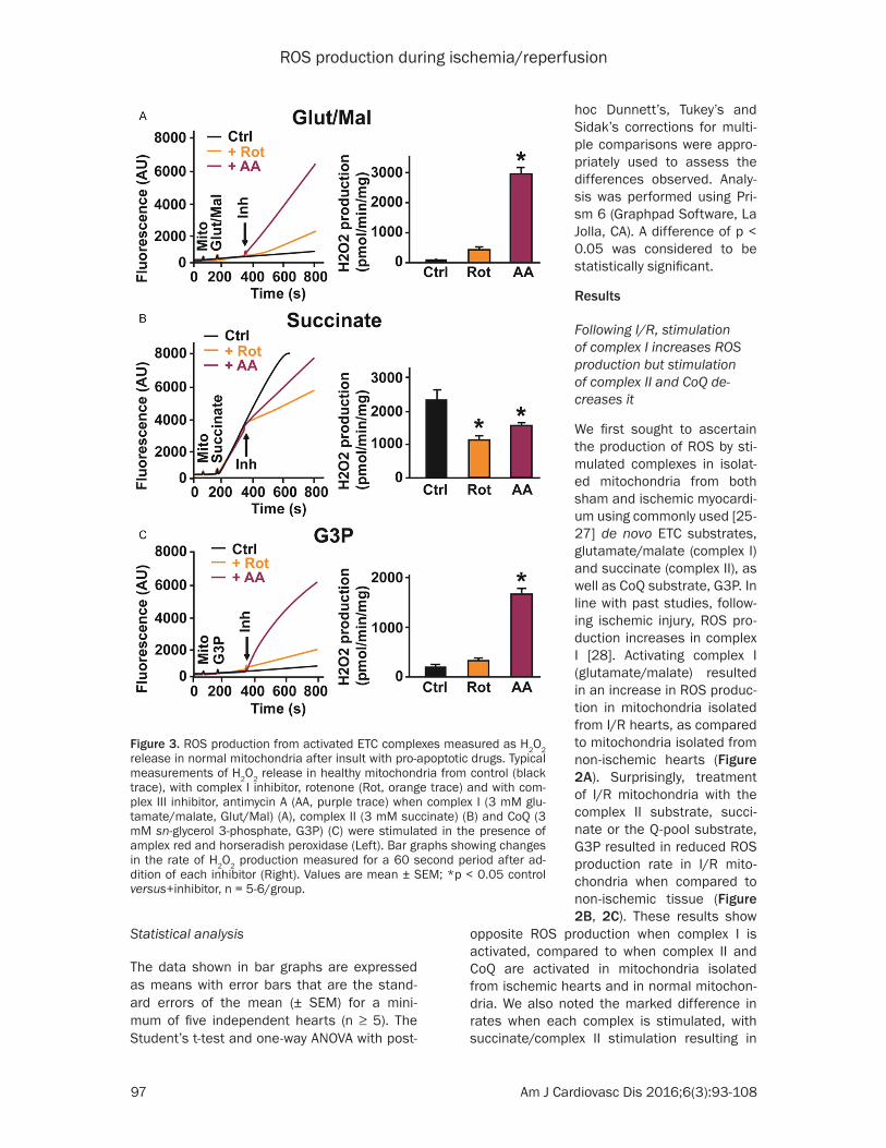
ROS production during ischemiareperfusion
105 Am J Cardiovasc Dis 20166(3)93-108
ide can directly reduce oxidative damage evoked by RET at complex I by inhibition of com-plex II However studies in H9c2 myoblasts by other investigators have suggested complex I being a source of signaling ROS for muscle dif-ferentiation [74] In our study our data agree with the Drose group in suggesting that the source of this lsquosignalingrsquo ROS in heart mito-chondria to be complex III while also highlight-ing the deleterious effects of ROS from com-plex I We extend this premise into the impact that both ETC sources have on cardiac tissue recovery following ischemic insult Despite this model offering some insights into ROS classifi-cation much is still needed to fully elucidate how cells respond to ROS from different ETC sites Multiple factors need consideration when defining such pathways including the varying needs and responses of mitochondria in differ-ent organs [25 50] and how for example in the brain glutamate is both an excitatory neu-rotransmitter and a cause of neurotoxicity [75] Understanding these mechanisms will be vital to designing targeted therapeutic agents for cardioprotection as current models of coen-zyme Q10 treatment [6] or applying indiscrimi-nant antioxidants as one-size fits all panaceas have proven to be somewhat fruitless as both good and bad ROS are destroyed [3]
In conclusion we demonstrated the opposite impact of ROS generated by stimulation of com-plexes I II and III after ischemia-reperfusion and also application of pro-apoptotic drugs in fresh mitochondria We propose an extension to the concept that the quantity of ROS is important by showing that the mitochondrial ETC source complex of that ROS is also pivotal We went on to demonstrate the positive impact of the ROS from mitochondrial complex III in cardioprotection via the inhibition of the mPTP opening and following IR reduction of myo-cardial infarct size and improvements in cardi-ac functional recovery while showing the car-dio-deleterious effects of ROS from complex I
Acknowledgements
Supported by Bopassarsquos AHA fellowship 09- POST2190008 Voelcker fund (Bopassa)
Disclosure of conflict of interest
None
Address correspondence to Jean C Bopassa De- partment of Physiology School of Medicine Uni- versity of Texas Health Science Center at San Antonio 7703 Floyd Curl Dr San Antonio TX 78- 229 USA Tel 210-567-0429 Fax 210-567-4410 E-mail bopassauthscsaedu
References
[1] Brown DI and Griendling KK Regulation of signal transduction by reactive oxygen species in the cardiovascular system Circ Res 2015 116 531-549
[2] Steinhubl SR Why have antioxidants failed in clinical trials Am J Cardiol 2008 101 14D-19D
[3] Madamanchi NR Vendrov A and Runge MS Oxidative stress and vascular disease Ar- terioscler Thromb Vasc Biol 2005 25 29-38
[4] Bjelakovic G Nikolova D Gluud LL Simonetti RG and Gluud C Mortality in randomized trials of antioxidant supplements for primary and secondary prevention systematic review and meta-analysis JAMA 2007 297 842-857
[5] Miller ER 3rd Pastor-Barriuso R Dalal D Riemersma RA Appel LJ and Guallar E Meta-analysis high-dosage vitamin E supplementa-tion may increase all-cause mortality Ann Intern Med 2005 142 37-46
[6] Pepe S Marasco SF Haas SJ Sheeran FL Krum H and Rosenfeldt FL Coenzyme Q10 in cardiovascular disease Mitochondrion 2007 7 Suppl S154-167
[7] Chandel NS and Tuveson DA The promise and perils of antioxidants for cancer patients N Engl J Med 2014 371 177-178
[8] Snow BJ Rolfe FL Lockhart MM Frampton CM OrsquoSullivan JD Fung V Smith RA Murphy MP Taylor KM and Protect Study G A double-blind placebo-controlled study to assess the mitochondria-targeted antioxidant MitoQ as a disease-modifying therapy in Parkinsonrsquos dis-ease Mov Disord 2010 25 1670-1674
[9] Smith RA and Murphy MP Animal and hu- man studies with the mitochondria-targeted antioxidant MitoQ Ann N Y Acad Sci 2010 1201 96-103
[10] Bartosz G Reactive oxygen species destroyers or messengers Biochem Pharmacol 2009 77 1303-1315
[11] Das DK Maulik N Sato M and Ray PS Reactive oxygen species function as second messenger during ischemic preconditioning of heart Mol Cell Biochem 1999 196 59-67
[12] Maslov LN Naryzhnaya NV Podoksenov YK Sementsov AS and Gorbunov AS [Signaling Mechanism of Cardioprotective Effect of Re- active Oxygen Species] Ross Fiziol Zh Im I M Sechenova 2015 101 377-385
ROS production during ischemiareperfusion
106 Am J Cardiovasc Dis 20166(3)93-108
[13] Halestrap AP Clarke SJ and Khaliulin I The role of mitochondria in protection of the heart by preconditioning Biochim Biophys Acta 2007 1767 1007-1031
[14] Zhou T Chuang CC and Zuo L Molecular Characterization of Reactive Oxygen Species in Myocardial Ischemia-Reperfusion Injury Biomed Res Int 2015 2015 864946
[15] Sun HY Wang NP Kerendi F Halkos M Kin H Guyton RA Vinten-Johansen J and Zhao ZQ Hypoxic postconditioning reduces cardiomyo-cyte loss by inhibiting ROS generation and in-tracellular Ca2+ overload Am J Physiol Heart Circ Physiol 2005 288 H1900-1908
[16] Kin H Zhao ZQ Sun HY Wang NP Corvera JS Halkos ME Kerendi F Guyton RA and Vinten-Johansen J Postconditioning attenuates myo-cardial ischemia-reperfusion injury by inhibit-ing events in the early minutes of reperfusion Cardiovasc Res 2004 62 74-85
[17] Penna C Rastaldo R Mancardi D Raimondo S Cappello S Gattullo D Losano G and Pag- liaro P Post-conditioning induced cardioprotec-tion requires signaling through a redox-sensi-tive mechanism mitochondrial ATP-sensitive K+ channel and protein kinase C activation Basic Res Cardiol 2006 101 180-189
[18] Feng Y and Bopassa JC Oxygen surrounding the heart during ischemic conservation deter-mines the myocardial injury during reperfu-sion Am J Cardiovasc Dis 2015 5 127-139
[19] Rahman S Li J Bopassa JC Umar S Iorga A Partownavid P and Eghbali M Phosphorylation of GSK-3beta mediates intralipid-induced car-dioprotection against ischemiareperfusion injury Anesthesiology 2011 115 242-253
[20] Bopassa JC Eghbali M Toro L and Stefani E A novel estrogen receptor GPER inhibits mito-chondria permeability transition pore opening and protects the heart against ischemia-reper-fusion injury Am J Physiol Heart Circ Physiol 2010 298 H16-23
[21] Bienert GP and Chaumont F Aquaporin-facilitated transmembrane diffusion of hydro-gen peroxide Biochim Biophys Acta 2014 1840 1596-1604
[22] Loschen G Azzi A Richter C and Flohe L Superoxide radicals as precursors of mito-chondrial hydrogen peroxide FEBS Lett 1974 42 68-72
[23] Bopassa JC Ferrera R Gateau-Roesch O Couture-Lepetit E and Ovize M PI 3-kinase regulates the mitochondrial transition pore in controlled reperfusion and postconditioning Cardiovasc Res 2006 69 178-185
[24] Ferrera R Bopassa JC Angoulvant D and Ovize M Post-conditioning protects from cardiople-gia and cold ischemia via inhibition of mito-
chondrial permeability transition pore J Heart Lung Transplant 2007 26 604-609
[25] Vial G Dubouchaud H Couturier K Cottet-Rousselle C Taleux N Athias A Galinier A Casteilla L and Leverve XM Effects of a high-fat diet on energy metabolism and ROS pro-duction in rat liver J Hepatol 2011 54 348-356
[26] Muller FL Song W Jang YC Liu Y Sabia M Richardson A and Van Remmen H Denervation-induced skeletal muscle atrophy is associated with increased mitochondrial ROS production Am J Physiol Regul Integr Comp Physiol 2007 293 R1159-1168
[27] Liu Y Fiskum G and Schubert D Generation of reactive oxygen species by the mitochondrial electron transport chain J Neurochem 2002 80 780-787
[28] Tahara EB Navarete FD and Kowaltowski AJ Tissue- substrate- and site-specific character-istics of mitochondrial reactive oxygen species generation Free Radic Biol Med 2009 46 1283-1297
[29] Wolvetang EJ Johnson KL Krauer K Ralph SJ and Linnane AW Mitochondrial respiratory chain inhibitors induce apoptosis FEBS Lett 1994 339 40-44
[30] Argaud L Gateau-Roesch O Muntean D Chalabreysse L Loufouat J Robert D and Ovize M Specific inhibition of the mitochon-drial permeability transition prevents lethal re-perfusion injury J Mol Cell Cardiol 2005 38 367-374
[31] Kabir ME Singh H Lu R Olde B Leeb-Lundberg LM and Bopassa JC G Protein-Coupled Estrogen Receptor 1 Mediates Acute Estrogen-Induced Cardioprotection via MEKERKGSK-3beta Pathway after IschemiaReperfusion PLoS One 2015 10 e0135988
[32] Lemasters JJ The mitochondrial permeability transition and the calcium oxygen and pH par-adoxes one paradox after another Cardiovasc Res 1999 44 470-473
[33] Tsutsui H Kinugawa S and Matsushima S Mitochondrial oxidative stress and dysfunction in myocardial remodelling Cardiovasc Res 2009 81 449-456
[34] Rolo AP and Palmeira CM Diabetes and mito-chondrial function role of hyperglycemia and oxidative stress Toxicol Appl Pharmacol 2006 212 167-178
[35] Weinberg F Hamanaka R Wheaton WW Weinberg S Joseph J Lopez M Kalyanaraman B Mutlu GM Budinger GR and Chandel NS Mitochondrial metabolism and ROS genera-tion are essential for Kras-mediated tumorige-nicity Proc Natl Acad Sci U S A 2010 107 8788-8793
ROS production during ischemiareperfusion
107 Am J Cardiovasc Dis 20166(3)93-108
[36] Chen YR and Zweier JL Cardiac mitochondria and reactive oxygen species generation Circ Res 2014 114 524-537
[37] Bopassa JC Protection of the ischemic myo-cardium during the reperfusion between hope and reality Am J Cardiovasc Dis 2012 2 223-236
[38] Chen Q Moghaddas S Hoppel CL and Les- nefsky EJ Ischemic defects in the electron transport chain increase the production of re-active oxygen species from isolated rat heart mitochondria Am J Physiol Cell Physiol 2008 294 C460-466
[39] Lambert AJ and Brand MD Superoxide produc-tion by NADHubiquinone oxidoreductase (complex I) depends on the pH gradient across the mitochondrial inner membrane Biochem J 2004 382 511-517
[40] Chance B Williams GR and Hollunger G Inhibition of electron and energy transfer in mi-tochondria I Effects of Amytal thiopental ro-tenone progesterone and methylene glycol J Biol Chem 1963 238 418-431
[41] Lambert AJ and Brand MD Inhibitors of the quinone-binding site allow rapid superoxide production from mitochondrial NADH ubiqui-none oxidoreductase (complex I) J Biol Chem 2004 279 39414-39420
[42] Quinlan CL Orr AL Perevoshchikova IV Treberg JR Ackrell BA and Brand MD Mitochondrial complex II can generate reactive oxygen spe-cies at high rates in both the forward and re-verse reactions J Biol Chem 2012 287 27255-27264
[43] Muller FL Liu Y and Van Remmen H Complex III releases superoxide to both sides of the in-ner mitochondrial membrane J Biol Chem 2004 279 49064-49073
[44] Jastroch M Divakaruni AS Mookerjee S Treberg JR and Brand MD Mitochondrial pro-ton and electron leaks Essays Biochem 2010 47 53-67
[45] Zhang DX and Gutterman DD Mitochondrial reactive oxygen species-mediated signaling in endothelial cells Am J Physiol Heart Circ Physiol 2007 292 H2023-2031
[46] Han D Antunes F Canali R Rettori D and Cadenas E Voltage-dependent anion channels control the release of the superoxide anion from mitochondria to cytosol J Biol Chem 2003 278 5557-5563
[47] St-Pierre J Buckingham JA Roebuck SJ and Brand MD Topology of superoxide production from different sites in the mitochondrial elec-tron transport chain J Biol Chem 2002 277 44784-44790
[48] Lee HL Chen CL Yeh ST Zweier JL and Chen YR Biphasic modulation of the mitochondrial electron transport chain in myocardial isch-
emia and reperfusion Am J Physiol Heart Circ Physiol 2012 302 H1410-1422
[49] Rottenberg H Covian R and Trumpower BL Membrane potential greatly enhances super-oxide generation by the cytochrome bc1 com-plex reconstituted into phospholipid vesicles J Biol Chem 2009 284 19203-19210
[50] Kwong LK and Sohal RS Substrate and site specificity of hydrogen peroxide generation in mouse mitochondria Arch Biochem Biophys 1998 350 118-126
[51] Mracek T Pecinova A Vrbacky M Drahota Z and Houstek J High efficiency of ROS pro-duction by glycerophosphate dehydrogenase in mammalian mitochondria Arch Biochem Biophys 2009 481 30-36
[52] Kalogeris T Bao Y and Korthuis RJ Mitochon- drial reactive oxygen species a double edged sword in ischemiareperfusion vs precondi-tioning Redox Biol 2014 2 702-714
[53] Victor VM and Rocha M Targeting antioxidants to mitochondria a potential new therapeutic strategy for cardiovascular diseases Curr Pharm Des 2007 13 845-863
[54] Taniyama Y and Griendling KK Reactive oxy-gen species in the vasculature molecular and cellular mechanisms Hypertension 2003 42 1075-1081
[55] Chen Q Moghaddas S Hoppel CL and Lesnefsky EJ Reversible blockade of electron transport during ischemia protects mitochon-dria and decreases myocardial injury following reperfusion J Pharmacol Exp Ther 2006 319 1405-1412
[56] Chen CH Budas GR Churchill EN Disatnik MH Hurley TD and Mochly-Rosen D Activation of aldehyde dehydrogenase-2 reduces isch-emic damage to the heart Science 2008 321 1493-1495
[57] Bernardi P and Petronilli V The permeability transition pore as a mitochondrial calcium re-lease channel a critical appraisal J Bioenerg Biomembr 1996 28 131-138
[58] Jones DP and Go YM Redox compartmental-ization and cellular stress Diabetes Obes Metab 2010 12 Suppl 2 116-125
[59] Cardoso AR Chausse B da Cunha FM Lue- vano-Martinez LA Marazzi TB Pessoa PS Queliconi BB and Kowaltowski AJ Mitochon- drial compartmentalization of redox process-es Free Radic Biol Med 2012 52 2201-2208
[60] Hansen JM Go YM and Jones DP Nuclear and mitochondrial compartmentation of oxi- dative stress and redox signaling Annu Rev Pharmacol Toxicol 2006 46 215-234
[61] Okado-Matsumoto A and Fridovich I Sub- cellular distribution of superoxide dismutases (SOD) in rat liver CuZn-SOD in mitochondria J Biol Chem 2001 276 38388-38393
ROS production during ischemiareperfusion
108 Am J Cardiovasc Dis 20166(3)93-108
[62] Drose S Differential effects of complex II on mitochondrial ROS production and their rela-tion to cardioprotective pre- and postcondition-ing Biochim Biophys Acta 2013 1827 578-587
[63] Drose S Hanley PJ and Brandt U Ambivalent effects of diazoxide on mitochondrial ROS pro-duction at respiratory chain complexes I and III Biochim Biophys Acta 2009 1790 558-565
[64] Drose S and Brandt U The mechanism of mito-chondrial superoxide production by the cyto-chrome bc1 complex J Biol Chem 2008 283 21649-21654
[65] Bernardi P Broekemeier KM and Pfeiffer DR Recent progress on regulation of the mito-chondrial permeability transition pore a cyclo-sporin-sensitive pore in the inner mitochondri-al membrane J Bioenerg Biomembr 1994 26 509-517
[66] Inoue K Zhuang L and Ganapathy V Human Na+ -coupled citrate transporter primary structure genomic organization and trans- port function Biochem Biophys Res Commun 2002 299 465-471
[67] Nakayama T Kawakami H Tanaka K and Nakamura S Expression of three glutamate transporter subtype mRNAs in human brain regions and peripheral tissues Brain Res Mol Brain Res 1996 36 189-192
[68] Hara-Chikuma M and Verkman AS Physio- logical roles of glycerol-transporting aquapo-rins the aquaglyceroporins Cell Mol Life Sci 2006 63 1386-1392
[69] Bartoloni L Wattenhofer M Kudoh J Berry A Shibuya K Kawasaki K Wang J Asakawa S Talior I Bonne-Tamir B Rossier C Michaud J McCabe ER Minoshima S Shimizu N Scott HS and Antonarakis SE Cloning and character-ization of a putative human glycerol 3-phos-phate permease gene (SLC37A1 or G3PP) on 21q223 mutation analysis in two candidate phenotypes DFNB10 and a glycerol kinase de-ficiency Genomics 2000 70 190-200
[70] Fletcher GF Ades PA Kligfield P Arena R Balady GJ Bittner VA Coke LA Fleg JL Forman DE Gerber TC Gulati M Madan K Rhodes J Thompson PD Williams MA American Heart Association Exercise CR Prevention Com- mittee of the Council on Clinical Cardiology CoNPA Metabolism CoC Stroke N Council on E and Prevention Exercise standards for test-ing and training a scientific statement from the American Heart Association Circulation 2013 128 873-934
[71] Pasdois P Parker JE Griffiths EJ and Halestrap AP Hexokinase II and reperfusion injury TAT-HK2 peptide impairs vascular function in Langendorff-perfused rat hearts Circ Res 2013 112 e3-7
[72] Cadete VJ Arcand SA Chaharyn BM Doroszko A Sawicka J Mousseau DD and Sawicki G Matrix metalloproteinase-2 is activated during ischemiareperfusion in a model of myocardial infarction Can J Cardiol 2013 29 1495-1503
[73] Chouchani ET Pell VR Gaude E Aksentijevic D Sundier SY Robb EL Logan A Nadtochiy SM Ord EN Smith AC Eyassu F Shirley R Hu CH Dare AJ James AM Rogatti S Hartley RC Eaton S Costa AS Brookes PS Davidson SM Duchen MR Saeb-Parsy K Shattock MJ Robinson AJ Work LM Frezza C Krieg T and Murphy MP Ischaemic accumulation of succi-nate controls reperfusion injury through mito-chondrial ROS Nature 2014 515 431-435
[74] Lee S Tak E Lee J Rashid MA Murphy MP Ha J and Kim SS Mitochondrial H2O2 generated from electron transport chain complex I stimu-lates muscle differentiation Cell Res 2011 21 817-834
[75] Atlante A Calissano P Bobba A Giannattasio S Marra E and Passarella S Glutamate neuro-toxicity oxidative stress and mitochondria FEBS Lett 2001 497 1-5
ROS production during ischemiareperfusion
107 Am J Cardiovasc Dis 20166(3)93-108
[36] Chen YR and Zweier JL Cardiac mitochondria and reactive oxygen species generation Circ Res 2014 114 524-537
[37] Bopassa JC Protection of the ischemic myo-cardium during the reperfusion between hope and reality Am J Cardiovasc Dis 2012 2 223-236
[38] Chen Q Moghaddas S Hoppel CL and Les- nefsky EJ Ischemic defects in the electron transport chain increase the production of re-active oxygen species from isolated rat heart mitochondria Am J Physiol Cell Physiol 2008 294 C460-466
[39] Lambert AJ and Brand MD Superoxide produc-tion by NADHubiquinone oxidoreductase (complex I) depends on the pH gradient across the mitochondrial inner membrane Biochem J 2004 382 511-517
[40] Chance B Williams GR and Hollunger G Inhibition of electron and energy transfer in mi-tochondria I Effects of Amytal thiopental ro-tenone progesterone and methylene glycol J Biol Chem 1963 238 418-431
[41] Lambert AJ and Brand MD Inhibitors of the quinone-binding site allow rapid superoxide production from mitochondrial NADH ubiqui-none oxidoreductase (complex I) J Biol Chem 2004 279 39414-39420
[42] Quinlan CL Orr AL Perevoshchikova IV Treberg JR Ackrell BA and Brand MD Mitochondrial complex II can generate reactive oxygen spe-cies at high rates in both the forward and re-verse reactions J Biol Chem 2012 287 27255-27264
[43] Muller FL Liu Y and Van Remmen H Complex III releases superoxide to both sides of the in-ner mitochondrial membrane J Biol Chem 2004 279 49064-49073
[44] Jastroch M Divakaruni AS Mookerjee S Treberg JR and Brand MD Mitochondrial pro-ton and electron leaks Essays Biochem 2010 47 53-67
[45] Zhang DX and Gutterman DD Mitochondrial reactive oxygen species-mediated signaling in endothelial cells Am J Physiol Heart Circ Physiol 2007 292 H2023-2031
[46] Han D Antunes F Canali R Rettori D and Cadenas E Voltage-dependent anion channels control the release of the superoxide anion from mitochondria to cytosol J Biol Chem 2003 278 5557-5563
[47] St-Pierre J Buckingham JA Roebuck SJ and Brand MD Topology of superoxide production from different sites in the mitochondrial elec-tron transport chain J Biol Chem 2002 277 44784-44790
[48] Lee HL Chen CL Yeh ST Zweier JL and Chen YR Biphasic modulation of the mitochondrial electron transport chain in myocardial isch-
emia and reperfusion Am J Physiol Heart Circ Physiol 2012 302 H1410-1422
[49] Rottenberg H Covian R and Trumpower BL Membrane potential greatly enhances super-oxide generation by the cytochrome bc1 com-plex reconstituted into phospholipid vesicles J Biol Chem 2009 284 19203-19210
[50] Kwong LK and Sohal RS Substrate and site specificity of hydrogen peroxide generation in mouse mitochondria Arch Biochem Biophys 1998 350 118-126
[51] Mracek T Pecinova A Vrbacky M Drahota Z and Houstek J High efficiency of ROS pro-duction by glycerophosphate dehydrogenase in mammalian mitochondria Arch Biochem Biophys 2009 481 30-36
[52] Kalogeris T Bao Y and Korthuis RJ Mitochon- drial reactive oxygen species a double edged sword in ischemiareperfusion vs precondi-tioning Redox Biol 2014 2 702-714
[53] Victor VM and Rocha M Targeting antioxidants to mitochondria a potential new therapeutic strategy for cardiovascular diseases Curr Pharm Des 2007 13 845-863
[54] Taniyama Y and Griendling KK Reactive oxy-gen species in the vasculature molecular and cellular mechanisms Hypertension 2003 42 1075-1081
[55] Chen Q Moghaddas S Hoppel CL and Lesnefsky EJ Reversible blockade of electron transport during ischemia protects mitochon-dria and decreases myocardial injury following reperfusion J Pharmacol Exp Ther 2006 319 1405-1412
[56] Chen CH Budas GR Churchill EN Disatnik MH Hurley TD and Mochly-Rosen D Activation of aldehyde dehydrogenase-2 reduces isch-emic damage to the heart Science 2008 321 1493-1495
[57] Bernardi P and Petronilli V The permeability transition pore as a mitochondrial calcium re-lease channel a critical appraisal J Bioenerg Biomembr 1996 28 131-138
[58] Jones DP and Go YM Redox compartmental-ization and cellular stress Diabetes Obes Metab 2010 12 Suppl 2 116-125
[59] Cardoso AR Chausse B da Cunha FM Lue- vano-Martinez LA Marazzi TB Pessoa PS Queliconi BB and Kowaltowski AJ Mitochon- drial compartmentalization of redox process-es Free Radic Biol Med 2012 52 2201-2208
[60] Hansen JM Go YM and Jones DP Nuclear and mitochondrial compartmentation of oxi- dative stress and redox signaling Annu Rev Pharmacol Toxicol 2006 46 215-234
[61] Okado-Matsumoto A and Fridovich I Sub- cellular distribution of superoxide dismutases (SOD) in rat liver CuZn-SOD in mitochondria J Biol Chem 2001 276 38388-38393
ROS production during ischemiareperfusion
108 Am J Cardiovasc Dis 20166(3)93-108
[62] Drose S Differential effects of complex II on mitochondrial ROS production and their rela-tion to cardioprotective pre- and postcondition-ing Biochim Biophys Acta 2013 1827 578-587
[63] Drose S Hanley PJ and Brandt U Ambivalent effects of diazoxide on mitochondrial ROS pro-duction at respiratory chain complexes I and III Biochim Biophys Acta 2009 1790 558-565
[64] Drose S and Brandt U The mechanism of mito-chondrial superoxide production by the cyto-chrome bc1 complex J Biol Chem 2008 283 21649-21654
[65] Bernardi P Broekemeier KM and Pfeiffer DR Recent progress on regulation of the mito-chondrial permeability transition pore a cyclo-sporin-sensitive pore in the inner mitochondri-al membrane J Bioenerg Biomembr 1994 26 509-517
[66] Inoue K Zhuang L and Ganapathy V Human Na+ -coupled citrate transporter primary structure genomic organization and trans- port function Biochem Biophys Res Commun 2002 299 465-471
[67] Nakayama T Kawakami H Tanaka K and Nakamura S Expression of three glutamate transporter subtype mRNAs in human brain regions and peripheral tissues Brain Res Mol Brain Res 1996 36 189-192
[68] Hara-Chikuma M and Verkman AS Physio- logical roles of glycerol-transporting aquapo-rins the aquaglyceroporins Cell Mol Life Sci 2006 63 1386-1392
[69] Bartoloni L Wattenhofer M Kudoh J Berry A Shibuya K Kawasaki K Wang J Asakawa S Talior I Bonne-Tamir B Rossier C Michaud J McCabe ER Minoshima S Shimizu N Scott HS and Antonarakis SE Cloning and character-ization of a putative human glycerol 3-phos-phate permease gene (SLC37A1 or G3PP) on 21q223 mutation analysis in two candidate phenotypes DFNB10 and a glycerol kinase de-ficiency Genomics 2000 70 190-200
[70] Fletcher GF Ades PA Kligfield P Arena R Balady GJ Bittner VA Coke LA Fleg JL Forman DE Gerber TC Gulati M Madan K Rhodes J Thompson PD Williams MA American Heart Association Exercise CR Prevention Com- mittee of the Council on Clinical Cardiology CoNPA Metabolism CoC Stroke N Council on E and Prevention Exercise standards for test-ing and training a scientific statement from the American Heart Association Circulation 2013 128 873-934
[71] Pasdois P Parker JE Griffiths EJ and Halestrap AP Hexokinase II and reperfusion injury TAT-HK2 peptide impairs vascular function in Langendorff-perfused rat hearts Circ Res 2013 112 e3-7
[72] Cadete VJ Arcand SA Chaharyn BM Doroszko A Sawicka J Mousseau DD and Sawicki G Matrix metalloproteinase-2 is activated during ischemiareperfusion in a model of myocardial infarction Can J Cardiol 2013 29 1495-1503
[73] Chouchani ET Pell VR Gaude E Aksentijevic D Sundier SY Robb EL Logan A Nadtochiy SM Ord EN Smith AC Eyassu F Shirley R Hu CH Dare AJ James AM Rogatti S Hartley RC Eaton S Costa AS Brookes PS Davidson SM Duchen MR Saeb-Parsy K Shattock MJ Robinson AJ Work LM Frezza C Krieg T and Murphy MP Ischaemic accumulation of succi-nate controls reperfusion injury through mito-chondrial ROS Nature 2014 515 431-435
[74] Lee S Tak E Lee J Rashid MA Murphy MP Ha J and Kim SS Mitochondrial H2O2 generated from electron transport chain complex I stimu-lates muscle differentiation Cell Res 2011 21 817-834
[75] Atlante A Calissano P Bobba A Giannattasio S Marra E and Passarella S Glutamate neuro-toxicity oxidative stress and mitochondria FEBS Lett 2001 497 1-5