ros-dependent apoptosis by gugulipid extract of ayurvedic
TRANSCRIPT
MOL #68551
1
ROS-dependent apoptosis by gugulipid extract of Ayurvedic medicine plant
Commiphora mukul in human prostate cancer cells is regulated by c-JUN N-
terminal kinase*
Dong Xiao, Yan Zeng, Lakshmi Prakash, Vladmir Badmaev, Muhammed Majeed and
Shivendra V. Singh
Department of Pharmacology & Chemical Biology, University of Pittsburgh School of Medicine
(D.X., S.V.S.) and University of Pittsburgh Cancer Institute (D.X., S.V.S., Y.Z.), Pittsburgh, PA.
Sabinsa Corporation (L.P., V.B., M.M.), Piscataway, NJ.
Molecular Pharmacology Fast Forward. Published on November 29, 2010 as doi:10.1124/mol.110.068551
Copyright 2010 by the American Society for Pharmacology and Experimental Therapeutics.
This article has not been copyedited and formatted. The final version may differ from this version.Molecular Pharmacology Fast Forward. Published on November 29, 2010 as DOI: 10.1124/mol.110.068551
at ASPE
T Journals on M
arch 14, 2022m
olpharm.aspetjournals.org
Dow
nloaded from

MOL #68551
2
Running head: JNK activation in gugulipid-induced apoptosis
Correspondence author: Dr. Dong Xiao, 2.32b Hillman Cancer Center Research Pavilion, 5117
Centre Avenue, Pittsburgh, PA 15213. Phone: 412-623-3262; Fax: 412-623-7828; E-mail:
The number of text pages: 29
The number of tables: 0
The number of figures: 6
The number of references: 40
The number of words in the abstract: 239
The number of words in the introduction: 599
The number of words in the discussion: 1083
ABBREVIATIONS: CDCFDA, (5-(and-6)-carboxy-2',7'-dichloroluorescein diacetate,
succinimidyl ester; HE, hydroethidine; PARP, poly-(ADP-ribose)-polymerase; DMSO, dimethyl
sulfoxide; PBS, phosphate buffered saline; BSA, bovine serum albumin; DCF, 2',7'-
dichlorodihydrofluorescein; JNK, c-Jun N-terminal kinase; MAPK, mitogen-activated protein
kinase; JNKK2, JNK kinase 2; PrEC, normal human prostate epithelial cell line.
This article has not been copyedited and formatted. The final version may differ from this version.Molecular Pharmacology Fast Forward. Published on November 29, 2010 as DOI: 10.1124/mol.110.068551
at ASPE
T Journals on M
arch 14, 2022m
olpharm.aspetjournals.org
Dow
nloaded from

MOL #68551
3
Abstract
Gugulipid (GL), extract of Indian Ayurvedic medicinal plant Commiphora mukul, has been used
to treat a variety of ailments. We now report anti-cancer effect and mechanism of GL against
human prostate cancer cells. Treatment with GL significantly inhibited viability of human
prostate cancer cell line LNCaP (androgen-dependent) and its androgen-independent variant
(C81) with an IC50 ~ 1 µM (24 h treatment) at pharmacologically relevant concentrations
standardized to its major active constituent z-guggulsterone. The GL-induced growth inhibition
correlated with apoptosis induction as evidenced by an increase in cytoplasmic histone-
associated DNA fragmentation and sub-G0/G1DNA fraction, and cleavage of poly-(ADP-ribose)-
polymerase (PARP). The GL-induced apoptosis was associated with reactive oxygen species
(ROS) production and c-Jun NH2-terminal kinase (JNK) activation. The induction of
proapoptotic Bcl-2 family proteins Bax and Bak and a decrease of antiapoptotic Bcl-2 protein
Bcl-2 were observed in GL-treated cells. SV40 immortalized mouse embryonic fibroblasts
derived from Bax-Bak double knockout mice were significantly more resistant to GL-induced
cell killing compared with wild type cells. It is interesting to note that a representative normal
prostate epithelial cell line PrEC was relatively more resistant to GL-mediated cellular responses
compared with prostate cancer cells. The GL treatment caused activation of JNK that functioned
upstream of Bax activation in apoptosis response. The GL-induced conformational change of
Bax and apoptosis were significantly suppressed by genetic suppression of JNK activation. In
conclusion, the present study indicates that ROS-dependent apoptosis by GL is regulated by JNK
signaling axis.
This article has not been copyedited and formatted. The final version may differ from this version.Molecular Pharmacology Fast Forward. Published on November 29, 2010 as DOI: 10.1124/mol.110.068551
at ASPE
T Journals on M
arch 14, 2022m
olpharm.aspetjournals.org
Dow
nloaded from

MOL #68551
4
Introduction
Novel strategies for prevention of prostate cancer are highly desirable because prostate
cancer continues to be leading cause of cancer-related deaths among men in the United States
(Jemal et al., 2009; Whittemore et al., 1995). Prostate cancer is usually diagnosed in the sixth or
seventh decades of life, which allows a large window of opportunity for intervention to prevent
or slow progression of the disease (Whittemore et al., 1995; Gilligan and Kantoff, 2002). Thus,
clinical development of agents from natural products that are relatively safe but can delay onset
and/or progression of human prostate cancer is highly desirable.
Gugulipid (GL), extract of Commiphora mukul, has been safely used in the Indian
Ayurvedic medicine practice for treatment of different ailments (Shishodia et al, 2008; Urizar
and Moore, 2003; Badmaev et al., 2003). Several products of standardized formulations of
Commiphora mukul are already in human use as cholesterol-lowering agents (Shishodia et al,
2008; Urizar and Moore, 2003; Badmaev et al., 2003). The z- and E-forms of guggulsterone
(4,17(20)-pregnadie-3, 16-dione) have been identified as major active components of GL, which
has been used in many clinical trials that have focused on its cholesterol-lowering effect
(Shishodia et al, 2008; Urizar and Moore, 2003; Badmaev et al., 2003). Although the antitumor
activity of GL has not been studied yet, the studies, including ours, have shown that z-
guggulsterone (z-Gug) inhibits proliferation, induces apoptosis; and suppresses angiogenesis as
well as the invasion and metastasis of cancer cells (Shishodia et al, 2008; Urizar and Moore,
2003; Badmaev et al., 2003; Gujral et al., 1960; Sinal and Gonzalez, 2002; Shishodia and
Aggarwal, 2004; Samudio et al., 2005; Ichikawa and Aggarwal, 2006; Cheon et al., 2006; Xiao
and Singh, 2008; Singh et al., 2005a, 2007; Urizar et al., 2002; Cui et al., 2003; Wu et al, 2002).
Apoptosis induction by Gug has been reported in leukemia, multiple myeloma, melanoma, head
This article has not been copyedited and formatted. The final version may differ from this version.Molecular Pharmacology Fast Forward. Published on November 29, 2010 as DOI: 10.1124/mol.110.068551
at ASPE
T Journals on M
arch 14, 2022m
olpharm.aspetjournals.org
Dow
nloaded from
MOL #68551
5
and neck, lung, ovarian, prostate and breast cancer cells (Sinal and Gonzalez, 2002; Shishodia
and Aggarwal, 2004; Samudio et al., 2005; Ichikawa and Aggarwal, 2006; Cheon et al., 2006;
Xiao and Singh, 2008; Singh et al., 2005a, 2007; Urizar et al., 2002; Cui et al., 2003; Wu et al,
2002). We have shown previously that z- and E-Gug inhibit growth of PC-3, DU145 and LNCaP
human prostate cancer cells in culture by causing apoptosis (Singh et al., 2005a, 2007).
Interestingly, a normal prostate epithelial cell line (PrEC) is significantly more resistant to
growth inhibition and apoptosis induction by z-Gug compared with prostate cancer cells (Singh
et al., 2005a, 2007). The z-Gug-induced cell death in PC-3 cells was not influenced by Bcl-2
protein level but correlated with induction of proapoptotic multidomain Bcl-2 family members
Bax and Bak and activation of caspases (Singh et al., 2005a). The z-Gug-induced apoptosis in
human prostate cancer cells was initiated by reactive oxygen intermediate-mediated activation of
c-Jun NH2-terminal kinase (Singh et al., 2007). Our previous study demonstrated that z-Gug and
E-Gug inhibit angiogenic features (capillary-like tube formation and/or migration) of human
umbilical vein endothelial cells (HUVEC) and DU145 human prostate cancer cells in vitro at
pharmacologically relevant concentrations (Xiao and Singh, 2008). Furthermore, oral gavage of
3 µmol z-Gug to male nude mice (five times per week) inhibits in vivo angiogenesis (Xiao and
Singh, 2008).
Based on these data, we hypothesized that GL might be more effective apoptosis induced in
prostate cancer cells because it contains a number of steroids, including the two isomers z- and
E-Gugs (Shishodia et al, 2008; Urizar and Moore, 2003; Badmaev et al., 2003). In present
studies, we tested this hypothesis by examining the effect of GL standardized to z-Gug.
This article has not been copyedited and formatted. The final version may differ from this version.Molecular Pharmacology Fast Forward. Published on November 29, 2010 as DOI: 10.1124/mol.110.068551
at ASPE
T Journals on M
arch 14, 2022m
olpharm.aspetjournals.org
Dow
nloaded from
MOL #68551
6
Materials and Methods
Reagents. GL, derived from the gum guggul resin (gum guggul) produced in the soft bark
ducts of the Commiphora mukul tree, is a registered product of Sabinsa Corporation (Registration
date: July 21, 1992; US Patent# 6436991 B1). A manufacturing flow chart for gum guggul resin
to GL was described by us previously (Badmaev et al., 2003). Standardization of GL was
performed by high-performance liquid chromatography (HPLC) and found to contain ~3.75% z-
Gug (Badmaev et al., 2003). The GL was stored at 40C and found to be stable for at least 6
months. The z-Gug was from Steraloids (Newport, RI). Reagents for cell culture including
medium, penicillin and streptomycin antibiotic mixture, and fetal bovine serum were purchased
from Invitrogen (Carlsbad, CA). The hydroethidine (HE) and (5-(and-6)-carboxy-2',7'-
dichloroluorescein diacetate, succinimidyl ester (CDCFDA) were from Molecular Probes
(Eugene, OR). The ELISA kit for quantitation of cytoplasmic histone-associated DNA
fragmentation was from Roche Diagnostics (Mannheim, Germany). The p38 MAPK and
p44/p42 MAPK (Erk1/2)-targeted small interfering RNA (siRNA) were from Cell Signaling
Technology (Danvers, MA). The anti-Bax (6A7) monoclonal antibody was from Pharmingen
(Palo Alto, CA), antibodies against Bax (polyclonal anti-Bax), Akt, phosphor-Akt and phosphor-
mTOR were from Cell Signaling Technology, the antibody against α-Tubulin was from Sigma,
the antibodies specific for detection of poly-(ADP-ribose)-polymerase (PARP), Bak, total JNK,
phospho-(Thr183/Tyr185)-JNK, total p38 MAPK, phospho-(Tyr182)-p38 MAPK, extracellular-
signal-related kinase 1/2 (ERK1/2), phospho-ERK1/2 and phospho-(Ser63/730)-c-Jun were from
Santa Cruz Biotechnology (Santa Cruz, CA), the anti-Bcl-2 antibody was from
DAKOCytomation (Carpinteria, CA), and anti-actin antibody was from Oncogene Research
This article has not been copyedited and formatted. The final version may differ from this version.Molecular Pharmacology Fast Forward. Published on November 29, 2010 as DOI: 10.1124/mol.110.068551
at ASPE
T Journals on M
arch 14, 2022m
olpharm.aspetjournals.org
Dow
nloaded from
MOL #68551
7
Products (San Diego, CA). N-acetyl-L-cysteine (NAC) was obtained from Sigma-Aldrich (St.
Louis, MO).
Cell culture and cell survival assays. Monolayer cultures of LNCaP and C81 cells were
maintained in RPMI1640 medium supplemented with 10% (v/v) FBS, 10 mM HEPES, 1 mM
sodium pyruvate, 0.2% glucose and antibiotics. Normal prostate epithelial cell line PrEC
(Clonetics, San Diego, CA) was maintained in PrEBM (Cambrex, Walkersville, MD). The MEFs
derived from wild type, and Bax-Bak double knockout (DKO) mice and immortalized by
transfection with a plasmid containing SV40 genomic DNA were generously provided by the
late Dr. Stanley Korsmeyer (Dana-Farber Cancer Institute, Boston, MA), and maintained as
described by us previously (Singh et al., 2005a). Each cell line was maintained in an atmosphere
of 95% air and 5% CO2 at 370C. The effect of GL and z-Gug on cell viability was determined by
(a) colonogenic survival assay and (b) trypan blue dye exclusion assays as described by us
previously (Xiao and Singh, 2007; Kim et al., 2007; Xiao et al., 2006a, 2008). For the
colonogenic survival assay, cells (1.5 × 10⁵) were plated in 6-well-plates for incubation
overnight and then were treated with 0.1 % DMSO (control group) or 1, 2.5 and 5 µM GL for 24
h. The treated cells were re-seeded in 6-well plates (500 cells/well) in complete medium without
drug. The medium were changed every two days. After culture 10 days, the cells were fixed and
stained with 0.5 % crystal violet in 20% MeOH for colony counting.
Detection of apoptosis. Apoptosis induction was assessed by (a) analysis of cytoplasmic
histone-associated DNA fragmentation, (b) flow cytometric analysis of cells with sub-G0/G1
DNA content following staining with propidium iodide and (c) immunoblotting analysis of
cleavage of PARP as described by us previously (Kim et al., 2007; Xiao et al., 2006a).
This article has not been copyedited and formatted. The final version may differ from this version.Molecular Pharmacology Fast Forward. Published on November 29, 2010 as DOI: 10.1124/mol.110.068551
at ASPE
T Journals on M
arch 14, 2022m
olpharm.aspetjournals.org
Dow
nloaded from

MOL #68551
8
Immunoblotting. The cells were treated with GL and were lysed as described by us
previously (Xiao and Singh, 2008; Singh et al., 2007). The lysate proteins were resolved by 6-
12.5% sodium dodecyl sulfate polyacrylamide gel electrophoresis and transferred onto
membrane. Immunoblotting was performed as described by us previously (Xiao and Singh,
2008; Singh et al., 2007). The blots were stripped and re-probed with anti-actin antibody to
correct for differences in protein loading. Change in protein level was determined by
densitometric scanning of the immunoreactive band and corrected for actin loading control.
Immunoblotting for each protein was performed at least twice using independently prepared
lysates to ensure reproducibility of the results.
ROS generation assay. Intracellular ROS generation was measured by flow cytometry
following staining with HE and CDCFDA essentially as described by us previously (Xiao et al.,
2008). Briefly, 2×10⁵ cells were plated in 60-mm culture dishes, allowed to attach by overnight
incubation, and exposed to DMSO (control) or desired concentration of GL for specified time
intervals. The cells were stained with 2 μM HE and 5 µM CDCFDA for 30 min at 37°C. The
cells were collected and the fluorescence was measured using a Coulter Epics XL Flow
Cytometer. In some experiments, cells were pretreated for specified time periods with NAC (10
mM) prior to GL exposure and analysis of ROS generation.
Genetic suppression of JNK in LNCaP cells. The LNCaP cells were transiently transfected
with the plasmid encoding for catalytically inactive mutant of JNK kinase 2 [JNKK2(AA)], a
generous gift from Dr. Michael Karin (University of California at San Diego, La Jolla, CA), or
empty pcDNA3.1 vector as described by us previously (Xiao et al., 2008). The cells were then
treated with DMSO (control) or 5 µM GL for specified time periods and processed for analysis
This article has not been copyedited and formatted. The final version may differ from this version.Molecular Pharmacology Fast Forward. Published on November 29, 2010 as DOI: 10.1124/mol.110.068551
at ASPE
T Journals on M
arch 14, 2022m
olpharm.aspetjournals.org
Dow
nloaded from

MOL #68551
9
of DNA fragmentation, immunoblotting for phospho-JNK and phospho-c-Jun or conformational
change of Bax.
Analysis of Bax conformation change. The cells were treated with 5 μM GL or DMSO
(control) for specified time interval, and lysed using a solution containing 10 mM HEPES (pH
7.4), 150 mM NaCl, 1% CHAPS, and protease inhibitor cocktail. Aliquots containing 200 μg
lysate proteins were incubated overnight at 4°C with 4 μg of anti-Bax 6A7 monoclonal antibody.
Protein G-agarose beads (50 μl; Santa Cruz Biotechnology) were then added to each sample and
the incubation was continued for an additional 2 h at 4°C. The immunoprecipitates were washed
five times with lysis buffer and subjected to electrophoresis followed by immunoblotting using
polyclonal anti-Bax antibody.
RNA interference of p38 MAPK and Erk1/2. The cells (1×10⁵) were seeded in six-well
plates and allowed to attach by overnight incubation. The cells were transfected with 200 nmol/L
of control non-specific siRNA or p38 MAPK or Erk1/2-targeted siRNA using Oligofectamine
(Invitrogen) according to the manufacturer's recommendations. Twenty-four hours after
transfection, the cells were treated with DMSO (control) or 5 µmol/L GL for specified time
period. The cells were collected, washed with phosphate-buffered saline (PBS), and processed
for immunoblotting or analysis of cytoplasmic histone-associated DNA fragmentation as
described by us previously (Xiao and Singh, 2010; 2007; Xiao et al., 2008).
Statistical analysis. Statistical significance of difference in measured variables between
control and treated groups was determined by t-test or one-way ANOVA. Difference was
considered significant at P<0.05.
This article has not been copyedited and formatted. The final version may differ from this version.Molecular Pharmacology Fast Forward. Published on November 29, 2010 as DOI: 10.1124/mol.110.068551
at ASPE
T Journals on M
arch 14, 2022m
olpharm.aspetjournals.org
Dow
nloaded from

MOL #68551
10
Results
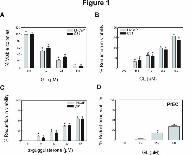
GL inhibited viability of human prostate cancer cells. The effect of GL standardized to
z-Gug on cell viability was determined by the colonogenic assay. By following the colony
formation assaying procedure, the cells were cultured for 10 days after 24 h exposure to GL and
the colony formation (>50 cells/colony) were determined. The viability of both LNCaP and its
androgen-independent variant C81 (Fig. 1A) was decreased significantly in a concentration-
dependent manner with an IC50 of GL ~1 μM, which is at pharmacologically achievable
concentrations (~3 µM, Verma et al., 1999). Growth inhibitory effect of GL was confirmed by
trypan blue dye exclusion assay. Treatment with GL for 24 h resulted in a significant reduction
in cell viability in both cells (Fig. 1B). Even though viability of LNCaP and C81 cells was also
decreased in the presence of z-Gug (Fig. 1C), the GL appeared relatively more effective
compared with z-Gug against both cell lines. Growth inhibitory effect of GL to the cancer cells
was ~10-fold stronger compared with z-Gug (Fig. 1). The results indicate that the anti-cancer
effect of GL against prostate cancer cells is most likely attributable to z-Gug as well as other
constituent(s). Interestingly, a normal prostate epithelial cell line (PrEC) was significantly more
resistant to growth inhibition by GL compared with prostate cancer cells (Fig. 1D). For instance,
2.5 μM GL, which inhibited viability of LNCaP and C-81 cells by about 50% (Fig. 1B), had
minimal effect on PrEC cell viability (Fig. 1D). These data indicated that human prostate cancer
cells, but not normal prostate epithelial cell PrEC, were sensitive to inhibition of cell viability by
GL. Because the LNCaP and C81 cells exhibited comparable sensitivity, we can also conclude
that androgen-responsiveness is not a critical factor in GL-mediated growth inhibition in prostate
cancer cells.
This article has not been copyedited and formatted. The final version may differ from this version.Molecular Pharmacology Fast Forward. Published on November 29, 2010 as DOI: 10.1124/mol.110.068551
at ASPE
T Journals on M
arch 14, 2022m
olpharm.aspetjournals.org
Dow
nloaded from
MOL #68551
11
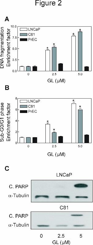
GL-mediated suppression of cancer cell growth correlated with apoptotic DNA
fragmentation. To gain further insights into the mechanism of GL-mediated inhibition of
prostate cancer cell growth, we determined its effect on cytoplasmic histone-associated DNA
fragmentation, a widely used technique for detection of apoptosis. The GL treatment resulted in a
dose-dependent increase in cytoplasmic histone-associated DNA fragmentation in both LNCaP
and C81 cells (Fig. 2A). Consistent with cell viability data (Fig. 1D), the PrEC cells were
resistant to GL-induced cytoplasmic histone associated DNA fragmentation (2A). To conform
the results of GL-induced apoptotic cell death, we further investigated whether GL treatment
increased sub-G0/G1 DNA content. A dose-dependent increase in the proportion of cells with
sub-G0/G1 content was observed in GL-treated LNCaP and C81 cells, but not in PrEC, compared
with DMSO-treated control (Fig. 2B). Furthermore, an immunoreactive band corresponding to
cleaved PARP was observed in both of cancer cells following treatment with GL (Fig. 2C).
Taken together, these observations clearly indicated that antiproliferative effect of GL against
prostate cancer cells was associated with apoptosis induction and this effect was selective for
prostate cancer cells.
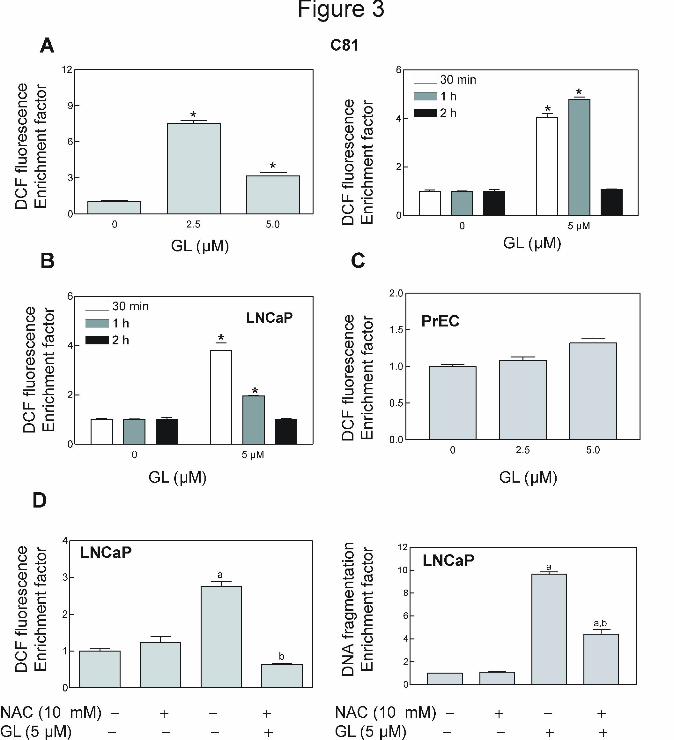
GL treatment caused ROS production in prostate cancer cells but not in a normal
prostate epithelial cell PrEC. Our previous studies have shown that many natural products,
such as, phenethyl isothiocyanate (Xiao and Singh, 2010; Xiao et al., 2006b), benzyl
isothiocyanate (Xiao et al., 2006c, 2008), sulforaphane (Xiao et al, 2009; Singh et al., 2005a),
diallyl trisulfide (Xiao et al., 2005b), and z-Gug (Singh et al., 2007), cause apoptosis through
mediation of ROS. We tested whether GL-induced apoptosis was ROS-dependent using flow
cytometry following staining with HE and CDCFDA. As can be seen in Figure 3A, GL-treated
C81 cells exhibited a dose- and time-dependent increase in mean DCF fluorescence compared
This article has not been copyedited and formatted. The final version may differ from this version.Molecular Pharmacology Fast Forward. Published on November 29, 2010 as DOI: 10.1124/mol.110.068551
at ASPE
T Journals on M
arch 14, 2022m
olpharm.aspetjournals.org
Dow
nloaded from

MOL #68551
12
with DMSO-treated (control) cells. For example, the DCF fluorescence in DCF in C81 cells
treated for 30 min with 2.5 and 5 µM GL was increased by about 7.5- and 3.2-fold compared
with control group (Fig. 3A). In time-course experiments, the ROS production was observed as
early as 30 min and peaked between 1 h and 2 h post-exposure (Fig.3A). The LNCaP cells had
almost a same response to GL treatment (Fig. 3B). It is important note that ROS generation was
not determined in the normal prostate epithelial cell PrEC treated with (Fig.3C). These
observations clearly indicated that GL treatment resulted in ROS production selectively in
human prostate cancer cells.
NAC, an antioxidant, attenuated GL-induced ROS production and apoptosis in
prostate cancer cells. Next, we designed experiments to determine whether GL-induced ROS
generation and apoptotic cell death were attenuated by NAC, an antioxidant. The present results
showed that pretreatment with NAC conferred significant protection against GL-induced ROS
production and apoptosis in LNCaP cells (Fig. 3D).
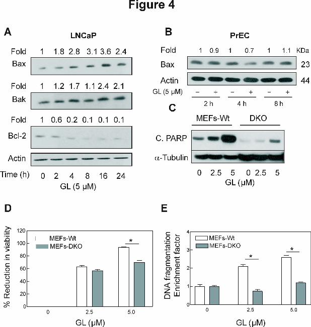
Effect of GL treatment on levels of Bcl-2 family proteins. The Bcl-2 family proteins have
emerged as critical regulators of mitochondria-mediated apoptosis by functioning as either
promoters (e.g., Bax and Bak) or inhibitors (e.g., Bcl-2 and Bcl-xL) of the cell death process
(Chao and Korsmeyer, 1998; Xiao et al., 2004, 2005c). We proceeded to test whether GL-
induced apoptosis was regulated by Bcl-2 family proteins. The effect of GL treatment on levels
of Bcl-2 family proteins in LNCaP cells was determined by immunoblotting and representative
blots are shown in Figure 4A. The levels of multidomain proapoptotic proteins Bax and Bak
were increased on treatment of LNCaP cells with GL. For example, GL-treated cells for 2-24 h
resulted in an increase of 2-4 folds for Bax and about 2 folds for Bak protein expression (Fig,
4A). In addition, the level of antiapoptotic proteins Bcl-2 was significantly decreased on
This article has not been copyedited and formatted. The final version may differ from this version.Molecular Pharmacology Fast Forward. Published on November 29, 2010 as DOI: 10.1124/mol.110.068551
at ASPE
T Journals on M
arch 14, 2022m
olpharm.aspetjournals.org
Dow
nloaded from
MOL #68551
13
treatment of LNCaP cells with GL (Fig. 4A). However, expression of Bax proteins was not
altered by same treatment of GL in human normal prostate epithelial cell PrEC (Fig. 4B). These
results indicated that GL treatment altered ratio of proapoptotic to antiapoptotic Bcl-2 family
proteins in LNCaP cells.
Bak and Bax deficiency conferred significant protection against GL-induced
apoptosis. Because GL treatment increased the Bax and Bak levels in the cancer cells (Fig. 4A),
we hypothesized that these proteins might play an important role in the regulation of GL-induced
apoptosis. The SV40-immortalized MEFs derived from WT and DKO mice were selected to test
the hypothesis. Because of immortalization by transfection with SV40 genomic DNA, the MEFs
cannot be regarded as normal fibroblasts. Initially, we used the MEFs from WT and DKO mice
to determine the effect of GL treatment on the cleavage of PARP. Similar to LNCaP cells, GL
treatment caused a significant increase in the cleavage of PARP in WT MEFs, but much less in
the DKO MEFs (Fig. 4C). The GL treatment caused concentration-dependent and statistically
significant inhibition of cell growth, and increase in apoptotic cells in WT MEFs as judged by
trypan blue dye exclusion assays (Fig. 4D) and the analysis of cytoplasmic histone-associated
DNA fragmentation (Fig. 4E ), respectively. On the other hand, the MEFs derived from DKO
MEFs mice were significantly more resistant to GL-induced growth inhibition and cytoplasmic
histone-associated DNA fragmentation compared with WT MEFs (Fig. 4C-E). Collectively,
these results indicated that Bax and Bak play an important role in the execution of GL-induced
apoptosis.
GL activated JNKs in human prostate cancer cells but not in PrEC. We, and others,
have demonstrated previously that z-Gug-induced apoptosis is regulated by JNKs (Singh et al.,
2007; Sarfaraz et al., 2008). However, role of JNK as well as other MAPK kinases, such as p38-
This article has not been copyedited and formatted. The final version may differ from this version.Molecular Pharmacology Fast Forward. Published on November 29, 2010 as DOI: 10.1124/mol.110.068551
at ASPE
T Journals on M
arch 14, 2022m
olpharm.aspetjournals.org
Dow
nloaded from

MOL #68551
14
MAPK and ERK in GL-induced apoptosis has not been studied. To elucidate the mechanism of
GL-induced apoptosis in human prostate cancer cells, we investigated its effect on MAPKs. The
LNCaP (Fig 5A) and C81 cells (results not shown) with 5 µM GL exhibited a rapid but sustained
activation of JNK for at least 8 h. JNK phosphorylation in GL-treated LNCaP cells could be
detected as early as 2 h (1.4 fold compared the control cells) after treatment, which was not
attributable to an increase in total JNK protein level (data not shown). However, the GL
treatment did not activate p38 MAPK and ERK kinases (Fig. 5A) or affected their total protein
level (data not shown). In addition, 5 µM z-Gug treatment was not found to activate JNK in
LNCaP (Fig. 5A) or C81 (data not shown) cells. We raised the question of whether GL-mediated
JNK activation was selective for cancer cells. In contrast to prostate cancer cells, GL did not
result in the activation of JNK in normal human prostate epithelial cell PrEC (Fig. 5B). The NAC
significantly reduced the phospho-c-Jun (Fig. 5C) protein expression by GL in LNCaP cells.
These results indicated that JNK activation mediated by ROS may be involved in GL-induced
apoptosis that seemed selective towards prostate cancer cells.
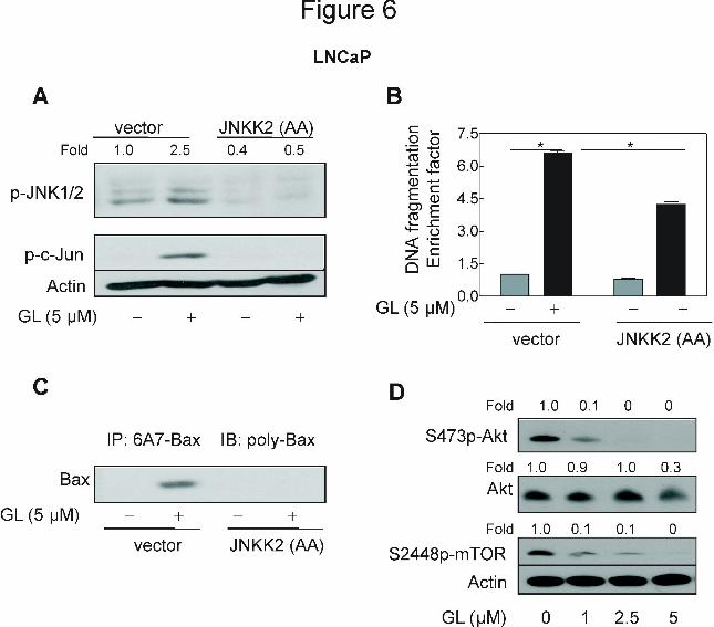
GL-mediated activation of Bax was inhibited by ectopic expression of JNKK2(AA).
The results shown above indicated critical roles of JNK and Bax activation in GL-induced
apoptosis but did not provide any insight into the signaling pathways linking these effects. Next,
we questioned if JNK activation contributed to GL-mediated activation of Bax. We addressed
this question by determining the effect of ectopic expression of catalytically inactive mutant of
JNKK2, which is a JNK-specific upstream kinase (Singh et al., 2007; Chen et al., 1998). GL
treatment caused an increase in phosphorylations of JNK and its downstream target c-Jun in the
empty-vector transfected LNCaP cells (Fig. 6A). In contrast, GL-mediated hyperphosphorylation
of both JNK and c-Jun were fully abolished by ectopic expression of catalytically inactive
This article has not been copyedited and formatted. The final version may differ from this version.Molecular Pharmacology Fast Forward. Published on November 29, 2010 as DOI: 10.1124/mol.110.068551
at ASPE
T Journals on M
arch 14, 2022m
olpharm.aspetjournals.org
Dow
nloaded from
MOL #68551
15
JNKK2(AA) in the LNCaP cells (Fig. 6A). In addition, statistically significant increase in
cytoplasmic histone-associated DNA fragmentation (6.6 fold of control) resulting from 24 h
exposure to GL 5 µM was observed in the empty-vector transfected LNCaP cells but partially
and significantly decreased in the cells transiently transfected with JNKK2(AA) (Fig. 6B).
Genetic suppression of JNK also attenuated the inhibition of LNCaP cell growth by GL (data not
shown). Furthmore, immunoprecipitation analysis of activate Bax from cell lysates was
performed by using a monoclonal antibody (6A7) that recognizes an epitope at the N-terminus of
the active Bax followed by immunoblotting using polyclonal anti-Bax antibody. The GL
treatment caused a remarkable increase in Bax conformational change in the empty-vector
transfected LNCaP cells (Fig. 6C). More importantly, overexpression of JNKK2(AA) conferred
protection against GL-mediated conformational change of Bax (Fig. 6C). Collectively, these
results indicated that the GL-mediated conformational change of Bax was regulated by JNK
signaling axis.
However, inhibition of the JNK signaling was not fully protective against the GL-induced
cell death (Fig. 6B). Therefore, the roles of the other MAPK signaling such as p38 MAPK and
ERK in the apoptosis induction by GL were determined by using the siRNA technology. As can
seen in Supplemental Figures. S1 and S2, the protein levels of p38 MAPK and ERK were
knocked down ~50% -90% by transient transfection of LNCaP cells with p38 MAPK or ERK-
targeted siRNA compared with cells transfected with a control nonspecific siRNA. However,
p38 MAPK-siRNA or ERK-siRNA did not have any protection against the apoptotic cell death
induced by GL (Figs. S1-2). The data indicate that the GL-induced apoptosis is not mediated by
p38 MAPK or ERK.
This article has not been copyedited and formatted. The final version may differ from this version.Molecular Pharmacology Fast Forward. Published on November 29, 2010 as DOI: 10.1124/mol.110.068551
at ASPE
T Journals on M
arch 14, 2022m
olpharm.aspetjournals.org
Dow
nloaded from

MOL #68551
16
Moreover, we investigated whether Akt signaling was affected in GL-treated LNCaP cells.
Exposure of LNCaP cells to GL resulted in a concentration-dependent and significant
inactivation of Akt and its substrate mTOR (Fig. 6D). These results suggested that Akt signaling
pathway may be involved in regulation of GL-induced cell death.
Discussion
GL, extract of Commiphora mukul, has been safely used for thousands of years in the Indian
Ayurvedic medicine practice for treatment of different ailments and has been used recently in
many clinical trials focused on its cholesterol-lowering effect (Shishodia et al, 2008; Urizar and
Moore, 2003; Badmaev et al., 2003). In the present study, we, for the first time, show that GL
has a stronger anti-cancer potential in human prostate cancer cells as evidenced by inhibition of
cell growth and induction of apoptotic cell death compared with one of its active constituents (z-
Gug). Statistically significant inhibition of cell survival by GL was evident at IC50 ~ 1 μmol/L
concentrations standardized to z-Gug. The effect on growth inhibition by GL was ~10-fold
stronger compared with z-Gug (Fig. 1). Interestingly, a normal prostate epithelial cell line PrEC
was significantly more resistant to growth inhibition by GL compared with prostate cancer cells.
Based on these results, we conclude that (a) GL treatment decreases survival of human prostate
cancer cells irrespective of their androgen-responsiveness, (b) a normal prostate epithelial cell
line is significantly more resistant to growth inhibition by GL, and (c) uncharacterized
constituent(s) of GL may interact additively or synergistically to inhibit viability of human
prostate cancer cells. Even though pharmacokinetic parameters for GL have not been determined
in humans, the maximal plasma concentration of z-Gug (Cmax) in rats was shown to be 3.3- and
18.3 µmol/L following oral gavage with 50 mg z-Gug/Kg body weight and intravenous injection
with 18 mg z-Gug/Kg body weight (Verma et al., 1999). Based on these pharmacokinetic
This article has not been copyedited and formatted. The final version may differ from this version.Molecular Pharmacology Fast Forward. Published on November 29, 2010 as DOI: 10.1124/mol.110.068551
at ASPE
T Journals on M
arch 14, 2022m
olpharm.aspetjournals.org
Dow
nloaded from
MOL #68551
17
observations, it is possible that the concentrations of GL needed to inhibit cancer cell growth
may be achievable in humans. We, more recently (Leeman-Neill et al., 2009) reported that
treatment with GL (3 µmol standardized to z-Gug, daily for 3 weeks) resulted in enhancement of
cetuximab activity in xenograft model of head and neck cancer.
Apoptosis has emerged as an important mechanism for anticancer effects of many
naturally occurring and synthetic agents (Shishodia and Aggarwal, 2004; Samudio et al., 2005;
Ichikawa and Aggarwal, 2006; Cheon et al., 2006; Xiao and Singh, 2008; Singh et al., 2007;
Xiao and Singh, 2007; Kim et al., 2007; Xiao et al., 2006a-c, 2008, 2009, 2010; Singh et al.,
2005a). Our present study indeed indicated that GL inhibits prostate cancer cell viability by
causing apoptosis that is characterized by appearance of cytoplasmic histone-associated DNA
fragmentation, subdiploid cells, and cleavage of PARP (Fig. 2). In contrast, PrEC is more
resistant to apoptosis induction by GL (Fig. 2). These results clearly indicate that antitumor
activity of GL against prostate cancer cells is associated with apoptosis induction.
Tumor-specific induction of oxidative stress is expected to offer a powerful therapeutic
modality. In fact, many anticancer agents and naturally occurring and synthetic agents exhibit
antitumor activity via ROS-dependent activation of apoptotic cell death (Singh et al., 2007; Kim
et al., 2007; Xiao et al., 2005b-c, 2006b-c; Fang et al., 2007). The present results indicate that the
cell death caused by GL in human prostate cancer cells is triggered by ROS generation. This
conclusion is based on following observations: (a) GL treatment caused a dose- and time-
dependent ROS production in LNCaP and C81 cells (Fig3. A-B); (b) GL-mediated ROS
generation and apoptotic cell death was significantly attenuated by antioxidant NAC (Fig.3D);
and (c) same treatment with GL did not affect the ROS induction (Fig. 3C) and cause apoptotic
cell death (Fig. 2A-B) in PrEC. It was also reported by us (Singh et al, 2007) that ROS is
This article has not been copyedited and formatted. The final version may differ from this version.Molecular Pharmacology Fast Forward. Published on November 29, 2010 as DOI: 10.1124/mol.110.068551
at ASPE
T Journals on M
arch 14, 2022m
olpharm.aspetjournals.org
Dow
nloaded from
MOL #68551
18
indispensable for z-Gug, one of the important active components of GL, caused apoptosis in
human prostate cancer PC-3 cells.
The proapoptotic Bcl-2 family proteins, which can be subdivided into the Bax subfamily
of multidomain proteins (e.g., Bax and Bak) or BH3-only subfamily (e.g., Bid and Bim), induce
mitochondrial membrane permeabilization and release of apoptogenic molecules from
mitochondria to the cytosol (Singh et al., 2005a; Xiao et al., 2005b-c). An increase in protein
levels of Bax and Bak was observed in GL-treated human prostate cancer cells (Fig. 4A), but not
in normal prostate epithelial PrEC cell line (Fig 4B). Furthmone, the SV40-immortalized MEFs
derived from Bax- and Bak- DKO mice are statistically significantly more resistant toward GL-
induced cell viability and apoptotic cell death compared with MEFs derived from WT mice (Fig.
4C-E). The present study indicates that the multidomain proapoptotic Bcl-2 family members Bax
and/or Bak play a critical role in regulation of GL-induced apoptosis. The GL-induced apoptosis
in human prostate cancer cells may be exacerbated because of down-regulation of Bcl-2 (Fig
4A). Therefore, further studies are needed to determine the role of antiapoptotic Bcl-2 family
members, such as Bcl-2 in regulation of GL-mediated cell death in prostate cancer cells.
The involvement of MAPKs signaling pathways in carcinogenesis and cancer prevention
and therapy is well documented (Xiao et al., 2004; 2005a). An interesting observation of the
present study is that GL-induced ROS-dependent apoptosis in our model is regulated by JNK
signaling axis (Figs. 5 and 6): we show that (a) GL treatment causes a time-dependent JNK
activation in human prostate cancer LNCaP (Fig. 5A) and C81 (date not shown) cells; (b)
pharmacological inhibition of JNK confers significant protection against GL-mediated apoptosis
in LNCaP and C81 cells (data not shown); (c) GL-induced JNK activation, Bax conformational
change (activation) and apoptosis induction were observed in empty vector-transfected LNCaP
This article has not been copyedited and formatted. The final version may differ from this version.Molecular Pharmacology Fast Forward. Published on November 29, 2010 as DOI: 10.1124/mol.110.068551
at ASPE
T Journals on M
arch 14, 2022m
olpharm.aspetjournals.org
Dow
nloaded from
MOL #68551
19
cells, but were inhibited in the cells after ectopic expression of catalytically inactive mutant of
JNKK2 (Fig. 6A-C); and (d). GL treatment could not activate the JNK activation in PrEC
(Fig.5B); JNK was reported to regulate Bax translocation through phosphorylation of Bim (Lei
and Davis, 2003) and to promote Bax translocation through phosphorylation of 14-3-3 proteins
(Tsuruta et al., 2003). Thus, it is reasonable to conclude from the present study that GL-inducted
Bax activation is regulated by JNK signaling axis. However, it was reported by Sarfaraz et al.
that z-Gug-inhibited skin tumorigenesis in SENCAR mice via inhibition of JNK activation.
Further studies are needed to systematically explore the role of JNK activation in GL anticancer
potential in vivo and other cancers in the future studies. These results indicate also that other
MAPK signaling such as p38MAPK and Erk are not but the survival signaling Akt may be a
mediator for the apoptotic induced by GL.
In conclusion, the present study reveals that GL is a potent inhibitor of prostate cancer cell
growth. The GL-mediated antitumor activity is associated with ROS-dependent apoptotic cell
death, and is regulated by JNK signaling axis.
Acknowledgments We thank the late Dr. Stanley Korsmeyer for the generous gift of MEFs and Dr. Michael Karin
for the generous gift of JNKK2(AA) plasmid.
Authorship contributions
D.X. provided oversight for the project, conducted the experiments and wrote the manuscript.
Y.Z. performed the experiments. L.P., V.B. and M.M. provided gugulipid and carried out quality
control. S.V.S. participated in the research design.
This article has not been copyedited and formatted. The final version may differ from this version.Molecular Pharmacology Fast Forward. Published on November 29, 2010 as DOI: 10.1124/mol.110.068551
at ASPE
T Journals on M
arch 14, 2022m
olpharm.aspetjournals.org
Dow
nloaded from
MOL #68551
20
References
Badmaev V, Majeed M, Pacchetti B, and Prakash L (2003) Standardiation of Commiphora
Mukul extract in dislipidemia and cardiovascular disease. NUTRA Foods 2: 45-51.
Chao DT and Korsmeyer SJ (1998) BCL-2 family: regulators of cell death. Annu Rev Immunol
16: 395–419.
Chen CY, Del Gatto-Konczak F, Wu z, and Karin M (1998) Stabilization of interleukin-2 mRNA
by the c-Jun NH2-terminal kinase pathway. Science 280: 1945-1949.
Cheon JH, Kim JS, Kim JM, Kim N, Jung HC, and Song IS (2006) Plant sterol guggulsterone
inhibits nuclear factor-κB signaling in intestinal epithelial cells by blocking IκB kinase and
ameliorates acute murine colitis. Inflamm Bowel Dis 12:1152-1161.
Cui J, Huang L, Zhao A, Lew JL, Yu J, Sahoo S, Meinke PT, Royo I, Pelaez F, and Wright SD
(2003) Guggulsterone is a farnesoid X receptor antagonist in coactivator association assays
but acts to enhance transcription of bile salt export pump. J Biol Chem 278:10214-10220.
Fang J, Nakamura H, and Lyer AK (2007) Tumaor-targeted induction of oxystress for cancer
therapy. J Drug Target 15: 475-486.
Gilligan T, and Kantoff PW (2002) Chemotherapy for prostate cancer. Urol 60 (suppl. 3A): 94-
100.
Gujral ML, Sareen K, Tangri KK, Amma MK, and Roy AK (1960) Antiarthritic and anti-
inflammatory activity of gum guggul (Balsamodendron mukul Hook). Ind J Physiol
Pharmacol 4:267-273.
Ichikawa H and Aggarwal B (2006) Guggulsterone inhibits osteoclastogenesis induced by
receptor activator of nuclear factor-κB ligand and by tumor cells by suppressing nuclear
factor-κB activation. Clin Cancer Res 12:662-668.
Jemal A, Siegel R, Ward E, How Y, Xu J, and Thun MJ (2009) Cancer Statistics. 2009. CA
Cancer J Clin 59: 225-249.
This article has not been copyedited and formatted. The final version may differ from this version.Molecular Pharmacology Fast Forward. Published on November 29, 2010 as DOI: 10.1124/mol.110.068551
at ASPE
T Journals on M
arch 14, 2022m
olpharm.aspetjournals.org
Dow
nloaded from

MOL #68551
21
Kim BJ, Ryu SW, and Song BJ (2006) JNK- and p38 kinase-mediated phosphorylation of Bax
leads to its activation and mitochondrial translocation and to apoptosis of human hepatoma
HepG2 cells. J Biol Chem 281: 21256-21265.
Kim YA, Xiao D, Xiao H, Powolny AA, Lew KL, Reilly ML, Zeng Y, Wang Z, and Singh SV
(2007) Mitochondria-mediated apoptosis by diallyl trisulfide in human prostate cancer cells
is associated with generation of reactive oxygen species and regulated by Bax/Bak. Mol.
Cancer Ther 6:1599-1609.
Leeman-Neill RJ, Wheeler SE, Singh SV, Thomas SM, Seethala RR, Neill DB, Panahanden MC,
Hahm ER, Joyce SC, Sen M, et al. (2009) Guggulsterone enhances head and neck cancer
therapies viainhibition of signal transducer and activator of transcription-3. Carcinegesis 11:
1848-1856.
Lei K and Davis RJ (2003) JNK phosphorylation of Bim-related members of the Bcl2 family
induces Bax-dependent apoptosis. PNAS 100: 2432–2437.
Samudio I, Konopleva M, Safe S, McQueen T, and Andreeff M (2005) Guggulsterone induce
apoptosis and differentiation in acute myeloid leukemia: identification of isomer-specific
antileukemic activities of the pregnadienedione structure. Mol Cancer Ther 4:1982-1992.
Sarfaraz S, Siddiqui IA, Syed DN, Afaq F, and Mukhtar H (2008) Guggulsterone modulates
MAPK and NF-κB pathways and inhibits skin tumorigenesis in SENCAR mice.
Carcinogenesis 29: 2011-2018.
Shishodia S and Aggarwal BB (2004) Guggulsterone inhibits NF-kappaB and IkappaBalpha
kinase activation, suppresses expression of anti-apoptotic gene products, and enhances
apoptosis. J Biol Chem 279:47148-47158.
Shishodia S, Harikumar KB, Dass S, Ramawat KG, and Aggarwal BB (2008) The guggul for
chronic disease: ancient medicine, modern targets. Anticancer Res 28: 3647-3664.
Sinal CJ and Gonzalez FJ (2002) Guggulsterone: an old approach to a new problem. Trends
Endocrinol Metab 13:275-276.
This article has not been copyedited and formatted. The final version may differ from this version.Molecular Pharmacology Fast Forward. Published on November 29, 2010 as DOI: 10.1124/mol.110.068551
at ASPE
T Journals on M
arch 14, 2022m
olpharm.aspetjournals.org
Dow
nloaded from

MOL #68551
22
Singh SV, Choi S, Zeng Y, Hahm ER, and Xiao D (2007) Guggulsterone-induced apoptosis in
human prostate cancer cells is caused by reactive oxygen intermediate-dependent activation
of c-Jun NH2-terminal kinase. Cancer Res 67:7439-7449.
Singh SV, Srivastava SK, Choi S, Lew KL, Antosiewicz J, Xiao D, Zeng Y, Watkins SC,
Johnson CS, Trump DL, Leey J, Xiao H, and Herman-Antosiewicz A (2005a Sulforaphane-
induced cell death in human prostate cancer cells is initiated by reactive oxygen species. J
Biol Chem 280: 19911-19924.
Singh SV, Zeng Y, Xiao D, Vogel VG, Nelson JB, Dhir R, and Tripathi YB (2005b Caspase-
dependent apoptosis induction by guggulsterone, a constituent of Ayurvedic medicinal plant
Commiphora mukul, in PC-3 human prostate cancer cells is mediated by Bax and Bak. Mol
Cancer Ther 4:1747-1754.
Tsuruta F, Sunayama J, Mori Y, Hatt6ori S, Shimizu S, Tsujjmoto Y, Yoshioka K, Masuyama N,
and Gotoh Y (2003) JNK promotes Bax translocation to mitochondria through
phosphorylation of 14-3-3 proteins. EMBO J 23: 1889–1899.
Urizar NL, Liverman AB, Dodds DT, Silv FV, Ordentlich P, Yan Y, Gonzaleg FJ, Heyman RA,
Mangelsdorf DJ, and Moore DD (2002) A natural product that lowers cholesterol as an
antagonist ligand for FXR. Science 296:1703-1706.
Urizar NL and Moore DD (2003) GUGULIPID: a natural cholesterol-lowering agent. Annu Rev
Nutr 23:303-313.
Verma N, Singh SK, and Gupta RC (1999) Pharmacokinetics of guggulsterone after intravenous
and oral administration in rats. Pharm Pharmacol Comm 5: 349-354.
Whittemore AS, Kolonel LN, Wu AH, John EM, Gallagher RP, Howe GR, Burch JD, Hankin J,
Dreon DM, West DW, et al. (1995) Prostate cancer in relation to diet, physical activity, and
body size in blacks, whites and Asians in the United States and Canada. J. Natl. Cancer Ins
87: 652-661.
This article has not been copyedited and formatted. The final version may differ from this version.Molecular Pharmacology Fast Forward. Published on November 29, 2010 as DOI: 10.1124/mol.110.068551
at ASPE
T Journals on M
arch 14, 2022m
olpharm.aspetjournals.org
Dow
nloaded from
MOL #68551
23
Wu J, Xia C, Meier J, Li S, Hu X, and Lala DS (2002) The hypolipidemic natural product
guggulsterone acts as an antagonist of the bile acid receptor. Mol Endocrinol 16:1590-1597.
Xiao D, Choi S, Johnson DE, Vogel VG, Johnson CS, Trump DL, Lee YJ, and Singh SV (2004)
Diallyl trisulfide-induced apoptosis in human prostate cancer cells involves c-Jun N-terminal
kinase and extracellular-signal regulated kinase-mediated phosphorylation of Bcl-
2.Oncogene 23:5594-5606.
Xiao D, Choi S, Lee YJ, and Singh SV (2005a) Role of mitogen-activated protein kinases in
phenethyl isothiocyanate-induced apoptosis in human prostate cancer cells. Mol
Carcinogenesis 43: 130-140. Xiao D, Herman-Antosiewicz A, Antosiewicz J, Xiao H, Brisson M, Lazo JS, and Singh SV
(2005b Diallyl trisulfide-induced G2-M phase cell cycle arrest in human prostate cancer cells
is caused by reactive oxygen species-dependent destruction and hyperphosphorylation of
Cdc25C. Oncogene 24: 6256-6268.
Xiao D, Lew KL, Kim YA, Zeng Y, Hahm ER, Dhir R, and Singh SV (2006a) Diallyl trisulfide
suppresses growth of PC-3 human prostate cancer xenograft in vivo in association with Bax
and Bak induction. Clin Cancer Res 12: 6836-6843.
Xiao D, Lew KL, Zeng Y, Xiao H, Marynowski SW, Dhir R, and Singh SV (2006b) Phenethyl
isothiocyanate-induced apoptosis in PC-3 human prostate cancer cells is mediated by reactive
oxygen species-dependent disruption of the mitochondrial membrane potential.
Carcinogenesis 27: 2223-2234.
Xiao D, Powolny AA, Antosiewicz J, Hahm ER, Bommareddy A, Zeng Y, Desai D, Amin S,
Herman-Antosiewicz A, and Singh SV (2009) Cellular responses to dietary cancer
chemopreventive agent D,L-sulforaphane in human prostate cancer cells are initiated by
mitochondria-derived reactive oxygen species. Pharm Res 26:1729-1738.
Xiao D, Powolny AA, and Singh SV (2008) Benzyl isothiocyanate targets mitochondrial
respiratory chain to trigger reactive oxygen species-dependent apoptosis in human breast
cancer cells. J Biol Chem 283: 30151-30163.
Xiao D and Singh SV (2010) P66Shc is indispensable for phenethyl isothiocyanate-induced
apoptosis in human prostate cancer cells. Cancer Res 70:3150-3158.
This article has not been copyedited and formatted. The final version may differ from this version.Molecular Pharmacology Fast Forward. Published on November 29, 2010 as DOI: 10.1124/mol.110.068551
at ASPE
T Journals on M
arch 14, 2022m
olpharm.aspetjournals.org
Dow
nloaded from

MOL #68551
24
Xiao D and Singh SV (2008) Guggulsterone, a constituent of Indian Ayurvedic medicinal plant
Commiphora mukul, inhibits angiogenesis in vitro and in vivo. Mol.Cancer Ther 7: 171-180.
Xiao D and Singh SV (2007) Phenethyl isothiocyanate inhibits angiogenesis in vitro and ex vivo.
Cancer Res 67: 2239-2246.
Xiao D, Vogel V and Singh SV (2006c) Benzyl isothiocyanate-induced cell death in MDA-MB-
231 and MCF-7 human breast cancer cells is initiated by reactive oxygen species and
regulated by Bax and Bak. Mol.Cancer Ther 5:2931-2945.
Xiao D, Zeng Y, Choi S, Lew KL, Nelson JB, and Singh SV (2005c Caspase-dependent
apoptosis induction by phenethyl isothiocyanate, a cruciferous vegetable-derived cancer
chemopreventive agent, is mediated by bak and bax. Clin Cancer Res 11:2670-2679.
This article has not been copyedited and formatted. The final version may differ from this version.Molecular Pharmacology Fast Forward. Published on November 29, 2010 as DOI: 10.1124/mol.110.068551
at ASPE
T Journals on M
arch 14, 2022m
olpharm.aspetjournals.org
Dow
nloaded from

MOL #68551
25
Footnotes
This work was supported by the National Institutes of Health National Cancer Institute [grant
R21-CA143104 (DX)].
This article has not been copyedited and formatted. The final version may differ from this version.Molecular Pharmacology Fast Forward. Published on November 29, 2010 as DOI: 10.1124/mol.110.068551
at ASPE
T Journals on M
arch 14, 2022m
olpharm.aspetjournals.org
Dow
nloaded from

MOL #68551
26
FIGURE LEGENDS
Figure 1. Effect of GL (GL contains ~3.75% z-Gug and was standardized to z-Gug (µM), A, B
and D) and z-Gug (C) on survival of LNCaP, C81 and PrEC cells determined by the colonogenic
assay (A) and trypan blue dye exclusion assay (B-D). Cells were treated with different
concentrations of GL or z-Gug for 24 h. Columns, mean of three determinations; bars, SE.
*Significantly different (P < 0.05) compared with DMSO-treated control by one-way ANOVA
followed by Dunnett’s test. Similar results were observed in two independent experiments.
Representative data from a single experiment are shown.
Figure 2. GL induced apoptosis in LNCaP and C81 cells, but not in normal human prostate
epithelial cells PrEC, determined by (A) quantitation of cytoplasmic histone associated DNA
fragmentation, (B) flow cytomitry analysis of Sub-G0/G1 cell phase, and (C) immunoblotting
cleavage of PARP. Cells were treated with the indicated concentrations of GL or DMSO
(control) for 24 Hours. Results in panels A and B are expressed as enrichment factor relative to
cells treated with DMSO (control). Results are mean ± SE (n= 3). *Significantly different
(P<0.05) between the indicated groups by one-way ANOVA followed by by Dunnett’s test. In
panel C, the cleaved PARP by immunoblotting using lysates from GL-treated or DMSO-treated
LNCaP and C81 cells. The blot was stripped and re-probed with anti-α-Tubulin antibody to
ensure equal protein loading. Similar results were observed in at least two independent
experiments. Representative data from a single experiment are shown.
Figure 3. GL-induced ROS production was involved in apoptotic cell death caused by GL. GL
caused ROS generation in C81 (A) and LNCaP (B) cells in dose- (left panel of A for C81) and
This article has not been copyedited and formatted. The final version may differ from this version.Molecular Pharmacology Fast Forward. Published on November 29, 2010 as DOI: 10.1124/mol.110.068551
at ASPE
T Journals on M
arch 14, 2022m
olpharm.aspetjournals.org
Dow
nloaded from

MOL #68551
27
time- (right panel of A for C81 and B for LNCaP) dependent manner, but not in PrEC (C). (D)
NAC protected against GL-mediated ROS production and apoptosis. LNCaP cells were treated
with 10 mM NAC for 2 h, and then exposed to 5µM GL standardized to z-Gug for 30 min (left
panel of D) or 24 h (right panel of D). In panels A- D, results are mean ± SE (n= 3).
*Significantly different (P<0.05) between the indicated groups by one-way ANOVA followed by
Dunnett’s test (left panel of A and C) and Bonferroni’s multiple comparison test (D), and by
paired t-test (right panel of A and B. Experiments were repeated twice with triplicate
measurements in each experiment. The results were consistent and representative data from a
single experiment are shown.
Figure 4. A, Immunoblotting for Bax, Bak, and Bcl-2 proteins using lysates from LNCaP cells
treated with DMSO (control) or 5 μmol/L GL standardized to z-Gug for the indicated time
periods. B, Immunoblotting for Bax protein using lysates from PrEC treated with DMSO
(control) or 5 μmol/L GL standardized to z-Gug for the indicated time periods. C,
Immuniblotting for cleaved PARP protein from the SV 40-immoratalized mouse embryonic
fibroblasts derived –Wild Tipe (WT) and Bax and Bak double knockout mice treated with
DMSO (control) or 2.5 and 5 μmol/L GL standardized to z-Gug for 24 h. For A - C, the blots
were stripped and re-probed with anti-actin antibody to normalize for differences in protein level.
The numbers on top of the immunoreactive bands represent change in protein levels relative to
DMSO-treated cells. Immunoblotting for each protein was performed at least twice using
independently prepared lysates. D, the cell survival and E, cytoplasmic histone-associated DNA
fragmentation in LNCaP cells treated with DMSO (control) or 2.5 and 5 μmol/L GL standardized
to z-Gug for 24 h. For D and E, Columns, mean (n= 3); bars, SE. *Significantly different (P
This article has not been copyedited and formatted. The final version may differ from this version.Molecular Pharmacology Fast Forward. Published on November 29, 2010 as DOI: 10.1124/mol.110.068551
at ASPE
T Journals on M
arch 14, 2022m
olpharm.aspetjournals.org
Dow
nloaded from

MOL #68551
28
<0.05) compared with corresponding DMSO treated control by one-way ANOVA followed by
Dunnett’s test. Each experiment was performed at least twice with triplicate measurements in
each experiment. The results were consistent and representative data from a single experiment
are shown.
Figure 5. The GL treatment increased activating phosphorylation of c-Jun N-terminal kinase
(JNK) in LNCaP cells, but not in PrEC. A, Immunoblotting for phospho-JNK, phosphor-Erk
and phospho-p38 MAPK using lysates from LNCaP cells and B, Immunoblotting for phospho-
JNK using lysates from PrEC treated with DMSO (control) or 5 µM GL standardized to z-Gug
or 5 µM z-Gug for the indicated time periods. The blots were stripped and re-probed with anti-
actin antibody to ensure equal protein loading. Immunoblotting for each protein was performed
twice using independently prepared lysates and the results were similar. Representative data
from a single experiment are shown. Fold change in phospho/total protein level relative to
DMSO-treated control at each time point is shown on top of the immunoreactive band. C, NAC
protected against GL-mediated JNK activation. LNCaP cells were treated with 10 mM NAC for
2 h, and then with or without 2.5 µM GL standardized to z-Gug for 8 h. The cellular lysates
from these groups were performed for immunoblotting of phospho-c-Jun. The blots were
stripped and re-probed with anti-actin antibody to ensure equal protein loading. The numbers on
top of the immunoreactive bands represent change in protein levels relative to corresponding
DMSO-treated control. Immunoblotting for the protein was performed twice using independently
prepared lysates and the results were similar. Representative data from a single experiment are
shown.
This article has not been copyedited and formatted. The final version may differ from this version.Molecular Pharmacology Fast Forward. Published on November 29, 2010 as DOI: 10.1124/mol.110.068551
at ASPE
T Journals on M
arch 14, 2022m
olpharm.aspetjournals.org
Dow
nloaded from

MOL #68551
29
Figure 6. A, immunoblotting for phospho-JNK and phospho-c-Jun using lysates from LNCaP
cells transiently transfected with the empty pcDNA3.1 vector or pcDNA3.1 vector encoding
catalytically inactive mutant of JNKK2 [JNKK2(AA)] and treated for 8 h with DMSO (control)
or 5 μM GL. B, cytoplasmic histone-associated DNA fragmentation in LNCaP cells transiently
transfected with the empty pcDNA3.1 vector or pcDNA3.1 vector encoding JNKK2(AA) and
treated for 24 h with DMSO (control) or 5 μM GL. Results are mean ± SE (n= 3). *Significantly
different (P<0.05) between the indicated groups by one-way ANOVA followed by Bonferroni’s
multiple comparison test. C, analysis of conformational change of Bax using lysates from
LNCaP cells transiently transfected with the empty pcDNA3.1 vector or pcDNA3.1 vector
encoding JNKK2(AA) and treated for 8 h with DMSO (control) or 5 μM GL. Bax protein was
immunoprecipitated from equal amounts of lysate proteins using ant-Bax 6A7 monoclonal
antibody. The immunoprecipitated complexes were subjected to immunoblotting using anti-Bax
polyclonal antibody. D, Immunoblotting for Akt, S473phosphor-Akt and S2448phospho-mTOR
proteins using lysates from LNCaP cells treated with DMSO (control) or indicated
concentrations of GL standardized to z-Gug for 24 h. For A and D, the blots were stripped and
re-probed with anti-actin antibody to ensure equal protein loading. The numbers on top of the
immunoreactive bands represent change in protein levels relative to corresponding DMSO-
treated control. Each experiment was repeated twice with comparable results. Representative
data from a single experiment are shown.
This article has not been copyedited and formatted. The final version may differ from this version.Molecular Pharmacology Fast Forward. Published on November 29, 2010 as DOI: 10.1124/mol.110.068551
at ASPE
T Journals on M
arch 14, 2022m
olpharm.aspetjournals.org
Dow
nloaded from
This article has not been copyedited and formatted. The final version may differ from this version.Molecular Pharmacology Fast Forward. Published on November 29, 2010 as DOI: 10.1124/mol.110.068551
at ASPE
T Journals on M
arch 14, 2022m
olpharm.aspetjournals.org
Dow
nloaded from
This article has not been copyedited and formatted. The final version may differ from this version.Molecular Pharmacology Fast Forward. Published on November 29, 2010 as DOI: 10.1124/mol.110.068551
at ASPE
T Journals on M
arch 14, 2022m
olpharm.aspetjournals.org
Dow
nloaded from
This article has not been copyedited and formatted. The final version may differ from this version.Molecular Pharmacology Fast Forward. Published on November 29, 2010 as DOI: 10.1124/mol.110.068551
at ASPE
T Journals on M
arch 14, 2022m
olpharm.aspetjournals.org
Dow
nloaded from
This article has not been copyedited and formatted. The final version may differ from this version.Molecular Pharmacology Fast Forward. Published on November 29, 2010 as DOI: 10.1124/mol.110.068551
at ASPE
T Journals on M
arch 14, 2022m
olpharm.aspetjournals.org
Dow
nloaded from

This article has not been copyedited and formatted. The final version may differ from this version.Molecular Pharmacology Fast Forward. Published on November 29, 2010 as DOI: 10.1124/mol.110.068551
at ASPE
T Journals on M
arch 14, 2022m
olpharm.aspetjournals.org
Dow
nloaded from
This article has not been copyedited and formatted. The final version may differ from this version.Molecular Pharmacology Fast Forward. Published on November 29, 2010 as DOI: 10.1124/mol.110.068551
at ASPE
T Journals on M
arch 14, 2022m
olpharm.aspetjournals.org
Dow
nloaded from