oral pigmentation.docx my
TRANSCRIPT

Thilanka Umesh Sugathadasa
ORAL
PIGMENTATION
Thilanka Umesh Sugathadasa

Thilanka Umesh Sugathadasa
Pigmentation
Exogenous
Dental hard tissue
Oral mucosal
Endogenous
Oral mucosal
Dental hard tissues
Oral Pigmentation
Classification
Endogenous – Produce by the body itself.(eg- Hb, Melanin, Hemosiderin)
Exogenous – Taken in to the body from outside.(eg- tobacco, pigments in the vegetables)
Some disease processes leads in formation of
- Pseudomembranes
- Increased keratinization(White lesions)
- Increase vascularization (Red lesions)
Color
Focal Multifocal
Solitary Diffuse
Blue/ Purple
Varix
Hemangioma
Hemangioma Kaposi’s sarcoma
Hereditory hemorrhagic telengiectasia
Brown
Melanotic macule
Nevus
Melanoma
Melanoma
Drug induced pigmentation
Ecchymosis
Hairy tongue
Physiological pigmentation
Neurofibromatosis
Melanoma hemochromatosis
LP
Addison’s disease.
Drug induced pigmentation
Peutz- Jeghers syndrome
Petechia
Gray/Black Amalgum/ Graphite tattoo
Nevus
Melanoma
Amalgum tattoo
Melanoma
Hairy tongue
Heavy metal ingestion pigmentation.
Thilanka Umesh Sugathadasa
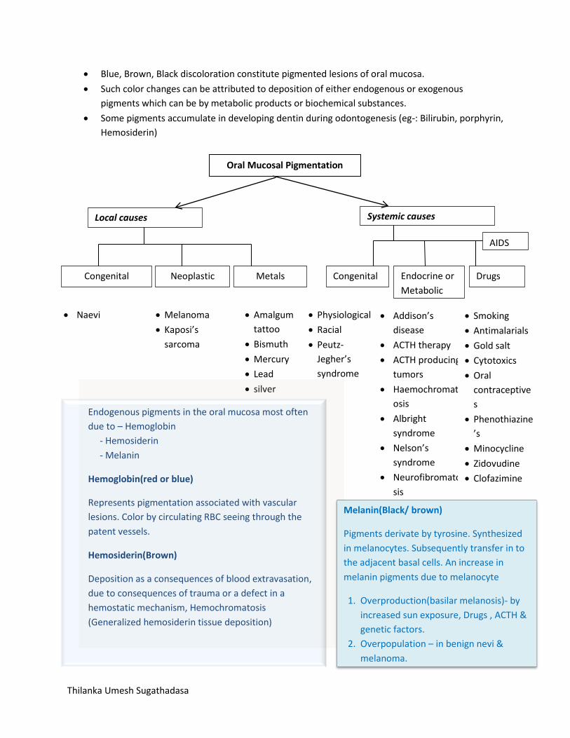
Blue, Brown, Black discoloration constitute pigmented lesions of oral mucosa.
Such color changes can be attributed to deposition of either endogenous or exogenous
pigments which can be by metabolic products or biochemical substances.
Some pigments accumulate in developing dentin during odontogenesis (eg-: Bilirubin, porphyrin,
Hemosiderin)
Oral Mucosal Pigmentation
Local causes
Systemic causes
Congenital Neoplastic Metals Endocrine or
Metabolic
Drugs
AIDS
Congenital
Naevi Melanoma
Kaposi’s
sarcoma
Amalgum
tattoo
Bismuth
Mercury
Lead
silver
Physiological
Racial
Peutz-
Jegher’s
syndrome
Addison’s
disease
ACTH therapy
ACTH producing
tumors
Haemochromat
osis
Albright
syndrome
Nelson’s
syndrome
Neurofibromato
sis
Smoking
Antimalarials
Gold salt
Cytotoxics
Oral
contraceptive
s
Phenothiazine
’s
Minocycline
Zidovudine
Clofazimine
Endogenous pigments in the oral mucosa most often
due to – Hemoglobin
- Hemosiderin
- Melanin
Hemoglobin(red or blue)
Represents pigmentation associated with vascular
lesions. Color by circulating RBC seeing through the
patent vessels.
Hemosiderin(Brown)
Deposition as a consequences of blood extravasation,
due to consequences of trauma or a defect in a
hemostatic mechanism, Hemochromatosis
(Generalized hemosiderin tissue deposition)
Melanin(Black/ brown)
Pigments derivate by tyrosine. Synthesized
in melanocytes. Subsequently transfer in to
the adjacent basal cells. An increase in
melanin pigments due to melanocyte
1. Overproduction(basilar melanosis)- by
increased sun exposure, Drugs , ACTH &
genetic factors.
2. Overpopulation – in benign nevi &
melanoma.

Thilanka Umesh Sugathadasa
Blue/Purple vascular lesions
Condition Features
Hemangioma
Tumor like hamartomas.
Mostly can see in childrens
Found on skin, scalp, & within CT of mucous membrane.
85% spontaneous regression after puberty.
Color depend on the depth of the vascular proliferation with in the oral submucosa. Reddish blue- Vessels close to the overlying epithelium. vDeep blue – Deeper in the CT No discoloration – Intramuscular hemangiomas
Mostly raised & nodular.
Some may be flat, macular & diffuse specially on facial skin (“port- wine stains”)
Clinical appearance – quite variable ranging from a flat reddish/ blue macule to a blue nodule.
Most of the time oral hemangiomas can be seen in the tongue as multinodular bluish red lesion.
Tongue angiomas are frequently extend deeply between the intrinsic muscles of the tongue
Lip mucosa is also the another commen site usually localized, blue & raised.
Port-wine stain present in facial skin & is flat & magenta in color present in the Sturge- weber syndrome*.(Here vascular lesions occur in the face as well as brain.)
Many hemangiomas aare spontaneously involute during teenage years. Treatment may be withheld in children. Hemodynamics - Stasis with thrombosis is common. - Most lesion will blanch under pressure, glass side test - Intraluminal clots become palpable but then lesion will not blanch - Thrombi may calcify so then can be seen hard on palpation - Calcified nodules/ Phleboliths may be radiographically evident.
Treatments – Conventional surgery/ Laser surgery/ cryosurgery - Larger lesions which are extends in to the muscles are very difficult
to eradicate surgically. - Intralesional injections of sclerosing agents such as 1% sodium
tetradecyl sulphate. - Cutaneous port- wine stains can be treated by subcutaneous
tattooing or by argon laser.

Thilanka Umesh Sugathadasa
Varix
Pathological dilatation of the veins/venules
Chief site is ventral surface of the tongue.
Become progressively prominent with the age
Lingual varicosities appear as tortuous serpentine blue/red & purple elevations.
Represent degenerative changes in the adventitia of the venous wall.
No clinical consequence.
Are painless & are not subject to rupture & hemorrhage.
This focal dilatation of the veins or group of venules tend to occur in the elderly persons.
Primarily located on the lower lip.
Focal raised pigmentation
Blue, red or purple
Surface mucosa is often lobulated or nodular
Some can be blanched,others are not due to the formation of intravascular thrombi.
Resembles hemangioma clinically & histologically
It is distinguish mainly using two features 1. The patients age at its onset 2. It’s etiology
Hemangiomas are usually congenital & have a tendency to regress spontaneously, Whereas a varix arises in the older individuals & once formed it does not regress.
Varix has finite growth potential, So once a varix has formed further enlargement is uncommon.
Hemangioma is vascular hamartomatous condition of unknown etiology. Varix may from trauma such as lip or check biting.
Varices presents in the lips & buccal mucosa are esthetically unacceptable & can be interfere with the mastication.
Rx - Can be excised or remove by other methods such as cryosurgery & electrosurgery. - Intralesional 1% sodium tetradecyl sulfate injection is effective but it is more painful than the simple excision.(Sclerosing agents should be injected directly in to the lumina with a tuberculin syringe)
Kaposi’s sarcoma
Arise from endothelial cells
Has been called “gay cancer” by some since it is transmitted sexually, seen rarely in HIV infected children’s or haemophiliacs.
It is caused by HHV-8, which is transmitted sexually often as co-infection with the HIV Like all herpes viruses this is DNA virus, which is seen more commonly where hygiene is poor. This remains latent after the infection & It is found in the saliva.
Classic form generally appeared in two distinct clinical settings 1. Elderly men (in oral mucosa & on skin of lower extremities) 2. Children in equatorial Africa(in lymph nodes)
Thilanka Umesh Sugathadasa
Slowly progressive growth
Classic Kaposi’s sarcoma does not show a great tendency for metastasis & probably has never caused the death of the patients.
Oral & cutaneous tumors are considered to be of multifocal origin rather than metastases from a distant primary tumor.
Presents initially as red, blue or purple macules
Then progress to papules, nodules or ulcers & may become painful.
Hard palate is the favored site.(Hard palate & soft palate junction also) Anterior maxillary gingiva also a preferable site. - Oral lesions begins as flat red macules of variable size & irregular configuration. - They may appear as focal lesions, but typically oral KS lesions are multifocal with numerous isolated & coalescing plaques. - Sometimes this nodular growth can exhibit the entire palate, protruding below the plane of occlusion. - facial gingiva is the second most-favored oral site - Uncommon for AIDS associated KS to arise in Tongue & lips - Thus even in the context of HIV infection, KS should be considered a low grade sarcoma.
Typically extravasation of RBC is a prominent feature and hemosiderin granules are commonly encountered, though more hemosiderin present browner tumor will appear clinically
Pattern of growth in larger lesions are multinodular.
Often involves the skin or mucosa in the head & neck, Whereas most common in the face (specially on the nose & the mouth)
Also skin tumors tend to localized in the dorsal aspect of the feet & great toe. Initially cutaneous lesions begins as red macules & enlarge to become blue, purple, & ultimately brown nodular tumefaction(swelling) Also can appear in the arms, face , scalp, or trunk
Diagnosis Must be supported by the biopsy. DD includes – Pyogenic granuloma* - Giant cell granuloma* - Hemangioma - Purpura - Epithelioid angiomatosis - Lymphoma
Rx - Early plaque / macular lesions are painless & do not require treatments. - Nodular lesions may become unsightly and interfere with mastication: Therapy may be indicated. - Surgical excision- severe hemorrhage can be occur, electrocautery* is recommended. - Intralesional injection of 1% sodium tetradecyl sulfate – necrosis of the lesion but painful - Intralesional vinblastine sulfate- not a sclerosing agent, not associated with significant pain.
Thilanka Umesh Sugathadasa
Hereditory Hemorrhagic Telengiectasia
Characterized by multiple round or oval purple papules measuring less than 0.5cm in diameter.
HHT is a genetically transmitted disease. Inherited as an autosomal dominant trait.
Represent multiple microaneurysms, due to weakening defects in the adventitial coat of venules.
More than 100 such purple papules on vermilion & mucosal surfaces of lips, tongue & BM, Facial skin & neck also
Lesions in nasal mucosa- epistaxis(Death can occur)
Lesions may be seen during infancy, but most common in adults.
DD - Petechial hemorrhages due to platelet disorder, petechiae are macular rather than popular & red or brown rather than purple.
No Rx presents
Angiosarcoma
Malignant vascular neoplasms & distinct from the KS
Not related to HIV
Can arise anywhere in the body oral cavity is an rare site.
Appear red, blue & purple
Rapidly proliferative & presents as nodular tumor. Tend to ulcerate
Can arise from blood or lymph vessels endothelial cells or from pericytic cells if the vasculature.
Poor prognosis & rx by radical excision+ radiation
*Sturge- weber syndrome (Encephalotrigeminal angiomatosis) - A congenital hamartomatous
condition of the upper face (unilateral), oral mucosa & the underlying bone (with hemi hypertrophy of
bone & accelerated eruption of associated teeth), which also extending intracranially to cause
convulsion, & contralateral hemiplegia & intracerebral calcification (In radiographs bilaminar radiopaque
tracks reffered to as “Tram line”calcification) & sometimes learning disability.
*Pyogenic granuloma – A possibly reactive vascular lesion, sometimes associated with the pregnancy.
Typically the pyogenic granuloma is a small(<3cm),red painless mass that bleeds easily, ulcerates &
grows rapidly & frequently seen on the gingival margin or tongue. Rx is excision to exclude angiomatous
proliferations, chancre, carcinoma or KS
*Giant cell granuloma- The central giant cell granuloma is the uncommon lesion only seen in the tooth
bearing areas of the jaws, mostly in the mandible & typically in the second & third decade of life. This is
destructive condition. The lesion may symptomless or stimulate the malignant neoplasms clinically &
radiographically occasionally. Occasionally lesion erode the cortical bone & present as domed shape
purplish submucosal swelling. Radiographically showing ill-defined areas of the radiolucency & there
may be resorption of the roots of related teeth. Microscopy shows multinucleated giant cells irregularly
distributed in a cellular stroma & spindle shape cells often highly vascular. There may be areas of new &
old hemorrhages with hemosiderin pigment deposition. These microscopic features are

Thilanka Umesh Sugathadasa
indistinguishable from the focal lesions of hyperparathyroidism. This can be only excluded by the
serological tests(Calcium & phosphate levels & ALP level). Can be recur following curettage. Virtually
never metastasize.
Brown melanotic lesions
Conditions Features
Ephelis & Oral Melanotic Macule
Ephelis is the cutaneous freckle (ephelis) represents an increase in melanin pigments synthesis by basal-layer melanocytes. Without an increase in the no of melanocytes.
On skin- due to actinic exposure
Ephelids means conditions which encountered on vermilion border of the lips, Lower lip being the favored site since it tends to receive more solar exposure than the upper lip.
Lip ephelids are asymptomatic
Occur equally in men & women
Rarely seen in childrens
Intraoral counterparts to the ephelis is oral melanotic macule.
Macular lesions range from quite small to over a centimeter in diameter. Oval & irregular in outline.
Tend to occur in the gingiva, palate, & BM
Once they reach a certain size, they do not tend to enlarge further.
DD - Nevus - Early superficially spreading melanoma - Amalgam tattoo - Focal ecchymosis Biopsy should be taken
Innocent
Does not represent melanocytic proliferation & no predispose to melanoma.
Once it removed no further surgery is required.
Nevocellular
Are due to benign proliferation of melanocytes
Two major types 1. Nevocellular Nevus 2. Blue Nevus.
These are not birth marks.
In oral mucosa both nevocellular & blue Nevi tend to be brown & may be macular or Nodular.
At any age.
Found most frequently on the palate & gingiva also may present in BM & lips
Once they reach a given size, their growth ceases & remain static.
Biopsy is necessary for diagnostic confirmation. Because high amount of DD

Thilanka Umesh Sugathadasa
Nevus & Blue Nevus.
Amalgam tatoo Melanotic macule Melanoma
Histopathology A benign, unencapsulated proliferation of small ovoid cells(Nevus cells) Junctional Nevus
In early stages nevus cells are found only along the basal cell layer of the epithelium specially at the tip of rete ridges(Junction between the epi & CT)
Compound Nevus
Nevus cells proliferates – Then drop in to the underlying dermis or lamina propria
Intradermal nevus/ Intramucosal nevus
Nevus cells only found in the CT
Simple excision is the Rx of choice
Malignant Melanoma
Two types presents -Cutaneous melanoma - Mucosal melanoma
Cutaneous Melanoma - On facial skin common at malar region. - Cutaneous melanomas are common among the white - Can be macular or nodular - Coloration can be vary ranging from brown to black to blue, with zones of depigmentation. - Jagged(rough) irregular margins - Common among elderly persons. - Male predilection presents. - “Lentigo maligna melanoma” or “Hutchinson’s freckle”- facial skin lesions that exhibit atypical melanocytic hyperplasia or melanoma in situ. - Tumor cells spread laterally therefor superficially(Radial growth phase) - Good prognosis if detected & treated before appearance of nodular lesions (Vertical growth phase) Level of invasion is determined by the Breslow method by which millimeter depths of invasion are measured (Depth correlating with prognosis)
Mucosal Melanoma - Extremely rare

Thilanka Umesh Sugathadasa
- prevalence highest among japaneese peoples - Common at anterior labial gingiva & anterior aspect of the hard palate. - Early lesions are macular brown & black plaques with an irregular outline - May be focal or diffuse & mosaic
DD - Nevi - Melanotic macule - Amalgam tattoo
Any pigmented lesion with an irregular margin with a history of growth should be suspect, and a biopsy of it should be performed without delay.
In melanoma latterly it become diffuse , nodular, & tumefactive
Grading systems are based on the quantification of vertical penetration of the submucosa.
Breslow classification has not being applied- generally quite advanced & invasive when biopsy specimens are initially obtained
Excision with wide margins is the Rx of choice: once nodularity has evolved, however the lesion has probably already metastasized.
CT & MRI – explore the regional metastases
Chemo- and immunotherapeutic strategies can be used once metastases have been identified.
Pigmented LP
Rarely erosive LP can be associated with diffuse melanosis.
A classic lesion of the LP remains recognizable, usually in the buccal mucosa & the vestibule.
Diffuse brown macular foci
Increase in melanogenesis may be stimulated by the infiltrate in to the basal layer of the T lymphocytes that contribute to basal cell degeneration.
Leaking pigments are eats by the macrophages. Then those are called as Melanophages.
Endocrinopathic pigmentation
Skin & patchy melanosis of the oral mucosa are signs of Addison’s disease & pituitary based Cushing’s syndrome. Autoimmune disease or any other condition which affecting the adrenal gland(Destruction).- Addison’s disease & TB Not produce enough corticosteroids Stimulate Hypothalamus Increase pituitary ACTH secretion due to feedback mechanisms. ACTH is having MSH like activity. More pigmentation by stimulating melanocytes.
Diagnosis by Serum steroid level & ACTH level.

Thilanka Umesh Sugathadasa
Pigments will disappear once the appropriate therapy started.
Nelson’s disease This condition is similar to the Addison’s disease. Here that condition occurs when the adrenal cortex is surgically removed mostly as a treatment measures for the metastatic breast cancers.
ACTH therapy
Hemochromatosis
Disease of iron metabolism characterized by high serum iron levels & deposition of iron (Haemossiderin) in various organ system in the body
The bronze pigmentation is said to affects the skin as well as the oral mucosa
Cirrhosis, Diabetes, Adrenocortical insufficiency are complication of the disease.
Affects only males.
Other brown heme associated lesions Ecchymosis Petechia
Albright syndrome
Café-au-lait pigmentation
Triad of symptoms - Café-au-lait pigmentation - polyostatic fibrous dysplasia - precocious puberty
Neurofibromatosis Café –au-lait pigmentation
Peutz- Jegher’s syndrome
It is an inherited condition associated with perioral & intraoral melanotic macules & developments of the polyps in the small intestine
Inherited as an Autosomal dominent trait. Melanotic macules are most of the times concentrated in the lips & rest of the facial skin is having less involvement.
Macules appear as freckles or ephlides. Usually measuring < 0.5cm in diameter.
Similar lesions may occur in anterior tongue,BM also in fingers & hands
Small intestine polyps are suspected to have malignant potebtial.
Bronchial cancers In these conditions secrete ACTH due to abnormal cells without any feed-back mechanism.

Thilanka Umesh Sugathadasa
Grey/ Black Pigmentations
Conditions Features
Amalgam tattoo
Common source of solitary or focal pigmentation
Macular & bluish grey or even black.
Usually seen in the buccal mucosa, gingiva, or palate
Found with relation to the teeth with large amalgam
Consequence of an iatrogenic event.
Metallic particles are quite fine, but when large enough they are identifiable on radiographs
Amalgam fragments can also be deposited in oral tissues during multiple tooth extractions
Metal particles may fall unnoticed in to extraction sockets, during the healing phase, the amalgam become entombed within the CT while reepithelialization occurs
Radiography almost always demonstrates the presence of a metal
Removal is not indicated
Not harmful
Bx is indicated grey pigmented lesions suddenly appears or when such a lesion arises distant from any restored teeth
The DD - Melanoma - Nevi
This occurs when amalgam particles engulf by the macrophages.
Graphite tattoo
Tend to occur on palate
Represent traumatic implantation from a lead pencil.
Usually macular, focal & grey or black
Many patients may not recall injury.
Hairy Tongue
Relatively common condition of unknown etiology
Various food, coffee & tea probably contribute to the diffuse discoloration
Classic clinical presentation
Bx not required
RX Brush tongue & avoid coffee & tea for week. For more informations see Tongue lesions
Heavy metals
Drugs containing bismuth, silver , or mercury secreted in saliva or crevicular fluids combine with sulphides in bacterial plaques & deposited as black pigmentation on gingival margins.

Thilanka Umesh Sugathadasa
Physiological pigmentation
Occurs particularly in females during pregnancy & puberty.
Racial pigmentation is seen In black & coloured races & also in some whites particularly those of Mediterranean extraction
Normal pigmentation of the gingiva.
Amalgam tattoo.
Bismuth deposition within the gingival papillae.
Pigmentation of the buccal mucosa caused by chloroquine
.
Smoker’s melanosis of the gingiva.
Smoker’s Melanosis
Thilanka Umesh Sugathadasa
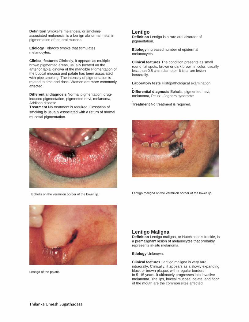
Definition Smoker’s melanosis, or smoking-
associated melanosis, is a benign abnormal melanin pigmentation of the oral mucosa. Etiology Tobacco smoke that stimulates
melanocytes. Clinical features Clinically, it appears as multiple
brown pigmented areas, usually located on the anterior labial gingiva of the mandible Pigmentation of the buccal mucosa and palate has been associated with pipe smoking. The intensity of pigmentation is related to time and dose. Women are more commonly affected. Differential diagnosis Normal pigmentation, drug-
induced pigmentation, pigmented nevi, melanoma, Addison disease Treatment No treatment is required. Cessation of
smoking is usually associated with a return of normal
mucosal pigmentation.
. Ephelis on the vermilion border of the lower lip.
Lentigo of the palate.
Lentigo Definition Lentigo is a rare oral disorder of
pigmentation. Etiology Increased number of epidermal
melanocytes. Clinical features The condition presents as small
round flat spots, brown or dark brown in color, usually less than 0.5 cmin diameter It is a rare lesion intraorally. Laboratory tests Histopathological examination
. Differential diagnosis Ephelis, pigmented nevi,
melanoma, Peutz– Jeghers syndrome . Treatment No treatment is required.
Lentigo maligna on the vermilion border of the lower lip.
Lentigo Maligna Definition Lentigo maligna, or Hutchinson’s freckle, is
a premalignant lesion of melanocytes that probably represents in-situ melanoma. Etiology Unknown.
Clinical features Lentigo maligna is very rare
intraorally. Clinically, it appears as a slowly expanding black or brown plaque, with irregular borders In 5–15 years, it ultimately progresses into invasive melanoma. The lips, buccal mucosa, palate, and floor of the mouth are the common sites affected.
Thilanka Umesh Sugathadasa
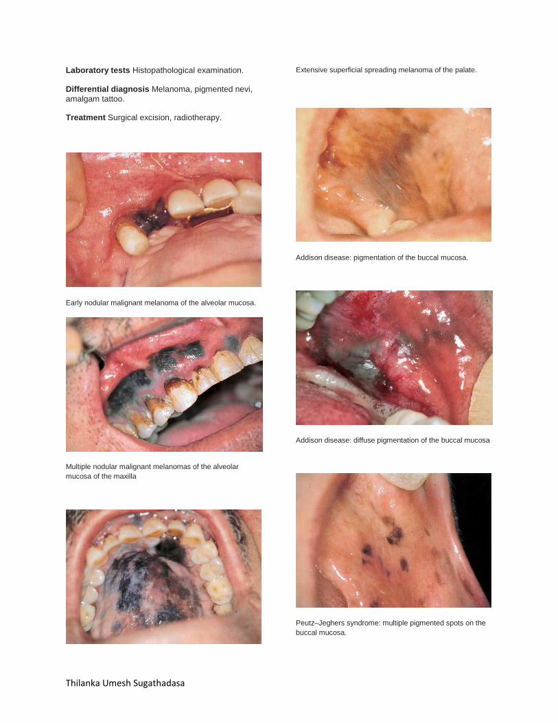
Laboratory tests Histopathological examination.
Differential diagnosis Melanoma, pigmented nevi,
amalgam tattoo. Treatment Surgical excision, radiotherapy.
Early nodular malignant melanoma of the alveolar mucosa.
Multiple nodular malignant melanomas of the alveolar
mucosa of the maxilla
Extensive superficial spreading melanoma of the palate.
Addison disease: pigmentation of the buccal mucosa.
Addison disease: diffuse pigmentation of the buccal mucosa
Peutz–Jeghers syndrome: multiple pigmented spots on the
buccal mucosa.
Thilanka Umesh Sugathadasa
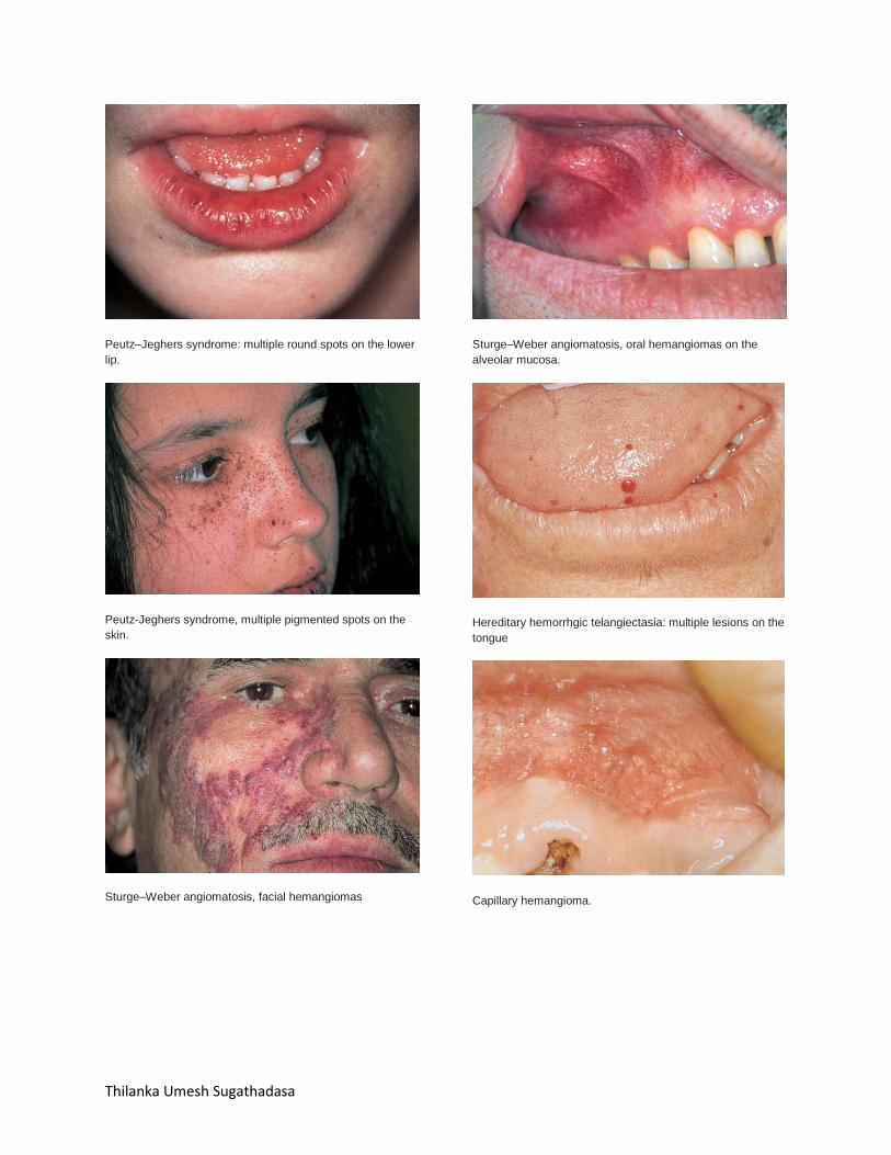
Peutz–Jeghers syndrome: multiple round spots on the lower
lip.
Peutz-Jeghers syndrome, multiple pigmented spots on the
skin.
Sturge–Weber angiomatosis, facial hemangiomas
Sturge–Weber angiomatosis, oral hemangiomas on the
alveolar mucosa.
Hereditary hemorrhgic telangiectasia: multiple lesions on the
tongue
Capillary hemangioma.

Thilanka Umesh Sugathadasa
Cavernous hemangioma.