ophthalmoscopy dr. shahab shaikh powerpoint courtesy dr mujeeb
TRANSCRIPT
OPHTHALMOSCOPYDR. SHAHAB SHAIKH
POWERPOINT COURTESY DR MUJEEB

OBJECTIVES1. To explain the general principles of ophthalmoscopy2. Describe the normal appearance of the fundus.3. Describe the changes in the fundus that occur commonly
in disease.4. Appreciate the importance of performing ophthalmoscopy
as a part of the routine physical examination.


OPHTALMOSCOPE

PRICIPLE OF OPHTHALMOSCOPE
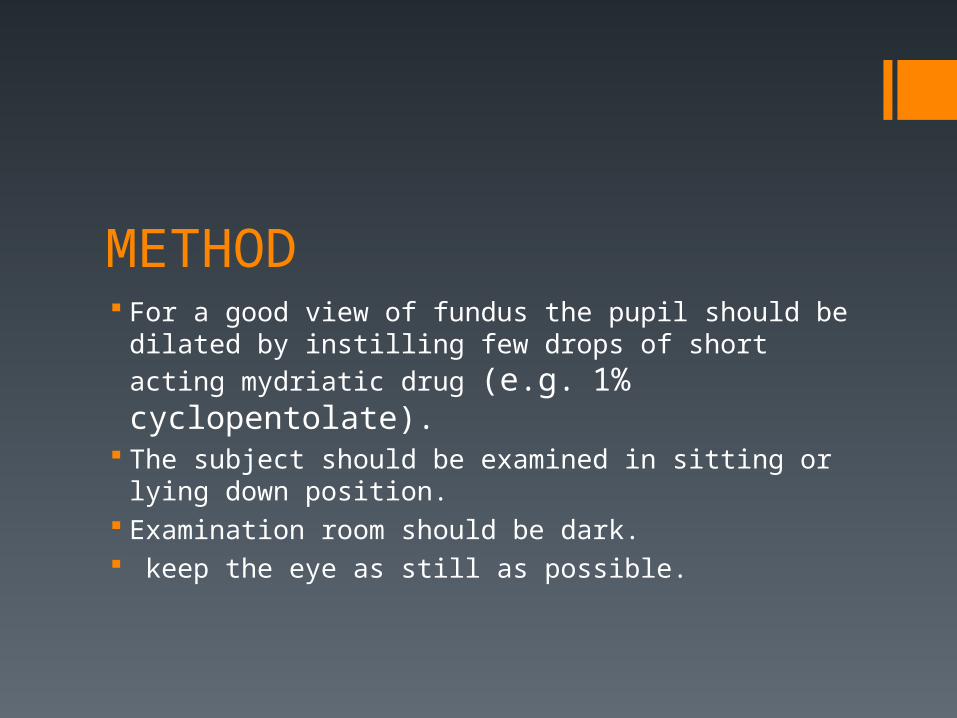
METHOD For a good view of fundus the pupil should be dilated by
instilling few drops of short acting mydriatic drug (e.g. 1% cyclopentolate).
The subject should be examined in sitting or lying down position.
Examination room should be dark. keep the eye as still as possible.

Position of the examiner For examining right eye of the patient, Examiner should stand on right side of the patient. Hold the instrument in his right hand. Use examiner’s right eye.
If examining left eye, stand on left side, hold instrument in left hand use left eye.
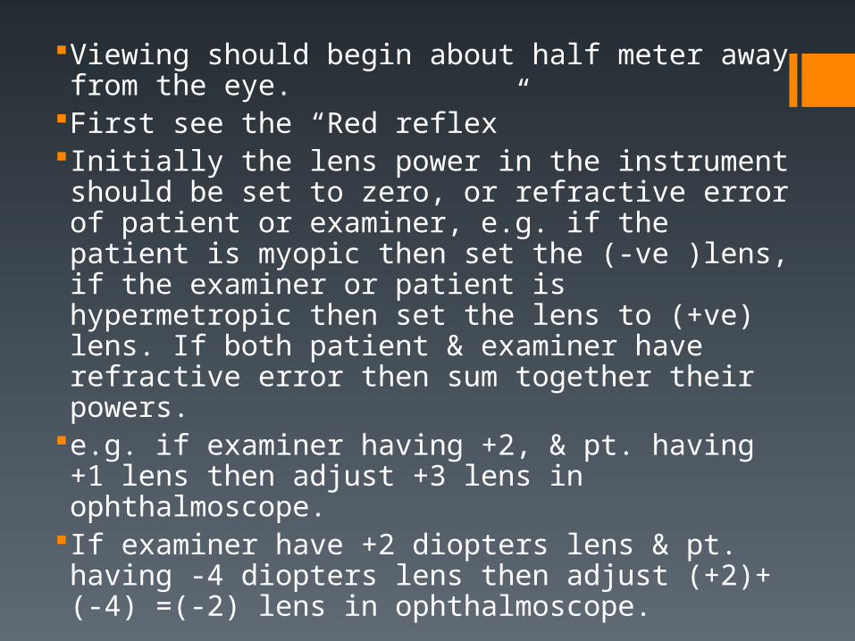
Viewing should begin about half meter away from the eye.
First see the “Red reflex”Initially the lens power in the instrument should be set to zero, or refractive error of patient or examiner, e.g. if the patient is myopic then set the (-ve )lens, if the examiner or patient is hypermetropic then set the lens to (+ve) lens. If both patient & examiner have refractive error then sum together their powers.
e.g. if examiner having +2, & pt. having +1 lens then adjust +3 lens in ophthalmoscope.
If examiner have +2 diopters lens & pt. having -4 diopters lens then adjust (+2)+(-4) =(-2) lens in ophthalmoscope.
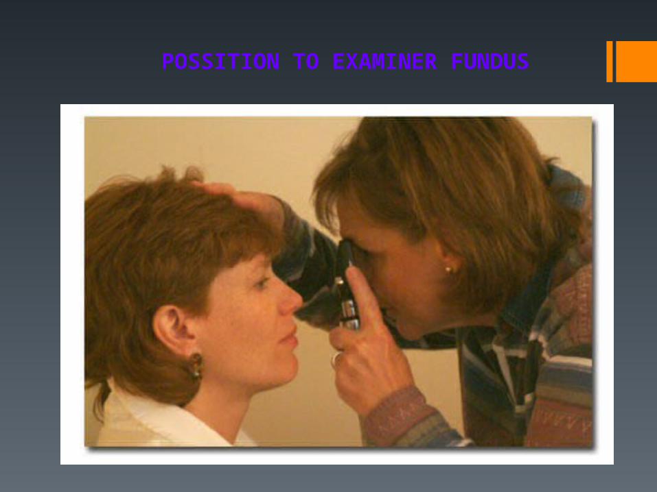
POSSITION TO EXAMINER FUNDUS


NORMAL HUMAN RETINA
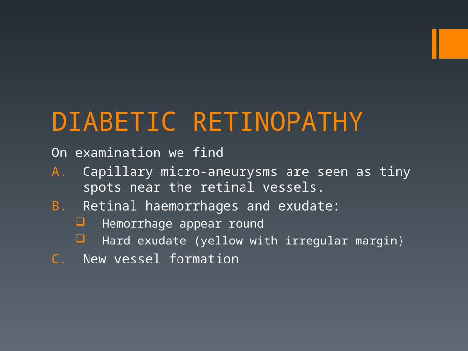
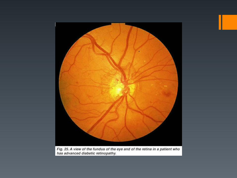
DIABETIC RETINOPATHYOn examination we find
A. Capillary micro-aneurysms are seen as tiny spots near the retinal vessels.
B. Retinal haemorrhages and exudate: Hemorrhage appear round Hard exudate (yellow with irregular margin)
C. New vessel formation


HYPERTENSIVE RETINOPATHYOn examination we findA. Generalized narrowing of retinal arteries.B. Arterio venous nipping i.e. indentation of the veins when
they are crossed by the arteries. C. Retinal haemorrhages and exudate:
Flame shaped hemorrhages Soft exudate (cotton wool)
C. Papilloedema.


MACULAR STAR (HYPERTENSIVE RETINOPATHY)

OPTIC ATROPHY

OPTIC ATROPHY

OPTIC ATROPHY

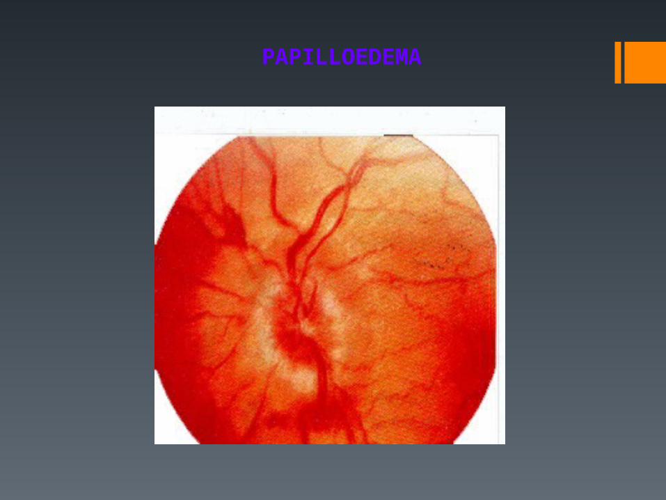
PapilloedemaEdema of optic nerve head, most commonly due to increased
intracranial pressure. eg. Brain tumor.
On examination of fundus we find;Increased redness of disc with blurring of its margins.Physiological cup disappears.Retinal vessels are distended.

PAPILLOEDEMA

PAPILLOEDEMA
PAPILLOEDEMA

NORMAL OPTIC CUP DEEP OPTIC CUP

GLAUCOMATOUS CHANGES

MYOPIC CRESCENT

MYOPIC CRESCENT

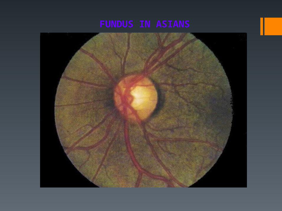
FUNDUS IN ASIANS